Role of Pasteurella granulomatis and Dermatobia horninis in the Etiology of Lechiguana in Cattle" S~LVIA L. LADEIRA, FRANKLIN RIET-CORREA, DANIELA B. PEREIRA, AND GORDON R. CARTER Regional Diagnostic Laboratory Faculty of Veterinary Medicine University of Pelotas 96010-900, Pelotas RS Brazil bVirginia-Maryland College of Veterinary Medicine Virginia Tech Blacksburg, Virginia 24061 INTRODUCTION A new disease of cattle, defined as a focal proliferative fibrogranulomatous panniculitis, has been reported from the State of Rio Grande do Sul, southern Brazil.' The disease, known as lechiguana, is characterized by a large subcutaneous swelling that increases in size, and if untreated causes a progressive weakness resulting in death. Histologically, the lesion consists of focal proliferation of fibrous tissue, infiltrated by plasma cells, eosinophils, lymphocytes, and sometimes neutrophils. The primary lesion is an eosinophilic lymphangitis which results in eosinophilic abscesses which may become small granulomas with occasional rosettes in their centers.' More recently, lechiguana was also reported from other Brazilian states including Santa Catarina, Parani, and Minas Gerais (unpublished data). The name Pasteurella granulomatis has been given to the bacterium that has been consistently isolated from deep biopsies of lesion^.^ Because the area of anatomic distribution of lechiguana lesions is similar to that of infection by Dermatobia horninis, it was postulated that this insect may transmit the causative agent and produce lesions that lead to lechiguana. In attempts to reproduce the disease, a lesion similar to those observed in natural cases was reproduced in 1 of 12 cattle inoculated with P. granulomatis. I The objectives of these experiments were (1) to determine if lechiguana can be reproduced by the inoculation of P. granulomatis: (2) to determine if the failure to reproduce the disease is due to loss of pathogenicity by P. granulomatis; (3) to determine if there is an association between P. granulomatis and D. hominis in the etiology of lechiguana; (4) to determine if the lechiguana lesion is due to the 'This work was funded by grants of the Conselho Nacional de Desenvolvimento Cientkco e Tecnol6gico (CNPq) and Fundqio de Amparo B Pesquisa do Rio Grande do Sul (FAPERGS). 359

Welcome message from author
This document is posted to help you gain knowledge. Please leave a comment to let me know what you think about it! Share it to your friends and learn new things together.
Transcript
Role of Pasteurella granulomatis and Dermatobia horninis in the Etiology
of Lechiguana in Cattle" S~LVIA L. LADEIRA, FRANKLIN RIET-CORREA,
DANIELA B. PEREIRA, AND GORDON R. CARTER Regional Diagnostic Laboratory Faculty of Veterinary Medicine
University of Pelotas 96010-900, Pelotas RS Brazil
bVirginia-Maryland College of Veterinary Medicine Virginia Tech
Blacksburg, Virginia 24061
INTRODUCTION
A new disease of cattle, defined as a focal proliferative fibrogranulomatous panniculitis, has been reported from the State of Rio Grande do Sul, southern Brazil.' The disease, known as lechiguana, is characterized by a large subcutaneous swelling that increases in size, and if untreated causes a progressive weakness resulting in death. Histologically, the lesion consists of focal proliferation of fibrous tissue, infiltrated by plasma cells, eosinophils, lymphocytes, and sometimes neutrophils. The primary lesion is an eosinophilic lymphangitis which results in eosinophilic abscesses which may become small granulomas with occasional rosettes in their centers.' More recently, lechiguana was also reported from other Brazilian states including Santa Catarina, Parani, and Minas Gerais (unpublished data).
The name Pasteurella granulomatis has been given to the bacterium that has been consistently isolated from deep biopsies of lesion^.^ Because the area of anatomic distribution of lechiguana lesions is similar to that of infection by Dermatobia horninis, it was postulated that this insect may transmit the causative agent and produce lesions that lead to lechiguana. In attempts to reproduce the disease, a lesion similar to those observed in natural cases was reproduced in 1 of 12 cattle inoculated with P. granulomatis. I
The objectives of these experiments were (1) to determine if lechiguana can be reproduced by the inoculation of P. granulomatis: ( 2 ) to determine if the failure to reproduce the disease is due to loss of pathogenicity by P. granulomatis; (3) to determine if there is an association between P. granulomatis and D. hominis in the etiology of lechiguana; (4) to determine if the lechiguana lesion is due to the
'This work was funded by grants of the Conselho Nacional de Desenvolvimento Cientkco e Tecnol6gico (CNPq) and Fundqio de Amparo B Pesquisa do Rio Grande do Sul (FAPERGS).
359
360 ANNALS NEW YORK ACADEMY OF SCIENCES
TABLE 1. Inoculation of Pasteurella granulomatis in Cattle in Experiments 1 to 12
Cattle Inoculum
Experiment TY Pe Amount and Route (Number) Age
1
2
3
4 5
6
7 8 9
10
1 1
12
Suspension of P.g" with oil adjuvant
Suspension of P.g with ground D.h'
Suspension of P.g with gastric mucin
Suspension of P.g BHI culture of P.g
with saponin BHI culture of P.g
with a previous passage in mice
Suspension of P.g Suspension of P.g Suspension of P.g
with gastric mucin Suspension of P.g
BHI culture of P.g
BHI culture of P.g
2 ml Sc" 4 ml SC 2 ml SC 4 ml SC 4 ml SC
2 ml SC 4 ml SC
5 ml SC
3 ml SC 4 ml IL" 4 ml IL
Injection of 0.2-0.4 ml in lesions of D.h
Lesions of D.h moistened with the P.g culture
Lesions of cattle 9 (experi- mentally) infected with D.h moistened with P.g culture
3 >3 years 3 >3 years 3 >3 years 3 >3 years 6 23 years
6 >3 years 6 10 months
1 2b 23 years
6' >3 years 3 10 months 3 10 months
11 >3 years
6 >3 years
9 >3 years
a SC = subcutaneous; IL = intralymphatic; P.g = P. grandomatis; D.h = D. horninis. 'These cattle had been inoculated previously in experiments 3 and 4. These cattle were immunodepressed.
multiplication of P. granulomatis in a concurrent inflammatory process, or to an immunologic inflammation; and (5) to determine the possibility of reproducing the disease in immunodepressed cattle.
MATERIAL AND METHODS
Znoculation of Cattle with P. granulomatis
In experiments 1 to 12, P. granulomatis (strain 145/91) in different preparations was inoculated into 68 cattle. Details of these inoculations are presented in TABLE 1.
The bacteria inoculated in experiments 1, 2, 3, 4, 7, 8, 9, and 10 were from a 24-hour culture of P. granulomatis on blood agar, suspended in saline solution or
LADEIRA el al.: ETIOLOGY OF LECHIGUANA 361
nutrient broth to a concentration of approximately 2.3 x lo* CFU/ml. The inoculum used in experiments $ 6 , 1 I , and 12 was a 24-hour brain heart infusion (BHI) broth culture of P. granulornatis containing approximately 1.68 x lo9 CFU/ml.
In experiment 1, the inoculum was a suspension of P. granulomatis in saline solution (0.85% NaCI) mixed (v/v) with complete Freund’s adjuvant. In experiment 2, the inoculum was prepared with ground larvae of D. hominis, diluted in saline solution, centrifuged, and the supernatant fluid mixed (v/v) with the saline solution of P. granulomatis used previously. In experiments 3 and 9, the inoculum was the same suspension of P. granulomatis in saline solution mixed (vlv) with a 5% mucin (porcine stomach, Sigma Chemical Company, St. Louis, MO) solution. In experiment 5, the inoculum was a solution of 1 mg of saponin mixed (v/v) with a BHI broth culture of P. grunulomatis. In experiment 6, BHI broth cultures of P. grunulornatis with a previous passage in mice were inoculated into 12 cattle. These cattle had been inoculated 68 days before in experiments 3 and 4. The 6 cattle used in experiment 7 were immunodepressed by the daily inoculation of dexamethasone at 0.1 mg per kg of body weight, for 5 days. Three cattle were inoculated with P. grunulomatis on the first day of treatment with dexamethasone and the other 3 on the last day. Thirty days after the initiation of the experiment, the 6 animals were treated again with 0.1 mg of dexamethasone per kg for 5 days.
In experiments 8 and 9, P. grunulornatis was inoculated intralymphatically. For this purpose, methylene blue (5% aqueous solution) was injected in the interdigital space of the left hind limb. Five to ten minutes later, 2 incisions at right angles were made in the external lateral face of the metatarsus, and the skin was raised for the location of a lymphatic vessel. P. granulomatis was inoculated into the lymphatic vessel with an endovenous infusion set (Venescalp 23G, Sio Paulo, SP, Brazil).
In experiment 10, a suspension of P. granulornatis in nutrient broth was inoculated into lesions of D. hominis in 11 cattle. In each animal, 2 to 5 lesions of D. hominis were inoculated with 0.2 to 0.5 ml of inoculum. The cattle had been treated for D. hominis 4 days before inoculation, and thus the larvae were dead at the time of inoculation.
Experiment 11 involved 6 cattle with severe natural infection by D. horninis. In each animal, 50 to 100 lesions of D. hominis were moistened with swabs that had been dipped in a BHI broth culture of P. granulomatis.
In experiment 12, each of 9 cattle were infected experimentally in the right prescapular region with 30 larvae of D. hominis. Before inoculation, the skin of the region had been moistened with a BHI broth culture of P. granulomatis. After infection with the larvae at weekly intervals for 4 weeks, swabs saturated with similar BHI broth cultures of P. granulomatis were applied to the larval lesions.
Five to 25 days after inoculation, needle biopsies were taken from the abscesses induced by P. granulomatis in 1 animal in experiment 3, 2 in experiment 4, 2 in experiment 5,4 in experiment 6, and 1 in experiment 7, the purulent exudate obtained was examined bacteriologically.
In experiment 1 1 , 2 surgical biopsies were obtained 49 and 55 days after inocula- tion from the swelling produced in one animal. The material obtained was examined bacteriologically and histologically. The biopsies were fixed, sectioned, and stained by standard procedure^.^
362 ANNALS NEW YORK ACADEMY OF SCIENCES
TABLE 2. Isolation of P. granulomatis from Lesions Caused by D. hominis and from Larvae
No. of Isolates of P. granulomatis Obtained No. of Lesions and
Larvae Cultured from:
Farm Cattle Larvae Lesions Larvae Lesions
- - 1 cow 1 I 1 Calf 12 12 4 2 2 Steer 4 4 2 Steer 3 3 2 Cow" 1 1 3 Steer 1 1 3 cow 1 1
- -
- - - -
- -
- -
a Cow with a spontaneous lesion of lechiguana.
In experiment 1 1 , 35 days after inoculation, exudate from the lesions caused by D. hominis in each of the cattle was collected with swabs for bacteriological examina- tion. The hemolymph from one larva of D. hominis was also examined bacteriologi- cally. For this purpose, the larva was washed in saline solution and the hemolymph was obtained with sterile syringe and needle.
Bacteriologic Study of D. hominis and Lesions Caused by the Larvae in Cattle
Larvae of D. hominis and exudate from lesions caused by the fly in cattle were collected from 7 cattle of 3 farms (TABLE 2 ) and examined bacteriologically. On farms 1 and 3, lechiguana had never been observed. On farm 2, one cow had a lechiguana lesion at the time of collection. Collection and culturing of exudate from the lesions and hemolymph from the larvae were performed by the methods previously described.
RESULTS
Cattle Inoculation
In the 36 cattle inoculated with P. granulomatis in experiments 1 to 5 and 7, and in the 12 cattle reinoculated in experiment 6, small abscesses were produced at the injection site. The abscesses regressed spontaneously in 15 to 60 days. P. granulomatis was isolated from the purulent exudate in experiments 3 to 7. Only temporary subcutaneous edema was observed in the 6 calves inoculated by the intralymphatic route in experiments 8 and 9.
LADEIRA et al.: ETIOLOGY OF LECHIGUANA 363
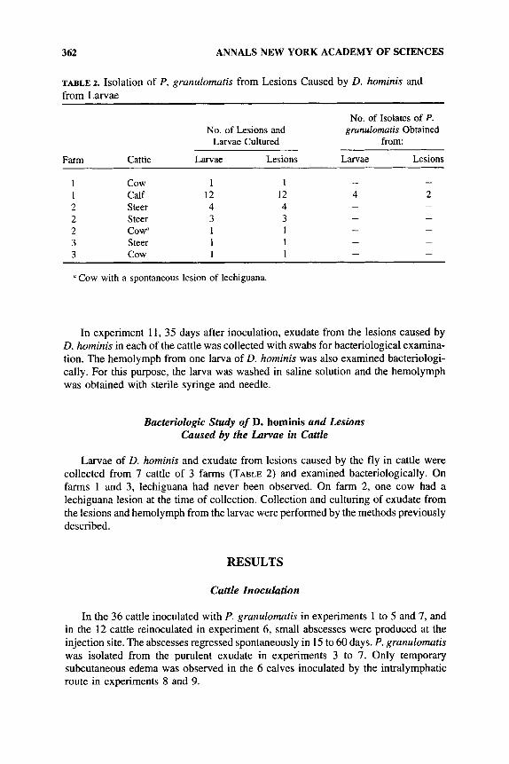
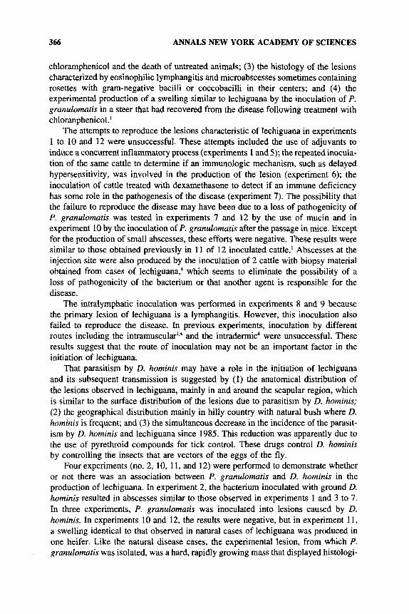
FIGURE 1. Heifer inoculated in experiment 11. Note the subcutaneous mass over the withers.
No significant changes were observed in the 20 cattle with lesions of D. hominis that had been inoculated with P. granulomatis in experiments 10 and 12.
Bacteriologic examination of the lesions caused by D. hominis in experiment 10 yielded various bacteria but not P. granulomatis. P. granulomatis was recovered from a D. horninis larva obtained from one animal at the end of experiment 12.
In experiment 11, no lesions were observed in the 6 cattle until day 3 I postinocula- tion. On day 34, a hard, rounded subcutaneous swelling was observed on the withers in one animal. This region had many D. hominis larvae at the time of the inoculation. Ten days after the first observation, the lesion measured 22 cm craniocaudal, 8 cm dorsoventral, and 25 cm transversal (FIG. 1); 1 1 days later it measured 23 cm craniocau- dal, 8 cm dorsoventral, and 27 cm transversal. At this time, the animal was treated, for 5 days, with 1.5 g of chloramphenicol daily. The swelling regressed rapidly, and regression was almost complete 8 days after the start of treatment. P. granulomatis was isolated in pure culture from the biopsies performed 10 and 21 days after the first observation of the swelling.
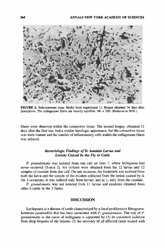
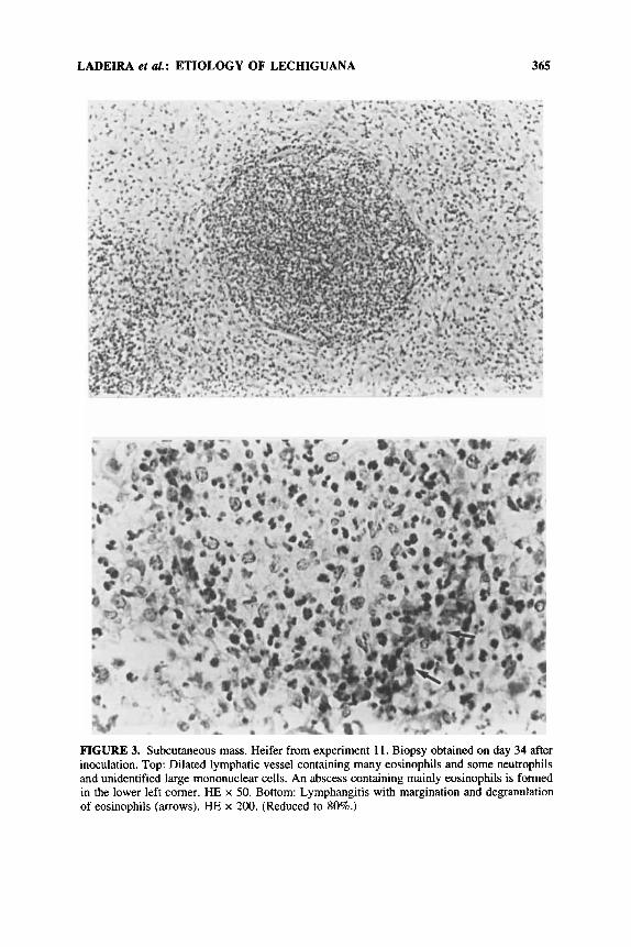
The histologic appearance of the first biopsy was characterized by proliferation of loose connective tissue infiltrated by numerous eosinophils and some neutrophils and lymphocytes. Many collagenous fibers were calcified (FIG. 2). Dilated lymphatic vessels containing eosinophils, neutrophils, and unidentified large mononuclear cells were observed within the connective tissue (FIG. 3 top). Deposits of eosinophilic granules were present along lymphatic walls (FIG. 3 bottom). Microabscesses consisted of mainly eosinophils with some neutrophils, mononuclear cells, and occasionally some eosinophilic acellular debris was also present (FIG. 3 top). Rosettelike structures were observed in some microabscesses, and colonies of gram-negative bacteria, either bacilli or coccobacilli, were present in some of the rosettes. Edema and some muscle
364 ANNALS NEW YORK ACADEMY OF SCIENCES
FIGURE 2. Subcutaneous mass. Heifer from experiment 11. Biopsy obtained 34 days after inoculation. The collagenous fibers are heavily calcified. HE x 200. (Reduced to 80%.)
fibers were observed within the connective tissue. The second biopsy, obtained 11 days after the first one, had a similar histologic appearance, but the connective tissue was more mature and the number of inflammatory cells within the collagenous fibers was reduced.
Bacteriologic Findings of D. hominis Larvae and Lesions Caused by the Fly in Cattle
P. grunulomutis was isolated from one calf on farm 1, where lechiguana had never occurred (TABLE 2). Six isolates were obtained from the 12 larvae and 12 samples of exudate from this calf. On one occasion, the bacterium was isolated from both the larva and the sample of the exudate collected from the lesion caused by it. On 3 occasions, it was isolated only from larvae; and in 1, only from the exudate.
P. grunulomatis was not isolated from 11 larvae and exudates obtained from other 6 cattle in the 3 farms.
DISCUSSION
Lechiguana is a disease of cattle characterized by a focal proliferative fibrogranu- lomatous panniculitis that has been associated with P. grunulomutis. The role of P. grunulomutis as the cause of lechiguana is supported by (1) its consistent isolation from deep biopsies of the lesions; ( 2 ) the recovery of all affected cattle treated with
LADEIRA et aZ.: ETIOLOGY OF LECHIGUANA 365
FIGURE 3. Subcutaneous mass. Heifer from experiment 11. Biopsy obtained on day 34 after inoculation. Top: Dilated lymphatic vessel containing many eosinophils and some neutrophils and unidentified large mononuclear cells. An abscess containing mainly eosinophils is formed in the lower left comer. HE x 50. Bottom: Lymphangitis with margination and degranulation of eosinophils (mows). HE x 200. (Reduced to SO%.)
366 ANNALS NEW YORK ACADEMY OF SCIENCES
chloramphenicol and the death of untreated animals; (3) the histology of the lesions characterized by eosinophilic lymphangitis and microabscesses sometimes containing rosettes with gram-negative bacilli or coccobacilli in their centers; and (4) the experimental production of a swelling similar to lechiguana by the inoculation of P. granulomatis in a steer that had recovered from the disease following treatment with chloranphenicol.'
The attempts to reproduce the lesions characteristic of lechiguana in experiments 1 to 10 and 12 were unsuccessful. These attempts included the use of adjuvants to induce a concurrent inflammatory process (experiments 1 and 5); the repeated inocula- tion of the same cattle to determine if an immunologic mechanism, such as delayed hypersensitivity, was involved in the production of the lesion (experiment 6); the inoculation of cattle treated with dexamethasone to detect if an immune deficiency has some role in the pathogenesis of the disease (experiment 7). The possibility that the failure to reproduce the disease may have been due to a loss of pathogenicity of P. granulomatis was tested in experiments 7 and 12 by the use of mucin and in experiment 10 by the inoculation of P. granulomatis after the passage in mice. Except for the production of small abscesses, these efforts were negative. These results were similar to those obtained previously in 11 of 12 inoculated cattle.' Abscesses at the injection site were also produced by the inoculation of 2 cattle with biopsy material obtained from cases of lechiguana; which seems to eliminate the possibility of a loss of pathogenicity of the bacterium or that another agent is responsible for the disease.
The intralymphatic inoculation was performed in experiments 8 and 9 because the primary lesion of lechiguana is a lymphangitis. However, this inoculation also failed to reproduce the disease. In previous experiments, inoculation by different routes including the intramu~cular'*~ and the intradermic4 were unsuccessful. These results suggest that the route of inoculation may not be an important factor in the initiation of lechiguana.
That parasitism by D. horninis may have a role in the initiation of lechiguana and its subsequent transmission is suggested by (1) the anatomical distribution of the lesions observed in lechiguana, mainly in and around the scapular region, which is similar to the surface distribution of the lesions due to parasitism by D. hominis; (2) the geographical distribution mainly in hilly country with natural bush where D. hominis is frequent; and (3) the simultaneous decrease in the incidence of the parasit- ism by D. hominis and lechiguana since 1985. This reduction was apparently due to the use of pyrethroid compounds for tick control. These drugs control D. hominis by controlling the insects that are vectors of the eggs of the fly.
Four experiments (no. 2, 10, 11, and 12) were performed to demonstrate whether or not there was an association between P. granulomatis and D. hominis in the production of lechiguana. In experiment 2, the bacterium inoculated with ground D. hominis resulted in abscesses similar to those observed in experiments 1 and 3 to 7. In three experiments, P. granulomatis was inoculated into lesions caused by D. horninis. In experiments 10 and 12, the results were negative, but in experiment 11, a swelling identical to that observed in natural cases of lechiguana was produced in one heifer. Like the natural disease cases, the experimental 'lesion, from which P. granulornatis was isolated, was a hard, rapidly growing mass that displayed histologi-
LADEIRA et al.: ETIOLOGY OF LECHIGUANA 367
cally a focal proliferative fibrogranulomatous panniculitis characteristic of lechiguana. The lesion regressed rapidly after treatment with chloramphenicol.
The experimental production of the disease in one animal with severe infection by D. hominis (50 to 100 larvae in 100 to 200 cm2 of skin) and the fact that these lesions are almost always situated in the same anatomic region in lechiguana (mainly in and around the scapular region) suggest that in natural cases, the large lesions characteristic of lechiguana occur when P. grunulomatis infects severe lesions caused by D. hominis. However, at least experimentally, such previous larval lesions are not strictly necessary for the development of lechiguana lesions, because in a previous experimental production of lechiguana in cattle, the bacterium was inoculated subcuta- neously in an animal not infected by D. horninis.'
The isolation of P. grunulomatis from larvae and lesions caused by D. hominis in one calf from a farm where lechiguana had never occurred suggests that D. horninis can carry and transmit P. granulomatis. This fact was also evidenced in experiment 12 with the isolation of P. granulomatis from one D. hominis larva.
The lechiguana lesion produced in experiment 11 in one animal was observed 34 days postinoculation. In a previous experimental production of the disease, the lesion also appeared 34 days postinoculation.' This apparent long incubation period, the negative results obtained in the other experiments, and also the infrequent occur- rence of the natural disease suggest that lechiguana is a disease for which Koch's postulates are not easily fulfilled. Nevertheless, the experimental production of the disease and the recovery of P. granulomatis from D. hominis larvae suggest that lechiguana is caused by P. granulomatis and that D. hominis has a role in the initiation and transmission of the disease.
SUMMARY
Attempts were made to reproduce bovine lechiguana, a disease associated with Dermatobia hominis and Pasteurella granulornatis infections. Suspensions of Paste- urella granulomatis were mixed with each of the following: saponin, oil adjuvant, ground Dermatobia hominis, or 5% mucin. Each preparation was inoculated into 6 cattle. Twelve more cattle, 6 of which received dexamethasone, were inoculated with bacterial suspension alone. Abscesses but no lechiguana was produced in all 36 cattle. After abscess regression, 12 cattle were reinoculated with a suspension of mouse- passed P. granulomatis. Only abscesses were produced. The intralymphatic inocula- tion of P. grunulomutis in 6 cattle did not produce the disease. Eleven cattle infected naturally with D. hominis had lesions containing dead larvae. These lesions were inoculated with P. granulomatis. Nine cattle were experimentally infected with larvae of D. hominis that had been contaminated with the bacteria. No lechiguana lesions were produced in these 20 cattle. Six cattle with severe natural D. hominis infection were inoculated in the larval lesions with P. granulomatis. One developed lesions indistinguishable from those of natural lechiguana. The lesions regressed after treat- ment with chloramphenicol. D. hominis larvae and exudate from lesions caused by the fly were collected from 7 cattle on 3 farms and examined bacteriologically. P. grunulomutis was isolated from the larvae and the exudate of a healthy calf from a farm where lechiguana had never been observed. These results suggest that P.
368 ANNALS NEW YORK ACADEMY OF SCIENCES
grunulornutis has a causal role in lechiguana, and that D. hominis may be a carrier of the bacterium. These observations suggest that lechiguana occurs when severe D. hominis lesions are infected with P. granulomatis. The apparent long incubation period, the negative results obtained in the other experiments, and also the infrequent occurrence of the natural disease suggest that lechiguana is a disease for which Koch’s postulates are not easily fulfilled.
REFERENCES
1. RIET-CORREA, F., M. C. MENDEZ, A. L. SCHILD, G. A. RIBEIRO & S. M. ALMEIDA. 1992. Bovine focal proliferative fibrogranulomatous panniculitis (lechiguana) associated with P. granulomatis. Vet. Pathol. 29: 93-103.
RIBEIRO, G. A., G. R. CARTER, W. FREDERIKSEN & F. RIET-CORREA. 1989. Pasteurella haemolitica-like bacterium from a progressive granuloma of cattle in Brazil. J. Clin. Microbiol. 27: 1401-1402.
LADEIRA, S. L. 1993. Participaqiio de Pasteurella granulomatis e Dermatobia hominis na etiologia da lechiguana (paniculite fibrogranulomatosa proliferativa) em bovinos. Ms Thesis. University of Pelotas.
ALMEIDA, S. M. 1986. Estudo sobre a epidemiologia, patologia e etiologia de urn granuloma em bovinos conhecido como lechiguana. Ms Thesis. University of Pelotas.
2.
3 .
4.
Related Documents