Role of Myosin VI in the Differentiation of Cochlear Hair Cells Tim Self,* Tama Sobe,² Neal G. Copeland,‡ Nancy A. Jenkins,‡ Karen B. Avraham,² and Karen P. Steel* ,1 *MRC Institute of Hearing Research, University Park, Nottingham NG7 2RD, United Kingdom; ²Department of Human Genetics and Molecular Medicine, Sackler School of Medicine, Tel Aviv University, Tel Aviv 69978, Israel; and ‡Mammalian Genetics Laboratory, ABL–Basic Research Program, National Cancer Institute–Frederick Cancer Research and Development Center, Frederick, Maryland 21702 The mouse mutant Snell’s waltzer (sv) has an intragenic deletion of the Myo6 gene, which encodes the unconventional myosin molecule myosin VI (K. B. Avraham et al., 1995, Nat. Genet. 11, 369 –375). Snell’s waltzer mutants exhibit behavioural abnormalities suggestive of an inner ear defect, including lack of responsiveness to sound, hyperactivity, head tossing, and circling. We have investigated the effects of a lack of myosin VI on the development of the sensory hair cells of the cochlea in these mutants. In normal mice, the hair cells sprout microvilli on their upper surface, and some of these grow to form a crescent or V-shaped array of modified microvilli, the stereocilia. In the mutants, early stages of stereocilia development appear to proceed normally because at birth many stereocilia bundles have a normal appearance, but in places there are signs of disorganisation of the bundles. Over the next few days, the stereocilia become progressively more disorganised and fuse together. Practically all hair cells show fused stereocilia by 3 days after birth, and there is extensive stereocilia fusion by 7 days. By 20 days, giant stereocilia are observed on top of the hair cells. At 1 and 3 days after birth, hair cells of mutants and controls take up the membrane dye FM1-43, suggesting that endocytosis occurs in mutant hair cells. One possible model for the fusion is that myosin VI may be involved in anchoring the apical hair cell membrane to the underlying actin-rich cuticular plate, and in the absence of normal myosin VI this apical membrane will tend to pull up between stereocilia, leading to fusion. © 1999 Academic Press Key Words: myosin VI; genetic deafness; Snell’s waltzer; mouse mutant; cochlear hair cells; microvilli; unconventional myosin. INTRODUCTION Of the handful of genes involved in deafness identified so far, three have been found to encode unconventional myo- sin molecules. Mutations in Myo7a have been found in shaker1 mouse mutants, Usher type 1B in humans, and both dominant and recessive forms of nonsyndromic deaf- ness in humans (Gibson et al., 1995; Mburu et al., 1997; Weil et al., 1995, 1997; Liu et al., 1997a,b). Myo15 has been reported to underlie both the shaker2 mouse mutant and another form of human recessively inherited nonsyndromic deafness (Probst et al., 1998; Wang et al., 1998). Finally, Myo6, encoding the myosin VI molecule, is mutated in the Snell’s waltzer (sv) mouse mutant (Avraham et al., 1995). The Snell’s waltzer mouse mutant was first described by Deol and Green in 1966 and displays the typical shaker– waltzer behaviour often associated with inner ear defects: deafness, hyperactivity, head tossing, and circling. Using light microscopy, Deol and Green (1966) found hair cell degeneration and loss to progress rapidly from 12 days after birth. The responsible gene was identified by Avraham and colleagues in 1995, and the mutation in the Snell’s waltzer mouse was found to be an intragenic deletion leading to an absence of detectable myosin VI protein in homozygotes. Immunofluorescence studies localised myosin VI to the inner and outer hair cells of the sensory epithelium in the normal adult mouse cochlea (Avraham et al., 1995, 1997). Hasson et al. (1997) further reported that myosin VI was 1 To whom correspondence should be addressed. Fax: 144 115 951 8503. E-mail: [email protected]. Developmental Biology 214, 331–341 (1999) Article ID dbio.1999.9424, available online at http://www.idealibrary.com on 0012-1606/99 $30.00 Copyright © 1999 by Academic Press All rights of reproduction in any form reserved. 331

Welcome message from author
This document is posted to help you gain knowledge. Please leave a comment to let me know what you think about it! Share it to your friends and learn new things together.
Transcript

Wrad
Developmental Biology 214, 331–341 (1999)Article ID dbio.1999.9424, available online at http://www.idealibrary.com on
Role of Myosin VI in the Differentiationof Cochlear Hair Cells
Tim Self,* Tama Sobe,† Neal G. Copeland,‡ Nancy A. Jenkins,‡Karen B. Avraham,† and Karen P. Steel*,1
*MRC Institute of Hearing Research, University Park, Nottingham NG7 2RD, UnitedKingdom; †Department of Human Genetics and Molecular Medicine, Sackler Schoolof Medicine, Tel Aviv University, Tel Aviv 69978, Israel; and ‡Mammalian GeneticsLaboratory, ABL–Basic Research Program, National Cancer Institute–FrederickCancer Research and Development Center, Frederick, Maryland 21702
The mouse mutant Snell’s waltzer (sv) has an intragenic deletion of the Myo6 gene, which encodes the unconventionalmyosin molecule myosin VI (K. B. Avraham et al., 1995, Nat. Genet. 11, 369–375). Snell’s waltzer mutants exhibitbehavioural abnormalities suggestive of an inner ear defect, including lack of responsiveness to sound, hyperactivity, headtossing, and circling. We have investigated the effects of a lack of myosin VI on the development of the sensory hair cellsof the cochlea in these mutants. In normal mice, the hair cells sprout microvilli on their upper surface, and some of thesegrow to form a crescent or V-shaped array of modified microvilli, the stereocilia. In the mutants, early stages of stereociliadevelopment appear to proceed normally because at birth many stereocilia bundles have a normal appearance, but in placesthere are signs of disorganisation of the bundles. Over the next few days, the stereocilia become progressively moredisorganised and fuse together. Practically all hair cells show fused stereocilia by 3 days after birth, and there is extensivestereocilia fusion by 7 days. By 20 days, giant stereocilia are observed on top of the hair cells. At 1 and 3 days after birth,hair cells of mutants and controls take up the membrane dye FM1-43, suggesting that endocytosis occurs in mutant haircells. One possible model for the fusion is that myosin VI may be involved in anchoring the apical hair cell membrane tothe underlying actin-rich cuticular plate, and in the absence of normal myosin VI this apical membrane will tend to pullup between stereocilia, leading to fusion. © 1999 Academic Press
Key Words: myosin VI; genetic deafness; Snell’s waltzer; mouse mutant; cochlear hair cells; microvilli; unconventional
myosin.MS
DwdldbcmaIi
INTRODUCTION
Of the handful of genes involved in deafness identified sofar, three have been found to encode unconventional myo-sin molecules. Mutations in Myo7a have been found inshaker1 mouse mutants, Usher type 1B in humans, andboth dominant and recessive forms of nonsyndromic deaf-ness in humans (Gibson et al., 1995; Mburu et al., 1997;
eil et al., 1995, 1997; Liu et al., 1997a,b). Myo15 has beeneported to underlie both the shaker2 mouse mutant andnother form of human recessively inherited nonsyndromiceafness (Probst et al., 1998; Wang et al., 1998). Finally,
nH
1 To whom correspondence should be addressed. Fax: 144 115951 8503. E-mail: [email protected].
0012-1606/99 $30.00Copyright © 1999 by Academic PressAll rights of reproduction in any form reserved.
yo6, encoding the myosin VI molecule, is mutated in thenell’s waltzer (sv) mouse mutant (Avraham et al., 1995).The Snell’s waltzer mouse mutant was first described byeol and Green in 1966 and displays the typical shaker–altzer behaviour often associated with inner ear defects:eafness, hyperactivity, head tossing, and circling. Usingight microscopy, Deol and Green (1966) found hair cellegeneration and loss to progress rapidly from 12 days afterirth. The responsible gene was identified by Avraham andolleagues in 1995, and the mutation in the Snell’s waltzerouse was found to be an intragenic deletion leading to an
bsence of detectable myosin VI protein in homozygotes.mmunofluorescence studies localised myosin VI to thenner and outer hair cells of the sensory epithelium in the
ormal adult mouse cochlea (Avraham et al., 1995, 1997).asson et al. (1997) further reported that myosin VI was331

d2fG
r1
bwtTHFpe
cabm
5osasecgcCc
umsvcmCrdaPtflawtqmio
t
332 Self et al.
highly enriched in cuticular plates and the pericuticularnecklace around the cuticular plate, present in the hair cellcytoplasm, but was not detected in the stereocilia in adultguinea pigs. Recently, myosin VI has been reported to bedetected in presumptive hair cells from as early as 13.5 daysof gestation, although the localisation within hair cells atearly stages was not described (Xiang et al., 1998).
It is estimated that sensory hair cells in the cochleadetect movements as small as 1 angstrom unit at thethreshold of hearing, so it is not surprising that mainte-nance of their cytoskeletal structure to a high degree ofprecision is critical to their function. Unconventional myo-sins may participate in this maintenance, which mightexplain why hair cells are particularly susceptible to muta-tions in the corresponding genes. However, despite recentreports of stereocilia bundle defects in the shaker1 andshaker2 mutant mice (Self et al., 1998; Probst et al., 1998),we still do not understand the role of unconventionalmyosins in hair cell development or function. In this report,we describe the development of hair cells in Snell’s waltzermutants, along with some further observations of cochlearresponses in these mutants.
MATERIALS AND METHODS.
Genotyping. Homozygous sv mutant mice can be distin-guished from littermate controls from 12 days of age by theirabnormal behaviour, but all mice were subjected to genotyping byPCR. Genomic DNA was amplified with primers that flank thedeletion in the sv allele (Avraham et al., 1995). Wild-type miceisplayed a genomic fragment of 4.0 kb, sv/sv mice displayed a.9-kb fragment, and DNA from heterozygote mice generated bothragments. Primers used were as follows: primer M2404 is 59-GAA-TTGACTACAAGTTTGGG-39 (mouse Myo6 cDNA nucleotides
2404–2424) and primer M2615R is 59-GCTTCAGCTCGATAT-TTTATT-39 (mouse Myo6 cDNA nucleotides 2615–2595). Theeaction mixture contained 200 ng genomic DNA, PCR buffer with.75 mM MgCl2, 350 mM each dNTP, 300 nM each primer, and
Expand Long-Template polymerase (Taq and Pwo DNA poly-merases; Boehringer Mannheim). PCR was performed as follows: 2min denaturing at 94°C; followed by 4 cycles of 10 s denaturationat 94°C, 30 s annealing at 56°C, 5 min extension at 68°C; followedby 24 cycles of 10 s denaturation at 94°C, 30 s annealing at 52°C, 5min extension at 68°C; and finally an additional 5 min at 68°C.
Scanning electron microscopy. A total of 25 sv homozygotesand 32 littermate controls were examined at birth and 1, 3, 7, 12,and 20 days after birth (DAB); all controls were 1/sv except one1/1 at 7 DAB. Inner ears were rapidly removed under a fixative of2.5% glutaraldehyde in 0.1 M phosphate buffer with 0.25% tannicacid and 3 mM CaCl2, pH 7.2. Openings into the coils were madey piercing the apex and rupturing both the oval and the roundindows. The specimens were fixed for 5 h, buffer washed, and
hen dissected further to reveal the surface of the organ of Corti.he osmium tetroxide–thiocarbohydrazide procedure adapted fromunter-Duvar (1978), Furness and Hackney (1986), and Davies andorge (1987) was used to stain prior to dehydrating and critical
oint drying. Specimens were finally sputter coated with gold andxamined in a Jeol 6400 Winsem electron microscope.Copyright © 1999 by Academic Press. All right
Transmission electron microscopy. A total of 47 (13 inner hairell, IHC, and 34 outer hair cell, OHC) homozygote sv/sv hair cellsnd 30 (8 IHC and 22 OHC) control hair cells were examined atirth and 20 days after birth from two mutants and two controlice (one 1/1 and one 1/sv). Cochleas were fixed in 2.5%
glutaraldehyde, 1% paraformaldehyde in 0.1 M phosphate bufferwith 3 mM CaCl2, 0.25% tannic acid, and 1% sucrose, pH 7.2 for
h at 4°C. After buffer washing the cochleas were postfixed in 1%smium tetroxide in phosphate buffer, dehydrated in ethanol, andtained en bloc with 1% phosphotungstic acid and 1% uranylcetate, before being embedded in Araldite epoxy resin. Ultrathinections were stained with uranyl acetate and lead citrate beforexamination on a Jeol 1010 electron microscope at 80 kV. Forounts of raised membranes between stereocilia, sections withood profiles of the cuticular plate and stereocilia bases werehosen (i.e., stereocilia were shown in longitudinal section).ounts were made from 8 control and 9 mutant IHCs plus 22ontrol and 21 mutant OHCs in newborn specimens.
Endocytosis. We assessed the ability of mutant hair cells tondergo endocytosis by using the fluorescent dye FM1-43, in 6utants and 15 control mice at 1 and 3 days of age. FM1-43 is a
tyryl dye used extensively to investigate activity-dependentesicle recycling, endocytosis, and membrane recycling (e.g., Co-hilla et al., 1999). The dye fluoresces brightly when bound to aembrane, but shows little fluorescence in aqueous solution.ochlear coils were dissected in Hanks’ balanced salt solution at
oom temperature, transferred to Dulbecco’s modified Eagle’s me-ium containing 10 mM Hepes (GIBCO BRL), and cultured for 2 ht 37°C. They were then stained with the dye FM1-43 (Molecularrobes) at a concentration of 3 mM for 15 s at 37°C. This exposureime was chosen because it gave optimal labelling of focal spots ofuorescence presumed to represent newly endocytosed vesicles,nd longer periods of labelling led to an intense, diffuse labellingithin hair cells without the focal pattern that gives confidence
hat the labelling was indeed of vesicles. Samples were subse-uently washed with PBS, fixed with 4% paraformaldehyde, andounted before viewing on a Leica confocal microscope. Confocal
mages were examined and assessed blind by two independentbservers in addition to the authors.
Electrophysiology. Eight sv/sv mutants and eight of their lit-ermate controls (1/sv or 1/1) at 20 or 30 days after birth (four
pairs at each age) were used for measuring cochlear responses. Themice were kept in full accordance with UK Home Office regula-tions. The mice were anaesthetised with urethane, a recordingelectrode was positioned on the round window of the cochlea, andresponses recorded as described previously (Steel and Smith, 1992).Round window responses are a well-established method for assess-ing cochlear activity and give response thresholds close to those ofsingle units (Johnstone et al., 1979). Three types of response weremeasured. Compound action potentials (CAPs) consist of one ortwo sharp negative deflections at the start of the tone burst,representing synchronous firing of many cochlear neurones. Sum-mating potentials (SPs) are DC shifts in the waveform sustained forthe duration of the tone burst and are thought to represent the grosscounterpart of intracellular dc responses of sensory hair cells in thecochlea (e.g., Dallos et al., 1972; Dallos, 1986), and they may bepositive or negative in polarity depending on stimulus frequencyand intensity. Cochlear microphonics (CMs) are an AC responsewith a frequency identical to that of the stimulus, and the CMmeasured from the round window is considered to be primarily
generated by the basal turn outer hair cells (e.g., Patuzzi et al.,1989; Dallos and Cheatham, 1976).s of reproduction in any form reserved.

irtmaTlncc
haeHrrowarf
333Myosin VI in Hair Cell Differentiation
RESULTS
Stereocilia Start to Develop Normally, but ThenFuse Together to Form Giant Stereocilia
In the littermate controls, stereocilia bundles show anormal pattern of development, with basal turn hair cellsmore advanced than apical turn hair cells. At birth, thecluster of microvilli at the top of each hair cell is alreadypolarised, with a crescent-shaped array of microvilli on thelateral (outer) edge of each cell growing taller. Later, thesemicrovilli become the stereocilia, while microvilli in thecentre and modiolar (inner) edge of each hair cell are shorterand in the process of being resorbed. A kinocilium is locatedat the lateral pole of each hair cell. Over the next few days,the stereocilia grow to form rows of graded heights, withthe tallest on the lateral edge; the short microvilli arecleared from the surface; and the kinocilium is lost (Fig. 1).During these few days after birth, outer hair cell stereociliaform a distinct V-shaped array of thin, apparently more rigidstereocilia, generally with three rows per hair cell, whileinner hair cell stereocilia retain their crescent-shaped or-ganisation and grow thicker than those of outer hair cells(Fig. 1).
In the Snell’s waltzer mutants, the general organisation ofthe stereocilia bundles appears broadly normal at birth,with the crescent-shaped arrays of growing stereocilia lo-cated at the lateral poles of hair cells, but there are someminor signs of disorganisation (Fig. 1B shows 1 DAB). Inplaces, the stereocilia bundles show a disorganised, swirledappearance, and close inspection by scanning electron mi-croscopy reveals early signs of fusion of stereocilia, startingat their bases, in a few hair cells (Figs. 1B and 2A). Therewere no obvious abnormalities in the cuticular plates of thenewborn mutant hair cells when examined by transmissionelectron microscopy, but occasionally the apical plasmamembrane between adjacent stereocilia was raised slightly,and this appeared to occur more often in the mutant than inthe control (Fig. 3). For IHCs, no interstereocilia gapsshowed raised membranes in controls (0/49), while 22% ofthese gaps (10/46) showed visibly raised membranes inmutants. For OHCs, 8% of gaps showed raised membranesin controls (15/194), compared with 20% in mutants (37/185). Material similar in appearance to the cuticular platefilled the area below the raised membranes. A day later,further signs of disorganisation and fusion are seen, and by3 DAB most hair cells are affected (Figs. 1D and 2B). At 7DAB, there is extensive fusion and excess growth of stereo-cilia, resulting in just one or a few large protrusions on thetop of each hair cell (Figs. 1F and 2C). The process of fusionand extension of these protrusions, or giant stereocilia,continues. By 20 DAB, there are no hair cells anywhere inthe cochlear duct showing normal stereocilia, and somegaps are seen in the regular array of hair cells, suggestingthat they are beginning to degenerate, almost certainly as a
secondary effect (Avraham et al., 1995) (Fig. 1H). Theprotrusions often lie across the surface of the organ of Corti,wc
Copyright © 1999 by Academic Press. All right
ncluding across neighbouring support cells, rather thanemaining perpendicular to the upper surface, and some-imes appear to be fused to the underlying apical surfaceembrane (Fig. 2D). The protrusions can show bifurcations
nd often have bulbous regions at or near their tips (Fig. 1H).hese giant stereocilia appeared to contain diffuse actin-
ike material, membrane whorls, and other cell contentsot normally found inside stereocilia (Fig. 4). Many hairells had disappeared by 20 DAB, and degenerating hairells were commonly seen in sections of a 20-DAB mutant.
Mutant Hair Cells Show Evidence of Endocytosis
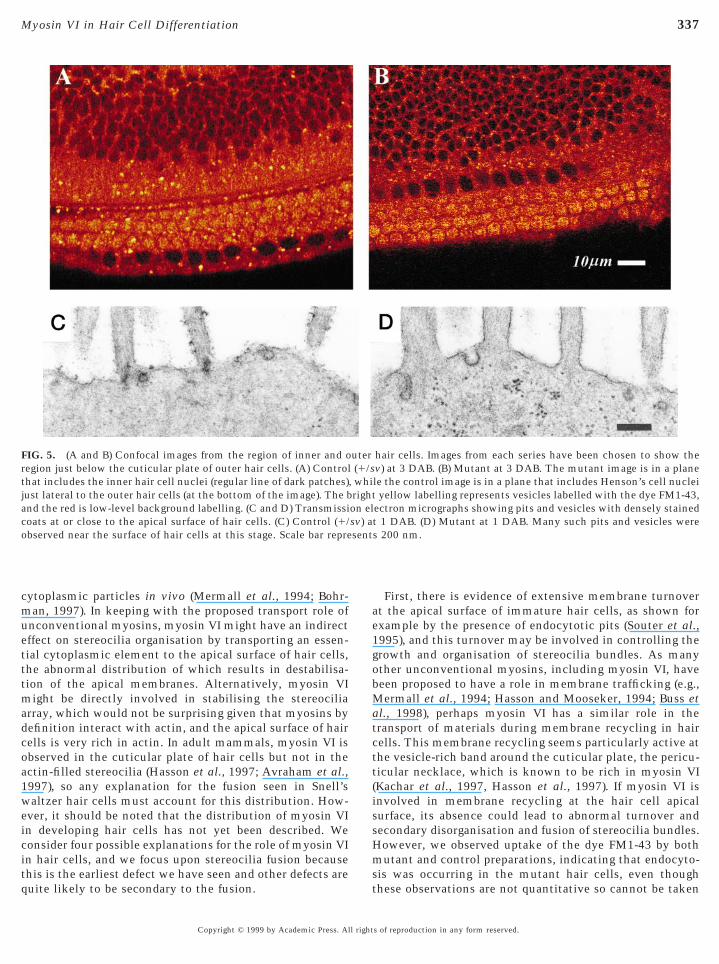
As unconventional myosins have been implicated inmembrane trafficking, and apical membrane turnovermight influence stereocilia organisation, we used a mem-brane dye, FM1-43, to assess whether mutant hair cellsshowed evidence of endocytosis. Serial confocal imagesthrough the organ of Corti showed that there were focalspots of intense fluorescence located within the hair cellcytoplasm, both above and below the nuclei, and they wereparticularly numerous just below the apical surface of thehair cells (Fig. 5). The intensity of these focal spots variedbetween samples, so we do not suggest that these observa-tions are quantitative or can be taken to indicate thatmembrane trafficking is completely normal. Nonetheless,these spots were seen in both mutant and control prepara-tions at 1 and 3 days of age and can be taken to indicate thatendocytosis has occurred. In transmission electron micro-graphs of newborn specimens, pits looking like coated pitswere seen at the apical surface of both mutants and controlhair cells (Fig. 5).
Snell’s waltzer Mutants Show Minimal Responsesto Sound Stimulation
The control mice showed compound action potentials,summating potentials, and cochlear microphonics thatwere essentially identical to previously described responsesin mice (e.g., Steel and Smith, 1992; Harvey and Steel, 1992)(data not shown). For example, the controls showed best(lowest) CAP thresholds at 18 kHz (mean 25.3 dB SPL 6 1.6SEM), which is around the usual best frequency for mice. Asreported previously (Avraham et al., 1995), Snell’s waltzer
omozygotes showed no evidence of a CAP or CM responset any frequency or intensity of stimulus used. Six of theight mutants also showed no sign of an SP response.owever, in 2 of the 30 DAB mutants, SP responses were
ecorded (Fig. 6). In both cases, these SP responses wereecorded only in response to 18-kHz stimuli and not to anyther frequency used in the range 3 to 30 kHz, and theyere recorded in response to very high intensity stimuli,
bove 125 dB SPL. These responses were considered true SPesponses despite the high thresholds because (a) the wave-orms were very similar in shape, size, and latency to
aveforms close to threshold in control mice (e.g., compareontrol waveforms at 25 and 30 dB with mutant waveforms
s of reproduction in any form reserved.

334 Self et al.
Copyright © 1999 by Academic Press. All rights of reproduction in any form reserved.

nsttfabcdr
awsobF parat as B
335Myosin VI in Hair Cell Differentiation
at 128 and 130 dB in Fig. 6), (b) the SP responses could berepeatedly measured in the two mutant mice, and so werenot spurious noise-related artefact, and (c) the amplitude ofthe response increased with increasing stimulus intensity,indicating that it was a genuine biological response.
DISCUSSION
We found that in Snell’s waltzer mutants with no detect-able myosin VI, cochlear hair cells appear to start to develop
FIG. 1. Scanning electron microscopy to show maturation of stermutants (right column), at ages 1 DAB (A, B, basal turn), 3 DAB (C, D
FIG. 2. Detail of stereocilia fusion in inner hair cells of Snell’s wppears to have an abnormal orientation, with its tallest microvillihile the bundles on the left and right have a normal appearance
tarting from the bases of the stereocilia in these three inner hair cef the image. (C) 7 DAB mutant, basal turn. Fusion is much moree distinguished is reduced. Some irregular bulges in the fused stereusion of these inner hair cell stereocilia is extensive, and they liehe underlying cell membrane. Scale bars represent 2 mm (A same
turn). The basal turn is more advanced in its development than the apexrepresents 10 mm (shown on A).
Copyright © 1999 by Academic Press. All right
ormally, forming correctly oriented arrays of growingtereocilia on their upper surface, but from around birthhese arrays become progressively more disorganised andhe stereocilia fuse. This process occurs rapidly, with veryew abnormal cells seen at birth, but by 3 DAB, practicallyll hair cells are affected, and stereocilia fusion is extensivey 7 DAB. By 20 DAB, giant stereocilia are seen and hairells are starting to degenerate. Our observation of hair cellegeneration in the mutants is consistent with the earliereports of Deol and Green (1966) and Avraham et al. (1995),
ia bundles in control littermates (left column) and Snell’s waltzerdbasal turn), 7 DAB (E, F, apical turn), and 20 DAB (G, H, midapical
er mutants. (A) Newborn mutant, basal turn. The middle bundlee right rather than at the lateral pole towards the top of the image,his stage. (B) 3 DAB mutant, basal turn. There are signs of fusionome disorganised outer hair cell bundles are just visible at the topsive than at earlier stages, and the number of stereocilia that cana can be found (e.g., extreme left). (D) 12 DAB mutant, apical turn.llel with the upper surface of the epithelium, apparently fused to, C same as D).
eocil, mi
altzon thfor tlls. Sextenocili
, so mutant and control images are matched for location. Scale bar
s of reproduction in any form reserved.

hmibtmS
336 Self et al.
although neither work described the ultrastructure of thehair cells or looked at their development. Our ultrastruc-tural findings suggest that myosin VI is essential for orga-nising and maintaining the structural integrity of the ste-reocilia bundle upon the apical surface of the hair cell.
The fusion and growth of giant stereocilia appear to be aspecific response to the lack of myosin VI, rather than ageneral feature of hair cell degeneration. In other deafmouse mutants such as Myo7a mutants (shaker1) andMyo15 mutants (shaker2), no stereocilia fusion is seen (Selfet al., 1998; Probst et al., 1998). However, extensive stereo-cilia fusion early in development has been reported in thewaltzing guinea pig (Sobin and Anniko, 1983); the respon-
FIG. 3. Transmission electron micrographs of upper surface ofair cells of a newborn control (A, basal turn) and a newbornutant (B, midapical turn). The bases of the stereocilia are shown
nserting into the organelle-free cuticular plate. A region of mem-rane between adjacent stereocilia is shown in the mutant (B, left);hese regions of raised membrane are seen in mutants and litter-ate controls, but seem to occur more frequently in the mutants.
cale bar represents 500 nm.
sible gene has not yet been identified, but Myo6 is anobvious candidate. Fusion of stereocilia and formation of
Copyright © 1999 by Academic Press. All right
giant stereocilia have been reported previously as a rareevent in damaged cochleas (e.g., Engstrom, 1983; Wright,1982) and from experimental manipulations of surfacecharges (Neugebauer and Thurm, 1987), but these do notoccur during development as we find in Snell’s waltzermutants. Nonetheless, the gross similarity between thegiant stereocilia seen in Snell’s waltzer and in damaged earssuggests that disturbance of myosin VI might be implicatedin the process of damage observed previously in maturemammals.
Little is known about the function of unconventionalmyosins such as myosin VI, although a number have beensuggested to have a role in vesicle transport in diverse celltypes (e.g., Durrbach et al., 1996; Hill et al., 1996; Govindanet al., 1995; Geli and Riezman, 1996; Mochida et al., 1994;Hasson and Mooseker, 1994; Mermall et al., 1998). ADrosophila myosin VI, 95F, has been shown to transport
FIG. 4. Transmission electron micrographs showing sectionsthrough the fused stereocilia of a 20-DAB mutant. Much extrane-ous material, such as membrane whorls, is present in places (A),while other sections appear to show dense arrays of actin-likematerial (dark-stained material) (B). One stereocilium is shown in(A) and sections through two in (B); all are membrane bound. The
extracellular material at the top of each image is the tectorialmembrane. Scale bars represent 500 nm.s of reproduction in any form reserved.
muetttmadcoa1weicitq
ae1gobMatctt(issHm
sv) asent
337Myosin VI in Hair Cell Differentiation
cytoplasmic particles in vivo (Mermall et al., 1994; Bohr-an, 1997). In keeping with the proposed transport role of
nconventional myosins, myosin VI might have an indirectffect on stereocilia organisation by transporting an essen-ial cytoplasmic element to the apical surface of hair cells,he abnormal distribution of which results in destabilisa-ion of the apical membranes. Alternatively, myosin VIight be directly involved in stabilising the stereocilia
rray, which would not be surprising given that myosins byefinition interact with actin, and the apical surface of hairells is very rich in actin. In adult mammals, myosin VI isbserved in the cuticular plate of hair cells but not in thectin-filled stereocilia (Hasson et al., 1997; Avraham et al.,997), so any explanation for the fusion seen in Snell’saltzer hair cells must account for this distribution. How-
ver, it should be noted that the distribution of myosin VIn developing hair cells has not yet been described. Weonsider four possible explanations for the role of myosin VIn hair cells, and we focus upon stereocilia fusion because
FIG. 5. (A and B) Confocal images from the region of inner and oregion just below the cuticular plate of outer hair cells. (A) Controlthat includes the inner hair cell nuclei (regular line of dark patches)just lateral to the outer hair cells (at the bottom of the image). The band the red is low-level background labelling. (C and D) Transmissicoats at or close to the apical surface of hair cells. (C) Control (1/observed near the surface of hair cells at this stage. Scale bar repre
his is the earliest defect we have seen and other defects areuite likely to be secondary to the fusion.
st
Copyright © 1999 by Academic Press. All right
First, there is evidence of extensive membrane turnovert the apical surface of immature hair cells, as shown forxample by the presence of endocytotic pits (Souter et al.,995), and this turnover may be involved in controlling therowth and organisation of stereocilia bundles. As manyther unconventional myosins, including myosin VI, haveeen proposed to have a role in membrane trafficking (e.g.,ermall et al., 1994; Hasson and Mooseker, 1994; Buss et
l., 1998), perhaps myosin VI has a similar role in theransport of materials during membrane recycling in hairells. This membrane recycling seems particularly active athe vesicle-rich band around the cuticular plate, the pericu-icular necklace, which is known to be rich in myosin VIKachar et al., 1997, Hasson et al., 1997). If myosin VI isnvolved in membrane recycling at the hair cell apicalurface, its absence could lead to abnormal turnover andecondary disorganisation and fusion of stereocilia bundles.owever, we observed uptake of the dye FM1-43 by bothutant and control preparations, indicating that endocyto-
hair cells. Images from each series have been chosen to show thev) at 3 DAB. (B) Mutant at 3 DAB. The mutant image is in a planele the control image is in a plane that includes Henson’s cell nucleiyellow labelling represents vesicles labelled with the dye FM1-43,
ectron micrographs showing pits and vesicles with densely stainedt 1 DAB. (D) Mutant at 1 DAB. Many such pits and vesicles weres 200 nm.
uter(1/s
, whirighton el
is was occurring in the mutant hair cells, even thoughhese observations are not quantitative so cannot be taken
s of reproduction in any form reserved.

sdtptmwho
mtosechnspApmmmm(mbatwbtnelt
338 Self et al.
to indicate that membrane trafficking is completely normalin mutants. Thus, we were unable to obtain experimentalsupport for this explanation for stereocilia fusion.
Second, myosin VI is present around the rootlets of thestereocilia, where they insert into the cuticular plate. Thislocalisation led to the suggestion that myosin VI may serveto pull down on the rootlets, using actin as a substrate, thushelping to anchor the stereocilia and provide resistance todeflection (Hasson et al., 1997; Mermall et al., 1998). Ifstereocilia are not well anchored, they could presumablymore easily dislocate and pull out from their attachment inthe cuticular plate, which might account for the extensionof stereocilia observed in the mutants. However, this expla-nation does not account for the fusion of stereocilia seen.
Third, the localisation of myosin VI in the cuticular plateand around the rootlets of stereocilia may result from a rolefor this myosin in transporting essential components to thestereocilia. Although the preferred end for addition of newactin monomers is at the tip of the growing stereocilia,
FIG. 6. Waveforms from an sv/sv mutant (right) and a littermatecontrol (left), both at 30 DAB, in response to an 18-kHz tone burstat varying stimulus intensity levels. 40 ms of the averaged wave-form is shown, and the summating potential is the DC offsetsustained for the 15-ms duration of the tone burst. For thesestimulus conditions, the summating potential is a downward,negative deflection. Vertical axes are scaled to the same height, andthe scale bars represent 1 mV. Top waveform in each columnshows no response, and increasing sound levels (dB SPL) giveprogressively larger amplitude summating potentials. The controlwaveforms for 30 and 40 dB SPL stimuli also show a compoundaction potential response, shown by the two sharp negative deflec-tions at the start of the response.
much of the growth (at least in chicken stereocilia) resultsfrom addition to the actin filaments at the root of the
Copyright © 1999 by Academic Press. All right
tereocilia (Pickles et al., 1996). This suggests that theelivery of stereocilia components to the site of construc-ion is controlled, and myosin VI may be involved in thisrocess. If delivery of components is not in correct balance,he result may be the uncontrolled growth observed inutant stereocilia and inclusion of cell components thatould not normally be found in stereocilia. Again, thisypothesis fails to explain the initial fusion of stereociliabserved.Fourth, myosin VI might be acting to pin down the apicalembranes between stereocilia, via the tail, to the actin in
he cuticular plate, via the motor domain. The natural statef a lipid bilayer in an aqueous environment is to form aphere to minimise surface tension, and energy must bexpended to maintain any other shape. Thus, the array oflosely packed stereocilial membranes must be activelyeld in place to avoid their tendency to fuse, particularlyear the bases where the ankle-shaped insertions of thetereocilia into the cuticular plate (Figs. 3 and 7) alreadyrovide a curved area of membrane close to a sphere.nother unconventional myosin, myosin I, has been pro-osed to perform the function of anchoring the actin fila-ents in the core of brush border microvilli to the plasmaembrane (Heintzelman and Mooseker, 1992). Myosin VIay act in a similar way, attaching the apical plasmaembrane to the actin of the cuticular plate of hair cells
Fig. 7). In the absence of myosin VI, as in the Snell’s waltzerutant, inadequate anchoring of the apical membrane
etween stereocilia could result in the membrane betweendjacent stereocilia rising up by surface tension and effec-ively zipping up the stereocilia (Fig. 7). This explanationould account for the progressive fusion, starting from theases of stereocilia, seen in Snell’s waltzer mutants and forhe early disorganisation of stereocilia bundles, but notecessarily the extension to form giant stereocilia. How-ver, if the stereocilia bases are fused, there may be fewerimitations on access of cell contents to stereocilia, leadingo the gross stereocilia extensions and abnormal cytoplas-
FIG. 7. One model for the role of myosin VI in maintainingstereocilia structure. Left: In the normal hair cell, myosin VI mayhelp to hold down the surface membrane by anchoring the mem-brane with its tail domain and using the abundant actin in thecuticular plate (dashes) to attach the motor domain. Right: In theabsence of myosin VI, there may be insufficient force to hold down
the surface membranes, which may then proceed to fuse starting atthe base and continuing to zip up to the stereocilia tips.s of reproduction in any form reserved.

e
tmvi
Vtrsmnl
lts
twivessmrilFabfedwumbi
brp
A
B
339Myosin VI in Hair Cell Differentiation
mic inclusions observed within them. The zipping upmodel has some support from observations on Drosophilambryos treated with antibody to 95F, the Drosophila
myosin VI (Mermall and Miller, 1995). In these developingsyncytia, deep furrows in the plasma membrane normallyform to separate adjacent dividing nuclei, but in theantibody-treated embryos, the furrows are not so deep,resulting in inappropriate spindle interactions and disor-ganisation of the blastoderm. There is an obvious parallel inthe formation or maintenance of deep furrows, in which themyosin VI may help to hold down the membrane againstsurface tension forces, and the maintenance of the closelyapposed stereocilia membranes of hair cells. Further sup-port for this suggestion comes from the recent observationby Buss et al. (1998) that chicken myosin VI is enriched athe plasma membrane of membrane ruffles, filopodia, andicrovilli in cultured fibroblasts, regions of extensive in-
aginations of cell membrane; myosin VI may be involvedn helping to anchor membranes here as well.
If myosin VI is required to anchor apical hair cell mem-brane to the actin cytoskeleton of the cuticular plate andthus permit the stereocilia to remain as separate entities,how do the microvilli, which later develop into stereocilia,form in the first place? We presume there must be anotheranchoring mechanism which is sufficient for early develop-ment of microvilli, but which is not adequate to preventfusion of membranes at the time that the microvilli acquirethe characteristic features of stereocilia around the time ofbirth. Any mutation that prevented the formation of mi-crovilli might be expected to be lethal, as these structuresare so important in diverse organs such as kidney and gut.Myosin VI has a distribution in the gut brush border similarto that in hair cells, being localised mainly to the terminalweb, an actin-rich region just below the microvilli formingthe brush border (Hasson and Mooseker, 1994). In mamma-lian brush border, no myosin VI is found in the microvilli,just as none is found in mammalian stereocilia (Hasson andMooseker, 1994). In brush border, myosin VI has a gener-alised distribution within the enterocyte cell body duringearly development, but it becomes concentrated to theterminal web during maturation of microvilli, at the sametime as they are growing longer (Heintzelman et al., 1994;Hasson and Mooseker, 1994; Smith et al., 1984). If myosin
I does have a critical role in the maturation of microvilli,hen we might expect Snell’s waltzer brush border toemain in an immature state, and our preliminary evidenceuggests that this may be the case. The brush bordericrovilli of the mature mutant gut are indeed shorter than
ormal (at 18 weeks, one sv/sv gut sample had a meanength of 1.41 mm, compared with 2.10 mm in the littermate
control, half-way along the villus), and microvillus fusionsnear their roots occur more frequently in young mutantsthan in controls (at 3 DAB, there were 28.7 fusions per 100mm in one sv/sv mutant compared with 5.4 fusions per 100mm in the littermate control). The rapid turnover time of
enterocytes (2.7 days; Smith et al., 1984) may ensure thatany tendency for brush border microvilli to fuse is notB
Copyright © 1999 by Academic Press. All right
ethal to the mouse, but hair cells differ from enterocytes inhat they are never replaced, and continued fusion oftereocilia is not life threatening.The primary function of sensory hair cells is reflected in
he electrical potentials they generate when stimulatedith sound, and the ultrastructural abnormalities we saw
n the sv/sv mutant hair cells suggested that there may beery little physiological response from them. Six of theight sv/sv mutant mice examined showed no sign of anytimulus-related cochlear responses even at very highound intensities, indicating a profound cochlear impair-ent. However, the finding of a summating potential
esponse in two of the mutant mice is surprising in that itndicates that the cochlear hair cells can, for certain stimu-us conditions, still respond to sound by depolarising.urthermore, the fact that an AC stimulus is converted intoDC response in the mutants indicates that the nonlinearehaviour of the hair cells, an important aspect of theirunction (e.g., Cheatham and Dallos, 1993), is to somextent preserved. As a careful examination of the cochlearuct by scanning electron microscopy revealed no hair cellsith a normal stereocilia array (e.g., Fig. 1H), it seemsnlikely that this summating potential in the mutantsight be generated by the normal transduction mechanism,
ut might instead be mediated by putative stretch receptorsn the basolateral cell membranes.
Finally, as myosin VI is widely expressed throughout theody (Hasson and Mooseker, 1994), it may have differentoles in different places, so we should not rule out theossibility that it may have more than one role in hair cells.
ACKNOWLEDGMENTS
We thank Ian Russell, Andy Forge, Carole Hackney, Rick Libby,and Ralph Holme for helpful suggestions. Supported by the MRC,Defeating Deafness, European Commission Contract CT97-2715,the United States–Israel Binational Science Foundation (BSF), andthe Israel Science Foundation founded by the Israel Academy ofArts and Sciences.
REFERENCES
Avraham, K. B., Hasson, T., Steel, K. P., Kingsley, D. M., Russell,L. B., Mooseker, M. S., Copeland, N. G., and Jenkins, N. A.(1995). The mouse Snell’s waltzer deafness gene encodes anunconventional myosin required for structural integrity of innerear hair cells. Nat. Genet. 11, 369–375.vraham, K. B., Hasson, T., Sobe, T., Balsara, B., Testa, J. R.,Skvorak, A. B., Morton, C. C., Copeland, N. G., and Jenkins,N. A. (1997). Characterisation of unconventional MYO6, thehuman homologue of the gene responsible for deafness in Snell’swaltzer mice. Hum. Mol. Genet. 6,1225–1231.
ohrman, J. (1997). Drosophila unconventional myosin VI is in-volved in intra- and intercellular transport during oogenesis. CellMol. Life Sci. 53, 652–662.
uss, F., Kendrick-Jones, J., Lionne, C., Knight, A. E., Cote, G. P.,and Luzio, J. P. (1998). The localization of myosin VI at the Golgi
s of reproduction in any form reserved.

C
C
D
D
D
D
D
D
E
F
G
G
G
H
H
H
H
H
H
H
J
K
L
L
M
M
M
M
M
N
P
P
S
S
340 Self et al.
complex and leading edge of fibroblasts and its phosphorylationand recruitment into membrane ruffles of A431 cells aftergrowth factor stimulation. J. Cell Biol. 143, 1535–1545.heatham, M. A., and Dallos, P. (1993). Longitudinal comparisonsof IHC ac and dc receptor potentials recorded from the guinea pigcochlea. Hear. Res. 68, 107–114.ochilla, A. J., Angleson, J. K., and Betz, W. J. (1999). Monitoringsecretory membrane with FM1-43 fluorescence. Annu. Rev.Neurosci. 22,1–10.allos, P. (1986). Neurobiology of cochlear inner and outer haircells: Intracellular recordings. Hear. Res. 22, 185–198.allos, P., and Cheatham, M. A. (1976). Production of cochlearpotentials by inner and outer hair cells. J. Acoust. Soc. Am. 60,510–512.allos, P., Schoeny, Z. G., and Cheatham, M. A. (1972). Cochlearsummating potentials: Descriptive aspects. Acta Otolaryngol.Suppl. 302, 1–46.avies, S., and Forge, A. (1987). Preparation of the mammalianorgan of Corti for scanning electron microscopy. J. Microsc. 147,89–101.eol, M. S., and Green, M. C. (1966). Snell’s waltzer, a newmutation affecting behaviour and the inner ear of the mouse.Genet. Res. 8, 339–345.urrbach, A., Collins, K., Matsudaira, P., Louvard, D., andCoudrier, E. (1996). Brush border myosin-1 truncated in themotor domain impairs the distribution and the function ofendocytic compartments in a hepatoma cell line. Proc. Natl.Acad. Sci. USA 93, 7053–7058.
ngstrom, B. (1983). Stereocilia of sensory cells in normal andhearing impaired ears. A morphological, physiological and be-havioural study. Scand. Audiol. Suppl. 19.
urness, D. N., and Hackney, C. M. (1986). High resolutionscanning-electron microscopy of stereocilia using the osmium–thiocarbohydrazide coating technique. Hear. Res. 21, 243–249.eli, M. I., and Reizman, H. (1996). Role of type I myosins inreceptor-mediated endocytosis in yeast. Science 272, 533–535.ibson, F., Walsh, P., Mburu, P., Varela, A., Brown, K. A., Antonio,M. K., Beisel, K. W., Steel, K. P., and Brown, S. D. M. (1995). Atype VII myosin encoded by the mouse deafness gene, shaker 1.Nature 374, 62–64ovindan, B., Bowser, R., and Novick, P. (1995). The role of Myo2,a yeast class V myosin, in vesicular transport. J. Cell Biol. 128,1055–1068.arvey, D., and Steel, K. P. (1992). The development and interpre-tation of the summating potential response. Hear. Res. 61,137–146.asson, T., Gillespie, P. G., Garcia, J. A., MacDonald, R. B., Zhao,Y., Yee, A. G., Mooseker, M. S., and Corey, D. P. (1997).Unconventional myosins in inner-ear sensory epithelia. J. CellBiol. 137, 1287–1307.asson, T., and Mooseker, M. S. (1994). Porcine myosin-VI: Char-acterization of a new mammalian unconventional myosin. J. CellBiol. 127, 425–440.eintzelman, M. B., Hasson, T., and Mooseker, M. S. (1994).Multiple unconventional myosin domains of the intestinal brushborder cytoskeleton. J. Cell Sci. 107, 3535–3543.eintzelman, M. B., and Mooseker, M. S. (1992). Assembly of theintestinal brush border cytoskeleton. Curr. Top. Dev. Biol. 26,93–122.
ill, K. L, Catlett, N. L., and Weisman, L. S. (1996). Actin andmyosin function in directed vacuole movement during cellCopyright © 1999 by Academic Press. All right
division in Saccharomyces cerevisiae. J. Cell Biol. 135, 1535–1549.unter-Duvar, I. M. (1978). Electron microscopic assessment of thecochlea. Acta Otolaryngol. Suppl. 351, 3–23.
ohnstone, J. R., Alder, V. A., Johnstone, B. M., Robertson, D., andYates, G. K. (1979). Cochlear action potential threshold andsingle unit thresholds. J. Acoust. Soc. Am. 65, 254–257.achar, B., Battaglia, A., and Fex, J. (1997). Compartmentalisedvesicular traffic around the hair cell cuticular plate. Hear. Res.107,102–112.
iu, X. Z., Walsh, J., Mburu, P., Kendrick-Jones, J., Cope, M. J. T. V.,Steel, K. P., and Brown, S. D. M. (1997a). Mutations in themyosin VIIA gene cause non-syndromic recessive deafness. Nat.Genet. 16, 188–190.
iu, X. Z., Walsh, J., Tamagawa, Y., Kitamura, K., Nishizawa, M.,Steel, K. P., and Brown, S. D. M. (1997b). Autosomal dominantnon-syndromic deafness caused by a mutation in the myosinVIIA gene. Nat. Genet. 17, 268–269.buru, P., Liu, X. Z., Walsh, J., Saw, D., Cope, M. J. T. V., Gibson,F., Kendrick-Jones, J., Steel, K. P., and Brown, S. D. M. (1997).Mutation analysis of the mouse myosin VIIA deafness gene—Aputative hybrid myosin–kinesin motor. Genes Funct. 1, 191–203.ermall, V., McNally, J. G., and Miller, K. G. (1994). Transport ofcytoplasmic particles catalysed by an unconventional myosin inliving Drosophila embryos. Nature 369, 560–562.ermall, V., and Miller, K. G. (1995). The 95F unconventionalmyosin is required for proper organisation of the Drosophilasyncytial blastoderm. J. Cell Biol. 129, 1575–1588.ermall, V., Post, P. L., and Mooseker, M. S. (1998). Unconven-tional myosins in cell movement, membrane traffic, and signaltransduction. Science 279, 527–533.ochida, S., Kobayashi, H., Matsuda, Y., Yuda, Y., Muramoto, K.,and Nonomura, Y. (1994). Myosin II is involved in neurotrans-mitter release at synapses formed between rat sympatheticneurons in culture. Neuron 13, 1131–1142.eugebauer, D. C., and Thurm, U. (1987). Surface charges of themembrane and cell adhesion substances determine the structuralintegrity of hair bundles from the inner ear of fish. Cell TissueRes. 249, 199–207.
atuzzi, R. B., Yates, G. K., and Johnstone, B. M. (1989). The originof the low-frequency microphonic in the first cochlear turn ofguinea pig. Hear. Res. 39, 177–188.
ickles, J. O., Billieux-Hawkins, D. A., and Rouse, G. W. (1996).The incorporation and turnover of radiolabelled amino acids indeveloping stereocilia of the chick cochlea. Hear. Res. 101,45–54.
Probst, F. J., Fridell, R. A., Raphael, Y., Saunders, T. L., Wang, A.,Liang, Y., Morell, R. J., Touchman, J. W., Lyons, R. H., Noben-Trauth, K., Friedman, T. B., and Camper, S. A. (1998). Correctionof deafness in shaker-2 mice by an unconventional myosin in aBAC transgene. Science 280, 1444–1447.
elf, T., Mahony, M., Fleming, J., Walsh, J., Brown, S. D. M., andSteel, K. P. (1998). Shaker-1 mutations reveal roles for myosinVIIA in both development and function of cochlear hair cells.Development 125, 557–566.
mith, M. W., Paterson, J. Y. F., and Peacock, M. A. (1984). Acomprehensive description of brush border membrane develop-
ment applying to enterocytes taken from a wide variety ofmammalian species. Comp. Biochem. Physiol. 77A, 655–662.s of reproduction in any form reserved.

341Myosin VI in Hair Cell Differentiation
Sobin, A., and Anniko, M. (1983). Embryonic development of thespecific vestibular hair cell pathology in a strain of the waltzingguinea pig. Acta Otolaryngol. 96, 397–405.
Souter, M., Nevill, G., and Forge, A. (1995). Postnatal developmentof membrane specialisations of gerbil outer hair cells. Hear. Res.91, 43–62.
Steel, K. P., and Smith, R. J. H. (1992). Normal hearing in Splotch(Sp/1), the mouse homologue of Waardenburg syndrome type 1.Nat. Genet. 2, 75–79.
Wang, A., Liang, Y., Fridell, R. A., Probst, F. J., Wilcox, E. R.,Touchman, J. W., Morton, C. C., Morell, R. J., Noben-Trauth, K.,Camper, S. A., and Friedman, T. B. (1998). Association of uncon-ventional myosin MYO15 mutations with human nonsyndromicdeafness DFNB3. Science 280, 1447–1451.
Weil, D., Blanchard, S., Kaplan, J., Guilford, P., Gibson, F., Walsh,
J., Mburu, P., Varela, A., Levilliers, J., Weston, M. D., Kelley,P. M., Kimberling, W. J., Wagenaar, M., Levi-Acobas, F., Larget-Copyright © 1999 by Academic Press. All right
Piet, D., Munnich, A., Steel, K. P., Brown, S. D. M., and Petit, C.(1995). Defective myosin VIIA gene responsible for Usher syn-drome type 1B. Nature 374, 60–61.
Weil, D., Kussel, P., Blanchard, S., Levy, G., Levi-Acobas, F., Drira,M., Ayadi, H., and Petit, C. (1997). The autosomal recessiveisolated deafness, DFNB2, and the Usher 1B syndrome are allelicdefects of the myosin-VIIA gene. Nat. Genet. 16, 191–193.
Wright, A. (1982). Giant stereocilia in the human organ of Corti.Clin. Otolaryngol. 7, 193–199.
Xiang, M., Gao, W. Q., Hasson, T., and Shin, J. J. (1998). Require-ment for Brn-3c in maturation and survival, but not in fatedetermination of inner ear hair cells. Development 125, 3935–3946.
Received for publication April 20, 1999
Revised June 25, 1999Accepted July 26, 1999
s of reproduction in any form reserved.
Related Documents