SPOTLIGHT REVIEW Role of microRNAs in diabetes and its cardiovascular complications Saran Shantikumar, Andrea Caporali, and Costanza Emanueli * Laboratory of Vascular Pathology and Regeneration, Bristol Heart Institute and School of Clinical Science-Regenerative Medicine Section, University of Bristol, Bristol Royal Infirmary, Marlborough Street, BS2 8HW Bristol, UK Received 2 August 2011; revised 7 October 2011; accepted 3 November 2011; online publish-ahead-of-print 7 November 2011 Diabetes is the most common metabolic disorder and is recognized as one of the most important health threats of our time. MicroRNAs (miRNAs) are a novel group of non-coding small RNAs that have been implicated in a variety of physiological processes, including glucose homeostasis. Recent research has suggested that miRNAs play a critical role in the pathogenesis of diabetes and its related cardiovascular complications. This review focuses on the aberrant expression of miRNAs in diabetes and examines their role in the pathogenesis of endo- thelial dysfunction, cardiovascular disease, and diabetic retinopathy. Furthermore, we discuss the potential role of miRNAs as blood biomar- kers and examine the potential of therapeutic interventions targeting miRNAs in diabetes. ----------------------------------------------------------------------------------------------------------------------------------------------------------- Keywords MicroRNAs † Diabetes † Cardiovascular complications ----------------------------------------------------------------------------------------------------------------------------------------------------------- This article is part of the Review Focus on: The Role of MicroRNA in Cardiovascular Biology and Disease 1. Introduction Diabetes mellitus (DM) is a complex, multisystem disease that repre- sents the most common metabolic disorder, 1,2 affecting around 8% of the US population. 3 Type 1 diabetes mellitus (T1DM) results from insulin deficiency, 4 usually secondary to autoimmune b-cell destruc- tion; and type 2 diabetes mellitus (T2DM) is characterized by insulin resistance, with or without abnormal insulin secretion. 5 Al- though T2DM is far more prevalent, both types can result in compli- cations. Many of the complications of diabetes are vascular in origin, be they macrovascular and/or microvascular (nephropathy, retinop- athy, and microangiopathy in several organs), and this puts diabetics at an increased risk of ischaemic heart disease, renal failure, stroke, lower limb amputations, and blindness. 6 MicroRNAs (miRNAs) are a family of small ( 22 nucleotide), non- coding single-strand RNA molecules that were first discovered in the nematode Caenorhabditis elegans in 1993. 7,8 Transcription of miRNAs occurs through RNA polymerase II 9 and subsequent processing is mediated by the nuclear ribonuclease III (RNase III) enzyme Drosha to form precursor miRNAs (70–100 nucleotides). Following trans- portation to the cytoplasm by exportin 5, a further cleavage occurs via another RNase III enzyme, Dicer, to form the mature miRNA. 10 miRNAs modulate both physiological and pathological pathways by post-transcriptionally inhibiting the expression of a plethora of target genes. 11 Much work has been done on the role of miRNAs in human disease, especially in cancers and infections. 12,13 The aim of this review is to describe the role of miRNAs in diabetes and its cardiovascular complications, with reference to the recent re- search. Specifically, we look at changes in miRNA expression in dia- betes, as well as their role in endothelial dysfunction, angiogenesis, cardiac disease, and retinopathy. We do not discuss diabetic nephro- pathy, as the role of miRNAs in this complication is well-described in another review. 14 We also discuss the potential role of miRNAs as biomarkers in diabetes and how aberrant pathways could be cor- rected therapeutically. A literature search was performed using PubMed and Embase to look for relevant papers. The last search was performed in September 2011. The key search words used were diabetes, glycaemia, microRNA, and miRNA. References of eli- gible papers were screened for further relevant studies. 2. MicroRNAs, insulin secretion, and b-cell function The pancreatic b-cell and its endocrine product insulin play a central role in glucose homeostasis and the pathogenesis of diabetes. A large number of miRNAs have been implicated in normal pancreatic development and function. Moreover, given the complex interplay * Corresponding author. Tel: +44 117 3423512; fax: +44 117 3423904, Email: [email protected] Published on behalf of the European Society of Cardiology. All rights reserved. & The Author 2011. For permissions please email: [email protected]. The online version of this article has been published under an open access model. Users are entitled to use, reproduce, disseminate, or display the open access version of this article for non- commercial purposes provided that the original authorship is properly and fully attributed; the Journal, Learned Society and Oxford University Press are attributed as the original place of publication with correct citation details given; if an article is subsequently reproduced or disseminated not in its entirety but only in part or as a derivative work this must be clearly indicated. For commercial re-use, please contact [email protected]. Cardiovascular Research (2012) 93, 583–593 doi:10.1093/cvr/cvr300

Welcome message from author
This document is posted to help you gain knowledge. Please leave a comment to let me know what you think about it! Share it to your friends and learn new things together.
Transcript

SPOTLIGHT REVIEW
Role of microRNAs in diabetes and itscardiovascular complicationsSaran Shantikumar, Andrea Caporali, and Costanza Emanueli*
Laboratory of Vascular Pathology and Regeneration, Bristol Heart Institute and School of Clinical Science-Regenerative Medicine Section, University of Bristol, Bristol Royal Infirmary,Marlborough Street, BS2 8HW Bristol, UK
Received 2 August 2011; revised 7 October 2011; accepted 3 November 2011; online publish-ahead-of-print 7 November 2011
Diabetes is the most common metabolic disorder and is recognized as one of the most important health threats of our time. MicroRNAs(miRNAs) are a novel group of non-coding small RNAs that have been implicated in a variety of physiological processes, including glucosehomeostasis. Recent research has suggested that miRNAs play a critical role in the pathogenesis of diabetes and its related cardiovascularcomplications. This review focuses on the aberrant expression of miRNAs in diabetes and examines their role in the pathogenesis of endo-thelial dysfunction, cardiovascular disease, and diabetic retinopathy. Furthermore, we discuss the potential role of miRNAs as blood biomar-kers and examine the potential of therapeutic interventions targeting miRNAs in diabetes.- - - - - - - - - - - - - - - - - - - - - - - - - - - - - - - - - - - - - - - - - - - - - - - - - - - - - - - - - - - - - - - - - - - - - - - - - - - - - - - - - - - - - - - - - - - - - - - - - - - - - - - - - - - - - - - - - - - - - - - - - - - - - - - - - - - - - - - - - - - - - - - - - - - - - - - - - - -Keywords MicroRNAs † Diabetes † Cardiovascular complications- - - - - - - - - - - - - - - - - - - - - - - - - - - - - - - - - - - - - - - - - - - - - - - - - - - - - - - - - - - - - - - - - - - - - - - - - - - - - - - - - - - - - - - - - - - - - - - - - - - - - - - - - - - - - - - - - - - - - - - - - - - - - - - - - - - - - - - - - - - - - - - - - - - - - - - - - - -This article is part of the Review Focus on: The Role of MicroRNA in Cardiovascular Biology and Disease
1. IntroductionDiabetes mellitus (DM) is a complex, multisystem disease that repre-sents the most common metabolic disorder,1,2 affecting around 8% ofthe US population.3 Type 1 diabetes mellitus (T1DM) results frominsulin deficiency,4 usually secondary to autoimmune b-cell destruc-tion; and type 2 diabetes mellitus (T2DM) is characterized byinsulin resistance, with or without abnormal insulin secretion.5 Al-though T2DM is far more prevalent, both types can result in compli-cations. Many of the complications of diabetes are vascular in origin,be they macrovascular and/or microvascular (nephropathy, retinop-athy, and microangiopathy in several organs), and this puts diabeticsat an increased risk of ischaemic heart disease, renal failure, stroke,lower limb amputations, and blindness.6
MicroRNAs (miRNAs) are a family of small (�22 nucleotide), non-coding single-strand RNA molecules that were first discovered in thenematode Caenorhabditis elegans in 1993.7,8 Transcription of miRNAsoccurs through RNA polymerase II9 and subsequent processing ismediated by the nuclear ribonuclease III (RNase III) enzyme Droshato form precursor miRNAs (70–100 nucleotides). Following trans-portation to the cytoplasm by exportin 5, a further cleavage occursvia another RNase III enzyme, Dicer, to form the mature miRNA.10
miRNAs modulate both physiological and pathological pathways bypost-transcriptionally inhibiting the expression of a plethora of
target genes.11 Much work has been done on the role of miRNAsin human disease, especially in cancers and infections.12,13
The aim of this review is to describe the role of miRNAs in diabetesand its cardiovascular complications, with reference to the recent re-search. Specifically, we look at changes in miRNA expression in dia-betes, as well as their role in endothelial dysfunction, angiogenesis,cardiac disease, and retinopathy. We do not discuss diabetic nephro-pathy, as the role of miRNAs in this complication is well-described inanother review.14 We also discuss the potential role of miRNAs asbiomarkers in diabetes and how aberrant pathways could be cor-rected therapeutically. A literature search was performed usingPubMed and Embase to look for relevant papers. The last searchwas performed in September 2011. The key search words usedwere diabetes, glycaemia, microRNA, and miRNA. References of eli-gible papers were screened for further relevant studies.
2. MicroRNAs, insulin secretion,and b-cell functionThe pancreatic b-cell and its endocrine product insulin play a centralrole in glucose homeostasis and the pathogenesis of diabetes. Alarge number of miRNAs have been implicated in normal pancreaticdevelopment and function. Moreover, given the complex interplay
* Corresponding author. Tel: +44 117 3423512; fax: +44 117 3423904, Email: [email protected]
Published on behalf of the European Society of Cardiology. All rights reserved. & The Author 2011. For permissions please email: [email protected] online version of this article has been published under an open access model. Users are entitled to use, reproduce, disseminate, or display the open access version of this article for non-commercial purposes provided that the original authorship is properly and fully attributed; the Journal, Learned Society and Oxford University Press are attributed as the original place ofpublication with correct citation details given; if an article is subsequently reproduced or disseminated not in its entirety but only in part or as a derivative work this must be clearly indicated.For commercial re-use, please contact [email protected].
Cardiovascular Research (2012) 93, 583–593doi:10.1093/cvr/cvr300
between many miRNAs in normal pancreatic cells, it is expected that ab-errant miRNA expression or mutations could result inb-cell pathology.miR-375, one of the most abundant miRNAs present in islet cells, isextensively studied in this context. miR-375 negatively regulatesglucose-stimulated insulin secretion (GSIS).15 In fact, inhibition ofmiR-375 enhances insulin secretion, while miR-375 overexpressionimpairs the insulin secretory pathway by reducing expression of myo-trophin (Mtpn), a protein involved in insulin–granule fusion.15,16 Theeffects of miR-375 on Mtpn expression may involve the transcriptionfactor nuclear factor-kappaB (NF-kB), the activation of which is asso-ciated with improved GSIS.17 Indeed, myotrophin functions as a tran-scription activator of NF-kB in cardiomyocytes, suggesting that theregulation of myotrophin by miR-375 may lead to changes in NF-kB ac-tivity.18 miR-375 also targets insulin gene expression, downregulating ex-pression of phosphoinositide-dependent protein kinase-1, a keycomponent in the phosphatidylinositol 3-kinase (PI 3-kinase) signallingcascade, thus resulting in decreased insulin-induced phosphorylationof AKT and GSK3 (glycogen synthase kinase 3).19 High levels ofmiR-375 are found in the pancreatic islet of ob/ob mice (which representa model of obesity, insulin resistance, and T2DM),20 and in patients withT2DM.21 Surprisingly, miR-375 knockout mice are hyperglycaemic andglucose intolerant; changes that occurred secondary to a decrease inb-cell mass. They also show increased numbers of a-cells and anelevated plasma glucagons.20
Several other miRNAs have been shown to have inhibitory roles ininsulin secretion. miR-9 targets the transcription factor Onecut 2,which inhibits the expression of Granuphilin—a negative regulatorof insulin exocytosis.22 Overexpression of miR-9 therefore decreasesGSIS. miR-96 also downregulates insulin secretion by decreasing theexpression of nucleolar complex protein 2 (Noc2), a Rab GTPase ef-fector required for insulin exocytosis, as well as by upregulation ofgranuphilin, although this appears to occur via a distinct mechanismnot involving Onecut-2.23 miR-124a, which is thought to be vital forpancreatic b-cell development,24 also modulates several componentsof the exocytotic system by directly targeting forkhead box proteinA2 (Foxa2)—a transcription factor involved in glucose metabolismand insulin secretion.25 Modulation of miR-124a in MIN6 (mouse insu-linoma) cells causes changes in Foxa2 and its downstream target genePDX-1 (which regulates insulin transcription). miR-124a overexpres-sion also correlates with downregulation of Kir-6.2 and Sur-1 (sulfo-nylurea receptor 1), both significant regulators of pancreaticdevelopment and function. Overexpression of mir-124a in MIN6cells leads to increased insulin secretion in response to basalglucose concentrations and reduced secretion in response to stimula-tory glucose concentrations.23
The destruction of pancreatic b-cells is the primary cause of T1DM.In the early stages, pancreatic islets are infiltrated by immune cells,hence b-cells are exposed to proinflammatory cytokines, resultingin altered insulin content, insulin secretion, and sensitisation to apop-tosis.26 High levels of miR-34a were seen in islets from T2DM db/dbmice.27 Moreover, MIN6 cells treated with proinflammatory cytokinesshow significant induction of miR-21, miR-34a, and miR-146.28 Subse-quent blockade of these miRs prevented cytokine-induced reductionin GSIS and protected b-cells from cytokine-induced cell death. Ex-perimental chronic exposure to the free fatty-acid palmitate mimicsthe adverse environmental conditions that promote failure ofb-cells, arising in defective GSIS. A further study found that exposureof insulin-secreting cell lines or pancreatic islets to palmitate led to anincrease in miR-34a and miR-146 expression.27 Overexpression of
miR-34a in MIN6 cells resulted in a decreased GSIS, along with a re-duction in the expression of the antiapoptotic protein Bcl2 and ofVAMP2 (vesicle-associated membrane protein 2), which is involvedin b-cell exocytosis.27 While antagonism of either miR-34a ormiR-146 activity partially prevented palmitate-induced b-cell apop-tosis, normal secretory activity was not restored,27 suggesting thatpalmitate may affect other components of the exocytotic machinerythat are not targeted by the two studied miRNAs.
Diabetes results in prolonged periods of hyperglycaemia. Prolongedexposure of MIN6 cells to high glucose (HG) results in differentialexpression of large numbers of miRNAs, including upregulation ofmiR-30d.29 Overexpression of miR-30d increases insulin gene expres-sion, whereas its inhibition attenuates glucose-stimulated insulin genetranscription.29 In contrast, miR-30d does not modulate insulin secre-tion.29 miR-15a promotes insulin biosynthesis by inhibiting endogen-ous UCP-2 (uncoupling protein-2) expression in mouse b-cells.30
UCP-2 is a member of the mitochondrial inner membrane family,and has previously been shown to inhibit GSIS.31 miR-15a levelshave been shown to increase in parallel with insulin in mouse isletsafter short-term exposure to HG concentrations.30 However, pro-longed exposure to hyperglycaemia resulted in a downregulation ofboth miR-15a and insulin. Transfection of MIN6 cells with miR-15aincreases insulin secretion, and inhibition of miR-15a decreasesinsulin levels.30 Esguerra et al.32 studied the differential expressionof miRNAs in the pancreatic islets of Wistar and Goto-Kakizaki(GK) rats—a non-obese model of T2DM that displays hypergly-caemia, impaired glucose tolerance (IGT), insulin resistance, anddefects in insulin secretion. miR-335 was upregulated in the pancreaticislets of GK rats, and was shown to target the messenger RNA(mRNA) for the exocytotic protein Stxbp1.32 However, many ofthe differentially expressed miRNAs had predicted target genes thatare known to be involved in insulin exocytosis.
Figure 1 summarizes the known targets of miRNAs in b-cellfunction.
3. MicroRNAs in insulin targettissues: energy metabolism andinsulin resistancemiRNAs also control insulin signalling in target tissues, including theliver, skeletal muscle, and adipose tissues. Insulin resistance describesthe failure of target tissues to respond adequately to circulatinginsulin. Insulin resistance in adipose tissue and skeletal musclereduces glucose uptake and the local storage of triglycerides andglycogen. Insulin resistance in liver cells leads to reduced glycogen syn-thesis and storage and concomitant failure to suppress glucose pro-duction. The end results are elevated blood glucose and free fattyacid levels, along with raised insulin levels—hallmarks of the metabolicsyndrome. If there is no sufficient compensatory increase in pancreat-ic b-cell function to counteract the insulin resistance, then T2DMresults.33
3.1 Adipose tissueEarly studies in Drosophila melanogaster implicated miRNAs in energymetabolism. Xu et al.34 found that miR-14 regulates adipocyte dropletsize and triglyceride levels, with miR-14-null flies showing increasedlipid droplet accumulation in adipose tissue that reverted onre-introduction of the miRNA. Another miRNA of Drosophila
S. Shantikumar et al.584

energy homeostasis, miR-278, may control insulin sensitivity inadipose tissue: miR-278-knockout flies are lean and display hypergly-caemia despite having an elevated insulin production, indicating aloss of insulin sensitivity.35 This idea is supported by the inappropri-ately high levels of insulin target genes found in the mutants.miR-278 acts by regulating the target gene expanded, and overexpres-sion of this gene resulted in the same features. Although miR-14 andmiR-278 have only been found in insects, a loss of insulin signalling inmammalian adipose tissue may result in a similar insulin-resistantphenotype. For example, mice that have had the insulin receptorgene deleted from adipose tissue are lean.36
He et al.37 examined the miRNA expression profiles of skeletalmuscle from healthy Wistar and GK rats.37 The miR-29 family was sig-nificantly upregulated in the context of diabetes. Further in vitro studyon 3T3-L1 adipocytes suggested that miR-29a and miR-29b wereupregulated by HG and insulin levels. Moreover, miR-29a/b/c overex-pression reduced insulin-induced glucose import by 3T3-L1 adipo-cytes, signifying a role in insulin resistance, and this was paralleledby a decrease in Akt activation, suggesting that the miR-29 familyacts by silencing components of the insulin signalling pathway.37
However, while in vitro inhibition of the miR-29 family improved Aktactivation, it had little effect on glucose uptake. This may be due in
part to a relatively low endogenous expression of miR-29, butcould also be explained by miR-29 acting on other targets withinthe insulin signalling pathway, as yet undetermined, rather thanhaving a direct effect on Akt.37 Two genes were also validated asdirect targets of the miR-29 family: Insig1 (insulin-induced gene 1)—anendoplasmic reticulum membrane protein involved in the control ofcholesterol biosynthesis; and Cav2 (caveolin 2), which is involvedwith lipid metabolism, cellular growth, and apoptosis.37
Insulin stimulates lipogenesis in adipose tissue, transforming bloodglucose into fatty acids for storage of energy. Obesity triggers macro-phage infiltration and cytokine release in adipose tissue, and many ofthese cytokines, such as TNFa, interfere with insulin signalling andinhibit adipogenesis.38 Some miRNAs that are induced during adipo-genesis are downregulated in obesity.39 For example, miR-103 andmiR-143 are upregulated during in vitro and in vivo adipogenesis, andinhibition of miR-143 inhibits adipocyte differentiation.39,40 BothmiR-103 and miR-143 are downregulated in the adipocytes from ob/ob mice.39 miR-143 antagonism in adipocytes results inupregulation of the miR-143 target mitogen-activated protein kinaseERK5/MAPK7,40 which is known to promote cell proliferation anddifferentiation, although the role of ERK5/MAPK7 in adipocytes hasnot been investigated. miR-107 has also been shown to accelerate
Figure 1 Schematic overview of the role of microRNAs in b-cell function.
miRNA in diabetes-related cardiovascular diseases 585

adipogenesis and is predicted to target pathways that regulate lipidlevels.41 Levels of miR-143, miR-103, and miR-107 are reduced in adi-pocytes after treatment with TNFa, suggesting that cytokines contrib-ute to reduced adipogenesis in obesity.39 These data indicate thatobesity leads to a loss of miRNA function that is required for adipo-genesis, and suggest a mechanism for obesity-induced insulin resist-ance. Other miRNAs that may be involved in insulin resistanceinclude miR-320 and miR-27b. miR-320 expression in insulin-resistantadipocytes is 50-fold that of normal 3T3-L1 adipocytes. Ling et al.42
found that treatment of insulin-resistant adipocytes withanti-miR-320 increases the insulin sensitivity by targeting p85, whichcontributes to cell growth by increasing Akt phosphorylation andGLUT4 levels. miR-27b is downregulated during adipogenesis fromhuman multipotent adipose-derived stem cells.43 Moreover, overex-pression of miR-27b impairs human adipocyte differentiation and inhi-bits the peroxisome proliferator-activated receptor (PPARg), thereceptor target for thiazolidinediones—insulin-sensitising agentsused for treating T2DM.43,44 More recently, miR-130 overexpressionwas also found to impair adipogenesis and to repress PPARg.45 Theseconnections between miRNA expression and adipogenesis may beexploited as therapeutic targets in the management of insulinresistance.
3.2 LiverOnce secreted from the islet, insulin travels via the portal circulationto the liver, to control hepatic glucose and lipid metabolism, and liverinsulin resistance contributes to the development of the metabolicsyndrome. miR-122 is the most abundant miRNA in the liver.46 Inhib-ition of miR-122 in mice results in decreased hepatic fatty acid andcholesterol synthesis, along with a reduction in plasma cholesterol.47
In addition, hepatic inhibition of miR-122 in diet-induced obese miceled to decreased plasma cholesterol, as well as a significant improve-ment in hepatic steatosis. While the authors noted an increase inphosphorylated AMP-activated protein kinase (AMPK) in the liversof these mice, they were unable to clarify whether miR-122 directlyregulated AMPK signalling. In any case, targeting miR-122 therapeutic-ally may correct the imbalances seen in liver insulin resistance.
miR-33a and miR-33b have been shown to regulate cholesterolhomeostasis through interaction with sterol regulatory element-binding proteins.48 Davalos et al.49 have recently reported the roleof these two miRNAs in regulating fatty acid metabolism and insulinsignalling. miR-33a/b inhibit the expression of insulin receptorsubstrate-2 (IRS-2) in hepatic cells, subsequently reducing the activa-tion of downstream insulin signalling pathways, including AKT andERK. Antagonism of endogenous miR-33-a/b upregulates fatty acidoxidation and the response to insulin in hepatocytes, suggesting itstherapeutic potential in the metabolic syndrome.49
The insulin receptor substrate-1 (IRS-1), like IRS-2, is a significantmediator of insulin signalling. Indeed, IRS-1 knockout mice are insulin-resistant.50 Mitochondrial dysfunction is associated with the develop-ment of insulin resistance as well as with downregulation of IRS-1 inmyocytes.51 Ryu et al.52 found that miR-126 was upregulated in thecontext of mitochondrial dysfunction in SK-Hep-1 (hepatic cancer)cells, and reduced expression of IRS-1. miR-145 has been shown todownregulate IRS-1 protein expression in human colon cancer cells,resulting in adverse growth and proliferation,53 although thismiRNA needs further study in the context of diabetes.
A summary of the miRNAs involved in insulin resistance is given inFigure 2.
3.3 Skeletal muscleHuang et al.54 found that miR-24 was significantly downregulated inthe skeletal muscle of GK rats. p38 mitogen activated protein kinase(MAPK) is activated by hyperglycaemia,55 is overexpressed in GKrats,56 and is involved in early diabetic nephropathy in T2DM.57
Huang et al.54 confirmed that p38 MAPK is a direct target ofmiR-24 in rat tissue.
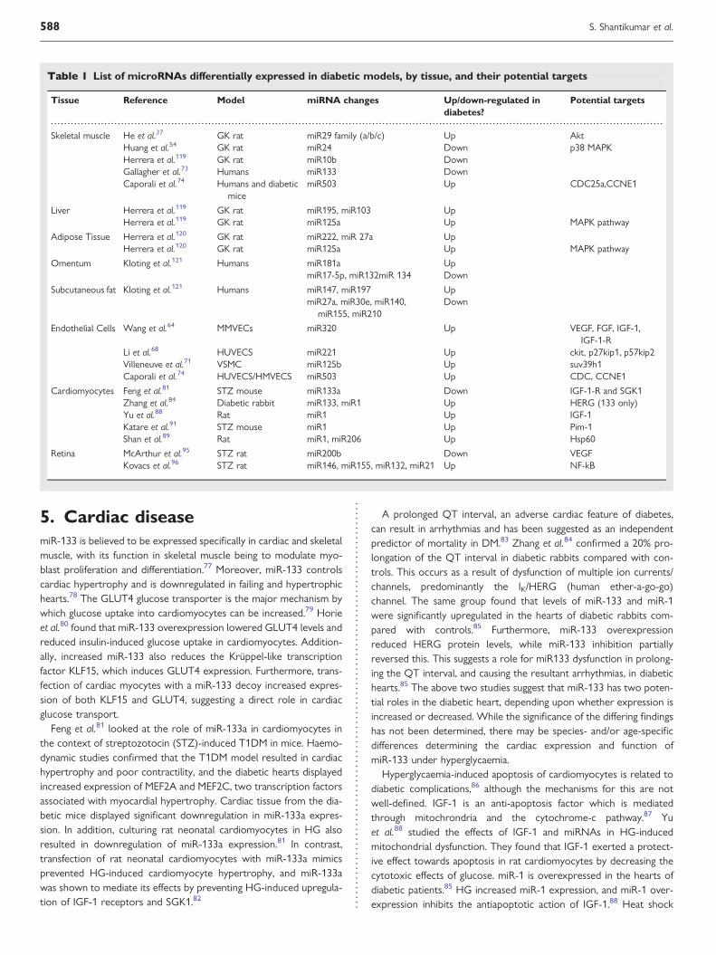
A list of miRNAs differentially expressed in diabetic models can befound in Table 1.
4. Endothelial functionand angiogenesisVascular complications associated with hyperglycaemia in diabetesoften begin with endothelial dysfunction.58 Altered expression of mul-tiple factors results in capillary and arteriole rarefaction in limbs andheart modifications59 and a reduced post-ischaemic angiogenic andcollateral vessel formation.60–62 Conversely, a pro-angiogenic patho-genic phenotype is found in the retina.63 Wang et al.64 were the firstto describe that miRNAs are differently expressed in endothelial cells(ECs) in the presence of HG. They studied myocardial microvascularECs (MMVEC) and compared miRNA expression in GK and Wistarrats. Of those differentially expressed, miR-320 may target severalangiogenic factors and their receptors, including vascular endothelialgrowth factor (VEGF)-A, fibroblast growth factors (FGFs), insulin-likegrowth factor 1 (IGF-1), and the IGF-1 receptor.65,66 Moreover, ele-vated miR-320 level in diabetic MMVECs was accompanied bydecreased cell proliferation and migration. Subsequent transfectionof an miRNA-320 inhibitor in MMVECs of GK rats improved bothproliferation and migration of these cells and increased the expressionof IGF-1, which is known to promote angiogenesis.67 The correlationbetween elevated miR-320 and suppressed IGF-1 may play a role inthe impaired angiogenesis in diabetes.
Li et al.68 studied the role of miR-221 in diabetes-induced endothe-lial dysfunction. miR-221, which is present in human umbilical vein ECs(HUVECs), participates in angiogenesis regulation by altering the ex-pression of c-kit (CD117), the receptor for stem cell factor whichadditionally promotes endothelial progenitor cell (EPC) migrationand homing.69 Incubating HUVECs in HG increased miR-221 expres-sion and reduced c-kit expression, while a miR-221 inhibitor reversedthis inhibitory effect of HG on c-kit expression.68 Another study byTogliatto et al.70 examined the role of miR-221 and miR-222 in HGand advanced glycation end-product (AGE)-mediated vasculardamage, both in HUVECs and in a model of angiogenesis (Matrigelplugs) in diabetic mice. They found that HG and high AGEs inhibitedcell cycle progression and resulted in impaired EC and EPC prolifer-ation as well as reduced angiogenesis. These conditions were alsoassociated with downregulation of both miR-221 and miR-222 ex-pression. Additionally, miR-221 and miR-222 were found to directlyinhibit P27KIP1 and P57KIP2 (cyclin-dependent kinase inhibitor pro-teins that inhibit the cell cycle). Hence, these miRNAs are likely tobe directly involved in AGE/HG-related cell cycle changes.70 Theresults regarding the regulation of miR-221 by glucose levels inHUVECs are contradictory. While there were some differences inthe cell culture methods, the results in Togliatto et al.70 were con-firmed in vivo. Clearly, further research is needed regarding the roleof miR-221 in the context of HG. Villeneuve et al.71 examined therole of miR-125b in vascular smooth muscle cells (VSMCs) cultured
S. Shantikumar et al.586

from T2DM db/db mice. They found that HG upregulated miR-125b,with parallel downregulation of its predicted target Suv39h1, ahistone-lysine N-methyltransferase. The reduced recruitment ofSuv39h1 at inflammatory gene promoters is a key mechanism under-lying the enhanced inflammatory gene expression in db/db micro-vascular VSMCs (MVSMCs).72 miR-125b mimics inhibited Suv39h1protein levels and miR-125b inhibitors had the opposite effect. Fur-thermore, miR-125b-mediated Suv39h1 knockdown resulted in theincreased expression of inflammatory proteins (interleukin 6 andmonocyte chemotactic protein 1) and increased monocyte-MVSMCbinding in hyperglycaemia, thus exhibiting a role for miR-125b in ac-celerating atherosclerosis.71
miR-503 is upregulated in myocardial ECs from GK rats,64 in3T3-L1 insulin-resistant adipocytes,42 and in the muscles of T2DMand insulin-resistant patients.73 We further examined the role ofmiR-503 in angiogenesis in diabetes.74 miR-503 was upregulated inHUVECs and human microvascular ECs (HMVECs) cultured in HG/low growth factor conditions (that mimic ischaemia-induced tissuestarvation in the context of diabetes). HUVECs infected with a lenti-viral vector expressing premiR-503 showed impaired proliferation, mi-gration, and cell networking capacities. Under similar conditions,miR-503 reduced expression of the cell cycle regulators cdc25A
and cyclin E1 (CCNE1). Furthermore, miR-503 inhibition in HG/lowgrowth factor conditions restored normal EC proliferation and angio-genesis.74 We went on to examine the role of miR-503 in diabeticlimb ischaemia to find that diabetes increased miR-503 expressionin the ischaemic muscles and ECs extracted from them in comparisonto non-diabetic/non-ischaemic controls. Moreover, in diabetic micewith induced limb ischaemia, local miR-503 inhibition by injection ofan adenoviral vector containing a decoy sequence for miR-503improved capillary and arteriolar density, promoted blood flow recov-ery, and normalized the expression of cdc25 and CCNE1. Important-ly, miR-503 expression is also increased in the limb muscles andplasma of diabetic patients undergoing amputation for critical limb is-chaemia compared with calf biopsies of non-diabetic/non-ischaemiccontrols.74 Our study demonstrates that miR-503 may be a suppres-sor of post-ischaemic neovascularization in diabetes and thus a poten-tial therapeutic target. The roles of miRNAs in endothelial dysfunctionand angiogenesis in diabetes are summarized in Figure 3A.
Atherosclerosis is a major vascular complication of diabetes.Although there is little research on miRNAs in atherosclerosis inthe context of diabetes, the role of miRNAs in the pathogenesis ofatherosclerosis in general has been the subject of two recentreviews.75,76
Figure 2 MicroRNAs involved in insulin resistance.
miRNA in diabetes-related cardiovascular diseases 587
5. Cardiac diseasemiR-133 is believed to be expressed specifically in cardiac and skeletalmuscle, with its function in skeletal muscle being to modulate myo-blast proliferation and differentiation.77 Moreover, miR-133 controlscardiac hypertrophy and is downregulated in failing and hypertrophichearts.78 The GLUT4 glucose transporter is the major mechanism bywhich glucose uptake into cardiomyocytes can be increased.79 Horieet al.80 found that miR-133 overexpression lowered GLUT4 levels andreduced insulin-induced glucose uptake in cardiomyocytes. Addition-ally, increased miR-133 also reduces the Kruppel-like transcriptionfactor KLF15, which induces GLUT4 expression. Furthermore, trans-fection of cardiac myocytes with a miR-133 decoy increased expres-sion of both KLF15 and GLUT4, suggesting a direct role in cardiacglucose transport.
Feng et al.81 looked at the role of miR-133a in cardiomyocytes inthe context of streptozotocin (STZ)-induced T1DM in mice. Haemo-dynamic studies confirmed that the T1DM model resulted in cardiachypertrophy and poor contractility, and the diabetic hearts displayedincreased expression of MEF2A and MEF2C, two transcription factorsassociated with myocardial hypertrophy. Cardiac tissue from the dia-betic mice displayed significant downregulation in miR-133a expres-sion. In addition, culturing rat neonatal cardiomyocytes in HG alsoresulted in downregulation of miR-133a expression.81 In contrast,transfection of rat neonatal cardiomyocytes with miR-133a mimicsprevented HG-induced cardiomyocyte hypertrophy, and miR-133awas shown to mediate its effects by preventing HG-induced upregula-tion of IGF-1 receptors and SGK1.82
A prolonged QT interval, an adverse cardiac feature of diabetes,can result in arrhythmias and has been suggested as an independentpredictor of mortality in DM.83 Zhang et al.84 confirmed a 20% pro-longation of the QT interval in diabetic rabbits compared with con-trols. This occurs as a result of dysfunction of multiple ion currents/channels, predominantly the IK/HERG (human ether-a-go-go)channel. The same group found that levels of miR-133 and miR-1were significantly upregulated in the hearts of diabetic rabbits com-pared with controls.85 Furthermore, miR-133 overexpressionreduced HERG protein levels, while miR-133 inhibition partiallyreversed this. This suggests a role for miR133 dysfunction in prolong-ing the QT interval, and causing the resultant arrhythmias, in diabetichearts.85 The above two studies suggest that miR-133 has two poten-tial roles in the diabetic heart, depending upon whether expression isincreased or decreased. While the significance of the differing findingshas not been determined, there may be species- and/or age-specificdifferences determining the cardiac expression and function ofmiR-133 under hyperglycaemia.
Hyperglycaemia-induced apoptosis of cardiomyocytes is related todiabetic complications,86 although the mechanisms for this are notwell-defined. IGF-1 is an anti-apoptosis factor which is mediatedthrough mitochrondria and the cytochrome-c pathway.87 Yuet al.88 studied the effects of IGF-1 and miRNAs in HG-inducedmitochondrial dysfunction. They found that IGF-1 exerted a protect-ive effect towards apoptosis in rat cardiomyocytes by decreasing thecytotoxic effects of glucose. miR-1 is overexpressed in the hearts ofdiabetic patients.85 HG increased miR-1 expression, and miR-1 over-expression inhibits the antiapoptotic action of IGF-1.88 Heat shock
. . . . . . . . . . . . . . . . . . . . . . . . . . . . . . . . . . . . . . . . . . . . . . . . . . . . . . . . . . . . . . . . . . . . . . . . . . . . . . . . . . . . . . . . . . . . . . . . . . . . . . . . . . . . . . . . . . . . . . . . . . . . . . . . . . . . . . . . . . . . . . . . . . . . . . . . . . . . . . . . . . . . . . . . . . . . . . . . . . . . . . .
Table 1 List of microRNAs differentially expressed in diabetic models, by tissue, and their potential targets
Tissue Reference Model miRNA changes Up/down-regulated indiabetes?
Potential targets
Skeletal muscle He et al.37 GK rat miR29 family (a/b/c) Up AktHuang et al.54 GK rat miR24 Down p38 MAPKHerrera et al.119 GK rat miR10b DownGallagher et al.73 Humans miR133 DownCaporali et al.74 Humans and diabetic
micemiR503 Up CDC25a,CCNE1
Liver Herrera et al.119 GK rat miR195, miR103 UpHerrera et al.119 GK rat miR125a Up MAPK pathway
Adipose Tissue Herrera et al.120 GK rat miR222, miR 27a UpHerrera et al.120 GK rat miR125a Up MAPK pathway
Omentum Kloting et al.121 Humans miR181a UpmiR17-5p, miR132miR 134 Down
Subcutaneous fat Kloting et al.121 Humans miR147, miR197 UpmiR27a, miR30e, miR140,
miR155, miR210Down
Endothelial Cells Wang et al.64 MMVECs miR320 Up VEGF, FGF, IGF-1,IGF-1-R
Li et al.68 HUVECS miR221 Up ckit, p27kip1, p57kip2Villeneuve et al.71 VSMC miR125b Up suv39h1Caporali et al.74 HUVECS/HMVECS miR503 Up CDC, CCNE1
Cardiomyocytes Feng et al.81 STZ mouse miR133a Down IGF-1-R and SGK1Zhang et al.84 Diabetic rabbit miR133, miR1 Up HERG (133 only)Yu et al.88 Rat miR1 Up IGF-1Katare et al.91 STZ mouse miR1 Up Pim-1Shan et al.89 Rat miR1, miR206 Up Hsp60
Retina McArthur et al.95 STZ rat miR200b Down VEGFKovacs et al.96 STZ rat miR146, miR155, miR132, miR21 Up NF-kB
S. Shantikumar et al.588

protein (Hsp) 60 prevents apoptotic cardiomyocyte death, but hasbeen shown to be underexpressed in the diabetic heart.89
Shan et al.89 examined miR-1 and miR-206 expression in thehyperglycaemic rat myocardium as well as in rat neonatal cardiomyo-cytes exposed to HG. They found that HG induced upregulation ofmiR-1 and miR-206, and that both miRNAs negatively regulatedHsp60 expression. Pim-1 (proviral integration site for Moloneymurine leukaemia virus-1) plays a key role in the cardiac responseto stressors.90 We found that in STZ-T1DM mice, Pim-1 levelsdecline during progression of diabetic cardiomyopathy, andthis was associated with a rise in miR-1 expression.91 Furthermore,
both forced Pim-1 expression and miR-1 inhibition rescuedPim-1 levels in cardiomyocytes under HG conditions, and thisresulted in a restoration of prosurvival signalling and reduction incardiomyocyte apoptosis, suggesting a direct role of miR-1 in inhibit-ing Pim-1.91
Another study looking into cardiomyocyte glucose metabolismfound that miR-223 was upregulated in the left ventricle of T2DMpatients. Moreover, miR-223 overexpression induces GLUT4protein levels in cardiomyocytes,92 and in vivo miR-223 inhibition sig-nificantly decreased GLUT4 expression. Given that GLUT4 is down-regulated in the diabetic heart, it is possible that increasing miR-223
Figure 3 (A) MicroRNAs involved in endothelial dysfunction and angiogenesis in the hyperglycaemic environment. Note the conflicting effects ofHG on miR-221 from two separate studies (green arrows68 and red arrows70). (B) microRNAs in cardiovascular disease in diabetes.
miRNA in diabetes-related cardiovascular diseases 589
levels is an adaptive response aimed to restore normal glucose uptakein the insulin-resistant heart.
The miRNAs involved in cardiovascular disease in diabetes aresummarized in Figure 3B.
6. RetinopathyDiabetic retinopathy (DR), one of the leading causes of blindness indeveloped countries,93 is a microvascular complication of diabeteswith multiple underlying pathogenic mechanisms. The altered expres-sion of growth factors in sustained hyperglycaemia results in bothstructural and functional changes in the retina.63 Elevated VEGF-Alevels, increasing both vessel permeability and neovascularization,have long been associated with diabetic retinopathy.94 Only two pub-lished studies have specifically looked at the role of miRNAs in DR.McArthur et al.95 investigated whether miRNA alterations areinvolved in DR in a STZ-T1DM rat model, which is known to resultin early changes of DR. miR-200b, which targets VEGF-A, wasfound downregulated in the retinas of diabetic rats. Moreover, invitro exposure of HUVECs and bovine retinal capillary ECs (BRECs)to HG resulted in a downregulation of miR-200b and an upregulationin VEGF-A mRNA.95 Subsequent transfection of a miR-200b inhibitorin HUVECs cultured under normal conditions resulted in a gluco-mimetic effect of upregulating VEGF-A.95 VEGF-A-mediated vascularpermeability and tube formation in HUVECs exposed to HG werereduced upon miR-200b mimic transfection.95 Additional experimen-tation was performed in animal models: miR-200b mimic injection intothe vitreous humour of one eye of diabetic mice resulted in localdecreased VEGF-A expression and vascular. Conversely, intravitrealinjection of miR-200b antagomir increased VEGF-A expression, thussuggesting a role for miR-200b in the pathogenesis of diabetic retinop-athy and raising support for intravitreal injections as a possible thera-peutic route for this condition.95
Kovacs et al.96 performed miRNA expression profiling in the retinaand retinal ECs of STZ-T1DM rats. NF-kB is a regulator of theimmune response and is known to play an important role in theearly pathogenesis of DR by triggering a pro-apoptotic program inretinal pericytes.97 miRNAs which are thought to be transcriptionallyregulated by NF-kB (miR-146, miR-155, and miR- 21)98–100 were alsodemonstrated to be upregulated in the retinal ECs of diabetic rats.96
The authors confirmed that NF-kB was able to directly activatemiR-146 expression. Furthermore, miR-146 overexpression inhibitedinterleukin-1b-induced NF-kB activation in retinal ECs, representing aregulatory negative feedback loop to control NF-kB and miR-146expression.96 Thus, overexpression of miR-146 could be exploitedtherapeutically through inhibition of NF-kB activation in DR.
7. BiomarkersThere are currently no reliable plasma biomarkers for vascular diseaseand endothelial dysfunction. Furthermore, current imaging methodscan visualize the latter stages of macrovascular disease, e.g. athero-sclerotic plaque, but not endothelial dysfunction, particularly at thelevel of microcirculation. Levels of miRNAs in the serum of humanshave been shown to be stable, reproducible, and consistentamongst healthy individuals, allowing them to be of potential valueas biomarkers of disease.101 The stability of circulating miRNAs maybe explained by their being carried in membrane-bound vesicles,102
complexed with Argonaute2,103,104 or transported by high-density
lipoproteins.104 The potential role of miRNAs as biomarkers inwider cardiovascular disease is the subject of another review in thisissue. The discovery of potential-specific biomarkers may helppredict or detect the development and progression of diabetes and/or diabetes complications at an early stage, and therefore allowtimely intervention to delay severe complications.
Zampetaki et al.105 aimed to understand if there is a plasma miRNAsignature for T2DM using a prospective population-based cohort.They studied 80 patients with T2DM and a further 80 age- andgender-matched controls.105 In T2DM, miR-28-3p wasoverexpressed, and a further 12 miRNAs were underexpressed.These findings were confirmed in T2DM ob/ob mice. A decrease incirculating miR-126 in non-diabetic people was found to be a signifi-cant predictor of DM. In fact, the authors detected a gradualdecline in plasma levels of miR-126 from normal glucose tolerance,through IGT, to diabetes. Some additional miRNAs were predictiveof the future development of diabetes: miR-15a, miR-29b, miR-126,and miR-223 were lower in those subjects who went on thedevelop DM.105 Using expression profiles of the five most significantlydifferent miRNAs (miR-15a, miR-126, miR-320, miR-223, andmiR-28-3p), controls and diabetics were correctly identified in 92and 70% of cases, respectively.105 Of note, the diabetic patientswho were not detected by the profiling method had lower fastingglucose levels or had well-controlled diabetes. This research suggeststhat the use of a miRNAs signature as a biomarker may be a usefulpredictive tool in diabetes, although this theory will require confirm-ation in larger prospective populations and in groups with T1DM.miR-126 is known to be highly expressed in ECs and to regulateangiogenesis.106,107 It facilitates VEGF-A signalling by inhibiting twoof the negative regulators of the VEGF pathway, the Sprouty-relatedprotein SPRED1 and phosphoinositol-3 kinase regulatory subunit 2(PIK3R2).106 miR-126 is also the most abundant miRNA in endothelialapoptotic bodies, which are thought to be a novel form of communi-cation between cells.108 Zampetaki et al. showed that HG conditionsreduced the miR-126 content in endothelial apoptotic bodies pro-duced by HUVECs in vitro. The authors speculate that, as a conse-quence of the aforementioned, low delivery of miR-126 tomonocytes in diabetes may result in the previously describedreduced VEGF-A and subsequent endothelial dysfunction, resultingin defective collateral vessel development.109 Furthermore, decreasingmiR-126 levels in plasma were associated with a low ankle brachialpressure index and subsequent new-onset peripheral vascular disease.
Another study explored seven diabetes-related serum miRNAs(miR-9, miR-29a, miR-34a, miR-30d, miR-124a, miR-146a, andmiR-375) in patients with diabetes, IGT, and in people susceptibleto T2DM with a normal oral glucose tolerance test.110 While allseven of the tested miRNAs were significantly upregulated in T2DMcases, there was no difference in the expressed levels between IGTcases and normal susceptible controls.110 However, the use of con-trols that were susceptible to T2DM, rather than those without thissusceptibility, may have had a bearing on this result.
8. Therapeutic applicationsThe deregulation of miRNA function has been linked to diabetes, al-though it is not yet fully certain whether this is a cause or effect of thepathology. If miRNAs are indeed active in the pathogenesis of diabetesand its related complications, the restoration of normal function bymodifying the expression of specific miRNAs may be a therapeutic
S. Shantikumar et al.590

target for managing this disease. Chemically modified siRNA-like oli-gonucleotides have been used to decrease miRNA expression (antag-omirs) in vivo.111,112 However, due to the hypothetical transient natureof their effects, it is likely that frequent doses may be required tosustain benefit. Given the chronic nature of diabetes, this wouldrequire the need for repeated injections with their associated costs.However, a recent study treating chimpanzees with locked nucleicacid-modified oligonucleotides complimentary to miR-122 foundthat serum cholesterol remained low for over 10 weeks after cessa-tion of treatment, suggesting that longer-lasting effects may be pos-sible.113 Adeno-associated virus (AAV) vectors containing miRNAmimics have been found to promote miRNA expression in vivo.114
For example, Kota et al.115 injected mice with AAV.miR-26a in thecontext of hepatic cancer. The rise in miR-26a resulted in protectionfrom cancer progression without signs of toxicity. Delivery of theseagents to specific tissue targets poses a further problem. DifferentAAV serotypes have been shown to favour specific tissues, forexample serotypes 6, 8, and 9 are predominantly directed to skeletalmuscle, liver, and the heart, respectively.116 These results suggest thepromises of AAV-mediated miRNA therapeutics. We have alsosuccessfully used an adenoviral vector to convey a decoy to inhibitmiR-503, which is pathogenic in the setting of diabetic limb ischae-mia.74 However, for their short transgene expression, adenovirusmay not be the best approach to treat diabetes and its chroniccomplications in the clinical arena. Another therapeutic strategyinvolves ‘miRNA sponges’. These artificial miRNA decoys bindnative miRNA to create a loss of function of a particular miRNA.These sponges contain multiple binding sites directed against aparticular miRNA or against an miRNA seed sequence family.117
miRNA sponges have already been used in vivo to decrease the activityof miR-31 and its role in cancer development.118 The use of miRNAsponges in an AAV vector delivery system maybe a potentialnovel strategy for miRNA therapeutics. While the first reports onmiRNA therapeutics are encouraging, the fact that a typical miRNAtargets several genes suggests that clinical intervention may be verycomplex.
9. ConclusionsmiRNAs belong to a class of non-coding RNAs which are involved inthe pathogenesis of several diseases. Although many miRNAs havealready been identified, their predicted target genes need to be fullyresearched and functionally characterized. The complexities in thepathogenesis of diabetes make this more challenging. Emerging evi-dence suggests that miRNAs are differentially expressed, and indeedhave a potential causative role, in diabetes and its related cardiovascu-lar complications. In future, these mechanisms may be exploited tohelp define specific clinical biomarkers and allow appropriate thera-peutic intervention in the management of diabetes.
Conflict of interest: none declared.
FundingS.S. is a British Heart Foundation (BHF) PhD student and C.E. is a BHFSenior Research Fellow. This article was supported by BHF grants(FS/10/001/27959 and FS/10/61/28566 to C.E.). Funding to pay theOpen Access publication charges for this article was provided by the BHF.
References1. Adams KF, Schatzkin A, Harris TB, Kipnis V, Mouw T, Ballard-Barbash R et al. Over-
weight, obesity, and mortality in a large prospective cohort of persons 50 to 71years old. N Engl J Med 2006;355:763–778.
2. Shaw JE, Sicree RA, Zimmet PZ. Global estimates of the prevalence of diabetes for2010 and 2030. Diabetes Res Clin Pract 9999;87:4–14.
3. Centers for Disease Control and Prevention. National Diabetes Factsheet: NationalEstimates and General Information on Diabetes and Prediabetes in the United States.Atlanta, GA: Department of Health and Human Services, Centers for DiseaseControl and Prevention; 2011.
4. Ilonen J, Akerblom HK. New technologies and genetics of type 1 diabetes. DiabetesTechnol Ther 1999;1:205–207.
5. Kahn SE. Clinical review 135: the importance of beta-cell failure in the developmentand progression of type 2 diabetes. J Clin Endocrinol Metab 2001;86:4047–4058.
6. Winer N, Sowers JR. Epidemiology of diabetes. J Clin Pharmacol 2004;44:397–405.7. Lee RC, Feinbaum RL, Ambros V. The C. elegans heterochronic gene lin-4 encodes
small RNAs with antisense complementarity to lin-14. Cell 1993;75:843–854.8. Wightman B, Ha I, Ruvkun G. Posttranscriptional regulation of the heterochronic
gene lin-14 by lin-4 mediates temporal pattern formation in C. elegans. Cell 1993;75:855–862.
9. Zeng Y, Cullen BR. Recognition and cleavage of primary microRNA transcripts.Methods Mol Biol 2006;342:49–56.
10. He L, Hannon GJ. MicroRNAs: small RNAs with a big role in gene regulation. NatRev Genet 2004;5:522–531.
11. van Rooij E. The art of microRNA research. Circ Res 2011;108:219–234.12. Yang W, Lee DY, Ben-David Y. The roles of microRNAs in tumorigenesis and angio-
genesis. Int J Physiol Pathophysiol Pharmacol 2011;3:140–155.13. Nicolas FE, Lopez-Martinez AF. MicroRNAs in human diseases. Recent Pat DNA Gene
Seq 2010;4:142–154.14. Lorenzen JM, Haller H, Thum T. MicroRNAs as mediators and therapeutic targets in
chronic kidney disease. Nat Rev Nephrol 2011;7:286–294.15. Poy MN, Eliasson L, Krutzfeldt J, Kuwajima S, Ma X, Macdonald PE et al. A pancreatic
islet-specific microRNA regulates insulin secretion. Nature 2004;432:226–230.16. Li Y, Xu X, Liang Y, Liu S, Xiao H, Li F et al. miR-375 enhances palmitate-induced
lipoapoptosis in insulin-secreting NIT-1 cells by repressing myotrophin (V1)protein expression. Int J Clin Exp Pathol 2010;3:254–264.
17. Norlin S, Ahlgren U, Edlund H. Nuclear factor-{kappa}B activity in {beta}-cells isrequired for glucose-stimulated insulin secretion. Diabetes 2005;54:125–132.
18. Gupta S, Sen S. Myotrophin-kappaB DNA interaction in the initiation process ofcardiac hypertrophy. Biochim Biophys Acta 2002;1589:247–260.
19. El Ouaamari A, Baroukh N, Martens GA, Lebrun P, Pipeleers D, van Obberghen E.miR-375 targets 3′-phosphoinositide-dependent protein kinase-1 and regulatesglucose-induced biological responses in pancreatic beta-cells. Diabetes 2008;57:2708–2717.
20. Poy MN, Hausser J, Trajkovski M, Braun M, Collins S, Rorsman P et al. miR-375 main-tains normal pancreatic alpha- and beta-cell mass. Proc Natl Acad Sci USA 2009;106:5813–5818.
21. Zhao H, Guan J, Lee HM, Sui Y, He L, Siu JJ et al. Up-regulated pancreatic tissuemicroRNA-375 associates with human type 2 diabetes through beta-cell deficitand islet amyloid deposition. Pancreas 2010;39:843–846.
22. Plaisance V, Abderrahmani A, Perret-Menoud V, Jacquemin P, Lemaigre F, Regazzi R.MicroRNA-9 controls the expression of Granuphilin/Slp4 and the secretory re-sponse of insulin-producing cells. J Biol Chem 2006;281:26932–26942.
23. Lovis P, Gattesco S, Regazzi R. Regulation of the expression of components of theexocytotic machinery of insulin-secreting cells by microRNAs. Biol Chem 2008;389:305–312.
24. Baroukh N, Ravier MA, Loder MK, Hill EV, Bounacer A, Scharfmann R et al.MicroRNA-124a regulates Foxa2 expression and intracellular signaling in pancreaticbeta-cell lines. J Biol Chem 2007;282:19575–19588.
25. Puigserver P, Rodgers JT. Foxa2, a novel transcriptional regulator of insulin sensitiv-ity. Nat Med 2006;12:38–39.
26. Guay C, Roggli E, Nesca V, Jacovetti C, Regazzi R. Diabetes mellitus, amicroRNA-related disease? Transl Res 2011;157:253–264.
27. Lovis P, Roggli E, Laybutt DR, Gattesco S, Yang JY, Widmann C et al. Alterations inmicroRNA expression contribute to fatty acid-induced pancreatic beta-cell dysfunc-tion. Diabetes 2008;57:2728–2736.
28. Roggli E, Britan A, Gattesco S, Lin-Marq N, Abderrahmani A, Meda P et al. Involve-ment of microRNAs in the cytotoxic effects exerted by proinflammatory cytokineson pancreatic beta-cells. Diabetes 2010;59:978–986.
29. Tang X, Muniappan L, Tang G, Ozcan S. Identification of glucose-regulated miRNAsfrom pancreatic {beta} cells reveals a role for miR-30d in insulin transcription. RNA2009;15:287–293.
30. Sun LL, Jiang BG, Li WT, Zou JJ, Shi YQ, Liu ZM. MicroRNA-15a positively regulatesinsulin synthesis by inhibiting uncoupling protein-2 expression. Diabetes Res Clin Pract2011;91:94–100.
miRNA in diabetes-related cardiovascular diseases 591

31. Chan CB, De Leo D, Joseph JW, McQuaid TS, Ha XF, Xu F et al. Increased uncoup-ling protein-2 levels in beta-cells are associated with impaired glucose-stimulatedinsulin secretion: mechanism of action. Diabetes 2001;50:1302–1310.
32. Esguerra JL, Bolmeson C, Cilio CM, Eliasson L. Differential glucose-regulation ofMicroRNAs in pancreatic islets of non-obese type 2 diabetes model Goto-Kakizakirat. PLoS ONE 2011;6:e18613.
33. Goossens GH. The role of adipose tissue dysfunction in the pathogenesis ofobesity-related insulin resistance. Physiol Behav 2008;94:206–218.
34. Xu P, Vernooy SY, Guo M, Hay BA. The Drosophila microRNA Mir-14 suppressescell death and is required for normal fat metabolism. Curr Biol 2003;13:790–795.
35. Teleman AA, Maitra S, Cohen SM. Drosophila lacking microRNA miR-278 are de-fective in energy homeostasis. Genes Dev 2006;20:417–422.
36. Bluher M, Michael MD, Peroni OD, Ueki K, Carter N, Kahn BB et al. Adipose tissueselective insulin receptor knockout protects against obesity and obesity-relatedglucose intolerance. Dev Cell 2002;3:25–38.
37. He A, Zhu L, Gupta N, Chang Y, Fang F. Overexpression of micro ribonucleic acid29, highly up-regulated in diabetic rats, leads to insulin resistance in 3T3-L1 adipo-cytes. Mol Endocrinol 2007;21:2785–2794.
38. Hotamisligil GS, Murray DL, Choy LN, Spiegelman BM. Tumor necrosis factor alphainhibits signaling from the insulin receptor. Proc Natl Acad Sci USA 1994;91:4854–4858.
39. Xie H, Lim B, Lodish HF. MicroRNAs induced during adipogenesis that accelerate fatcell development are downregulated in obesity. Diabetes 2009;58:1050–1057.
40. Esau C, Kang X, Peralta E, Hanson E, Marcusson EG, Ravichandran LV et al.MicroRNA-143 regulates adipocyte differentiation. J Biol Chem 2004;279:52361–52365.
41. Wilfred BR, Wang WX, Nelson PT. Energizing miRNA research: a review of the roleof miRNAs in lipid metabolism, with a prediction that miR-103/107 regulates humanmetabolic pathways. Mol Genet Metab 2007;91:209–217.
42. Ling HY, Ou HS, Feng SD, Zhang XY, Tuo QH, Chen LX et al. Changes in micro-RNA profile and effects of miR-320 in insulin-resistant 3T3-L1 adipocytes. ClinExp Pharmacol Physiol 2009;36:e32.
43. Karbiener M, Fischer C, Nowitsch S, Opriessnig P, Papak C, Ailhaud G et al. micro-RNA miR-27b impairs human adipocyte differentiation and targets PPARgamma.Biochem Biophys Res Commun 2009;390:247–251.
44. Krishnaswami A, Ravi-Kumar S, Lewis JM. Thiazolidinediones: a 2010 perspective.Perm J 2010;14:64–72.
45. Lee EK, Lee MJ, Abdelmohsen K, Kim W, Kim MM, Srikantan S et al. miR-130 sup-presses adipogenesis by inhibiting peroxisome proliferator-activated receptorgamma expression. Mol Cell Biol 2011;31:626–638.
46. Chang J, Nicolas E, Marks D, Sander C, Lerro A, Buendia MA et al. miR-122, a mam-malian liver-specific microRNA, is processed from PCR mRNA and may downregu-late the high affinity cationic amino acid transporter CAT-1. RNA Biol 2004;1:106–113.
47. Esau C, Davis S, Murray SF, Yu XX, Pandey SK, Pear M et al. miR-122 regulation oflipid metabolism revealed by in vivo antisense targeting. Cell Metab 2006;3:87–98.
48. Najafi-Shoushtari SH, Kristo F, Li Y, Shioda T, Cohen DE, Gerszten RE et al.MicroRNA-33 and the SREBP host genes cooperate to control cholesterol homeo-stasis. Science 2010;328:1566–1569.
49. Davalos A, Goedeke L, Smibert P, Ramirez CM, Warrier NP, Andreo U et al.miR-33a/b contribute to the regulation of fatty acid metabolism and insulin signaling.Proc Natl Acad Sci USA 2011;108:9232–9237.
50. White MF. Regulating insulin signaling and beta-cell function through IRS proteins.Can J Physiol Pharmacol 2006;84:725–737.
51. Kim JA, Wei Y, Sowers JR. Role of mitochondrial dysfunction in insulin resistance.Circ Res 2008;102:401–414.
52. Ryu HS, Park SY, Ma D, Zhang J, Lee W. The induction of microRNA targeting IRS-1is involved in the development of insulin resistance under conditions of mitochon-drial dysfunction in hepatocytes. PLoS ONE 2011;6:e17343.
53. Shi B, Sepp-Lorenzino L, Prisco M, Linsley P, deAngelis T, Baserga R. Micro RNA 145targets the insulin receptor substrate-1 and inhibits the growth of colon cancer cells.J Biol Chem 2007;282:32582–32590.
54. Huang B, Qin W, Zhao B, Shi Y, Yao C, Li J et al. MicroRNA expression profiling indiabetic GK rat model. Acta Biochim Biophys Sin (Shanghai) 2009;41:472–477.
55. Igarashi M, Wakasaki H, Takahara N, Ishii H, Jiang ZY, Yamauchi T et al. Glucose ordiabetes activates p38 mitogen-activated protein kinase via different pathways. J ClinInvest 1999;103:185–195.
56. Imai G, Satoh T, Kumai T, Murao M, Tsuchida H, Shima Y et al. Hypertension accel-erates diabetic nephropathy in Wistar fatty rats, a model of type 2 diabetes mellitus,via mitogen-activated protein kinase cascades and transforming growth factor-beta1.Hypertens Res 2003;26:339–347.
57. Susztak K, Raff AC, Schiffer M, Bottinger EP. Glucose-induced reactive oxygenspecies cause apoptosis of podocytes and podocyte depletion at the onset of diabet-ic nephropathy. Diabetes 2006;55:225–233.
58. Avogaro A, Albiero M, Menegazzo L, de Kreutzenberg S, Fadini GP. Endothelial dys-function in diabetes: the role of reparatory mechanisms. Diabetes Care 2011;34(Suppl. 2):S285–S290.
59. Emanueli C, Salis MB, Pinna A, Stacca T, Milia AF, Spano A et al. Prevention ofdiabetes-induced microangiopathy by human tissue kallikrein gene transfer. Circula-tion 2002;106:993–999.
60. Waltenberger J. Impaired collateral vessel development in diabetes: potential cellularmechanisms and therapeutic implications. Cardiovasc Res 2001;49:554–560.
61. Rivard A, Silver M, Chen D, Kearney M, Magner M, Annex B et al. Rescue ofdiabetes-related impairment of angiogenesis by intramuscular gene therapy withadeno-VEGF. Am J Pathol 1999;154:355–363.
62. Emanueli C, Graiani G, Salis MB, Gadau S, Desortes E, Madeddu P. Prophylactic genetherapy with human tissue kallikrein ameliorates limb ischemia recovery in type 1diabetic mice. Diabetes 2004;53:1096–1103.
63. Khan ZA, Chakrabarti S. Growth factors in proliferative diabetic retinopathy. ExpDiabesity Res 2003;4:287–301.
64. Wang XH, Qian RZ, Zhang W, Chen SF, Jin HM, Hu RM. MicroRNA-320 expressionin myocardial microvascular endothelial cells and its relationship with insulin-likegrowth factor-1 in type 2 diabetic rats. Clin Exp Pharmacol Physiol 2009;36:181–188.
65. Lewis BP, Shih IH, Jones-Rhoades MW, Bartel DP, Burge CB. Prediction of mamma-lian microRNA targets. Cell 2003;115:787–798.
66. John B, Enright AJ, Aravin A, Tuschl T, Sander C, Marks DS. Human MicroRNAtargets. PLoS Biol 2004;2:e363.
67. Rabinovsky ED, Draghia-Akli R. Insulin-like growth factor I plasmid therapy pro-motes in vivo angiogenesis. Mol Ther 2004;9:46–55.
68. Li Y, Song YH, Li F, Yang T, Lu YW, Geng YJ. MicroRNA-221 regulates highglucose-induced endothelial dysfunction. Biochem Biophys Res Commun 2009;381:81–83.
69. Poliseno L, Tuccoli A, Mariani L, Evangelista M, Citti L, Woods K et al. MicroRNAsmodulate the angiogenic properties of HUVECs. Blood 2006;108:3068–3071.
70. Togliatto G, Trombetta A, Dentelli P, Rosso A, Brizzi MF. MIR221/MIR222-drivenpost-transcriptional regulation of P27KIP1 and P57KIP2 is crucial for high-glucose-and AGE-mediated vascular cell damage. Diabetologia 2011;54:1930–1940.
71. Villeneuve LM, Kato M, Reddy MA, Wang M, Lanting L, Natarajan R. Enhanced levelsof microRNA-125b in vascular smooth muscle cells of diabetic db/db mice lead toincreased inflammatory gene expression by targeting the histone methyltransferaseSuv39h1. Diabetes 2010;59:2904–2915.
72. Villeneuve LM, Reddy MA, Lanting LL, Wang M, Meng L, Natarajan R. Epigenetichistone H3 lysine 9 methylation in metabolic memory and inflammatory phenotypeof vascular smooth muscle cells in diabetes. Proc Natl Acad Sci USA 2008;105:9047–9052.
73. Gallagher IJ, Scheele C, Keller P, Nielsen AR, Remenyi J, Fischer CP et al. Integrationof microRNA changes in vivo identifies novel molecular features of muscle insulinresistance in type 2 diabetes. Genome Med 2010;2:9.
74. Caporali A, Meloni M, Vollenkle C, Bonci D, Sala-Newby GB, Addis R et al. Deregu-lation of microRNA-503 contributes to diabetes mellitus-induced impairment ofendothelial function and reparative angiogenesis after limb ischemia. Circulation2011;123:282–291.
75. Vickers KC, Remaley AT. MicroRNAs in atherosclerosis and lipoprotein metabolism.Curr Opin Endocrinol Diabetes Obes 2010;17:150–155.
76. Najafi-Shoushtari SH. MicroRNAs in Cardiometabolic Disease. Curr Atheroscler Rep2011;13:202–207.
77. Chen JF, Mandel EM, Thomson JM, Wu Q, Callis TE, Hammond SM et al. The role ofmicroRNA-1 and microRNA-133 in skeletal muscle proliferation and differentiation.Nat Genet 2006;38:228–233.
78. Care A, Catalucci D, Felicetti F, Bonci D, Addario A, Gallo P et al. MicroRNA-133controls cardiac hypertrophy. Nat Med 2007;13:613–618.
79. Tian R, Abel ED. Responses of GLUT4-deficient hearts to ischemia underscore theimportance of glycolysis. Circulation 2001;103:2961–2966.
80. Horie T, Ono K, Nishi H, Iwanaga Y, Nagao K, Kinoshita M et al. MicroRNA-133regulates the expression of GLUT4 by targeting KLF15 and is involved in metaboliccontrol in cardiac myocytes. Biochem Biophys Res Commun 2009;389:315–320.
81. Feng B, Chen S, George B, Feng Q, Chakrabarti S. miR133a regulates cardiomyocytehypertrophy in diabetes. Diabetes Metab Res Rev 2010;26:40–49.
82. Aoyama T, Matsui T, Novikov M, Park J, Hemmings B, Rosenzweig A. Serum andglucocorticoid-responsive kinase-1 regulates cardiomyocyte survival and hyper-trophic response. Circulation 2005;111:1652–1659.
83. Rossing P, Breum L, Major-Pedersen A, Sato A, Winding H, Pietersen A et al. Pro-longed QTc interval predicts mortality in patients with type 1 diabetes mellitus.Diabet Med 2001;18:199–205.
84. Zhang Y, Xiao J, Lin H, Luo X, Wang H, Bai Y et al. Ionic mechanisms underlyingabnormal QT prolongation and the associated arrhythmias in diabetic rabbits: arole of rapid delayed rectifier K+ current. Cell Physiol Biochem 2007;19:225–238.
85. Xiao J, Luo X, Lin H, Zhang Y, Lu Y, Wang N et al. MicroRNA miR-133 repressesHERG K+ channel expression contributing to QT prolongation in diabetic hearts.J Biol Chem 2007;282:12363–12367.
86. Yoon YS, Uchida S, Masuo O, Cejna M, Park JS, Gwon HC et al. Progressive attenu-ation of myocardial vascular endothelial growth factor expression is a seminal eventin diabetic cardiomyopathy: restoration of microvascular homeostasis and recoveryof cardiac function in diabetic cardiomyopathy after replenishment of local vascularendothelial growth factor. Circulation 2005;111:2073–2085.
S. Shantikumar et al.592

87. Li Y, Higashi Y, Itabe H, Song YH, Du J, Delafontaine P. Insulin-like growth factor-1receptor activation inhibits oxidized LDL-induced cytochrome C release and apop-tosis via the phosphatidylinositol 3 kinase/Akt signaling pathway. Arterioscler ThrombVasc Biol 2003;23:2178–2184.
88. Yu XY, Song YH, Geng YJ, Lin QX, Shan ZX, Lin SG et al. Glucose induces apoptosisof cardiomyocytes via microRNA-1 and IGF-1. Biochem Biophys Res Commun 2008;376:548–552.
89. Shan YX, Liu TJ, Su HF, Samsamshariat A, Mestril R, Wang PH. Hsp10 and Hsp60modulate Bcl-2 family and mitochondria apoptosis signaling induced by doxorubicinin cardiac muscle cells. J Mol Cell Cardiol 2003;35:1135–1143.
90. Muraski JA, Fischer KM, Wu W, Cottage CT, Quijada P, Mason M et al. Pim-1 kinaseantagonizes aspects of myocardial hypertrophy and compensation to pathologicalpressure overload. Proc Natl Acad Sci USA 2008;105:13889–13894.
91. Katare R, Caporali A, Zentilin L, Avolio E, Sala-Newby G, Oikawa A et al. Intraven-ous gene therapy with PIM-1 via a cardiotropic viral vector halts the progression ofdiabetic cardiomyopathy through promotion of prosurvival signaling. Circ Res 2011;108:1238–1251.
92. Lu H, Buchan RJ, Cook SA. MicroRNA-223 regulates Glut4 expression and cardio-myocyte glucose metabolism. Cardiovasc Res 2010;86:410–420.
93. Klein R, Klein BE, Moss SE, Davis MD, DeMets DL. The Wisconsin epidemiologicstudy of diabetic retinopathy. III. Prevalence and risk of diabetic retinopathy whenage at diagnosis is 30 or more years. Arch Ophthalmol 1984;102:527–532.
94. Shibuya M. Differential roles of vascular endothelial growth factor receptor-1 andreceptor-2 in angiogenesis. J Biochem Mol Biol 2006;39:469–478.
95. McArthur K, Feng B, Wu Y, Chen S, Chakrabarti S. MicroRNA-200b regulates vas-cular endothelial growth factor-mediated alterations in diabetic retinopathy. Diabetes2011;60:1314–1323.
96. Kovacs B, Lumayag S, Cowan C, Xu S. microRNAs in Early Diabetic Retinopathy inStreptozotocin-induced Diabetic Rats. Invest Ophthalmol Vis Sci 2011;52:4402–4409.
97. Kowluru RA, Koppolu P, Chakrabarti S, Chen S. Diabetes-induced activation ofnuclear transcriptional factor in the retina, and its inhibition by antioxidants. FreeRadic Res 2003;37:1169–1180.
98. Taganov KD, Boldin MP, Chang KJ, Baltimore D. NF-kappaB-dependent induction ofmicroRNA miR-146, an inhibitor targeted to signaling proteins of innate immuneresponses. Proc Natl Acad Sci USA 2006;103:12481–12486.
99. Gatto G, Rossi A, Rossi D, Kroening S, Bonatti S, Mallardo M. Epstein-Barr viruslatent membrane protein 1 trans-activates miR-155 transcription through theNF-kappaB pathway. Nucleic Acids Res 2008;36:6608–6619.
100. Sheedy FJ, Palsson-McDermott E, Hennessy EJ, Martin C, O’Leary JJ, Ruan Q et al.Negative regulation of TLR4 via targeting of the proinflammatory tumor suppressorPDCD4 by the microRNA miR-21. Nat Immunol 2010;11:141–147.
101. Gilad S, Meiri E, Yogev Y, Benjamin S, Lebanony D, Yerushalmi N et al. Serum micro-RNAs are promising novel biomarkers. PLoS ONE 2008;3:e3148.
102. Hunter MP, Ismail N, Zhang X, Aguda BD, Lee EJ, Yu L et al. Detection of microRNAexpression in human peripheral blood microvesicles. PLoS ONE 2008;3:e3694.
103. Arroyo JD, Chevillet JR, Kroh EM, Ruf IK, Pritchard CC, Gibson DF et al. Argonaute2complexes carry a population of circulating microRNAs independent of vesicles inhuman plasma. Proc Natl Acad Sci USA 2011;108:5003–5008.
104. Vickers AC, Palmisano BT, Shoucri BM, Shamburek RD, Remaley AT. MicroRNAsare transported in plasma and delivered to recipient cells by high-density lipopro-teins. Nat Cell Biol 2001;13:423–433.
105. Zampetaki A, Kiechl S, Drozdov I, Willeit P, Mayr U, Prokopi M et al. Plasma micro-RNA profiling reveals loss of endothelial miR-126 and other microRNAs in type 2diabetes. Circ Res 2010;107:810–817.
106. Fish JE, Santoro MM, Morton SU, Yu S, Yeh RF, Wythe JD et al. miR-126 regulatesangiogenic signaling and vascular integrity. Dev Cell 2008;15:272–284.
107. Wang S, Aurora AB, Johnson BA, Qi X, McAnally J, Hill JA et al. The endothelial-specific microRNA miR-126 governs vascular integrity and angiogenesis. Dev Cell2008;15:261–271.
108. Zernecke A, Bidzhekov K, Noels H, Shagdarsuren E, Gan L, Denecke B et al. Deliv-ery of microRNA-126 by apoptotic bodies induces CXCL12-dependent vascularprotection. Sci Signal 2009;2:ra81.
109. Waltenberger J, Lange J, Kranz A. Vascular endothelial growth factor-A-inducedchemotaxis of monocytes is attenuated in patients with diabetes mellitus: a potentialpredictor for the individual capacity to develop collaterals. Circulation 2000;102:185–190.
110. Kong L, Zhu J, Han W, Jiang X, Xu M, Zhao Y et al. Significance of serum microRNAsin pre-diabetes and newly diagnosed type 2 diabetes: a clinical study. Acta Diabetol2011;48:61–69.
111. Kolfschoten IG, Roggli E, Nesca V, Regazzi R. Role and therapeutic potential ofmicroRNAs in diabetes. Diabetes Obes Metab 2009;11(Suppl. 4):118–129.
112. Krutzfeldt J, Rajewsky N, Braich R, Rajeev KG, Tuschl T, Manoharan M et al. Silencingof microRNAs in vivo with ‘antagomirs’. Nature 2005;438:685–689.
113. Lanford RE, Hildebrandt-Eriksen ES, Petri A, Persson R, Lindow M, Munk ME et al.Therapeutic silencing of microRNA-122 in primates with chronic hepatitis C virusinfection. Science 2010;327:198–201.
114. Snove O Jr, Rossi JJ. Expressing short hairpin RNAs in vivo. Nat Methods 2006;3:689–695.
115. Kota J, Chivukula RR, O’Donnell KA, Wentzel EA, Montgomery CL, Hwang HWet al. Therapeutic microRNA delivery suppresses tumorigenesis in a murine livercancer model. Cell 2009;137:1005–1017.
116. Alexander IE, Cunningham SC, Logan GJ, Christodoulou J. Potential of AAV vectorsin the treatment of metabolic disease. Gene Ther 2008;15:831–839.
117. Ebert MS, Neilson JR, Sharp PA. MicroRNA sponges: competitive inhibitors of smallRNAs in mammalian cells. Nat Methods 2007;4:721–726.
118. Valastyan S, Reinhardt F, Benaich N, Calogrias D, Szasz AM, Wang ZC et al. A pleio-tropically acting microRNA, miR-31, inhibits breast cancer metastasis. Cell 2009;137:1032–1046.
119. Herrera BM, Lockstone HE, Taylor JM, Ria M, Barrett A, Collins S et al. GlobalmicroRNA expression profiles in insulin target tissues in a spontaneous rat modelof type 2 diabetes. Diabetologia 2010;53:1099–1109.
120. Herrera BM, Lockstone HE, Taylor JM, Wills QF, Kaisaki PJ, Barrett A et al. Micro-RNA-125a is over-expressed in insulin target tissues in a spontaneous rat model ofType 2 Diabetes. BMC Med Genomics 2009;2:54.
121. Kloting N, Berthold S, Kovacs P, Schon MR, Fasshauer M, Ruschke K et al. MicroRNAexpression in human omental and subcutaneous adipose tissue. PLoS One 2009;4:e4699.
miRNA in diabetes-related cardiovascular diseases 593
Related Documents