ORIGINAL ARTICLE Role of macrophage sialoadhesin in host defense against the sialylated pathogen group B Streptococcus Yung-Chi Chang & Joshua Olson & Aaron Louie & Paul R. Crocker & Ajit Varki & Victor Nizet Received: 12 February 2014 /Revised: 23 March 2014 /Accepted: 14 April 2014 # Springer-Verlag Berlin Heidelberg 2014 Abstract Several bacterial pathogens decorate their surfaces with sialic acid (Sia) residues within cell wall components or capsular exopolysaccharides. Sialic acid expression can promote bacterial virulence by blocking complement activation or by engagement of inhibitory sialic acid-binding immunoglobulin-like lectins (Siglecs) on host leukocytes. Expressed at high levels on splenic and lymph node macrophages, sialoadhesin (Sn) is a unique Siglec with an elongated structure that lacks intracellular signal- ing motifs. Sialoadhesin allows macrophage to engage certain sialylated pathogens and stimulate inflammatory responses, but the in vivo significance of sialoadhesin in infection has not been shown. We demonstrate that macrophages phagocytose the sialylated pathogen group B Streptococcus (GBS) and increase bactericidal activity via sialoadhesin-sialic-acid-mediated recog- nition. Sialoadhesin expression on marginal zone metallophillic macrophages in the spleen trapped circulating GBS and restrict- ed the spread of the GBS to distant organs, reducing mortality. Specific IgM antibody responses to GBS challenge were also impaired in sialoadhesin-deficient mice. Thus, sialoadhesin rep- resents a key bridge to orchestrate innate and adaptive immune defenses against invasive sialylated bacterial pathogens. Key message & Sialoadhesin is critical for macrophages to phagocytose and clear GBS. & Increased GBS organ dissemination in the sialoadhesin- deficient mice. & Reduced anti-GBS IgM production in the sialoadhesin- deficient mice. Keywords Metallophillic macrophage . Antibody . Siglec . Sialic acid Introduction The spleen is the largest secondary lymphoid organ in our body, receiving 5 % of the cardiac output in blood perfusion, and capable of screening the entire blood volume for patho- gens within 20–30 min [1]. The crucial role of the spleen in blood filtering is evident in asplenic individuals, who are particularly vulnerable to sepsis with encapsulated bacteria such as Neisseria meningitidis, Streptococcus pneumoniae, Electronic supplementary material The online version of this article (doi:10.1007/s00109-014-1157-y) contains supplementary material, which is available to authorized users. Y.<C. Chang : A. Varki (*) : V. Nizet Glycobiology Research and Training Center, University of California, San Diego, La Jolla, CA 92093, USA e-mail: [email protected] Y.<C. Chang : J. Olson : A. Louie : V. Nizet Department of Pediatrics, University of California, San Diego, La Jolla, CA 92093, USA A. Varki Department of Cellular and Molecular Medicine, University of California, San Diego, La Jolla, CA 92093, USA A. Varki Department of Medicine, University of California, San Diego, La Jolla, CA 92093, USA V. Nizet Skaggs School of Pharmacy and Pharmaceutical Sciences, University of California, San Diego, La Jolla, CA 92093, USA P. R. Crocker Division of Cell Signalling and Immunology, College of Life Sciences, University of Dundee, Dundee, UK V. Nizet (*) Division of Pediatric Pharmacology and Drug Discovery, University of California, San Diego, 9500 Gilman Drive, Mail Code 0687, La Jolla, CA 92093-0687, USA e-mail: [email protected] J Mol Med DOI 10.1007/s00109-014-1157-y

Welcome message from author
This document is posted to help you gain knowledge. Please leave a comment to let me know what you think about it! Share it to your friends and learn new things together.
Transcript
ORIGINAL ARTICLE
Role of macrophage sialoadhesin in host defenseagainst the sialylated pathogen group B Streptococcus
Yung-Chi Chang & Joshua Olson & Aaron Louie &
Paul R. Crocker & Ajit Varki & Victor Nizet
Received: 12 February 2014 /Revised: 23 March 2014 /Accepted: 14 April 2014# Springer-Verlag Berlin Heidelberg 2014
AbstractSeveral bacterial pathogens decorate their surfaces with sialicacid (Sia) residues within cell wall components or capsularexopolysaccharides. Sialic acid expression can promote bacterialvirulence by blocking complement activation or by engagementof inhibitory sialic acid-binding immunoglobulin-like lectins(Siglecs) on host leukocytes. Expressed at high levels on splenicand lymph node macrophages, sialoadhesin (Sn) is a unique
Siglec with an elongated structure that lacks intracellular signal-ing motifs. Sialoadhesin allows macrophage to engage certainsialylated pathogens and stimulate inflammatory responses, butthe in vivo significance of sialoadhesin in infection has not beenshown. We demonstrate that macrophages phagocytose thesialylated pathogen group B Streptococcus (GBS) and increasebactericidal activity via sialoadhesin-sialic-acid-mediated recog-nition. Sialoadhesin expression on marginal zone metallophillicmacrophages in the spleen trapped circulating GBS and restrict-ed the spread of the GBS to distant organs, reducing mortality.Specific IgM antibody responses to GBS challenge were alsoimpaired in sialoadhesin-deficient mice. Thus, sialoadhesin rep-resents a key bridge to orchestrate innate and adaptive immunedefenses against invasive sialylated bacterial pathogens.
Key message& Sialoadhesin is critical for macrophages to phagocytose
and clear GBS.& Increased GBS organ dissemination in the sialoadhesin-
deficient mice.& Reduced anti-GBS IgM production in the sialoadhesin-
deficient mice.
Keywords Metallophillic macrophage . Antibody . Siglec .
Sialic acid
Introduction
The spleen is the largest secondary lymphoid organ in ourbody, receiving 5 % of the cardiac output in blood perfusion,and capable of screening the entire blood volume for patho-gens within 20–30 min [1]. The crucial role of the spleen inblood filtering is evident in asplenic individuals, who areparticularly vulnerable to sepsis with encapsulated bacteriasuch as Neisseria meningitidis, Streptococcus pneumoniae,
Electronic supplementary material The online version of this article(doi:10.1007/s00109-014-1157-y) contains supplementary material,which is available to authorized users.
Y.<C. Chang :A. Varki (*) :V. NizetGlycobiology Research and Training Center, University ofCalifornia, San Diego, La Jolla, CA 92093, USAe-mail: [email protected]
Y.<C. Chang : J. Olson :A. Louie :V. NizetDepartment of Pediatrics, University of California, San Diego, LaJolla, CA 92093, USA
A. VarkiDepartment of Cellular and Molecular Medicine, University ofCalifornia, San Diego, La Jolla, CA 92093, USA
A. VarkiDepartment of Medicine, University of California, San Diego, LaJolla, CA 92093, USA
V. NizetSkaggs School of Pharmacy and Pharmaceutical Sciences,University of California, San Diego, La Jolla, CA 92093, USA
P. R. CrockerDivision of Cell Signalling and Immunology, College of LifeSciences, University of Dundee, Dundee, UK
V. Nizet (*)Division of Pediatric Pharmacology and Drug Discovery, Universityof California, San Diego, 9500 Gilman Drive, Mail Code 0687, LaJolla, CA 92093-0687, USAe-mail: [email protected]
J Mol MedDOI 10.1007/s00109-014-1157-y
and Haemophilus influenzae [2, 3]. Opening of the arterialblood stream into the marginal sinuses of the spleen reducesblood flow so that pathogens in the systemic circulation can beefficiently phagocytosed by marginal zone macrophages(MZMs) and marginal metallophilic macrophages (MMMs)[4, 5]. Depletion of MZMs and MMMs can result in patho-gens escaping to the blood, potentially triggering uncontrolledbacteremia and sepsis [6, 7].
MMMs, but typically not MZMs, express high levels ofsialoadhesin (Sn, sialic acid-binding immunoglobulin-likelectin-1 (Siglec-1), CD169) and form an inner ring borderingthe marginal zone and the white pulp follicular areas. AmongSiglecs, Sn is unique in possessing 17 immunoglobulin-likeextracellular domains that may extend its length ∼40 nmbeyond the cell surface and recognize sialylated ligands foundon many sialylated pathogens, such as sialylated envelopedviruses [8–10], bacteria [11, 12], and protozoa [13]. Morerecently, Sn has been shown to capture killed sialylatedCampylobacter jejuni and able to promote rapid proinflam-matory cytokine and type I interferon responses [14].However, the consequences of Sn-dependent recognition ofan invasive sialylated bacteria pathogen on infection outcomehave not been addressed.
In this work, we used the sialylated pathogen group BStreptococcus (GBS), a leading cause of human neonatalsepsis and meningitis, as a model for in vitro and in vivoanalysis of Sn function. Specifically, we studied whetherexpression of Sn in MMMs could facilitate splenic trappingof GBS during early infection and/or influence later phasehumoral responses to coordinately combat this invasiveblood-borne pathogen.
Materials and methods
Bacteria and growth condition
GBS of serotypes Ia (A909), Ib (UAB), II (DK23), III (COH1,K79 and D136), and VI (NT-6) are human neonate isolates.COH1ΔNeuA mutant and isogenic COH1 mutants express-ing different levels ofO-acetylation on the terminal sialic acid(Sia) were previously described [15, 16]. GBS were grown inTodd-Hewitt broth (Difco, BD Diagnostics) at 37 °C withoutshaking. For functional assays, bacteria were cultivated at37 °C to early log phase and resuspended to OD600 of 0.4 inappropriate buffer to the desired concentrations.
Culture of mouse bone marrow-derived macrophages(MBDMs) and in vitro stimulation with GBS
MBDMs were derived from marrow cells collected fromfemur and tibia and cultured with conditioned media contain-ing macrophage colony-stimulating factors (M-CSF) for
7 days. MBDMs were then seeded onto 24-well plates at 5×10⁵/well with M-CSF (10 ng/ml, R&D Systems) andinterferon-α (500 U/ml, PBL IFN Source) to optimize Snexpression. For phagocytosis assays, 2.5×10⁶ CFU GBSDK23 was added to 5×10⁵ macrophages (MOI=5).Following a 30-min incubation, cells were washed, addedmedium containing penicillin (5 μg/ml) and gentamicin(100 μg/ml) to kill extracellular bacteria, detached by trypsin,lysed with 0.025 % Triton X-100, and plated to enumerateintracellular bacteria. For bacterial killing, 105 GBS DK23was added to MBDMs at MOI=0.2 for 1 h, followed byaddition of 50 μl of 0.6 % Triton X-100 to lyse cells.Recovered GBS were plated for CFU enumeration. For ex-periments using Sn-neutralizing mAb (clone SER-4,10 μg/ml), the mAb was added to cells 10 min prior toinoculation with GBS. This neutralizing mAb has been shownto block the sialic-acid-mediated interaction between Sn anderythrocytes as well as the interaction between Sn andsialylated meningococcal lipopolysaccharide [11, 17].
Flow cytometry analysis of Sn expression
MBDMs were stimulated with IFN-α (500 U/ml), LPS(1 μg/ml, InvivoGen), lipoteichoic acid (1 μg/ml,InvivoGen), GBS capsule (1 μg/ml, purified as described[15]), or heat-killed GBS (108 CFU) for 2 days. Cells weredetached by 5mMEDTA/PBS, stained with FITC-conjugatedrat anti-mouse Sn (clone 3D6, Serotec), and analyzed by flowcytometer (FACSCalibur, BD Biosciences).
Siglec-Fc binding assay
Siglec-Fc and FITC-labeled GBS binding studies were mod-ified from a described method [18]. GBS was labeled with0.1 % FITC/PBS for 30 min at 37 °C with rotation. Ninety-six-well plates were coated with 1 μg/well protein A in50 mM carbonate buffer (pH 9.5) overnight at 4 °C, washed,and blocked with 1 % BSA/PBS for 1 h at room temperature.Human and murine Sn-Fc (0.5 μg) (produced as described[11]) was immobilized to individual wells for >3 h at roomtemperature. FITC-labeled GBS (107) was added to each well,and a 96-well plate was centrifuged and incubated for 30 minat 37 °C. Initial fluorescence intensity was verified, and theresidual fluorescence intensity (excitation, 488 nm; emission,530 nm) was measured using a CytoFluorII fluorescence platereader after extensive wash to remove unbound bacteria.
Immunofluorescence microscopy-based phagocytosisanalysis
GBS DK23 was labeled with PKH26 (Sigma) per manufac-turer’s instruction. Labeled GBS (2.5×10⁶ CFU) were added to5×105 macrophages and incubated for 30 min. Macrophages
J Mol Med
were washed, fixed, permeabilized, blocked with BSA, andstained with AlexaFluor 488® phalloidin (Life Technologies)and DAPI for 30 min at room temperature. Images wereacquired using the Olympus FV1000 confocal microscopeand FV1000 Viewer software.
Intravenous GBS infection and visualization of GBSin the spleen
All studies were approved by the UCSD Committee on theUse and Care of Animals and performed using acceptedveterinary standards. Generation of Sn-deficient mice haspreviously been described [19]. GBS DK23 was labeled with5-(and-6)-carboxyfluorescein (Invitrogen) per manufacturer’sinstructions. Mice were intravenously infected with 2×10⁸ CFU of labeled GBS via tail vein. Kidney, lung, andspleen were collected for CFU enumeration or frozen inOCT solution 1 h after infection. Spleen cryostat sections(5 μm) were fixed in acetone and blocked with 1 % BSA/PBST and AVIDIN/BIOTIN blocking kit (VectorLaboratories). The sections were stained with rat mAb anti-mouse Sn (clone SER-4) with Alexa Fluor 647-conjugatedgoat anti-rat IgG (Life Technologies) followed by second-stepstaining with biotinylated rat mAb anti-mouse F4/80 (cloneCI:A3-1, Serotec) or B220 (clone RA3-6B2, BD Biosciences)with Alexa Fluor 555-conjugated streptavidin (LifeTechnologies). Images were from an Olympus FV1000 con-focal microscope using FV1000 Viewer software.
GBS intraperitoneal infection mouse model
Wild-type (WT) and Sn-deficient mice (10–12 weeks) wereinfected intraperitoneally with 108 CFU of GBS DK23 andbacterial CFU in the blood monitored 4 h post-infection. Todetect anti-GBS antibody responses, mice (10–12 weeks)were first injected with 0.5 mg of anti-Ly-6G mAb (clone1A8, BioXcell) intraperitoneally to deplete neutrophils 24 hprior to infection with 2×10⁷ CFU of GBS DK23. Mousesurvival was monitored twice per day for 14 days at whichtime mice were euthanized and their blood was collected forantibody titer analysis.
ELISA of anti-GBS antibody isotypes
Fifty microliters of GBS (OD600=1.0) in 0.1 M sodium car-bonate buffer (pH 9.5) was added into 96-well plates, spun at2,000 rpm for 10 min, and incubated overnight at 4 °C. GBS-coated plates were blocked with 3 % BSA/PBST for 1 h atroom temperature and then mouse sera was added in 3-foldserial dilutions and incubated for 2 h at room temperature.Antibody titers were determined with horseradish peroxidase-conjugated immunoglobulin isotype-specific antibodies(Southern Biotechnology) and TMB substrate solution. OD
450 nm was measured with a SpectraMax M3 microplatereader.
ELISA of total antibody isotypes
Ninety-six-well plates were coated with goat anti-mouseIgG+IgA+IgM (Southern Biotechnology, 0.5 μg/well in0.1 M sodium carbonate buffer, pH 9.5) at 4 °C overnight.Plates were blocked with 3 % BSA/PBST for 1 h, and seracollected from GBS-infected mice was added (1:5,000 dilu-tion for IgM and IgA; 1:50,000 dilution for IgG) andincubated for 2 h at room temperature. Antibodies weredetected by horseradish peroxidase-conjugated immuno-globulin isotype-specific antibodies and TMB substratesolution. OD 450 nm was measured with a SpectraMaxM3 microplate reader.
Results
Sialoadhesin (Sn) binds to sialylated group B Streptococcus(GBS), and its expression is upregulated by bacterialcomponents and inflammatory stimuli
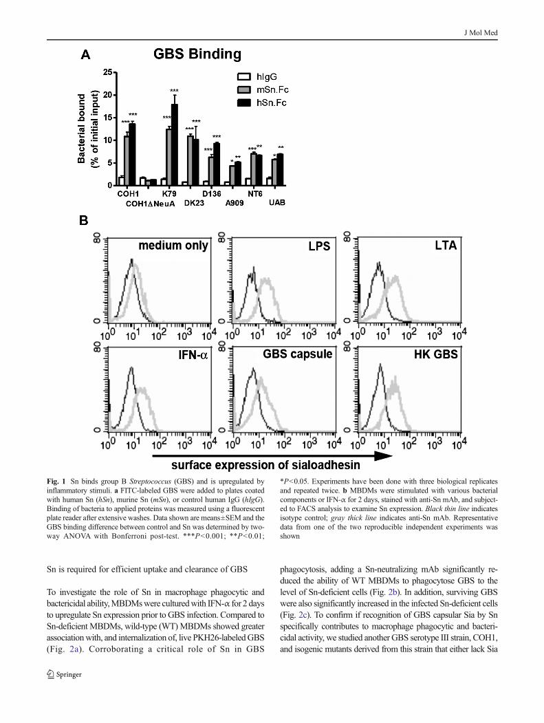
GBS is a leading cause of neonatal pneumonia and sepsis, andits surface capsular polysaccharide is invariably capped with aterminal α2-3-linked sialic acid (Sia) known to impair phago-cytosis and dampen neutrophil bactericidal activities via en-gaging inhibitory Siglecs and to block complement deposition[18, 20–23]. Sn, a unique Siglec possessing 17immunoglobulin-like extracellular domains, has been reportedto recognize sialylated ligands found on many sialylated path-ogens.We sought to determine if Sn could recognize GBS in aSia-dependent manner as a defense strategy to counteractsuppressive signals transduced by inhibitory Siglecs. SevenGBS strains (A909, UAB, DK23, COH1, K79, D136, andNT-6) tested here all bound to human Sn (hSn) and murine Sn(mSn), but the Sia-negative COH1ΔNeuA mutant did not(Fig. 1a).
Expression of Sn on human monocytes is upregulated byIFN-α and agonists for Toll-like receptors [24]. In addition,under infection or pathological conditions, expression of Snon circulating human monocytes and tissue macrophages canbe increased and correlated to disease progression [24–29].We observed that IFN-α significantly increased Sn expressionon mouse bone marrow-derived macrophages (MBDMs), asdid lipopolysaccharide (LPS), lipoteichoic acid (LTA), GBScapsular polysaccharide preparation, and heat-killed GBS(HK GBS) (Fig. 1b). Upregulation of Sn expression duringinfection could theoretically influence macrophage phagocyt-ic and bactericidal activity against GBS.
J Mol Med
Sn is required for efficient uptake and clearance of GBS
To investigate the role of Sn in macrophage phagocytic andbactericidal ability,MBDMswere culturedwith IFN-α for 2 daysto upregulate Sn expression prior to GBS infection. Compared toSn-deficient MBDMs, wild-type (WT) MBDMs showed greaterassociationwith, and internalization of, live PKH26-labeledGBS(Fig. 2a). Corroborating a critical role of Sn in GBS
phagocytosis, adding a Sn-neutralizing mAb significantly re-duced the ability of WT MBDMs to phagocytose GBS to thelevel of Sn-deficient cells (Fig. 2b). In addition, surviving GBSwere also significantly increased in the infected Sn-deficient cells(Fig. 2c). To confirm if recognition of GBS capsular Sia by Snspecifically contributes to macrophage phagocytic and bacteri-cidal activity, we studied another GBS serotype III strain, COH1,and isogenic mutants derived from this strain that either lack Sia
Fig. 1 Sn binds group B Streptococcus (GBS) and is upregulated byinflammatory stimuli. a FITC-labeled GBS were added to plates coatedwith human Sn (hSn), murine Sn (mSn), or control human IgG (hIgG).Binding of bacteria to applied proteins was measured using a fluorescentplate reader after extensive washes. Data shown are means±SEM and theGBS binding difference between control and Sn was determined by two-way ANOVA with Bonferroni post-test. ***P<0.001; **P<0.01;
*P<0.05. Experiments have been done with three biological replicatesand repeated twice. b MBDMs were stimulated with various bacterialcomponents or IFN-α for 2 days, stained with anti-Sn mAb, and subject-ed to FACS analysis to examine Sn expression. Black thin line indicatesisotype control; gray thick line indicates anti-Sn mAb. Representativedata from one of the two reproducible independent experiments wasshown
J Mol Med
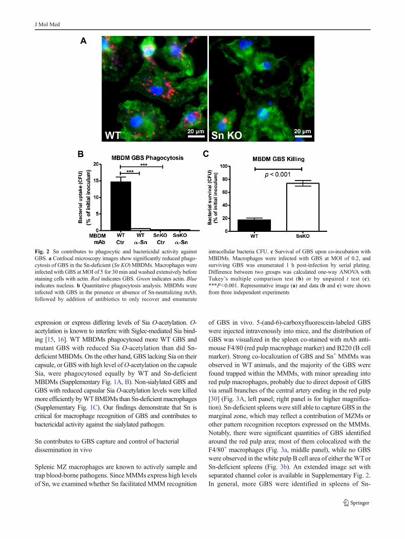
expression or express differing levels of Sia O-acetylation. O-acetylation is known to interfere with Siglec-mediated Sia bind-ing [15, 16]. WT MBDMs phagocytosed more WT GBS andmutant GBS with reduced Sia O-acetylation than did Sn-deficientMBDMs. On the other hand, GBS lacking Sia on theircapsule, or GBSwith high level ofO-acetylation on the capsuleSia, were phagocytosed equally by WT and Sn-deficientMBDMs (Supplementary Fig. 1A, B). Non-sialylated GBS andGBS with reduced capsular Sia O-acetylation levels were killedmore efficiently byWTBMDMs than Sn-deficientmacrophages(Supplementary Fig. 1C). Our findings demonstrate that Sn iscritical for macrophage recognition of GBS and contributes tobactericidal activity against the sialylated pathogen.
Sn contributes to GBS capture and control of bacterialdissemination in vivo
Splenic MZ macrophages are known to actively sample andtrap blood-borne pathogens. SinceMMMs express high levelsof Sn, we examined whether Sn facilitated MMM recognition
of GBS in vivo. 5-(and-6)-carboxyfluorescein-labeled GBSwere injected intravenously into mice, and the distribution ofGBS was visualized in the spleen co-stained with mAb anti-mouse F4/80 (red pulp macrophage marker) and B220 (B cellmarker). Strong co-localization of GBS and Sn+ MMMs wasobserved in WT animals, and the majority of the GBS werefound trapped within the MMMs, with minor spreading intored pulp macrophages, probably due to direct deposit of GBSvia small branches of the central artery ending in the red pulp[30] (Fig. 3A, left panel; right panel is for higher magnifica-tion). Sn-deficient spleens were still able to capture GBS in themarginal zone, which may reflect a contribution of MZMs orother pattern recognition receptors expressed on the MMMs.Notably, there were significant quantities of GBS identifiedaround the red pulp area; most of them colocalized with theF4/80+ macrophages (Fig. 3a, middle panel), while no GBSwere observed in the white pulp B cell area of either theWTorSn-deficient spleens (Fig. 3b). An extended image set withseparated channel color is available in Supplementary Fig. 2.In general, more GBS were identified in spleens of Sn-
Fig. 2 Sn contributes to phagocytic and bactericidal activity againstGBS. a Confocal microscopy images show significantly reduced phago-cytosis of GBS in the Sn-deficient (Sn KO) MBDMs. Macrophages wereinfected with GBS at MOI of 5 for 30 min and washed extensively beforestaining cells with actin. Red indicates GBS. Green indicates actin. Blueindicates nucleus. b Quantitative phagocytosis analysis. MBDMs wereinfected with GBS in the presence or absence of Sn-neutralizing mAb,followed by addition of antibiotics to only recover and enumerate
intracellular bacteria CFU. c Survival of GBS upon co-incubation withMBDMs. Macrophages were infected with GBS at MOI of 0.2, andsurviving GBS was enumerated 1 h post-infection by serial plating.Difference between two groups was calculated one-way ANOVA withTukey’s multiple comparison test (b) or by unpaired t test (c).***P<0.001. Representative image (a) and data (b and c) were shownfrom three independent experiments
J Mol Med
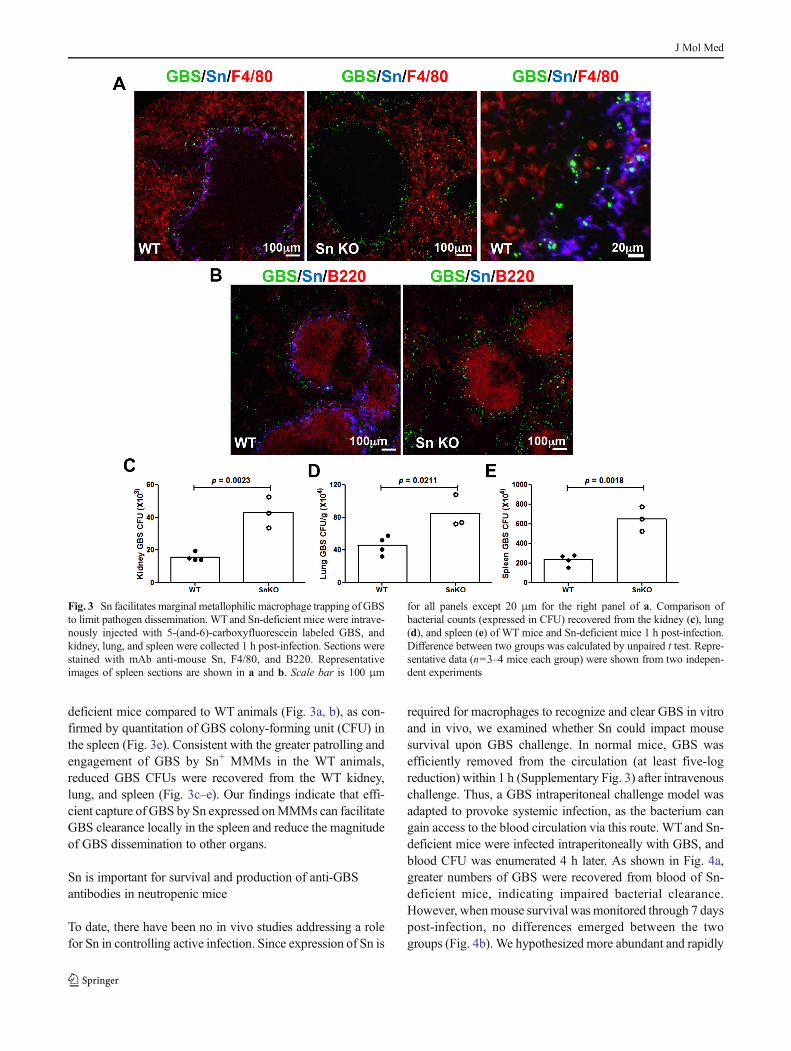
deficient mice compared to WT animals (Fig. 3a, b), as con-firmed by quantitation of GBS colony-forming unit (CFU) inthe spleen (Fig. 3e). Consistent with the greater patrolling andengagement of GBS by Sn+ MMMs in the WT animals,reduced GBS CFUs were recovered from the WT kidney,lung, and spleen (Fig. 3c–e). Our findings indicate that effi-cient capture of GBS by Sn expressed onMMMs can facilitateGBS clearance locally in the spleen and reduce the magnitudeof GBS dissemination to other organs.
Sn is important for survival and production of anti-GBSantibodies in neutropenic mice
To date, there have been no in vivo studies addressing a rolefor Sn in controlling active infection. Since expression of Sn is
required for macrophages to recognize and clear GBS in vitroand in vivo, we examined whether Sn could impact mousesurvival upon GBS challenge. In normal mice, GBS wasefficiently removed from the circulation (at least five-logreduction) within 1 h (Supplementary Fig. 3) after intravenouschallenge. Thus, a GBS intraperitoneal challenge model wasadapted to provoke systemic infection, as the bacterium cangain access to the blood circulation via this route. WTand Sn-deficient mice were infected intraperitoneally with GBS, andblood CFU was enumerated 4 h later. As shown in Fig. 4a,greater numbers of GBS were recovered from blood of Sn-deficient mice, indicating impaired bacterial clearance.However, whenmouse survival was monitored through 7 dayspost-infection, no differences emerged between the twogroups (Fig. 4b). We hypothesized more abundant and rapidly
Fig. 3 Sn facilitates marginal metallophilic macrophage trapping of GBSto limit pathogen dissemination. WT and Sn-deficient mice were intrave-nously injected with 5-(and-6)-carboxyfluorescein labeled GBS, andkidney, lung, and spleen were collected 1 h post-infection. Sections werestained with mAb anti-mouse Sn, F4/80, and B220. Representativeimages of spleen sections are shown in a and b. Scale bar is 100 μm
for all panels except 20 μm for the right panel of a. Comparison ofbacterial counts (expressed in CFU) recovered from the kidney (c), lung(d), and spleen (e) of WT mice and Sn-deficient mice 1 h post-infection.Difference between two groups was calculated by unpaired t test. Repre-sentative data (n=3–4 mice each group) were shown from two indepen-dent experiments
J Mol Med
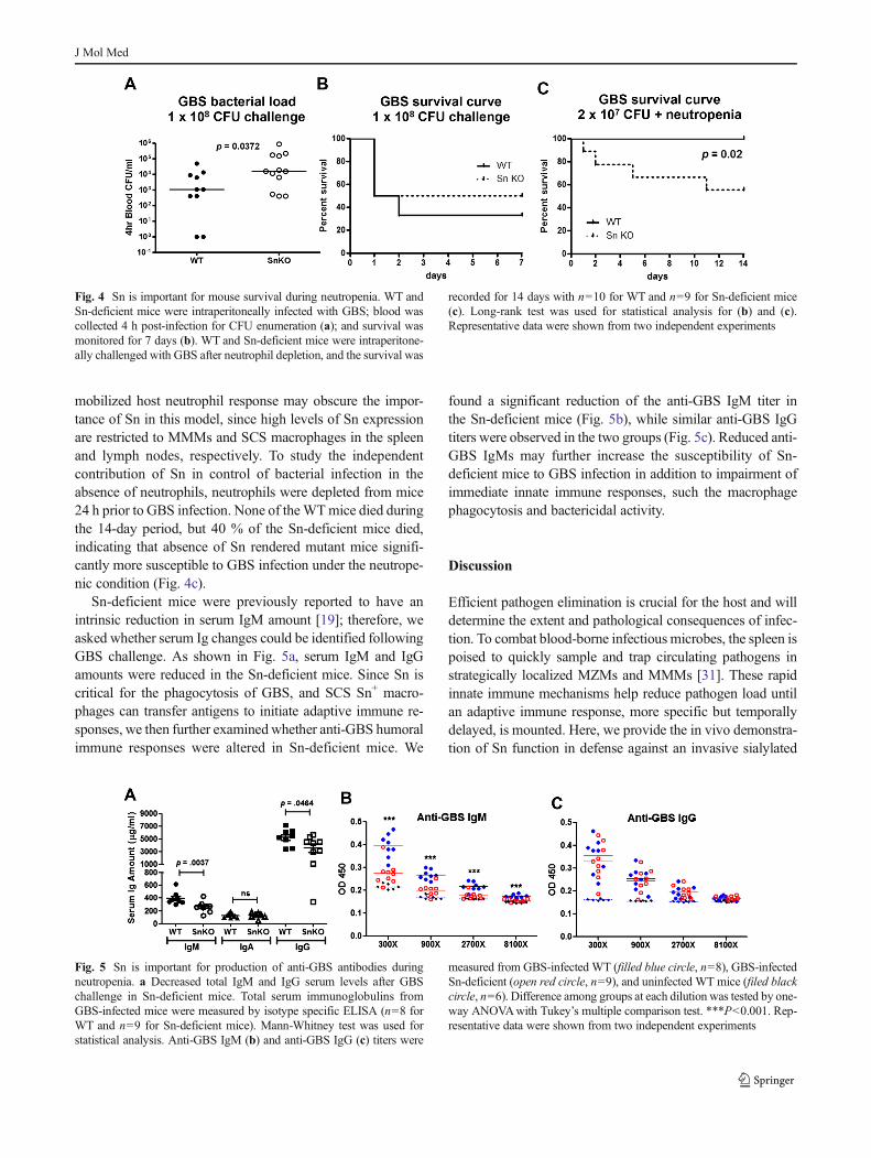
mobilized host neutrophil response may obscure the impor-tance of Sn in this model, since high levels of Sn expressionare restricted to MMMs and SCS macrophages in the spleenand lymph nodes, respectively. To study the independentcontribution of Sn in control of bacterial infection in theabsence of neutrophils, neutrophils were depleted from mice24 h prior to GBS infection. None of theWTmice died duringthe 14-day period, but 40 % of the Sn-deficient mice died,indicating that absence of Sn rendered mutant mice signifi-cantly more susceptible to GBS infection under the neutrope-nic condition (Fig. 4c).
Sn-deficient mice were previously reported to have anintrinsic reduction in serum IgM amount [19]; therefore, weasked whether serum Ig changes could be identified followingGBS challenge. As shown in Fig. 5a, serum IgM and IgGamounts were reduced in the Sn-deficient mice. Since Sn iscritical for the phagocytosis of GBS, and SCS Sn+ macro-phages can transfer antigens to initiate adaptive immune re-sponses, we then further examinedwhether anti-GBS humoralimmune responses were altered in Sn-deficient mice. We
found a significant reduction of the anti-GBS IgM titer inthe Sn-deficient mice (Fig. 5b), while similar anti-GBS IgGtiters were observed in the two groups (Fig. 5c). Reduced anti-GBS IgMs may further increase the susceptibility of Sn-deficient mice to GBS infection in addition to impairment ofimmediate innate immune responses, such the macrophagephagocytosis and bactericidal activity.
Discussion
Efficient pathogen elimination is crucial for the host and willdetermine the extent and pathological consequences of infec-tion. To combat blood-borne infectious microbes, the spleen ispoised to quickly sample and trap circulating pathogens instrategically localized MZMs and MMMs [31]. These rapidinnate immune mechanisms help reduce pathogen load untilan adaptive immune response, more specific but temporallydelayed, is mounted. Here, we provide the in vivo demonstra-tion of Sn function in defense against an invasive sialylated
Fig. 4 Sn is important for mouse survival during neutropenia. WT andSn-deficient mice were intraperitoneally infected with GBS; blood wascollected 4 h post-infection for CFU enumeration (a); and survival wasmonitored for 7 days (b). WT and Sn-deficient mice were intraperitone-ally challenged with GBS after neutrophil depletion, and the survival was
recorded for 14 days with n=10 for WT and n=9 for Sn-deficient mice(c). Long-rank test was used for statistical analysis for (b) and (c).Representative data were shown from two independent experiments
Fig. 5 Sn is important for production of anti-GBS antibodies duringneutropenia. a Decreased total IgM and IgG serum levels after GBSchallenge in Sn-deficient mice. Total serum immunoglobulins fromGBS-infected mice were measured by isotype specific ELISA (n=8 forWT and n=9 for Sn-deficient mice). Mann-Whitney test was used forstatistical analysis. Anti-GBS IgM (b) and anti-GBS IgG (c) titers were
measured from GBS-infected WT (filled blue circle, n=8), GBS-infectedSn-deficient (open red circle, n=9), and uninfected WT mice (filed blackcircle, n=6). Difference among groups at each dilution was tested by one-way ANOVAwith Tukey’s multiple comparison test. ***P<0.001. Rep-resentative data were shown from two independent experiments
J Mol Med
bacterial pathogen. This specialized macrophage Siglec notonly provides innate immune functions such as phagocytosisand bactericidal activities but also supports the developmentof proper humoral responses against the microbe.
Most Siglecs have one or more immunoreceptor tyrosine-based inhibitory motifs (ITIMs) in the cytosolic tail and func-tion as inhibitory receptors. Sialylated pathogens can evenexploit ITIM-containing Siglecs to dampen innate immuneresponses therefore to enhance their survival [18, 32, 33], inaddition to reduce complement deposition and activation onthe bacterial surface to impair opsonophagocytosis [20, 21]. Incontrast, Sn, upregulated upon infection, recognizes the verysame Sia epitope and helps preserve a critical innate immunefunction of these phagocytic cells. As a consequence, Sn hasthe right properties to mediate critical initial contacts withsialylated pathogens to function directly as a phagocytic re-ceptor or to coordinate with other pattern recognition recep-tors to mediate efficient phagocytosis. Our data supports thisidea showing that applying anti-Sn-neutralizing mAb nearlyeliminated the phagocytic ability and significantly reduced thebactericidal activity of macrophages, even as they are expect-ed to express numerous other pattern recognition and scaven-ger receptors (Fig. 2b).
The humoral response provides protective circulating anti-bodies to neutralize or coat pathogens and facilitate subse-quent opsonophagocytosis. The ability of B lymphocytes tointeract with an antigen involves antigen-trapping, depositing,and transport systems localized to the major antigen entrancesite. Splenic MZ macrophages can facilitate antibody produc-tion by transferring antigen captured from the circulation toMZ B cells [4, 34]. In addition, migration of splenic follicularB cells to the outer follicle is induced upon binding ofoxysterol produced by MMMs [35–37]. SCS Sn+ macro-phages in the LNs transport antigens to invariant natural killerT cells, dendritic cells, and B cells and thus promote theimmune responses against invaders [38–40]. Taking all thefactors together, we speculate that MMMs are not only re-quired for early antigen trapping and clearance but may alsorepresent a crucial compartment for antigen transferring to Bcells and follicular dendritic cells. Although Sn-deficient micehad MMMs located in the right location, they exhibited asmall decrease of B220+ cells in the spleen and reduction ofserum IgM levels [19]. In response to GBS challenge, Sn-deficient animals showed reduced total serum IgM levels andspecific anti-GBS IgM antibodies. Although the production ofanti-GBS IgG antibodies was not altered in those Sn-deficientanimals, they had reduced total serum IgG levels after GBSchallenge. Our findings suggest that expression of Sn onMMMs is required for them to elicit specific humoral re-sponses to counteract invading pathogens.
Whether through specific de novo biosynthesis, scaveng-ing, or precursor scavenging, many pathogens including GBS,C. jejuni, H. influenzae, Neisseria gonorrhoeae, Neisseria
meningitides, Escherichia coli K1, and Pasteurella multocidacan display Sias on their cell surface as a method of molecularmimicry, counteracting complement activation and/or engag-ing inhibitory ITIM-bearing Siglecs on leukocytes. The spe-cialized macrophage Sn receptor has a conserved bindingspecificity that happens to mirror the type and linkages ofSias expressed by the pathogens mentioned above.Understanding how Sn promotes Sia-dependent phagocytosisand stimulates antibody responses during such infectionshighlights an additional complexity in the evolutionary armsrace between pathogen and host immune defense, whereinmicrobial glycan expression and its recognition strongly in-fluence outcome.
Acknowledgments This work was supported by the NIH/NHLBI Pro-grams of Excellence in Glycosciences grant P01HL107150 to A.V. andV.N. and by a Wellcome Trust Senior Fellowship WT081882 to P.R.C.We thank technical support from the UCSD Histology Core (Nissi Varki,Director) and Patrick Secrest for mouse husbandry.
Conflict of interest The authors have no financial conflict of interestwith this work.
References
1. Eichner ER (1979) Splenic function: normal, too much and too little.Am J Med 66:311–320
2. Davidson RN, Wall RA (2001) Prevention and management ofinfections in patients without a spleen. Clin Microbiol Infect 7:657–660
3. Ram S, Lewis LA, Rice PA (2010) Infections of people with com-plement deficiencies and patients who have undergone splenectomy.Clin Microbiol Rev 23:740–780
4. Mebius RE, Kraal G (2005) Structure and function of the spleen. NatRev Immunol 5:606–616
5. den Haan JM, Kraal G (2012) Innate immune functions of macro-phage subpopulations in the spleen. J Innate Immun 4:437–445
6. Aichele P, Zinke J, Grode L, Schwendener RA, Kaufmann SH, SeilerP (2003) Macrophages of the splenic marginal zone are essential fortrapping of blood-borne particulate antigen but dispensable for in-duction of specific T cell responses. J Immunol 171:1148–1155
7. Seiler P, Aichele P, Odermatt B, Hengartner H, Zinkernagel RM,Schwendener RA (1997) Crucial role of marginal zone macrophagesand marginal zone metallophils in the clearance of lymphocyticchoriomeningitis virus infection. Eur J Immunol 27:2626–2633
8. Rempel H, Calosing C, Sun B, Pulliam L (2008) Sialoadhesinexpressed on IFN-induced monocytes binds HIV-1 and enhancesinfectivity. PLoS ONE 3:e1967
9. Vanderheijden N, Delputte PL, Favoreel HW,Vandekerckhove J, VanDamme J, van Woensel PA, Nauwynck HJ (2003) Involvement ofsialoadhesin in entry of porcine reproductive and respiratory syn-drome virus into porcine alveolar macrophages. J Virol 77:8207–8215
10. Zou Z, Chastain A, Moir S, Ford J, Trandem K, Martinelli E, CicalaC, Crocker P, Arthos J, Sun PD (2011) Siglecs facilitate HIV-1infection of macrophages through adhesion with viral sialic acids.PLoS ONE 6:e24559
11. Jones C, Virji M, Crocker PR (2003) Recognition of sialylatedmeningococcal lipopolysaccharide by Siglecs expressed on myeloid
J Mol Med
cells leads to enhanced bacterial uptake. Mol Microbiol 49:1213–1225
12. Heikema AP, Bergman MP, Richards H, Crocker PR, Gilbert M,Samsom JN, van Wamel WJ, Endtz HP, van Belkum A (2010)Characterization of the specific interaction between sialoadhesinand sialylated Campylobacter jejuni lipooligosaccharides. InfectImmun 78:3237–3246
13. Monteiro VG, Lobato CS, Silva AR, Medina DV, de Oliveira MA,Seabra SH, de SouzaW, DaMatta RA (2005) Increased association ofTrypanosoma cruzi with sialoadhesin positive mouse macrophages.Parasitol Res 97:380–385
14. Klaas M, Oetke C, Lewis LE, Erwig LP, Heikema AP, Easton A,Willison HJ, Crocker PR (2012) Sialoadhesin promotes rapid proin-flammatory and type I IFN responses to a sialylated pathogen,Campylobacter jejuni. J Immunol 189:2414–2422
15. Lewis AL, Nizet V, Varki A (2004) Discovery and characterization ofsialic acid O-acetylation in group B Streptococcus. Proc Natl AcadSci U S A 101:11123–11128
16. Weiman S, Dahesh S, Carlin AF, Varki A, Nizet V, Lewis AL (2009)Genetic and biochemical modulation of sialic acid O-acetylation ongroup B Streptococcus: phenotypic and functional impact.Glycobiology 19:1204–1213
17. Crocker PR, Gordon S (1989) Mouse macrophage hemagglutinin(sheep erythrocyte receptor) with specificity for sialylatedglycoconjugates characterized by a monoclonal antibody. J ExpMed 169:1333–1346
18. Carlin AF, Uchiyama S, Chang YC, Lewis AL, Nizet V, Varki A(2009) Molecular mimicry of host sialylated glycans allows a bacte-rial pathogen to engage neutrophil Siglec-9 and dampen the innateimmune response. Blood 113:3333–3336
19. Oetke C, Vinson MC, Jones C, Crocker PR (2006) Sialoadhesin-deficient mice exhibit subtle changes in B- and T-cell populations andreduced immunoglobulin M levels. Mol Cell Biol 26:1549–1557
20. Rubens CE, Wessels MR, Heggen LM, Kasper DL (1987)Transposon mutagenesis of type III group B Streptococcus: correla-tion of capsule expression with virulence. Proc Natl Acad Sci U S A84:7208–7212
21. Wessels MR, Rubens CE, Benedi VJ, Kasper DL (1989) Definitionof a bacterial virulence factor: sialylation of the group B streptococcalcapsule. Proc Natl Acad Sci U S A 86:8983–8987
22. Marques MB, Kasper DL, Pangburn MK, Wessels MR (1992)Prevention of C3 deposition by capsular polysaccharide is a virulencemechanism of type III group B streptococci. Infect Immun 60:3986–3993
23. Vimr E, Lichtensteiger C (2002) To sialylate, or not to sialylate: thatis the question. Trends Microbiol 10:254–257
24. York MR, Nagai T, Mangini AJ, Lemaire R, van Seventer JM,Lafyatis R (2007) A macrophage marker, Siglec-1, is increased oncirculating monocytes in patients with systemic sclerosis and inducedby type I interferons and Toll-like receptor agonists. Arthritis Rheum56:1010–1020
25. Bao G, Han Z, Yan Z,Wang Q, Zhou Y, Yao D, GuM, Chen B, ChenS, Deng A et al (2010) Increased Siglec-1 expression in monocytes ofpatients with primary biliary cirrhosis. Immunol Invest 39:645–660
26. Biesen R, Demir C, Barkhudarova F, Grun JR, Steinbrich-Zollner M,Backhaus M, Haupl T, Rudwaleit M, Riemekasten G, Radbruch Aet al (2008) Sialic acid-binding Ig-like lectin 1 expression in inflam-matory and resident monocytes is a potential biomarker for monitor-ing disease activity and success of therapy in systemic lupus erythe-matosus. Arthritis Rheum 58:1136–1145
27. Cornelissen M, van der Kuyl AC, van den Burg R, Zorgdrager F, vanNoesel CJ, Goudsmit J (2003) Gene expression profile of AIDS-related Kaposi’s sarcoma. BMC Cancer 3:7
28. van der Kuyl AC, van den Burg R, Zorgdrager F, Groot F, BerkhoutB, Cornelissen M (2007) Sialoadhesin (CD169) expression inCD14+ cells is upregulated early after HIV-1 infection and increasesduring disease progression. PLoS ONE 2:e257
29. Xiong YS, Zhou YH, Rong GH, Wu WL, Liang Y, Yang ZX, GengHL, Zhong RQ (2009) Siglec-1 on monocytes is a potential riskmarker for monitoring disease severity in coronary artery disease.Clin Biochem 42:1057–1063
30. Kraal G (1992) Cells in themarginal zone of the spleen. Int Rev Cytol132:31–74
31. Junt T, Moseman EA, Iannacone M, Massberg S, Lang PA, Boes M,Fink K, Henrickson SE, Shayakhmetov DM, Di Paolo NC et al(2007) Subcapsular sinus macrophages in lymph nodes clearlymph-borne viruses and present them to antiviral B cells. Nature450:110–114
32. Cao H, Crocker PR (2011) Evolution of CD33-related siglecs: regu-lating host immune functions and escaping pathogen exploitation?Immunology 132:18–26
33. Khatua B, Bhattacharya K, Mandal C (2012) Sialoglycoproteinsadsorbed by Pseudomonas aeruginosa facilitate their survival byimpeding neutrophil extracellular trap through Siglec-9. J LeukocBiol 91:641–655
34. Kraal G, Mebius R (2006) New insights into the cell biology of themarginal zone of the spleen. Int Rev Cytol 250:175–215
35. Pereira JP, Kelly LM, Cyster JG (2010) Finding the right niche: B-cell migration in the early phases of T-dependent antibody responses.Int Immunol 22:413–419
36. Hannedouche S, Zhang J, Yi T, Shen W, Nguyen D, Pereira JP,Guerini D, Baumgarten BU, Roggo S, Wen B et al (2011)Oxysterols direct immune cell migration via EBI2. Nature 475:524–527
37. Liu C, Yang XV,Wu J, Kuei C, Mani NS, Zhang L, Yu J, Sutton SW,Qin N, Banie H et al (2011) Oxysterols direct B-cell migrationthrough EBI2. Nature 475:519–523
38. Barral P, Polzella P, Bruckbauer A, van Rooijen N, Besra GS,Cerundolo V, Batista FD (2010) CD169(+) macrophages presentlipid antigens to mediate early activation of iNKT cells in lymphnodes. Nat Immunol 11:303–312
39. Asano K, Nabeyama A, Miyake Y, Qiu CH, Kurita A, Tomura M,Kanagawa O, Fujii S, Tanaka M (2011) CD169-positive macro-phages dominate antitumor immunity by crosspresenting dead cell-associated antigens. Immunity 34:85–95
40. Martinez-Pomares L, Gordon S (2012) CD169+ macrophagesat the crossroads of antigen presentation. Trends Immunol 33:66–70
J Mol Med
Related Documents