REVIEW Role of M. tuberculosis RD-1 region encoded secretory proteins in protective response and virulence Niladri Ganguly, Imran Siddiqui, Pawan Sharma* International Centre for Genetic Engineering and Biotechnology, Aruna Asaf Ali Marg, P.O. Box-10504, New Delhi 110067, India Received 28 December 2007; received in revised form 25 April 2008; accepted 6 May 2008 KEYWORDS CFP-10; Dendritic cells; ESAT-6; RD-1; Macrophage; M. tuberculosis Summary A gene fragment corresponding to the region of difference-1 (RD-1) of the Mycobacterium tuberculosis genome, spanning open reading frames Rv3871 to Rv3879c, is missing in all ba- cillus CalmetteeGuerin (BCG) vaccine strains of M. bovis, indicating that this was perhaps the primary deletion event responsible for attenuation of virulent M. bovis. The RD-1 locus has, therefore, been considered crucial in the pathogenesis of M. tuberculosis. Two most predominant secretory proteins encoded by this region viz. CFP-10 (Rv3874) and ESAT-6 (Rv3875) are being widely evaluated as candidate vaccine(s) and used as antigens in the diagnosis of tuberculosis. However, several recent reports have implicated their putative role in deactivation of the macrophage and dendritic cell functions. A large body of recent liter- ature provides an inkling of various mechanisms these proteins might use to down regulate normal macrophage functions and their possible contribution to virulence of M. tuberculosis. This review re-emphasizes the suggestion about the dual function of these two secreted mycobacterial proteins, viz., they have both T-cell activation and macrophage deactivation functions. ª 2008 Elsevier Ltd. All rights reserved. Introduction Tuberculosis remains a global health problem with 2e3 million deaths every year. 1 Emergence of multi-drug resis- tance (MDR) strains of Mycobacterium tuberculosis (Mtb) and enhanced susceptibility of patients infected with human * Corresponding author. Tel.: þ91 11 2674 1358; fax: þ91 11 2674 2316. E-mail address: [email protected] (P. Sharma). 1472-9792/$ - see front matter ª 2008 Elsevier Ltd. All rights reserved. doi:10.1016/j.tube.2008.05.002 available at www.sciencedirect.com journal homepage: http://intl.elsevierhealth.com/journals/tube Tuberculosis (2008) 88, 510e517

Welcome message from author
This document is posted to help you gain knowledge. Please leave a comment to let me know what you think about it! Share it to your friends and learn new things together.
Transcript
Tuberculosis (2008) 88, 510e517
ava i lab le a t www.sc iencedi rec t .com
journa l homepage : h t tp : / / i n t l . e l sev ierhea l th . com/ journa ls / tube
REVIEW
Role of M. tuberculosis RD-1 region encodedsecretory proteins in protective responseand virulence
Niladri Ganguly, Imran Siddiqui, Pawan Sharma*
International Centre for Genetic Engineering and Biotechnology, Aruna Asaf Ali Marg,P.O. Box-10504, New Delhi 110067, India
Received 28 December 2007; received in revised form 25 April 2008; accepted 6 May 2008
KEYWORDSCFP-10;Dendritic cells;ESAT-6;RD-1;Macrophage;M. tuberculosis
* Corresponding author. Tel.: þ91 112316.
E-mail address: [email protected]
1472-9792/$ - see front matter ª 200doi:10.1016/j.tube.2008.05.002
Summary
A gene fragment corresponding to the region of difference-1 (RD-1) of the Mycobacteriumtuberculosis genome, spanning open reading frames Rv3871 to Rv3879c, is missing in all ba-cillus CalmetteeGuerin (BCG) vaccine strains of M. bovis, indicating that this was perhapsthe primary deletion event responsible for attenuation of virulent M. bovis. The RD-1 locushas, therefore, been considered crucial in the pathogenesis of M. tuberculosis. Two mostpredominant secretory proteins encoded by this region viz. CFP-10 (Rv3874) and ESAT-6(Rv3875) are being widely evaluated as candidate vaccine(s) and used as antigens in thediagnosis of tuberculosis. However, several recent reports have implicated their putative rolein deactivation of the macrophage and dendritic cell functions. A large body of recent liter-ature provides an inkling of various mechanisms these proteins might use to down regulatenormal macrophage functions and their possible contribution to virulence of M. tuberculosis.This review re-emphasizes the suggestion about the dual function of these two secretedmycobacterial proteins, viz., they have both T-cell activation and macrophage deactivationfunctions.ª 2008 Elsevier Ltd. All rights reserved.
2674 1358; fax: þ91 11 2674
om (P. Sharma).
8 Elsevier Ltd. All rights reserved
Introduction
Tuberculosis remains a global health problem with 2e3million deaths every year.1 Emergence of multi-drug resis-tance (MDR) strains of Mycobacterium tuberculosis (Mtb)and enhanced susceptibility of patients infected with human
.
Mtb RD-1 region encoded secretory proteins in protective response and virulence 511
immunodeficiency virus (HIV) to tuberculosis has fuelled thespread of the disease.2,3 Mtb gets inhaled as air-borne drop-let nuclei released during coughing, sneezing and talking bypatients suffering from cavitary tuberculosis. In the lungs,the alveolar macrophages are equipped to engulf, digestand destroy a majority of the invading pathogens, but the tu-bercle bacillus has an extraordinary ability to persist andreplicate in this hostile environment.
In addition, the persistence of pathogenic mycobacte-rium inside the macrophage occurs through modulation ofhost cell signaling which allows them, unlike the non-pathogenic mycobacterial species, to survive inside thehost. Modulation of host cell-signaling is a dynamic processinvolving the interference of signaling pathways by bacte-rial molecules. Several bacterial pathogens secrete virulentmediator molecules that modulate the host cell signaling.4
Macrophages are a common target for these pathogens thatbenefit from avoiding an encounter with the immunesystem, as well as for those that are aiming to secure a sys-temic spread.5 The secretory proteins of Mtb have gainedattention in recent years both as vaccine candidates anddiagnostic tools.6,7 Secretory proteins are also targets ofthe host immune system and, besides triggering a protectiveresponse, they may also be involved in the mycobacterialvirulence and pathogenesis.8,9 This idea is supported bythe observation that only the live and not the dead mycobac-teria can down regulate the macrophage immune function.10
Genomics of the region of difference-1 (RD-1)
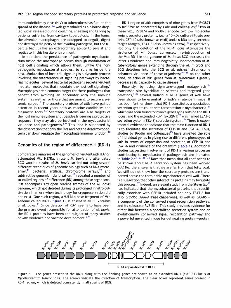
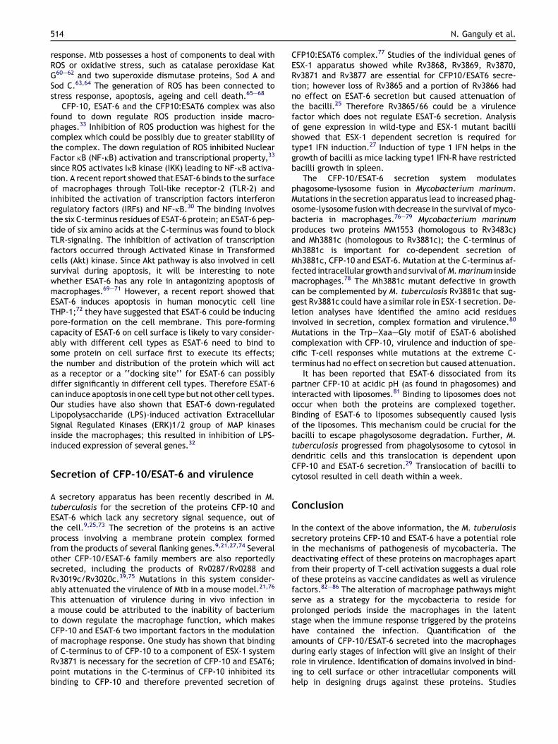
Comparative analyses of the genomes of virulent Mtb H37Rv,attenuated Mtb H37Ra, virulent M. bovis and attenuatedBCG vaccine strains of M. bovis carried out using severaldifferent techniques of genomic biology such as DNA micro-array,11 bacterial artificial chromosome arrays,12 andsubtractive genomic hybridization,13 revealed a number ofso-called regions of difference (RD) among these organisms.RDs encompass 129 open reading frames of the M. bovisgenome, which got deleted during its prolonged in vitro cul-tivation in an era when technology for cryopreservation didnot exist. One such region, a 9.5 kilo base fragment of Mtbgenome called RD-1 (Figure 1), is absent in all BCG strainsof M. bovis.11 Since deletion of RD-1 seems to have beenthe primary event responsible for attenuation of M. bovis,the RD-1 proteins have been the subject of many studieson Mtb virulence and vaccine development.8,9
Rv3
865
Rv3
866
Rv3
867
Rv3
868
Rv3
869
Rv3
870
Rv3
871
Rv3
872
Rv
Rv3
864
Figure 1 The genes present in the RD-1 along with the flankiMycobacterium tuberculosis. The arrows indicate the direction oRD-1 region, which is deleted consistently in all strains of BCG.
RD-1 region of Mtb comprises of nine genes from Rv3871to Rv3879c as annotated by Cole and colleagues;14 two ofthese viz., Rv3874 and Rv3875 encode two low molecularweight secretory proteins, i.e., a 10-kDa culture filtrate pro-tein, CFP-10 (also known as esxB) and a 6-kDa early-secretedtarget antigen, ESAT-6 (also known as esxA),15 respectively.Not only the deletion of the RD-1 locus attenuates thevirulence of M. bovis, conversely, re-introduction ofthe Mtb RD-1 in the genome of M. bovis BCG increases thelatter’s virulence and immunogenicity. Incorporation of M.tuberculosis genes extending through the M. microti andBCG deletions into the BCG or M. microti chromosomeenhances virulence of these organisms;16e19 on the otherhand, deletion of RD1 genes from M. tuberculosis greatlydecreases its capacity to cause disease.20
Recently, by using signature-tagged mutagenesis,21
transposon site hybridization screens and targeted genedeletions,9,22 several individual RD-1 genes of Mtb havebeen shown to be essential for Mtb’s virulence in mice. Ithas been further shown that RD-1 constitutes a specializedsecretion system called snm for secretion in mycobacteria,21
which was soon found to involve genes lying outside the RD-1locus, and the extended RD-1 (extRD-1)23 was named ESAT-6secretion system (ESX-1) secretion system.24 There is exper-imental evidence to indicate that the main function of RD-1is to facilitate the secretion of CFP-10 and ESAT-6. Thus,studies by Brodin and colleagues25 have unveiled the roleof individual genes in giving rise to different phenotypes ofMtb in terms of expression and secretion of CFP-10 andESAT-6 and virulence of the organism (Table 1). Additionalstudies suggesting involvement of RD-1 in various processescontributing to mycobacterial pathogenesis are indicatedin Table 2.21e23,26e36 Does that mean that all that needs tobe known about RD-1 secretion system has been workedout? No, the answer is that we are far from that lofty goal.We still do not know how the secretory proteins are trans-ported across the formidable mycobacterial cell wall. Thereis a suggestion that other interacting proteins may facilitatethis process.37 Indeed, an elegant study from the Steyn lab38
has indicated that the mycobacterial proteins that specifi-cally associate with CFP10 included not only ESAT-6 butalso Rv3596c (AAA-ATPase chaperone), as well as Rv0686 ea component of the conserved signal recognition pathway,and its substrate Rv2151c. This study provides evidence fordirect link between a specialized secretion system and anevolutionarily conserved signal recognition pathway anda powerful novel technique for delineating proteineprotein
Rv3
874(
Cfp
-10)
3873
Rv3
875(
Esat
6)R
v387
6R
v387
7R
v387
8
Rv3
879c
Rv3
880c
Rv3
881c
Rv3
882c
Rv3
883c
RD-1 region deleted in BCG
ng genes are shown as an extended RD-1 (extRD-1) locus off transcription. The clear boxes represent genes present in
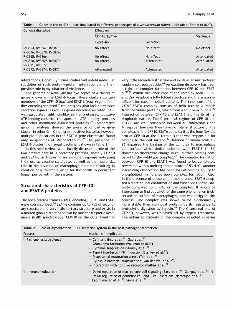
Table 1 Genes in the extRD-1 locus implicated in different phenotypes of Mycobacterium tuberculosis (after Brodin et al.25).
Gene(s) disrupted Effect on
CFP-10/ESAT-6 Virulence
Expression Secretion
Rv3864, Rv3867, Rv3873 No effect No effect No effectRv3876, Rv3878, Rv3879cRv3865, Rv3866 No effect No effect AttenuatedRv3868, Rv3869, Rv3870 No effect Attenuated AttenuatedRv3871, Rv3877Rv3872, Rv3874, Rv3875 Attenuated Attenuated Attenuated
512 N. Ganguly et al.
interactions. Hopefully future studies will unfold molecularsubtleties of such proteineprotein interactions and theirpossible role in mycobacterial virulence.
The genome of MtbH37Rv has five copies of a cluster ofgenes known as the ESAT-6 locus. These clusters containmembers of the CFP-10 (lhp) and ESAT-6 (esat-6) gene fam-ilies encoding secreted T-cell antigens (that lack detectablesecretion signals) as well as genes encoding secreted, cell-wall-associated subtilisin-like serine proteases, putativeATP-binding-cassette transporters, ATP-binding proteinsand other membrane-associated proteins.39 Comparativegenomic analyses showed the presence of ESAT-6 genecluster in other G þ C rich gram-positive bacteria; howevermultiple duplications in the ESAT-6 gene cluster are foundonly in genomes of Mycobacterium.39 The presence ofESAT-6 cluster in different bacteria is shown in Table 3.
In this mini-review, we primarily discuss the role of thetwo predominant RD-1 secretory proteins, namely CFP-10and ESAT-6 in triggering an immune response indicatingtheir use as vaccine candidates as well as their potentialrole in deactivation of macrophage function resulting increation of a favorable niche for the bacilli to persist forlonger period within the system.
Structural characteristics of CFP-10and ESAT-6 proteins
The open reading frames (ORFs) encoding CFP-10 and ESAT-6 are cotranscribed.14 ESAT-6 contains up to 75% of second-ary structure and very little tertiary structure and exists ina molten globule state as shown by Nuclear Magnetic Reso-nance (NMR) spectroscopy; CFP-10 on the other hand has
Table 2 Role of mycobacterial RD-1 secretion system in the ho
Process Mechanism implicated
I. Pathogenesis/virulence e Cell lysis (Hsu et ale Granuloma formatie Cytokine suppressioe Type I Interferon (Ie Phagosomal maturae Cytosolic bacteriale Interaction with To
II. Immunomodulation e Down regulation ofe Down regulation of
Latchumanan et al.
very little secondary structure and exists as an unstructuredrandom coil polypeptide.40 An exciting discovery has beena tight 1:1 complex formation between CFP-10 and ESAT-6.40,41 Within the inner core of the complex both CFP-10and ESAT-6 adopt a fully folded structure and there is a sig-nificant increase in helical content. The inner core of theCFP10:ESAT6 complex consists of helix-turn-helix motifsfrom individual proteins, which form a four helix bundle.42
Interaction between CFP-10 and ESAT-6 is primarily of hy-drophobic nature. The C-terminal regions of CFP-10 andESAT-6 are well conserved between M. tuberculosis andM. leprae; however they have no role in structure of thecomplex. In the CFP10:ESAT6 complex it is the long flexiblearm of CFP-10 at the C-terminus that was responsible forbinding to the cell surface.42 Deletion of amino acids 1e86 impaired the binding of the complex to macrophagecell surface while similar deletion with ESAT-6 (1e84)showed no discernible change in cell surface binding com-pared to the wild-type complex.42 The complex formationbetween CFP-10 and ESAT-6 was found to be completelyreversible with a melting temperature of 53.4 �C. Anotherinteresting observation has been loss of binding ability tophospholipid membranes upon complex formation. Also,in the presence of phospholipid membranes, ESAT-6 adop-ted a more helical conformation and enhanced thermal sta-bility compared to CFP-10 or the complex. It would beinteresting to find out whether the same phenomenon is ob-served on surface of macrophages, and what triggers thisprocess. The complex was shown to be biochemicallymore stable than individual proteins by its resistance toproteolytic digestion by trypsin.43 The C-terminal end ofCFP-10, however, was cleaved off by trypsin treatment.The enhanced stability of the complex resulted in lesser
st-pathogen interaction.
.22; Gao et al.23)on (Volkman et al.26)n (Stanley et al.21)FN) induction (Stanley et al.27)tion arrest (Tan et al.28)translocation (van der Wel et al.29)ll-like receptor (Pathak et al.30)
macrophage cell signaling (Basu et al.31; Ganguly et al.32,33)dendritic cell and T-cell functions (Natarajan et al.34;
35; Sinha et al.36)
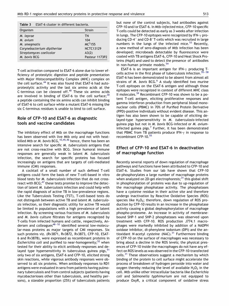
Table 3 ESAT-6 cluster in different bacteria.
Organism Strain
M. leprae TNM. avium 104M. smegmatis MC2155Corynebacterium diptheriae NCTC13129Streptomyces coelicolor A3(2)M. bovis BCG Pasteur 1173P2
Mtb RD-1 region encoded secretory proteins in protective response and virulence 513
T-cell activation compared to ESAT-6 alone due to lower ef-ficiency of proteolytic digestion and peptide presentationwith Major Histocompatibility Complex (MHC) complex onthe cell surface.44 It was also found that ESAT-6 had auto-proteolytic activity and the last six amino acids at theC-terminus can be cleaved off.40 These six amino acidsare involved in binding of ESAT-6 to the cell surface;30
a peptide containing the six amino acids can inhibit bindingof ESAT-6 to cell surface while a mutant ESAT-6 missing thesix C-terminus residues is unable to bind to cell surface.
Role of CFP-10 and ESAT-6 as diagnostictools and vaccine candidates
The inhibitory effect of Mtb on the macrophage functionshas been observed with live Mtb only and not with heat-killed Mtb or M. bovis BCG,10,45,46 hence, there has been anintensive search for specific M. tuberculosis antigens thatare not cross-reactive with BCG. Since humoral immuneresponses are generally weak in latent M. tuberculosisinfection, the search for specific proteins has focusedincreasingly on antigens that are targets of cell-mediatedimmune (CMI) responses.
A cocktail of a small number of such defined T-cellantigens could form the basis of new T-cell-based in vitroblood tests for M. tuberculosis infection that do not cross-react with BCG;47 these tests promise to improve the detec-tion of latent M. tuberculosis infection and could help withthe rapid diagnosis of active TB in low-prevalence regions.Like the Tuberculosis Testing (TST), T-cell-based tests donot distinguish between active TB and latent M. tuberculo-sis infection, so their diagnostic utility for active TB wouldbe limited in populations with a high prevalence of latentinfection. By screening various fractions of M. tuberculosisand M. bovis culture filtrates for antigens recognized byT-cells from infected humans and cattle, respectively, An-dersen and colleagues48e52 identified several low-molecu-lar-mass proteins as major targets of CMI responses. Sixsuch proteins viz. (Rv3871, Rv3872, Rv3873, CFP-10, ESAT-6 and Rv3878), were expressed as recombinant proteins inEscherichia coli and purified to near-homogeneity;52 whentested for their ability to elicit antibody responses and de-layed type hypersensitivity in tuberculous guinea pigs,51
only two of six antigens, ESAT-6 and CFP-10, elicited strongskin reactions, while vigorous antibody responses were ob-served to all six proteins. When antibody responses to RD1antigens were evaluated in sera from patients having pulmo-nary tuberculosis and from control subjects (patients havingmycobacterioses other than tuberculosis, and healthy per-sons), a sizeable proportion (25%) of tuberculosis patients
but none of the control subjects, had antibodies againstCFP-10 and/or ESAT-6. In Mtb-infected mice, CFP-10 specificT-cells could be detected as early as 3 weeks after infectionin lungs. The CFP-10 epitopes were recognized by IFN-g pro-ducing CD-4þ and CD-8þT-cells which was recruited in largenumbers in the lungs of the infected mice.53 Recently,a new method of sero-diagnosis of Mtb infection has beendeveloped; microbeads detectable by fluorescence werecoated with TB antigens ESAT-6, CFP-10 and Heat Shock Pro-teins (HspX) and used to detect the presence of antibodiesin non-human primate models.54
ESAT-6 is an important antigen for IFN-g producing T-cells active in the first phase of tuberculosis infection.55e58
ESAT-6 has been demonstrated to be absent from almost allstrains of M. bovis BCG.6 A study identified two murineT-cell epitopes on the ESAT-6 antigen and although thoseepitopes were recognized in context of different MHC classII molecules.59 Recombinant CFP-10 was shown to be a po-tent T-cell antigen, eliciting proliferative responses andgamma interferon production from peripheral blood mono-nuclear cells (PBMC) in 70% of Purified Protein Derivative(PPD)-positive individuals without evident disease. This an-tigen has also been shown to be capable of eliciting de-layed-type hypersensitivity in M. tuberculosis-infectedguinea pigs but not in M. bovis BCG-infected or M. avium-infected guinea pigs.7 Further, it has been demonstratedthat PBMC from TB patients produce IFN-g in response torecombinant CFP-10.55
Effect of CFP-10 and ESAT-6 in deactivationof macrophage function
Recently several reports of down regulation of macrophagepathways and functions have been attributed to CFP-10 andESAT-6. Studies from our lab have shown that CFP-10de-phosphorylates a large number of macrophage proteinswhen analyzed on 2D-gel electrophoresis;31 this global shiftin phosphorylation of proteins was due to up-regulation ofthe macrophage phosphatase activity. The phosphataseshave a cysteine residue in their active site and thereforeundergo inactivation by Reactive Oxidative Species (ROS)species like H2O2; therefore, down regulation of ROS pro-duction by CFP-10 results in an increase in the phosphataseactivity causing a global dephosphorylation of macrophagephospho-proteome. An increase in activity of membrane-bound SHP-1 and SHP-2 phosphatases was observed upontreatment with CFP-10. ROS production J774.1 macro-phages were markedly inhibited by addition of a NADPHoxidase inhibitor, di-phenylene iodonium (DPI) and the an-tioxidant N-acetyl cysteine (NAC).31 Furthermore bindingof CFP-10 on the surface of macrophages was necessary tobring about a decline in the ROS levels; the physical pres-ences of CFP-10 inside the macrophages do not have any ef-fect on ROS levels as was observed in the CFP-10-transfectedcells.31 These observations suggest a mechanism by whichbinding of the protein to cell surface might accelerate theprocess of breakdown of hydrogen peroxide into water andoxygen thereby reducing the level of available H2O2 in thecell. Mtb unlike other intracellular bacteria like Escherichiacoli and Salmonella typhimurium are not equipped toproduce OxyR, a critical component of oxidative stress
514 N. Ganguly et al.
response. Mtb possesses a host of components to deal withROS or oxidative stress, such as catalase peroxidase KatG60e62 and two superoxide dismutase proteins, Sod A andSod C.63,64 The generation of ROS has been connected tostress response, apoptosis, ageing and cell death.65e68
CFP-10, ESAT-6 and the CFP10:ESAT6 complex was alsofound to down regulate ROS production inside macro-phages.33 Inhibition of ROS production was highest for thecomplex which could be possibly due to greater stability ofthe complex. The down regulation of ROS inhibited NuclearFactor kB (NF-kB) activation and transcriptional property,33
since ROS activates IkB kinase (IKK) leading to NF-kB activa-tion. A recent report showed that ESAT-6 binds to the surfaceof macrophages through Toll-like receptor-2 (TLR-2) andinhibited the activation of transcription factors interferonregulatory factors (IRFs) and NF-kB.30 The binding involvesthe six C-terminus residues of ESAT-6 protein; an ESAT-6 pep-tide of six amino acids at the C-terminus was found to blockTLR-signaling. The inhibition of activation of transcriptionfactors occurred through Activated Kinase in Transformedcells (Akt) kinase. Since Akt pathway is also involved in cellsurvival during apoptosis, it will be interesting to notewhether ESAT-6 has any role in antagonizing apoptosis ofmacrophages.69e71 However, a recent report showed thatESAT-6 induces apoptosis in human monocytic cell lineTHP-1;72 they have suggested that ESAT-6 could be inducingpore-formation on the cell membrane. This pore-formingcapacity of ESAT-6 on cell surface is likely to vary consider-ably with different cell types as ESAT-6 need to bind tosome protein on cell surface first to execute its effects;the number and distribution of the protein which will actas a receptor or a ‘‘docking site’’ for ESAT-6 can possiblydiffer significantly in different cell types. Therefore ESAT-6can induce apoptosis in one cell type but not other cell types.Our studies have also shown that ESAT-6 down-regulatedLipopolysaccharide (LPS)-induced activation ExtracellularSignal Regulated Kinases (ERK)1/2 group of MAP kinasesinside the macrophages; this resulted in inhibition of LPS-induced expression of several genes.32
Secretion of CFP-10/ESAT-6 and virulence
A secretory apparatus has been recently described in M.tuberculosis for the secretion of the proteins CFP-10 andESAT-6 which lack any secretory signal sequence, out ofthe cell.9,25,73 The secretion of the proteins is an activeprocess involving a membrane protein complex formedfrom the products of several flanking genes.9,21,27,74 Severalother CFP-10/ESAT-6 family members are also reportedlysecreted, including the products of Rv0287/Rv0288 andRv3019c/Rv3020c.39,75 Mutations in this system consider-ably attenuated the virulence of Mtb in a mouse model.21,76
This attenuation of virulence during in vivo infection ina mouse could be attributed to the inability of bacteriumto down regulate the macrophage function, which makesCFP-10 and ESAT-6 two important factors in the modulationof macrophage response. One study has shown that bindingof C-terminus to of CFP-10 to a component of ESX-1 systemRv3871 is necessary for the secretion of CFP-10 and ESAT6;point mutations in the C-terminus of CFP-10 inhibited itsbinding to CFP-10 and therefore prevented secretion of
CFP10:ESAT6 complex.77 Studies of the individual genes ofESX-1 apparatus showed while Rv3868, Rv3869, Rv3870,Rv3871 and Rv3877 are essential for CFP10/ESAT6 secre-tion; however loss of Rv3865 and a portion of Rv3866 hadno effect on ESAT-6 secretion but caused attenuation ofthe bacilli.25 Therefore Rv3865/66 could be a virulencefactor which does not regulate ESAT-6 secretion. Analysisof gene expression in wild-type and ESX-1 mutant bacillishowed that ESX-1 dependent secretion is required fortype1 IFN induction.27 Induction of type 1 IFN helps in thegrowth of bacilli as mice lacking type1 IFN-R have restrictedbacilli growth in spleen.
The CFP-10/ESAT-6 secretion system modulatesphagosome-lysosome fusion in Mycobacterium marinum.Mutations in the secretion apparatus lead to increased phag-osome-lysosome fusionwith decrease in the survival of myco-bacteria in macrophages.76e79 Mycobacterium marinumproduces two proteins MM1553 (homologous to Rv3483c)and Mh3881c (homologous to Rv3881c); the C-terminus ofMh3881c is important for co-dependent secretion ofMh3881c, CFP-10 and ESAT-6. Mutation at the C-terminus af-fected intracellular growth and survival of M. marinum insidemacrophages.78 The Mh3881c mutant defective in growthcan be complemented by M. tuberculosis Rv3881c that sug-gest Rv3881c could have a similar role in ESX-1 secretion. De-letion analyses have identified the amino acid residuesinvolved in secretion, complex formation and virulence.80
Mutations in the TrpeXaaeGly motif of ESAT-6 abolishedcomplexation with CFP-10, virulence and induction of spe-cific T-cell responses while mutations at the extreme C-terminus had no effect on secretion but caused attenuation.
It has been reported that ESAT-6 dissociated from itspartner CFP-10 at acidic pH (as found in phagosomes) andinteracted with liposomes.81 Binding to liposomes does notoccur when both the proteins are complexed together.Binding of ESAT-6 to liposomes subsequently caused lysisof the liposomes. This mechanism could be crucial for thebacilli to escape phagolysosome degradation. Further, M.tuberculosis progressed from phagolysosome to cytosol indendritic cells and this translocation is dependent uponCFP-10 and ESAT-6 secretion.29 Translocation of bacilli tocytosol resulted in cell death within a week.
Conclusion
In the context of the above information, the M. tuberulosissecretory proteins CFP-10 and ESAT-6 have a potential rolein the mechanisms of pathogenesis of mycobacteria. Thedeactivating effect of these proteins on macrophages apartfrom their property of T-cell activation suggests a dual roleof these proteins as vaccine candidates as well as virulencefactors.82e86 The alteration of macrophage pathways mightserve as a strategy for the mycobacteria to reside forprolonged periods inside the macrophages in the latentstage when the immune response triggered by the proteinshave contained the infection. Quantification of theamounts of CFP-10/ESAT-6 secreted into the macrophagesduring early stages of infection will give an insight of theirrole in virulence. Identification of domains involved in bind-ing to cell surface or other intracellular components willhelp in designing drugs against these proteins. Studies
Mtb RD-1 region encoded secretory proteins in protective response and virulence 515
with Mtb having RD-1 region knocked out from their genomewill give an idea of the precise functions of these proteins.
Acknowledgments
NG was recipient of senior research fellowship from Councilfor Scientific and Industrial Research (CSIR). Sharma labreceives generous funding support from DBT, Government ofIndia. IS is funded through a project from Department ofBiotechnology (DBT).
Funding: None.
Competing interests: None declared.
Ethical approval: Not required.
References
1. Raviglione MC. The TB epidemic from 1992 to 2002. Tuberculo-sis (Edinb) 2003;83:4e14.
2. Elliot AM, Halwindii B, Hayes R, Luo N, Mwinga AG, Tembo G,et al. The impact of human immunodeficiency virus on mortal-ity of patients treated for tuberculosis in a cohort study inZambia. Trans R Soc Trop Med Hyg 1995;89:78e82.
3. Chintu C, Mwinga A. An African perspective on the threat oftuberculosis and HIV/AIDS- can despair be turned into hope?Lancet 1999;353:997.
4. Koul A, Herget T, Klebl B, Ullrich A. Interplay betweenmycobacteria and host signalling pathways. Nat Rev Microbiol2004;2:189e202.
5. Rosenberger CM, Finlay BB. Phagocyte sabotage: disruption ofmacrophage signalling by bacterial pathogens. Nat Rev MolCell Biol 2003;4:385e96.
6. Harboe M, Oettinger T, Wiker HG, Rosenkrands I, Andersen P.Evidence for occurrence of the ESAT-6 protein in Mycobacte-rium tuberculosis and virulent Mycobacterium bovis and forits absence in Mycobacterium bovis BCG. Infect Immun 1996;64:16e22.
7. Colangeli R, Spencer JS, Bifani P, Williams A, Lyashchenko K,Keen MA, et al. MTSA-10, the product of the Rv3874 gene ofMycobacterium tuberculosis, elicits tuberculosis-specific,delayed-type hypersensitivity in guinea pigs. Infect Immun2000;68:990e3.
8. Pym AS, Brodin P, Majlessi L, Brosch R, Demangel C, Williams A,et al. Recombinant BCG exporting ESAT-6 confers enhancedprotection against tuberculosis. Nat Med 2003;9:533e9.
9. Guinn KM, Hickey MJ, Mathur SK, Zakel KL, Grotzke JE,Lewinsohn DM, et al. Individual RD1-region genes are requiredfor export of ESAT-6/CFP-10 and for virulence of Mycobacte-rium tuberculosis. Mol Microbiol 2004;51:359e70.
10. Malik ZA, Iyer SS, Kusner DJ. Mycobacterium tuberculosis phag-osomes exhibit altered calmodulin-dependent signal transduc-tion: contribution to inhibition of phagosome-lysosome fusionand intracellular survival in human macrophages. J Immunol2001;166:3392e401.
11. Behr MA, Wilson MA, Gill WP, Salamon H, Schoolnik GK, Rane S,et al. Comparative genomics of BCG vaccines by whole-genomeDNA microarray. Science 1999;284:1520e3.
12. Gordon SV, Brosch R, Billault A, Garnier T, Eiglmeier K, Cole ST.Identification of variable regions in the genomes of tuberclebacilli using BAC arrays. Mol Microbiol 1999;32:643e55.
13. Mahairas GG, Sabo PJ, Hickey MJ, Singh DC, Stover CK. Molecu-lar analysis of genetic differences between Mycobacteriumbovis BCG and virulent M. bovis. J Bacteriol 1996;178:1274e82.
14. Cole ST, Brosch R, Parkhill J, Garnier T, Churcher C, Harris D,et al. Deciphering the biology of Mycobacterium tuberculosisfrom the complete genome sequence. Nature 1998;393:537e44.
15. Berthet FX, Rasmussen PB, Rosenkrands I, Andersen P,Gicquel B. A Mycobacterium tuberculosis operon encodingESAT-6 and a novel low-molecular-mass culture filtrate protein(CFP-10). Microbiology 1998;144:3195e203.
16. Wards BJ, de Lisle GW, Collins DM. An esat6 knockout mutantof Mycobacterium bovis produced by homologous recombina-tion will contribute to the development of a live tuberculosis.
17. Behr MA. BCG-different strains, different vaccines? LancetInfect Dis 2002;2:86e92.
18. Demangel C, Garnier T, Rosenkrands I, Cole ST. Differential ef-fects of prior exposure to environmental mycobacteria on vacci-nation with Mycobacterium bovis BCG or a recombinant BCGstrain expressing RD1 antigens. Infect Immun 2005;73:2190e6.
19. Pym AS, Brodin P, Brosch R, Huerre M, Cole ST. Loss of RD1contributed to the attenuation of the live tuberculosis vaccinesMycobacterium bovis BCG and Mycobacterium microti. MolMicrobiol 2002;46:709e17.
20. Lewis KN, Liao R, Guinn KM, Hickey MJ, Smith S, Behr MA, et al.Deletion of RD1 from Mycobacterium tuberculosis mimicsbacille CalmetteeGuerin attenuation. J Infect Dis 2003;187:117e23.
21. Stanley SA, Raghavan S, Hwang WW, Cox JS. Acute infectionand macrophage subversion by Mycobacterium tuberculosisrequire a specialized secretion system. Proc Natl Acad SciUSA 2003;100:13001e6.
22. Hsu T, Hingley-Wilson SM, Chen B, Chen M, Dai AZ, Morin PM,et al. The primary mechanism of attenuation of bacillusCalmetteeGuerin is a loss of secreted lytic function requiredfor invasion of lung interstitial tissue. Proc Natl Acad Sci USA2003;100:12420e5.
23. Gao LY, Guo S, McLaughlin B, Morisaki H, Engel JN, Brown EJ. Amycobacterial virulence gene cluster extending RD1 is requiredfor cytolysis, bacterial spreading and ESAT-6 secretion. MolMicrobiol 2004;53:1677e93.
24. Brodin P, Rosenkrands I, Andersen P, Cole ST, Brosch R. ESAT-6proteins: protective antigens and virulence factors? TrendsMicrobiol 2004;12:500e8.
25. Brodin P, Majlessi L, Marsollier L, de Jonge MI, Bottai D,Demangel C, et al. Dissection of ESAT-6 system 1 of Mycobacte-rium tuberculosis and impact on immunogenicity andvirulence. Infect Immun 2006;74:88e98.
26. Volkman HE, Clay H, Beery D, Chang JC, Sherman DR,Ramakrishnan L. Tuberculous granuloma formation is enhancedby a mycobacterium virulence determinant. PLoS Biol 2004;2:e367.
27. Stanley SA, Johndrow JE, Manzanillo P, Cox JS. The type I IFNresponse to infection with Mycobacterium tuberculosisrequires ESX-1-mediated secretion and contributes to patho-genesis. J Immunol 2007;178:3143e52.
28. Tan T, Lee WL, Alexander DC, Grinstein S, Liu J. The ESAT-6/CFP-10 secretion system of Mycobacterium marinum modu-lates phagosome maturation. Cell Microbiol 2006;8:1417e29.
29. Van der Wel N, Hava D, Houben D, Fluitsma D, van Zon M,Pierson J, et al. M. tuberculosis and M. leprae translocatefrom the phagolysosome to the cytosol in myeloid cells. Cell2007;129:1287e98.
30. Pathak SK, Basu S, Basu KK, Banerjee A, Pathak S,Bhattacharyya A, et al. Directextracellular interaction betweenthe early secreted antigen ESAT-6 of Mycobacterium tuberculo-sis and TLR2 inhibits TLR signaling in macrophages. Nat Immunol2007;8:610e8.
31. Basu SK, Kumar D, Singh DK, Ganguly N, Siddiqui Z, Rao KVS,et al. Mycobacterium tuberculosis secreted antigen (MTSA-10) modulates macrophage function by redox regulation ofphosphatases. FEBS J 2006;273:5517e34.
516 N. Ganguly et al.
32. Ganguly N, Giang PH, Basu SK, Mir FA, Siddiqui I, Sharma P.Mycobacterium tuberculosis 6-kDa Early Secreted AntigenicTarget (ESAT-6) protein downregulates Lipopolysaccharideinduced c-myc expression by modulating the ExtracellularSignal Regulated Kinases1/2 (ERK1/2). BMC Immunol 2007;8:24. doi:10.1186/1471-2172-8-24.
33. Ganguly N, Giang PH, Gupta C, Basu SK, Siddiqui I, Salunke DM,et al. Mycobacterium tuberculosis secretory proteins CFP-10,ESAT-6 and CFP10:ESAT6 complex inhibit lipopolysaccharideinduced NF-kB transactivation by downregulation of reactiveoxidative species (ROS) production. Immunol Cell Biol 2008;86:98e106.
34. Natarajan K, Latchumanan VK, Singh B, Singh S, Sharma P.Down regulation of T helper1 responses to mycobacterialantigens due to maturation of dendritic cells by 10-kDa Myco-bacterium tuberculosis secretory antigen. J Infect Dis 2003;187:914e28.
35. Latchumanan VK, Balkhi MY, Sinha A, Singh B, Sharma P,Natarajan K. Regulation of immune responses to Mycobacte-rium tuberculosis secretory antigens by dendritic cells. Tuber-culosis 2005;85:377e83.
36. Sinha A, Salam N, Gupta S, Natarajan K. Mycobacterium tuber-culosis and dendritic cells: recognition, activation and func-tional implications. Indian J Biochem Biophys 2008;44:279e88.
37. Champion PAD, Cox JS. Protein secretion systems n mycobacte-ria. Cell Microbiol 2007;9:1376e84.
38. Singh A, Mai D, Kumar A, Steyn AJ. Dissecting virulence path-ways of Mycobacterium tuberculosis through proteineproteinassociation. Proc Natl Acad Sci USA 2006;103:11346e51.
39. Gey Van Pittius NC, Gamieldien J, Hide W, Brown GD,Siezen RJ, Beyers AD. The ESAT-6 gene cluster of Mycobacte-rium tuberculosis and other high GþC Gram-positive bacteria.Genome Biology 2001;2(10): research 0044.1-0044.18.
40. Renshaw PS, Panagiotidou P, Whelan A, Gordon SV,Hewinson RG, Williamson RA, et al. Conclusive evidence thatthe major T-cell antigens of the Mycobacterium tuberculosiscomplex ESAT-6 and CFP-10 form a tight, 1:1 complex andcharacterization of the structural properties of ESAT-6,CFP-10, and the ESAT-6*CFP-10 complex. Implications for path-ogenesis and virulence. J Biol Chem 2002;277:21598e603.
41. Lightbody KL, Renshaw PS, Collins ML, Wright RL, Hunt DM,Gordon SV, et al. Characterisation of complex formationbetween members of the Mycobacterium tuberculosis complexCFP-10/ESAT-6 protein family: towards an understanding of therules governing complex formation and thereby functionalflexibility. FEMS Microbiol Lett 2004;238:255e62.
42. Renshaw PS, Lightbody KL, Veverka V, Muskett FW, Kelly G,Frenkiel TA, et al. Structure and function of the complexformed by the tuberculosis virulence factors CFP-10 andESAT-6. EMBO J 2005;24:2491e8.
43. Meher AK, Bal NC, Chary KV, Arora A. Mycobacterium tuber-culosis H37RvESAT-6-CFP-10 complex formation confersthermodynamic and biochemical stability. FEBS J 2006;273:1445e62.
44. Marei A, Ghaemmaghami A, Renshaw P, Wiselka M, Barer M,Carr M, et al. Superior T cell activation by ESAT-6 as comparedwith the ESAT-6-CFP-10 complex. Int Immunol 2005;17:1439e46.
45. Malik ZA, Denning GM, Kusner DJ. Inhibition of Ca(2þ) signalingby Mycobacterium tuberculosis is associated with reducedphagosome-lysosome fusion and increased survival withinhuman macrophages. J Exp Med 2000;191:287e302.
46. Malik ZA, Thompson CR, Hashimi S, Porter B, Iyer SS, Kusner DJ.Cutting edge: Mycobacterium tuberculosis blocks Ca2þ signal-ing and phagosome maturation in human macrophages viaspecific inhibition of sphingosine kinase. J Immunol 2003;170:2811e5.
47. Chee CB, KhinMar KW, Gan SH, Barkham TM, Pushparani M,Wang YT. Latent tuberculosis infection treatment and T-cell
responses to Mycobacterium tuberculosis-specific antigens.Am J Respir Crit Care Med 2007;175:282e7.
48. Andersen P, Askgaard D, Ljungqvist L, Bennedsen J, Heron I.Proteins released from Mycobacterium tuberculosis duringgrowth. Infect Immun 1991;59:1905e10.
49. Andersen P, Askgaard D, Ljungqvist L, Bentzon MW, Heron I.T-cell proliferative response to antigens secreted by Mycobac-terium tuberculosis. Infect Immun 1991;59:1558e63.
50. Andersen P. Effective vaccination of mice against Mycobacte-rium tuberculosis infection with a soluble mixture of secretedmycobacterial proteins. Infect Immun 1994;62:2536e44.
51. Weldingh K, Andersen P. Immunological evaluation of novelMycobacterium tuberculosis culture filtrate proteins. FEMSImmunol Med Microbiol 1999;23:159e64.
52. Weldingh K, Rosenkrands I, Jacobsen S, Rasmussen PB,Elhay MJ, Andersen P. Two-dimensional electrophoresis foranalysis of Mycobacterium tuberculosis culture filtrate andpurification and characterization of six novel proteins. InfectImmun 1998;66:3492e500.
53. Kamath AB, Woodworth J, Xiong X, Taylor C, Weng Y, Behar SM.Cytolytic CD8þT cells recognizing CFP10 are recruited to thelung after Mycobacterium tuberculosis infection. J Exp Med2004;200:1479e89.
54. Khan IH, Ravindran R, Yee J, Ziman M, Lewinsohn DM,Genaro ML, et al. Profiling antibodies to Mycobacterium tuber-culosis (M.tb.) by multiplex microbead suspension arrays forserodiagnosis of TB. Clin Vaccine Immunol; 2007. Epub aheadof print.
55. Porsa E, Cheng L, Seale MM, Delclos GL, Ma X, Reich R, et al.Comparison of a new ESAT-6/CFP-10 peptide-based gammainterferon assay and a tuberculin skin test for tuberculosisscreening in a moderate-risk population. Clin Vaccine Immunol2006;13:53e8.
56. Ravn P, Demissi A, Eguale T, Wondwosson H, Lein D, Amoudy HA,et al. Human T cell responses to the ESAT-6 antigen fromMycobacterium tuberculosis. J Infect Dis 1999;179:637e45.
57. Skjot RL, Oettinger T, Rosenkrands I, Ravn P, Brock I,Jacobsen S, et al. Comparative evaluation of low-molecular-mass proteins from Mycobacterium tuberculosis identifiesmembers of the ESAT-6 family as immunodominant T-cellantigens. Infect Immun 2000;68:214e20.
58. de Jong BC, Hill PC, Brookes RH, Gagneux S, Jeffries DJ,Otu JK, et al. Mycobacterium africanum elicits an attenuatedT cell response to early secreted antigenic target, 6 kDa, inpatients with tuberculosis and their household contacts.J Infect Dis 2006;193:1279e86.
59. Dietrich J, Aagaard C, Leah R, Olsen AW, Stryhn A, Doherty TM,et al. Exchanging ESAT6 with TB10.4 in an Ag85B fusionmolecule-based tuberculosis subunit vaccine: efficient protec-tion and ESAT6-based sensitive monitoring of vaccine efficacy.J Immunol 2005;174:6332e9.
60. Sherman DR, Sabo PJ, Hickey MJ, Arain TM, Mahairas GG,Yuan Y, et al. Disparate responses to oxidative stress in sapro-phytic and pathogenic mycobacteria. Proc Natl Acad Sci USA1995;92:6625e9.
61. Manca C, Paul S, Baryy 3rd CE, Freedman VH, Kaplan G. Myco-bacterium tuberculosis catalase and peroxidase activities andresistance to oxidative killing in human monocytes in vitro.Infect Immun 1999;67:74e9.
62. Ng VH, Cox JS, Sousa AO, MacMicking JD, McKinney JD. Role ofKatG catalase-peroxidase in mycobacterial pathogenesis:countering the phagocyte oxidative burst. Mol Microbiol2004;52:1291e302.
63. Piddington DL, Fang FC, Laessig T, Cooper AM, Orme IM,Buchmeier NA. Cu,Zn superoxide dismutase of Mycobacteriumtuberculosis contributes to survival in activated macrophagesthat are generating an oxidative burst. Infect Immun 2001;69:4980e7.
Mtb RD-1 region encoded secretory proteins in protective response and virulence 517
64. Zhang Y, Lathigra R, Garbe T, Catty D, Young D. Geneticanalysis of superoxide dismutase, the 23 kilodalton antigenof Mycobacterium tuberculosis. Mol Microbiol 1991;5:381e91.
65. Adler V, Yin Z, Tew KD, Ronai Z. Role of redox potential andreactive oxygen species in stress signaling. Oncogene 1999;18:6104e11.
66. Finkel T, Holbrook NJ. Oxidants, oxidative stress and thebiology of ageing. Nature 2000;408:239e47.
67. Finkel T. Oxidant signals and oxidative stress. Curr Opin CellBiol 2003;15:247e54.
68. Buttke TM, Sandstrom PA. Redox regulation of programmedcell death in lymphocytes. Free Radic Res 1995;22:389e97.
69. Balcewicz-Sablinska MK, Keane J, Kornfeld H, Remold HG.Pathogenic Mycobacterium tuberculosis evades apoptosis ofhost macrophages by release of TNF-R2, resulting in inactiva-tion of TNF-alpha. J Immunol 1998;161:2636e41.
70. Keane J, Remold HG, Kornfeld H. Virulent Mycobacteriumtuberculosis strains evade apoptosis of infected alveolarmacrophages. J Immunol 2000;164:2016e20.
71. Bhattacharyya A, Pathak S, Basak C, Law S, Kundu M, Basu J.Execution of macrophage apoptosis by Mycobacterium aviumthrough apoptosis signal-regulating kinase 1/p38 mitogen-activated protein kinase signaling and caspase 8 activation.J Biol Chem 2003;278:26517e25.
72. Derrick SC, Morris SL. The ESAT6 protein of Mycobacteriumtuberculosis induces apoptosis of macrophages by activatingcaspase expression. Cell Microbiol 2007;9:1547e55.
73. Fortune SM, Jaeger A, Sarracino DA, Chase MR, Sassetti CM,Sherman DR, et al. Mutually dependent secretion of proteinsrequired for mycobacterial virulence. Proc Natl Acad Sci USA2005;102:10676e81.
74. Converse SE, Cox JS. A protein secretion pathway critical for My-cobacterium tuberculosis virulence is conserved and functionalin Mycobacterium smegmatis. J Bacteriol 2005;187:1238e45.
75. Arend SM, de Haas P, Leyten E, Rosenkrands I, Rigouts L,Andersen P, et al. ESAT-6 and CFP-10 in clinical versus environ-mental isolates of Mycobacterium kansasii. J Infect Dis 2000;191:1301e10.
76. Majlessi L, Brodin P, Brosch R, Rojas MJ, Khun H, Huerre M,et al. Influence of ESAT-6 secretion system 1 (RD1) of
Mycobacterium tuberculosis on the interaction between myco-bacteria and the host immune system. J Immunol 2005;174:3570e9.
77. Champion PA, Stanley SA, Champion MM, Brown EJ, Cox JS.C-terminal signal sequence promotes virulence factor secre-tion in Mycobacterium tuberculosis. Science 2006;313:1632e6.
78. Xu J, Laine O, Masciocchi M, Manoranjan J, Smith J, Du SJ,et al. A unique Mycobacterium ESX-1 protein co-secretes withCFP-10/ESAT-6 and is necessary for inhibiting phagosomematuration. Mol Microbiol 2007;66:787e800.
79. Lee VT, Schneewind O. Protein secretion and the pathogenesisof bacterial infections. Genes Dev 2001;15:1725e52.
80. Brodin P, de Jonge MI, Majlessi L, Leclerc C, Nilges M,Cole ST, et al. Functional analysis of early secreted antigenictarget-6, the dominant T-cell antigen of Mycobacteriumtuberculosis, reveals key residues involved in secretion, com-plex formation, virulence, and immunogenicity. J Biol Chem2005;280:33953e9.
81. de Jonge MI, Pehau-Arnaudet G, Fretz MM, Romain F, Bottai D,Brodin P, et al. ESAT-6 from Mycobacterium tuberculosisdissociates from its putative chaperone CFP-10 under acidicconditions and exhibits membrane-lysing activity. J Bacteriol2007;189:6028e34.
82. Brandt L, Elhay M, Rosenkrands I, Lindblad EB, Andersen P.ESAT-6 subunit vaccination against Mycobacterium tuberculo-sis. Infect Immun 2000;68:791e5.
83. Andersen P, Doherty TM. TB subunit vaccinesdputting thepieces together. Microbes Infect 2005;7:911e21.
84. Taggart EW, Hill HR, Ruegner RG, Litwin CM. Evaluation of an invitro assay for interferon gamma production in response to theMycobacterium tuberculosis-synthesized peptide antigensESAT-6 and CFP-10 and the PPD skin test. Am J Clin Pathol2006;125:467e73.
85. Smith I. Mycobacterium tuberculosis pathogenesis and molec-ular determinants of virulence. Clin Microbiol Rev 2003;16:463e96.
86. Alderson MR, Bement T, Day CH, Zhu L, Molesh D, Skeiky YA,et al. Expression cloning of an immunodominant family ofMycobacterium tuberculosis antigens using human CD4(þ) Tcells. J Exp Med 2000;191:551e60.
Related Documents