Role of CXC Chemokine Ligand 13 in Oral Squamous Cell Carcinoma Associated Osteolysis in Athymic Mice Subramanya N.M. Pandruvada 1 , Yuvaraj Sambandam 1 , Xiang Liu 2 , Kumaran Sundaram 1 , Srinivasan Shanmugarajan 1 , William L. Ries 3 , James S. Norris 2 , Steven D. London 3 , and Sakamuri V. Reddy 1 1 Charles P. Darby Children’s Research Institute, Medical University of South Carolina, Charleston SC, USA 2 Department of Microbiology and Immunology, Medical University of South Carolina, Charleston SC, USA 3 College of Dental Medicine, Medical University of South Carolina, Charleston SC, USA Abstract Oral squamous cell carcinomas (OSCC) are malignant tumors with a potent activity of local bone invasion; however the molecular mechanisms of tumor osteolysis are unclear. In this study, we identified high level expression of chemokine ligand, CXCL13 and RANK ligand (RANKL) in OSCC cells (SCC1, SCC12 and SCC14a). OSCC cell conditioned media (20%) induced osteoclast differentiation which was inhibited by OPG in peripheral blood monocyte cultures indicating that OSCC cells produce soluble RANKL. Recombinant hCXCL13 (10 ng/ml) significantly enhanced RANKL stimulated osteoclast differentiation in these cultures. Trans-well migration assay identified that CXCL13 induces chemotaxis of peripheral blood monocytes in vitro which was inhibited by addition of anti-CXCR5 receptor antibody. Zymogram analysis of conditioned media from OSCC cells revealed matrix metalloproteinase-9 (MMP-9) activity. Interestingly, CXCL13 treatment to OSCC cells induced CXCR5 and MMP-9 expression suggesting an autocrine regulatory function in OSCC cells. To examine the OSCC tumor cell bone invasion/osteolysis, we established an in vivo model for OSCC by subcutaneous injection of OSCC cells onto the surface of calvaria in NCr-nu/nu athymic mice, which developed tumors in 4–5 weeks. μCT analysis revealed numerous osteolytic lesions in calvaria from OSCC tumor-bearing mice. Histochemical staining of calvarial sections from these mice revealed a significant increase in the numbers of TRAP-positive osteoclasts at the tumor-bone interface. Immunohistochemical analysis confirmed CXCL13 and MMP-9 expression in tumor cells. Thus, our data implicate a functional role for CXCL13 in bone invasion and may be a potential therapeutic target to prevent osteolysis associated with OSCC tumors in vivo. Keywords Oral squamous cell carcinoma (OSCC); Chemokine; MMP-9; Bone invasion; RANK ligand; Osteoclast; Mouse model * Address for Correspondence: Sakamuri V. Reddy, Ph.D, Charles P. Darby Children’s Research Institute, 173 Ashley Avenue, Charleston, SC 29425; Tel: Ph. 843-792-8373; Fax: 843-792-7466; [email protected]. NIH Public Access Author Manuscript Int J Cancer. Author manuscript; available in PMC 2011 May 15. Published in final edited form as: Int J Cancer. 2010 May 15; 126(10): 2319–2329. doi:10.1002/ijc.24920. NIH-PA Author Manuscript NIH-PA Author Manuscript NIH-PA Author Manuscript

Welcome message from author
This document is posted to help you gain knowledge. Please leave a comment to let me know what you think about it! Share it to your friends and learn new things together.
Transcript

Role of CXC Chemokine Ligand 13 in Oral Squamous CellCarcinoma Associated Osteolysis in Athymic Mice
Subramanya N.M. Pandruvada1, Yuvaraj Sambandam1, Xiang Liu2, Kumaran Sundaram1,Srinivasan Shanmugarajan1, William L. Ries3, James S. Norris2, Steven D. London3, andSakamuri V. Reddy1
1 Charles P. Darby Children’s Research Institute, Medical University of South Carolina,Charleston SC, USA2 Department of Microbiology and Immunology, Medical University of South Carolina, CharlestonSC, USA3 College of Dental Medicine, Medical University of South Carolina, Charleston SC, USA
AbstractOral squamous cell carcinomas (OSCC) are malignant tumors with a potent activity of local boneinvasion; however the molecular mechanisms of tumor osteolysis are unclear. In this study, weidentified high level expression of chemokine ligand, CXCL13 and RANK ligand (RANKL) inOSCC cells (SCC1, SCC12 and SCC14a). OSCC cell conditioned media (20%) induced osteoclastdifferentiation which was inhibited by OPG in peripheral blood monocyte cultures indicating thatOSCC cells produce soluble RANKL. Recombinant hCXCL13 (10 ng/ml) significantly enhancedRANKL stimulated osteoclast differentiation in these cultures. Trans-well migration assayidentified that CXCL13 induces chemotaxis of peripheral blood monocytes in vitro which wasinhibited by addition of anti-CXCR5 receptor antibody. Zymogram analysis of conditioned mediafrom OSCC cells revealed matrix metalloproteinase-9 (MMP-9) activity. Interestingly, CXCL13treatment to OSCC cells induced CXCR5 and MMP-9 expression suggesting an autocrineregulatory function in OSCC cells. To examine the OSCC tumor cell bone invasion/osteolysis, weestablished an in vivo model for OSCC by subcutaneous injection of OSCC cells onto the surfaceof calvaria in NCr-nu/nu athymic mice, which developed tumors in 4–5 weeks. μCT analysisrevealed numerous osteolytic lesions in calvaria from OSCC tumor-bearing mice. Histochemicalstaining of calvarial sections from these mice revealed a significant increase in the numbers ofTRAP-positive osteoclasts at the tumor-bone interface. Immunohistochemical analysis confirmedCXCL13 and MMP-9 expression in tumor cells. Thus, our data implicate a functional role forCXCL13 in bone invasion and may be a potential therapeutic target to prevent osteolysisassociated with OSCC tumors in vivo.
KeywordsOral squamous cell carcinoma (OSCC); Chemokine; MMP-9; Bone invasion; RANK ligand;Osteoclast; Mouse model
*Address for Correspondence: Sakamuri V. Reddy, Ph.D, Charles P. Darby Children’s Research Institute, 173 Ashley Avenue,Charleston, SC 29425; Tel: Ph. 843-792-8373; Fax: 843-792-7466; [email protected].
NIH Public AccessAuthor ManuscriptInt J Cancer. Author manuscript; available in PMC 2011 May 15.
Published in final edited form as:Int J Cancer. 2010 May 15; 126(10): 2319–2329. doi:10.1002/ijc.24920.
NIH
-PA Author Manuscript
NIH
-PA Author Manuscript
NIH
-PA Author Manuscript

IntroductionHead and neck squamous cell carcinoma (HNSCC) are the most common malignantneoplasms, with a prevalence estimated to be greater than 40,000 cases annually in the US.Oral squamous cell carcinoma (OSCC), which contributes to 40% of all HNSCC isassociated with mucosal surfaces of the oral cavity and oropharynx 1. The etiology of OSCCinvolves both a genetic predisposition and exposure to environmental carcinogens such astobacco, alcohol, chronic inflammation and viral infection 2. Curcumin has been shown tosuppress HNSCC growth both in vitro and in vivo 3, and Fas-mediated apoptosis in HNSCCis a proven and efficient therapeutic approach in a xenograft animal model 4. Furthermore,tamoxifen inhibition of OSCC cell growth in vitro 5 and a role for human longevityassurance gene 1 (LASS1) and C18-ceramide in chemotherapy induced cell death in HNSCChave been reported 6. Malignant HNSCC tumors are known to have a potent activity of localbone invasion; however the molecular mechanisms of tumor-associated osteolysis areunclear.
The osteoclast is hematopoietic in origin and is the bone-resorbing cell derived frommonocyte/macrophage lineage. Tumor necrosis factor (TNF) family member, RANK ligand(RANKL), which is expressed on marrow stromal/osteoblast cells in response to severalosteotropic factors, is critical for osteoclast precursor differentiation to form multinucleatedosteoclasts, which resorb bone 7. Osteoclast activity is controlled by local factors producedin the bone microenvironment. In addition, the osteoclast is an autocrine/paracrine,intracrine regulatory cell that produces factors such as IL-6, annexin II, TGF-beta andOIP-1/hSca, which influence its own formation and activity. Matrix metalloproteinase-9(MMP-9), a type IV collagenase is highly expressed in osteoclast cells and plays animportant role in degradation of the extracellular matrix 8. Osteoclast activation plays animportant role in several malignancies including oral cancers invasion of bone andsubsequent metastasis 9. Further, studies using a murine mandibular bone invasion modelfor OSCC demonstrated mRNA expression of cytokines associated with osteoclastactivation such as IL-6, TNF-α and PTHrP in tumor tissue as well as high bone resorption 9.Also, conditioned media from OSCC cells derived from patients with bone involvementstimulated osteoclast differentiation in vitro 10.
Chemokines are a superfamily of small, cytokine-like proteins that selectively attract andactivate different cell types 11. CXC chemokines are known to promote angiogenesis 12 andhave a characteristic heparin-binding domain. Chemokines interact with seven-transmembrane-domain glycoprotein receptors coupled to the G protein signaling pathway11. In several studies, tumor cells were shown to express functionally active chemokinereceptors which regulate cellular functions and metastasis 13. HNSCC has been reported topredominantly expressed chemokine receptors such as CCR7 and CXCR5; however,CXCR4 expression is low or undetectable 14. CXCL13 (BCA-1) which bindsmonogamously to the CXCR5 receptor and is involved in B-cell chemotaxis and is inducedunder inflammatory conditions 15. Microarray analysis for gene expression profiling inOSCC identified gene signatures which include chemokine (CXC motif) ligand-13 andmatrix-metalloproteinases (MMPs) that are highly relevant to OSCC development andprogression 16. However, a functional role for CXCL13 in HNSCC tumor cell invasion andosteolysis is unknown. In this study, we showed CXCL13 expression and an autocrineregulation of MMP-9 production in tumor cells. We further show CXCL13 and RANKLexpression in OSCC cells support osteoclastogenesis. We developed an in vivo model forOSCC by subcutaneous injection of SCC 14a cells onto the surface of calvaria in NCr-nu/nuathymic mice which showed osteolytic lesions. Our data implicate CXCL13 a potentialtherapeutic target to prevent OSCC tumor-associated osteolysis in vivo.
Pandruvada et al. Page 2
Int J Cancer. Author manuscript; available in PMC 2011 May 15.
NIH
-PA Author Manuscript
NIH
-PA Author Manuscript
NIH
-PA Author Manuscript

Materials and MethodsCell lines and culture
Human OSCC-derived cell lines SCC1, SCC12 and SCC14a were generously provided byDr. Thomas E. Carey (University of Michigan, Ann Arbor, MI). The OSCC, normal humanfibroblast cell lines, WI-38 and IMR-90 (ATCC, Manassas, VA) were maintained inDulbecco’s Modified Eagle Medium containing 10% fetal bovine serum (FBS) andsupplemented with L-glutamine, penicillin, and streptomycin. The RWPE-1 (normal humanprostatic epithelial cells) (ATCC, Manassas, VA) cells were grown in keratinocyte serumfree medium (K-SFM) containing 50 μg/ml bovine pituitary extract and 5 ng/ml epidermalgrowth factor. All cells were incubated at 37 °C in 5% CO2.
Quantitative real-time RT-PCRCXCL13, CXCR5, RANKL and MMP-9 mRNA expression levels were determined byquantitative real-time reverse transcription polymerase chain reaction (RT-PCR) asdescribed previously 17. Briefly, total RNA was isolated from OSCC cells using RNAzolreagent (Tel-Test Inc, Friendswood, TX). A reverse transcription reaction was performedusing poly-dT primer and Moloney murine leukemia virus reverse transcriptase (AppliedBiosystems) in a 25 μl reaction volume containing total RNA (2 μg), 1x PCR buffer and 2mM MgCl2, at 42 °C for 15 min followed by 95 °C for 5 min. The real-time PCR wasperformed using SYBR Green Supermix in an iCycler (iCycler iQ Single-color Real Time-PCR detection system; Bio-Rad, Hercules, CA). The primer sequences used to amplifyGAPDH mRNA were 5′-TGC ACC ACC AAC TGC TTA GC-3′ (sense) and 5′-GGC ATGGAC TGT GGT CAT GAG-3′ (anti-sense); CXCL13 mRNA 5′-CAG TCC AAG GTG TTCTGG-3′ (sense) and 5′-CAA TGA AGC GTC TAG GGA TAA AG-3′ (anti-sense); CXCR5mRNA 5′-CTT CGC CAA AGT CAG CCA AG -3′ (sense) and 5′-TGG TAG AGG AATCGG GAG GT-3′ (anti-sense); RANKL mRNA 5′-ACC AGC ATC AAA ATC CCA AG -3′(sense) and 5′-TAA GGA GGG GTT GGA GAC CT-3′ (anti-sense); MMP-9 mRNA 5′-TGA CAG CGA CAA GAA GTG-3′ (sense) and 5′-CAG TGA AGC GGT ACA TAG G-3′(anti-sense). Thermal cycling at 94 °C for 3 min, followed by 40 cycles of amplifications at94 °C for 30 s, 60 °C for 1 min, 72 °C for 1 min and 72 °C for 5 min as the final elongationstep. Relative mRNA expression was normalized in all the samples analyzed with respect tothe levels of GAPDH mRNA amplification.
Osteoclast differentiation assayHuman peripheral blood mononuclear cells (PBMC) were isolated from heparinized bloodusing Histopaque 1077 (Sigma, St Louis, MO) and resuspended in α-MEM with 10% FBSas described 18. Briefly, 15 ml of whole blood was mixed with 15 ml of warm (37 °C) α-MEM, layered over 15 ml of histopaque and centrifuged (1,500 × g, 30 min) at roomtemperature. The cell layer on top of the histopaque was collected, resuspended in 10 ml ofα-MEM and centrifuged. The mononuclear cells collected were plated in 96-well plates at6×105 cells per well in 0.2 ml of medium (α-MEM, pH 7.4, containing 10% FBS)supplemented with 10 ng/ml hM-CSF and presence of 100 ng/ml hRANKL or OSCC cellconditioned media (CM). The cells were re-fed twice weekly by semi-depletion (half of themedium withdrawn and replaced with fresh medium). At the end of culture period (10 days)the cells were fixed with 2% glutaraldehyde in PBS and stained for tartrate-resistant acidphosphatase (TRAP) activity using a histochemical staining kit (Sigma, St. Louis, MO) asdescribed 19. TRAP-positive multinucleated cells containing three or more nuclei werescored as osteoclast cells under a microscope.
Pandruvada et al. Page 3
Int J Cancer. Author manuscript; available in PMC 2011 May 15.
NIH
-PA Author Manuscript
NIH
-PA Author Manuscript
NIH
-PA Author Manuscript

Chemotaxis assayChemotaxis of freshly isolated PBMC as described above was assessed in a 24-well platecontaining polycarbonate membrane inserts with an 8-μm pore size (Costar Corning, NY) asdescribed 20. Briefly, cell suspensions (4.5×10³) of monocytes in culture medium wereadded to the upper chemotaxis chambers overlying wells containing medium with or withoutrecombinant human CXCL13 (0–10 ng/ml). After 1 h incubation at 37 °C in 5% CO2, cellsthat migrated into the lower chamber were counted using a hemocytometer.
Western blot analysisOSCC cells were seeded in 6-well plates at a density of 5×10⁵ cells per ml of DMEMcontaining 10% FBS and cultured for 48 h in the presence or absence of CXCL13 (10 ng/ml). The cells were lysed in RIPA buffer containing 1x protease inhibitor cocktail (Sigma,St. Louis, MO). Protein content of the samples was measured using the BCA reagent(Pierce, Madison, WI). Protein (20 μg) samples were then subjected to SDS-PAGE using12% Tris HCl gels and blot transferred on to a nitrocellulose membrane. Blocking wasperformed with 5% non-fat dry milk in TBST buffer (50 mM Tris, pH 7.2, 150 mM NaCl;0.1%Tween 20) for 1 h. The membrane was incubated with a primary anti-MMP-9 and anti-CXCR5 antibody at 1: 1000 dilution overnight at 4 °C. The blots were then incubated for 1 hwith horseradish peroxidase conjugated secondary antibody and developed using an ECLsystem (Pierce, Madison, WI). β-actin expression levels in all the samples were used tonormalize loading variations. The band intensity was quantified by densitometric analysisusing the NIH ImageJ Program.
Gelatin zymography for MMP-9 activityThe conditioned media obtained from OSCC cells stimulated with and without CXCL13 (10ng/ml) for 48 h were analyzed for MMP-9 activity by gelatin substrate gel electrophoresis asdescribed earlier 21. Serum free cell cultured media samples were applied without reductionto a 10% polyacrylamide gel containing 0.1% gelatin. After electrophoresis, the gels werewashed in washing buffer (50 mM Tris-HCl, pH 7.5; 5 mM CaCl2, 1μM ZnCl2 and 2.5%Triton X-100) for 30 min at room temperature, and then incubated overnight at 37 °C in theabove buffer without Triton X-100. The gels were stained with a solution containing 0.1%Coomassie Brilliant Blue R-250. Formation of clear zone against the blue background on thepolyacrylamide gels indicated the gelatinolytic activity, which was quantified bydensitometric analysis using the NIH ImageJ Program.
CXCL13 shRNA knock-downCXCL13 expression in SCC14a cells was knock-down using SureSilencing shRNA plasmidobtained from SABiosciecnes Frederick, MD. CXCL13 targeting sequence in the plasmidwas 5′-ATCCCTAGACGCTTCATTGAT-3′ and the scrambled sequence in the controlshRNA plasmid was 5′-GGAATCTCATTCGATGCATAC-3′. SCC14a cells were stablytransfected with shRNA containing vectors by lipofectamine and clonal cell lines wereestablished by selection with neomycin (800 μg/ml). shRNA suppression of CXCL13production and mRNA expression were confirmed by ELISA and real-time RT-PCR asdescribed.
In vivo model for OSCCAthymic NCr-nu/nu male mice, aged 4 to 6 weeks (NCI, Frederick, MD) were used todevelop an in vivo model for OSCC tumor cell invasion into bone and osteolysis. Understerile conditions, 7×10⁶ OSCC cells in phosphate buffered saline (PBS) were injectedsubcutaneously (n=10) overlaying the calvaria and PBS alone injected were as servedcontrol group (n=8). Tumor development over calvaria was monitored weekly using vernier
Pandruvada et al. Page 4
Int J Cancer. Author manuscript; available in PMC 2011 May 15.
NIH
-PA Author Manuscript
NIH
-PA Author Manuscript
NIH
-PA Author Manuscript
calipers. Animals were sacrificed when the tumor reached 2000 mm3. At the end ofexperimental period, the animals were sacrificed and calvaria were collected for μCTanalysis. Tumor were surgically removed and fixed in formalin for histological analysis.
Micro-computed tomography (μCT) imagingCalvaria were surgically removed from PBS treated control, SCC14a, SCC12 tumors-bearing athymic mice were fixed in 70% ethanol and scanned using a Skyscan 1072 μCTinstrument (Skyscan, Antwerp, Belgium). μCT-Analyser software (from SkyScan) was usedto analyze the structure of the sample using the global segmentation method. Two-dimensional images were used to generate three-dimensional reconstructions with thesoftware supplied with the instrument.
Histologic analysisFormalin-fixed SCC14a tumor specimens collected from athymic mice were processed forparaffin sectioning. Serial 5-μm sections were cut on a modified Leica RM 2155 rotarymicrotome (Leica Microsystems, Ontario, Canada) and stained with hematoxylin and eosin.Immunocytochemical staining of the sections were performed by incubation of serialsections with anti-CXCR5, anti-CXCL13 or anti-MMP-9 antibody in 5% BSA overnightfollowed by HRP labeled secondary antibody and DAB staining. Specimens treated withnon-specific IgG served as control.
To perform histochemical staining, tumor-bearing calvaria were decalcified in 0.5 M EDTA(pH 7.4) for a 1–3 week period and processed for paraffin embedding. Serial 4 to 6-μmsections of paraffin embedded calvaria were stained for TRAP activity. Histomorphometricanalysis of TRAP-positive osteoclast numbers at the tumor-bone interface was performedwith OsteoMeasure version 2.2 software.
Quantification of CXCL13CXCL13 levels in OSCC, RWPE-1, WI-38 and IMR-90 cells-conditioned media (CM) weremeasured using an ELISA kit (R&D systems, Minneapolis, MO) following themanufacturer’s protocol.
Statistical analysisResults are represented as mean±SE of at least three independent experiments. Differencesbetween experimental groups were analyzed by ANOVA using Graphpad Prism software.Values were considered significantly different for *p<0.05.
ResultsCXCL13 and CXCR5 expression in OSCC cells
Chemokines are implicated in tumor progression and metastasis 22. Recent evidenceindicated down regulation of CXCL5 expression inhibits HNSCC development and boneinvasion 23. In this study, we examined CXCL13 and CXCR5 expression in OSCC derivedcell lines (SCC1, SCC12 and SCC14a). ELISA analysis of conditioned media (CM)obtained from OSCC cell lines revealed CXCL13 at significantly high (38 to 118 pg/ml)concentration. In contrast, normal epithelial and fibroblast cells produced low levels (18 to25 pg/ml) of CXCL13 (Fig. 1A). We further examined CXCL13 and CXCR5 receptormRNA expression in OSCC cells by real-time RT-PCR analysis. CXCL13 mRNAexpression was abundant in SCC1 and SCC14a; but at very low levels in SCC12 cells.CXCR5 mRNA is expressed at high levels in all the OSCC cell lines analyzed (Fig. 1B). Wefurther examined whether CXCL13 stimulates CXCR5 mRNA expression in OSCC cells.
Pandruvada et al. Page 5
Int J Cancer. Author manuscript; available in PMC 2011 May 15.
NIH
-PA Author Manuscript
NIH
-PA Author Manuscript
NIH
-PA Author Manuscript
OSCC cells were stimulated with different concentration of CXCL13 (0–25 ng/ml) for 48 h.Real-time PCR analysis of total RNA isolated from these cells identified a dose-dependentstimulation of CXCR5 mRNA expression (Fig. 1C). Western blot analysis of total celllysates further confirmed CXCL13 stimulation of CXCR5 expression in OSCC cells (Fig.1D). Thus, our results implicate CXCL13/CXCR5 expression in OSCC and suggest anautocrine regulatory role for CXCL13 for enhancing CXCR5 receptor expression in OSCCcells.
OSCC cells support osteoclast formationTumor cells activate bone resorbing osteoclasts, thereby facilitating osteolytic process andbone invasion24. RANKL is a critical osteoclastogenic factor in the bone microenvironment7. To determine the potential of OSCC cells to stimulate osteoclast formation, CM collectedfrom these cells was tested at 1 and 20% concentrations for its capacity to stimulateosteoclast formation in peripheral blood mononuclear cell (PBMC) cultures as described inthe methods. Treatment of PBMC with SCC14a cell CM at a 20% concentration in thepresence of M-CSF (10 ng/ml) significantly increased (190±23) TRAP positivemultinucleated cell formation compared to control cultures treated with M-CSF alone (8±2)in the absence of RANKL. Osteoclasts formed in PBMC cultures stimulated with RANKL(100 ng/ml) and M-CSF (10 ng/ml) served as a positive control (321±32) (Fig. 2A).Osteoprotegerin (OPG) is a decoy receptor of RANKL which inhibits RANKL stimulatedosteoclast differentiation25 and addition of OPG significantly inhibited osteoclast formationcompared to OSCC-CM alone in these cultures (Fig. 2B). These data suggest that the OSCCcells produce significant levels of RANKL, thereby stimulating osteoclast differentiation.Real-time RT-PCR analysis further confirmed RANKL mRNA expression in OSCC cells(Fig. 2C).
CXCL13 stimulates MMP-9 expression in OSCC cellsTumor cell production of MMP-9 facilitates bone invasion 26. Therefore, we furtherexamined MMP-9 expression and the potential of CXCL13 to up regulate MMP-9expression in OSCC cells. Zymogram analysis of CM obtained from OSCC cell lines(SCC14a, SCC1 and SCC12) stimulated with CXCL13 for 48 h demonstrated a significantincrease in the levels of MMP-9 activity (Fig. 3A). Total RNA isolated from OSCC cellstreated with recombinant CXCL13 (0–25 ng/ml) for a 48 h period were subjected to real-time PCR analysis for MMP-9 mRNA expression. As shown in Fig. 3B, OSCC cellsstimulated with CXCL13 demonstrated a dose-dependent increase in the levels of MMP-9mRNA expression. Western blot analysis of total cell lysates obtained from these cellsfurther confirmed CXCL13 stimulation of MMP-9 expression in a dose-dependent manner(Fig. 3C). Further, OSCC cells stimulated with CXCL13 in the presence of anti-CXCR5antibody showed a significant decrease in the levels of MMP-9 expression (Fig. 3D). Thesedata indicate that the CXCL13/CXCR5 axis play an important role in bone invasion throughupregulation of MMP-9 expression in OSCC cells.
CXCL13 promotes chemotactic recruitment of preosteoclastsChemokines are chemotactic and modulate cellular functions in bone remodeling. Moreover,abundant levels of CXCR5, but very low levels of CXCL13 expression in differentiatingosteoclasts have been reported 27. We therefore, examined a potential chemotactic role forCXCL13 expression in OSCC tumor cells in chemotaxis for human peripheral blood-derivedosteoclast progenitor cells using a transwell system as described in methods. RecombinantCXCL13 protein dose-dependently stimulated chemotaxis of peripheral blood monocytes in1 h at 10 ng/ml concentration (126±15 versus 37±6 in control) (Fig. 4A). This migration wassignificantly inhibited in the presence of anti-CXCR5 receptor antibody (Fig. 4B). These
Pandruvada et al. Page 6
Int J Cancer. Author manuscript; available in PMC 2011 May 15.
NIH
-PA Author Manuscript
NIH
-PA Author Manuscript
NIH
-PA Author Manuscript

results indicate that the CXCL13/CXCR5 axis may be critical in chemo-attraction ofpreosteoclast cells which facilitates osteoclast differentiation.
OSCC tumor invasion/osteolysis in vivoTo further examine the molecular mechanisms associated with OSCC tumor cell invasionand osteolysis of bone, we established an in vivo model for OSCC by subcutaneous injectionof OSCC cells (SCC14a, SCC1 and SCC12) onto the surface of calvaria (7×10⁶ cells inPBS) in NCr-nu/nu athymic mice. Tumors appeared by day seven and thereafter continuedto grow for 4–5 weeks. The tumor size was measured at regular intervals with verniercalipers. All the OSCC cell lines demonstrated tumor development, however SCC14a cellswith high tumor growth compared to SCC1 and SCC12 cell lines (Fig. 5A) and athymicmice with vehicle (PBS) control and SCC14a tumor-bearing mice are shown in Fig. 5B.After tumors reached 2000 mm3 the mice were sacrificed and the isolated calvaria weresubjected to μCT and histological analysis. μCT imaging analysis revealed numerousosteolytic lesions in calvaria isolated from OSCC tumor bearing mice. The increased rate ofosteolysis in SCC14a tumor bearing mice compared to SCC12 could be related to high levelproduction of CXCL13 (118 pg/ml) in these cells (Fig. 5C).
Histologic analysis of calvaria derived from tumor-bearing mice revealed tumor cellinvasion into the bone matrix. Histochemical staining for TRAP activity identified asignificant increase in the numbers of TRAP-positive osteoclasts at the tumor-bone interfacecompared to control mice. (Fig. 6A–D). The sections were analyzed byimmunohistochemical staining to further confirm the expression of CXCL13/CXCR5 intumor cells in vivo. As shown in Fig. 6E, tumor cells showed positive immunocytochemicalstaining with antibodies against CXCL13 and CXCR5. Tumor cells also stained positive forMMP-9 expression. In contrast, a control IgG did not stain. To further determine thepotential of CXCL13 in OSCC tumor osteolysis, we developed SCC14a cells stablytransfected with vectors containing control scrambled shRNA and shRNA against CXCL13by lipofectamine and clonal cell lines were established by selection with neomycin (800 μg/ml). shRNA suppression of CXCL13 production and mRNA expression were confirmed byELISA and real-time RT-PCR respectively (Fig. 5E&D). As shown in Fig. 5F, calvaria fromSCC14a cells knock-down of CXCL13 expression demonstrated a significant decrease(52%) in tumor osteolysis compared to control scrambled shRNA transfected cells, howeverno change in tumor size. Further, histologic analysis of calvaria from SCC14a cells shRNAknock-down for CXCL13 tumor bearing mice showed inhibition of tumor invasion intobone matrix and significant decrease in osteoclasts number at tumor-bone interfacecompared to control scrambled shRNA transfected cells (Fig. 6F&G). Taken together, ourresults implicate CXCL13 play an important role in OSCC tumor invasion and osteolysis invivo.
DiscussionChemokines have a profound influence on bone remodeling in pathologic conditions. Recentstudies indicated that down regulation of CXCL5 inhibits SCC carcinogenesis and thatCXCL8 modulates cellular proliferation and migration involved in HNSCC tumorprogression 28. Studies also implicated chemokine CXCL12 (SDF-1) and its receptorCXCR4 in migration and homing of multiple myeloma cells to the bone environment 20.SDF-1/CXCR4 signaling has been shown to be involved in lymph node metastasis in OSCC29. Therefore, it is necessary to define a pathologic role for tumor-derived factors in boneinvasion and the osteolysis process. In this study, we identified CXCL13/CXCR5 receptorexpression and an autocrine regulation of CXCR5 receptor expression in OSCC cells.Furthermore, autocrine/paracrine activation of chemokine receptor-7 (CCR7) in HNSCC hasbeen shown to mediate cell survival and metastasis 30. Inflammatory cytokines such as
Pandruvada et al. Page 7
Int J Cancer. Author manuscript; available in PMC 2011 May 15.
NIH
-PA Author Manuscript
NIH
-PA Author Manuscript
NIH
-PA Author Manuscript

IL-1β have been shown to induce CXCL13 production in differentiated osteoblasts 31 andhuman osteoblasts have been shown to express functional CXCR3 and 5 receptors 32. Also,CXCL12 (SDF-1) and CXCL13 (BCA-1) chemokines have been shown to directly modulateproliferation and type I collagen expression in osteoblastic cells in osteoarthritis 33.Therefore, high levels of CXCL13 production by OSCC cells could modulate osteoclastactivity and gene expression in the tumor-bone environment. NF-κB has been shown to beconstitutively activated in SCC and inactivation of NF-κB has been shown to suppress amalignant phenotype 34. In the present study, we show OSCC production of solubleRANKL and that CXCL13 chemotaxis of preosteoclast cells stimulated osteoclastdifferentiation. Similarly, SDF-1 binding to the CXCR4 receptor on human osteoclastprecursor cells has been shown to` promote chemotactic recruitment, development andsurvival of osteoclasts 20. However, co-cultures of human OSCC cells (BHY and HSC-2cells) with primary osteoblasts indicated suppression of OPG expression which is implicatedin osteoclast differentiation35. Thus, it is possible that OSCC cells may inhibit OPGexpression in the tumor-bone environment to enhance osteoclastogenesis and osteolyticactivity. Conversely, bone derived SDF-1 from osteoblastic cells has been shown tostimulate IL-6 release from oral cancer cells which promotes osteoclastogenesis 36.
Previously it has been reported that IL-8 in saliva and IL-6 in serum are potential biomarkersfor oral cancers 37. MMPs have been implicated in tumor invasion and metastasis ofcarcinomas. IL-8 secreted by OSCC has been shown to contribute to tumor cell invasionthrough modulation of MMP-7 expression in tumor cells 38. Furthermore, MMP-7 producedby osteoclasts at the prostate tumor-bone interface in a rodent model has been shown topromote osteolysis through solubilization of RANKL 39. Therefore, our findings that OSCCproduces soluble RANKL which stimulated osteoclast differentiation and that CXCL13induces high levels of MMP-9 expression in tumor cells suggests that CXCL13 plays animportant role in OSCC bone invasion/osteolysis. We found no significant change in thelevels of MMP-9 mRNA expression in CXCL13 treated cells in the presence of actinomycinD (data not shown), which suggests transcriptional regulation of MMP-9 expression.However, we cannot exclude the possibility that other factors produced by OSCC cells suchas PTHrP and TNF-α may potentiate the actions of CXCL13 to stimulate osteoclastformation. Cytokines such as TNF-α and TGF-β1 have been shown to promote oral cancerinvasion through upregulation of MMP-9 expression 40. The in vivo model that wedeveloped of OSCC over calvaria in athymic mice mimicked tumor invasion and osteolysisby activated osteoclasts and abundant expression of CXCL13 and MMP-9 by tumor cells inthe bone environment. Furthermore, increased rate of osteolysis observed with SCC14a cellswhich produce high levels of CXCL13 compared to SCC12 could implicate CXCL13 as aprognostic marker of OSCC tumor growth/osteolysis. Also, our findings that shRNA knock-down of CXCL13 expression in OSCC cells results in a significant decrease in tumorosteolysis further suggests CXCL13 as a therapeutic target for OSCC tumor osteolysis invivo. The calvaria model of OSCC offers advantages for further unraveling the molecularmechanisms associated with osteoclast activation at the tumor-bone interface in a stage-specific manner. Thus, our results implicate a functional role for CXCL13 in OSCC tumorinvasion into bone and may be a potential therapeutic target for prevention of osteolysisassociated with OSCC in vivo.
AcknowledgmentsGrant support: This work was conducted in a facility constructed with support from the National Institutes ofHealth, Grant Number C06 RR015455 from the Extramural Research Facilities Program of the National Center forResearch Resources.
We thank Saeed ElOjeimy for his assistance with the athymic mice.
Pandruvada et al. Page 8
Int J Cancer. Author manuscript; available in PMC 2011 May 15.
NIH
-PA Author Manuscript
NIH
-PA Author Manuscript
NIH
-PA Author Manuscript
References1. Funk GF, Karnell LH, Robinson RA, Zhen WK, Trask DK, Hoffman HT. Presentation, treatment,
and outcome of oral cavity cancer: a National Cancer Data Base report. Head Neck. 2002; 24:165–80. [PubMed: 11891947]
2. Choi S, Myers JN. Molecular pathogenesis of oral squamous cell carcinoma: implications fortherapy. J Dent Res. 2008; 87:14–32. [PubMed: 18096889]
3. LoTempio MM, Veena MS, Steele HL, Ramamurthy B, Ramalingam TS, Cohen AN, ChakrabartiR, Srivatsan ES, Wang MB. Curcumin suppresses growth of head and neck squamous cellcarcinoma. Clin Cancer Res. 2005; 11:6994–7002. [PubMed: 16203793]
4. ElOjeimy S, McKillop JC, El-Zawahry AM, Holman DH, Liu X, Schwartz DA, Day TA, Dong JY,Norris JS. FasL gene therapy: a new therapeutic modality for head and neck cancer. Cancer GeneTher. 2006; 13:739–45. [PubMed: 16543918]
5. Nelson K, Helmstaedter V, Lage H. The influence of tamoxifen on growth behavior and cell-celladhesion in OSCC in vitro. Oral Oncol. 2007; 43:720–7. [PubMed: 17112777]
6. Senkal CE, Ponnusamy S, Rossi MJ, Bialewski J, Sinha D, Jiang JC, Jazwinski SM, Hannun YA,Ogretmen B. Role of human longevity assurance gene 1 and C18-ceramide in chemotherapy-induced cell death in human head and neck squamous cell carcinomas. Mol Cancer Ther. 2007;6:712–22. [PubMed: 17308067]
7. Hsu H, Lacey DL, Dunstan CR, Solovyev I, Colombero A, Timms E, Tan HL, Elliott G, Kelley MJ,Sarosi I, Wang L, Xia XZ, et al. Tumor necrosis factor receptor family member RANK mediatesosteoclast differentiation and activation induced by osteoprotegerin ligand. Proc Natl Acad Sci U SA. 1999; 96:3540–5. [PubMed: 10097072]
8. Reddy SV. Regulatory mechanisms operative in osteoclasts. Crit Rev Eukaryot Gene Expr. 2004;14:255–70. [PubMed: 15663356]
9. Nomura T, Shibahara T, Katakura A, Matsubara S, Takano N. Establishment of a murine model ofbone invasion by oral squamous cell carcinoma. Oral Oncol. 2007; 43:257–62. [PubMed:16920384]
10. Deyama Y, Tei K, Yoshimura Y, Izumiyama Y, Takeyama S, Hatta M, Totsuka Y, Suzuki K. Oralsquamous cell carcinomas stimulate osteoclast differentiation. Oncol Rep. 2008; 20:663–8.[PubMed: 18695921]
11. Zlotnik A, Yoshie O. Chemokines: a new classification system and their role in immunity.Immunity. 2000; 12:121–7. [PubMed: 10714678]
12. Belperio JA, Keane MP, Arenberg DA, Addison CL, Ehlert JE, Burdick MD, Strieter RM. CXCchemokines in angiogenesis. J Leukoc Biol. 2000; 68:1–8. [PubMed: 10914483]
13. Muller A, Homey B, Soto H, Ge N, Catron D, Buchanan ME, McClanahan T, Murphy E, Yuan W,Wagner SN, Barrera JL, Mohar A, et al. Involvement of chemokine receptors in breast cancermetastasis. Nature. 2001; 410:50–6. [PubMed: 11242036]
14. Muller A, Sonkoly E, Eulert C, Gerber PA, Kubitza R, Schirlau K, Franken-Kunkel P, Poremba C,Snyderman C, Klotz LO, Ruzicka T, Bier H, et al. Chemokine receptors in head and neck cancer:association with metastatic spread and regulation during chemotherapy. Int J Cancer. 2006;118:2147–57. [PubMed: 16331601]
15. Legler DF, Loetscher M, Roos RS, Clark-Lewis I, Baggiolini M, Moser B. B cell-attractingchemokine 1, a human CXC chemokine expressed in lymphoid tissues, selectively attracts Blymphocytes via BLR1/CXCR5. J Exp Med. 1998; 187:655–60. [PubMed: 9463416]
16. Ziober AF, Patel KR, Alawi F, Gimotty P, Weber RS, Feldman MM, Chalian AA, Weinstein GS,Hunt J, Ziober BL. Identification of a gene signature for rapid screening of oral squamous cellcarcinoma. Clin Cancer Res. 2006; 12:5960–71. [PubMed: 17062667]
17. Srinivasan S, Ito M, Kajiya H, Key LL Jr, Johnson-Pais TL, Reddy SV. Functional characterizationof human osteoclast inhibitory peptide-1 (OIP-1/hSca) gene promoter. Gene. 2006; 371:16–24.[PubMed: 16380218]
18. Susa M, Luong-Nguyen NH, Cappellen D, Zamurovic N, Gamse R. Human primary osteoclasts: invitro generation and applications as pharmacological and clinical assay. J Transl Med. 2004; 2:6.[PubMed: 15025786]
Pandruvada et al. Page 9
Int J Cancer. Author manuscript; available in PMC 2011 May 15.
NIH
-PA Author Manuscript
NIH
-PA Author Manuscript
NIH
-PA Author Manuscript

19. Shanmugarajan S, Irie K, Musselwhite C, Key LL Jr, Ries WL, Reddy SV. Transgenic mice withOIP-1/hSca overexpression targeted to the osteoclast lineage develop an osteopetrosis bonephenotype. J Pathol. 2007; 213:420–8. [PubMed: 17940999]
20. Wright LM, Maloney W, Yu X, Kindle L, Collin-Osdoby P, Osdoby P. Stromal cell-derivedfactor-1 binding to its chemokine receptor CXCR4 on precursor cells promotes the chemotacticrecruitment, development and survival of human osteoclasts. Bone. 2005; 36:840–53. [PubMed:15794931]
21. Sundaram K, Nishimura R, Senn J, Youssef RF, London SD, Reddy SV. RANK ligand signalingmodulates the matrix metalloproteinase-9 gene expression during osteoclast differentiation. ExpCell Res. 2007; 313:168–78. [PubMed: 17084841]
22. Krieg C, Boyman O. The role of chemokines in cancer immune surveillance by the adaptiveimmune system. Seminars in cancer biology. 2008
23. Miyazaki H, Patel V, Wang H, Edmunds RK, Gutkind JS, Yeudall WA. Down-regulation ofCXCL5 inhibits squamous carcinogenesis. Cancer Res. 2006; 66:4279–84. [PubMed: 16618752]
24. Tada T, Shin M, Fukushima H, Okabe K, Ozeki S, Okamoto M, Jimi E. Oral squamous cellcarcinoma cells modulate osteoclast function by RANKL-dependent and -independentmechanisms. Cancer letters. 2008
25. Blair JM, Zhou H, Seibel MJ, Dunstan CR. Mechanisms of disease: roles of OPG, RANKL andRANK in the pathophysiology of skeletal metastasis. Nature clinical practice. 2006; 3:41–9.
26. Sun L, Diamond ME, Ottaviano AJ, Joseph MJ, Ananthanarayan V, Munshi HG. Transforminggrowth factor-beta 1 promotes matrix metalloproteinase-9-mediated oral cancer invasion throughsnail expression. Mol Cancer Res. 2008; 6:10–20. [PubMed: 18234959]
27. Grassi F, Piacentini A, Cristino S, Toneguzzi S, Cavallo C, Facchini A, Lisignoli G. Humanosteoclasts express different CXC chemokines depending on cell culture substrate: molecular andimmunocytochemical evidence of high levels of CXCL10 and CXCL12. Histochem Cell Biol.2003; 120:391–400. [PubMed: 14600836]
28. Christofakis EP, Miyazaki H, Rubink DS, Yeudall WA. Roles of CXCL8 in squamous cellcarcinoma proliferation and migration. Oral Oncol. 2008; 44:920–6. [PubMed: 18282785]
29. Uchida D, Begum NM, Almofti A, Nakashiro K, Kawamata H, Tateishi Y, Hamakawa H, YoshidaH, Sato M. Possible role of stromal-cell-derived factor-1/CXCR4 signaling on lymph nodemetastasis of oral squamous cell carcinoma. Exp Cell Res. 2003; 290:289–302. [PubMed:14567988]
30. Wang J, Seethala RR, Zhang Q, Gooding W, van Waes C, Hasegawa H, Ferris RL. Autocrine andparacrine chemokine receptor 7 activation in head and neck cancer: implications for therapy. JNatl Cancer Inst. 2008; 100:502–12. [PubMed: 18364504]
31. Lisignoli G, Cristino S, Toneguzzi S, Grassi F, Piacentini A, Cavallo C, Facchini A, Mariani E.IL1beta and TNFalpha differently modulate CXCL13 chemokine in stromal cells and osteoblastsisolated from osteoarthritis patients: evidence of changes associated to cell maturation. ExpGerontol. 2004; 39:659–65. [PubMed: 15050303]
32. Lisignoli G, Toneguzzi S, Piacentini A, Cattini L, Lenti A, Tschon M, Cristino S, Grassi F,Facchini A. Human osteoblasts express functional CXC chemokine receptors 3 and 5: activationby their ligands, CXCL10 and CXCL13, significantly induces alkaline phosphatase and beta-N-acetylhexosaminidase release. J Cell Physiol. 2003; 194:71–9. [PubMed: 12447991]
33. Lisignoli G, Toneguzzi S, Piacentini A, Cristino S, Grassi F, Cavallo C, Facchini A. CXCL12(SDF-1) and CXCL13 (BCA-1) chemokines significantly induce proliferation and collagen type Iexpression in osteoblasts from osteoarthritis patients. J Cell Physiol. 2006; 206:78–85. [PubMed:15965952]
34. Loercher A, Lee TL, Ricker JL, Howard A, Geoghegen J, Chen Z, Sunwoo JB, Sitcheran R,Chuang EY, Mitchell JB, Baldwin AS Jr, Van Waes C. Nuclear factor-kappaB is an importantmodulator of the altered gene expression profile and malignant phenotype in squamous cellcarcinoma. Cancer Res. 2004; 64:6511–23. [PubMed: 15374962]
35. Tada T, Jimi E, Okamoto M, Ozeki S, Okabe K. Oral squamous cell carcinoma cells induceosteoclast differentiation by suppression of osteoprotegerin expression in osteoblasts. Int J Cancer.2005; 116:253–62. [PubMed: 15800904]
Pandruvada et al. Page 10
Int J Cancer. Author manuscript; available in PMC 2011 May 15.
NIH
-PA Author Manuscript
NIH
-PA Author Manuscript
NIH
-PA Author Manuscript

36. Tang CH, Chuang JY, Fong YC, Maa MC, Way TD, Hung CH. Bone-derived SDF-1 stimulatesIL-6 release via CXCR4, ERK and NF-kappaB pathways and promotes osteoclastogenesis inhuman oral cancer cells. Carcinogenesis. 2008; 29:1483–92. [PubMed: 18310089]
37. St John MA, Li Y, Zhou X, Denny P, Ho CM, Montemagno C, Shi W, Qi F, Wu B, Sinha U,Jordan R, Wolinsky L, et al. Interleukin 6 and interleukin 8 as potential biomarkers for oral cavityand oropharyngeal squamous cell carcinoma. Arch Otolaryngol Head Neck Surg. 2004; 130:929–35. [PubMed: 15313862]
38. Watanabe H, Iwase M, Ohashi M, Nagumo M. Role of interleukin-8 secreted from human oralsquamous cell carcinoma cell lines. Oral Oncol. 2002; 38:670–9. [PubMed: 12167419]
39. Lynch CC, Hikosaka A, Acuff HB, Martin MD, Kawai N, Singh RK, Vargo-Gogola TC, BegtrupJL, Peterson TE, Fingleton B, Shirai T, Matrisian LM, et al. MMP-7 promotes prostate cancer-induced osteolysis via the solubilization of RANKL. Cancer Cell. 2005; 7:485–96. [PubMed:15894268]
40. Hohberger L, Wuertz BR, Xie H, Griffin T, Ondrey F. TNF-alpha drives matrixmetalloproteinase-9 in squamous oral carcinogenesis. Laryngoscope. 2008; 118:1395–9. [PubMed:18496150]
Pandruvada et al. Page 11
Int J Cancer. Author manuscript; available in PMC 2011 May 15.
NIH
-PA Author Manuscript
NIH
-PA Author Manuscript
NIH
-PA Author Manuscript

Figure 1.CXCL13/CXCR5 expression in human OSCC cells. (A). CXCL13 levels in conditionedmedia (CM) obtained from OSCC cells (SCC14a, SCC12, SCC1), normal epithelial cells(RWPE-1) and normal fibroblast cells (WI-38 and IMR-90) as measured by ELISA. (B).Real-time RT-PCR analysis of CXCL13 and CXCR5 receptor expression relative to thelevel of GAPDH amplification in OSCC cells. (C) CXCL13 stimulates CXCR5 mRNAexpression in OSCC cells. The cells were stimulated with different concentrations ofCXCL13 (0–25 ng/ml) for 48 h. Total RNA was isolated from these cells and CXCR5mRNA expression was quantified by Real time RT-PCR analysis (*p<0.05). (D) Westernblot analysis of CXCR5 expression in CXCL13 stimulated OSCC cells.
Pandruvada et al. Page 12
Int J Cancer. Author manuscript; available in PMC 2011 May 15.
NIH
-PA Author Manuscript
NIH
-PA Author Manuscript
NIH
-PA Author Manuscript

Figure 2.Conditioned media (CM) from OSCC cells stimulates osteoclastogenesis. (A) SCC14a cellCM induced osteoclast differentiation in human peripheral blood monocyte (PBMC) culture.PBMC were incubated with RANKL (100 ng/ml) or SCC-CM (1 and 20%) in the presenceof M-CSF (10 ng/ml) for 10 days. Cells cultured with M-CSF alone served as control.TRAP-positive multinucleated osteoclasts formed at the end of the culture period werescored (*p<0.05). (B) Osteoprotegerin (OPG) inhibition of OSCC-CM stimulated osteoclastdifferentiation in PBMC cultures. PBMC cultured with OSCC-CM (20%) with and withoutOPG (100 ng/ml) for 10 days. PBMC cultured with M-CSF alone served as control. TRAPpositive multinucleated cells formed at the end of the culture period were scored (*p<0.05).(C) Real-time RT-PCR analysis of RANKL mRNA expression in OSCC cells. RelativemRNA expression level was normalized with respect to GAPDH amplification (*p<0.05).
Pandruvada et al. Page 13
Int J Cancer. Author manuscript; available in PMC 2011 May 15.
NIH
-PA Author Manuscript
NIH
-PA Author Manuscript
NIH
-PA Author Manuscript
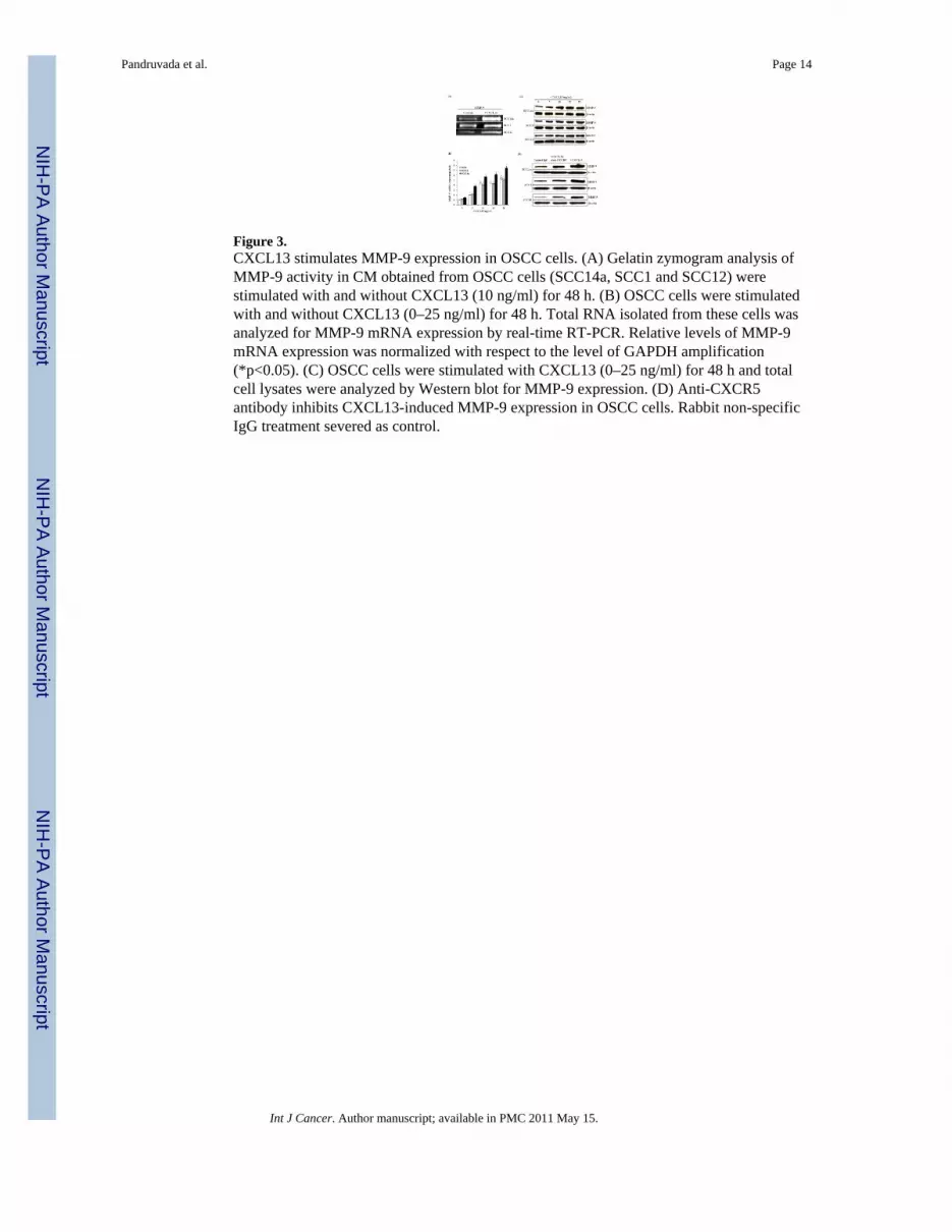
Figure 3.CXCL13 stimulates MMP-9 expression in OSCC cells. (A) Gelatin zymogram analysis ofMMP-9 activity in CM obtained from OSCC cells (SCC14a, SCC1 and SCC12) werestimulated with and without CXCL13 (10 ng/ml) for 48 h. (B) OSCC cells were stimulatedwith and without CXCL13 (0–25 ng/ml) for 48 h. Total RNA isolated from these cells wasanalyzed for MMP-9 mRNA expression by real-time RT-PCR. Relative levels of MMP-9mRNA expression was normalized with respect to the level of GAPDH amplification(*p<0.05). (C) OSCC cells were stimulated with CXCL13 (0–25 ng/ml) for 48 h and totalcell lysates were analyzed by Western blot for MMP-9 expression. (D) Anti-CXCR5antibody inhibits CXCL13-induced MMP-9 expression in OSCC cells. Rabbit non-specificIgG treatment severed as control.
Pandruvada et al. Page 14
Int J Cancer. Author manuscript; available in PMC 2011 May 15.
NIH
-PA Author Manuscript
NIH
-PA Author Manuscript
NIH
-PA Author Manuscript

Figure 4.Recombinant hCXCL13 protein induced chemotaxis of peripheral blood monocyte cells(PBMC). (A). PBMC were treated with CXCL13 (0–10 ng/ml) and chemotaxis was assayedas described in the methods. (B). PBMC were incubated with anti-CXCR5 antibody (25 ng/ml) for 1 h and stimulated with CXCL13 (10 ng/ml) (*p<0.05). Cells cultured in the absenceof CXCL13 or CXCL13 treatment in the presence of rabbit non-specific IgG served ascontrols.
Pandruvada et al. Page 15
Int J Cancer. Author manuscript; available in PMC 2011 May 15.
NIH
-PA Author Manuscript
NIH
-PA Author Manuscript
NIH
-PA Author Manuscript
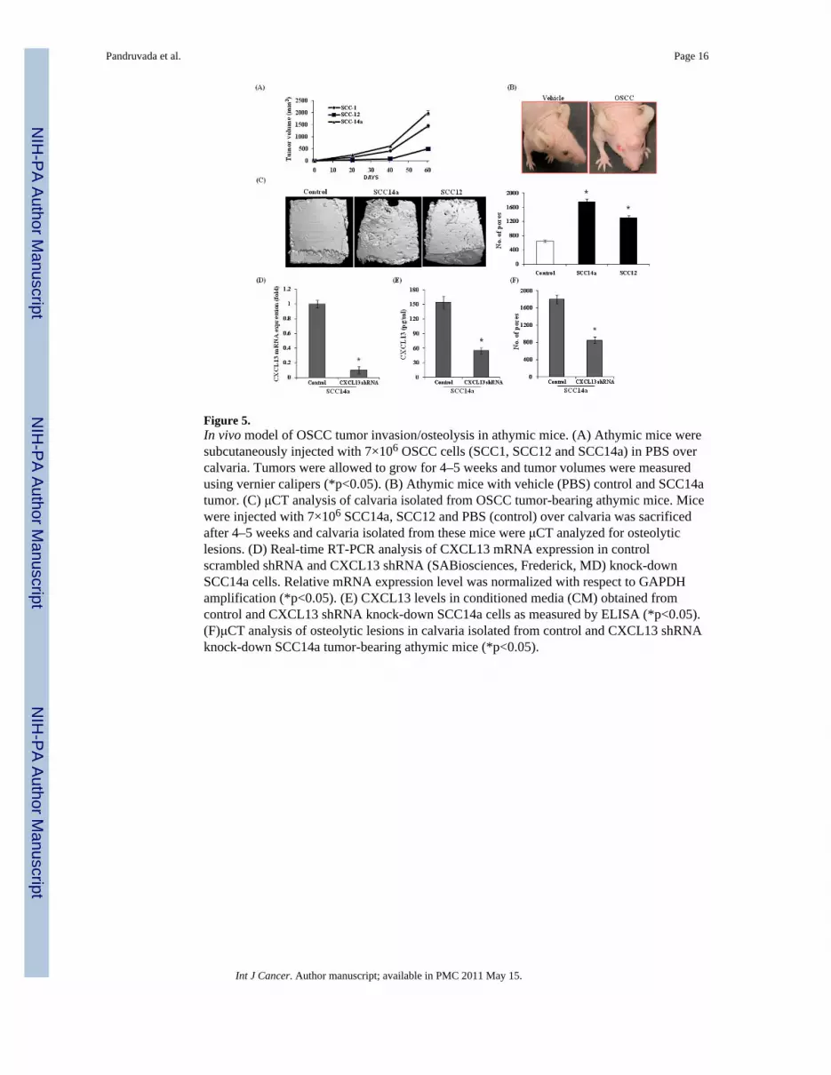
Figure 5.In vivo model of OSCC tumor invasion/osteolysis in athymic mice. (A) Athymic mice weresubcutaneously injected with 7×10⁶ OSCC cells (SCC1, SCC12 and SCC14a) in PBS overcalvaria. Tumors were allowed to grow for 4–5 weeks and tumor volumes were measuredusing vernier calipers (*p<0.05). (B) Athymic mice with vehicle (PBS) control and SCC14atumor. (C) μCT analysis of calvaria isolated from OSCC tumor-bearing athymic mice. Micewere injected with 7×10⁶ SCC14a, SCC12 and PBS (control) over calvaria was sacrificedafter 4–5 weeks and calvaria isolated from these mice were μCT analyzed for osteolyticlesions. (D) Real-time RT-PCR analysis of CXCL13 mRNA expression in controlscrambled shRNA and CXCL13 shRNA (SABiosciences, Frederick, MD) knock-downSCC14a cells. Relative mRNA expression level was normalized with respect to GAPDHamplification (*p<0.05). (E) CXCL13 levels in conditioned media (CM) obtained fromcontrol and CXCL13 shRNA knock-down SCC14a cells as measured by ELISA (*p<0.05).(F)μCT analysis of osteolytic lesions in calvaria isolated from control and CXCL13 shRNAknock-down SCC14a tumor-bearing athymic mice (*p<0.05).
Pandruvada et al. Page 16
Int J Cancer. Author manuscript; available in PMC 2011 May 15.
NIH
-PA Author Manuscript
NIH
-PA Author Manuscript
NIH
-PA Author Manuscript

Figure 6.Histological analysis of calvaria excised from control and OSCC tumor bearing mice.Histochemical staining was performed with hematoxylin-eosin (H&E) and for TRAPactivity. (A). Calvaria from control mice. (B). OSCC tumor bearing mouse calvaria. (C)OSCC tumor invasion into calvarial bone (arrows point to TRAP- positive osteoclasts; b=bone, t= tumor). (D) Osteoclasts at the tumor-bone interface and in calvaria from controlmice treated with PBS alone were counted using a micrometer scale and expressed per mm2
(*p<0.05) (E) Immuno histochemical analysis of CXCR5, CXCL13 and MMP-9 expressionin SCC14a tumors from athymic mice. Immunostaining with antibodies specific to CXCR5,CXCL13 and MMP-9 was performed and a rabbit non-specific IgG served as control. (F)Histochemical staining of calvaria isolated from control scrambled shRNA, CXCL13shRNA knock-down SCC14a cell tumor-bearing athymic mice were performed with H&Eand TRAP activity staining for osteoclasts (G) Osteoclasts numbers in the control andCXCL13 shRNA knock-down tumor-bone interface were counted using a micrometer scaleand expressed per mm2 (*p<0.05).
Pandruvada et al. Page 17
Int J Cancer. Author manuscript; available in PMC 2011 May 15.
NIH
-PA Author Manuscript
NIH
-PA Author Manuscript
NIH
-PA Author Manuscript
Related Documents

![Growth Inhibition of Human Tumor Cells in Athymic Mice by ...[CANCER RESEARCH 44, 1002-1007, March 1984] Growth Inhibition of Human Tumor Cells in Athymic Mice by Anti-Epidermal Growth](https://static.cupdf.com/doc/110x72/5e7bcebb508ec15dc92ee12e/growth-inhibition-of-human-tumor-cells-in-athymic-mice-by-cancer-research-44.jpg)