81,9(56,7( &/$8'( %(51$5' /<21 (FROH GRFWRUDOH %0,& %LRORJLH 0ROpFXODLUH ,QWpJUDWLYH HW &HOOXODLUH 'RFWRUDW 3RXU O¶REWHQWLRQ GX JUDGH GH 'RFWHXU HQ 6FLHQFHV GH O¶8QLYHUVLWp /\RQ 1 3UpVHQWpH HVW VRXWHQXH SXEOLTXHPHQW OH 'pFHPEUH 3DU )DWPD %(55, 5{OH GH O¶KpPRVWDVH GDQV O¶LQIODPPDWLRQ LQGXLWH SDU OHV YLUXV LQIOXHQ]D $ 7KqVH GLULJpH SDU OH 'U %pDWULFH 5,7($8 /DERUDWRLUH ($ 8QLWp 9LU3DWK )DFXOWp GH 0pGHFLQH GH /DHQQHF /\RQ )UDQFH -XU\ 3URIHVVHXU %UXQR /,1$ 3UpVLGHQW 'RFWHXU 1DWKDOLH 528$6)5(,66 5DSSRUWHXU 'RFWHXU 0XVWDSKD 6,7$+$5 5DSSRUWHXU 'RFWHXU 0DUWLQH -$1'5273(5586 ([DPLQDWHXU 'RFWHXU -HDQ&ODXGH %25'(7 ([DPLQDWHXU 'RFWHXU %pDWULFH 5,7($8 'LUHFWHXU GH 7KqVH

Welcome message from author
This document is posted to help you gain knowledge. Please leave a comment to let me know what you think about it! Share it to your friends and learn new things together.
Transcript


Research article
The Journal of Clinical Investigation http://www.jci.org 1
PAR1 contributes to influenza A virus pathogenicity in mice
Khaled Khoufache,1,2 Fatma Berri,1 Wolfgang Nacken,3 Annette B. Vogel,4,5 Marie Delenne,1 Eric Camerer,6,7 Shaun R. Coughlin,8 Peter Carmeliet,9,10 Bruno Lina,1 Guus F. Rimmelzwaan,11
Oliver Planz,4 Stephan Ludwig,3 and Béatrice Riteau1,2
1Virologie et Pathologie Humaine, EA 4610, Université Lyon1, Faculté de Médecine RTH Laennec, Lyon, France. 2INRA Tours, Nouzilly, France. 3Institute of Molecular Virology, ZMBE, Westfälische-Wilhelms-University, Münster, Germany. 4Friedrich-Loeffler-Institute, Institute of Immunology, University Hospital, Tuebingen, Germany. 5Institute of Immunology, Friedrich-Loeffler-Institut, Greifswald-Insel Riems, Germany. 6INSERM U970, Paris Cardiovascular Centre, Paris, France. 7Université Paris-Descartes, Paris, France. 8Cardiovascular Research Institute, UCSF, San Francisco,
California, USA. 9Laboratory of Angiogenesis and Neurovascular link, Vesalius Research Center, VIB, Leuven, Belgium. 10Laboratory of Angiogenesis and Neurovascular link, Vesalius Research Center, KU Leuven, Leuven, Belgium.
11Department of Virology, Erasmus Medical Center, Rotterdam, the Netherlands.
Influenza causes substantial morbidity and mortality, and highly pathogenic and drug-resistant strains are likely to emerge in the future. Protease-activated receptor 1 (PAR1) is a thrombin-activated receptor that con-tributes to inflammatory responses at mucosal surfaces. The role of PAR1 in pathogenesis of virus infections is unknown. Here, we demonstrate that PAR1 contributed to the deleterious inflammatory response after influ-enza virus infection in mice. Activating PAR1 by administering the agonist TFLLR-NH2 decreased survival and increased lung inflammation after influenza infection. Importantly, both administration of a PAR1 antago-nist and PAR1 deficiency protected mice from infection with influenza A viruses (IAVs). Treatment with the PAR1 agonist did not alter survival of mice deficient in plasminogen (PLG), which suggests that PLG permits and/or interacts with a PAR1 function in this model. PAR1 antagonists are in human trials for other indica-tions. Our findings suggest that PAR1 antagonism might be explored as a treatment for influenza, including that caused by highly pathogenic H5N1 and oseltamivir-resistant H1N1 viruses.
IntroductionInfluenza is an ineradicable contagious disease that occurs in sea-sonal epidemics and sporadic pandemic outbreaks that pose sig-nificant morbidity and mortality for humans and animals (1–3). The continuous sporadic infections of humans with highly patho-genic avian influenza viruses of the H5N1 subtype and the recent pandemic caused by swine-origin H1N1 viruses highlight the permanent threat caused by these viruses (4–6). The pathogenesis of influenza A virus (IAV) infection is not fully understood, but involves both viral traits and the host immune response (3). Full understanding of the host response may aid in the development of intervention strategies that target these host factors.
Both innate and adaptive components of the immune system are activated shortly after virus infection, which provides an efficient line of defense against IAV (7). However, excessive inflammation may also result in lung damage that limits respiratory capacity and may account for IAV pathogenesis in humans (1, 8, 9). Recruit-ment of inflammatory cells to inflamed sites is controlled by a number of cellular components, including proteases (10). These proteases not only cleave extracellular substrates, but also medi-ate signal transduction in part via protease-activated receptors (PARs) (11–14). PAR1, which links local protease activity to cellu-lar responses involved in thrombosis, inflammation, and cytopro-tection (15, 16), shows increased expression in the airways of IAV-infected mice (17). The role of PAR1 in the context of IAV infection
has not been studied. We report evidence that PAR1 signaling con-tributed to the deleterious inflammation that followed influenza virus infection in mice in a manner dependent on plasminogen (PLG). While administration of a PAR1 agonist to mice increased severity of IAV infection, PAR1 deficiency protected mice from fatal outcome. Administration of the PAR1 antagonist SCH79797 (18) to mice decreased inflammation and improved survival after infection with multiple IAV strains, including a highly pathogenic avian H5N1 strain and 2009 pandemic H1N1 virus. Importantly, administration of SCH79797 improved survival in mice even when administered 48 or 72 hours after inoculation. PAR1 antagonists are currently in clinical trials for potential use as antithrombotic drugs (19–22). Because an intervention strategy aimed at a host cellular protein would be effective against virus strains that devel-op resistance to existing antiviral drugs, PAR1 antagonists might be explored for the treatment of IAV in additional preclinical mod-els and, if appropriate, in humans.
ResultsPAR1 contributes to the pathogenesis of IAV infection. To investigate the role of PAR1 in the pathogenesis of IAV infection, WT mice were inoculated with 50 or 500 PFU of H1N1 strain A/PR/8/34 (referred to herein as H1N1) and either left untreated or stimulated with 50 M of the PAR1 agonist TFLLR-NH2 (referred to herein as PAR1-activating peptide; PAR1-AP). Mice treated with PAR1-AP displayed enhanced weight loss and higher mortality rates after infection compared with untreated control mice, differences that were statistically significant at both doses (Figure 1A). In contrast, treatment of uninfected mice with PAR1-AP did not affect survival or body weight of mice (Figure 1B), which indicates that the effect of PAR1-AP on survival and weight loss requires IAV infection.
Authorship note: Khaled Khoufache and Fatma Berri contributed equally to this work.
Conflict of interest: Khaled Khoufache and Béatrice Riteau have a patent concerning the use of PAR1 antagonist against influenza.
Citation for this article: J Clin Invest. doi:10.1172/JCI61667.

research article
2 The Journal of Clinical Investigation http://www.jci.org
Moreover, treatment with a control peptide did not impair survival or increase weight loss in IAV-infected mice (Figure 1C), militating against nonspecific effects of peptide administration. Thus, PAR1 activation led to increased pathogenicity of IAV infection.
To further explore the role of PAR1 in IAV pathogenesis, we investigated the consequence of PAR1 deficiency. Par1+/– mice were intercrossed to generate WT and Par1–/– mice, which were infected with 100 PFU H1N1, and weight loss and survival rates were mon-itored. Compared with WT littermates, Par1–/– mice were more resistant to IAV infection (Figure 1D). Thus, PAR1 contributed to death and weight loss caused by IAV infection.
PAR1-AP increases cytokine release and neutrophil recruitment in the lungs of infected mice. Because PAR1 can trigger cytokine production in endothelial and other cell types (14), we next investigated the effects of PAR1-AP in the inflammatory response induced by IAV infection. Mice infected with 50 PFU H1N1 were treated or not with 50 M PAR1-AP, and bronchoalveolar lavages (BALs) were collected to assess the presence of cytokines and polymorpho-
nuclear neutrophils (PMNs) in the lungs at different time points after inoculation. IAV infection resulted in increased levels of all cytokines tested (RANTES, IL-6, and KC) in a time course–depen-dent manner, and PAR1-AP treatment augmented this response (Figure 2A). Similar results were obtained when the effect of PAR1 was compared with that of a control peptide (Supplemental Fig-ure 1; supplemental material available online with this article; doi:10.1172/JCI61667DS1), confirming PAR1-AP specificity. PAR1-AP treatment also increased the occurrence of BAL PMNs 24 and 48 hours after infection, but had little effect in uninfected mice (Figure 2B). By 72 hours after infection, the PMN content of BAL in PAR1-AP–treated and control mice was not different. These results suggest that PAR1 activation can increase IAV-induced pro-duction of cytokines and increase early recruitment of neutrophils in the lungs of infected mice.
Virus replication in the lungs. We then investigated whether the effect of PAR1 activation on the outcome of IAV infection in mice corre-lates with an increase of virus production in the lungs. To this end,
Figure 1Effect of PAR1 activation and PAR1 deficiency on IAV pathogenicity. (A) Time course of IAV-induced pathogenesis and death in mice in response to PAR1 stimulation. Mice were inoculated intrana-sally with H1N1 (50 PFU, n = 22 per group; 500 PFU, n = 18 per group) and treated with either vehicle or 50 M PAR1-AP. (B) Time course of uninfected mice treated or not with 50 M PAR1-AP (n = 13 per group). (C) Mice were infected with 50 PFU H1N1 and treated with control peptide or vehicle (n = 10 per group). Results are average percent survival or weight loss from 3 indepen-dent experiments. (D) Survival and weight loss of Par1–/– mice and WT littermates after infection with 100 PFU H1N1 (n = 12 per group). Results are average percent survival or weight loss from 2 experiments. P < 0.05, PAR1-AP vs. untreated or Par1–/– vs. WT, Kaplan-Meier test.

research article
The Journal of Clinical Investigation http://www.jci.org 3
infectious virus titers were determined in lungs collected from mice treated with PAR1-AP (50 M) or control peptide at different time points after inoculation. At 24 and 48 hours after inoculation, virtu-ally no virus replication was detected (101 was the detection limit of the assay), but lung virus titers significantly increased after PAR1-AP treatment (Figure 2C). No significant differences were observed 3 and 5 days after infection. These data suggest that PAR1 activa-tion promotes an early increase in virus production in mouse lungs.
The effect of PAR1 activation on virus production, weight loss, and sur-vival after IAV infection is PLG dependent. To decipher the mechanism by which PAR1 accelerated virus production in vivo, we performed in vitro experiments to assess the effect of PAR1 activation on virus replication in alveolar epithelial A549 cells. PAR1-AP triggered ERK phosphorylation in these cells, with a maximal effect at about 40 M (Figure 3A); this concentration was used in all subsequent in vitro experiments. Because proteolytic cleavage of HA is essen-tial for IAV infectivity, and PLG promotes IAV replication through HA cleavage (23, 24), we examined the effect of adding PLG — alone or in combination with PAR1-AP — on virus production. As expected, viral production was barely detectable in untreated A549 cultures, but was markedly increased by the addition of PLG (Figure 3B). Importantly, addition of PAR1-AP augmented this effect 8 and 24 hours after infection. The effect of PAR1-AP was not seen when trypsin was used as an alternative protease for IAV replication (data not shown), and PAR1 signaling did not affect virus entry into cells (Supplemental Figure 2). However, inclu-sion of PAR1-AP appeared to increase PLG-dependent cleavage of HA. Thus, we next infected A549 cells (MOI 0.5) in the presence or absence of PLG, with or without PAR1-AP, and evaluated HA cleavage by Western blot analysis 16 hours after infection. In the absence of PLG, similar amounts of uncleaved HA (HA0) accumu-lated in infected cells, and PAR1-AP was without effect (Figure 3C). In the presence of PLG, in addition to HA0, a 25-kDa band corre-sponding to HA2 was observed. Importantly, in PAR1-AP–treated cultures, the intensity of HA2 increased and HA0 decreased relative to that in control cultures. Thus, viral HA was cleaved in a PLG-dependent manner that was enhanced by PAR1-AP and correlated with increased viral production.
PLG is an important mediator of lung inflammation (25, 26) and is known to influence IAV virulence (27, 28). Importantly, PLG bind-ing to cells and activation may be controlled by PAR1 signaling (29, 30). In combination with the findings outlined above, these observa-tions prompted us to investigate whether the effect of PAR1 signal-ing on the pathogenicity of IAV infection also depends on PLG in vivo. We therefore inoculated Plg–/– mice with 50 PFU H1N1 with or without PAR1-AP treatment. In contrast to WT mice, treatment of Plg–/– mice with PAR1-AP did not increase mortality rates, weight loss, or virus titers in lungs after IAV infection (Figure 3, D and E).
Histopathological examination showed that treatment with PAR1-AP increased cellular infiltrates in lungs from infected WT mice, but not Plg–/– mice (Supplemental Figure 3). These results suggest that PAR1 activation increased early virus production, inflammation, and pathogenicity of IAV infection in a PLG-dependent fashion. Notably, when this low 50-PFU dose was used, virtually no virus replication was detected in the lungs of WT or Plg–/– mice at the indicated time points after inoculation (Figure 3E). Additionally, leukocyte infiltration in IAV-infected WT or Plg–/– mice was barely detectable (Supplemental Figure 3). However, when a higher virus dose was used for inoculation, leukocyte infiltration and lung virus titers of Plg–/– mice were substantially lower than those of WT mice (F. Berri, unpublished observations), which sug-gests that PLG promotes IAV replication and inflammation. While the finding that PAR1-AP increased PLG-dependent cleavage of HA in vitro suggests that PAR1 signaling might promote viral replica-tion by enhancing PLG/plasmin function, our data do not exclude a PAR1-independent permissive role for PLG or PLG-independent roles for PAR1 activation in IAV infection and pathogenesis.
PAR1 antagonist protects against H1N1 and H3N2 infection. We next investigated whether pharmacological inhibition of PAR1 signal-ing alters the course of IAV infection. The pharmacology of PARs is not well developed, and inhibitors capable of blocking PAR1 function in mouse models have not been well characterized with respect to off-target effects. Nonetheless, SCH79797 has been used to probe PAR1 function in rodent models (31–33); thus, encour-aged by the protection against IAV seen in Par1–/– mice, we exam-ined the effects of this compound on the course of IAV infection.
Figure 2PAR1-AP increases inflammation and virus replication during 50 PFU H1N1 infection in mice. (A) Cytokines in the BAL of infected mice treated or not with PAR1-AP were mea-sured by ELISA 24, 48, and 72 hours after inoculation. Data are mean ± SD from 5–11 individual animals per group from 3 experi-ments. (B) Relative PMN numbers in BAL from infected mice treated or not with PAR1-AP. PMN percentage was determined by May-Grünwald–Giemsa staining 24, 48, or 72 hours after inoculation. Results are mean ± SD from 4–5 individual mice per group from 2 individual experiments. Noninfected mice were used as control (n = 2–4 per group). (C) H1N1 virus titers in the lungs at the indicated times after infection of mice treated or not with 50 M PAR1-AP. Data are average ± SD from 3–5 individual animals per group. *P < 0.05, treated vs. untreated, Mann-Whitney test.

research article
4 The Journal of Clinical Investigation http://www.jci.org
SCH79797 inhibited PAR1-AP–induced ERK activation in mouse NIH3T3 cells (Figure 4A), which suggests that it is capable of blocking signaling by the mouse homolog of PAR1. SCH79797 treatment prevented decreased survival and increased weight loss associated with administration of PAR1-AP to IAV-infected mice (Figure 4B). More strikingly, when mice were infected with lethal doses of H1N1 (500 and 5,000 PFU), SCH79797 treatment pro-tected mice from weight loss and death: 47% and 16% survival, respectively, was observed in untreated control mice, whereas 84%–94% of SCH79797-treated mice survived the infections (Fig-ure 4C). Moreover, when SCH79797 was administered beginning 2 or 3 days after infection, mice were also significantly protected from H1N1 and from H3N2 strain A/Hong-Kong/68 (referred to herein as H3N2; Figure 4, D and E). Treatment of uninfected mice with SCH79797 did not affect their survival rates or body weight (Supplemental Figure 4), which suggests that PAR1 antagonists do not cause side effects. Thus, SCH79797 treatment protected mice from IAV infection, consistent with the notion that PAR1 contributes to IAV pathogenesis in this model.
Inflammation and virus replication are attenuated by SCH79797. Since PAR1 activation promoted inflammation in the lungs dur-ing IAV infection, we determined whether blockade of PAR1 sig-naling would result in reduced IAV-induced inflammation in vivo. Mice were infected with 500 PFU H1N1 and treated or not with SCH79797, and BAL was collected at different times after inocu-
lation. SCH79797 treatment significantly reduced the levels of RANTES, IL-6, and KC in BAL 24, 48, and 72 hours after inocula-tion, as measured by ELISA (Figure 5A). 5 days after inoculation, cytokine levels were still high in the BAL from untreated mice, but barely detectable in the BAL from SCH79797-treated mice (Supplemental Figure 5). SCH79797 treatment also significantly decreased PMN frequency in the BAL of infected mice: 24 and 48 hours after inoculation, PMNs were hardly detectable in the BAL of SCH79797-treated mice, whereas they represented 10% of cells in BAL from untreated mice (Figure 5B). Accordingly, histopathologi-cal examination revealed a reduction of cell infiltration in the lungs of infected mice treated with SCH79797 (Supplemental Figure 6).
Finally, a reduction in lung virus titers was observed 24 and 48 hours after 500 PFU H1N1 inoculation compared with untreated controls (Figure 5C). At day 3 after inoculation, lung virus titers were similar in SCH79797-treated and untreated mice (approximately 104 PFU/ml), which suggests that SCH79797 delayed, but did not prevent, virus production. Lung virus titers dropped to less than 102 PFU/ml at days 5 and 7 in both SCH79797-treated and control mice (Figure 5C). The observation that SCH79797 suppressed markers of inflammation, but not viral titers, at day 3 suggests that inhibition of PAR1 signaling may inhibit inflammation and early virus replica-tion by at least partially independent mechanisms.
SCH79797 protects against highly pathogenic H1N1v and H5N1 infec-tion. To test whether inhibition of PAR1 signaling by SCH79797
Figure 3Effect of PLG and PLG deficiency on IAV production and PAR1-AP effects. (A) ERK phosphorylation after stimulation of A549 cells with the indicated PAR1-AP concentrations. Anti–phospho-Erk and anti-Erk antibodies were used. (B) Infectious virus titers in the supernatant of infected cells after stimulation with 40 M PAR1-AP or control peptide in the presence or absence of PLG. (C) Noninfected (NI) or infected (INF) cells were stimulated with 40 M PAR1-AP or control peptide in the presence or absence of PLG. After cell lysis, proteins were analyzed by Western blot for HA cleavage. (D) Time course of IAV-induced pathogenesis in Plg–/– and WT littermates after treatment or not with PAR1-AP (n = 9–10 mice per group from 2 experiments). (E) Virus titers 48 hours after infection (50 PFU) in lungs of WT or Plg–/– mice stimulated or not with 50 M PAR1-AP. Data are average ± SD from 5 individual animals per group from 2 experiments. *P < 0.05, treated vs. untreated, Kaplan-Meier test (D), Mann Whitney test (B and E).

research article
The Journal of Clinical Investigation http://www.jci.org 5
also affects infection with other IAV strains, mice were infected with a highly pathogenic H5N1 strain or a pandemic H1N1v strain that had acquired oseltamivir resistance during treatment of a severe infection (see Methods and ref. 34), then treated or not with SCH79797. After lethal infection with 5,000 PFU H5N1 and 500 PFU H1N1v, 60% and 100% of untreated control mice died, respectively, whereas almost full protection was observed in SCH79797-treated animals of both inoculation groups (P < 0.05; Figure 6, A and B). In addition to mortality and body weight, the onset of clinical signs was also inhibited when H5N1-infected mice were treated with SCH79797 compared with untreated mice (data not shown). Mouse mortality was monitored until day 21 after inoculation, and sustained survival was observed after SCH79797 treatment (data not shown), which indicated that SCH79797 protection was durable. Thus, inhibition of PAR1 sig-
naling protected mice against infection with various IAVs, includ-ing highly pathogenic strains.
DiscussionOur present findings support an important role for PAR1 in mouse models of IAV infection. Studies with PAR1-AP indicated that PAR1 activation increased inflammation, early virus produc-tion, weight loss, and mortality after infection (Figures 1 and 2), and studies using Par1–/– mice indicated that PAR1 contributed to the pathogenesis of IAV infection (Figure 1). The observation that SCH79797, a drug that inhibits PAR1 signaling, decreased inflammation, early virus production, weight loss, and mortality after infection was in accord with the PAR1-AP and Par1–/– results. Moreover, the observation that SCH79797 decreased mortal-ity after infection with multiple IAV strains (H1N1, H3N2, and
Figure 4PAR1 antagonist protects mice against infection with H1N1 and H3N2. (A) Treatment of NIH3T3 cells with SCH79797 blocked ERK activation by 10 M PAR1-AP. (B) SCH79797 treatment prevented PAR1-AP–induced mouse mortality in a dose-dependent manner. (C) IAV-induced patho-genesis in infected mice treated or not with SCH79797. Mice were inoculated with 500 PFU (n = 17–19 per group) or 5,000 PFU (n = 14 per group) H1N1 and treated or not with 50 M SCH79797 on days 0–2 after infection. (D) SCH79797 treatment on days 2–4 after infection with 5,000 PFU H1N1 (n = 12 per group) or 100 PFU H3N2 (n = 7 per group). (E) SCH79797 treatment on days 3–5 after infection with 5,000 PFU H1N1 (n = 7 per group) or 100 PFU H3N2 (n = 7 per group). *P < 0.05, treated vs. control, Kaplan-Meier test.

research article
6 The Journal of Clinical Investigation http://www.jci.org
H5N1), and was effective even when dosing was initiated at day 3 after inoculation, suggests that PAR1 inhibition should be explored in additional preclinical studies and, if appropriate, in humans as a possible treatment for influenza.
To our knowledge, a role for PAR1 in the response to, and the pathogenesis of, virus infections has not been previously described. PAR1 activation in endothelial cells, fibroblasts, and other cell types triggers various responses, many of which are proinflammatory (e.g., chemokine and cytokine production, adhesion molecule display, prostaglandin production, and per-meability increases; refs. 14, 15). In accord with our observations, intratracheal delivery of PAR1 agonist was not sufficient to trig-ger inflammation in the lungs of otherwise normal mice (35), but did exacerbate ventilation injury–induced pulmonary edema (36). Additionally, Par1–/– mice are protected from ventilation injury–induced and bleomycin-induced lung injury (36–38). Like our results, these observations suggest that PAR1 signaling contrib-utes to inflammatory responses to injury in the lung, the major target in our IAV infection model.
PAR1 activation did not exacerbate the effects of IAV infection in Plg–/– mice (Figure 3). It is possible that PLG is simply playing a permissive role for the effect of PAR1 activation in IAV infec-tion; that is, PLG supports infection and injury, and PAR1 acti-vation exacerbates their effects. Interestingly, however, PAR1-AP did promote PLG-dependent HA cleavage in lung epithelial cul-tures, suggestive of a possible interaction of PAR1 signaling with the ability of IAV to become infectious and hence replicate. These findings are consistent with the prior observation that PLG con-tributes to the pathogenesis of IAV infection (27, 28). Additionally, PAR1 signaling may promote PLG activation to plasmin (29, 30), thereby providing a possible link to increased HA cleavage and IAV
production. It is also possible that PAR1 activation contributes to proinflammatory functions of PLG (25, 39–41), by promoting its conversion to plasmin or by other mechanisms.
Additional considerations suggest that PAR1 activation’s abili-ties to promote early virus replication and to enhance a harmful inflammatory response in the respiratory tract are, at least in part, independent of each other. When PAR1-AP was delivered 3 days after infection, despite similar virus replication in the lungs, treat-ment still had a deleterious effect (data not shown). Additionally, based on critical residues in HA involved for cleavage by plasmin, it is unlikely that the replication of highly pathogenic H5N1 and 2009 pandemic H1N1 are modulated by plasmin (42), yet SCH79797 treatment still decreased mortality.
As noted above, we found that in IAV-infected A549 cells, activa-tion of PAR1 increased PLG-dependent HA cleavage, an essential step for virus infectivity. Indeed, only the cleaved form of HA per-mits pH-dependent fusion of the viral envelope within the endo-somal membranes and subsequent release of the genome into the cytosol and virus replication. In vivo, PAR1 also promoted virus replication shortly after infection. However, at 48 hours after infection, no difference in lung virus titers was observed between PAR1-AP–stimulated and unstimulated mice, which suggests that HA cleavage could be compensated by other proteases that are either recruited or activated by infection in the lungs.
Therefore, we propose a model (Figure 7) in which PAR1 pro-motes activation of PLG into plasmin. Subsequently, plasmin acts on virus replication through HA cleavage, enhancement of which likely enhances inflammation via pathogen-associated molecular patterns. Simultaneously, plasmin also acts as a proinflammatory mediator that accounts for the deleterious lung inflammation. Additionally, PAR1 triggers a variety of proinflammatory respons-
Figure 5PAR1 antagonist inhibits lung inflammation and virus repli-cation. (A) Cytokines in the BAL of infected mice treated or not with SCH79797 were measured by ELISA 24, 48, and 72 hours after inoculation. Data are average ± SD from 7–11 individual animals per group, representative of 3 experi-ments. (B) Relative PMN frequency in BAL from infected mice treated or not with SCH79797. PMN percentage was determined by May-Grünwald–Giemsa staining 24, 48, and 72 hours after inoculation. Data are average ± SD from 3–5 individual mice per group. Noninfected mice were used as control (n = 3–5 per group). Results are representative of 2 individual experiments. (C) Virus titers in lungs of infected mice at the indicated times after infection with 500 PFU H1N1 and treatment with SCH79797. Data are average ± SD from 3–5 individual animals per group. *P < 0.05, treated vs. control, Mann-Whitney test.

research article
The Journal of Clinical Investigation http://www.jci.org 7
es, independent of PLG and virus, that may exacerbate inflamma-tion and injury. Because PAR1 couples coagulation to inflamma-tion (14, 15) and coagulation to fibrinolysis (30), further studies are needed to investigate the overall impact of hemostasis dysregu-lation in PAR1-mediated inflammation during IAV infection.
Our observation that a PAR1 agonist (43, 44) exacerbated the effects of IAV infection suggests that PAR1 activation is capable of promoting inflammation and tissue damage in this setting. More-over, our observation that Par1–/– mice and SCH79797-treated mice were protected from IAV infection suggests that PAR1 activation contributes to the pathogenesis of IAV infection and that PAR1 is endogenously activated during IAV infection. Accordingly, the nat-ural PAR1 activator thrombin was generated in IAV-infected lungs (45), and elevated levels of PAR1 were observed in the airways of IAV-infected mice (17). It is worth noting, however, that SCH79797 is known to have off-target effects on cell proliferation and sur-vival (46, 47); thus, we cannot exclude PAR1-independent effect of SCH79797. However, SCH79797 was capable of inhibiting PAR1 signaling (Figure 4A and ref. 18), and the concordance of our KO and inhibitor studies — and the fact that their effects were opposite from those of PAR1-AP — suggest that the effects of SCH79797 in our model could be related to its ability to block PAR1 signaling.
Besides PAR1, other PARs may be involved in the pathogenesis of IAV infection (48–50). Identification of the exact nature and amount of proteases present at the site of infection, and how virus strain dif-ferences alter the immune response and its interactions with PARs, may advance our understanding of the pathogenesis of IAV infection.
Current treatments for IAV infection target the viral proteins M2 and NA. These drugs suffer from a number of disadvantages, including the rapid development of resistant virus variants as a result of selective pressure, which highlights the need for new pharmacological strategies against IAV infection. Because target-ing host proteins would not be subject to resistance, and because severe infections with IAV are associated with a deleterious host inflammatory response, drugs regulating inflammation are appealing as potential treatments for IAV infection (51, 52). In our present study, blocking PAR1 signaling almost fully protected mice from a highly pathogenic, oseltamivir-resistant 2009 pandemic H1N1v virus isolated from a severely diseased oseltamivir-treat-ed patient (34). Additionally, inhibition of PAR1 signaling up to 3 days after inoculation protected mice from a detrimental out-come of infection with various IAVs, including H1N1 and H3N2 strains. Because IAVs of the H1N1 and H3N2 subtypes are currently circulating in the human population, it is reasonable to assume that PAR1 antagonists are most likely also effective against season-al influenza viruses. Interestingly, the PAR1 antagonist vorapaxar has been studied as a potential antithrombotic drug in approxi-mately 40,000 patients over 3 years (53, 54). The most serious side effect, increased incidence of intracranial bleeding, occurred mainly in patients with a history of prior stroke. In the absence of such a history, the increase in the incidence of intracranial bleeding was less than 1 per 1,000 treatment-years. Thus, short periods of PAR1 antagonism would appear to be relatively safe. This observation, in consideration with our results, suggests that PAR1 antagonism
Figure 6PAR1 antagonist protects mice from lethal infection with H5N1 or H1N1v. Mice were inoculated intranasally with (A) 5,000 PFU H5N1 (n = 10 per group) or (B) 500 PFU H1N1v (n = 10–11 per group) and treated or not with 50 M SCH79797. Results are expressed as percent survival or weight loss from 2 experiments. *P < 0.05, treated vs. control, Kaplan-Meier test.
Figure 7Proposed model for PAR1-mediated influenza virus pathogenesis. Dur-ing IAV infection, PAR1 is activated and increases conversion of PLG into plasmin. On the one hand, plasmin cleaves and activates the viral HA, promoting IAV replication, which contributes to inflammation. On the other hand, plasmin directly promotes inflammation, and PAR1 pro-motes inflammation via mechanisms that are independent of PLG and virus. These likely interact with other host responses to viral infection to exacerbate inflammation and injury.

research article
8 The Journal of Clinical Investigation http://www.jci.org
Mouse infection and treatment. Mice were anesthetized and inoculated intranasally with 25 l of a solution containing different doses of virus in the presence or absence of 50 M PAR1-AP, 50 M control peptide, and/or 50 M SCH79797. 500 M SCH79797 was also used for blocking experi-ments in Figure 4B. Intranasal treatments with PAR1-AP, control peptide, and/or SCH79797 were also repeated at days 2 and 3 after infection. Alter-natively, mice were inoculated, and SCH79797 was administered on days 2–4 or days 3–5 after infection. Mice were then monitored for weight loss and mortality. For assessing virus replication, lungs were obtained from scarified mice, and infectious virus titers were determined by plaque assay as described previously (56).
Cytokine detection by ELISA and PMN recruitment. Production of the cytokines RANTES, IL-6, and KC in the lungs was determined by ELISA (R&D Systems), using BAL from mice, as previously described (60). For PMN recruitment, BAL was collected in PBS (Invitrogen) supplemented with 1 mM EDTA (Invitrogen). After cytocentrifugation, the percentage of PMNs was determined by counting a total of 500 cells per sample by microscopic examination of May-Grünwald– and Giemsa-stained cytocentrifuge slides.
Lung histology. At 3 days after virus inoculation and treatment, mice were killed, and lung tissue was harvested, fixed in 10% formaldehyde, and subsequently embedded in paraffin. Tissues were sectioned at 12 M, and sections were examined after staining with hematoxylin and eosin for histopathological changes.
Statistics. Mann-Whitney test was used for statistical analysis of lung virus titers and cytokine ELISA results. Kaplan-Meier test was used for statistical analysis of survival rates. XLSTAT software was used to analyze differences between groups; a P value less than 0.05 was considered statistically significant.
Study approval. Experiments were performed according to recommen-dations of the National Commission of Animal Experiment (CNEA) and the National Committee on the Ethic Reflexion of Animal Experiments (CNREEA) in compliance with European animal welfare regulation. The protocol was approved by the committee of animal experiments of the Uni-versity Claude Bernard Lyon I (permit no. BH2008-13). All animal experi-ments were also carried out under the authority of licence issued by “la direction des services Vétérinaires” (accreditation no. 78-114). All efforts were made to minimize suffering.
AcknowledgmentsWe are grateful to N. Lejal for technical assistance. This work was supported by the Agence Nationale de la Recherche (ANR; to B. Riteau); Inserm Avenir (to E. Camerer); Marie Curie actions (to E. Camerer); and Long-term structural funding—Methusalem by the Flemish government (to P. Carmeliet).
Received for publication November 7, 2011, and accepted in revised form October 4, 2012.
Address correspondence to: Beatrice Riteau, EMR 4610 Vir-Path, Virologie et Pathologie Humaine, Faculté de médecine RTH Laennec, Université Claude Bernard Lyon 1, Université de Lyon, F-69008, Lyon, France. Phone: 33.1.0478771008; Fax: 33.1.0478778751; E-mail: [email protected].
should be further explored for the treatment of IAV in additional preclinical models and, if appropriate, human studies.
MethodsCells, virus strain, and reagents. The NIH3T3 mouse cell line was a gift from D. Décimo (INSERM U758, Lyon, France). The human alveolar type II (A549) and MDCK cell lines used in this study were obtained from ATCC and grown as previously described (55). H1N1 (strain A/PR/8/34) was obtained from the ATCC. H3N2 (strain A/Hong-Kong/2/68) was obtained from the Dutch National Influenza Centre. The strain was originally obtained from the National Institute for Biological Standards and Control (NIBSC). The highly pathogenic H5N1 avian influenza virus (strain A/mallard/Bavaria/ 1/2006; also known as MB1) and the pandemic H1N1v influenza virus (strain A/Nordrhein-Westfalen/173/09) were used in this study. H1N1v, isolated from a severe H1N1pdm09 case and obtained through the Ger-man National Reference Centre for Influenza of the Robert Koch Insti-tute, had acquired oseltamivir resistance during treatment (34). H5N1 was propagated in chicken eggs for 2 days, and the other viruses were propa-gated in confluent MDCK cells. After 2 days, cytopathic changes were com-plete, and culture supernatants were harvested and cleared by low-speed centrifugation and stored at –80°C. PAR1-AP and control peptide (TFLLR-NH2 and FTLLR-NH2, respectively) were purchased from Bachem. The PAR1 antagonist (SCH79797 dihydrochloride) was purchased from Axon Medchem. PLG was purchased from Sigma-Aldrich, and the following antibodies were used: monoclonal anti-HA (C102; Santa Cruz Biotechnol-ogy), monoclonal anti-tubulin (Sigma-Aldrich), and polyclonal anti-ERK and phospho-ERK (Cell Signaling Technology).
In vitro stimulation. A549 cells were preincubated for 5 minutes with 40 M PAR1-AP or control peptide or for 1 hour with 5 M SCH79797. Cells were then infected with H1N1 (MOI 0.001) in MEM supplemented with 0.5 M PLG (Sigma-Aldrich) in the presence of the drug. At the indi-cated times after stimulation, virus titers were analyzed by classical plaque assays as performed previously, using MDCK cells (56).
Western blot analysis of ERK activation and HA cleavage. A549 or NIH3T3 cells were stimulated or not with the indicated concentrations of PAR1-AP for 5 minutes at 37°C. Where indicated, cells were preincubated for 1 hour with SCH79797. Cells were then lysed, and proteins from the lysate were analyzed by Western blot for ERK activation, as previously described (57). For the HA cleavage experiments, A549 cells were stimulated or not with 40 M PAR1-AP and infected with IAV (MOI 0.5) for 16 hours in the pres-ence or absence of 0.5 M PLG. Cells were then lysed, and proteins from the lysate were analyzed by Western blot, as described previously (57).
Mice. Plg–/– mice (with a disrupted Plg gene) and their WT littermates (58) and 6-week-old C57BL/6 female mice (Charles River Laboratories) were used in this study. Par1–/– mice (with a disrupted Par1 gene) and their WT littermates were described previously (59). Heterozygous mice were crossed, and WT and KO offspring were used. Mouse ages ranged from 5 weeks to a maximum of 4 months, since the number of mice that could be obtained was limited. Male and female mice were used in the experiments. Groups of WT and KO mice were stratified for these differences in age and gender. Polymerase chain reaction of tail-tip genomic DNA was performed (60) for determination of the absence or presence of a functional Plg or Par1 gene.
1. La Gruta NL, Kedzierska K, Stambas J, Doherty PC. A question of self-preservation: immunopathol-ogy in influenza virus infection. Immunol Cell Biol. 2007;85(2):85–92.
2. Bouvier NM, Palese P. The biology of influenza viruses. Vaccine. 2008;26(suppl 4):D49–D53.
3. Kuiken T, Riteau B, Fouchier RA, Rimmelzwaan GF. Pathogenesis of influenza virus infections: the good, the bad and the ugly. Curr Opin Virol.
2012;2(3):276–286. 4. Webby RJ, Webster RG. Are we ready for pandemic
influenza? Science. 2003;302(5650):1519–1522. 5. Foucault ML, Moules V, Rosa-Calatrava M, Riteau
B. Role for proteases and HLA-G in the pathoge-nicity of influenza A viruses. J Clin Virol. 2011; 51(3):155–159.
6. Solorzano A, Song H, Hickman D, Perez DR. Pan-demic influenza: preventing the emergence of novel
strains and countermeasures to ameliorate its effects. Infect Disord Drug Targets. 2007;7(4):304–317.
7. Schmolke M, Garcia-Sastre A. Evasion of innate and adaptive immune responses by influenza A virus. Cell Microbiol. 2010;12(7):873–880.
8. de Jong MD, et al. Fatal outcome of human influ-enza A (H5N1) is associated with high viral load and hypercytokinemia. Nat Med. 2006;12(10):1203–1207.
9. Peiris JS, Cheung CY, Leung CY, Nicholls JM. Innate

research article
The Journal of Clinical Investigation http://www.jci.org 9
immune responses to influenza A H5N1: friend or foe? Trends Immunol. 2009;30(12):574–584.
10. Heutinck KM, ten Berge IJ, Hack CE, Hamann J, Rowshani AT. Serine proteases of the human immune system in health and disease. Mol Immu-nol. 2010;47(11–12):1943–1955.
11. Mackie EJ, Pagel CN, Smith R, de Niese MR, Song SJ, Pike RN. Protease-activated receptors: a means of converting extracellular proteolysis into intracellular signals. IUBMB Life. 2002;53(6):277–281.
12. Hollenberg MD. Proteinase-mediated signaling: proteinase-activated receptors (PARs) and much more. Life Sci. 2003;74(2–3):237–246.
13. Riteau B, de Vaureix C, Lefevre F. Trypsin increases pseudorabies virus production through activa-tion of the ERK signalling pathway. J Gen Virol. 2006;87(pt 5):1109–1112.
14. Vu TK, Hung DT, Wheaton VI, Coughlin SR. Molecular cloning of a functional thrombin recep-tor reveals a novel proteolytic mechanism of recep-tor activation. Cell. 1991;64(6):1057–1068.
15. Coughlin SR. Thrombin signalling and protease-activated receptors. Nature. 2000;407(6801):258–264.
16. Coughlin SR, Camerer E. PARticipation in inflam-mation. J Clin Invest. 2003;111(1):25–27.
17. Lan RS, Stewart GA, Goldie RG, Henry PJ. Altered expression and in vivo lung function of protease-activated receptors during influenza A virus infec-tion in mice. Am J Physiol Lung Cell Mol Physiol. 2004;286(2):L388–L398.
18. Ahn HS, Foster C, Boykow G, Stamford A, Manna M, Graziano M. Inhibition of cellular action of thrombin by N3-cyclopropyl-7-[[4-(1-methylethyl)phenyl]methyl]-7H-pyrrolo[3, 2-f]quinazoline-1,3-diamine (SCH 79797), a nonpeptide throm-bin receptor antagonist. Biochem Pharmacol. 2000; 60(10):1425–1434.
19. Goto S, Yamaguchi T, Ikeda Y, Kato K, Yamaguchi H, Jensen P. Safety and exploratory efficacy of the novel thrombin receptor (PAR-1) antagonist SCH530348 for non-ST-segment elevation acute coronary syn-drome. J Atheroscler Thromb. 2010;17(2):156–164.
20. White HD. Oral antiplatelet therapy for athero-thrombotic disease: current evidence and new directions. Am Heart J. 2011;161(3):450–461.
21. Oestreich J. SCH-530348, a thrombin receptor (PAR-1) antagonist for the prevention and treat-ment of atherothrombosis. Curr Opin Investig Drugs. 2009;10(9):988–996.
22. Shinohara Y, Goto S, Doi M, Jensen P. Safety of the novel protease-activated receptor-1 antagonist vora-paxar in Japanese patients with a history of ischemic stroke. J Stroke Cerebrovasc Dis. 2012;21(4):318–324.
23. LeBouder F, Lina B, Rimmelzwaan GF, Riteau B. Plasminogen promotes Influenza A virus replica-tion through an annexin II-dependent pathway in absence of neuraminidase. J Gen Virol. 2010; 91(pt 11):2753–2761.
24. LeBouder F, et al. Annexin II incorporated into influenza virus particles supports virus replication by converting plasminogen into plasmin. J Virol. 2008;82(14):6820–6828.
25. Wygrecka M, et al. Enolase-1 promotes plasminogen-mediated recruitment of monocytes to the acutely inflamed lung. Blood. 2009;113(22):5588–5598.
26. Gong Y, Hart E, Shchurin A, Hoover-Plow J. Inflammatory macrophage migration requires MMP-9 activation by plasminogen in mice. J Clin Invest. 2008;118(9):3012–3024.
27. Goto H, Wells K, Takada A, Kawaoka Y. Plasmino-gen-binding activity of neuraminidase determines the pathogenicity of influenza A virus. J Virol. 2001; 75(19):9297–9301.
28. Goto H, Kawaoka Y. A novel mechanism for the acquisition of virulence by a human influenza A virus. Proc Natl Acad Sci U S A. 1998;95(17):10224–10228.
29. Peterson EA, Sutherland MR, Nesheim ME, Pryz-dial EL. Thrombin induces endothelial cell-surface exposure of the plasminogen receptor annexin 2. J Cell Sci. 2003;116(pt 12):2399–2408.
30. McEachron TA, Pawlinski R, Richards KL, Church FC, Mackman N. Protease-activated receptors mediate crosstalk between coagulation and fibri-nolysis. Blood. 2010;116(23):5037–5044.
31. Strande JL, Hsu A, Su J, Fu X, Gross GJ, Baker JE. SCH 79797, a selective PAR1 antagonist, limits myocardial ischemia/reperfusion injury in rat hearts. Basic Res Cardiol. 2007;102(4):350–358.
32. Cao C, Gao Y, Li Y, Antalis TM, Castellino FJ, Zhang L. The efficacy of activated protein C in murine endotoxemia is dependent on integrin CD11b. J Clin Invest. 2010;120(6):1971–1980.
33. Lo HM, Chen CL, Tsai YJ, Wu PH, Wu WB. Thrombin induces cyclooxygenase-2 expression and prostaglandin E2 release via PAR1 activation and ERK1/2- and p38 MAPK-dependent path-way in murine macrophages. J Cell Biochem. 2009; 108(5):1143–1152.
34. Seyer R, et al. Synergistic adaptive mutations in the HA and PA lead to increased virulence of pandemic 2009 H1N1 influenza A virus in mice. J Infect Dis. 2012;205(2):262–271.
35. Su X, Camerer E, Hamilton JR, Coughlin SR, Mat-thay MA. Protease-activated receptor-2 activation induces acute lung inflammation by neuropep-tide-dependent mechanisms. J Immunol. 2005; 175(4):2598–2605.
36. Jenkins RG, et al. Ligation of protease-activated receptor 1 enhances alpha(v)beta6 integrin-depen-dent TGF-beta activation and promotes acute lung injury. J Clin Invest. 2006;116(6):1606–1614.
37. Mercer PF, Deng X, Chambers RC. Signaling path-ways involved in proteinase-activated receptor1-induced proinflammatory and profibrotic media-tor release following lung injury. Ann N Y Acad Sci. 2007;1096:86–88.
38. Chen D, et al. Protease-activated receptor 1 activa-tion is necessary for monocyte chemoattractant protein 1-dependent leukocyte recruitment in vivo. J Exp Med. 2008;205(8):1739–1746.
39. Busuttil SJ, Ploplis VA, Castellino FJ, Tang L, Eaton JW, Plow EF. A central role for plasminogen in the inflammatory response to biomaterials. J Thromb Haemost. 2004;2(10):1798–1805.
40. Syrovets T, Tippler B, Rieks M, Simmet T. Plas-min is a potent and specific chemoattractant for human peripheral monocytes acting via a cyclic guanosine monophosphate-dependent pathway. Blood. 1997;89(12):4574–4583.
41. O’Connell PA, Surette AP, Liwski RS, Svenningsson P, Waisman DM. S100A10 regulates plasminogen-dependent macrophage invasion. Blood. 2010; 116(7):1136–1146.
42. Sun X, Tse LV, Ferguson AD, Whittaker GR. Modi-fications to the hemagglutinin cleavage site con-trol the virulence of a neurotropic H1N1 influenza virus. J Virol. 2010;84(17):8683–8690.
43. Zhao A, et al. Immune regulation of protease-acti-
vated receptor-1 expression in murine small intes-tine during Nippostrongylus brasiliensis infection. J Immunol. 2005;175(4):2563–2569.
44. Cunningham MA, Rondeau E, Chen X, Coughlin SR, Holdsworth SR, Tipping PG. Protease-activat-ed receptor 1 mediates thrombin-dependent, cell-mediated renal inflammation in crescentic glo-merulonephritis. J Exp Med. 2000;191(3):455–462.
45. Keller TT, et al. Effects on coagulation and fibrino-lysis induced by influenza in mice with a reduced capacity to generate activated protein C and a defi-ciency in plasminogen activator inhibitor type 1. Circ Res. 2006;99(11):1261–1269.
46. Di Serio C, et al. Protease-activated receptor 1-selec-tive antagonist SCH79797 inhibits cell prolifera-tion and induces apoptosis by a protease-activated receptor 1-independent mechanism. Basic Clin Phar-macol Toxicol. 2007;101(1):63–69.
47. Pawlinski R, et al. Response to letter by Strande regarding article “Protease-activated receptor-1 contributes to cardiac remodeling and hypertro-phy”. Circulation. 2008;117(24):e496.
48. Khoufache K, et al. Protective role for protease-activated receptor-2 against influenza virus patho-genesis via an IFN-gamma-dependent pathway. J Immunol. 2009;182(12):7795–7802.
49. Nhu QM, et al. Novel signaling interactions between proteinase-activated receptor 2 and Toll-like recep-tors in vitro and in vivo. Mucosal Immunol. 2010; 3(1):29–39.
50. Feld M, et al. Agonists of proteinase-activated receptor-2 enhance IFN-gamma-inducible effects on human monocytes: role in influenza A infec-tion. J Immunol. 2008;180(10):6903–6910.
51. Garcia CC, et al. Platelet-activating factor receptor plays a role in lung injury and death caused by Influ-enza A in mice. PLoS Pathog. 2010;6(11):e1001171.
52. Walsh KB, et al. Suppression of cytokine storm with a sphingosine analog provides protection against pathogenic influenza virus. Proc Natl Acad Sci U S A. 2011;108(29):12018–12023.
53. Tricoci P, et al. Thrombin-receptor antagonist vora-paxar in acute coronary syndromes. N Engl J Med. 2012;366(1):20–33.
54. Morrow DA, et al. Vorapaxar in the secondary pre-vention of atherothrombotic events. N Engl J Med. 2012;366(15):1404–1413.
55. Riteau B, et al. Characterization of HLA-G1, -G2, -G3, and -G4 isoforms transfected in a human melanoma cell line. Transplant Proc. 2001;33(3):2360–2364.
56. LeBouder F, et al. Immunosuppressive HLA-G molecule is upregulated in alveolar epithelial cells after influenza A virus infection. Hum Immunol. 2009;70(12):1016–1019.
57. Riteau B, Barber DF, Long EO. Vav1 phosphoryla-tion is induced by beta2 integrin engagement on natural killer cells upstream of actin cytoskeleton and lipid raft reorganization. J Exp Med. 2003; 198(3):469–474.
58. Ploplis VA, et al. Effects of disruption of the plas-minogen gene on thrombosis, growth, and health in mice. Circulation. 1995;92(9):2585–2593.
59. Griffin CT, Srinivasan Y, Zheng YW, Huang W, Coughlin SR. A role for thrombin receptor signal-ing in endothelial cells during embryonic develop-ment. Science. 2001;293(5535):1666–1670.
60. Bernard D, et al. Costimulatory receptors in a tele-ost fish: typical CD28, elusive CTLA4. J Immunol. 2006;176(7):4191–4200.


Plasminogen Controls Inflammation and Pathogenesis ofInfluenza Virus Infections via FibrinolysisFatma Berri1, Guus F. Rimmelzwaan2, Michel Hanss3, Emmanuel Albina4, Marie-Laure Foucault-
Grunenwald1, Vuong B. Le1, Stella E. Vogelzang-van Trierum2, Patrica Gil4, Eric Camerer5,6,
Dominique Martinez4, Bruno Lina1, Roger Lijnen7, Peter Carmeliet8,9, Beatrice Riteau1,10*
1 VirPath, EA4610 Virologie et Pathologie Humaine, Faculte de Medecine RTH Laennec, Universite Claude Bernard Lyon 1, Lyon, France, 2Department of Virology, Erasmus
Medical Center, Rotterdam, The Netherlands, 3 Laboratoire d’Hematologie, CBPE, Hospices Civils de Lyon, Lyon, France, 4CIRAD, UMR CMAEE, Montpellier, France INRA,
UMR1309 CMAEE, Montpellier, France, 5 INSERM U970, Paris Cardiovascular Centre, Paris, France, 6Universite Paris-Descartes, Paris, France, 7Center for Molecular and
Vascular Biology, KU Leuven, Leuven, Belgium, 8 Laboratory of Angiogenesis & Neurovascular Link, Vesalius Research Center, VIB, Leuven, Belgium, 9 Laboratory of
Angiogenesis & Neurovascular Link, Vesalius Research Center, KU Leuven, Leuven, Belgium, 10 INRA, Nouzilly, Indre-et-Loire, France
Abstract
Detrimental inflammation of the lungs is a hallmark of severe influenza virus infections. Endothelial cells are the source ofcytokine amplification, although mechanisms underlying this process are unknown. Here, using combined pharmacologicaland gene-deletion approaches, we show that plasminogen controls lung inflammation and pathogenesis of infections withinfluenza A/PR/8/34, highly pathogenic H5N1 and 2009 pandemic H1N1 viruses. Reduction of virus replication was notresponsible for the observed effect. However, pharmacological depletion of fibrinogen, the main target of plasminogenreversed disease resistance of plasminogen-deficient mice or mice treated with an inhibitor of plasminogen-mediatedfibrinolysis. Therefore, plasminogen contributes to the deleterious inflammation of the lungs and local fibrin clot formationmay be implicated in host defense against influenza virus infections. Our studies suggest that the hemostatic system mightbe explored for novel treatments against influenza.
Citation: Berri F, Rimmelzwaan GF, Hanss M, Albina E, Foucault-Grunenwald M-L, et al. (2013) Plasminogen Controls Inflammation and Pathogenesis of InfluenzaVirus Infections via Fibrinolysis. PLoS Pathog 9(3): e1003229. doi:10.1371/journal.ppat.1003229
Editor: Andrew Pekosz, Johns Hopkins University - Bloomberg School of Public Health, United States of America
Received September 22, 2012; Accepted January 20, 2013; Published March 21, 2013
Copyright: � 2013 Berri et al. This is an open-access article distributed under the terms of the Creative Commons Attribution License, which permits unrestricteduse, distribution, and reproduction in any medium, provided the original author and source are credited.
Funding: This work was supported by the Agence Nationale de la Recherche (ANR, BR), Long term Structural funding - Methusalem by the Flemish Government(PC), and INSERM avenir (EC). The funders had no role in study design, data collection and analysis, decision to publish, or preparation of the manuscript.
Competing Interests: The authors have declared that no competing interests exist.
* E-mail: [email protected]
Introduction
Influenza A viruses (IAV) are an important cause of outbreaks of
respiratory tract infections and are responsible for significant
morbidity and mortality in the human population [1]. Upon
infection with IAV, innate and adaptive immune responses are
induced that restrict viral replication and that afford protection
against infection with these viruses. However, excessive inflam-
mation, particularly in the lower respiratory tract, may result in
alveolar damage limiting respiratory capacity and deteriorate the
clinical outcome of IAV infections [2,3]. Dys-regulation of
cytokine production in the lungs is thus often associated with a
fatal outcome of IAV [4]. The sites of virus replication in the
respiratory tract represent complex microenvironments, in which
extracellular proteases are present abundantly [5,6]. Some of these
proteases can play a role in innate immune responses since they
are important mediators of inflammatory processes [7] and
influence virus replication [8,9]. To date, however, the elucidation
of host proteases contributing to pathogenesis of IAV infections in
vivo has been hampered by the lack of experimental models.
One of the proteases of interest is plasmin, which is a serine
protease involved in fibrinolysis, the biological process of dissolving
fibrin polymers into soluble fragments. Plasmin is generated
through cleavage of the proenzyme plasminogen, produced in the
liver and present in the blood. Specific binding and conversion of
plasminogen into plasmin by IAV may afford the virus an
alternative protease for cleavage of its hemagglutinin molecule
[10,11]. This is an essential step in the virus replication cycle and
this may contribute to the pathogenesis of IAV infection [12,13].
In addition, plasminogen/plasmin plays a central role in
fibrinolysis-mediated inflammation [14] and there is evidence of
fibrinolysis activation during IAV infections [15]. Thus, plasmin-
ogen could contribute to the pathogenesis of IAV infections by
promoting virus replication or by inducing a fibrinolysis-depen-
dent harmful inflammatory response in the respiratory tract. At
present it is unknown whether one or both of these two
mechanisms of plasminogen activity contribute to pathogenesis
of IAV infections in vivo. In the present study we address this
research question and using plasminogen-deficient mice (PLG-
KO) and pharmacological approaches the role of plasminogen
during IAV infections was investigated.
Our findings show that plasminogen plays an important role in
lung inflammation upon IAV infections, mainly through fibrino-
lysis. Therefore, targeting host factors, such as the fibrinolytic
molecule plasminogen may be of interest for the development of
new therapeutics against IAV infections.
PLOS Pathogens | www.plospathogens.org 1 March 2013 | Volume 9 | Issue 3 | e1003229

Results
Plasminogen promotes IAV pathogenesisTo explore the role of plasminogen in IAV pathogenesis, we
investigated the consequence of plasminogen-deficiency. Plasmin-
ogen +/2 mice were intercrossed to generate wild-type (WT) and
plasminogen2/2 (PLG-KO) mice, which were infected with IAV
A/PR/8/34 (H1N1; 50,000 or 500 PFU) and weight loss and
survival rates were monitored. As shown in Figure 1A, compared
to WT mice, PLG-KO mice were significantly more resistant to
IAV-induced weight loss and death. In PLG-KO mice substantial
protection was also observed against infection with 2009 pandemic
virus A/Netherlands/602/09 (30,000 PFU, Figure 1B) and highly
pathogenic H5N1 virus A/chicken/Ivory-Coast/1787/2006 (10
EID50 H5N1, Figure 1C). Of note, the latter was not adapted to
replicate in mammals, which could explain the delay in weight loss
observed upon infection, as also observed by others [16]. Thus, we
concluded that without plasminogen, pathogenesis of IAV
infections was dampened and mortality reduced in a subtype-
independent manner.
Protection conferred by PLG-deficiency is independenton virus replicationTo gain further insight into the role of plasminogen in virus
replication, A549 cells were infected with IAV in the absence or
presence of plasminogen. Interestingly, plasminogen supported the
replication of IAV A/PR/8/34 but not that of A/Netherlands/
602/09 (Figure 2A). In contrast, trypsin supported replication of
both viruses while no replication was observed in absence of
proteases. Since plasminogen promotes IAV replication through
HA cleavage [11], plasminogen-mediated HA cleavage of both
viruses was compared (Figure 2B). In absence of proteases (2),
HA0 precursor protein was detected in A549 cells infected with
either virus. In presence of plasminogen (PLG), an additional
band, corresponding to HA2 [11] was detected at 25 kDa in A/
PR/8/34, but not in A/Netherlands/602/09 infected cells. In
presence of trypsin (Try), HA2 was detected in cells infected with
either virus. Similar levels of tubulin were detected, which was
included as control cellular protein. Thus, plasminogen promotes
cleavage of HA of IAV A/PR/8/34 but not that of A/
Netherlands/602/09, which correlated with differences in repli-
cative capacity of these viruses in presence of plasminogen.
On day 2 post-inoculation with IAV A/PR/8/34, mean lung
virus titer of PLG-KO mice was significantly lower than that of
WT mice (Figure 2C). This difference was not observed for IAV
A/Netherlands/602/09. For both viruses, and at the other days
post-infection, no significant differences in lung virus titers were
observed between PLG-KO and WT mice. Thus, in vivo,
plasminogen promoted early virus replication of IAV A/PR/8/
34 but not of A/Netherlands/602/09. Since the absence of
plasminogen protected mice against both viruses, the deleterious
effect of plasminogen was most likely independent of virus
replication in the lungs.
Pulmonary injury and virus disseminationTo assess possible other contributions of plasminogen to the
pathogenesis of IAV infections, inflammation of the lungs and viral
dissemination were examined after infection of mice with IAV A/
PR/8/34 or A/Netherlands/602/09. At day 3 post-infection,
extensive alveolar damage and marked cellular infiltrates were
observed in lungs of WT mice in contrast to those of PLG-KO
mice (HE) after A/PR/8/34 virus infection (Figure 3A, left panel).
This difference was also observed upon infection with A/
Netherlands/602/09 virus, at day 5 (Figure 3A, right panel) but
not at day 3 post-inoculation (data not shown). For all conditions,
in WT and PLG-KO mice, similar numbers of IAV-infected cells
were detected by immunohistochemistry (IHC). Also, no lesions
were observed in Mock-infected mice (data not shown). Thus,
plasminogen-deficiency protected mice against inflammation
induced by A/PR/8/34 and A/Netherlands/602/09 viruses,
showing that plasminogen plays a deleterious role in lung
inflammation, independent of virus replication in the lungs.
To investigate the difference in pulmonary inflammation
between PLG-KO and WT mice, cytokine levels in the
bronchoalveolar lavages (BALs) were assessed by ELISA
(Figure 3B) or a luminex-based cytokine detection assays
(Figure 4A) at various time point post-infection. Upon inoculation
of A/PR/8/34 virus, both in PLG-KO and WT mice, BAL
cytokine levels increased 2 and 5 days post-inoculation. However,
in BAL of PLG-KO mice cytokine levels were considerably and
significantly lower than in those of WT littermates (see scale
differences for Figure 4A), which correlated with reduced IAV-
induced lung inflammation in absence of plasminogen. Upon A/
Netherlands/602/09 virus infection, release of cytokines in the
BAL was also significantly higher in WT mice compared to PLG-
KO mice at day 5 but not at day 2 post-inoculation (Figure 3B,
right panel). Thus in concordance with the histological analysis,
plasminogen promoted lung inflammation of IAV A/PR/8/34
and A/Netherlands/602/09 viruses, showing that the effect is
most likely independent of virus replication in the lungs.
Furthermore, in PLG-KO mice the virus failed to disseminate to
extra pulmonary organs unlike in WT mice, upon intranasal
infection with A/PR/8/34 virus (500 PFU) (Figure 4B). Especially
high virus titers were detected in the liver, the source of
plasminogen. Collectively, these results suggest that plasminogen
plays an important role in promoting the inflammatory response
and virus dissemination to extra-pulmonary organs during IAV-
infection.
Fibrinolysis and IAV pathogenesisSince degradation of fibrin is one of the main functions of
plasminogen/plasmin, we hypothesized that the host fibrinolytic
system plays a role in the pathogenesis of IAV infection. First, we
investigated whether IAV infection induced fibrinolysis. To this
end, mice were inoculated with IAV A/PR/8/34 and at various
time points post-inoculation, the level of fibrinolysis markers in
BALs was assessed by ELISA (Figure 5A). Plasminogen and active
plasmin levels were barely detectable in the BAL of uninfected
mice but their levels significantly increased during the course of
infection. Levels of fibrinogen also significantly increased at day 4
post-infection and then dropped at days 5 and 6, suggesting a
recruitment of fibrinogen to the lungs and a rapid consumption of
Author Summary
Influenza viruses, including H5N1 bird influenza virusescontinue to form a major threat for public health. Availableantiviral drugs for the treatment of influenza are effectiveto a limited extent and the emergence of resistant virusesmay further undermine their use. The symptoms associat-ed with influenza are caused by replication of the virus inthe respiratory tract and the host immune response. Here,we report that a molecule of the fibrinolytic system,plasminogen, contributes to inflammation caused byinfluenza. Inhibiting the action of plasminogen protectedmice from severe influenza infections, including thosecaused by H5N1 and H1N1 pandemic 2009 viruses andmay be a promising novel strategy to treat influenza.
Plasminogen and Influenza A Virus Infections
PLOS Pathogens | www.plospathogens.org 2 March 2013 | Volume 9 | Issue 3 | e1003229
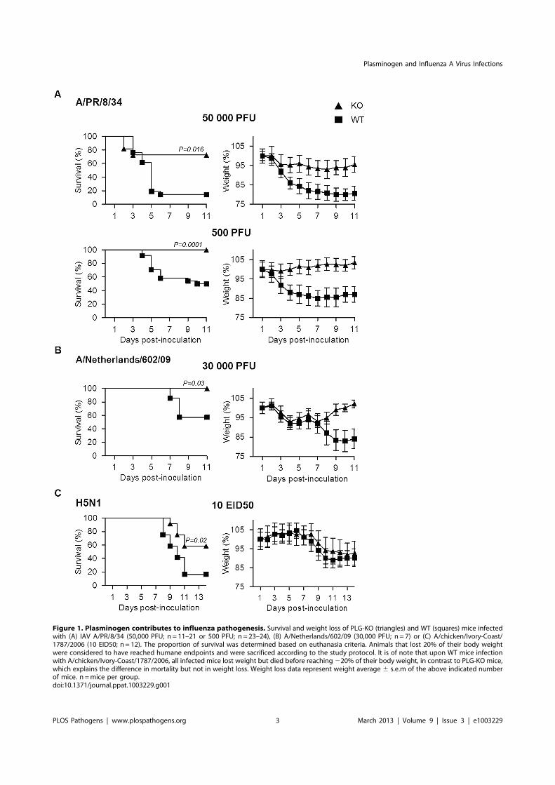
Figure 1. Plasminogen contributes to influenza pathogenesis. Survival and weight loss of PLG-KO (triangles) and WT (squares) mice infectedwith (A) IAV A/PR/8/34 (50,000 PFU; n = 11–21 or 500 PFU; n = 23–24), (B) A/Netherlands/602/09 (30,000 PFU; n = 7) or (C) A/chicken/Ivory-Coast/1787/2006 (10 EID50; n = 12). The proportion of survival was determined based on euthanasia criteria. Animals that lost 20% of their body weightwere considered to have reached humane endpoints and were sacrificed according to the study protocol. It is of note that upon WT mice infectionwith A/chicken/Ivory-Coast/1787/2006, all infected mice lost weight but died before reaching220% of their body weight, in contrast to PLG-KO mice,which explains the difference in mortality but not in weight loss. Weight loss data represent weight average 6 s.e.m of the above indicated numberof mice. n =mice per group.doi:10.1371/journal.ppat.1003229.g001
Plasminogen and Influenza A Virus Infections
PLOS Pathogens | www.plospathogens.org 3 March 2013 | Volume 9 | Issue 3 | e1003229

the molecule and fibrinolysis. Finally, levels of FDP and D-dimers,
degradation products of fibrinolysis, significantly increased upon
infection, reaching 45 and 13 ng/ml respectively on day 6 post-
inoculation. Similar results were also obtained upon infection with
influenza virus A/Netherlands/602/09 (Figure 5A). As expected,
in the BAL of infected PLG-KO mice, used as negative control,
Figure 2. The deleterious role of plasminogen is independent on virus replication. (A) Virus replication of IAV A/PR/8/34 and A/Netherlands/602/09 after inoculation of A549 cells in presence or absence (triangle) of plasminogen (square) or trypsin (circle). Data represent mean6 s.e.m of three independent experiments. (B) Western blot analysis of A/PR/8/34 and A/Netherlands/602/09 HA cleavage after infection of A549 cellsin presence or absence of plasminogen (PLG) or trypsin (Try). Membranes were probed with anti-HA and anti-tubulin antibodies. kDa (apparentmolecular weight). NI stands for uninfected. (C) Infectious A/PR/8/34 (n = 3–5) and A/Netherlands/602/09 (n = 3) lung virus titers at the indicated timepoints post-inoculation of WT (black bars) or PLG-KO mice (white bars). Data represent mean 6 s.e.m of 3–5 individual mice per group. n =mice pergroup and per time-point.doi:10.1371/journal.ppat.1003229.g002
Plasminogen and Influenza A Virus Infections
PLOS Pathogens | www.plospathogens.org 4 March 2013 | Volume 9 | Issue 3 | e1003229

fibrinolysis markers were barely detectable. Thus, IAV infection
induced fibrinolysis. These results were confirmed by Western blot
analysis using an antibody directed against the mouse Aa chain of
fibrinogen (Figure 5B), which recognizes purified mouse fibrinogen
at a molecular weight of 66 kDa (data not shown). Compared to
uninfected mice (2), fibrinogen was readily detectable 2–6 days
post-inoculation in the lungs of infected mice. In the tissues, no
marked fibrinogen consumption was detected but during the
course of IAV infection, additional smaller bands corresponding to
FDP were observed in mouse lungs. These findings confirmed that
fibrinolysis took place during IAV infections in vivo.
To simulate the depletion of fibrin (and therefore fibrinolysis),
mice were treated with the snake venom Ancrod, a thrombin-like
protease that cleaves the Aa chain of fibrinogen, enhancing its
degradation and severely reducing its plasma levels (Figure 5C).
Treatment with Ancrod significantly increased IAV-induced
weight loss and mortality compared to vehicle-treated mice, but
had no effect on uninfected control mice (Figure 6A). This
increased mortality was also associated with an increase in
inflammation of the lungs, as detected by elevated cytokine levels
in the BAL (Figure 6B, WT). Of particular interest, the level of
interferon-gamma was barely detectable in untreated mice but
severely increased upon ancrod treatment. Thus, degradation of
fibrin(ogen) contributed to inflammation and increased pathoge-
nicity of IAV infection.
Plasminogen promotes IAV pathogenesis throughfibrinolysisNext, we investigated whether Ancrod treatment could reverse
the protective effect of plasminogen-deficiency in terms of
inflammation and mortality rate. Again, PLG-KO mice were
protected from lung inflammation (p,0,05, between WT versus
PLG-KO), as judged from cytokine responses (Figure 6B) and
from IAV-induced mortality (Figure 6C). Interestingly, Ancrod-
treatment reversed the protection observed in the absence of
plasminogen and cytokine responses and mortality rates were
similar to those of Ancrod treated WT mice (Figure 6B and C,
p.0.05, between WT-treated and PLG-KO-treated ancrod).
Ancrod had no effect in uninfected mice (Figure S1). Thus,
fibrinolysis contributes to inflammation and pathogenesis of IAV
infections, which is mediated by plasminogen.
To further confirm if the deleterious role of plasminogen is
caused by fibrinolysis, we tested the outcome of infection of mice
after treatment with Ancrod and/or 6-aminohexanoic acid (6-
AHA). Indeed, 6-AHA is a lysine analogue that binds to the lysine
binding sites of plasminogen, inhibiting plasminogen-binding to
fibrin(ogen) and plasmin-mediated fibrinolysis [17]. First, 6-AHA
treated mice inoculated with 5,000 or 500 PFU of A/PR/8/34
were significantly more resistant to infection than untreated mice
(Figure 7A) and this protection correlated with reduced inflam-
mation in 6-AHA treated animals (Figure S2). Also, lung virus
titers were significantly lower in 6-AHA-treated mice compared to
untreated mice, at day 2 but not at days 3 or 5 post-infection
(Figure 7B). Thus, inhibition of plasminogen fibrinolytic activity
protected mice from developing pneumonitis and severe disease.
Furthermore, Ancrod-treatment of 6-AHA treated mice over-rode
the protective effect of 6-AHA, again resulting in IAV-induced
mortality (Figure 7A, lower panel). Administration of Ancrod and/
or 6-AHA had no effect in uninfected mice (Figure S3). Thus, the
protective effect of 6-AHA was reversed by Ancrod-mediated
fibrinogen degradation, demonstrating that plasminogen contrib-
uted to pathogenesis of IAV infection through fibrinolysis
activation.
6-AHA protects against influenzaPreventing deleterious inflammation after IAV infection could
be a promising new strategy to treat IAV infections. Therefore, we
investigated whether blocking the fibrinbolytic activity of plasmin-
ogen by 6-AHA administration at a later time point post-
inoculation was still protective. WT mice were inoculated with
IAV A/PR/8/34 and treated or not with 6-AHA, two days later.
As shown in Figure 7C, treatment with 6-AHA improved the
outcome of infection and prevented mortality. 6-AHA treatment
also protected mice from infection with A/Netherlands/602/09
and highly pathogenic H5N1 viruses (Figure 7C, lower panels).
Thus, blocking plasminogen-mediated fibrinolysis protected mice
against infections with various and highly pathogenic IAVs.
Discussion
The present study showed for the first time that fibrinolysis plays
a central role in the inflammatory response and the pathogenesis
Figure 3. Plasminogen-deficiency prevents severe inflamma-tion. (A) Histopathological analysis of lungs from infected WT and PLG-KO mice inoculated with A/PR/8/34 virus (day 3 post-infection) or A/Netherlands/602/09 virus (day 5 post-infection). Thin sections of lungsobtained from infected and uninfected WT and PLG-KO mice (asindicated) were stained with hematoxilin end eosin (HE) to evaluatehistopathological changes. Note the marked infiltration of inflammatorycells in the lungs of infected WT mice, which was largely absent in thelungs of PLG-KO mice. The results shown are representative for two-three mice for both groups. Immunohistochemistry (IHC) using amonoclonal antibody for the influenza A virus nucleoprotein was usedto detect virus-infected cells. Cells positive for the presence of viralantigen stained red. (B) Cytokine levels in BAL were assessed by ELISAon the indicated days post inoculation of WT (black bars) and PLG-KOmice (white bars) with IAV A/PR/8/34 or A/Netherlands/602/09. Datarepresent mean 6 s.e.m. of 3–6 mice per group.doi:10.1371/journal.ppat.1003229.g003
Plasminogen and Influenza A Virus Infections
PLOS Pathogens | www.plospathogens.org 5 March 2013 | Volume 9 | Issue 3 | e1003229

Figure 4. Plasminogen-deficiency prevents severe inflammation and virus dissemination. (A) Cytokine levels in BAL were assessed by 23-multiplex Luminex kit (uninfected, white bars; infected, black bars) on the indicated days post inoculation of WT (top panel) and PLG-KO mice(bottom panel) with IAV A/PR/8/34. The levels of IL-2, IL-3, IL-4, IL-5, IL-9, IL-12(p70), IL-13, IL-17 and eotaxin were below the detection limit (notshown). Data represent mean 6 s.e.m. of 2 individual mice per group from one experiment and is representative of 2 individual experiments (totaln = 3–6 mice per group). (B) A/PR/8/34 virus titers in the indicated organs of WT (closed symbols) and PLG-KO mice (open symbols) was assessed 2and 5 days post-inoculation.doi:10.1371/journal.ppat.1003229.g004
Plasminogen and Influenza A Virus Infections
PLOS Pathogens | www.plospathogens.org 6 March 2013 | Volume 9 | Issue 3 | e1003229

Figure 5. Fibrinolysis is induced following severe influenza infections. (A) Levels of Plasminogen, Active Plasmin, FDP, D-dimer andFibrinogen, were determined by ELISA in the BAL of A/PR/8/34 infected or uninfected (2) C57BL/6 mice after the indicated days post-inoculation.Markers were also evaluated in the BAL of WT or PLG-KO mice infected with A/Netherlands/602/09 virus. Data represent mean 6 s.e.m of n = 3–6mice per group. (B) Western blot analysis for the detection of fibrinogen and FDP in the lungs of IAV-infected mice on the indicated days postinoculation (representative of n = 3). kDa: apparent molecular weight. n =mice per group. (C) Presence of fibrinogen was assessed in the blood ofmice treated or not with Ancrod by ELISA (left panel) or Western blot analysis (right panel). The results represent the mean values 6 s.e.m from 3individual animals per group for the ELISA. The western blot analysis is representative for results of 3 mice per group.doi:10.1371/journal.ppat.1003229.g005
Plasminogen and Influenza A Virus Infections
PLOS Pathogens | www.plospathogens.org 7 March 2013 | Volume 9 | Issue 3 | e1003229
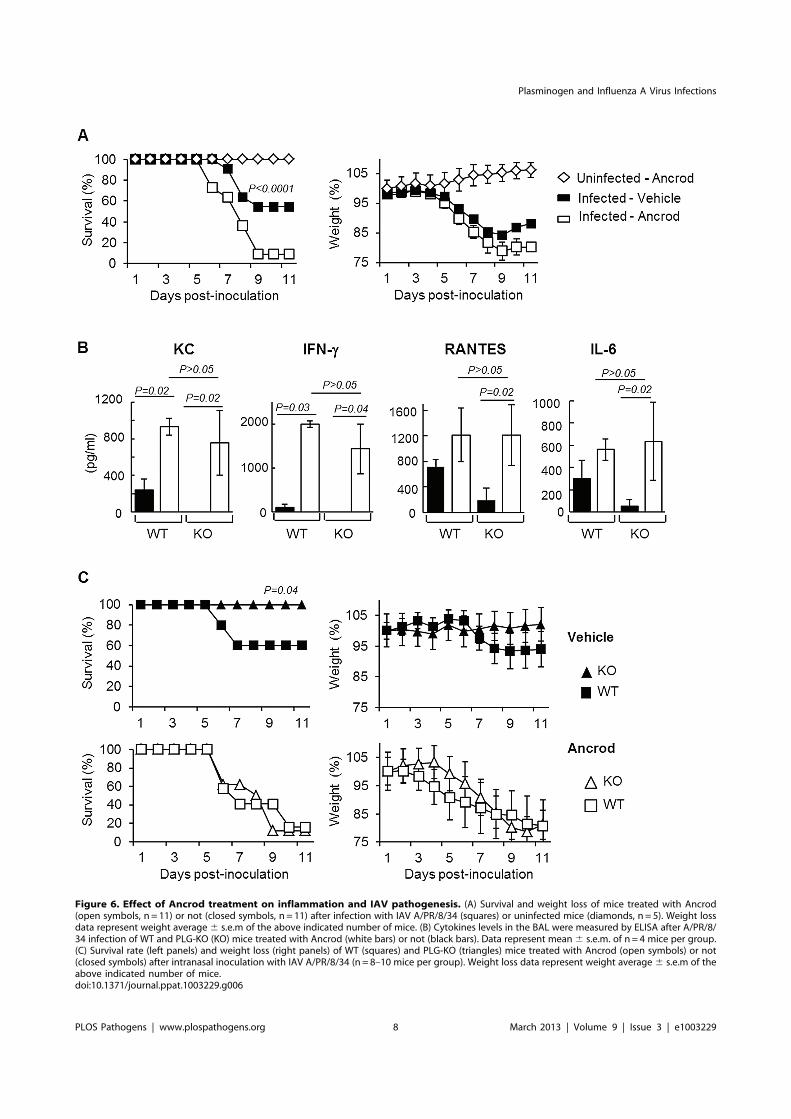
Figure 6. Effect of Ancrod treatment on inflammation and IAV pathogenesis. (A) Survival and weight loss of mice treated with Ancrod(open symbols, n = 11) or not (closed symbols, n = 11) after infection with IAV A/PR/8/34 (squares) or uninfected mice (diamonds, n = 5). Weight lossdata represent weight average 6 s.e.m of the above indicated number of mice. (B) Cytokines levels in the BAL were measured by ELISA after A/PR/8/34 infection of WT and PLG-KO (KO) mice treated with Ancrod (white bars) or not (black bars). Data represent mean 6 s.e.m. of n = 4 mice per group.(C) Survival rate (left panels) and weight loss (right panels) of WT (squares) and PLG-KO (triangles) mice treated with Ancrod (open symbols) or not(closed symbols) after intranasal inoculation with IAV A/PR/8/34 (n = 8–10 mice per group). Weight loss data represent weight average 6 s.e.m of theabove indicated number of mice.doi:10.1371/journal.ppat.1003229.g006
Plasminogen and Influenza A Virus Infections
PLOS Pathogens | www.plospathogens.org 8 March 2013 | Volume 9 | Issue 3 | e1003229

of IAV infections. Consistently, evidence is accumulating that the
fibrinolytic molecule plasminogen and plasmin are critical host
factors for immune cell infiltration and cytokine production upon
injury [18–20]. The absence of plasminogen blunts inflammation
in response to several inflammatory stimuli and suppresses
development of lesions [21–23]. In our study, absence of
plasminogen also considerably reduced the extent of lung
inflammation upon IAV infection. Since severe inflammation
contributes to the pathogenicity of IAV infections of humans [2,4],
most likely the proinflammatory properties of plasminogen play a
role in the pathogenesis of these infections. IAV have the capacity
to bind plasminogen and convert it into its active form plasmin
through viral or cellular proteins like annexin-2 [11,12]. However,
the extent of plasminogen activation is strain-dependent [11],
which may explain differences in pathogenicity of IAV strains.
Mechanistically, the mode of action of plasminogen-driven lung
inflammation was through fibrinolysis. Indeed, degradation of
fibrinogen by Ancrod treatment increased pathogenicity of IAV
infection and compensated the protective effect in PLG-KO mice
or in mice in which plasminogen fibrinolytic activity was blocked
by 6-AHA treatment. Consistently, Keller et al showed an
activation of the fibrinolytic system during non-pathogenic IAV
infection in mice [15]. Remarkably, in humans increased
production of D-dimer, a marker of fibrinolysis was found to be
a risk factor for fatal outcome of H5N1 and pandemic H1N1 virus
infections [24,25]. Furthermore, IAV infections have been
associated with bleeding medical disorders [26,27]. Thus, as for
bacteria [28], the dysregulation of hemostasis by virus infections
may cause serious complications. Consistent with our results, it
was recently demonstrated that endothelial cells are central
orchestrators of cytokine amplification during IAV infections
[29]. Interestingly, plasminogen-dependent inflammation appears
early after infection with influenza virus A/PR/8/34, of which
virus replication is promoted by plasminogen. In contrast,
Figure 7. Effect of 6-aminohexanoic acid and/or Ancrod treatment on the course of IAV infection. Survival and weight loss of IAVinoculated C57BL/6 mice treated with 6-AHA (circle) or not (squares). (A) Mice were inoculated with IAV A/PR8/34 (5,000 PFU, n = 28 or 500 PFU;n = 11) in presence (open symbols) or absence (closed symbols) of Ancrod. 6-AHA treatment was initiated on the day of inoculation. (B) Infectious A/PR/8/34 lung virus titers in 6-AHA treated or untreated mice. Data represent mean 6 s.e.m of 3 individual mice per group. (C) Mice were inoculatedwith IAV A/PR/8/34 (n = 10), A/Netherlands/602/09 (n = 16) or A/chicken/Ivory-Coast/1787/2006 (n = 10) as indicated. 6-AHA treatment was initiatedtwo days post-inoculation. n = per group. Weight loss data represent weight average 6 s.e.m of the above indicated number of mice.doi:10.1371/journal.ppat.1003229.g007
Plasminogen and Influenza A Virus Infections
PLOS Pathogens | www.plospathogens.org 9 March 2013 | Volume 9 | Issue 3 | e1003229

replication of influenza virus A/Netherlands/602/09 is indepen-
dent of plasminogen and control of plasminogen activity has a
delayed impact on inflammation and disease. Thus, the capability
of plasminogen to cleave HA and promote virus replication may
also contribute to lung inflammation for some IAV strains.
Possibly, a sustained high degree of inflammation is deleterious for
the host.
Collectively, we propose a model (Figure 8) in which
plasminogen-mediated fibrinolysis increases FDP production and
vascular permeability allowing increase recruitment of inflamma-
tory cells at the site of infection. As a positive feedback loop,
plasminogen mediated virus replication may also further contrib-
ute to lung inflammation. Fibrinolysis may also allow systemic
haematogenous spread of virus. Consistently, we and others
detected IAV replication in extrapulmonary organs in plasmino-
gen-competent mice [30]. Since plasminogen is omnipresent in the
blood, it may provide certain IAV an alternative mechanism of
HA cleavage in extra-pulmonary organs [10,11]. For example, the
plasminogen-binding property of the neuraminidase of A/WSN/
33 strain is a determinant of its neurotropism and pathogenicity in
mice [12,13]. Interestingly, particular high virus titers were found
in the liver, which is the primary source of plasminogen. This may
explain why IAV can replicate in hepatocarcinoma liver HEPG-2
cells in the absence of exogenous proteases (Figure S4). Whether
plasminogen-dependent IAV replication contributes to damage of
the liver or other extra-pulmonary organs, as observed in Reye’s
syndrome or other postinfluenza complications [31] requires
further investigation. Interestingly, differences in virus replication
were not at the basis of plasminogen-dependent differences in
pathogenesis of IAV infection although it also can contribute to
exacerbation of inflammation. Indeed, A/Netherland/602/09
virus replication in the lungs was not affected by plasminogen
deficiency, while infected PLG-KO mice were protected from
infection. This is consistent with a recent report showing that
presence of critical residues in HA, necessary for cleavage by
plasmin is strain-dependent [32]. In addition, the HA of A/
chicken/Ivory-Coast/1787/2006 contains a polybasic site, which
is cleaved by furin-type proteases. This suggests that plasminogen
plays a minor role in replication of this virus, while plasminogen
deficiency still protected from infection with this virus. Alternative
proteases may thus play a more dominant role in HA cleavage and
virus replication in vivo than plasminogen [33–36].
For the clinical management of influenza patients, a limited
number of antiviral drugs are available. The use of these currently
available drugs is compromised by the emergence of virus strains
that developed resistance to these drugs. Therefore, intervention
strategies that aim at preventing deleterious inflammatory
responses after IAV infection are of interest and do not suffer
from resistance to antiviral drugs. Specifically, blocking protease
activity may be an efficient way to achieve this, as previously
suggested [37–39]. Our results are consistent with these studies but
differ in term of mechanism of action. Indeed, our results suggest a
more predominant role for proteases in lung hemostasis compared
to virus replication and HA cleavage.
In summary, our findings reveal a previously unrecognized role
for fibrinolysis and plasminogen in the pathogenesis of IAV
infections. Thus, targeting plasminogen, its conversion into
plasmin or regulating fibrinolysis may be a venue for the
development of novel intervention strategies for the treatment of
severe IAV infections.
Figure 8. Schematic overview of the proposed model for Plasminogen-mediated influenza virus pathogenesis. During IAV infection,plasminogen is converted into plasmin. On the one hand, plasmin cleaves and activates the viral hemagglutinin, promoting IAV replication for someinfluenza strains. On the other hand, plasmin promotes inflammation via fibrinolysis and increases permeability.doi:10.1371/journal.ppat.1003229.g008
Plasminogen and Influenza A Virus Infections
PLOS Pathogens | www.plospathogens.org 10 March 2013 | Volume 9 | Issue 3 | e1003229

Materials and Methods
Ethics statementExperiments were performed according to recommendations of
the ‘‘National Commission of Animal Experiment (CNEA)’’ and
the ‘‘National Committee on the Ethic Reflexion of Animal
Experiments (CNREEA)’’. The protocol was approved by the
committee of animal experiments of the University Claude
Bernard Lyon I (Permit number: BH2008-13). All animal
experiments were also carried out under the authority of license
issued by ‘‘la direction des services Veterinaires’’ (accreditation
number 78–114). All efforts were made to minimize suffering.
ReagentViruses, cells, and reagents used, were: IAV A/Netherlands/
602/09 [40], A/chicken/Ivory-Coast/1787/2006 [41], A/PR/8/
34 (American Type Culture Collection, ATCC), A549 cells
(ATCC), Madin-Darby Canine Kidney cells (MDCK, ATCC),
trypsin (Becton Dickinson), plasminogen and 6-AHA (Sigma),
Ancrod (NIBSC), 23-Plex Mouse Cytokine Assay (Bio-Rad),
ELISA kits for mouse -IL-6, -KC, -–RANTES, -IFN-a -IFN-
c(R&D Systems), -plasminogen (Mybiosource), -active plasmin
(Kordia), -D-dimer, -fibrinogen and -FDP (Genway), antibodies
anti-HA (Santa Cruz), anti-tubulin (Sigma), anti-NP (ATCC), anti-
fibrinogen (Genway).
In vitro experiments and proteins detectionBlood fibrinogen and lung proteins were extracted as described
[42,43] and proteins were analyzed by western blot [44]. A549
experiments were performed as described previously [11].
MiceMice with a disrupted PLG gene (PLG-KO) and their WT
littermates were bred as described previously [45]. Briefly, PLG
heterozygous mice (C57BL/6 and 25% 129Sv) were crossed and
WT and PLG-KO mice offspring were genotyped by polymerase
chain reaction, which was performed, as previously described [46]
using primers amplifying the WT PLG gene (59ACTGCTGCC-
CACTGTTTGGAG 39 and 59 GATAACCTTGTAGAATT-
CAGGTC39) or the inactivated PLG gene (59ATGAACTGCAG-
GACGAGGCAG39 and 59 GCGAACAGTTCGGCTGGCGC
39). Most of the experiments were performed using 5–6 weeks old
mice. Also, males and females were used in the experiments.
Groups between WT and PLG KO mice were homogenized for
these different parameters. Except when PLG-WT and PLG-KO
mice were used, experiments were performed with six-week-old
C57BL/6 female mice (Charles River Laboratories).
Mice infection and treatmentMice were anesthetized with ketamine (42,5 mg/kg) and inocu-
lated by the intranasal route with the indicated IAV in a volume of
25 ml. Upon inoculation, survival rates and loss of body weight was
scored daily, as previously described [47]. For weight loss curves, the
last measured value was carried forward until the end of the
observation period. Alternatively, mice were sacrificed at various pre-
fixed time points post-inoculation to perform bronchoalveolar lavages
(BAL) or to sample organs. Virus titers in organs were determined by
classical plaque assay using MDCK cells [47]. ELISA and luminex
assays were performed according to the instructions of the
manufacturer and virus titers were assessed as described [48]. Lungs
histology and immunohistochemistry were performed as described
[49]. Treatment with 6-AHA was injected intraperitoneally (30 mg
per mouse in 200 ml of physiological serum) every 6 hours for 4 days.
Ancrod was injected (1.75 unit per mouse) intraperitoneally two days
before infection for 7 days at 10 hours intervals.
Statistical analysisKaplan-Meier test was used for statistical analysis of survival
rates and Mann–Whitney’s test was used for lung virus titers and
ELISA results, p values,0.05, were considered statistically
significant. Two-tails analysis was performed. The number (n) of
animals per experimental group is mentioned in the figure legends.
Experiments were stratified in terms of weight, gender and age of
the mice.
Supporting Information
Figure S1 Effect of Ancrod treatment on uninfectedmice. Survival and weight loss of uninfected PLG-KO mice
treated with Ancrod (open triangle, n = 3).
(TIF)
Figure S2 Effect of 6-AHA on cytokine levels in the BAL.Cytokine levels in the BAL of IAV-infected C57BL/6 mice,
treated or not (upper panel) with 6-AHA (lower panel) was
evaluated by multiplex assay four days post-inoculation. Only
detectable levels are shown. n = 3 mice per group. Please note the
difference in scale of y-axis between treated and untreated
animals.
(TIF)
Figure S3 Effect of Ancrod treatment and/or 6-AHAtreatment on uninfected mice. Survival and weight loss of
uninfected C57BL/6 mice treated with Ancrod and 6-AHA (open
circles, n = 5) or 6-AHA only (closed circles, n = 5).
(TIF)
Figure S4 IAV replication kinetics in HEPG-2 cells.Replication kinetics of IAV A/PR/8/34 and A/Udorn/72 in
absence of proteases was assessed after inoculating HEPG-2 cells
at a MOI of 0.001.
(TIF)
Acknowledgments
We thank Renata de Almeida for assistance and discussions, Emmanuel
Couacy-Hymann for providing the Avian H5N1 influenza strain and
Martine Jandrot-Perrus for discussions.
Author Contributions
Conceived and designed the experiments: FB BR. Performed the
experiments: FB EA MLFG VBL SEVvT PG. Analyzed the data: FB
GFR MH EC BL RL PC BR. Contributed reagents/materials/analysis
tools: GFR MH DM PC. Wrote the paper: BR.
References
1. Knipe DM, Howley PM, editors (2006) Fields Virology. 5th edition. Philadelphia
(Pennsylvania): Lippincott, Williams, & Wilkins.2. Kuiken T, Riteau B, Fouchier RA, RimmelzwaanGF (2012) Pathogenesis of influenza
virus infections: the good, the bad and the ugly. Curr Opin Virol 2: 276–286.3. La Gruta NL, Kedzierska K, Stambas J, Doherty PC (2007) A question of self-
preservation: immunopathology in influenza virus infection. Immunol Cell Biol
85: 85–92.
4. de Jong MD, Simmons CP, Thanh TT, Hien VM, Smith GJ, et al. (2006) Fatal
outcome of human influenza A (H5N1) is associated with high viral load andhypercytokinemia. Nat Med 12: 1203–1207.
5. Reed CE, Kita H (2004) The role of protease activation of inflammation inallergic respiratory diseases. J Allergy Clin Immunol 114: 997–1008; quiz 1009.
6. Sokolova E, Reiser G (2007) A novel therapeutic target in various lung diseases:
airway proteases and protease-activated receptors. Pharmacol Ther 115: 70–83.
Plasminogen and Influenza A Virus Infections
PLOS Pathogens | www.plospathogens.org 11 March 2013 | Volume 9 | Issue 3 | e1003229

7. Steinhoff M, Buddenkotte J, Shpacovitch V, Rattenholl A, Moormann C, et al.
(2005) Proteinase-activated receptors: transducers of proteinase-mediatedsignaling in inflammation and immune response. Endocr Rev 26: 1–43.
8. Bradley LM, Douglass MF, Chatterjee D, Akira S, Baaten BJ (2012) Matrixmetalloprotease 9 mediates neutrophil migration into the airways in response to
influenza virus-induced toll-like receptor signaling. PLoS Pathog 8: e1002641.
9. Khoufache K, Berri F, Nacken W, Vogel AB, Delenne M, et al. (2013) PAR1
contributes to influenza A virus pathogenicity in mice. J Clin Invest 123: 206–214.
10. LeBouder F, Morello E, Rimmelzwaan GF, Bosse F, Pechoux C, et al. (2008)Annexin II incorporated into influenza virus particles supports virus replication
by converting plasminogen into plasmin. J Virol 82: 6820–6828.
11. LeBouder F, Lina B, Rimmelzwaan GF, Riteau B (2010) Plasminogen promotes
influenza A virus replication through an annexin 2-dependent pathway in theabsence of neuraminidase. J Gen Virol 91: 2753–2761.
12. Goto H, Kawaoka Y (1998) A novel mechanism for the acquisition of virulence
by a human influenza A virus. Proc Natl Acad Sci U S A 95: 10224–10228.
13. Goto H, Wells K, Takada A, Kawaoka Y (2001) Plasminogen-binding activity of
neuraminidase determines the pathogenicity of influenza A virus. J Virol 75:
9297–9301.
14. van Hinsbergh VW (2012) Endothelium–role in regulation of coagulation andinflammation. Semin Immunopathol 34: 93–106.
15. Keller TT, van der Sluijs KF, de Kruif MD, Gerdes VE, Meijers JC, et al. (2006)Effects on coagulation and fibrinolysis induced by influenza in mice with a
reduced capacity to generate activated protein C and a deficiency inplasminogen activator inhibitor type 1. Circ Res 99: 1261–1269.
16. Manz B, Brunotte L, Reuther P, Schwemmle M (2012) Adaptive mutations inNEP compensate for defective H5N1 RNA replication in cultured human cells.
Nat Commun 3: 802.
17. Prentice C (1980) Basis of antifibrinolytic therapy. J Clin Pathol 33: 35–40.
18. Gong Y, Hart E, Shchurin A, Hoover-Plow J (2008) Inflammatory macrophage
migration requires MMP-9 activation by plasminogen in mice. J Clin Invest 118:
3012–3024.
19. Wygrecka M, Marsh LM, Morty RE, Henneke I, Guenther A, et al. (2009)Enolase-1 promotes plasminogen-mediated recruitment of monocytes to the
acutely inflamed lung. Blood 113: 5588–5598.
20. Syrovets T, Tippler B, Rieks M, Simmet T (1997) Plasmin is a potent and
specific chemoattractant for human peripheral monocytes acting via a cyclicguanosine monophosphate-dependent pathway. Blood 89: 4574–4583.
21. O’Connell PA, Surette AP, Liwski RS, Svenningsson P, Waisman DM (2010)S100A10 regulates plasminogen-dependent macrophage invasion. Blood 116:
1136–1146.
22. Ploplis VA, French EL, Carmeliet P, Collen D, Plow EF (1998) Plasminogen
deficiency differentially affects recruitment of inflammatory cell populations inmice. Blood 91: 2005–2009.
23. Moons L, Shi C, Ploplis V, Plow E, Haber E, et al. (1998) Reduced transplantarteriosclerosis in plasminogen-deficient mice. J Clin Invest 102: 1788–1797.
24. Soepandi PZ, Burhan E, Mangunnegoro H, Nawas A, Aditama TY, et al. (2010)
Clinical course of avian influenza A(H5N1) in patients at the Persahabatan
Hospital, Jakarta, Indonesia, 2005–2008. Chest 138: 665–673.
25. Wang ZF, Su F, Lin XJ, Dai B, Kong LF, et al. (2011) Serum D-dimer changes
and prognostic implication in 2009 novel influenza A(H1N1). Thromb Res 127:198–201.
26. Urso R, Bevilacqua N, Gentile M, Biagioli D, Lauria FN (2011) Pandemic 2009
H1N1 virus infection associated with purpuric skin lesions: a case report. J Med
Case Reports 5: 132.
27. Okayama S, Arakawa S, Ogawa K, Makino T (2011) A case of hemorrhagiccolitis after influenza A infection. J Microbiol Immunol Infect 44(6): 480–483.
28. Degen JL, Bugge TH, Goguen JD (2007) Fibrin and fibrinolysis in infection andhost defense. J Thromb Haemost 5 Suppl 1: 24–31.
29. Teijaro JR, Walsh KB, Cahalan S, Fremgen DM, Roberts E, et al. (2011)
Endothelial Cells Are Central Orchestrators of Cytokine Amplification duringInfluenza Virus Infection. Cell 146: 980–991.
30. Fislova T, Gocnik M, Sladkova T, Durmanova V, Rajcani J, et al. (2009)
Multiorgan distribution of human influenza A virus strains observed in a mousemodel. Arch Virol 154: 409–419.
31. Sanchez-Lanier M, Davis LE, Blisard KS, Woodfin BM, Wallace JM, et al.(1991) Influenza A virus in the mouse: hepatic and cerebral lesions in a Reye’s
syndrome-like illness. Int J Exp Pathol 72: 489–500.
32. Sun X, Tse LV, Ferguson AD, Whittaker GR (2010) Modifications to thehemagglutinin cleavage site control the virulence of a neurotropic H1N1
influenza virus. J Virol 84: 8683–8690.33. Zhirnov OP, Ikizler MR, Wright PF (2002) Cleavage of influenza a virus
hemagglutinin in human respiratory epithelium is cell associated and sensitive toexogenous antiproteases. J Virol 76: 8682–8689.
34. Bertram S, Glowacka I, Blazejewska P, Soilleux E, Allen P, et al. (2010)
TMPRSS2 and TMPRSS4 facilitate trypsin-independent influenza virus spreadin Caco-2 cells. J Virol. 84: 10016–10025.
35. Bottcher E, Matrosovich T, Beyerle M, Klenk HD, Garten W, et al. (2006)Proteolytic activation of influenza viruses by serine proteases TMPRSS2 and
HAT from human airway epithelium. J Virol 80: 9896–9898.
36. Chaipan C, Kobasa D, Bertram S, Glowacka I, Steffen I, et al. (2009) Proteolyticactivation of the 1918 influenza virus hemagglutinin. J Virol 83: 3200–3211.
37. Zhirnov OP, Klenk HD, Wright PF (2011) Aprotinin and similar proteaseinhibitors as drugs against influenza. Antiviral Res 92: 27–36.
38. Bottcher-Friebertshauser E, Freuer C, Sielaff F, Schmidt S, Eickmann M, et al.(2010) Cleavage of influenza virus hemagglutinin by airway proteases
TMPRSS2 and HAT differs in subcellular localization and susceptibility to
protease inhibitors. J Virol 84: 5605–5614.39. Zhirnov OP, Ovcharenko AV, Bukrinskaya AG (1982) Protective effect of
protease inhibitors in influenza virus infected animals. Arch Virol 73: 263–272.40. Munster VJ, de Wit E, van den Brand JM, Herfst S, Schrauwen EJ, et al. (2009)
Pathogenesis and transmission of swine-origin 2009 A(H1N1) influenza virus in
ferrets. Science 325: 481–483.41. Couacy-Hymann E, Danho T, Keita D, Bodjo SC, Kouakou C, et al. (2009)
The first specific detection of a highly pathogenic avian influenza virus (H5N1)in Ivory Coast. Zoonoses Public Health 56: 10–15.
42. Hanss MM, Ffrench PO, Mornex JF, Chabuet M, Biot F, et al. (2003) Twonovel fibrinogen variants found in patients with pulmonary embolism and their
families. J Thromb Haemost 1: 1251–1257.
43. Luyendyk JP, Sullivan BP, Guo GL, Wang R (2010) Tissue factor-deficiency andprotease activated receptor-1-deficiency reduce inflammation elicited by diet-
induced steatohepatitis in mice. Am J Pathol 176: 177–186.44. Bernard D, Riteau B, Hansen JD, Phillips RB, Michel F, et al. (2006)
Costimulatory receptors in a teleost fish: typical CD28, elusive CTLA4.
J Immunol 176: 4191–4200.45. Ploplis VA, Carmeliet P, Vazirzadeh S, Van Vlaenderen I, Moons L, et al.
(1995) Effects of disruption of the plasminogen gene on thrombosis, growth, andhealth in mice. Circulation 92: 2585–2593.
46. Riteau B, Moreau P, Menier C, Khalil-Daher I, Khosrotehrani K, et al. (2001)Characterization of HLA-G1, -G2, -G3, and -G4 isoforms transfected in a
human melanoma cell line. Transplant Proc 33: 2360–2364.
47. Khoufache K, LeBouder F, Morello E, Laurent F, Riffault S, et al. (2009)Protective role for protease-activated receptor-2 against influenza virus
pathogenesis via an IFN-gamma-dependent pathway. J Immunol 182: 7795–7802.
48. LeBouder F, Khoufache K, Menier C, Mandouri Y, Keffous M, et al. (2009)
Immunosuppressive HLA-G molecule is upregulated in alveolar epithelial cellsafter influenza A virus infection. Hum Immunol 70: 1016–1019.
49. Riteau B, Faure F, Menier C, Viel S, Carosella ED, et al. (2003) Exosomesbearing HLA-G are released by melanoma cells. Hum Immunol 64: 1064–1072.
Plasminogen and Influenza A Virus Infections
PLOS Pathogens | www.plospathogens.org 12 March 2013 | Volume 9 | Issue 3 | e1003229


Annexin V Incorporated into Influenza Virus Particles InhibitsGamma Interferon Signaling and Promotes Viral Replication
Fatma Berri,a Ghina Haffar,a Vuong Ba Lê,a Anne Sadewasser,b Katharina Paki,b Bruno Lina,a Thorsten Wolff,b Béatrice Riteaua,c
VirPath, EMR4610 Virologie et Pathologie Humaine, Faculté de Médecine RTH Laennec, Université Claude Bernard Lyon 1, Université de Lyon, Lyon, Francea; Division of
Influenza Viruses and Other Respiratory Viruses, Robert Koch Institute, Berlin, Germanyb; INRA, Tours, Francec
ABSTRACT
During the budding process, influenza A viruses (IAVs) incorporate multiple host cell membrane proteins. However, for most ofthem, their significance in viral morphogenesis and infectivity remains unknown. We demonstrate here that the expression ofannexin V (A5) is upregulated at the cell surface upon IAV infection and that a substantial proportion of the protein is present inlipid rafts, the site of virus budding. Western blotting and immunogold analysis of highly purified IAV particles showed thepresence of A5 in the virion. Significantly, gamma interferon (IFN-�)-induced Stat phosphorylation and IFN-�-induced 10-kDaprotein (IP-10) production in macrophage-derived THP-1 cells was inhibited by purified IAV particles. Disruption of the IFN-�signaling pathway was A5 dependent since downregulation of its expression or its blockage reversed the inhibition and resultedin decreased viral replication in vitro. The functional significance of these results was also observed in vivo. Thus, IAVs can sub-vert the IFN-� antiviral immune response by incorporating A5 into their envelope during the budding process.
IMPORTANCE
Many enveloped viruses, including influenza A viruses, bud from the plasma membrane of their host cells and incorporate cellu-lar surface proteins into viral particles. However, for the vast majority of these proteins, only the observation of their incorpora-tion has been reported. We demonstrate here that the host protein annexin V is specifically incorporated into influenza virusparticles during the budding process. Importantly, we showed that packaged annexin V counteracted the antiviral activity ofgamma interferon in vitro and in vivo. Thus, these results showed that annexin V incorporated in the viral envelope of influenzaviruses allow viral escape from immune surveillance. Understanding the role of host incorporated protein into virions may re-veal how enveloped RNA viruses hijack the host cell machinery for their own purposes.
Influenza is an ineradicable contagious disease that constitutes amajor public health problem, occurring as a seasonal epidemic
of variable impact or sporadic pandemic outbreaks (1, 2). Theetiological agents of the disease, the single-stranded RNA influ-enza viruses, are classified into three types (A, B, and C), of whichinfluenza A virus (IAV) is clinically themost important. Annually,IAV causes 3 to 5 million clinical infections and 200,000 to500,000 fatal cases (3). Thus, these viruses are of great concern tohuman health and impose a considerable socioeconomic burden.Important factors in the pathogenesis of influenza include theefficient replication of the virus in the respiratory tract and thehost immune response, traits that are dependent on each other(4–6). While the immune response aims to control the spread ofthe virus, IAV has developed strategies for subverting host de-fenses, thereby facilitating their spread (7–10). Further knowledgeinto how IAV escapes the host immunosurveillance is critical forthe design of new treatments that are able to control the disease.
Similarly to other enveloped viruses, IAV exits the host cell bybudding from a cellular membrane (11, 12). Thereby, particlesreleased from infected cells can incorporate many host cellularproteins during the assembly and budding steps of morphogene-sis. Earlier study identified 36 host-encoded proteins in purifiedIAV particles in addition to viral virion components (13). Amongthem, the annexin family of proteins that bind to negativelycharged phospholipids is well represented (13, 14). However, thefunctional significance of host protein incorporation has not beendetermined yet, except for the role of annexin II, which promotesviral replication when incorporated into a virus particle (14, 15).
One protein of interest is annexin V (A5), which has recently beenfound to play a role in the regulation of the immune response (16,17). We address here the specific incorporation of A5 into IAVparticles and its functional relevance in viral replication.
We found that the host protein A5 was incorporated into IAVparticles and inhibited gamma interferon (IFN-�)-induced sig-naling and antiviral activity both in vitro and in vivo. Collectively,these results show that incorporation of A5 into IAV virions sup-ports influenza virus escape from immunosurveillance.
MATERIALS AND METHODS
Viruses and reagents. IAV A/PR/8/34 (H1N1) was a gift from G. F.Rimmelzwaan (Erasmus University, Rotterdam, Netherlands), andA/WSN/33 (H1N1) and A/Udorn/72 (H3N2) IAV were a gift from N.Naffakh (Pasteur Institute, Paris, France). The following reagents wereused in the study: small interfering RNA (siRNA) targeting A5 (SantaCruz Biotechnology), recombinant mouse IFN-� (Sigma-Aldrich), re-combinant human IFN-� and IFN-� (R&D Systems), trypsin (BectonDickinson), an enzyme-linked immunosorbent assay (ELISA) kit for
Received 14 May 2014 Accepted 14 July 2014
Published ahead of print 16 July 2014
Editor: B. Williams
Address correspondence to Béatrice Riteau, [email protected].
F.B. and G.H. contributed equally to this article.
Copyright © 2014, American Society for Microbiology. All Rights Reserved.
doi:10.1128/JVI.01405-14
October 2014 Volume 88 Number 19 Journal of Virology p. 11215–11228 jvi.asm.org 11215
on S
epte
mber 8
, 2014 b
y IN
IST
-CN
RS
Bib
lioV
iehttp
://jvi.a
sm
.org
/D
ow
nlo
aded fro
m

IFN-�-induced 10-kDa protein (IP-10) and interleukin-1� (IL-1�; R&DSystems), cholera toxin B subunit (Sigma-Aldrich, France), monoclonalanti-tubulin (Sigma), polyclonal anti-A5 (Santa Cruz Biotechnology),monoclonal anti-M2 (Santa Cruz Biotechnology),monoclonal anti-hem-agglutinin (anti-HA; Santa Cruz Biotechnology), polyclonal anti-ERK(Cell Signaling Technology, Saint Quentin, France), monoclonal anti-Stat1 (Santa Cruz Biotechnology), and polyclonal anti-p-Stat1 (R&D Sys-tems) antibodies. Rabbit polyclonal anti-A/PR/8/34 virus cross-reactingwith A/WSN/33 virus proteins (referred to as polyclonal anti-influenza)was a gift fromG. F. Rimmelzwaan (Erasmus University). Phorbol myris-tate acetate (PMA; Sigma) was used for human monocytic cell line(THP-1) differentiation.
Cell culture and raft isolation.Thehumanmonocytic THP-1, humanalveolar A549, human epithelial kidney 293T, HeLa, and Madin-Darbycanine kidney (MDCK) cell lines used in the present study were obtainedfrom theAmericanTypeCultureCollection.MDCKcells were cultured inEagle minimal essential medium (EMEM; Lonza, France) supplementedwith 5% fetal bovine serum (FBS; Lonza), 2 mM L-glutamine, and 100international units (IU)/ml penicillin-streptomycin (PS). A549 and 293Tcells were grown in Dulbecco modified Eagle medium (DMEM; Lonza)supplemented with 10% FBS, 2 mM L-glutamine, and 100 IU/ml PS.THP-1 cells were cultured in RPMI (Lonza) supplemented with 10%FBS,2mM L-glutamine, 100 IU/ml PS, 5ml of pyruvate sodium, 5ml of aminoacids, and �-mercaptoethanol. Raft isolations were performed as previ-ously described (18).
Virus production, titration, and purification. MDCK cells wereseeded at 13 � 106 cells per 175-cm2 tissue culture flask and then incu-bated at 37°C overnight. The next day, based on previous evaluations, cellconfluence was evaluated at 20 � 106 cells per 175 cm2, and the cells wereinfected with IAV at a multiplicity of infection (MOI) of 10�3 in EMEMcontaining 1 �g of trypsin/ml. At 2 days postinfection, the supernatantwas harvested and then clarified using low-speed centrifugation, and thevirus particles were titrated as previously described (19). Briefly, MDCKcells were infectedwith IAV for 1 h at 37°C. After viral adsorption, the cellswere overlaid with medium containing 2% agarose and 1 �g of trypsin/ml, followed by incubation for 3 days at 37°C. Viral plaques were thenvisualized using bromophenol blue staining. To purify the virus particles,the supernatants were clarified and concentrated 100-fold by ultracentrif-ugation at 60,000 � g for 105 min at 4°C. Concentrated viruses were thenpurified by centrifugation for 2 h at 80,000 � g at 4°C in a 20 to 60%sucrose density gradient. The virus particles were then separated into twodifferent tubes for pretreatment with 20 �g of either blocking anti-A5antibody (referred to as “AV-V”) or isotype control antibody (referred toas “V”)/ml for 1 h at 4°C. Viruses were thenwashed by ultracentrifugationat 31,000 � g for 2 h and suspended in medium. Infectious virus titerswere then evaluated in both virus preparations and used for experiments.AV-V or V particles were then used in the experiments.
Identification and quantification of cell surface proteins by SILAC(stable isotope labeling by amino acids in cell culture)-based mass spec-trometric (MS) analysis. A549 cells were grown in stable isotope-labeledDMEM(SILAC-DMEM,PAA) supplementedwith 10%dialyzed FBS (In-vitrogen), 2 mM L-glutamine, and antibiotics at 37°C with 5% CO2. Cellswere either cultivated in SILAC medium containing light (R0K0: R �12C6,
14N4; K � 12C6,14N2) or heavy (R10K8: R � 13C6,
15N4; K � 13C6,15N2) arginine and lysine for at least six cell doublings prior to infection. Atotal of 4� 107 heavy-labeled cells (R10K8) were infected with IAVA/PR/8/34 (H1N1) at anMOI of 5, while the same number of light-labeled cells(R0K0) served as a mock control. At 16 h postinfection (hpi) cells werewashed with phosphate-buffered saline (PBS) and incubated with 1mg/ml Sulfo-NHS-SS-Biotin (Thermo Fisher Scientific)/PBS for 40 minat 4°C, followed by quenching with 10 mM glycine-PBS buffer. Afterbiotinylation of cell surface proteins, the cell extract of each population(heavy or light) was prepared in 1ml of lysis buffer (50mMTris-HCl [pH8], 150mMNaCl, 1%Nonidet P-40, 2mMNa3VO4, 1mMPefabloc) andcleared by centrifugation. The protein concentration of each lysate was
determined by a BCA protein assay (Thermo Fisher Scientific), and thelysates were mixed at a 1:1 heavy/light ratio, followed by selection of bio-tinylated proteins on a streptavidin-agarose resin (Thermo Fisher Scien-tific) at 4°C for 16 h. The beads were washed once with 50 mM Tris-HCl(pH 7.4)–150 mM NaCl–5 mM EDTA, twice with 50 mM Tris-HCl (pH7.4)–500 mMNaCl–5 mMEDTA, three times with 20 mMTris-HCl (pH7.4)–500 mM NaCl, and once with 10 mM Tris-HCl (pH 7.4). The pre-cipitated proteins were eluted in 4� sodiumdodecyl sulfate (SDS) samplebuffer–20%�-mercaptoethanol for 30min at 37°C. Affinity-purified pro-teins were reduced and alkylated by the addition of 10 mM dithiothreitol(2 min, 95°C) and 50 mM iodacetamid (30 min, 22°C, in the dark), re-spectively. Proteins were separated by SDS–12.5% PAGE, and the gel lanewas cut into six slices, which were then subjected to in-gel tryptic digestusing a trypsin profile IGD kit (Sigma). The resulting peptides were sep-arated using a C18 capillary analytical column (10 cm [inner diameter, 75�m];ThermoFisher Scientific)with a linear gradient over 95min (solventA � 1% FA, 99% H2O and solvent B � 80% ACN, 1% FA) at a constantflow rate of 300 nl/min using an Easy Nano liquid chromatography IIsystem coupled to an LTQ Orbitrap discovery XL (Thermo Fisher Scien-tific). Eluting peptides were ionized by electrospray ionization at 1.4 kVand a capillary temperature of 200°C. Mass spectra (m/z range, 300 to2000)weremeasuredwith a resolution ofM/M� 30,000 atm/z 400. Thetop five precursor peptide ions were fragmented by collision-induced dis-sociation (normalized collision energy, 35%; activation Q, 0.250, activa-tion time, 30 ms) with a dynamic exclusion time of 30 s. The data wereacquired using Xcalibur software. Raw data files were evaluated usingProteome Discoverer (PD) software (version 1.4; Thermo Fisher Scien-tific). Proteins were identified by searching against the UniProt/Swiss-Prot Human and Influenza A/PR/8/34 database (89,454 entries) usingSEQUEST algorithm and the following search parameters: carbamidom-ethylation of cysteine (57.021) as a fixed modification, oxidation ofhistidine, methionine, and tryptophan (15.995); phosphorylation ofserine, threonine, and tyrosine (79.966) and appropriate SILAC labelsas variable modifications; tryptic digestion with a maximum of twomissed cleavages; a peptide precursor mass tolerance of 10 ppm; and afragment mass tolerance of 0.8 Da. The decoy database search option wasenabled and all peptides were filteredwith amaximum false discovery rate(FDR) of 1%. Protein quantification was performed with at least twounique and labeled peptides per protein and a mass precision of 4 ppm.The relative abundance of a protein at cell surface was derived from itsheavy/light (H/L) ratio in the differently labeled cell populations. Quan-tification values outside the range from 0.01 to 100 were recorded as 0.01(ratio � 0.01) and 100 (ratio � 100). Proteins were grouped by PD an-notation software tool and selected according to the gene ontology cellularcomponent (GOCC) categories “membrane,” “cell surface,” or “extracel-lular” (20). AnH/L ratio of�2 for a given proteinwas considered to signalincreased surface abundance.
Depletion of A5 from virions by siRNA-mediated knockdown. Spe-cific siRNA targeting A5 was used to knock down protein expression in293T cells. These cells were chosen because of their high transfectionefficiency. Nontargeted siRNA was used as a control. According to themanufacturer’s instructions, 293T cells (60 to 80% confluence, i.e., 2 �
105 cells per 10 cm2) were transfected with siRNA targeting A5 (1 �g/2 �
105 cells) or control-siRNA, diluted in transfection reagent (confidentiallipidic composition from Santa Cruz). AT 24 h posttransfection, DMEMcontaining 20% fetal calf serum, PS (200 IU/ml), and L-glutamine (4mM)was added to the cells, followed by incubation at 37°C for additional 48 h.At this step,Western blot analysis was performed to verify the transfectionefficiency (data not shown). Alternatively, cells were infected with IAV(MOI � 1), and supernatants containing the virus particles were har-vested at 16 hpi. The virus titers were evaluated by plaque assay and usedin experiments. Similar ratios of the different viral proteins in both prep-arations and reduced expression of packaged A5 in the virions released inthe supernatant of A5-specific siRNA-treated cells (referred to as A5siRNA v) compared to control viruses (referred to as Ctl siRNA v) were
Berri et al.
11216 jvi.asm.org Journal of Virology
on S
epte
mber 8
, 2014 b
y IN
IST
-CN
RS
Bib
lioV
iehttp
://jvi.a
sm
.org
/D
ow
nlo
aded fro
m
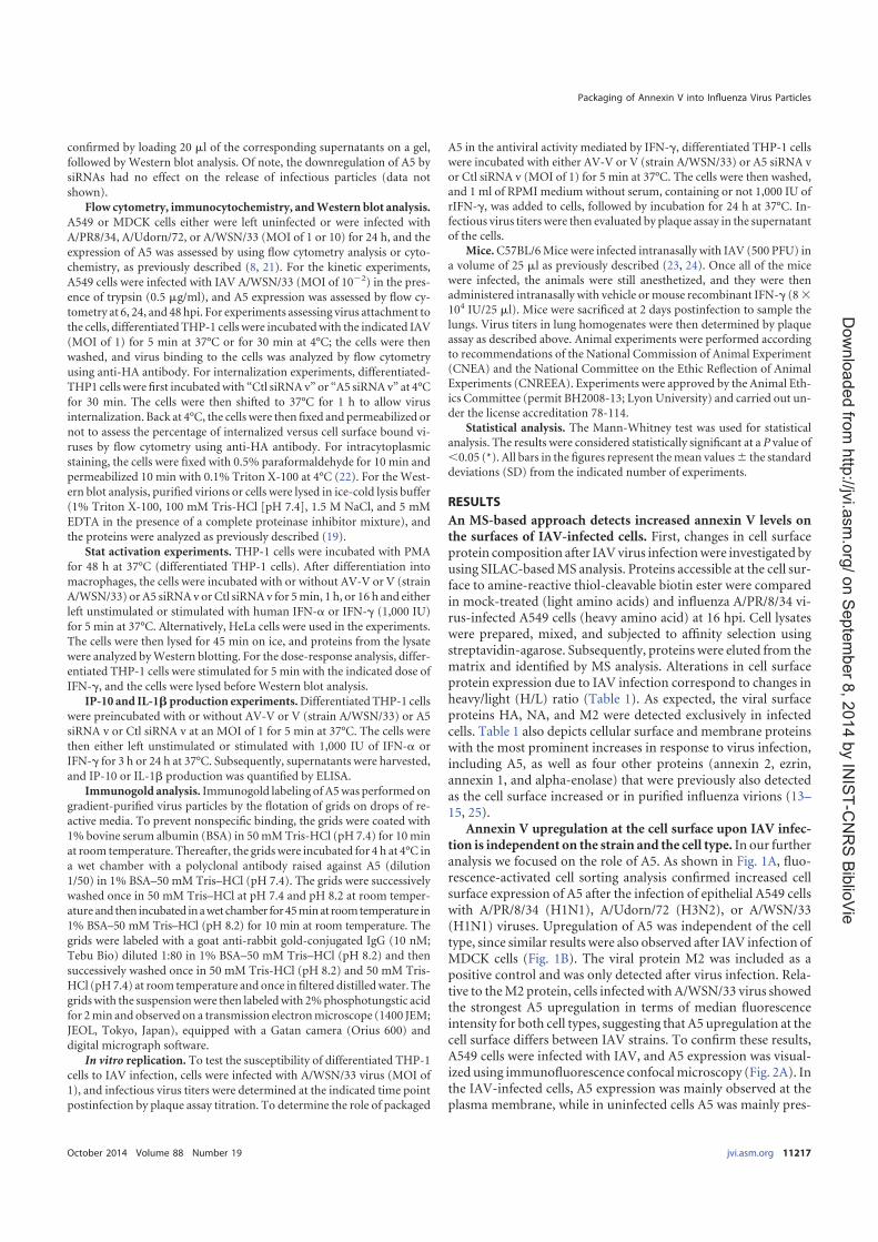
confirmed by loading 20 �l of the corresponding supernatants on a gel,followed by Western blot analysis. Of note, the downregulation of A5 bysiRNAs had no effect on the release of infectious particles (data notshown).
Flow cytometry, immunocytochemistry, and Western blot analysis.A549 or MDCK cells either were left uninfected or were infected withA/PR8/34, A/Udorn/72, or A/WSN/33 (MOI of 1 or 10) for 24 h, and theexpression of A5 was assessed by using flow cytometry analysis or cyto-chemistry, as previously described (8, 21). For the kinetic experiments,A549 cells were infected with IAV A/WSN/33 (MOI of 10�2) in the pres-ence of trypsin (0.5 �g/ml), and A5 expression was assessed by flow cy-tometry at 6, 24, and 48 hpi. For experiments assessing virus attachment tothe cells, differentiated THP-1 cells were incubatedwith the indicated IAV(MOI of 1) for 5 min at 37°C or for 30 min at 4°C; the cells were thenwashed, and virus binding to the cells was analyzed by flow cytometryusing anti-HA antibody. For internalization experiments, differentiated-THP1 cells were first incubatedwith “Ctl siRNAv” or “A5 siRNAv” at 4°Cfor 30 min. The cells were then shifted to 37°C for 1 h to allow virusinternalization. Back at 4°C, the cells were then fixed and permeabilized ornot to assess the percentage of internalized versus cell surface bound vi-ruses by flow cytometry using anti-HA antibody. For intracytoplasmicstaining, the cells were fixed with 0.5% paraformaldehyde for 10 min andpermeabilized 10 min with 0.1% Triton X-100 at 4°C (22). For the West-ern blot analysis, purified virions or cells were lysed in ice-cold lysis buffer(1% Triton X-100, 100 mM Tris-HCl [pH 7.4], 1.5 M NaCl, and 5 mMEDTA in the presence of a complete proteinase inhibitor mixture), andthe proteins were analyzed as previously described (19).
Stat activation experiments. THP-1 cells were incubated with PMAfor 48 h at 37°C (differentiated THP-1 cells). After differentiation intomacrophages, the cells were incubated with or without AV-V or V (strainA/WSN/33) or A5 siRNAv orCtl siRNAv for 5min, 1 h, or 16 h and eitherleft unstimulated or stimulated with human IFN-� or IFN-� (1,000 IU)for 5 min at 37°C. Alternatively, HeLa cells were used in the experiments.The cells were then lysed for 45 min on ice, and proteins from the lysatewere analyzed byWestern blotting. For the dose-response analysis, differ-entiated THP-1 cells were stimulated for 5 min with the indicated dose ofIFN-�, and the cells were lysed before Western blot analysis.
IP-10 and IL-1� production experiments. Differentiated THP-1 cellswere preincubated with or without AV-V or V (strain A/WSN/33) or A5siRNA v or Ctl siRNA v at an MOI of 1 for 5 min at 37°C. The cells werethen either left unstimulated or stimulated with 1,000 IU of IFN-� orIFN-� for 3 h or 24 h at 37°C. Subsequently, supernatants were harvested,and IP-10 or IL-1� production was quantified by ELISA.
Immunogold analysis. Immunogold labeling of A5was performed ongradient-purified virus particles by the flotation of grids on drops of re-active media. To prevent nonspecific binding, the grids were coated with1% bovine serum albumin (BSA) in 50mMTris-HCl (pH 7.4) for 10minat room temperature. Thereafter, the gridswere incubated for 4 h at 4°C ina wet chamber with a polyclonal antibody raised against A5 (dilution1/50) in 1% BSA–50 mM Tris–HCl (pH 7.4). The grids were successivelywashed once in 50 mM Tris–HCl at pH 7.4 and pH 8.2 at room temper-ature and then incubated inawet chamber for45minat roomtemperature in1% BSA–50 mM Tris–HCl (pH 8.2) for 10 min at room temperature. Thegrids were labeled with a goat anti-rabbit gold-conjugated IgG (10 nM;Tebu Bio) diluted 1:80 in 1% BSA–50 mM Tris–HCl (pH 8.2) and thensuccessively washed once in 50 mM Tris-HCl (pH 8.2) and 50 mM Tris-HCl (pH7.4) at room temperature and once in filtered distilledwater. Thegridswith the suspensionwere then labeledwith 2%phosphotungstic acidfor 2min and observed on a transmission electronmicroscope (1400 JEM;JEOL, Tokyo, Japan), equipped with a Gatan camera (Orius 600) anddigital micrograph software.
In vitro replication. To test the susceptibility of differentiated THP-1cells to IAV infection, cells were infected with A/WSN/33 virus (MOI of1), and infectious virus titers were determined at the indicated time pointpostinfection by plaque assay titration. To determine the role of packaged
A5 in the antiviral activity mediated by IFN-�, differentiated THP-1 cellswere incubated with either AV-V or V (strain A/WSN/33) or A5 siRNA vor Ctl siRNA v (MOI of 1) for 5 min at 37°C. The cells were then washed,and 1 ml of RPMI medium without serum, containing or not 1,000 IU ofrIFN-�, was added to cells, followed by incubation for 24 h at 37°C. In-fectious virus titers were then evaluated by plaque assay in the supernatantof the cells.
Mice. C57BL/6Mice were infected intranasally with IAV (500 PFU) ina volume of 25 �l as previously described (23, 24). Once all of the micewere infected, the animals were still anesthetized, and they were thenadministered intranasally with vehicle ormouse recombinant IFN-� (8 �
104 IU/25 �l). Mice were sacrificed at 2 days postinfection to sample thelungs. Virus titers in lung homogenates were then determined by plaqueassay as described above. Animal experiments were performed accordingto recommendations of the National Commission of Animal Experiment(CNEA) and the National Committee on the Ethic Reflection of AnimalExperiments (CNREEA). Experiments were approved by the Animal Eth-ics Committee (permit BH2008-13; Lyon University) and carried out un-der the license accreditation 78-114.
Statistical analysis. The Mann-Whitney test was used for statisticalanalysis. The results were considered statistically significant at a P value of�0.05 (*). All bars in the figures represent themean values the standarddeviations (SD) from the indicated number of experiments.
RESULTS
An MS-based approach detects increased annexin V levels onthe surfaces of IAV-infected cells. First, changes in cell surfaceprotein composition after IAVvirus infectionwere investigated byusing SILAC-basedMS analysis. Proteins accessible at the cell sur-face to amine-reactive thiol-cleavable biotin ester were comparedin mock-treated (light amino acids) and influenza A/PR/8/34 vi-rus-infected A549 cells (heavy amino acid) at 16 hpi. Cell lysateswere prepared, mixed, and subjected to affinity selection usingstreptavidin-agarose. Subsequently, proteins were eluted from thematrix and identified by MS analysis. Alterations in cell surfaceprotein expression due to IAV infection correspond to changes inheavy/light (H/L) ratio (Table 1). As expected, the viral surfaceproteins HA, NA, and M2 were detected exclusively in infectedcells. Table 1 also depicts cellular surface and membrane proteinswith the most prominent increases in response to virus infection,including A5, as well as four other proteins (annexin 2, ezrin,annexin 1, and alpha-enolase) that were previously also detectedas the cell surface increased or in purified influenza virions (13–15, 25).
Annexin V upregulation at the cell surface upon IAV infec-tion is independent on the strain and the cell type. In our furtheranalysis we focused on the role of A5. As shown in Fig. 1A, fluo-rescence-activated cell sorting analysis confirmed increased cellsurface expression of A5 after the infection of epithelial A549 cellswith A/PR/8/34 (H1N1), A/Udorn/72 (H3N2), or A/WSN/33(H1N1) viruses. Upregulation of A5 was independent of the celltype, since similar results were also observed after IAV infection ofMDCK cells (Fig. 1B). The viral protein M2 was included as apositive control and was only detected after virus infection. Rela-tive to theM2 protein, cells infectedwith A/WSN/33 virus showedthe strongest A5 upregulation in terms of median fluorescenceintensity for both cell types, suggesting that A5 upregulation at thecell surface differs between IAV strains. To confirm these results,A549 cells were infected with IAV, and A5 expression was visual-ized using immunofluorescence confocalmicroscopy (Fig. 2A). Inthe IAV-infected cells, A5 expression was mainly observed at theplasma membrane, while in uninfected cells A5 was mainly pres-
Packaging of Annexin V into Influenza Virus Particles
October 2014 Volume 88 Number 19 jvi.asm.org 11217
on S
epte
mber 8
, 2014 b
y IN
IST
-CN
RS
Bib
lioV
iehttp
://jvi.a
sm
.org
/D
ow
nlo
aded fro
m

ent in the cytoplasm. As controls, the infected but not uninfectedcells displayed detectable HA proteins. Also, nuclei were stainedwith DAPI, and the merged images are shown (Fig. 2A). We thenfurther investigated whether total A5 expression or simply its lo-
calization was affected by the infection. A549 cells were left unin-fected or infected with influenza A/WSN/33 virus, and total A5expression was assessed by flow cytometry analysis on permeabil-ized cells (Fig. 2B, left panel). The results indicated that total A5
TABLE 1 Upregulated cell surface proteins upon IAV infection compared to noninfected cells identified by LC-MS/MSa
Protein(accession no.) Description Score
Coverage(%)
H/Lratio
H/Lcount
H/L variability(%)
P60903 Protein S100-A10 (S100-A10) 31.21 36.08 5.319 6 4.9Q9BQE5 Apolipoprotein L2 (APOL2) 8.89 13.06 4.747 3 17.5P07355 Annexin A2 (ANXA2) 534.01 69.62 4.352 84 12.2P08758 Annexin A5 (ANXA5) 10.30 11.25 4.072 3 24.9Q9HCC0 Methylcrotonoyl-CoA carboxylase beta chain, mitochondrial (MCCC2) 17.35 18.29 2.940 8 12.6E7EQR4b Ezrin (EZR) 5.77 6.14 2.872 3 33.6O95994 Anterior gradient protein 2 homolog (AGR2) 9.26 24.57 2.743 4 11.1O00220 Tumor necrosis factor receptor superfamily member 10A (TNFRSF10A) 50.86 19.02 2.716 10 7.3P30510b HLA class I histocompatibility antigen, Cw-14 alpha chain (HLA-C) 262.98 39.88 2.595 9 5.0P04083 Annexin A1 (ANXA1) 65.75 46.82 2.444 17 12.6O60218 Aldo-keto reductase family 1 member B10 (AKR1B10) 91.28 60.76 2.406 21 9.8P06733 Alpha-enolase (ENO1) 29.77 21.89 2.310 6 10.0P06821 Matrix protein 2 {influenza A virus[(A/Puerto Rico/8/34(H1N1)]}) 176.41 39.18 100.000 7 0.0P03452 Hemagglutinin {influenza A virus [A/Puerto Rico/8/34(H1N1)]} 87.22 46.83 100.000 31 0.0P03468 Neuraminidase {influenza A virus [A/Puerto Rico/8/34(H1N1)]} 38.28 19.38 100.000 13 0.0a A549 cells were infected with A/PR/8/34 virus at an MOI of 5 and, at 16 h postinoculation, upregulated cell surface proteins were identified by liquid chromatography-tandem MS(LC-MS/MS). Heavy/light (H/L) ratios of cellular proteins are organized from the potential strongest change in cell surface abundance to minor changes upon IAV infection. Theaccession numbers, descriptions, and total scores for the cellular and three viral proteins are shown. The total score is the sum of the scores of the individual peptides that identifiedthe protein. “Coverage” indicates the percentage of the protein sequence covered by the identified peptides. The H/L count indicates the number of peptide ratios that were actuallyused to calculate a particular protein ratio, whereas H/L variability indicates the variability of these peptide ratios from the particular H/L protein ratio.b Uniprot accession number.
FIG 1 The host cellular protein A5 is upregulated at the cell surface after IAV infection. A549 (A) or MDCK (B) cells were either left uninfected or infected withA/PR/8/34,A/Udorn/72,orA/WSN/33viruses (MOIof10).At24hpi,flowcytometryanalysiswasperformedwithananti-A5antibody(closedhistograms)oran isotypecontrol (open histograms). The viral proteinM2 was used as a positive control for viral infection. The results are representative of two independent experiments.
Berri et al.
11218 jvi.asm.org Journal of Virology
on S
epte
mber 8
, 2014 b
y IN
IST
-CN
RS
Bib
lioV
iehttp
://jvi.a
sm
.org
/D
ow
nlo
aded fro
m

protein levels were similar in infected compared to uninfectedcells. Inmarked contrast, cytometry analysis performed onunper-meabilized cells, which only revealed cell surface protein, con-firmed a specific increase of A5 at the cell surface upon IAV infec-tion. These results are highlighted in the right panel of Fig. 2B bythe quantification of the mean fluorescence intensity (MFI) of A5labeling. Altogether, these results indicated that IAV infection in-duced A5 translocation to the cell surface, without affecting totalcellular A5 levels.
Cell surface expression of annexin V is dependent on viralreplication. Although we observed that upon IAV infection allstrains increased A5 expression at the cell surface, the levels of A5translocation differed between IAV strains. Thus, possibly, cellsurface A5 translocation was dependent on the rate of IAV repli-cation, which could differ between IAV strains. To test this hy-pothesis, we investigated whether A5 localization at the cell mem-brane would increase in a replication-dependent manner. A549
cells were thus infected with IAV A/WSN/33 at a lowMOI (10�2)in the presence of trypsin. Cell surface expression of A5 was thenassessed by flow cytometry experiments at 6, 24, and 48 hpi (Fig.3A). The results showed that, in marked contrast to noninfectedcells (NI), upon infection (INF) A5 was translocated at the cellsurface in a time course-dependent manner, showing that trans-location of A5 to the cell surface increases withmultiple rounds ofreplication. In these experiments, M2 expression was assessed as apositive control for IAV infection. Thus, translocation of A5 to thecell surface is dependent on viral replication.
A substantial proportion of annexin V is present in lipidrafts. Due to the functional importance of lipid rafts in IAV infec-tion and budding, we then investigated the association betweenA5 and these domains. Clustered rafts were thus floated by sucrosedensity gradient centrifugation, which by definition isolates deter-gent-resistant membrane (DRM or lipid raft) domains, and gra-dient fractions were analyzed by Western blotting (Fig. 3B).
FIG 2 The host cellular protein A5 is translocated to the cell surface. (A) A549 cells were either left uninfected or infected with IAV A/WSN/33 virus (MOI of 1).At 24 hpi, cellular A5 or viral HA proteins were visualized by immunofluorescence microscopy, using anti-A5 and anti-HA specific antibodies, respectively. Thenuclei were stained with DAPI (4=,6=-diamidino-2-phenylindole), and the merged images are shown (original magnification, �189). The results are represen-tative of two independent experiments. Please note the presence of A5 labeling in the cytoplasm in uninfected cells, which is largely absent in the infected ones(arrows) but rather detected at the plasmamembrane (stars). (B) A549 cells were either left uninfected or infected with A/WSN/33 virus (MOI of 10). At 24 hpi,flow cytometry analysis was performed using an anti-A5 antibody (closed histograms) or an isotype control (open histograms). Labeling of A5 was performedeither on unpermeabilized cells, showing cell surface A5 proteins, or on permeabilized cells, showing total A5 proteins (left panel). Quantification of the meanfluorescence intensity of A5 expression the SD from five independent experiments is shown on the right panel. *, P � 0.05 (NI versus WSN).
Packaging of Annexin V into Influenza Virus Particles
October 2014 Volume 88 Number 19 jvi.asm.org 11219
on S
epte
mber 8
, 2014 b
y IN
IST
-CN
RS
Bib
lioV
iehttp
://jvi.a
sm
.org
/D
ow
nlo
aded fro
m

ERK1/2 was present in the detergent-soluble fractions (Fig. 3B,lanes 7 to 10), while the ganglioside GM1, a resident raft compo-nent, detected by cholera toxin B subunit, was present in theDRMfractions (Fig. 3B, lanes 3 and 4). Only infected cells displayeddetectable viral HA and M2 proteins. HA protein (HA0 or HA2)was found almost exclusively in association with the DRM, whileM2 protein was predominantly associated with the soluble mem-brane fraction. More importantly, A5 was indifferently found inthe soluble membrane fraction and with the DRM in uninfectedcells or infected cells. Therefore, a substantial proportion of A5 ispresent in lipid rafts, although influenza virus infection did notalter its localization.
Annexin V is incorporated into virus particles. IAVs budfrom lipid rafts, and a substantial proportion of A5 is located inthese domains. Thus, we investigated whether A5 could be pack-aged into virions when released from the infected cell. To investi-gate this point, IAVA/PR/8/34, A/Udorn/72, andA/WSN/33werepurified from culture supernatants of infected MDCK cells, andthe resulting purified virions were probed by Western blotting
with anti-A5, anti-M2, and anti-ERK antibodies (Fig. 4A).MDCKcells were used because of their high susceptibility to infectionwith various IAV strains, allowing us to obtain a sufficient amountof virus particles in the supernatant for subsequent purification.The results showed the presence of A5 in all purified virions, inaddition to the viral proteinM2. In contrast, the cytoplasmic pro-tein extracellular signal-regulated kinase (ERK) was not detectedin the virions but was present in the lysates of uninfected or A/PR/8/34 virus-infectedMDCK cells, excluding a nonspecific incorpo-ration of cellular proteins into virus particles. It is of note thathigher quantities of purified A/PR/8/34 and A/Udorn/72 particleswere loaded onto the gel to detect A5 within these virions. Mostlikely, the level of A5 incorporation into virus particles is straindependent. To confirm that A5 was not a copurified contaminantof cellular origin, electron microscopic immunogold labeling wasperformed on purified virions with anti-A5 and secondary goldantibodies, followed by negative staining. Immunogold stainingconfirmed that A5 was indeed associated with the A/PR/8/34,A/Udorn/72, and A/WSN/33 IAV strains (Fig. 4B). Altogether,
FIG 3 Kinetic of cell surface expression of A5 after IAV infection and its expression in DRM fractions. (A) Time course experiment of cell surface expression ofA5 upon infection of A549 cells with A/WSN/33 virus (MOI of 10�2 in the presence of trypsin). Expression of the viral M2 protein was used as a positive controlof viral infection. (B) A549 cells were either left uninfected or infected with A/WSN/33 virus (MOI of 10) for 16 h. Cells were then lysed, and the DRM domainswere isolated by sucrose gradient ultracentrifugation. After centrifugation, 1-ml fractions were collected from the top of the tube and characterized by Westernblot analysis (fractions 1 to 10). Blots were probed with anti-ERK antibody (ERK), cholera toxin B subunit (GM1), and anti-HA (HA0-HA2), anti-M2 (M2), andanti-A5 (A5) antibodies. Fractions 3 to 5 correspond to the DRMs, whereas the soluble fractions correspond to fractions 7 to 10. The results are representativeof two independent experiments.
Berri et al.
11220 jvi.asm.org Journal of Virology
on S
epte
mber 8
, 2014 b
y IN
IST
-CN
RS
Bib
lioV
iehttp
://jvi.a
sm
.org
/D
ow
nlo
aded fro
m

these data demonstrated that the cellular protein A5 is incorpo-rated into IAV particles.
Packaged A5 inhibits IFN-� receptor signaling. A5 associateswith the IFN-� receptor and downregulates its signaling (16). Wetherefore investigated whether A5 incorporated into IAV particles(A/WSN/33 strain) could modulate the IFN-� response in differ-entiated THP-1 macrophages, which express the IFN-� receptorat the cell surface (data not shown). Although productive infec-tion of IAV by macrophages is a matter of debate (26–28), wefound that differentiatedTHP-1macrophageswere highly suscep-tible to IAV infection (Fig. 5A). First, stimulation of these cellswith recombinant IFN-� activated the Jak/Stat pathway in a dose-dependent manner, as demonstrated by increased Stat1 phos-phorylation byWestern blot analysis (Fig. 5B). Themaximal effectwas observed at around 1,000 IU of IFN-�, which was the concen-tration used in subsequent experiments. When differentiatedTHP-1 cells were preincubated with purified A/WSN/33 virions(V), Stat1 phosphorylation triggered by IFN-�was strongly inhib-ited (Fig. 5C). Thus, purified virions inhibited IFN-�-inducedStat1 phosphorylation. This effect was not observed when A5 onpurified virions was masked with a specific neutralizing antibody(AV-V), showing that inhibition of stat phosphorylation was A5dependent. In the absence of IFN-�, purified virions had no effect
on Stat phosphorylation. Thus, we concluded that A5 incorpo-rated into virus particles inhibits IFN-�-induced signaling.
SignaltransductionviatheJak/StatpathwayinitiatedbyIFN-�recep-tors leads to the release of C-X-Cmotif chemokine 10 (CXCL10), alsoknown as IP-10 (29). Therefore, to confirm that A5 blocked IFN-�receptor signaling, we next investigated whether packaged A5 couldalso interfere with IFN-�-induced IP-10 production. As expected,IFN-� triggered IP-10 production in differentiated THP-1 cells (Fig.5D). Cells preincubated with purified A/WSN/33 virus particles in-hibited this response. However, such an inhibition was not observedin the presence of purified A/WSN/33 viruses, in which packaged A5was masked with a specific antibody. In the absence of IFN-�, IP-10release was barely detectable. Importantly, flow cytometry experi-ments showed comparable attachment of the cells by the two viruses,V versus AV-V, as revealed by similar HA staining in both groups(Fig. 5E, leftpanel).QuantificationofMFI labelingofHAis shownontherightpanel (Fig. 5E).Also,bothviruspreparationsdisplayed iden-tical infectivity (Fig. 5F). Thus, we concluded that A5 incorporatedinto virus particles inhibits IFN-�-induced stat activation and IP-10release.
These findings were further confirmed by an approach usingsiRNA, allowing us to obtain viruses with reduced A5 levels (re-ferred to as A5 siRNA v) compared to control viruses (referred to
FIG 4 Cellular A5 protein is incorporated into IAV particles. (A) A/PR/8/34, A/Udorn/72, and A/WSN/33 viruses, produced on MDCK cells, were purified bysucrose ultracentrifugation and analyzed byWestern blotting with anti-A5, anti-M2, and anti-ERK antibodies. Aliquots of total proteins fromMDCK cells eitherleft uninfected or infected for 16 h with A/PR/8/34 strain were used as controls. The molecular mass is indicated in kilodaltons. (B) Electron microscopicimmunogold labeling was performed on purified virions using A5-specific antibodies or isotype control. Scale bar, 50 nm. The results presented in both panelsare representative of three independent experiments.
Packaging of Annexin V into Influenza Virus Particles
October 2014 Volume 88 Number 19 jvi.asm.org 11221
on S
epte
mber 8
, 2014 b
y IN
IST
-CN
RS
Bib
lioV
iehttp
://jvi.a
sm
.org
/D
ow
nlo
aded fro
m

as Ctl siRNA v) (Fig. 6A). Both virus preparations displayed iden-tical infectivity (Fig. 6B) and similar ratios of the different virusproteins, as shownbyWestern blot analysis with a polyclonal anti-influenza virus antibody (Fig. 6C). When differentiated THP-1cells were preincubated for 5 min with Ctl siRNA v, Stat phos-phorylation triggered by IFN-� was again inhibited. In marked
contrast, no effect was observed in the presence of A5 siRNA v(Fig. 6D). Similar results were obtained in HeLa cells, suggestingthat the inhibitory effect of virion-associated A5 was independentof the cell type (Fig. 6E). Packaged A5 also interfered with IFN-�-induced IP-10 production at 3 h poststimulation, but this effectwas lost after 24 h (Fig. 6F). In contrast, no effect of packaged A5
FIG 5 Packaged A5 inhibits IFN-� receptor signaling. (A) Macrophage-differentiated THP-1 cells were infected with A/WSN/33 virus (MOI of 1), and virustiters were determined in the supernatants of the cells at the indicated time points postinoculation. (B)Macrophage-differentiated THP-1 cells were treated withdifferent doses of human rIFN-� for 5 min at 37°C. The cells were lysed, and Stat phosphorylation was analyzed by Western blotting with an anti-phospho Statantibody (p-Stat). Tubulin was used as a control for loading. (C and D) Differentiated THP-1 cells were incubated for 5 min with purified A/WSN/33 particles(MOI of 1), which were either pretreated (AV-V) or not pretreated (V) with an anti-A5 antibody. The cells were then either left unstimulated or stimulated withIFN-� (1,000 IU). (C) After 5min, the cells were lysed, and Stat phosphorylationwas analyzed byWestern blotting. (D) Alternatively, IP-10 release was evaluatedin the supernatant at 3 h poststimulation by classical ELISA. *, P � 0.05 (between “�” versus “V” and “V” versus “AV-V”). The results in panels A to D arerepresentative of at least two independent experiments. (E) Differentiated THP-1 cells were incubated for 5 min with purified A/WSN/33 particles (MOI of 1),which were either pretreated (AV-V) or not pretreated (V) with an anti-A5 antibody. The cells were then analyzed for virus binding by flow cytometry with ananti-HA antibody (left panel). The MFI for HA staining was obtained from three replicates (right panel). (F) Infectious titers of V and AV-V preparations.
Berri et al.
11222 jvi.asm.org Journal of Virology
on S
epte
mber 8
, 2014 b
y IN
IST
-CN
RS
Bib
lioV
iehttp
://jvi.a
sm
.org
/D
ow
nlo
aded fro
m

was observed upon IL-1� release (Fig. 6F). Comparable attach-ment of the cells by the two viruses, A5 siRNA v versus Ctl siRNAv, was confirmed by flow cytometry experiments (Fig. 7A) afterbinding assays for 5 min at 37°C or 1 h at 4°C. Indeed, quantifica-tion of the MFI showed similar A5 labeling (Fig. 7B). Also, afterinternalization assays for 30 min at 37°C, cell surface-bound vi-ruses decreased, and both viruses showed similar internalizationwithin the cells (Fig. 7C, left panel). Quantification of the MFI of
cell surface versus the total (cell surface and internalized) viruses isshown in Fig. 7D. More importantly, inhibition mediated bypackagedA5 on IFN-� receptor signalingwas specific, and such aneffect was not detected in the presence of IFN-� (Fig. 7E and F).Altogether, these observations strengthen the previous findingsshowing that A5 incorporated into virus particles specificallyblocks intracellular signaling mediated by IFN-�.
Virus replication in vitro. IFN-� mediates a cellular antiviral
FIG 6 Packaged A5 inhibits IFN-� receptor signaling. (A) Western blot analysis of virions produced from 293T cells transfected with nontargeted siRNA (CtlsiRNA v) or specific siRNA targeting A5 (A5 siRNA v), using an anti-A5 antibody. Anti-HA antibody was used as a positive control for virus detection. (B)Infectious titers of Ctl siRNAv andA5 siRNAvpreparations. (C)Western blot analysis of control siRNAv andA5 siRNAv, using a polyclonal anti-influenza virusantibody. (D to F) Differentiated THP-1 cells (D) or HeLa cells (E) were incubated for 5 min with Ctl siRNA v or A5 siRNA v (MOI of 1). Cells were then eitherleft unstimulated or stimulated with IFN-� (, IU). After 5min, the cells were lysed, and Stat phosphorylation was analyzed byWestern blotting. (F) Alternatively,IP-10 release was evaluated in the supernatant at 3 or 24 h poststimulation by classical ELISA. *, P � 0.05 (between “�” versus “Ctl siRNA v” and “Ctl siRNA v”versus “A5 siRNA v”). The results are representative of at least two independent experiments.
Packaging of Annexin V into Influenza Virus Particles
October 2014 Volume 88 Number 19 jvi.asm.org 11223
on S
epte
mber 8
, 2014 b
y IN
IST
-CN
RS
Bib
lioV
iehttp
://jvi.a
sm
.org
/D
ow
nlo
aded fro
m

state that prevents further viral spread (30). Since packaged A5inhibits IFN-� receptor signaling, we then investigated whether itcould also block the antiviral activity of IFN-� and promote viralreplication. To address this point, viral growth was evaluated inthe supernatant of differentiated THP-1 cells infected with IAVparticles (Fig. 8). In the presence of IFN-� treatment, masking A5
with a specific antibody on A/WSN/33 virus particles inhibitedviral replication in differentiated THP-1 cells. Also, A5 siRNA vreplicated less efficiently than Ctl siRNA v in the presence ofIFN-�. Altogether, these results showed that A5 incorporated intoIAV particles triggers an intracellular process leading to increasedvirus production in the presence of IFN-�.
FIG 7 Packaged A5 does not inhibit IFN-� receptor signaling. (A) Differentiated THP-1 cells were incubated with Ctl siRNA v or A5 siRNA v (MOI of 1) for 5min at 37°C or for 30min at 4°C. (B) The cells were then analyzed for virus binding by flow cytometry with an anti-HA antibody, and theMFI of HA staining wasobtained from three triplicates. (C) Alternatively, cells were incubated with the virus for 30 min at 4°C and with a shift to 37°C to allow virus internalization.Labeling of HA was performed either on unpermeabilized cells, showing cell surface-bound viruses (left panel), or on permeabilized cells, showing total viruses,including the cell surface and internalized ones (right panel). (D) The MFI of HA staining was obtained from three triplicates. (E and F) Differentiated THP-1cells were incubated for 5 min with Ctl siRNA v or A5 siRNA v (MOI of 1). The cells were then either left unstimulated or stimulated with IFN-� (1,000 IU) orIFN-� (1,000 IU). (E) After 5min, the cells were lysed, and Stat1 phosphorylationwas analyzed byWestern blotting. (F) Alternatively, IP-10 release was evaluatedin the supernatant at 3 h poststimulation by classical ELISA. *, P � 0.05 (between “�” versus “Ctl siRNA v” and “Ctl siRNA v” versus “A5 siRNA v”). The resultsare representative of two independent experiments (B and C).
Berri et al.
11224 jvi.asm.org Journal of Virology
on S
epte
mber 8
, 2014 b
y IN
IST
-CN
RS
Bib
lioV
iehttp
://jvi.a
sm
.org
/D
ow
nlo
aded fro
m

Virus replication in vivo. Next, we investigated whether pack-aged A5 could also promote viral replication by subverting theIFN-� response in vivo. First, infectious virus titers were deter-mined in lungs collected from infectedmice treated with differentconcentrations of rIFN-�. On day 2 postinoculationwith IAV, themean lung virus titers in mice treated with IFN-� was lower thanthat of untreated mice, and this effect was dose dependent. A sig-nificant inhibition at 8 � 104 IU of rIFN-� per mouse was ob-served (Fig. 9A). Thus, in vivo, the administration of rIFN-� in-hibits virus production in mouse lungs. Next, mice were infectedwith a high dose of purified IAV particles that were preincubatedwith or without anti-A5 neutralizing antibodies. At 2 days postin-fection, the lung virus titers were evaluated.When purified virions(V) were used for infection, IFN-� treatment inhibited the meanlung virus titers obtained compared to untreated mice. However,this inhibition was much greater when purified virions in whichA5 was blocked were used to infect the mice (Fig. 9B). No differ-ence was observed in lung virus titers obtained frommice infectedwith V or AV-V in the absence of rIFN-� treatment. Thus, we
concluded that A5 incorporated into IAV particles increases lungviral replication in the presence of IFN-� in vivo.
DISCUSSION
Previous works have shown that virus infection can alter the con-tingent of proteins exposed at the surface of the host cell (31). It isinteresting that 5 of the 12 proteins with the strongest increase insurface abundance in influenza virus-infected cells have been pre-viously detected in purified IAV virions. Therefore, it is temptingto speculate that their augmented display at the cell surface is notmerely an incidental event but may be rather stimulated by theinfection to support virus propagation. In the present study, wehave demonstrated that incorporation of the host cellular proteinA5 into IAV particles provided the virus with a means to inhibitIFN-� signaling and increase its replication in vitro and in vivo.The in vitro data showed increased A5 cell surface expression afterIAV infection. Cellular programmed cell death is activated by IAVand, during such event, phosphatidylserine becomes exposed tothe cell surface (32). A5 has a strong affinity for phosphatidylser-
FIG 8 Packaged A5 inhibits the antiviral activity mediated by IFN-� in vitro. PMA-differentiated THP-1 macrophages were infected with purified A/WSN/33particles, in which A5 was previously masked (AV-V) or not masked (V) with anti-A5 antibody (A), or the supernatant of A/WSN/33-infected 293T cells, inwhich expression of A5 was downregulated by siRNA (A5 siRNAv) or not downregulated (Ctl siRNAv) (B). All viruses were used at anMOI of 1. The cells wereeither left in the presence or in the absence of rIFN-�. Infectious virus titers were then evaluated in the supernatant of the cells at 24 hpi. The results representmean virus titers the SD from three independent experiments. *, P � 0.05 (between “V” versus “AV-V” and “Ctl siRNA v” versus “A5 siRNA v”). The resultsare representative of three independent experiments.
FIG 9 Packaged A5 inhibits the antiviral activity mediated by IFN-� in vivo. (A) Mice were infected with purified A/PR/8/34 virus (500 PFU) and treated withthe indicated quantities of mouse rIFN-� by intranasal administration. At 2 days postinfection, virus titers were evaluated in the lungs by classical plaque assay.(B)Mice (n � 5 per group)were treatedwith 8� 104 IU of rIFN-� and infectedwith purifiedA/PR/8/34 viruses, inwhichA5was previously blockedwith anti-A5antibody (AV-V) or not blocked (V). At 2 days postinfection, lung virus titers were evaluated by plaque assay. *, P � 0.05 (between “V” and “AV-V”). The resultsare representative of two independent experiments.
Packaging of Annexin V into Influenza Virus Particles
October 2014 Volume 88 Number 19 jvi.asm.org 11225
on S
epte
mber 8
, 2014 b
y IN
IST
-CN
RS
Bib
lioV
iehttp
://jvi.a
sm
.org
/D
ow
nlo
aded fro
m

ine (33), making it a useful probe for the detection of apoptoticcells (34). Thus, most likely, cellular A5 is translocated from thecytoplasm to the cell surface through phosphatidylserine bindingandflip-flop transmembrane translocation of lipids (35). Interest-ingly, a substantial proportion of A5 was located in cholesterol-rich membrane domains, referred to as lipid rafts. HA was en-riched in these domains, whereas M2 and the host ERK moleculewere rather excluded, which is in line with other reports (36). Ithas been demonstrated that these domains are the platforms forIAV assembly and budding (36, 37). Since the viral envelope ofIAV is derived from the host cell plasmamembrane, it is likely thatenveloped viruses incorporate proteins enriched in lipid raftsfrom the host cell. Accordingly, wewere able to detect A5 in highlypurified IAV preparations by Western blot analysis, as well as byimmunogold labeling, indicating that cellular contaminants areprobably not responsible for the detection of A5. Consistently,alongwith annexin 2, A5 has also been detected bymatrix-assistedlaser desorption ionization–time of flight analysis of purified IAVparticles previously (data not shown; 14). These results are in ac-cordance with results obtained by others and show that A5 is oneof the 36 host proteins incorporated into influenza virus particles(13). Interestingly, A5 is also associated with other enveloped vi-ruses, such as human cytomegaloviruses (38), human immuno-deficiency viruses (39), herpes simplex viruses (40), vaccinia vi-ruses (41), and porcine reproductive and respiratory syndromeviruses (42). Thus, the acquisition of A5 from the host cell mem-brane during the budding process is not specific to IAV. However,to our knowledge, the present study is the first to show a func-tional role for packaged A5 in the context of viral infection. In-deed, our results showed that A5-associated with IAV inhibitedIFN-� receptor signaling and allowed for an increase in viral rep-lication, in vitro, using differentiated THP-1 macrophages andHeLa cells. These resultswere not observed in epithelial A549 cells,which surprisingly did not express the IFN-� receptor at the cellsurface (data not shown). Interestingly, however, we were able toconfirm the role of packaged A5 on virus replication in vivo after48 h of IFN-� administration in mice, a period during which itsbiological activity remains stable (43).
In our study, the role of packaged A5 was detected when thevirus was preincubated for 5 min but not 1 or 16 h before IFN-�treatment (data not shown). Preincubation for 5 min most likelycorresponds to virus binding to the cells, whereas after 1 h thevirusmay be internalized and after 16 h the virusmay have under-gone replication. Thus, virus binding to the cells, but not endocy-tosis or replication, was required for inhibition of IFN-� receptorsignaling. These results are consistent with a previous reportwhich showed that A5 associates with the IFN-� receptor andnegatively regulates IFN-� signaling (16).
IFN-� plays an important role in recovery from IAV infectionby helping to clear the virus (44–46). Thus, the incorporation ofA5 into IAV particles provides the virus a way to escape from hostimmune IFN-� responses and therefore is an opportunity for thevirus to become more infectious. In line with this hypothesis, ithas been shown that IAV abrogates the IFN-� response to evade itsantiviral activity (47). Thus, as previously suggested, strategiesattempting to restore IFN-� function may be of interest for ther-apeutic effects against IAV pathogenesis in humans (46).
We found that downregulation of A5 expression in 293T cellsor in A549 epithelial cells had no effect on viral replication (datanot shown), showing that A5 has no role in the viral replication
cycle, at least in our conditions. These results differ from a previ-ous study, which suggested that A5 could serve as a second recep-tor for viral entry (48). The precise physiological role of A5 re-mains to be determined. However, it has been proposed that A5inhibits blood coagulation by competing for phosphatidylserinebinding sites with prothrombin (49–51). Recently, we found thatthe thrombin protease-activated receptor 1 (PAR1) and hemosta-sis deregulation play a pivotal role in the inflammation and cyto-kine storm induced during severe virus infections (5, 23, 24, 52).Thus, themodulating function of A5 during IAV could go beyondIFN-�. Possibly, by modulating hemostasis, A5 expression mayalso play a role in the inflammation and cytokine storm that occurduring severe cases of influenza.
Altogether, this report suggests that specific incorporation ofA5 into virus particles is a strategy adopted by IAV for subvertinghost defenses, thereby facilitating viral spread. The differentialcapacity of IAV to upregulate A5 at the surfaces of infected cellsand to incorporate A5 during the budding process may be anadditional factor for differences in the virulence of IAV.
ACKNOWLEDGMENTS
This study was supported by the Agence Nationale de la Recherche (grantANR-13-BSV3-0011HemoFlu to B.R.), theVIROSIGNproject funded bythe German Ministry of Education and Research (to T.W.), and the Ger-man Research Foundation (grant DFG SFB-TR84 to T.W.).
We are grateful to G. F. Rimmelzwaan (Erasmus University, Rotter-dam, Netherlands) and N. Naffakh (Pasteur Institute, Paris, France) forthe IAV strains and to T. Schwecke (Robert Koch Institute) for initial helpwith the MS analysis.
REFERENCES
1. Fukuyama S, Kawaoka Y. 2011. The pathogenesis of influenza virusinfections: the contributions of virus and host factors. Curr. Opin. Immu-nol. 23:481–486. http://dx.doi.org/10.1016/j.coi.2011.07.016.
2. Webby RJ, Webster RG. 2003. Are we ready for pandemic influenza?Science 302:1519–1522. http://dx.doi.org/10.1126/science.1090350.
3. Stohr K. 2002. Influenza: WHO cares. Lancet Infect. Dis. 2:517. http://dx.doi.org/10.1016/S1473-3099(02)00366-3.
4. Kuiken T, Riteau B, Fouchier RA, Rimmelzwaan GF. 2012. Pathogenesisof influenza virus infections: the good, the bad, and the ugly. Curr. Opin.Virol. 2:276–286. http://dx.doi.org/10.1016/j.coviro.2012.02.013.
5. Berri F, Le VB, Jandrot-Perrus M, Lina B, Riteau B. 2014. Switch fromprotective to adverse inflammation during influenza: viral determinantsand hemostasis are caught as culprits. Cell. Mol. Life Sci. 71:885–898.http://dx.doi.org/10.1007/s00018-013-1479-x.
6. Foucault ML, Moules V, Rosa-Calatrava M, Riteau B. 2011. Role forproteases and HLA-G in the pathogenicity of influenza A viruses. J. Clin.Virol. 51:155–159. http://dx.doi.org/10.1016/j.jcv.2011.04.013.
7. Kochs G, Garcia-Sastre A, Martinez-Sobrido L. 2007. Multiple anti-interferon actions of the influenza A virus NS1 protein. J. Virol. 81:7011–7021. http://dx.doi.org/10.1128/JVI.02581-06.
8. LeBouder F, Khoufache K, Menier C, Mandouri Y, Keffous M, Lejal N,Krawice-Radanne I, Carosella ED, Rouas-Freiss N, Riteau B. 2009.ImmunosuppressiveHLA-Gmolecule is upregulated in alveolar epithelialcells after influenza A virus infection. Hum. Immunol. 70:1016–1019.http://dx.doi.org/10.1016/j.humimm.2009.07.026.
9. Garcia-Sastre A. 2011. Induction and evasion of type I interferon re-sponses by influenza viruses. Virus Res. 162:12–18. http://dx.doi.org/10.1016/j.virusres.2011.10.017.
10. Le Gal FA, Riteau B, Sedlik C, Khalil-Daher I, Menier C, Dausset J,Guillet JG, Carosella ED, Rouas-Freiss N. 1999. HLA-G-mediated inhi-bition of antigen-specific cytotoxic T lymphocytes. Int. Immunol. 11:1351–1356. http://dx.doi.org/10.1093/intimm/11.8.1351.
11. Nayak DP, Balogun RA, Yamada H, Zhou ZH, Barman S. 2009. Influ-enza virusmorphogenesis and budding. Virus Res. 143:147–161. http://dx.doi.org/10.1016/j.virusres.2009.05.010.
12. Nayak DP, Hui EK, Barman S. 2004. Assembly and budding of influenza
Berri et al.
11226 jvi.asm.org Journal of Virology
on S
epte
mber 8
, 2014 b
y IN
IST
-CN
RS
Bib
lioV
iehttp
://jvi.a
sm
.org
/D
ow
nlo
aded fro
m

virus. Virus Res. 106:147–165. http://dx.doi.org/10.1016/j.virusres.2004.08.012.
13. Shaw ML, Stone KL, Colangelo CM, Gulcicek EE, Palese P. 2008.Cellular proteins in influenza virus particles. PLoS Pathog. 4:e1000085.http://dx.doi.org/10.1371/journal.ppat.1000085.
14. LeBouder F, Morello E, Rimmelzwaan GF, Bosse F, Pechoux C, DelmasB, Riteau B. 2008. Annexin II incorporated into influenza virus particlessupports virus replication by converting plasminogen into plasmin. J. Vi-rol. 82:6820–6828. http://dx.doi.org/10.1128/JVI.00246-08.
15. LeBouder F, Lina B, Rimmelzwaan GF, Riteau B. 2010. Plasminogenpromotes influenza A virus replication through an annexin 2-dependentpathway in the absence of neuraminidase. J. Gen. Virol. 91:2753–2761.http://dx.doi.org/10.1099/vir.0.023804-0.
16. Leon C, Nandan D, Lopez M, Moeenrezakhanlou A, Reiner NE. 2006.Annexin V associates with the IFN-gamma receptor and regulates IFN-gamma signaling. J. Immunol. 176:5934–5942. http://dx.doi.org/10.4049/jimmunol.176.10.5934.
17. Yan X, Doffek K, Yin C, Krein M, Phillips M, Sugg SL, Johnson B,Shilyansky J. 2012. Annexin V promotes anti-tumor immunity and in-hibits neuroblastoma growth in vivo. Cancer Immunol. Immunother. 61:1917–1927. http://dx.doi.org/10.1007/s00262-012-1250-4.
18. Riteau B, Barber DF, Long EO. 2003. Vav1 phosphorylation is inducedby �2 integrin engagement on natural killer cells upstream of actin cyto-skeleton and lipid raft reorganization. J. Exp. Med. 198:469–474. http://dx.doi.org/10.1084/jem.20021995.
19. Riteau B, de Vaureix C, Lefevre F. 2006. Trypsin increases pseudorabiesvirus production through activation of the ERK signalling pathway. J.Gen. Virol. 87:1109–1112. http://dx.doi.org/10.1099/vir.0.81609-0.
20. Ashburner M, Ball CA, Blake JA, Botstein D, Butler H, Cherry JM,Davis AP, Dolinski K, Dwight SS, Eppig JT, Harris MA, Hill DP,Issel-Tarver L, Kasarskis A, Lewis S, Matese JC, Richardson JE, Ring-wald M, Rubin GM, Sherlock G. 2000. Gene ontology: tool for theunification of biology. Nat. Genet. 25:25–29.
21. Riteau B, Moreau P, Menier C, Khalil-Daher I, Khosrotehrani K,Bras-Goncalves R, Paul P, Dausset J, Rouas-Freiss N, Carosella ED.2001. Characterization of HLA-G1, -G2, -G3, and -G4 isoforms trans-fected in a human melanoma cell line. Transplant Proc. 33:2360–2364.http://dx.doi.org/10.1016/S0041-1345(01)02021-8.
22. Zilberman S, Schenowitz C, Agaugue S, Benoit F, Riteau B, Rouzier R,Carosella ED, Rouas-Freiss N, Menier C. 2012. HLA-G1 and HLA-G5active dimers are present in malignant cells and effusions: the influence ofthe tumor microenvironment. Eur. J. Immunol. 42:1599–1608. http://dx.doi.org/10.1002/eji.201141761.
23. Berri F, Rimmelzwaan GF, Hanss M, Albina E, Foucault-Grunenwald ML,Le VB, Vogelzang-van Trierum SE, Gil P, Camerer E, Martinez D, Lina B,Lijnen R, Carmeliet P, Riteau B. 2013. Plasminogen controls inflammationand pathogenesis of influenza virus infections via fibrinolysis. PLoS Pathog.9:e1003229. http://dx.doi.org/10.1371/journal.ppat.1003229.
24. Khoufache K, Berri F, Nacken W, Vogel AB, Delenne M, Camerer E,Coughlin SR, Carmeliet P, Lina B, Rimmelzwaan GF, Planz O, LudwigS, Riteau B. 2013. PAR1 contributes to influenza A virus pathogenicity inmice. J. Clin. Invest. 123:206–214. http://dx.doi.org/10.1172/JCI61667.
25. Sagara J, Tsukita S, Yonemura S, Tsukita S, Kawai A. 1995. Cellularactin-binding ezrin-radixin-moesin (ERM) family proteins are incorpo-rated into the rabies virion and closely associated with viral envelope pro-teins in the cell. Virology 206:485–494. http://dx.doi.org/10.1016/S0042-6822(95)80064-6.
26. Mok CK, Lee DC, Cheung CY, Peiris M, Lau AS. 2007. Differential onsetof apoptosis in influenza A virusH5N1- andH1N1-infected human bloodmacrophages. J. Gen. Virol. 88:1275–1280. http://dx.doi.org/10.1099/vir.0.82423-0.
27. Perrone LA, Plowden JK, Garcia-Sastre A, Katz JM, Tumpey TM. 2008.H5N1 and1918pandemic influenza virus infection results in early and exces-sive infiltration of macrophages and neutrophils in the lungs of mice. PLoSPathog. 4:e1000115. http://dx.doi.org/10.1371/journal.ppat.1000115.
28. Yu WC, Chan RW, Wang J, Travanty EA, Nicholls JM, Peiris JS, MasonRJ, Chan MC. 2011. Viral replication and innate host responses in pri-mary human alveolar epithelial cells and alveolar macrophages infectedwith influenza H5N1 andH1N1 viruses. J. Virol. 85:6844–6855. http://dx.doi.org/10.1128/JVI.02200-10.
29. Liu M, Guo S, Hibbert JM, Jain V, Singh N, Wilson NO, Stiles JK. 2011.CXCL10/IP-10 in infectious diseases pathogenesis and potential thera-peutic implications. Cytokine Growth Factor Rev. 22:121–130.
30. Garcia-Sastre A, Biron CA. 2006. Type 1 interferons and the virus-hostrelationship: a lesson in detente. Science 312:879–882. http://dx.doi.org/10.1126/science.1125676.
31. Gudleski-O’Regan N, Greco TM, Cristea IM, Shenk T. 2012. Increasedexpression of LDL receptor-related protein 1 during human cytomegalo-virus infection reduces virion cholesterol and infectivity. Cell Host Mi-crobe 12:86–96. http://dx.doi.org/10.1016/j.chom.2012.05.012.
32. Demchenko AP. 2012. The change of cellular membranes on apoptosis:fluorescence detection. Exp. Oncol. 34:263–268.
33. van Engeland M, Nieland LJ, Ramaekers FC, Schutte B, Reuteling-sperger CP. 1998. annexin V-affinity assay: a review on an apoptosisdetection system based on phosphatidylserine exposure. Cytometry 31:1–9. http://dx.doi.org/10.1002/(SICI)1097-0320(19980101)31:1�1::AID-CYTO1�3.0.CO;2-R.
34. Niu G, Chen X. 2010. Apoptosis imaging: beyond annexin V. J. Nucl.Med. 51:1659–1662. http://dx.doi.org/10.2967/jnumed.110.078584.
35. Boon JM, Lambert TN, Sisson AL, Davis AP, Smith BD. 2003. Facili-tated phosphatidylserine (PS) flip-flop and thrombin activation using asynthetic PS scramblase. J. Am. Chem. Soc. 125:8195–8201. http://dx.doi.org/10.1021/ja029670q.
36. Leser GP, Lamb RA. 2005. Influenza virus assembly and budding inraft-derivedmicrodomains: a quantitative analysis of the surface distribu-tion of HA, NA andM2 proteins. Virology 342:215–227. http://dx.doi.org/10.1016/j.virol.2005.09.049.
37. Rossman JS, Lamb RA. 2011. Influenza virus assembly and budding.Virology 411:229–236. http://dx.doi.org/10.1016/j.virol.2010.12.003.
38. Varnum SM, Streblow DN, Monroe ME, Smith P, Auberry KJ,Pasa-Tolic L, Wang D, Camp DG, 2nd, Rodland K, Wiley S, Britt W,Shenk T, Smith RD, Nelson JA. 2004. Identification of proteins inhuman cytomegalovirus (HCMV) particles: the HCMV proteome.J. Virol. 78:10960–10966. http://dx.doi.org/10.1128/JVI.78.20.10960-10966.2004.
39. Chertova E, Chertov O, Coren LV, Roser JD, Trubey CM, Bess JW, Jr,Sowder RC, II, Barsov E, Hood BL, Fisher RJ, Nagashima K, ConradsTP, Veenstra TD, Lifson JD, Ott DE. 2006. Proteomic and biochemicalanalysis of purified human immunodeficiency virus type 1 produced frominfected monocyte-derived macrophages. J. Virol. 80:9039–9052. http://dx.doi.org/10.1128/JVI.01013-06.
40. Loret S, Guay G, Lippe R. 2008. Comprehensive characterization ofextracellular herpes simplex virus type 1 virions. J. Virol. 82:8605–8618.http://dx.doi.org/10.1128/JVI.00904-08.
41. Jensen ON, Houthaeve T, Shevchenko A, Cudmore S, Ashford T, MannM, Griffiths G, Krijnse Locker J. 1996. Identification of the major mem-brane and core proteins of vaccinia virus by two-dimensional electropho-resis. J. Virol. 70:7485–7497.
42. Zhang C, Xue C, Li Y, Kong Q, Ren X, Li X, Shu D, Bi Y, Cao Y. 2010.Profiling of cellular proteins in porcine reproductive and respiratory syn-drome virus virions by proteomics analysis. Virol. J. 7:242. http://dx.doi.org/10.1186/1743-422X-7-242.
43. Miyakawa N, Nishikawa M, Takahashi Y, Ando M, Misaka M, Wa-tanabe Y, Takakura Y. 2011. Prolonged circulation half-life of interferongamma activity by gene delivery of interferon gamma-serum albumin fu-sion protein in mice. J. Pharm. Sci. 100:2350–2357. http://dx.doi.org/10.1002/jps.22473.
44. Karupiah G, Chen JH, Mahalingam S, Nathan CF, MacMicking JD.1998. Rapid interferon gamma-dependent clearance of influenza A virusand protection from consolidating pneumonitis in nitric oxide synthase2-deficient mice. J. Exp. Med. 188:1541–1546. http://dx.doi.org/10.1084/jem.188.8.1541.
45. Wiley JA, Cerwenka A, Harkema JR, Dutton RW, Harmsen AG. 2001.Production of interferon-gamma by influenza hemagglutinin-specificCD8 effector T cells influences the development of pulmonary immuno-pathology. Am. J. Pathol. 158:119–130. http://dx.doi.org/10.1016/S0002-9440(10)63950-8.
46. Khoufache K, LeBouder F, Morello E, Laurent F, Riffault S, Andrade-Gordon P, Boullier S, Rousset P, Vergnolle N, Riteau B. 2009. Protectiverole for protease-activated receptor-2 against influenza virus pathogenesisvia an IFN-gamma-dependent pathway. J. Immunol. 182:7795–7802.http://dx.doi.org/10.4049/jimmunol.0803743.
47. Uetani K, Hiroi M, Meguro T, Ogawa H, Kamisako T, Ohmori Y,Erzurum SC. 2008. Influenza A virus abrogates IFN-gamma response inrespiratory epithelial cells by disruption of the Jak/Stat pathway. Eur. J.Immunol. 38:1559–1573. http://dx.doi.org/10.1002/eji.200737045.
Packaging of Annexin V into Influenza Virus Particles
October 2014 Volume 88 Number 19 jvi.asm.org 11227
on S
epte
mber 8
, 2014 b
y IN
IST
-CN
RS
Bib
lioV
iehttp
://jvi.a
sm
.org
/D
ow
nlo
aded fro
m

48. Huang RT, Lichtenberg B, Rick O. 1996. Involvement of annexin V in theentry of influenza viruses and role of phospholipids in infection. FEBSLett. 392:59–62. http://dx.doi.org/10.1016/0014-5793(96)00783-1.
49. Rand JH, Wu XX, Quinn AS, Taatjes DJ. 2010. The annexin A5-mediated pathogenic mechanism in the antiphospholipid syndrome: rolein pregnancy losses and thrombosis. Lupus 19:460–469. http://dx.doi.org/10.1177/0961203310361485.
50. Rand JH. 2000. The pathogenic role of annexin V in the antiphospholipidsyndrome. Curr. Rheumatol. Rep. 2:246–251. http://dx.doi.org/10.1007/s11926-000-0086-7.
51. Joseph JE, Harrison P, Mackie IJ, Isenberg DA, Machin SJ. 2001.Increased circulating platelet-leucocyte complexes and platelet activationin patients with antiphospholipid syndrome, systemic lupus erythemato-sus and rheumatoid arthritis. Br. J. Haematol. 115:451–459. http://dx.doi.org/10.1046/j.1365-2141.2001.03101.x.
52. Aerts LHM, Rhéaume C, Lavigne S, Couture C, Kim W, Susan-ResigaD, Prat A, Seidah NG, Vergnolle N, Riteau B, Boivin G. 2013. Modu-lation of protease activated receptor 1 influences human metapneumovi-rus disease severity in a mouse model. PLoS One 28:e72529. http://dx.doi.org/10.1371/journal.pone.0072529.
Berri et al.
11228 jvi.asm.org Journal of Virology
on S
epte
mber 8
, 2014 b
y IN
IST
-CN
RS
Bib
lioV
iehttp
://jvi.a
sm
.org
/D
ow
nlo
aded fro
m


1 3
DOI 10.1007/s00018-013-1479-x Cellular and Molecular Life SciencesCell. Mol. Life Sci.
REVIEW
Switch from protective to adverse inflammation during influenza: viral determinants and hemostasis are caught as culprits
Fatma Berri · Vuong Ba Lê · Martine Jandrot-Perrus · Bruno Lina · Béatrice Riteau
Received: 28 May 2013 / Revised: 21 August 2013 / Accepted: 16 September 2013 © European Union 2013
Introduction
Influenza is one of the most important causes of respiratory tract infection and is responsible for widespread morbid-ity and mortality every winter in moderate climate zones [1, 2]. Worldwide, influenza epidemics result in about 200,000–500,000 deaths each year. In addition to the epi-demic outbreaks, a virus of animal origin (usually avian) can also be transmitted to humans and cause a pandemic, which can range from mild (200,000 deaths) to unusual but severe impacts in the population (40 million deaths for the Spanish 1918 pandemic). Thus, influenza is of great con-cern for human health.
The etiological agents of the disease, the enveloped single-stranded negative-sense RNA influenza viruses, are classified into three types (A, B, and C), of which influenza A virus (IAV) is clinically the most important [2–4]. IAV particles possess two viral surface glycoproteins, hemag-glutinin (HA, organized in trimers) and neuraminidase (NA, organized in tetramers) and one matrix-2 protein (M2, organized in tetramers) (Fig. 1). Inside the virion, eight segments of negative-sense RNA are independently encapsidated by the viral nucleoprotein (NP) and a poly-merase complex (PB2, PB1, PA), forming the ribonucleo-protein (RNP) complexes. The RNPs are surrounded by a layer of the matrix protein, M1, which line the envelope. In the initial phase of IAV infection, the homotrimer of HA binds to sialic acid on the surface of the host cell, allow-ing the endocytosis of the virus [4] (Fig. 2). In the endo-some, under external acidic pH, the tetrameric channel of M2 proteins conducts protons into the virion, resulting in the dissociation of M1 from the RNP. Fusion of the viral and endosome membranes is mediated by the cleaved HA, which exposes its fusion peptide under acid pH. The vRNPs are then released from the endosome and transported to
Abstract Influenza viruses cause acute respiratory infec-tions, which are highly contagious and occur as seasonal epidemic and sporadic pandemic outbreaks. Innate immune response is activated shortly after infection with influenza A viruses (IAV), affording effective protection of the host. However, this response should be tightly regulated, as insufficient inflammation may result in virus escape from immunosurveillance. In contrast, excessive inflammation may result in bystander lung tissue damage, loss of respira-tory capacity, and deterioration of the clinical outcome of IAV infections. In this review, we give a comprehensive overview of the innate immune response to IAV infection and summarize the most important findings on how the host can inappropriately respond to influenza.
Keywords Influenza virus · Inflammation · Hemostasis · Innate immune sensors · PAR1 · Plasminogen · Fibrinolysis · HLA-G
F. Berri · V. B. Lê · B. Lina · B. Riteau (*) VirPath, EA4610 Virologie et Pathologie Humaine, Faculté de médecine RTH Laennec, Université Claude Bernard Lyon 1, Université de Lyon, 69008 Lyon, Francee-mail: [email protected]
M. Jandrot-Perrus Inserm, U698, Paris, France
M. Jandrot-Perrus Université Paris 7, Paris, France
M. Jandrot-Perrus AP-HP, Hôpital Xavier Bichat, Paris, France
B. Riteau INRA, Nouzilly, France

F. Berri et al.
1 3
the nucleus, where replication occurs. The newly synthe-sized viral RNAs are produced through a complementary positive stranded intermediate RNA (cRNA), which repre-sents a full-length copy of the vRNA. In the nucleus, the polymerase also allows the transcription of the genome into mRNA, which is then transported back to the cyto-plasm and translated into viral proteins. Each RNA seg-ment (S) encodes one or two proteins. Proteins NP, PB1, PB2, and PA re-enter the nucleus to form the RNP com-plex with vRNA. M1 and NEP also re-enter the nucleus and their binding to the vRNPs allows their export to the cytoplasm. Instead, HA, NA, and M2 are transported to the plasma membrane via the reticulum/Golgi route. RNPs bud from the plasma membrane, which expresses the viral HA, NA, and M2 viral proteins to form the newly synthesized IAV virions [4] (Fig. 2). The glycoprotein content of viral proteins on the surface of IAV varies between IAV strains and is dependent on the viral genomic composition of the virus particles [5]. Also, because the envelope is derived from the plasma membrane of the host cell, host cellular proteins such as annexins are also incorporated into the virions [6, 7]. The neuraminidase plays an important role in the last steps of the budding, as it prevents direct re-asso-ciation of the viral HA with sialic acid of the host cells, so IAV particles can be released [4]. The HA and NA of IAV exhibit a high sequence variability and based on their anti-genic differences, IAV are divided into subtypes. To date, 17 HA and ten NA subtypes have been described for IAV [3]. While the bird is the reservoir of all IAV subtypes, only H1, H2, H3, and N1, N2 subtypes have caused infections in humans. Currently, only IAV of the H1N1 and H3N2
strains have established sustained human-to-human trans-mission. It is noteworthy that in addition to the epidemic outbreaks, a virus of animal origin (usually avian) can also be transmitted to humans and could cause a pandemic if the virus becomes transmissible from human to human. To date, recurrent human infections with IAV of the H5N1 virus subtype and more recently with the newly emerged H7N9 virus has highlighted the important threat caused by influenza [8–10].
Upon infection with IAV, immune responses are induced that protect the host efficiently [1]. However, when the response to the infection is inappropriately regulated, a deterioration of the respiratory capacity and the clinical outcome of IAV infections can be observed (Fig. 3) [11]. On one hand, if the response is low, the virus can escape immune-surveillance and replicate within the host, leading to a severe infection. On the other hand, hypercytokinemia and excessive recruitment of innate immune cells induce collateral damage of the lungs and increase the immuno-pathology of influenza. Thus, a better understanding of the mechanism by which inflammation is induced as well as how it fails or turns inappropriate for the host is neces-sary in order to develop more efficient means of treatment against influenza.
The innate immune response to IAV infection
During the first days of IAV infection, viral replication, particularly in epithelial cells but also in monocytes, mac-rophages, or dendritic cells, initiates a cascade of signaling
Fig. 1 Structure of the IAV particle. The virion consists of 8 vRNP (ssRNA, NP, PB1, PB2, PA) surrounded by M1 proteins and an enveloped derived from the plasma membrane of the host cell. The
viral HA, NA, and M2 as well as host proteins such as annexins (not shown) are incorporated into the enveloped

Innate immune response during influenza
1 3
pathways involving a myriad of innate immune sensors, called pattern-recognition receptors (PRRs) [12] (Fig. 4). Activation of these receptors results in the release of cytokines and chemokines, which promote a local antivi-ral state and the recruitment of immune cells to the site of infection. The innate immune response includes both the production of secretory molecules and the recruitment of the cellular components of the immune system. In this par-agraph, we will summarize our current knowledge on the innate immune response to influenza.
Secretory molecules and pattern-recognition receptors activation
Secretory molecules are key mediators of antiviral immu-nity. Type I-IFN are the major cytokines that limit viral replication [13]. However, they are not sufficient for effec-tive clearance of the virus, which evolved sophisticated
strategies to escape immune-surveillance [13]. Thus, local proinflammatory cytokines are also extremely important for immune cell recruitment to the site of infection and to promote adaptive immune response [1, 14]. Each cytokine is produced in a cell-type-dependent manner. Thus, the nature of the cytokines that are present in the respiratory tract varies as the infection progresses. It is also dependent on the strain of the virus since cell susceptibility is subtype-dependent. Although simplified, the first target of influenza is the epithelial cell and interleukin 6(IL-6), IL-8 and regu-lated on activation, normal T cell expressed and secreted (RANTES) will be first release. Then, in addition to IL-6 and IL-8, infected alveolar macrophages will release mac-rophage inflammatory proteins (MIP), IL-1 and tumor necrosis factor-α (TNF-α) while infected dendritic cells will produce additional TNF-α, IL-1, IL-6, and MIP [15]. Each cytokine has specific major functions and thus the relative level of each cytokine will drive the host response
Fig. 2 Schematic representation of the replication cycle of IAV. The viral HA binds to sialylated glycoprotein receptors (1) and upon bind-ing the virus becomes endocytosed (2). From the endosome, the virus genome is released following a low PH-dependent fusion event medi-ated by HA (3). The RNPs are transported to the nucleus (4) where the transcription (5) and replication (6) occur. The newly synthesized viral RNAs are produced through a complementary positive-stranded intermediate RNA (cRNA). The mRNA are transported to the cyto-
plasm and translated into protein (7). HA, NA, and M2 are trans-ported to the plasma membrane through the reticulum/Golgi route (8) while PB1, PB2, PA, NP, NEP, and M1 re-enter the nucleus (9). Association of M1 and NEP with the vRNA complex (vRNA, NP, PA, PB1, PB2) allows the translocation of the vRNPs (10). Budding of the vRNP/M1/NEP from the plasma membrane expressing host proteins and HA, NA, and M2 form the new virions (11)

F. Berri et al.
1 3
(Fig. 4). A high level of IL-1, IL-6, or TNF-α broadly pro-vokes the inflammatory response and causes fever. In con-trast, a high level of IL-8 (KC in mouse) or MIP proteins attract and activate neutrophils, while of MCP-1 promote monocytes recruitment [15]. Although the cytokines have specific functions and are released in a cell-type-depend-ent manner, all of them are produced/activated via a com-mon mechanism involving the activation of PRRs (Fig. 4). Three PRRs detect influenza via pathogen-associated molecular patterns and initiate the release of secretory mol-ecules. Those receptors are the Toll-like receptors (TLR), the RIG-I like receptors (RLR), and the Nod-like recep-tors (NLR). Thus, TLRs constitute the first group of PRRs that sense influenza and are themselves divided into two groups based on their localization and type of ligand. The first group includes TLR 1, 2, 4, 5, and 6 that are cell sur-face-expressed and are activated by non-nucleic acid patho-gen components. The role of the first group in the defense against IAV infection was poorly investigated and remains controversial [16–18]. The second group includes TLR 3, 7, 8, and 9, which are endosome-localized receptors, rec-ognizing nucleic acids. The intracellular localization of the
second group facilitates recognition of IAV, which enter host cells by endocytosis. All TLR, except TLR3 activate NF-κB (proinflammatory) and IRF3/7 (antiviral) through a common signaling adaptor MyD88. Instead, TLR3 recruits TRIF that can also be activated by TLR4. Upon IAV infec-tion, TLR7 or MyD88-deficient dendritic cells are unable to release type-I IFN, in marked contrast to infected wild-type or TLR9-deficient cells [19, 20]. Thus, TLR 7 and 8, which specifically recognize ssRNA, are the main sensors of the ssRNA influenza virus [12, 19–21], while TLR9 does not seem to play a role. In contrast, the antiviral effect of TLR3 (that recognizes dsRNA intermediates) and TRIF remain obscure [22, 23].
The RLRs constitute the second group of PRRs, which sense influenza. RLR are cytoplasm-based receptors that recognize dsRNA and comprise three members; RIG-I, the melanoma differentiation-associated gene 5 (MDA5), and the laboratory of genetics and physiology 2 (LGP2). RLR signal though the mitochondrial antiviral-signaling protein (MAVS) signalosome leading to NF-κB and IRF3 activation. All three receptors contain a helicase domain while RIG-I and MDA 5 also contain a caspase recruitment domain, which allow them to overlap the role of inflam-masome for IL1-β release (please see below). Upon IAV infection, RIG-I, which detects 5′ triphosphate RNA [24] and possibly containing short dsRNA structure motifs, but not MDA5, which recognizes stable dsRNA structures, are activated by IAV, while the role of LGP2 is not, so far, well defined. It was indeed demonstrated that RIG-I-deficiency but not MDA5-deficiency affects the release of IFN in response to IAV infection [25]. Finally, the cytosolic NLR receptors form the last group of PRRs that sense IAV. NLR are divided into subfamilies based on their difference in their effectors domains, leading to inflammatory response, autophagy, or cell death. Upon activation, NLR involved in inflammation assemble into platforms called inflammas-omes to activate caspase-1 and trigger the maturation and secretion of IL-1 and IL-18, cytokines that play an important role during Flu infections [26]. Those cytokines are synthe-sized as inactive molecules, which upon enzymatic cleavage by caspase-1 become active and are secreted. So far, four members of the NLR family have been reported to initi-ate inflammasome multimeric protein platforms: NLRP1, NLRP3, NLRP6, and NLRC4. During macrophages infec-tion by IAV, both the viral RNAs and the viral matrix 2 protein (M2) would be required to produce mature IL1-β via activation of two signals (Fig. 5) [27]. Signal 1 allows pro-IL1 synthesis through TLR7 activation and signal 2 activates the complex NLRP3/ASC/caspase-1 for cleav-age of pro-IL-1 into mature IL-1 by active caspase-1. The complex NLRP3/ASC/caspase-1 is activated when ionic concentration is modified by the proton channel activity of the viral M2 protein. In marked contrast to macrophages,
Fig. 3 Model of unbalanced inflammation following influenza infec-tion. When the response to influenza infection is low or excessive, immunopathology of influenza develops. Strong interplay may exist between insufficient versus excessive inflammation. Immune escape from immunosurveillance (low response) may increase viral repli-cation, which in turn induces strong release of secretory molecules (intensity of infection). When excessive inflammation is sustained by an uncontrolled host response, collateral lung damage increases IAV pathogeneses
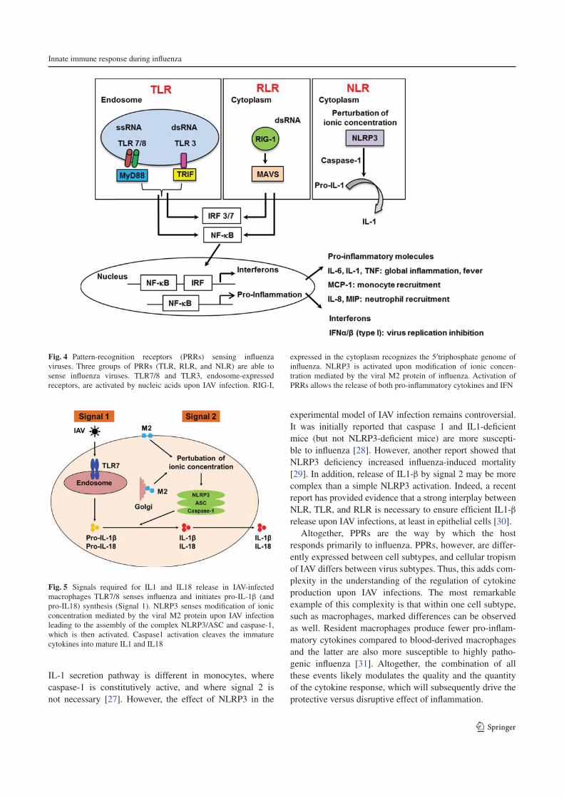
Innate immune response during influenza
1 3
IL-1 secretion pathway is different in monocytes, where caspase-1 is constitutively active, and where signal 2 is not necessary [27]. However, the effect of NLRP3 in the
experimental model of IAV infection remains controversial. It was initially reported that caspase 1 and IL1-deficient mice (but not NLRP3-deficient mice) are more suscepti-ble to influenza [28]. However, another report showed that NLRP3 deficiency increased influenza-induced mortality [29]. In addition, release of IL1-β by signal 2 may be more complex than a simple NLRP3 activation. Indeed, a recent report has provided evidence that a strong interplay between NLR, TLR, and RLR is necessary to ensure efficient IL1-β release upon IAV infections, at least in epithelial cells [30].
Altogether, PPRs are the way by which the host responds primarily to influenza. PPRs, however, are differ-ently expressed between cell subtypes, and cellular tropism of IAV differs between virus subtypes. Thus, this adds com-plexity in the understanding of the regulation of cytokine production upon IAV infections. The most remarkable example of this complexity is that within one cell subtype, such as macrophages, marked differences can be observed as well. Resident macrophages produce fewer pro-inflam-matory cytokines compared to blood-derived macrophages and the latter are also more susceptible to highly patho-genic influenza [31]. Altogether, the combination of all these events likely modulates the quality and the quantity of the cytokine response, which will subsequently drive the protective versus disruptive effect of inflammation.
Fig. 4 Pattern-recognition receptors (PRRs) sensing influenza viruses. Three groups of PRRs (TLR, RLR, and NLR) are able to sense influenza viruses. TLR7/8 and TLR3, endosome-expressed receptors, are activated by nucleic acids upon IAV infection. RIG-I,
expressed in the cytoplasm recognizes the 5′triphosphate genome of influenza. NLRP3 is activated upon modification of ionic concen-tration mediated by the viral M2 protein of influenza. Activation of PRRs allows the release of both pro-inflammatory cytokines and IFN
Fig. 5 Signals required for IL1 and IL18 release in IAV-infected macrophages TLR7/8 senses influenza and initiates pro-IL-1β (and pro-IL18) synthesis (Signal 1). NLRP3 senses modification of ionic concentration mediated by the viral M2 protein upon IAV infection leading to the assembly of the complex NLRP3/ASC and caspase-1, which is then activated. Caspase1 activation cleaves the immature cytokines into mature IL1 and IL18

F. Berri et al.
1 3
The cellular components of the inflammation
As mentioned above, cytokines and chemokines that are released upon infection contribute to the recruitment and activation of immune cells, thus facilitating the antiviral defense against the infection. Among the cellular compo-nents involved against influenza, three major components of the innate immune response stricto sensu can be men-tioned; i.e., neutrophils, macrophages, and natural killer (NK) cells. First, (1) neutrophils recruited in large numbers to the respiratory tract upon influenza infections are impli-cated in the protection of the host [32, 33]. Depletion of these cells, in IAV-infected mice, increases viral replica-tion, pulmonary inflammation, as well as mortality of the mice [32, 33]. Neutrophils eliminate the virus via differ-ent pathways, which include the phagocytosis of apoptotic IAV-infected cells and the degranulation and the production of reactive oxygen species, which assist in the clearance of infected cells [34, 35]. Another important additional weapon of neutrophils against pathogens is the release of neutrophil extracellular trap (NET), which arises from their nuclear contents into the extracellular space and are com-posed of decondensed chromatin and antimicrobial pro-teins. It was clearly demonstrated that NETs are formed upon IAV infections, although their role remains controver-sial [36, 37]. Altogether, neutrophils are important players against influenza. However, excessive recruitment of neu-trophils to the lungs is also a major contributor of severe IAV infections and is typically observed upon mice infec-tion with highly pathogenic H1N1 and H5N1 viruses [37, 38]. Their over-reaction further contributes to excessive lung inflammation and additional release of secretory mol-ecules and particularly IL-1, TNF, or MIP proteins.
In addition to neutrophils, alveolar macrophages as well as newly recruited monocytes, which differentiate into macrophages, also contribute to innate immunity against influenza [32]. Macrophages eliminate cellu-lar debris and apoptotic infected cells by phagocytosis. They also act as antigen-presenting cells and contribute to the induction of the adaptive immune response. Deple-tion of these cells increases lung viral replication as well as pathogenesis and death upon IAV infection [32, 39]. However, as for neutrophils, the presence of excessive macrophages in the lungs is a sign of severe IAV infec-tion, suggesting that these cells could also contribute to the immunopathology of influenza [38]. Finally, the third innate cellular component recruited to the lungs upon IAV infection and playing a key role in IAV immune-surveil-lance are NK cells [40]. Upon activation, NK cells secrete cytokines and chemokines, and kill sensitive target cells by releasing the content of cytolytic granules [41, 42]. NK cell activation is orchestrated through a balance of inhibi-tory receptors (KIR) versus activatory (KAR) receptors
[43]. First, NK cells detect the loss of human-leukocyte antigen (HLA) at the surface of infected cells via absence of engagement of KIRs, an activation known as the miss-ing self-signal. Secondly, NK cells sense infected targets that express ligands for activation receptors, known as the danger signal [44]. When positive signals tend to be dominant, the functional outcome is tilted in favor of NK responsiveness. Surprisingly, while most viruses down-regulate the expression of HLA molecules at the surface of infected cells, IAV does not alter HLA expression on infected target cells [45] or does so only slightly [46]. IAV even augments NK cell inhibition through reorgani-zation of HLA molecules into lipid rafts [45]. Thus, acti-vation of NK during influenza is not due to a missing self-signal. Instead, during influenza, the KARs, NKp44 and NKp46 (but not NKp30), are engaged by the HA of IAV, which leads to NK cell activation [47–49]. In vivo, mice deficient in NKp46 receptor are more susceptible to IAV infection, demonstrating the importance of NK cell func-tion against influenza [50]. However, as for all the compo-nents of the innate immune system, NK cells function can turn deleterious for the host. It was indeed demonstrated that NK cells can also contribute to the pathogenesis of IAV infection [51, 52]. Altogether, this illustrates the importance to consider the severity of infection regard-ing a protective or deleterious role for any component of the immune response. For example, the role of other cell types of the immune system such as the mucosal-associ-ated invariant T cells could be revised. Their function was initially shown to be restricted to bacterial infections [53] but their role during influenza may be crucial, depending on the type of infection and more importantly during IAV coinfection with bacteria.
Viral escape from immunosurveillance
To evade the immune system, IAV has adopted strategies to efficiently replicate within the host [54]. Viral determi-nants such as the nonstructural protein 1 (NS1) and PB1-F2 block the antiviral IFN response and induce apoptosis of the recruited cellular components of the immune system, which enable them to react [55]. In addition, IAV upregu-lates the expression of the powerful immunotolerant human leukocyte antigen-G molecule (HLA-G) [56]. We will here highlight our current knowledge on how IAV manipulate these powerful molecules to escape immune-surveillance.
Role of the NS1 viral protein
NS1 is a nonstructural viral protein that antagonizes host immune responses [13]. The segment 8 of influenza vRNA, also known as the NS gene, encodes two proteins. The

Innate immune response during influenza
1 3
primary transcript generated encodes the NS1 protein. The second protein, NS2 (or NEP), is generated by alter-native splicing of the primary transcript [57]. Recombi-nant viruses unable to express NS1 are viable but induce robust IFN secretion and show an attenuated phenotype in vitro and in vivo, which demonstrates the role of NS1 in the virulence of IAV [58]. The major function of NS1 is to limit the antiviral effect of IFN via different pathways. On one hand, NS1 blocks signaling by IRF3 and NF-κB [59, 60] and RIG-I activation [61, 62]. On the other hand, NS1 blocks IFN secretion at the posttranscriptional level with strong inhibition of IFN mRNA synthesis and post-tran-scriptional processing of IFN [63, 64]. In addition, NS1 is a key player in the manipulation of cell apoptotic machinery [65]. It was demonstrated that NS1 interact with tubulin, leading to disruption of normal cell division and apoptosis [66]. The length of NS1 is variable and strain-specific. In particular, at the C-terminus of NS1, truncations or exten-sions were observed [3]. While NS1 predominantly local-izes in the nucleus and cytoplasm, those modifications may have consequences in its localization and most likely in its function [67]. The fact that NS1 is a virulence factor of IAV makes it a good target to attenuate these viruses. Sev-eral studies demonstrated that IAV with partial deletions in NS1 proteins are attenuated and do not cause disease, but induce a protective immune response in different species. These IAV variants are excellent live-attenuated influenza vaccine candidates, which could be of high interest in the future [68].
Role of PB1-F2
PB1-F2 is a proapoptotic viral protein that is expressed from an alternative open reading frame in the PB1 gene of IAV [69]. Some influenza strains do not express PB1-F2 and thus it is not required for viral replication. Neverthe-less, PB1-F2 has been established as an important factor of virulence of influenza [70, 71]. Recombinant viruses una-ble to express PB1-F2 protein are less pathogenic in mice [72]. In addition, viruses with a single mutation in PB1-F2 (N66S) are highly pathogenic in mice as a consequence of increased viral replication [71]. The way by which PB1-F2 mediates increased viral replication is through inhibition of RIG-I-mediated type I IFN production at the level of the MAVS pathway [73–75]. The serine at position 66 (66S) in PB1-F2 further enhances IFN antagonism activity. PB1-F2 also induces apoptosis. After phosphorylation by protein kinase C, PB1-F2 interacts with the inner mitochondrial membrane adenine nucleotide translocase 3 and the outer mitochondrial membrane voltage-dependent anion channel 1, leading to permeabilization and destabilization of mito-chondrial membrane, which results in cell death [74–76]. Also, another interesting characteristic of PB1-F2 is its
contribution to the virulence of subsequent secondary bac-terial pneumonia [77].
Role of the nonclassical host HLA-G molecule
The major histocompatibility complex molecule, HLA-G, is a non-classical antigen, which expression is mainly restricted to the cytotrophoblast, during pregnancy [78]. Several isoforms of HLA-G have been described that exhibit immunotolerant properties and are key factors in maternal-fetal tolerance [78–80]. HLA-G inhibits the lytic activity of NK cells [81, 82] as well as antigen-specific cytotoxic T cells directed against influenza (CTL) and allo-geneic proliferative responses [83–85]. Recently, HLA-G has emerged as a key molecule in the evasion of immune response to several pathologic situations, such as tumors [86–90] and bacterial and viral infections, including influ-enza [56, 91–95]. HLA-G is upregulated at the surface of IAV-infected cells in a strain-dependent manner, at both the mRNA and protein levels [96]. These results suggest that the virulence of IAV may be caused by the differential capability of different strains to upregulate HLA-G. In line with this report, elevated HLA-G expression was ectopi-cally observed in pandemic and seasonal IAV-infected patients [97]. HLA-G has been found to play an important role in several other viral infections and its expression has been correlated with increased severity of infection and poor survival of infected patients [92–95]. Given its broad immune-tolerant properties, by upregulating HLA-G, IAV may efficiently escape from immune surveillance and this likely contributes to IAV pathogenesis.
Uncontrolled deleterious inflammation
Resolution of inflammation is an integral component of the program of acute inflammation. It is absolutely required to protect healthy cells from tissue damage and is a prereq-uisite for the return of tissue homeostasis. When inflam-mation is inappropriately regulated, it becomes persistent and excessive. This deregulated inflammation, known as a “cytokine storm”, exacerbates the immunopathology of influenza [98]. Compared to uncomplicated patients, abnor-mal elevated levels of cytokines and chemokines are com-monly detected in severe influenza infections [11]. Here, we will discuss the possible mechanism leading to the uncontrolled inflammation associated with severe influenza infections.
Role of the viral determinants
The role of viral replication in the virulence of IAV is still debated. Clinical studies showed that in severe influenza

F. Berri et al.
1 3
cases, a high level of virus replication and an excessive inflammatory response can be observed [11]. Whether a direct correlation exists between viral replication and the deregulated immune response remains an open question. An emerging idea is that a high level of virus replication likely contributes but is not the only culprit of excessive inflammation during influenza. The so-called “cytokine storm” would result from two components, which are (1) a high intensity of inflammation mediated by increased viral replication and PRR activation and (2) a sustained inflam-mation that results from an improper host response. Thus, some viral determinants are assumed to be associated with increased viral replication as well as excessive inflamma-tion. Not surprisingly, chimeric viruses expressing strong activity of the polymerase complex (PA, PB1, and PB2) replicate more efficiently and are a potent inducer of pro-inflammatory cytokines and chemokines [99]. Also, the PA gene of a highly pathogenic H5N1 virus contributes to its virulence through increased viral replication and subse-quent induction of an excessive innate immune response [100]. Another viral determinant that could impact viral replication and cytokine/chemokine release is the presence of a multibasic site in the HA of IAV [56]. After entry into the cell, the virus genome is released from the endosome following a low pH-dependent fusion event mediated by HA, and this fusion occurs only when HA is cleaved. The HA of low pathogenic strains contain a monobasic site that can only be cleaved by extracellular trypsin-like proteases, which thus represent a restricted factor for viral replica-tion. In contrast, the HA of highly pathogenic IAV contain a polybasic site that is cleaved by intracellular furin-type proteases that are present ubiquitously, facilitating viral replication [101]. Indeed, the production of excessive proinflammatory molecules was reported for strains with multibasic cleavage site in HA [102].
Also, as described above, viral proteins NS1 and PB1-F2 are also important determinants that can promote the deregulation of inflammation.
Role of host determinants and hemostasis deregulation
As just mentioned, virus replication is unlikely to be solely responsible for deregulation of innate immunity upon IAV infection. In particular, the crosstalk between the pathogen and the host is a crucial factor driving immunopathogenesis of IAV.
Role of PAR1 in the transition between protective versus deleterious inflammation
Proteases and their receptors have recently emerged as a contributor of immunopathogenesis during viral infections [56, 103, 104]. Protease-activated-receptor 1 (PAR1), a G
protein-coupled receptor, is activated as a result of proteo-lytic cleavage by thrombin, a protease central to the coagu-lation process. At a high concentration of thrombin, PAR1 plays a proinflammatory role, while at low concentration of thrombin, PAR1 mediates anti-inflammatory effects [105]. Using a mild IAV infection (observed by low levels of cytokine release in the broncho-alveolar lavages of infected WT mice), PAR1 was recently proposed to cooperate with TLR for IFN production [106]. Thus, according to the anti-viral effect of TLR and IFN during IAV infections, these results are consistent with a potential protective effect of PAR1 during IAV infections (Fig. 6). The role of PAR1 in promoting innate immunity is platelet-independent, which is in favor of the presence of low concentration of throm-bin and a moderate activation of endothelial cells [106]. Interestingly, and in marked contrast, we recently reported that during a severe lethal IAV infection, in which activa-tion of the coagulation is likely to occur, resulting in high thrombin concentrations, PAR1 signaling was deleterious for the host [104]. Administration of PAR1 antagonists or PAR1 deficiency protected mice from lethal inflammation of the lungs. In contrast, activating PAR1 with specific agonists increased the cytokine storm and decreased sur-vival. In addition, during severe infections, a cooperation between the activation of PAR1 and of the fibrinolytic sys-tem appeared to promote lethal inflammation [107] (Fig. 6 and discussed below). Similar deleterious role of PAR1 was also during meta-pneumovirus infections [108]. Thus, the severity of the infection likely determines the extent of IAV infection (epithelial versus endothelial cells) and the protective versus deleterious role of PAR-1-triggered anti versus pro-inflammatory responses. Accordingly, endothe-lial cells have recently emerged at the center of the uncon-trolled inflammatory response induced by influenza [109]. The S1P1 receptor has a key position in the control of endothelial cell integrity and the routing of PAR1 towards anti-inflammatory versus proinflammatory responses [105]. At a low concentration of thrombin, PAR1 mediates endothelial barrier protection and anti-inflammatory effects through cross-activation of S1P1 receptor [105]. At a high concentration of thrombin, S1P1 is no longer activated and PAR1 signaling turns pro-inflammatory [105]. In fact, sev-eral reports showed that administration of S1P1 receptor (S1P1R) agonists blunt influenza-induced cytokine storm in mice and protect them from mortality induced by several IAV strains [109–111]. Altogether, it is tempting to specu-late that modulation of the interactions between PAR1 and S1P1 contributes to regulate and orchestrate inflammation during influenza. More complex regulations of PAR1 may also involve cross-activation of (1) PAR2 [112], previously shown to protect against influenza [113] or (2) endothelial protein C receptor (EPCR) [114], although its role remains to be fully demonstrated [115, 116].

Innate immune response during influenza
1 3
Role of plasminogen and hyperfibrinolysis
Plasminogen is a zymogen that is activated into its active form plasmin by urokinase and tissue plasminogen activa-tors (uPA, tPA). The main function of plasmin is to break down blood clots by dissolving fibrin polymers into solu-ble fragments, a process called fibrinolysis. Pericellular plasmin contributes to the remodeling of the extracellular matrix directly or indirectly via the activation of metal-loproteases and could lead to cell anoïkis when exces-sive [117]. The generation of plasmin activity is a tightly regulated process. However, since ever, pathogens have exploited the function of plasminogen/plasmin for their own benefit. Particularly, activation of plasminogen by bac-teria increases extracellular matrix degradation and fibrinol-ysis, a way by which the pathogen disseminates within the host. At the same time, this dysregulation of plasminogen activation and fibrinolysis has been associated with exces-sive inflammation [118]. Not only bacteria but also viruses and IAV in particular have evolved several strategies to sequester and activate plasminogen, through viral or cellu-lar proteins [6, 119, 120]. Neuraminidase of the IAV strain A/WSN/33 can bind plasminogen, conferring this strain with the capacity to replicate efficiently in the brain [119, 121]. IAV can also activate plasminogen through the host cellular protein annexin 2 (A2), which is upregulated at the
surface of infected cells and which is incorporated into the virions [6, 120]. Recently, we provided the first evidence that plasminogen plays a central role in influenza patho-genesis and cytokine storm [107]. We found that plasmino-gen-deficient mice or pharmacological inhibition of plas-minogen activation in vivo protected mice from influenza infections and cytokine storm. Furthermore, pharmacologi-cal depletion of fibrinogen, the main target of plasmin had a profound deleterious effect on the survival of IAV-infected mice and this whether or not plasminogen activation is triggered (WT versus plasminogen-deficient mice), sug-gesting that fibrin is rather protective. Thus, these results pointed out for the first time that uncontrolled activation of the plasminergic system drives vascular permeability and excessive lung inflammation upon IAV infections. These results are consistent with clinical reports showing that fibrinolysis deregulation could be associated with fatal out-come of IAV infections in humans [122, 123]. In addition to fibrinolysis, it is well known that plasmin also promotes, in a strain-dependent manner, the proteolytic cleavage of the viral hemagglutinin, an essential step for the infectivity of IAV [2]. In vivo, viruses where HA can be cleaved by plasminogen replicate more efficiently in the lungs of plas-minogen-competent mice compared to the ones of plasmi-nogen-deficient mice [107]. Likely, this increased plasmi-nogen-dependent virus replication also contributes to more
Fig. 6 Model of protective and destructive inflammation during influenza. Upon IAV infection (non-severe infection), epithelial cells are infected and release secretory molecules promoting activation of the host immune response. Initial immune system activation is pro-tective and aims at the elimination of the invading pathogen. PAR1, expressed at the surface of epithelial cells, cooperates with PRRs for effective activation of innate immunity against influenza. However, if
the infection is not controlled (severe infection), endothelial cells are injured (1). Hemostasis is activated (2) and deregulation of fibrinoly-sis through hyperactivation of plasminogen/plasmin promotes exces-sive and deleterious inflammation (3). PAR1, which is also expressed at the surface of the endothelium, cooperates with plasminogen and further exacerbates inflammation and injury

F. Berri et al.
1 3
PPR activation, which may further nourish the vicious cir-cle of inflammation. Thus, these results point to a role for plasminergic and hemostasis deregulation in the control of the deleterious inflammation induced by influenza.
Conclusions
Influenza still causes significant morbidity and mortal-ity associated with severe immunopathology of the lungs, related to excessive innate immune response. However, the mechanisms of such immunopathogenesis remain poorly understood. Based on our recent understanding, a model of inflammation in response to influenza can be proposed (Fig. 6). First, infected epithelial cells sense influenza and activate the innate immune response. Cytokines and chemokines are released and immune cells are recruited to the site of infection to clear the virus (protective immu-nity). In this context and at the epithelial level, some mol-ecules such as PAR1 cooperate with PPR for protective innate immunity activation. A local and limited formation of fibrin could also be protective by limiting the diffusion of the infection. If the protective barriers are overwhelmed by the infection, endothelial cells are injured. Endothelium injury can result from (1) the acute phase of inflammation leading to increased endothelial cell permeability or (2) a direct infection of endothelial cells by IAV. In these con-ditions, protective molecules turn deleterious for the host. Deregulation of hemostasis, activation of PAR-1, or of the plasminergic system, then feed a vicious circle leading to malignant inflammation. This recent demonstration of the involvement of unbalanced hemostasis in the pathogenesis of influenza has to be replaced in a broader context. Indeed fibrinolysis plays a fundamental role in the clearance of blood clots and the clearance of extravascular fibrin. The major manifestation of plasminogen deficiency is the absence of fibrin resorption leading to the formation of pseudomembranes on inflamed mucosal surfaces in human [124] and impaired wound healing in mice [125]. In the context of sepsis, impairment of fibrin clearance is assumed to be pivotal in the pathogenesis of microvascular thrombo-sis and disseminated intravascular coagulation (DIC) [126]. Given the dual role of fibrinolysis, which may dependent on the severity of the infection, our results suggest that it will be essential to define in the next future specific mark-ers of non-severe versus severe IAV infections to direct therapeutics against influenza. During non-severe infec-tions, one could use the current and novel antivirals against influenza aiming at slowing down viral growth. In contrast, during severe IAV infections, where the hallmark of patho-genesis is the deleterious inflammation of the lungs, block-ing viral replication may have no effect. Instead, targeting hemostasis looks to be a promising novel strategy for the
future. Future research will aim at more precisely elucidat-ing the immune mechanism of protection and deregula-tion in order to design new intervention strategies against influenza. From our current knowledge, PAR1 antagonists, PAR2 agonists, plasminogen inhibitors, or S1P1 agonists might be explored as a new treatment for influenza. By maintaining the inflammatory responses in their protective role against viral replication, these new strategies would provide protection against severe IAV infections, without encouraging the emergence of virus resistance.
References
1. Kuiken T, Riteau B, Fouchier RA, Rimmelzwaan GF (2012) Pathogenesis of influenza virus infections: the good, the bad and the ugly. Curr Opin Virol 2(3):276–286
2. Horimoto T, Kawaoka Y (2005) Influenza: lessons from past pandemics, warnings from current incidents. Nat Rev Microbiol 3(8):591–600
3. Palese P, Shaw ML (2007) Orthomyxoviridae: the viruses and their replication. In: Knipe DM, Howley PM (eds) Fields virol-ogy, vol 2, 5th edn. Lippincott Williams & Wilkins, Philadel-phia, pp 1647–1689
4. Lamb RAKR (2001) Orthomyxoviridae: the viruses and their replication. In: Knipe DM, Howley PM, Griffin DE (eds) Fields virology. Lippincott Williams and Wilkins, Philadelphia, pp 1487–1531
5. Moules V, Terrier O, Yver M, Riteau B, Moriscot C, Ferraris O, Julien T, Giudice E, Rolland JP, Erny A, Bouscambert-Duchamp M, Frobert E, Rosa-Calatrava M, Pu Lin Y, Hay A, Thomas D, Schoehn G, Lina B (2011) Importance of viral genomic composition in modulating glycoprotein content on the surface of influenza virus particles. Virology 414(1):51–62
6. LeBouder F, Morello E, Rimmelzwaan GF, Bosse F, Pechoux C, Delmas B, Riteau B (2008) Annexin II incorporated into influenza virus particles supports virus replication by convert-ing plasminogen into plasmin. J Virol 82(14):6820–6828
7. Shaw ML, Stone KL, Colangelo CM, Gulcicek EE, Palese P (2008) Cellular proteins in influenza virus particles. PLoS Pat-hog 4(6):e1000085
8. Gao R, Cao B, Hu Y, Feng Z, Wang D, Hu W, Chen J, Jie Z, Qiu H, Xu K, Xu X, Lu H, Zhu W, Gao Z, Xiang N, Shen Y, He Z, Gu Y, Zhang Z, Yang Y, Zhao X, Zhou L, Li X, Zou S, Zhang Y, Li X, Yang L, Guo J, Dong J, Li Q, Dong L, Zhu Y, Bai T, Wang S, Hao P, Yang W, Zhang Y, Han J, Yu H, Li D, Gao GF, Wu G, Wang Y, Yuan Z, Shu Y (2013) Human infection with a novel avian-origin influenza A (H7N9) virus. N Engl J Med 368(20):1888–1897
9. Herfst S, Schrauwen EJ, Linster M, Chutinimitkul S, de Wit E, Munster VJ, Sorrell EM, Bestebroer TM, Burke DF, Smith DJ, Rimmelzwaan GF, Osterhaus AD, Fouchier RA (2012) Air-borne transmission of influenza A/H5N1 virus between ferrets. Science 336(6088):1534–1541
10. Imai M, Watanabe T, Hatta M, Das SC, Ozawa M, Shinya K, Zhong G, Hanson A, Katsura H, Watanabe S, Li C, Kawakami E, Yamada S, Kiso M, Suzuki Y, Maher EA, Neumann G, Kawaoka Y (2012) Experimental adaptation of an influenza H5 HA confers respiratory droplet transmission to a reassortant H5 HA/H1N1 virus in ferrets. Nature 486(7403):420–428
11. de Jong MD, Simmons CP, Thanh TT, Hien VM, Smith GJ, Chau TN, Hoang DM, Chau NV, Khanh TH, Dong VC, Qui PT, Cam BV, Ha do Q, Guan Y, Peiris JS, Chinh NT, Hien TT,

Innate immune response during influenza
1 3
Farrar J (2006) Fatal outcome of human influenza A (H5N1) is associated with high viral load and hypercytokinemia. Nat Med 12(10):1203–1207
12. Ichinohe T (2010) Respective roles of TLR, RIG-I and NLRP3 in influenza virus infection and immunity: impact on vaccine design. Expert Rev Vaccines 9(11):1315–1324
13. Garcia-Sastre A (2011) Induction and evasion of type I inter-feron responses by influenza viruses. Virus Res 162(1–2):12–18
14. Pascale F, Contreras V, Bonneau M, Courbet A, Chilmon-czyk S, Bevilacqua C, Epardaud M, Niborski V, Riffault S, Balazuc AM, Foulon E, Guzylack-Piriou L, Riteau B, Hope J, Bertho N, Charley B, Schwartz-Cornil I (2008) Plasmacy-toid dendritic cells migrate in afferent skin lymph. J Immunol 180(9):5963–5972
15. La Gruta NL, Kedzierska K, Stambas J, Doherty PC (2007) A question of self-preservation: immunopathology in influenza virus infection. Immunol Cell Biol 85(2):85–92
16. Shinya K, Ito M, Makino A, Tanaka M, Miyake K, Eisfeld AJ, Kawaoka Y (2012) The TLR4-TRIF pathway protects against H5N1 influenza virus infection. J Virol 86(1):19–24
17. Imai Y, Kuba K, Neely GG, Yaghubian-Malhami R, Perkmann T, van Loo G, Ermolaeva M, Veldhuizen R, Leung YH, Wang H, Liu H, Sun Y, Pasparakis M, Kopf M, Mech C, Bavari S, Pei-ris JS, Slutsky AS, Akira S, Hultqvist M, Holmdahl R, Nicholls J, Jiang C, Binder CJ, Penninger JM (2008) Identification of oxidative stress and Toll-like receptor 4 signaling as a key path-way of acute lung injury. Cell 133(2):235–249
18. Shirey KA, Lai W, Scott AJ, Lipsky M, Mistry P, Pletneva LM, Karp CL, McAlees J, Gioannini TL, Weiss J, Chen WH, Ernst RK, Rossignol DP, Gusovsky F, Blanco JC, Vogel SN (2013) The TLR4 antagonist Eritoran protects mice from lethal influenza infection. Nature 497(7450):498–502. doi:10.1038/nature12118
19. Lund JM, Alexopoulou L, Sato A, Karow M, Adams NC, Gale NW, Iwasaki A, Flavell RA (2004) Recognition of single-stranded RNA viruses by Toll-like receptor 7. Proc Natl Acad Sci USA 101(15):5598–5603
20. Diebold SS, Kaisho T, Hemmi H, Akira S, Reis e Sousa C (2004) Innate antiviral responses by means of TLR7-mediated recognition of single-stranded RNA. Science 303(5663):1529–1531
21. Geeraedts F, Goutagny N, Hornung V, Severa M, de Haan A, Pool J, Wilschut J, Fitzgerald KA, Huckriede A (2008) Supe-rior immunogenicity of inactivated whole virus H5N1 influenza vaccine is primarily controlled by Toll-like receptor signalling. PLoS Pathog 4(8):e1000138
22. Le Goffic R, Balloy V, Lagranderie M, Alexopoulou L, Escriou N, Flavell R, Chignard M, Si-Tahar M (2006) Detrimental con-tribution of the Toll-like receptor (TLR)3 to influenza A virus-induced acute pneumonia. PLoS Pathog 2(6):e53
23. Zhao J, Wohlford-Lenane C, Zhao J, Fleming E, Lane TE, McCray PB Jr, Perlman S (2012) Intranasal treatment with poly(I*C) protects aged mice from lethal respiratory virus infections. J Virol 86(21):11416–11424
24. Rehwinkel J, Tan CP, Goubau D, Schulz O, Pichlmair A, Bier K, Robb N, Vreede F, Barclay W, Fodor E, Reis e Sousa C (2010) RIG-I detects viral genomic RNA during negative-strand RNA virus infection. Cell 140(3):397–408
25. Kato H, Takeuchi O, Sato S, Yoneyama M, Yamamoto M, Matsui K, Uematsu S, Jung A, Kawai T, Ishii KJ, Yamaguchi O, Otsu K, Tsujimura T, Koh CS, Reis e Sousa C, Matsuura Y, Fujita T, Akira S (2006) Differential roles of MDA5 and RIG-I helicases in the recognition of RNA viruses. Nature 441(7089):101–105
26. Bauernfeind F, Ablasser A, Bartok E, Kim S, Schmid-Burgk J, Cavlar T, Hornung V (2011) Inflammasomes:
current understanding and open questions. Cell Mol Life Sci 68(5):765–783
27. Netea MG, Simon A, van de Veerdonk F, Kullberg BJ, Van der Meer JW, Joosten LA (2010) IL-1beta processing in host defense: beyond the inflammasomes. PLoS Pathog 6(2):e1000661
28. Ichinohe T, Lee HK, Ogura Y, Flavell R, Iwasaki A (2009) Inflammasome recognition of influenza virus is essential for adaptive immune responses. J Exp Med 206(1):79–87
29. Allen IC, Scull MA, Moore CB, Holl EK, McElvania-TeKippe E, Taxman DJ, Guthrie EH, Pickles RJ, Ting JP (2009) The NLRP3 inflammasome mediates in vivo innate immunity to influenza A virus through recognition of viral RNA. Immunity 30(4):556–565
30. Pothlichet J, Meunier I, Davis BK, Ting JP, Skamene E, von Messling V, Vidal SM (2013) Type I IFN triggers RIG-I/TLR3/NLRP3-dependent inflammasome activation in influ-enza A virus-infected cells. PLoS Pathog 9(4):e1003256
31. van Riel D, Leijten LM, van der Eerden M, Hoogsteden HC, Boven LA, Lambrecht BN, Osterhaus AD, Kuiken T (2011) Highly pathogenic avian influenza virus H5N1 infects alveolar macrophages without virus production or excessive TNF-alpha induction. PLoS Pathog 7(6):e1002099
32. Tumpey TM, Garcia-Sastre A, Taubenberger JK, Palese P, Swayne DE, Pantin-Jackwood MJ, Schultz-Cherry S, Solor-zano A, Van Rooijen N, Katz JM, Basler CF (2005) Pathogenic-ity of influenza viruses with genes from the 1918 pandemic virus: functional roles of alveolar macrophages and neutro-phils in limiting virus replication and mortality in mice. J Virol 79(23):14933–14944
33. Tate MD, Deng YM, Jones JE, Anderson GP, Brooks AG, Read-ing PC (2009) Neutrophils ameliorate lung injury and the devel-opment of severe disease during influenza infection. J Immunol 183(11):7441–7450
34. Hashimoto Y, Moki T, Takizawa T, Shiratsuchi A, Nakanishi Y (2007) Evidence for phagocytosis of influenza virus-infected, apoptotic cells by neutrophils and macrophages in mice. J Immunol 178(4):2448–2457
35. Peake J, Suzuki K (2004) Neutrophil activation, antioxidant supplements and exercise-induced oxidative stress. Exerc Immunol Rev 10:129–141
36. Hemmers S, Teijaro JR, Arandjelovic S, Mowen KA (2011) PAD4-mediated neutrophil extracellular trap formation is not required for immunity against influenza infection. PLoS ONE 6(7):e22043
37. Narasaraju T, Yang E, Samy RP, Ng HH, Poh WP, Liew AA, Phoon MC, van Rooijen N, Chow VT (2011) Excessive neutro-phils and neutrophil extracellular traps contribute to acute lung injury of influenza pneumonitis. Am J Pathol 179(1):199–210
38. Perrone LA, Plowden JK, Garcia-Sastre A, Katz JM, Tumpey TM (2008) H5N1 and 1918 pandemic influenza virus infection results in early and excessive infiltration of macrophages and neutrophils in the lungs of mice. PLoS Pathog 4(8):e1000115
39. Kim HM, Lee YW, Lee KJ, Kim HS, Cho SW, van Rooijen N, Guan Y, Seo SH (2008) Alveolar macrophages are indispen-sable for controlling influenza viruses in lungs of pigs. J Virol 82(9):4265–4274
40. Ennis FA, Meager A, Beare AS, Qi YH, Riley D, Schwarz G, Schild GC, Rook AH (1981) Interferon induction and increased natural killer-cell activity in influenza infections in man. Lancet 2(8252):891–893
41. Bryceson YT, Long EO (2008) Line of attack: NK cell specificity and integration of signals. Curr Opin Immunol 20(3):344–352
42. Riteau B, Barber DF, Long EO (2003) Vav1 phosphorylation is induced by beta2 integrin engagement on natural killer cells

F. Berri et al.
1 3
upstream of actin cytoskeleton and lipid raft reorganization. J Exp Med 198(3):469–474
43. Thielens A, Vivier E, Romagne F (2012) NK cell MHC class I specific receptors (KIR): from biology to clinical intervention. Curr Opin Immunol 24(2):239–245
44. Orr MT, Lanier LL (2010) Natural killer cell education and tol-erance. Cell 142(6):847–856
45. Achdout H, Manaster I, Mandelboim O (2008) Influenza virus infection augments NK cell inhibition through reorganization of major histocompatibility complex class I proteins. J Virol 82(16):8030–8037
46. Ronni T, Matikainen S, Sareneva T, Melen K, Pirhonen J, Kes-kinen P, Julkunen I (1997) Regulation of IFN-alpha/beta, MxA, 2′,5′-oligoadenylate synthetase, and HLA gene expression in influenza A-infected human lung epithelial cells. J Immunol 158(5):2363–2374
47. Mandelboim O, Lieberman N, Lev M, Paul L, Arnon TI, Bushkin Y, Davis DM, Strominger JL, Yewdell JW, Porgador A (2001) Recognition of haemagglutinins on virus-infected cells by NKp46 activates lysis by human NK cells. Nature 409(6823):1055–1060
48. Ho JW, Hershkovitz O, Peiris M, Zilka A, Bar-Ilan A, Nal B, Chu K, Kudelko M, Kam YW, Achdout H, Mandelboim M, Altmeyer R, Mandelboim O, Bruzzone R, Porgador A (2008) H5-type influenza virus hemagglutinin is functionally recog-nized by the natural killer-activating receptor NKp44. J Virol 82(4):2028–2032
49. Arnon TI, Lev M, Katz G, Chernobrov Y, Porgador A, Mandel-boim O (2001) Recognition of viral hemagglutinins by NKp44 but not by NKp30. Eur J Immunol 31(9):2680–2689
50. Gazit R, Gruda R, Elboim M, Arnon TI, Katz G, Achdout H, Hanna J, Qimron U, Landau G, Greenbaum E, Zakay-Rones Z, Porgador A, Mandelboim O (2006) Lethal influenza infection in the absence of the natural killer cell receptor gene Ncr1. Nat Immunol 7(5):517–523
51. Zhou G, Juang SW, Kane KP (2013) NK cells exacerbate the pathology of influenza virus infection in mice. Eur J Immunol 43(4):929–938
52. Abdul-Careem MF, Mian MF, Yue G, Gillgrass A, Chenoweth MJ, Barra NG, Chew MV, Chan T, Al-Garawi AA, Jordana M, Ashkar AA (2012) Critical role of natural killer cells in lung immunopathology during influenza infection in mice. J Infect Dis 206(2):167–177
53. Le Bourhis L, Martin E, Peguillet I, Guihot A, Froux N, Core M, Levy E, Dusseaux M, Meyssonnier V, Premel V, Ngo C, Riteau B, Duban L, Robert D, Rottman M, Soudais C, Lantz O (2010) Antimicrobial activity of mucosal-associated invariant T cells. Nat Immunol 11(8):701–708
54. Garcia-Sastre A, Biron CA (2006) Type 1 interferons and the virus-host relationship: a lesson in detente. Science 312(5775):879–882
55. Herold S, Ludwig S, Pleschka S, Wolff T (2012) Apoptosis signaling in influenza virus propagation, innate host defense, and lung injury. J Leukoc Biol 92(1):75–82
56. Foucault ML, Moules V, Rosa-Calatrava M, Riteau B (2011) Role for proteases and HLA-G in the pathogenicity of influenza A viruses. J Clin Virol 51(3):155–159
57. Robb NC, Jackson D, Vreede FT, Fodor E (2010) Splicing of influenza A virus NS1 mRNA is independent of the viral NS1 protein. J Gen Virol 91(Pt 9):2331–2340
58. Garcia-Sastre A, Egorov A, Matassov D, Brandt S, Levy DE, Durbin JE, Palese P, Muster T (1998) Influenza A virus lacking the NS1 gene replicates in interferon-deficient systems. Virol-ogy 252(2):324–330
59. Talon J, Horvath CM, Polley R, Basler CF, Muster T, Palese P, Garcia-Sastre A (2000) Activation of interferon regulatory
factor 3 is inhibited by the influenza A virus NS1 protein. J Virol 74(17):7989–7996
60. Wang X, Li M, Zheng H, Muster T, Palese P, Beg AA, Garcia-Sastre A (2000) Influenza A virus NS1 protein prevents acti-vation of NF-kappaB and induction of alpha/beta interferon. J Virol 74(24):11566–11573
61. Guo Z, Chen LM, Zeng H, Gomez JA, Plowden J, Fujita T, Katz JM, Donis RO, Sambhara S (2007) NS1 protein of influenza A virus inhibits the function of intracytoplasmic pathogen sensor, RIG-I. Am J Respir Cell Mol Biol 36(3):263–269
62. Mibayashi M, Martinez-Sobrido L, Loo YM, Cardenas WB, Gale M Jr, Garcia-Sastre A (2007) Inhibition of retinoic acid-inducible gene I-mediated induction of beta interferon by the NS1 protein of influenza A virus. J Virol 81(2):514–524
63. Kochs G, Garcia-Sastre A, Martinez-Sobrido L (2007) Multiple anti-interferon actions of the influenza A virus NS1 protein. J Virol 81(13):7011–7021
64. Qiu Y, Krug RM (1994) The influenza virus NS1 protein is a poly(A)-binding protein that inhibits nuclear export of mRNAs containing poly(A). J Virol 68(4):2425–2432
65. Schultz-Cherry S, Dybdahl-Sissoko N, Neumann G, Kawaoka Y, Hinshaw VS (2001) Influenza virus ns1 protein induces apoptosis in cultured cells. J Virol 75(17):7875–7881
66. Han X, Li Z, Chen H, Wang H, Mei L, Wu S, Zhang T, Liu B, Lin X (2012) Influenza virus A/Beijing/501/2009(H1N1) NS1 interacts with beta-tubulin and induces disruption of the microtubule network and apoptosis on A549 cells. PLoS ONE 7(11):e48340
67. Melen K, Kinnunen L, Fagerlund R, Ikonen N, Twu KY, Krug RM, Julkunen I (2007) Nuclear and nucleolar targeting of influ-enza A virus NS1 protein: striking differences between different virus subtypes. J Virol 81(11):5995–6006
68. Talon J, Salvatore M, O’Neill RE, Nakaya Y, Zheng H, Muster T, Garcia-Sastre A, Palese P (2000) Influenza A and B viruses expressing altered NS1 proteins: a vaccine approach. Proc Natl Acad Sci USA 97(8):4309–4314
69. Conenello GM, Palese P (2007) Influenza A virus PB1-F2: a small protein with a big punch. Cell Host Microbe 2(4):207–209
70. Schmolke M, Manicassamy B, Pena L, Sutton T, Hai R, Varga ZT, Hale BG, Steel J, Perez DR, Garcia-Sastre A (2011) Differ-ential contribution of PB1-F2 to the virulence of highly patho-genic H5N1 influenza A virus in mammalian and avian species. PLoS Pathog 7(8):e1002186
71. Conenello GM, Zamarin D, Perrone LA, Tumpey T, Palese P (2007) A single mutation in the PB1-F2 of H5N1 (HK/97) and 1918 influenza A viruses contributes to increased virulence. PLoS Pathog 3(10):1414–1421
72. Zamarin D, Ortigoza MB, Palese P (2006) Influenza A virus PB1-F2 protein contributes to viral pathogenesis in mice. J Virol 80(16):7976–7983
73. Varga ZT, Ramos I, Hai R, Schmolke M, Garcia-Sastre A, Fer-nandez-Sesma A, Palese P (2011) The influenza virus protein PB1-F2 inhibits the induction of type I interferon at the level of the MAVS adaptor protein. PLoS Pathog 7(6):e1002067
74. Zamarin D, Garcia-Sastre A, Xiao X, Wang R, Palese P (2005) Influenza virus PB1-F2 protein induces cell death through mito-chondrial ANT3 and VDAC1. PLoS Pathog 1(1):e4
75. Chen W, Calvo PA, Malide D, Gibbs J, Schubert U, Bacik I, Basta S, O’Neill R, Schickli J, Palese P, Henklein P, Ben-nink JR, Yewdell JW (2001) A novel influenza A virus mitochondrial protein that induces cell death. Nat Med 7(12):1306–1312
76. Chanturiya AN, Basanez G, Schubert U, Henklein P, Yewdell JW, Zimmerberg J (2004) PB1-F2, an influenza A virus-encoded proapoptotic mitochondrial protein, creates variably sized pores in planar lipid membranes. J Virol 78(12):6304–6312

Innate immune response during influenza
1 3
77. McAuley JL, Hornung F, Boyd KL, Smith AM, McKeon R, Bennink J, Yewdell JW, McCullers JA (2007) Expression of the 1918 influenza A virus PB1-F2 enhances the pathogenesis of viral and secondary bacterial pneumonia. Cell Host Microbe 2(4):240–249
78. Rouas-Freiss N, Khalil-Daher I, Riteau B, Menier C, Paul P, Dausset J, Carosella ED (1999) The immunotolerance role of HLA-G. Semin Cancer Biol 9(1):3–12
79. Menier C, Riteau B, Dausset J, Carosella ED, Rouas-Freiss N (2000) HLA-G truncated isoforms can substitute for HLA-G1 in fetal survival. Hum Immunol 61(11):1118–1125
80. Riteau B, Moreau P, Menier C, Khalil-Daher I, Khosrotehrani K, Bras-Goncalves R, Paul P, Dausset J, Rouas-Freiss N, Caro-sella ED (2001) Characterization of HLA-G1, -G2, -G3, and -G4 isoforms transfected in a human melanoma cell line. Trans-plant Proc 33(3):2360–2364
81. Khalil-Daher I, Riteau B, Menier C, Sedlik C, Paul P, Dausset J, Carosella ED, Rouas-Freiss N (1999) Role of HLA-G versus HLA-E on NK function: HLA-G is able to inhibit NK cytolysis by itself. J Reprod Immunol 43(2):175–182
82. Riteau B, Menier C, Khalil-Daher I, Martinozzi S, Pla M, Daus-set J, Carosella ED, Rouas-Freiss N (2001) HLA-G1 co-expres-sion boosts the HLA class I-mediated NK lysis inhibition. Int Immunol 13(2):193–201
83. Riteau B, Menier C, Khalil-Daher I, Sedlik C, Dausset J, Rouas-Freiss N, Carosella ED (1999) HLA-G inhibits the allogeneic proliferative response. J Reprod Immunol 43(2):203–211
84. Le Gal FA, Riteau B, Sedlik C, Khalil-Daher I, Menier C, Daus-set J, Guillet JG, Carosella ED, Rouas-Freiss N (1999) HLA-G-mediated inhibition of antigen-specific cytotoxic T lympho-cytes. Int Immunol 11(8):1351–1356
85. Riteau B, Rouas-Freiss N, Menier C, Paul P, Dausset J, Caro-sella ED (2001) HLA-G2, -G3, and -G4 isoforms expressed as nonmature cell surface glycoproteins inhibit NK and antigen-specific CTL cytolysis. J Immunol 166(8):5018–5026
86. Paul P, Rouas-Freiss N, Khalil-Daher I, Moreau P, Riteau B, Le Gal FA, Avril MF, Dausset J, Guillet JG, Carosella ED (1998) HLA-G expression in melanoma: a way for tumor cells to escape from immunosurveillance. Proc Natl Acad Sci USA 95(8):4510–4515
87. Adrian Cabestre F, Moreau P, Riteau B, Ibrahim EC, Le Danff C, Dausset J, Rouas-Freiss N, Carosella ED, Paul P (1999) HLA-G expression in human melanoma cells: protection from NK cytolysis. J Reprod Immunol 43(2):183–193
88. Riteau B, Faure F, Menier C, Viel S, Carosella ED, Amigor-ena S, Rouas-Freiss N (2003) Exosomes bearing HLA-G are released by melanoma cells. Hum Immunol 64(11):1064–1072
89. Menier C, Riteau B, Carosella ED, Rouas-Freiss N (2002) MICA triggering signal for NK cell tumor lysis is counter-acted by HLA-G1-mediated inhibitory signal. Int J Cancer 100(1):63–70
90. Zilberman S, Schenowitz C, Agaugue S, Benoit F, Riteau B, Rouzier R, Carosella ED, Rouas-Freiss N, Menier C (2012) HLA-G1 and HLA-G5 active dimers are present in malignant cells and effusions: the influence of the tumor microenviron-ment. Eur J Immunol 42(6):1599–1608
91. Fainardi E, Castellazzi M, Stignani M, Morandi F, Sana G, Gonzalez R, Pistoia V, Baricordi OR, Sokal E, Pena J (2011) Emerging topics and new perspectives on HLA-G. Cell Mol Life Sci 68(3):433–451
92. Li C, Toth I, Schulze Zur Wiesch J, Pereyra F, Rychert J, Rosenberg ES, van Lunzen J, Lichterfeld M, Yu XG (2013) Functional characterization of HLA-G(+) regulatory T cells in HIV-1 infection. PLoS Pathog 9(1):e1003140
93. Larsen MH, Zinyama R, Kallestrup P, Gerstoft J, Gomo E, Thorner LW, Berg TB, Erikstrup C, Ullum H (2013) HLA-G 3′
untranslated region 14-base pair deletion: association with poor survival in an HIV-1-infected Zimbabwean population. J Infect Dis 207(6):903–906
94. Segat L, Catamo E, Fabris A, Morgutti M, D’Agaro P, Campello C, Crovella S (2010) HLA-G*0105N allele is associated with augmented risk for HIV infection in white female patients. AIDS 24(12):1961–1964
95. Shi WW, Lin A, Xu DP, Bao WG, Zhang JG, Chen SY, Li J, Yan WH (2011) Plasma soluble human leukocyte antigen-G expres-sion is a potential clinical biomarker in patients with hepatitis B virus infection. Hum Immunol 72(11):1068–1073
96. LeBouder F, Khoufache K, Menier C, Mandouri Y, Keffous M, Lejal N, Krawice-Radanne I, Carosella ED, Rouas-Freiss N, Riteau B (2009) Immunosuppressive HLA-G molecule is upregulated in alveolar epithelial cells after influenza A virus infection. Hum Immunol 70(12):1016–1019
97. Chen HX, Chen BG, Shi WW, Zhen R, Xu DP, Lin A, Yan WH (2011) Induction of cell surface human leukocyte antigen-G expression in pandemic H1N1 2009 and seasonal H1N1 influ-enza virus-infected patients. Hum Immunol 72(2):159–165
98. Tsotsiashvilli M, Levi R, Arnon R, Berke G (1998) Activation of influenza-specific memory cytotoxic T lymphocytes by Con-canavalin A stimulation. Immunol Lett 60(2–3):89–95
99. Li OT, Chan MC, Leung CS, Chan RW, Guan Y, Nicholls JM, Poon LL (2009) Full factorial analysis of mammalian and avian influenza polymerase subunits suggests a role of an efficient polymerase for virus adaptation. PLoS ONE 4(5):e5658
100. Hu J, Hu Z, Song Q, Gu M, Liu X, Wang X, Hu S, Chen C, Liu H, Liu W, Chen S, Peng D, Liu X (2013) The PA-gene-medi-ated lethal dissemination and excessive innate immune response contribute to the high virulence of H5N1 avian influenza virus in mice. J Virol 87(5):2660–2672
101. Zeng H, Pappas C, Belser JA, Houser KV, Zhong W, Wad-ford DA, Stevens T, Balczon R, Katz JM, Tumpey TM (2012) Human pulmonary microvascular endothelial cells support pro-ductive replication of highly pathogenic avian influenza viruses: possible involvement in the pathogenesis of human H5N1 virus infection. J Virol 86(2):667–678
102. Suguitan AL Jr, Matsuoka Y, Lau YF, Santos CP, Vogel L, Cheng LI, Orandle M, Subbarao K (2012) The multibasic cleavage site of the hemagglutinin of highly pathogenic A/Vietnam/1203/2004 (H5N1) avian influenza virus acts as a virulence factor in a host-specific manner in mammals. J Virol 86(5):2706–2714
103. Riteau B, de Vaureix C, Lefevre F (2006) Trypsin increases pseudorabies virus production through activation of the ERK signalling pathway. J Gen Virol 87(Pt 5):1109–1112
104. Khoufache K, Berri F, Nacken W, Vogel AB, Delenne M, Camerer E, Coughlin SR, Carmeliet P, Lina B, Rimmelzwaan GF, Planz O, Ludwig S, Riteau B (2013) PAR1 contrib-utes to influenza A virus pathogenicity in mice. J Clin Invest 123(1):206–214
105. Feistritzer C, Riewald M (2005) Endothelial barrier pro-tection by activated protein C through PAR1-dependent sphingosine 1-phosphate receptor-1 crossactivation. Blood 105(8):3178–3184
106. Antoniak S, Owens AP 3rd, Baunacke M, Williams JC, Lee RD, Weithauser A, Sheridan PA, Malz R, Luyendyk JP, Esser-man DA, Trejo J, Kirchhofer D, Blaxall BC, Pawlinski R, Beck MA, Rauch U, Mackman N (2013) PAR-1 contributes to the innate immune response during viral infection. J Clin Invest 123(3):1310–1322
107. Berri F, Rimmelzwaan GF, Hanss M, Albina E, Foucault-Grunenwald ML, Le VB, Vogelzang-van Trierum SE, Gil P, Camerer E, Martinez D, Lina B, Lijnen R, Carme-liet P, Riteau B (2013) Plasminogen controls inflammation and

F. Berri et al.
1 3
pathogenesis of influenza virus infections via fibrinolysis. PLoS Pathog 9(3):e1003229
108. Aerts L, Hamelin MÈ, Rhéaume C, Lavigne S, Couture C, Kim W, Susan-Resiga D, Prat A, Seidah NG, Vergnolle N, Riteau B, Boivin G (2013) Modulation of protease activated receptor 1 influences human metapneumovirus disease severity in a mouse model. Plos One 8:e72529
109. Teijaro JR, Walsh KB, Cahalan S, Fremgen DM, Rob-erts E, Scott F, Martinborough E, Peach R, Oldstone MB, Rosen H (2011) Endothelial cells are central orchestrators of cytokine amplification during influenza virus infection. Cell 146(6):980–991
110. Walsh KB, Teijaro JR, Wilker PR, Jatzek A, Fremgen DM, Das SC, Watanabe T, Hatta M, Shinya K, Suresh M, Kawaoka Y, Rosen H, Oldstone MB (2011) Suppression of cytokine storm with a sphingosine analog provides protection against pathogenic influenza virus. Proc Natl Acad Sci USA 108(29):12018–12023
111. Marsolais D, Hahm B, Walsh KB, Edelmann KH, McGavern D, Hatta Y, Kawaoka Y, Rosen H, Oldstone MB (2009) A criti-cal role for the sphingosine analog AAL-R in dampening the cytokine response during influenza virus infection. Proc Natl Acad Sci USA 106(5):1560–1565
112. O’Brien PJ, Prevost N, Molino M, Hollinger MK, Woolkalis MJ, Woulfe DS, Brass LF (2000) Thrombin responses in human endothelial cells. Contributions from receptors other than PAR1 include the transactivation of PAR2 by thrombin-cleaved PAR1. J Biol Chem 275(18):13502–13509
113. Khoufache K, LeBouder F, Morello E, Laurent F, Riffault S, Andrade-Gordon P, Boullier S, Rousset P, Vergnolle N, Riteau B (2009) Protective role for protease-activated recep-tor-2 against influenza virus pathogenesis via an IFN-gamma-dependent pathway. J Immunol 182(12):7795–7802
114. Esmon CT (2012) Protein C anticoagulant system–anti-inflam-matory effects. Semin Immunopathol 34(1):127–132
115. Schouten M, Sluijs KF, Gerlitz B, Grinnell BW, Roelofs JJ, Levi MM, van’t Veer C, Poll T (2010) Activated protein C ameliorates coagulopathy but does not influence outcome in lethal H1N1 influenza: a controlled laboratory study. Crit Care 14(2):R65
116. Schouten M, van’t Veer C, Levi M, Esmon CT, van der Poll T (2011) Endogenous protein C inhibits activation of coagulation and transiently lowers bacterial outgrowth in murine Escheri-chia coli peritonitis. J Thromb Haemost 9(5):1072–1075
117. Meilhac O, Ho-Tin-Noe B, Houard X, Philippe M, Michel JB, Angles-Cano E (2003) Pericellular plasmin induces smooth muscle cell anoikis. FASEB J 17(10):1301–1303
118. Degen JL, Bugge TH, Goguen JD (2007) Fibrin and fibrinoly-sis in infection and host defense. J Thromb Haemost 5(Suppl 1):24–31
119. Goto H, Kawaoka Y (1998) A novel mechanism for the acquisi-tion of virulence by a human influenza A virus. Proc Natl Acad Sci USA 95(17):10224–10228
120. LeBouder F, Lina B, Rimmelzwaan GF, Riteau B (2010) Plas-minogen promotes influenza A virus replication through an annexin 2-dependent pathway in the absence of neuraminidase. J Gen Virol 91(Pt 11):2753–2761
121. Goto H, Wells K, Takada A, Kawaoka Y (2001) Plasminogen-binding activity of neuraminidase determines the pathogenicity of influenza A virus. J Virol 75(19):9297–9301
122. Wang ZF, Su F, Lin XJ, Dai B, Kong LF, Zhao HW, Kang J (2011) Serum D-dimer changes and prognostic implication in 2009 novel influenza A(H1N1). Thromb Res 127(3):198–201
123. Soepandi PZ, Burhan E, Mangunnegoro H, Nawas A, Aditama TY, Partakusuma L, Isbaniah F, Ikhsan M, Swidarmoko B, Sutiyoso A, Malik S, Benamore R, Baird JK, Taylor WR (2010) Clinical course of avian influenza A(H5N1) in patients at the Persahabatan Hospital, Jakarta, Indonesia, 2005–2008. Chest 138(3):665–673
124. Mehta R, Shapiro AD (2008) Plasminogen deficiency. Haemo-philia 14(6):1261–1268
125. Bugge TH, Kombrinck KW, Flick MJ, Daugherty CC, Dan-ton MJ, Degen JL (1996) Loss of fibrinogen rescues mice from the pleiotropic effects of plasminogen deficiency. Cell 87(4):709–719
126. Gando S (2013) Role of fibrinolysis in sepsis. Semin Thromb Hemost 39(4):392–399

Platelet dysfunction promotes influenza pathogenesis: Toward the Development of a 1
Universal Therapy 2
3
Vuong Ba Lê1, Jochen G. Schneider2,3, Yvonne Boergeling4, Fatma Berri1, Mariette Ducatez5,6, 4
Jean-Luc Guerin5,6, Iris Adrian3, Bruno Lina1, Jean-Claude Bordet7, Martine Jandrot-Perrus8, 5
Stephan Ludwig4, Béatrice Riteau1,9 * 6
7
1EA4610, Lyon, France; 2Luxembourg Centre for Systems Biomedicine, Esch-Sur-Alzette, 8
Luxembourg ; 3Saarland University Medical Center, Homburg/Saar, Germany; 4Institute 9
Molecular Virology, ZMBE, Münster, Germany; 5UMR 1225, IHAP, INRA Toulouse, France; 10
6INP, ENVT, Toulouse France; 7Unité d’Hémostase Clinique, Lyon, France; 8INSERM 11
UMR_S1148, Paris Diderot, CHU Xavier Bichat, Paris, France; 9INRA Nouzilly, France 12
* correspondance to Beatrice Riteau: EA4610, Université Lyon INRA, France. Phone : 13
04.78.77.87.11 - Fax: 04.78.77.87.51 [email protected]. 14
Author contributions: VBL, JGS, JCB, MJP, SL, BR designed the experiments. VBL, YB, 15
FB, IA performed the experiments. FB, JGS, SL, BL critically read the manuscript. VBL, MJP 16
and BR wrote the manuscript. Support: BR and MJP acquired funding from ANR HemoFlu 17
and JGS from DFG, FNR Core Itgb3VascIn. Short Head: Platelet dysfunction during 18
influenza; Classification: 10.15 Treatment. 19
Commentary: Our research shows that platelets play a key role in the pathogenesis of 20
influenza-induced acute lung injury. This may have strong impact for the development of novel 21
drugs for the treatment of these diseases. This article has an online data supplement, which is 22
accessible from this issue's table of content online at www.atsjournals.org. Total word count: 23
3436 24
Abstract 25

1
Rationale: The hallmark of severe influenza virus infections is an excessive inflammation of 26
the lungs. Platelets are activated during influenza but their role in influenza pathogenesis and 27
cytokine storm is unknown. 28
Objectives: To determine the role of platelet during influenza virus infections and propose new 29
therapeutics against influenza. 30
Methods: We used targeted gene deletion approaches and pharmacological interventions to 31
investigate the role of platelets during influenza virus infection, in mice. 32
Measurements and Main Results: Lungs of infected mice were massively infiltrated by 33
aggregates of activated platelets that have engulfed influenza viruses. Deficiency in the major 34
platelet receptor glyprotein IIIa (GPIIIa) protected mice from death caused by influenza viruses. 35
In contrast, activating Protease-Activated Receptor 4 (PAR4), a receptor crucial for platelet 36
activation exacerbated influenza-induced acute lung injury and death. Mechanistically, platelet 37
dysfunction was at the basis of this process since this effect was abolished in mice treated with 38
the specific anti-platelet drug eptifibatide or in mice deficient in GPIIIa. More interestingly, 39
mice treated with anti-platelet molecules (antagonists of PAR4 or eptifibatide) were protected 40
from severe lung injury and lethal infections induced by several influenza strains. 41
Conclusions: The intricate relationship between hemostasis and inflammation has major 42
consequences in influenza virus pathogenesis and anti-platelet drugs might be explored for 43
developing universal treatments against influenza virus infections. The anti-platelet drug 44
eptifibatide (Integrilin®), that was tested in this study is commercialized and used in humans, 45
permitted us to expect that such anti-platelet molecules would combine efficacy and safety in 46
humans. 47
Key words: Lung injury, novel drugs, Flu pathogenesis, pneumonia. Words: 245 48
49

2
Introduction 50
Influenza is one of the most common infectious diseases in humans, occurring as sporadic 51
pandemic and seasonal epidemic outbreaks, leading to significant fatal cases. Influenza 52
pathogenesis is a complex process involving both viral determinants and the immune system 53
(1-3). During severe influenza, dysregulation of cytokine production contributes to collateral 54
damage of the lungs, possibly leading to organ failure and death (4-7). The endothelium, which 55
lines the interior surface of blood vessels is thought to orchestrate the crescendo in cytokine 56
accumulation, although the mechanism involved is not fully identified (8). 57
Upon endothelial injury, platelets are recruited by inflamed endothelial cells, where they adhere 58
and get activated (9). Simultaneously, Protease-Activated Receptor (PAR) mediates activation 59
of platelets by thrombin. These events lead to the conformational change of the platelet 60
glycoprotein IIb/IIIa (GPIIb/IIIa) receptor for fibrinogen that bridges platelets, leading to their 61
aggregation and a reinforcement of their activation. Importantly, platelet activation is strongly 62
associated with enhanced inflammatory responses. Activated platelets release potent 63
inflammatory molecules and play a key role in leukocyte recruitment (10). Platelet activation 64
is finely tuned but its dysfunction is pathogenic and contributes to inflammatory disorders (11-65
13). Thus, uncontrolled platelet activation could contribute to the pathogenesis of IAV 66
infections by fuelling a harmful inflammatory response in the respiratory tract. However, the 67
role of platelets in the context of IAV infection has never been investigated. In the present study, 68
using pharmacological and gene deletion approaches, we investigated the role of platelets in 69
IAV pathogenesis, in vivo. Our findings showed that platelets feed the cytokine storm and 70
contribute to influenza virus pathogenicity in mice. More importantly, commercially available 71
anti-platelets drugs efficiently protected mice from IAV pathogenesis, induced by several 72
influenza strains. Because such a strategy targeting the host rather than the virus would limit 73
the emergence of virus resistance and would be universal, these results suggest that inhibitors 74

3
of platelet function should be explored for the development of a novel treatment of IAV 75
infections. 76
77

4
METHODS 78
79
Cells, Viruses, Antibodies, and Reagents 80
A549 cells and MDCK cells were purchased from ATCC. IAV A/PR/8/34 virus (H1N1), 81
A/HK/1/68 (H3N2) and A/NL/602/2009 (H1N1) (ATCC) were gifts from GF. Rimmelzwaan 82
(Erasmus, Rotterdam, Netherlands). Highly pathogenic avian influenza virus 83
A/FPV/Bratislava/79 (H7N7) was from the IMV Münster, Germany. The following reagents 84
were used: DAPI (Life Technologies), Alexa Fluo® secondary antibodies (Life Technologies), 85
eptifibatide (Integrilin®, GlaxoSmithKline), PAR4 antagonist pepducin p4pal-10 (Polypeptide 86
Laboratories), PAR4 agonist peptide (AYPGKF-NH2, Bachem), PAR4 control peptide 87
(YAPGKF-NH2, Bachem); antibodies: monoclonal anti-neutrophil Ly6G (Cedarlane), 88
polyclonal anti-platelet CD41 (Bioss), monoclonal anti-viral HA (Santa Cruz Biotechnology), 89
monoclonal anti-IAV NP (kind gift from Dr GF. Rimmelzwaan), monoclonal anti-p-Selectin 90
FITC-conjugated (Emfret), monoclonal anti-CD41/61 PE-conjugated (Emfret); Vectastain® 91
ABC kit (Vector Laboratories), 3,3’-diaminobenzidine (DAB) peroxidase substrate (Vector 92
Laboratories), ketamine/xylazine anesthesia (Virbac, Bayer HealthCare), May-Grünwald and 93
Giemsa solutions (Merck), Hematoxylin and Eosin solutions (Diapath), ELISA kits for mouse 94
IL-6, IL-1β, IFN-γ, MIP-2, RANTES (R&D Systems), serotonin (BlueGene), TXB2 95
(Elabscience) and sP-selectin (Qayee-Bio). Total protein was evaluated by using the Coomassie 96
Bradford Protein assay kit (Thermo Scientific). 97
98
Mice 99
Experiments were performed in accordance with the Guide for the Care and Use of Laboratory 100
Animals of “la Direction des Services Vétérinaires (DSV)”, the French regulations to which 101
our animal care and protocol adhered. icence authority was issued by the DSV and Lyon 102

5
university (accreditation 78-114). Protocols were approved by the Committee on Ethics of 103
Animal Experiments of Lyon University (Permit BH2008-13). 104
Balb/c, female, 7weeks old were used for H7N7 virus infections. Otherwise, 6-week-old 105
C57BL/6 female mice (Charles River Laboratories, Arbresle, France) and GPIIIa-/- mice or 106
wild-type littermates on a C57BL/6 background were used in this study. For the latter, 107
heterozygous mice were crossed, and WT and KO offspring (males and females) were used. 108
Polymerase chain reaction of tail-tip genomic DNA was performed (14) for determination of 109
the absence or presence of GPIIIa gene. Infection experiments were performed as previously 110
described (15). Mice were anesthetized with ketamine/xylazine (42.5/5 mg/kg) and inoculated 111
intranasally with IAV. Eptifibatide was injected intraperitoneally (10μg /200 μl per mouse) 112
every 3 days until the end of the experiment. For PAR4 stimulation experiments, mice were 113
anesthetized every day for 3 days. The first day, anesthetized mice were infected intranasally 114
in the presence or absence of PAR4-AP or control peptide (100 μg/mouse). Intranasal peptide 115
treatments were also repeated at days 2 and 3 after infection. For PAR4 antagonist treatment, 116
pepducin p4pal-10 was given intraperitoneally (0.5 mg/kg) two days post-infection and 117
treatments were repeated on the next two days. Upon inoculation, survival rates were followed. 118
Alternatively, mice were sacrificed at prefixed time points to perform BAL or harvest lungs. 119
ELISA was performed according to the manufacturer’s instructions and virus titers were 120
assessed by plaque assay using MDCK cells as previously described (16). Lung histology and 121
immunohistochemistry were also performed as previously (17). 122
123
Electron Microscopy 124
For ultrastructural analysis, lung tissues were cut into 1 mm3 pieces, fixed in 2% glutaraldehyde 125
at 4°C and tissues were washed in 0.2 M cacodylate-HCl buffer containing 0.4 M saccharose 126
and post-fixed in 0.3 M cacodylate-HCl buffer containing 2% osmium tetroxide for 1 hour. 127

6
After dehydration in a graded alcohol series, tissue samples were impregnated with 75% Epon 128
A/25% Epon B/1.7% DMP30 mixture. Tissue embedding was performed by polymerization at 129
60°C for 72 hours. Ultrathin sections (approximately 70 nm thick) were made using a Reichert 130
ultracut ultramicrotome (Leica Microsystems), mounted on 200 mesh copper grids coated with 131
1:1,000 polylysine, stabilized for 24 hours at room temperature and contrasted with uranyl 132
acetate/citrate. Sections were examined using a transmission electron microscope. 133
134
Immungold Staining 135
Immunogold staining was then performed, using the anti-HA antibody followed by 10 nm gold-136
conjugated secondary antibody, as previously described (18). 137
Evaluation of platelet and leukocyte numbers 138
Numbers of platelets were assessed using the Vet ABCTM Hematology Analyzer (SCIL). 139
Leukocytes and neutrophils in the BAL were determined by May-Grünwald Giemsa stained 140
cytospin preparations, as previously performed (15). 141
Flow Cytometry of blood platelets 142
Blood was collected by cardiac puncture in ACD buffer. CD41-positive cells and platelet 143
activation in whole blood were evaluated using FITC-conjugated P-selectin and PE-conjugated 144
CD41/CD61 antibodies, as previously described (19, 20). 145
146
Statistical Analysis 147
Kaplan-Meier test was used for statistical analysis of survival rates and the Mann-Whitney test 148
for lung virus titers and results of ELISA and total protein quantifications. Probabilities * (p) < 149
0.05, ** (p) < 0.01 were considered statistically significant. 150
7
RESULTS 151
152
Platelet recruitment to the lungs upon IAV infection 153
Platelet recruitment to the lungs was first examined after infection of mice with a sublethal or 154
a 50% lethal dose (LD50) of IAV A/PR/8/34. Immunohistochemistry of the lungs, using 155
monoclonal antibodies for IAV nucleoprotein (NP) and CD41, was used to detect virus-infected 156
cells and platelets, respectively (Figure 1A). At both doses, extensive numbers of IAV-infected 157
cells and marked platelet infiltrates were detected in the lungs of infected mice compared to 158
uninfected mice. To confirm these results, platelet counts in the broncho-alveolar lavages 159
(BAL) of infected (LD50) versus uninfected mice were assessed using a blood cell counter 160
(Figure 1B). In the BAL of infected mice, platelet levels were significantly higher than in those 161
of uninfected mice, reaching 50*109 cells/L on day 6 post-inoculation. These results show that 162
platelets are massively recruited to the lungs upon IAV infection. 163
164
Engulfment of viral particles by platelets 165
Next, we investigated whether platelets recruited to the lungs of IAV infected mice would 166
engulf IAV particles. To this end, the presence of IAV particles in platelets from the BAL of 167
infected mice was investigated, by immunofluorescence staining, using the platelet-specific 168
anti-CD41 and viral anti-hemagglutinin (HA) antibodies. Nuclei were counterstained with 169
DAPI. In contrast to uninfected mice (NI), upon infection (LD50), CD41-positive DAPI-170
negative platelets, stained positively for viral HA, demonstrating that platelets engulfed IAV 171
particles, in vivo (Figure 1C). CD41-negative/DAPI-positive cells were used as controls for 172
antibody specificity. To confirm these results, immunogold labeling of ultrathin cryosections 173
of lungs of uninfected or infected mice was performed using a specific anti-HA antibody. 174
Examination of platelets clearly showed a positive and specific staining of viruses, which were 175

8
located predominantly within platelet granules (Figure 1D). Altogether, these results show that 176
platelets recruited to the lungs take up IAV particles in the specific subcellular compartments 177
of granules. 178
179
Platelet activation and aggregation 180
Upon injury, platelets become immobilized, activate, secrete their granule content, and 181
aggregate. Thus we next analyzed these responses in the lungs of infected mice (sublethal or 182
LD50). Upon activation, serotonin is released from platelet dense granules and P-selectin is 183
rapidly translocated from the alpha granules to the plasma membrane and shed. Serotonin and 184
soluble P-selectin (sP-selectin) levels were respectively measured in BAL and plasma of the 185
mice by ELISA (Figure 2A). Levels of serotonin and sP-selectin were significantly higher in 186
the fluids of infected mice. Significant differences were only observed upon infection with IAV 187
at the LD50. Thus, upon lethal IAV infection, the presence of platelet activation markers in the 188
BAL and plasma indicates that platelets are activated in the lung. Furthermore, exposure of P-189
selectin at the surface of blood platelets isolated from IAV-infected mice was increased 190
compared to those of uninfected mice (Figure 2B, left panel). The average % of P-selectin-191
positive platelets reached 23% upon infection, versus 5% in uninfected mice (Figure 2B, right 192
panel). Moreover, transmission electron microscopic studies showed that platelets in the lungs 193
of influenza virus-infected mice were tightly packed, forming large extravascular aggregates 194
with signs of shape change and degranulation (Figure 2C). In contrast, in the lungs of uninfected 195
mice, only a few isolated platelets were detected. Together, these results show that IAV 196
infection induce infiltration of numerous activated platelets that form large aggregates within 197
the lung tissue. 198
199
200

9
Platelets contribute to influenza pathogenesis 201
To explore the contribution of platelets to the severity of IAV infection, we then investigated 202
the consequence of the deficiency of a major platelet receptor, GPIIIa. To this end, platelet 203
GPIIIa+/- mice were intercrossed to generate wild-type (WT) and platelet GPIIIa-/- mice, which 204
were then infected with IAV A/PR/8/34 and survival rates were monitored. As shown in Figure 205
2D, compared to WT mice, GPIIIa-/- mice were significantly more resistant to IAV-induced 206
death. Thus, in absence of platelet GPIIIa, the pathogenesis of IAV infection was dampened 207
and mortality reduced, indicating that platelets contribute to the fatal outcome of severe IAV 208
infections. 209
210
PAR4 promotes pathogenesis of IAV infection in a platelet-dependent pathway 211
In a complementary assay, we investigated the effect of promoting platelet activation. PAR4 is 212
a major receptor for platelet activation in the mouse model. Therefore, mice were inoculated 213
with a sublethal dose of IAV A/PR/8/34 and stimulated with 100 μg/mouse of the PAR4 agonist 214
peptide, AYPGKF-NH2 (PAR4-AP), or the inactive control peptide, YAPGKF-NH2. As 215
expected, treatment with PAR4-AP increased platelet activation, as observed by increased 216
serotonin and soluble P-selectin levels in the BAL and plasma of infected mice (Figure 3A). 217
More interestingly, upon infection, mice treated with PAR4-AP displayed significantly higher 218
mortality rates compared with mice treated with control peptide (Figure 3B). In contrast, 219
treatment with PAR4-AP did not affect the survival of uninfected mice. The effect was platelet 220
dependent, as treatment of mice with eptifibatide abrogated the deleterious effect of PAR4-AP 221
(Figure 3C), as also did the platelet GPIIIa-deficiency (Figure 3D). Thus, platelet activation 222
potentiates IAV pathogenesis. To assess whether PAR4 activation impacted virus replication, 223
infectious virus titers were evaluated in the lungs of infected mice treated or not with PAR4-224
AP. No significant differences in lung virus titers were observed 3 or 6 days post-inoculation 225

10
between mice treated or not with PAR4-AP, showing that the deleterious effect of PAR4 was 226
independent of virus replication in the lungs (Figure 3E). The contribution of PAR4 was further 227
assessed by measuring the amounts of total protein and the cytokine levels in the BAL of 228
infected mice treated or not with PAR4-AP. At day 6 post-infection, treatment with PAR4-AP 229
significantly increased total proteins in the BAL (Figure 3F). Response levels of IL-6, IL-1β 230
and MIP-2 were also enhanced, while those of interferon (IFN)-γ, RANTES and KC were 231
unaffected (Figure 4A). At day 3 post-infection, no difference was observed. Thus, PAR4 232
activation promoted IAV-induced inflammation of the lungs, at later time points post-infection. 233
In agreement, staining of lung section at day 6 post-infection revealed marked cellular infiltrates 234
of leukocytes (HE) and neutrophils (Ly6G) in the lungs of PAR4-AP-treated mice but not in 235
controls (Figure 4B). Similar numbers of IAV-infected cells were detected by 236
immunohistochemistry using an anti-viral NP antibody. No staining was observed in the lungs 237
of uninfected mice, used as controls. Thus, PAR4 contributes to deleterious lung inflammation 238
and IAV pathogenesis, via increased platelet activation. 239
240
PAR4 antagonism protects against influenza virus pathogenicity 241
We next examined the effect of pharmacological inhibition of PAR4, using pepducin p4pal-10 242
(21). When mice were infected with IAV A/PR/8/34 (LD50), treatment with pepducin p4pal-243
10 protected them from death (Figure 5A). Substantial protection was also observed against 244
infection with an H3N2 virus, A/HK/1/68. The protection conferred by PAR4 antagonism 245
correlated with the degree of inhibition of platelet activation. In the BAL of pepducin p4pal-246
10-treated mice, decreased levels of thromboxane B2 (TXB2), a specific marker of platelet 247
activation, were observed (Figure 5B). In contrast, no difference in mean lung virus titers was 248
detected on days 3 and 6 post-inoculation with IAV A/PR/8/34 (Figure 5C). However, treatment 249
with pepducin p4pal-10 significantly reduced the recruitment of leucocytes (Figure 5D), 250

11
including neutrophils, in BAL at day 6 post-inoculation. Total proteins (Figure 5E) and levels 251
of IL-6, IL-1β and MIP-2 (Figure 5F) were also decreased. Also, histopathological studies 252
revealed that treatment with pepducin p4pal-10 reduced infiltration of inflammatory cells (HE), 253
including neutrophils (Ly6G), in the lungs of infected mice (Figure 5G), while similar numbers 254
of IAV-infected cells (NP) were detected by immunohistochemistry. Thus, inhibition of PAR4 255
protects mice from IAV-induced pathogenesis. 256
257
The anti-platelet drug eptifibatide protects mice from lethal influenza infection 258
Preventing deleterious inflammation could be a promising new strategy to treat severe 259
influenza. Therefore, we investigated whether inhibition of platelet aggregation with 260
eptifibatide would have an incremental benefit on IAV infection outcome. Eptifibatide is an 261
approved anti-platelet drug and therefore of particular interest for its potential repositioning as 262
an anti-influenza treatment with accelerated regulatory registration. Mice were inoculated with 263
IAV A/PR/8/34 (LD50) and treated or not with eptifibatide. Eptifibatide treatment had a 264
dramatic effect on lung infiltration by platelets: platelet aggregation was totally prevented and 265
only isolated platelets were observed (Figure 6A). This effect was accompanied by a decrease 266
in the levels of TXB2 present in the BAL of infected mice (Figure 6B), showing that inhibition 267
of platelet aggregation also limited the extent of platelet activation. More importantly, treatment 268
with eptifibatide improved the outcome of infection with A/PR/8/34 virus and prevented 269
mortality of the mice (Figure 6C). Protection appeared to be independent of the strain, as it was 270
also observed upon infection with IAV pandemic A/NL/602/09 (H1N1) and A/HK/1/68 271
(H3N2). Similarly, a protective tendency was also observed upon infection with the highly 272
pathogenic avian H7N7 virus (FPV). Protective effect was independent of virus replication in 273
lungs (Figure 7A) but correlated with decreased total protein and cytokine levels in the BAL of 274
eptifibatide-treated mice (Figure 7B-C). Immunohistochemistry confirmed that treatment by 275

12
eptifibatide prevented IAV-induced lung alveolar damage (HE) and neutrophil infiltration 276
(Ly6G) but not viral replication (NP) at day 6 post-infection (Figure 7D). This effect was not 277
observed at day 2 post-infection (data not shown). Thus, eptifibatide treatment prevented IAV-278
induced cytokine storm and protected mice against infection by various strains of IAV. 279
280
DISCUSSION 281
The present study shows that platelets play an active role in fuelling the cytokine storm and 282
promote pathogenesis of influenza virus infections. 283
Histological analysis of lungs provided evidence that platelets massively infiltrate the lungs of 284
infected mice. Also, IAV particles were detected within platelet granules. This finding confirms 285
a previous report, showing that platelets engulf IAV particles, in vitro (22). This could consist 286
of a passive passage of particles through the open canalicular system, the tortuous invaginations 287
of platelet surface membrane tunnelling through the cytoplasm, in a manner similar to bacterial 288
ingestion (23). Alternatively, uptake of IAV may be compared to phagocytosis by macrophages 289
and neutrophils, as previously observed for human immunodeficiency viruses (24). 290
Ultrastructural analysis showed that features of platelets in the lungs of infected mice are those 291
of aggregates made of activated platelets: platelets were tightly stacked without interplatelet 292
spaces and with images of degranulation. Consistently, markers of platelet activation were 293
detected in the fluids of infected mice. More recently, a recent report also showed platelet 294
activation upon IAV infection (25). Platelets contribute to the host defence against bacterial 295
infectious agents by limiting vascular lesions and induce repair of injury (12, 26, 27). However, 296
platelet dysfunction may have pathological consequences. In our influenza model, platelet 297
function was deleterious. First, mice deficient in GPIIIa, a major receptor required for platelet 298
aggregation were protected from infections. Furthermore, stimulation of PAR4, a major 299
receptor for platelet activation increased lung inflammation and the severity of IAV infections. 300

13
In contrast, PAR4 antagonists protected mice from death. Our results indicate that PAR4 acted 301
through platelet activation since the effect of PAR4-AP was abrogated when infected mice were 302
treated with the platelet specific inhibitor, eptifibatide (28), or when mice were deficient in 303
platelet GPIIIa protein. 304
In several models of injury when platelet activation escapes control, it drives deleterious 305
inflammation (29). Activated platelets release an arsenal of potent pro-inflammatory molecules 306
(30, 31), which exacerbate neutrophil rolling, adhesion and recruitment (10, 32-34). In addition, 307
the physical interaction between platelets and neutrophils further contributes to neutrophil 308
retention and activation (35). Since cytokine storm is a hallmark of severe influenza virus 309
infections, it was likely that platelets should have a pro-inflammatory effect with a key role in 310
IAV pathogenesis. 311
Interestingly, exacerbation of cytokine production induced by platelet stimulation was only 312
observed at later time points after infection. Upon injury, inflammation is induced to activate 313
the repairing processes but should be resolved to allow recovery. Thus, it is possible that 314
cytokine storm results from a default in the resolution of inflammation more than in its 315
induction. Thus, cytokine storm could be depend on a loss of control of endothelial cells, 316
hemostasis and wound healing, rather than virus replication (8, 36, 37). In this scenario, 317
extravasation of large amounts of platelets and leucocytes would be at the basis of the defect in 318
the resolution phase of the inflammation and cytokine storm, as in the model proposed in Figure 319
8. Most likely this further promotes hemostasis dysregulation, such as fibrinolysis (19, 36) or 320
PAR1 activation (38, 39), fuelling the vicious circle of inflammation (36, 40). In accord, in 321
patients with severe IAV infection, dysregulation of hemostasis with thrombocytopenia and 322
cardiovascular complications were often observed (41, 42). The dissociation of the aggregates 323
should thus contribute to the restoration of pulmonary function. 324

14
Recurrent outbreaks of IAV that cause severe infections in humans have raised serious concern 325
about therapeutic strategies available for these pathogens. Current treatments target viral protein 326
that suffers from a number of disadvantages, including the rapid development of resistant virus 327
variants as a result of selective pressure (43, 44). Because targeting the host rather than the virus 328
would not easily lead to resistance, drugs regulating inflammation are appealing as potential 329
treatments for IAV infection (15, 38, 40, 45-47). It is noteworthy that one of the drugs that was 330
tested here, eptifibatide, is already commercialized and is currently in use clinically. This 331
provides the potential for immediate therapeutic impact with the development of new drugs for 332
treating influenza with an accelerated regulatory registration. 333
ACKNOWLEDGMENTS 334
Authors are grateful to Dr. P. Clézardin (Inserm UMR S1033, France) and Dr. C. Dumontet 335
(Cancer Center of Lyon, France) for help in immunohistochemistry and immunofluorescence. 336
337
REFERENCES 338 339 1. Kuiken T, Riteau B, Fouchier RA, Rimmelzwaan GF. Pathogenesis of influenza 340
virus infections: The good, the bad and the ugly. Curr Opin Virol 2012;2:276-286. 341
2. Fukuyama S, Kawaoka Y. The pathogenesis of influenza virus infections: The 342
contributions of virus and host factors. Current opinion in immunology 2011;23:481-486. 343
3. Foucault ML, Moules V, Rosa-Calatrava M, Riteau B. Role for proteases and hla-344
g in the pathogenicity of influenza a viruses. J Clin Virol 2011;51:155-159. 345
4. La Gruta NL, Kedzierska K, Stambas J, Doherty PC. A question of self-346
preservation: Immunopathology in influenza virus infection. Immunol Cell Biol 347
2007;85:85-92. 348
5. Cheung CY, Poon LL, Lau AS, Luk W, Lau YL, Shortridge KF, Gordon S, Guan 349
Y, Peiris JS. Induction of proinflammatory cytokines in human macrophages by influenza 350

15
a (h5n1) viruses: A mechanism for the unusual severity of human disease? Lancet 351
2002;360:1831-1837. 352
6. de Jong MD, Simmons CP, Thanh TT, Hien VM, Smith GJ, Chau TN, Hoang DM, 353
Chau NV, Khanh TH, Dong VC, Qui PT, Cam BV, Ha do Q, Guan Y, Peiris JS, Chinh 354
NT, Hien TT, Farrar J. Fatal outcome of human influenza a (h5n1) is associated with high 355
viral load and hypercytokinemia. Nature medicine 2006;12:1203-1207. 356
7. Kobasa D, Jones SM, Shinya K, Kash JC, Copps J, Ebihara H, Hatta Y, Kim JH, 357
Halfmann P, Hatta M, Feldmann F, Alimonti JB, Fernando L, Li Y, Katze MG, Feldmann 358
H, Kawaoka Y. Aberrant innate immune response in lethal infection of macaques with 359
the 1918 influenza virus. Nature 2007;445:319-323. 360
8. Teijaro JR, Walsh KB, Cahalan S, Fremgen DM, Roberts E, Scott F, 361
Martinborough E, Peach R, Oldstone MB, Rosen H. Endothelial cells are central 362
orchestrators of cytokine amplification during influenza virus infection. Cell 363
2011;146:980-991. 364
9. Rumbaut RE, Thiagarajan P. Platelet-vessel wall interactions in hemostasis and 365
thrombosis. San Rafael (CA); 2010. 366
10. Duerschmied D, Suidan GL, Demers M, Herr N, Carbo C, Brill A, Cifuni SM, 367
Mauler M, Cicko S, Bader M, Idzko M, Bode C, Wagner DD. Platelet serotonin promotes 368
the recruitment of neutrophils to sites of acute inflammation in mice. Blood 369
2013;121:1008-1015. 370
11. Cohen J. The immunopathogenesis of sepsis. Nature 2002;420:885-891. 371
12. Degen JL, Bugge TH, Goguen JD. Fibrin and fibrinolysis in infection and host 372
defense. J Thromb Haemost 2007;5 Suppl 1:24-31. 373
13. Medcalf RL. Fibrinolysis, inflammation, and regulation of the plasminogen 374
activating system. J Thromb Haemost 2007;5 Suppl 1:132-142. 375

16
14. Riteau B, Moreau P, Menier C, Khalil-Daher I, Khosrotehrani K, Bras-Goncalves 376
R, Paul P, Dausset J, Rouas-Freiss N, Carosella ED. Characterization of hla-g1, -g2, -g3, 377
and -g4 isoforms transfected in a human melanoma cell line. Transplant Proc 378
2001;33:2360-2364. 379
15. Khoufache K, LeBouder F, Morello E, Laurent F, Riffault S, Andrade-Gordon P, 380
Boullier S, Rousset P, Vergnolle N, Riteau B. Protective role for protease-activated 381
receptor-2 against influenza virus pathogenesis via an ifn-gamma-dependent pathway. J 382
Immunol 2009;182:7795-7802. 383
16. Riteau B, de Vaureix C, Lefevre F. Trypsin increases pseudorabies virus 384
production through activation of the erk signalling pathway. J Gen Virol 2006;87:1109-385
1112. 386
17. Riteau B, Faure F, Menier C, Viel S, Carosella ED, Amigorena S, Rouas-Freiss N. 387
Exosomes bearing hla-g are released by melanoma cells. Hum Immunol 2003;64:1064-388
1072. 389
18. LeBouder F, Morello E, Rimmelzwaan GF, Bosse F, Pechoux C, Delmas B, Riteau 390
B. Annexin ii incorporated into influenza virus particles supports virus replication by 391
converting plasminogen into plasmin. J Virol 2008;82:6820-6828. 392
19. LeBouder F, Lina B, Rimmelzwaan GF, Riteau B. Plasminogen promotes influenza 393
a virus replication through an annexin 2-dependent pathway in the absence of 394
neuraminidase. J Gen Virol 2010;91:2753-2761. 395
20. LeBouder F, Khoufache K, Menier C, Mandouri Y, Keffous M, Lejal N, Krawice-396
Radanne I, Carosella ED, Rouas-Freiss N, Riteau B. Immunosuppressive hla-g molecule 397
is upregulated in alveolar epithelial cells after influenza a virus infection. Hum Immunol 398
2009;70:1016-1019. 399

17
21. Covic L, Misra M, Badar J, Singh C, Kuliopulos A. Pepducin-based intervention 400
of thrombin-receptor signaling and systemic platelet activation. Nature medicine 401
2002;8:1161-1165. 402
22. Danon D, Jerushalmy Z, De Vries A. Incorporation of influenza virus in human 403
blood platelets in vitro. Electron microscopical observation. Virology 1959;9:719-722. 404
23. White JG. Platelets are covercytes, not phagocytes: Uptake of bacteria involves 405
channels of the open canalicular system. Platelets 2005;16:121-131. 406
24. Youssefian T, Drouin A, Masse JM, Guichard J, Cramer EM. Host defense role of 407
platelets: Engulfment of hiv and staphylococcus aureus occurs in a specific subcellular 408
compartment and is enhanced by platelet activation. Blood 2002;99:4021-4029. 409
25. Boilard E, Pare G, Rousseau M, Cloutier N, Dubuc I, Levesque T, Borgeat P, 410
Flamand L. Influenza virus h1n1 activates platelets through fcgammariia signaling and 411
thrombin generation. Blood 2014;123:2854-2863. 412
26. Petaja J. Inflammation and coagulation. An overview. Thromb Res 2011;127 Suppl 413
2:S34-37. 414
27. Engelmann B, Massberg S. Thrombosis as an intravascular effector of innate 415
immunity. Nat Rev Immunol 2013;13:34-45. 416
28. Tardiff BE, Jennings LK, Harrington RA, Gretler D, Potthoff RF, Vorchheimer 417
DA, Eisenberg PR, Lincoff AM, Labinaz M, Joseph DM, McDougal MF, Kleiman NS, 418
Investigators P. Pharmacodynamics and pharmacokinetics of eptifibatide in patients with 419
acute coronary syndromes: Prospective analysis from pursuit. Circulation 2001;104:399-420
405. 421
29. Henn V, Slupsky JR, Grafe M, Anagnostopoulos I, Forster R, Muller-Berghaus G, 422
Kroczek RA. Cd40 ligand on activated platelets triggers an inflammatory reaction of 423
endothelial cells. Nature 1998;391:591-594. 424

18
30. Lindemann S, Tolley ND, Dixon DA, McIntyre TM, Prescott SM, Zimmerman GA, 425
Weyrich AS. Activated platelets mediate inflammatory signaling by regulated interleukin 426
1beta synthesis. J Cell Biol 2001;154:485-490. 427
31. von Hundelshausen P, Weber KS, Huo Y, Proudfoot AE, Nelson PJ, Ley K, Weber 428
C. Rantes deposition by platelets triggers monocyte arrest on inflamed and atherosclerotic 429
endothelium. Circulation 2001;103:1772-1777. 430
32. Diacovo TG, Roth SJ, Buccola JM, Bainton DF, Springer TA. Neutrophil rolling, 431
arrest, and transmigration across activated, surface-adherent platelets via sequential 432
action of p-selectin and the beta 2-integrin cd11b/cd18. Blood 1996;88:146-157. 433
33. Kuijper PH, Gallardo Torres HI, van der Linden JA, Lammers JW, Sixma JJ, 434
Koenderman L, Zwaginga JJ. Platelet-dependent primary hemostasis promotes selectin- 435
and integrin-mediated neutrophil adhesion to damaged endothelium under flow 436
conditions. Blood 1996;87:3271-3281. 437
34. Mayadas TN, Johnson RC, Rayburn H, Hynes RO, Wagner DD. Leukocyte rolling 438
and extravasation are severely compromised in p selectin-deficient mice. Cell 439
1993;74:541-554. 440
35. Zarbock A, Polanowska-Grabowska RK, Ley K. Platelet-neutrophil-interactions: 441
Linking hemostasis and inflammation. Blood reviews 2007;21:99-111. 442
36. Berri F, Le VB, Jandrot-Perrus M, Lina B, Riteau B. Switch from protective to 443
adverse inflammation during influenza: Viral determinants and hemostasis are caught as 444
culprits. Cellular and molecular life sciences : CMLS 2013. 445
37. O'Brien KB, Vogel P, Duan S, Govorkova EA, Webby RJ, McCullers JA, Schultz-446
Cherry S. Impaired wound healing predisposes obese mice to severe influenza virus 447
infection. J Infect Dis 2012;205:252-261. 448

19
38. Khoufache K, Berri F, Nacken W, Vogel AB, Delenne M, Camerer E, Coughlin 449
SR, Carmeliet P, Lina B, Rimmelzwaan GF, Planz O, Ludwig S, Riteau B. Par1 450
contributes to influenza a virus pathogenicity in mice. J Clin Invest 2013;123:206-214. 451
39. Aerts L HM, Rhéaume C, Lavigne S, Couture C, Kim W, Susan-Resiga D, Prat A, 452
Seidah NG,Vergnolle N , Riteau B, Boivin G. Modulation of protease activated receptor 1 453
influences human metapneumovirus disease severity in a mouse model. Plos One 454
2013;28;8(8):e72529. 455
40. Berri F, Rimmelzwaan GF, Hanss M, Albina E, Foucault-Grunenwald ML, Le VB, 456
Vogelzang-van Trierum SE, Gil P, Camerer E, Martinez D, Lina B, Lijnen R, Carmeliet 457
P, Riteau B. Plasminogen controls inflammation and pathogenesis of influenza virus 458
infections via fibrinolysis. PLoS Pathog 2013;9:e1003229. 459
41. Warren-Gash C, Smeeth L, Hayward AC. Influenza as a trigger for acute 460
myocardial infarction or death from cardiovascular disease: A systematic review. Lancet 461
Infect Dis 2009;9:601-610. 462
42. Wiwanitkit V. Hemostatic disorders in bird flu infection. Blood Coagul Fibrinolysis 463
2008;19:5-6. 464
43. Song MS, Hee Baek Y, Kim EH, Park SJ, Kim S, Lim GJ, Kwon HI, Pascua PN, 465
Decano AG, Lee BJ, Kim YI, Webby RJ, Choi YK. Increased virulence of neuraminidase 466
inhibitor-resistant pandemic h1n1 virus in mice: Potential emergence of drug-resistant 467
and virulent variants. Virulence 2013;4:489-493. 468
44. Butler J, Hooper KA, Petrie S, Lee R, Maurer-Stroh S, Reh L, Guarnaccia T, Baas 469
C, Xue L, Vitesnik S, Leang SK, McVernon J, Kelso A, Barr IG, McCaw JM, Bloom JD, 470
Hurt AC. Estimating the fitness advantage conferred by permissive neuraminidase 471
mutations in recent oseltamivir-resistant a(h1n1)pdm09 influenza viruses. PLoS Pathog 472
2014;10:e1004065. 473

20
45. Garcia CC, Russo RC, Guabiraba R, Fagundes CT, Polidoro RB, Tavares LP, 474
Salgado AP, Cassali GD, Sousa LP, Machado AV, Teixeira MM. Platelet-activating factor 475
receptor plays a role in lung injury and death caused by influenza a in mice. PLoS Pathog 476
2010;6:e1001171. 477
46. Walsh KB, Teijaro JR, Wilker PR, Jatzek A, Fremgen DM, Das SC, Watanabe T, 478
Hatta M, Shinya K, Suresh M, Kawaoka Y, Rosen H, Oldstone MB. Suppression of 479
cytokine storm with a sphingosine analog provides protection against pathogenic 480
influenza virus. Proc Natl Acad Sci U S A 2011;108:12018-12023. 481
47. Lina B, Riteau B. [antagonists of par1: Towards a new antiviral strategy against 482
flu]. Med Sci (Paris) 2013;29:107-109. 483
484
FIGURE LEGENDS 485
Figure 1: Upon IAV infection, platelets infiltrate the lungs and engulf IAV particles. (A) 486
Immunohistochemistry analysis of lungs from uninfected (NI) or infected mice inoculated with 487
A/PR/8/34 virus, at a sublethal dose or LD50 (day 6 post-infection). Antibodies against the IAV 488
nucleoprotein (NP) and CD41 were used to detect virus infected cells and platelets, 489
respectively. The results shown are representative of three mice per group. (B) Platelet numbers 490
in BAL were assessed using a Vet ABCTM Hematology Analyzer at day 6 post inoculation of 491
mock or IAV-infected mice. Data are represented as means ± SEM of 4 mice per group. (C) 492
Immunofluorescence staining of viral particles in platelets from BAL was performed with anti-493
influenza HA antibody. Platelets were detected with anti-CD41 antibody and the nuclei were 494
counterstained with DAPI. The merged images are shown on the right panel. CD41-negative 495
cells from BAL were used as a negative control. (D) Immunogold labeling of ultrathin 496
cryosections of lungs of uninfected (NI) or A/PR/8/34 virus-infected mice (LD50, day 6 post-497

21
infection) was performed using the specific anti-HA antibody. Black arrows indicate viral 498
particles. 499
500
Figure 2: Upon IAV infection, platelets are stimulated and contribute to influenza 501
pathogenesis. (A) Levels of serotonin and sP-selectin were determined by ELISA in the BAL 502
and plasma of Mock (NI) or A/PR/8/34 virus-infected mice, respectively, at day 6 post 503
inoculation (sublethal dose or LD50). Data represent means ± SEM of 4 mice per group. (B) 504
Blood samples from uninfected (NI) or infected mice were double-stained with anti-P-selectin 505
and anti-CD41 antibody as a platelet identifier. The mean percentage ± SEM of activated 506
platelets (CD41 and P-selectin-positive) from n = 5 mice per group is shown on the right panel. 507
(C) Ultrastructural analysis of platelets in the lungs of uninfected and infected mice (A/PR/8/34, 508
LD50). Note the aggregation of platelets in the lungs of infected mice along with their 509
morphological change (arrows) and the absence of granules in some of them, which reflects 510
their degranulation (asterisks). (D) Survival of platelet GPIIIa-/- mice and WT littermates after 511
infection with A/PR/8/34 virus at a lethal dose (n=9-10 mice per group) or LD50 (n = 6 mice per 512
group). 513
514
Figure 3: Effect of PAR4 activation on IAV pathogenicity, virus replication and 515
inflammation. (A) Levels of serotonin and sP-selectin, were determined by ELISA, 516
respectively in the BAL and plasma of infected mice (A/PR/8/34, sublethal dose) after treatment 517
with PAR4-AP or control peptide, at day 6 post-inoculation. Columns represent means ± SEM 518
(n = 4-5). (B) Time course of IAV-induced death in mice in response to PAR4 stimulation. 519
Mice were Mock-infected or inoculated with A/PR/8/34virus (Sublethal dose, n = 18-19 mice 520
per group; LD50, n = 6-12 mice per group) and treated with either control peptide or PAR4-AP. 521
(C) Time course of IAV-induced death in mice (A/PR/8/34virus) in response to PAR4 522

22
stimulation and after treatment or not with eptifibatide (n = 6-18 mice per group). (D) Time 523
course of IAV-induced death in WT (n = 10 mice per group) and GPIIIa -/- mice (n = 7-9 mice 524
per group) in response to PAR4 stimulation (A/PR/8/34virus). Same mice were used in panel 525
A (dose LD50) (E) Lung virus titers after infection of mice with A/PR/8/34 virus (sublethal 526
dose) stimulated or not with PAR4-AP. (F) Total protein quantification in BAL of infected mice 527
in response to PAR4 stimulation. For E and F, columns represent means ± SEM (n= 3-5). 528
Figure 4: PAR4-AP increases lung inflammation upon A/PR/8/34 virus infection. (A) 529
Cytokines in the BAL of infected mice (sublethal dose), treated with PAR4-AP or control 530
peptide, were measured by ELISA 3 and 6 days after inoculation. Uninfected mice (NI) were 531
used as control. Columns represent means ± SEM (n = 3-5). (B) Histopathological analysis of 532
lungs from uninfected mice or mice infected with a sublethal dose of A/PR/8/34 virus after 533
treatment with PAR4-AP or control peptide, at day 6 post-infection. Thin sections of lungs were 534
stained with hematoxylin and eosin (HE). Note the marked infiltration of cells in the lungs of 535
infected mice stimulated with PAR4-AP. Immunohistochemistry using antibodies against 536
Ly6G, viral NP was used to detect neutrophils and virus-infected cells. Data are representative 537
of three mice per group. 538
539
Figure 5: PAR4 antagonist protects mice against IAV infection and deleterious lung 540
inflammation. (A) IAV-induced pathogenesis in mice treated or not with the PAR4 antagonist, 541
pepducin p4pal-10 (pepducin). Mice were inoculated with A/PR/8/34 virus (LD50 n = 13 mice 542
per group) or A/HK/1/68 (LD50, n = 12 mice per group) and treated with pepducin or vehicle. 543
Survival was then monitored for two weeks. (B) Levels of thromboxane B2 (TXB2) was 544
determined by ELISA in the BAL of infected mice (A/PR/8/34, LD50) after treatment with 545
pepducin or vehicle, at day 6 post-inoculation. Data represent mean ± SEM of 4-6 mice per 546
group. (C) Lung virus titers after infection of mice with A/PR/8/34 virus (LD50) treated with 547

23
pepducin or vehicle. Columns represent means ± SEM from 3 individual animals per group. 548
(D) Relative leukocyte and neutrophil numbers in BAL from mice treated with pepducin or 549
vehicle, determined by May-Grünwald-Giemsa staining 6 days after inoculation. Columns 550
represent means ± SEM from 6 individual mice per group. (E, F) Total proteins and levels of 551
cytokines were determined by ELISA in the BAL of infected mice (A/PR/8/34, LD50) after 552
treatment with pepducin or vehicle, at day 6 post-inoculation. Columns represent means ± SEM 553
of 4-6 mice per group. (G) Histopathological analysis of lungs from mice infected with 554
A/PR/8/34 virus (LD50) after treatment with pepducin or vehicle, at day 6 post infection. Lung 555
sections were stained with hematoxylin and eosin (HE). Immunohistochemistry using 556
antibodies against Ly6G, viral NP was used to detect neutrophils and virus-infected cells. Data 557
are representative of three mice per group. 558
559
Figure 6: Eptifibatide protects mice against IAV infection, independently of the strain. 560
(A) Ultrastructural analysis of platelets in the lungs of infected mice (A/PR/8/34, LD50), treated 561
or not with eptifibatide, was performed by transmission electron microscopy. Note the 562
aggregation of platelets in the lungs of infected mice, and their disaggregation after treatment 563
of mice with eptifibatide. (B) Levels of thromboxane B2 (TXB2) were determined by ELISA 564
in the BAL of infected mice (A/NL/602/09, LD50) after treatment with eptifibatide or vehicle. 565
Columns represent means ± SEM of 3-5 mice per group. (C) Survival of mice treated with 566
eptifibatide or vehicle after infection with IAV A/PR/8/34 (n = 13 mice per group), 567
A/NL/602/09 (n = 9-12 mice per group) or A/HK/1/68 (n = 12 mice per group) at their 568
respective LD50. A/FPV/Bratislava/79 was used at 5 Pfu/mouse (n = 6-7 mice per group). 569
570
Figure 7: Eptifibatide treatment prevents severe inflammation during influenza virus 571
infections. (A) Lung virus titers after infection of mice with the A/NL/602/09 virus (LD50) 572
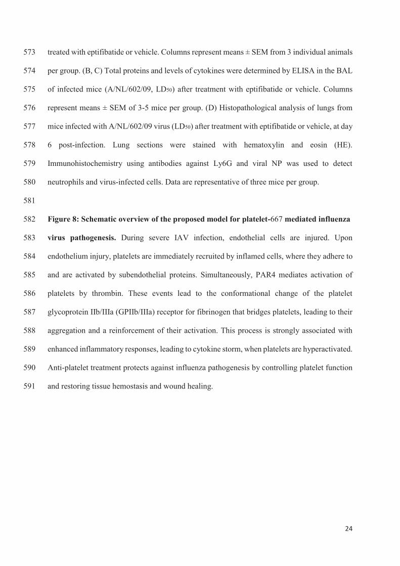
24
treated with eptifibatide or vehicle. Columns represent means ± SEM from 3 individual animals 573
per group. (B, C) Total proteins and levels of cytokines were determined by ELISA in the BAL 574
of infected mice (A/NL/602/09, LD50) after treatment with eptifibatide or vehicle. Columns 575
represent means ± SEM of 3-5 mice per group. (D) Histopathological analysis of lungs from 576
mice infected with A/NL/602/09 virus (LD50) after treatment with eptifibatide or vehicle, at day 577
6 post-infection. Lung sections were stained with hematoxylin and eosin (HE). 578
Immunohistochemistry using antibodies against Ly6G and viral NP was used to detect 579
neutrophils and virus-infected cells. Data are representative of three mice per group. 580
581
Figure 8: Schematic overview of the proposed model for platelet-667 mediated influenza 582
virus pathogenesis. During severe IAV infection, endothelial cells are injured. Upon 583
endothelium injury, platelets are immediately recruited by inflamed cells, where they adhere to 584
and are activated by subendothelial proteins. Simultaneously, PAR4 mediates activation of 585
platelets by thrombin. These events lead to the conformational change of the platelet 586
glycoprotein IIb/IIIa (GPIIb/IIIa) receptor for fibrinogen that bridges platelets, leading to their 587
aggregation and a reinforcement of their activation. This process is strongly associated with 588
enhanced inflammatory responses, leading to cytokine storm, when platelets are hyperactivated. 589
Anti-platelet treatment protects against influenza pathogenesis by controlling platelet function 590
and restoring tissue hemostasis and wound healing. 591

A B
N I L D 5 0
0
1 5
3 0
4 5
6 0
Pla
tele
tn
um
be
r(x
10
9/L
)
*
Figure 1
C
DNI LD50
SublethalNI
NP
CD41
LD50
CD41 HADAPI Merge
NI
LD50
LD50
Plat
elet
Plat
elet
Con
trol
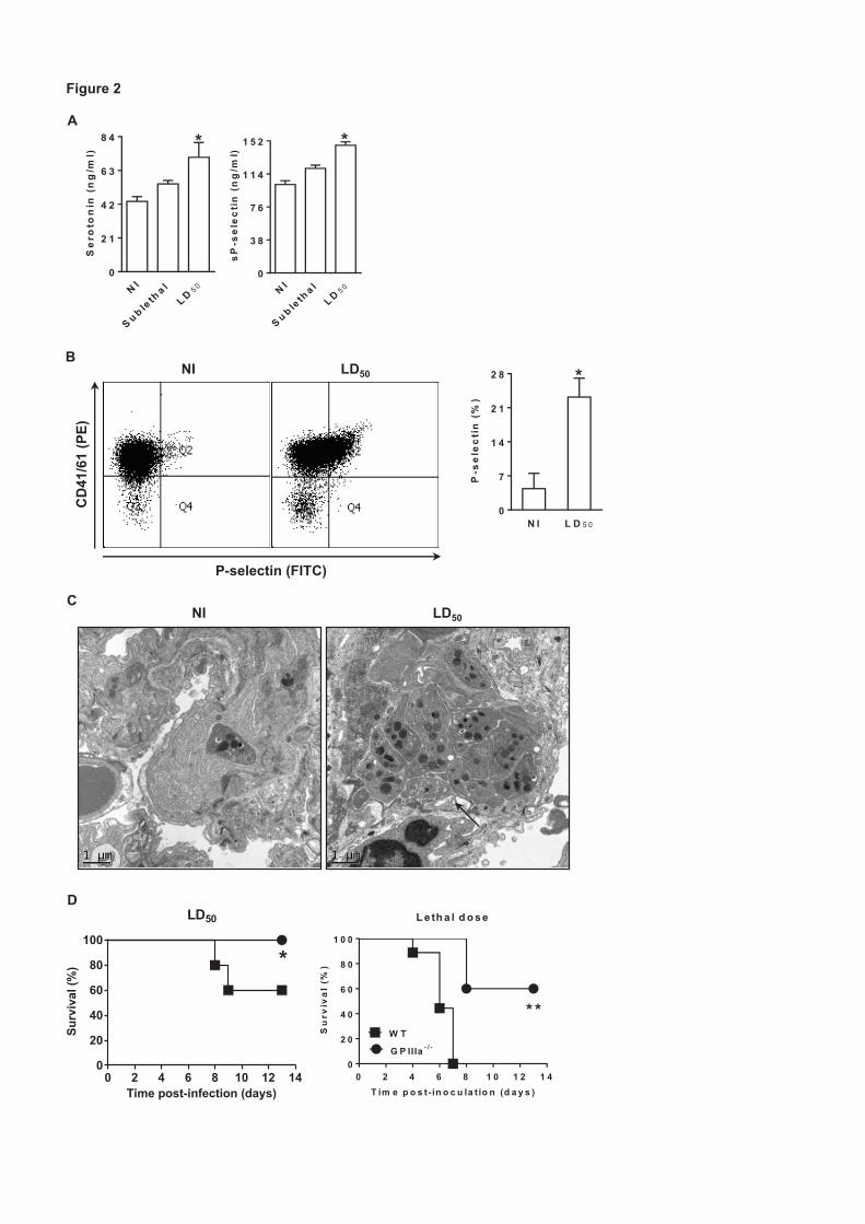
Figure 2
A
B
N I
S u b leth
a lL D 5 0
0
2 1
4 2
6 3
8 4
Se
roto
nin
(ng
/ml)
*
D
C
CD
41/6
1 (P
E)
P-selectin (FITC)
NI LD50
N I L D 5 0
0
7
1 4
2 1
2 8
P-s
ele
cti
n(%
)
*
N I
S u b leth
a lL D 5 0
0
3 8
7 6
1 1 4
1 5 2
sP
-se
lec
tin
(ng
/ml)
*
*
NI LD50
L e th a l d o s e
0 2 4 6 8 1 0 1 2 1 40
2 0
4 0
6 0
8 0
1 0 0
* *W T
G P IIIa - / -
T im e p o s t- in o c u la t io n (d a y s )
Su
rviv
al
(%)
LD50
Time post-infection (days)
Surv
ival
(%)
0 2 4 6 8 10 12 140
20
40
60
80
100
*

Figure 3
A
0
2 3
4 6
6 9
9 2
Se
roto
nin
(ng
/ml)
* *
0
7 0
1 4 0
2 1 0
2 8 0
sP
-se
lec
tin
(ng
/ml)
*
C o n tro lP A R 4 -A P
BL D 5 0
0 2 4 6 8 1 0 1 2 1 40
2 0
4 0
6 0
8 0
1 0 0
*T im e p o s t-in o c u la tio n (d a y s )
Su
rviv
al(%
)N I
0 2 4 6 8 1 0 1 2 1 40
2 0
4 0
6 0
8 0
1 0 0
T im e p o s t-in o c u la tio n (d a y s )
Su
rviv
al(%
)
C o n tro lP A R 4 -A P
S u b le th a l
0 2 4 6 8 1 0 1 2 1 40
2 0
4 0
6 0
8 0
1 0 0
* *
T im e p o s t-in o c u la tio n (d a y s )
Su
rviv
al(%
)
CC o n tro l
0 2 4 6 8 1 0 1 2 1 40
2 0
4 0
6 0
8 0
1 0 0
*
T im e p o s t-in o c u la tio n (d a y s )
Su
rviv
al(%
)
E p t if ib a tid e
0 2 4 6 8 1 0 1 2 1 40
2 0
4 0
6 0
8 0
1 0 0
C o n tro lP A R 4 -A P
T im e p o s t-in o c u la tio n (d a y s )
Su
rviv
al(%
)
3 61 0 0
1 0 1
1 0 2
1 0 3
1 0 4
1 0 5
T im e p o s t- in o c u la t io n (d a y s )
Vir
al
tite
rs(l
og
10
PF
U/m
l)
C o n tro lP A R 4 -A P
E F
N I 3 60
2 7 0
5 4 0
8 1 0
1 0 8 0 * *
T im e p o s t- in o c u la t io n (d a y s )
To
tal
pro
tein
(μg
/ml) C o n tro l
P A R 4 -A P
DG P IIIa -/-
T im e p o s t-in fe c tio n (d a y s )
Su
rviv
al(%
)
0 2 4 6 8 1 0 1 2 1 40
2 0
4 0
6 0
8 0
1 0 0
P A R 4 -A PC o n tro l
W T
T im e p o s t-in fe c tio n (d a y s )
Su
rviv
al(%
)
0 2 4 6 8 1 0 1 2 1 40
2 0
4 0
6 0
8 0
1 0 0
* * *

Figure 4
A
N I 3 60
1 9 5
3 9 0
5 8 5
7 8 0 * *
T im e p o s t- in o c u la t io n (d a y s )
IL-6
(pg
/ml)
N I 3 60
8 0
1 6 0
2 4 0
3 2 0
* *
T im e p o s t- in o c u la t io n (d a y s )
MIP
-2(p
g/m
l)
N I 3 60
7 0
1 4 0
2 1 0
2 8 0
T im e p o s t- in o c u la t io n (d a y s )
IL-1
(pg
/ml)
*
N I 3 60
9 0
1 8 0
2 7 0
3 6 0
T im e p o s t- in o c u la t io n (d a y s )
RA
NT
ES
(pg
/ml)
N I 3 60
4 0
8 0
1 2 0
1 6 0
T im e p o s t- in o c u la t io n (d a y s )
KC
(pg
/ml)
P A R 4 -A PC o n tro l
N I 3 60
1 5 5
3 1 0
4 6 5
6 2 0
T im e p o s t- in o c u la t io n (d a y s )
IFN
-(p
g/m
l)
B
Con
trol
HE
NI
NPLy6G
PAR
4-A
P
Sublethal
Con
trol
Sublethal

Figure 5
A
C
A /P R /8 /3 4
0 2 4 6 8 1 0 1 2 1 40
2 0
4 0
6 0
8 0
1 0 0
*
T im e p o s t- in o c u la t io n (d a y s )
Su
rviv
al
(%)
3 61 0 0
1 0 2
1 0 4
1 0 6
1 0 8C o n tro lP e p d u c in
T im e p o s t- in o c u la t io n (d a y s )
Vir
al
tite
rs(l
og
10
PF
U/m
l)
A /H K /1 /6 8
0 2 4 6 8 1 0 1 2 1 40
2 0
4 0
6 0
8 0
1 0 0
*
T im e p o s t- in o c u la t io n (d a y s )
Su
rviv
al
(%)
C o n tro l
P e p d u c in
F
0
3 0 0
6 0 0
9 0 0
1 2 0 0
*
IL-6
(pg
/ml)
0
1 0 0
2 0 0
3 0 0
4 0 0
*
IL-1
(pg
/ml)
0
3 2 5
6 5 0
9 7 5
1 3 0 0
*
MIP
-2(p
g/m
l)
C o n tro lP e p d u c in
G
D E
0
4 5 0
9 0 0
1 3 5 0
1 8 0 0
* *
To
tal
pro
tein
(μg
/ml) C o n tro l
P e p d u c in
0
3
6
9
1 2
* *N
eutr
op
hils
(x10
4/m
l) C o n tro lP e p d u c in
0
4
8
1 2
1 6
* *
Leu
kocy
tes
(x10
4/m
l)
0
1 1 0
2 2 0
3 3 0
4 4 0
TX
B2
(pg
/ml)
*
C o n tro lP e p d u c in
B
Control
Pepducin
HE NPLy6G

Figure 6
A
CA /H K /1 /6 8
0 2 4 6 8 1 0 1 2 1 40
2 0
4 0
6 0
8 0
1 0 0
* *
T im e p o s t- in o c u la t io n (d a y s )
Su
rviv
al
(%)
A /N L /6 0 2 /0 9
0 2 4 6 8 1 0 1 2 1 40
2 0
4 0
6 0
8 0
1 0 0
*
T im e p o s t- in o c u la t io n (d a y s )
Su
rviv
al
(%)
A /P R /8 /3 4
0 2 4 6 8 1 0 1 2 1 40
2 0
4 0
6 0
8 0
1 0 0
T im e p o s t- in o c u la t io n (d a y s )
Su
rviv
al
(%) * *
V e h ic le
E p tif ib a tid e
B
2 60
1 0 5
2 1 0
3 1 5
4 2 0
*
T im e p o s t- in o c u la t io n (d a y s )
TX
B2
(pg
/ml)
C o n tro lE p tif ib a tid e
Control Eptifibatide
A /F P V /B ra tis la v a /7 9
T im e p o s t- in fe c t io n (d a y s )
Su
rviv
al
(%)
0 2 4 6 8 1 0 1 2 1 40
2 0
4 0
6 0
8 0
1 0 0

Figure 7
D
C
A
2 60
2
4
6
8
T im e p o s t- in o c u la t io n (d a y s )
Vir
al
tite
rs(l
og
10
PF
U/m
l)
C o n tro lE p tif ib a tid e
2 60
1 9 5
3 9 0
5 8 5
7 8 0
*
T im e p o s t- in o c u la t io n (d a y s )
IL-6
(pg
/ml)
2 60
1 1 5
2 3 0
3 4 5
4 6 0
*
T im e p o s t- in o c u la t io n (d a y s )
IL-1
(pg
/ml)
2 60
1 2 0
2 4 0
3 6 0
4 8 0
* *
T im e p o s t- in o c u la t io n (d a y s )M
IP-2
(pg
/ml)
C o n tro lE p tif ib a tid e
B
2 60
1 9 5
3 9 0
5 8 5
7 8 0
*
C o n tro lE p tif ib a tid e
T im e p o s t- in o c u la t io n (d a y s )
To
tal
pro
tein
(μg
/ml)
Ly6G NP
Control
Eptifibatide
HE

Platelet hyper-activation Cytokine storm
Controlled platelet functionWound healing
Endothelial cell activation
Influenza virus
Endothelium
Epithelium
Blood vessel
Protection
Lethal
Anti-platelet treatment
Immune cell
Cytokine
Resting platelet
Activated platelet
Figure 8


Ex Vivo and In Vivo Inhibition of Human Rhinovirus Replicationby a New Pseudosubstrate of Viral 2A Protease
Nisrine Falah,a Sébastien Violot,b Didier Décimo,c Fatma Berri,a Marie-Laure Foucault-Grunenwald,a Théophile Ohlmann,c
Isabelle Schuffenecker,d Florence Morfin,d Bruno Lina,a,d Béatrice Riteau,a,e and Jean-Claude Cortaya
VirPath, EMR 4610, Virologie et Pathologie Humaine, Université Lyon 1, Université de Lyon, Faculté de Médecine Lyon-Est, Secteur Laennec, Lyon, Francea;
Biocristallographie et Biologie Structurale des Cibles Thérapeutiques, Université Lyon 1, Université de Lyon, Lyon, France, and CNRS, UMR 5086, Bases Moléculaires et
Structurales des Systèmes Infectieux, Vercors, Franceb; Ecole Normale Supérieure de Lyon, Unité de Virologie Humaine, INSERM U758, Université Lyon 1, Université de
Lyon, Lyon, Francec; Laboratoire de Virologie, Hospices Civils de Lyon, Lyon, Franced; and INRA, Tours, Francee
Human rhinoviruses (HRVs) remain a significant public health problem as they are the major cause of both upper and lowerrespiratory tract infections. Unfortunately, to date no vaccine or antiviral against these pathogens is available. Here, using ahigh-throughput yeast two-hybrid screening, we identified a 6-amino-acid hit peptide, LVLQTM, which acted as a pseudosub-strate of the viral 2A cysteine protease (2Apro) and inhibited its activity. This peptide was chemically modified with a reactiveelectrophilic fluoromethylketone group to form a covalent linkage with the nucleophilic active-site thiol of the enzyme. Ex vivoand in vivo experiments showed that thus converted, LVLQTM was a strong inhibitor of HRV replication in both A549 cells andmice. To our knowledge, this is the first report validating a compound against HRV infection in a mouse model.
Human rhinoviruses (HRVs) belong to the enterovirus groupof the Picornaviridae family and are themain causative agents
of the common cold, asthma exacerbations, and chronic obstruc-tive pulmonary disease in humans (22). To date, there is no vac-cine against HRV as there is almost no cross protection betweenthe nearly 100 serotypes identified so far (21). Furthermore, noantiviral treatment against HRV is currently available on themar-ket. Thus, there is an urgent need for validation of new com-pounds against HRV.
Among several antiviral strategies attempting to impair rhino-virus replication, one consists of blocking the activity of the viralHRV 2A protease (2Apro). Targeting 2Apro is of particular interestas it is a cysteine protease playing multifunctional roles necessaryfor viral replication. These roles include (i) autoprocessing by ciscleavage at the VP1-2Apro junction; (ii) inhibition of the host celltranslation through cleavage of the initiation factor eIF4G (17, 27)and the poly(A)-binding protein (PABP) (10); (iii) contributionto the deleterious overwhelming host cellular defense (3, 9, 23);and (iv) strengthening of viral polysome formation and stability(11, 12).
Several 2Apro inhibitors have already been described andinclude alkylating agents such as iodoacetamide or N-ethylmaleimide that can react with the catalytic cysteine ofthe enzyme and that have been shown to reduce 2Apro activity(15). Moreover, as the substrate-binding pocket of elastase is sim-ilar to that of 2Apro, two substrate-derived elastase inhibitors, elas-tinal and methoxysuccinyl-Ala-Ala-Pro-Val-chloromethylketonehave been reported to inhibit the in vitro proteolytic activity of2Apro and consequently reduce viral yields of HRV type 14(HRV-14) and poliovirus type 1 (PV-1) (18). Furthermore, it hasbeen demonstrated that the irreversible caspase inhibitorbenzyloxycarbonyl-Val-Ala-Asp(methoxy)-fluoromethylketone(z-VAD-fmk) (7) is also able to directly inactivate HRV and cox-sackie B virus type 4 (CBV4) 2Apro enzymes (5). However, none ofthese compounds has ever been tested in vivo, thus impairing thevalidation of their efficacy in preclinical assays and the possibilityto go further in the development of anti-HRV therapies.
The aim of this study was to design a peptide inhibitor ofHRV-2 2Apro and to test its antiviral activity ex vivo in A549 cellsand in vivo inmice which are susceptible to infection by theminorHRV group member HRV-2.
Here, we report the identification of the LVLQTMpeptide as adecoy substrate for 2Apro that blocked enzyme activity upon bind-ing and consequently affected HRV-2 replication ex vivo and invivo when administered to infected mice. This is the first studyvalidating such a compound in vivo in mice, opening new pros-pects for testing other drugs.
MATERIALS AND METHODS
Ethics statement. Experiments were performed according to recommen-dations of the National Commission of Animal Experiment (CNEA) andthe National Committee on the Ethic Reflexion of Animal Experiments(CNREEA). The protocol was approved by the Committee of AnimalExperiments of the University Claude Bernard Lyon I (permit numberBH2008-13). All animal experiments were also carried out under the au-thority of a license issued by laDirection des ServicesVétérinaires (accred-itation number 78-114). All efforts were made to minimize suffering.
Yeast two-hybrid analysis. Yeast two-hybrid screening was per-formed by Hybrigenics, S.A., Paris, France. The full-length coding se-quence for HRV-2 2Apro (GenBank accession number X02316) was am-plified by PCR and cloned into pB27 plasmid as a C-terminal fusion toLexA (LexA-p2A). The construct was validated by sequencing and used asbait to screen a random-primed human placenta cDNA library con-structed into pP6 plasmid. pB27 and pP6 plasmids were derived from theoriginal pBTM116 and pGADGH plasmids, respectively (34).
A total of 53.1 million clones (6-fold the complexity of the library)were screened using a mating approach with Y187 (mat�) and L40Gal4
Received 31 May 2011 Accepted 21 October 2011
Published ahead of print 9 November 2011
Address correspondence to Jean-Claude Cortay, [email protected].
Copyright © 2012, American Society for Microbiology. All Rights Reserved.
doi:10.1128/JVI.05263-11
0022-538X/12/$12.00 Journal of Virology p. 691–704 jvi.asm.org 691
on J
une 1
, 2013 b
y IN
IST
-CN
RS
Bib
lioV
iehttp
://jvi.a
sm
.org
/D
ow
nlo
aded fro
m

(mata) Saccharomyces cerevisiae yeast strains, as previously described (6).Fifty-oneHis-positive (His) colonies were selected on amedium lackingtryptophan, leucine, and histidine and supplemented with 100 mM3-aminotriazole to suppress bait autoactivation. The prey fragments ofthe positive clones were then amplified by PCR and sequenced at their 5=
and 3= junctions. The resulting sequences were used to identify the corre-sponding interacting proteins in the GenBank database (NCBI) using afully automated procedure.
Bacterial expression vectors and protein purification. Syntheticgenes (Eurofins MWG Operon) coding for HRV-2 2Apro or echovirus 6(EV-6) 2Apro (GenBank accession number AY302558) were cloned be-tween the NdeI and XhoI sites of the pSCodon1.2 vector (Eurogentec) infusion with either a Strep●Tag (WSHPQFEK) or a (His)6 tag at their Ctermini. Recombinant plasmids were used to transform the Escherichiacoli B SE1 strain [F� Cmr ompT lon hsdSB (rB� mB
�) gal dcm (DE3) (lacI,T7 polymerase under the control of the PlacUV5 promoter) ccdB]. Bac-teria were grown in the autoinductionmediumZYP-5052 (30) at 37°C for5 to 6 h with vigorous shaking in baffled flasks, before growing to satura-tion at 20°C within 16 to 18 h. Subsequent purification steps were per-formed at 4°C. Cells were lysed with BugBuster protein extraction reagent(Novagen), and clarified supernatants were applied to the correspondingaffinity chromatography resins. Strep●Tag proteins were purified using aStreptrap HP resin (GE Healthcare) and His-tagged proteins were puri-fied using aHIS-SelectHF nickel resin (Sigma) according to the respectivemanufacturers’ instructions. In each case, proteins were dialyzed againstbuffer D (100 mM Tris-HCl, pH 7.5, 200 mM NaCl, 4 mM dithiothreitol[DTT]) and concentrated using a Vivaspin centrifugal concentrator de-vice (Sartorius Stedim Biotech). Enzymes were stored at �20°C in bufferD containing 50% glycerol.
In vitro cleavage assays. Different protease recognition site-codingsequences were inserted between the NheI and BglII sites in a short poly-peptide linker that connects the native N and C termini of a circularlypermuted firefly luciferase in the pGloSensor-10F linear vector (Pro-mega). The resulting plasmids were then used as templates in a cell-freesystem for the expression of the corresponding GloSensor proteins con-taining the protease sites of interest. In vitro transcription/translation re-actions were carried out in a TNT SP6 high-yield wheat germ master mix(Promega) supplemented with [35S]methionine according to the manu-facturer’s protocol. Reaction mixtures were incubated for 2 h at 25°C.Two micrograms of recombinant tobacco etch virus (TEV) protease,HRV-2 2Apro, or EV-6 2Apro was added to 13 �l of the in vitro translationreactionmixture and 13 �l of 2� digestion buffer (100mMTris-HCl, pH8, 1 mM EDTA, 4 mMDTT [for TEV protease], 100 mMHEPES-NaOH,pH 7.9, 200mMNaCl, 2mMEDTA, 10mMDTT [forHRV-2 2Apro], 100mMTris-HCl, pH 7.5, 300mMNaCl, and 10mMDTT [for EV-6 2Apro])and incubated for 45min at 30°C. Aliquots of total proteins were removed0, 15, 30, and 45 min postincubation and separated in a 12% SDS-polyacrylamide gel. Autoradiography was performed after fluorographytreatment. The 61-kDa band intensity was determined by densitometricanalysis after background subtraction using Bio-Rad Quantity One one-dimension software. Luminescence detection was performed by dilutingthe remaining volume of each protease digestion and negative-controlreactionmixture 1:20 in nuclease-free water, and 100 �l of these dilutionswas added to each well of a white, flat-bottom 96-well plate. Each reactionwas analyzed in triplicate. After addition of 100 �l Bright-Glo assay re-agent to each well and incubation for 2 to 5 min at room temperature,luminescence was measured using a GloMax 96-well microplate lumi-nometer. According to the manufacturer’s instructions, the fold activa-tion of each luciferase activity was calculated as follows: [(luminescencefrom tube A) � (luminescence from tube C)]/[(luminescence from tubeB) � (luminescence from tube D)], where tube A contains a Plus-DNATNT reaction mixture and HRV-2 2Apro, tube B contains a Plus-DNATNT reaction mixture only, tube C contains a no-DNA TNT reactionmixture and HRV-2 2Apro, and tube D contains a no-DNA TNT reactionmixture only.
Pulldown experiments. For His pulldown assays, a PCR-amplifiedfragment corresponding to truncated proteins consisting of theC-terminal part (amino acids 274 to 520) of the functionally uncharacter-ized RBM66 protein (RBM66274-520) or RBM6274-660 was inserted intothe NcoI-XhoI sites of the expression vector pET-28 (Novagen). Trans-lated proteins were synthesized in vitro using a T7 RNApolymerase-basedTNT-coupled reticulocyte lysate system (Promega). HRV-2 2Apro–(His)6fusion protein was bound to nickel nitrilotriacetic acid (Ni-NTA) mag-netic agarose beads (Qiagen) and incubated for 1 h with 50 �l in vitro-translated [35S]methionine-labeled RBM66274-520 or RBM6274-660 in atotal volume of 1 ml of incubation buffer (25 mM sodium phosphate, pH8.0, 500 mM NaCl, 20 mM imidazole, and 0.005% Tween 20). Resin wascollected with a magnetic separator and washed twice with 500 �l incu-bation buffer.Washed beads were resuspended in 40�l of 2� SDS samplebuffer, heated for 5 min, and pelleted in a microcentrifuge. Proteins fromthe supernatant were then subjected to a 12% SDS-PAGE. Gels weretreated with Amplify reagent (GE Healthcare) for fluorography or sub-jected to Western blot analysis using a polyclonal antihistidine antibody(Cell Signaling). The amount of labeled proteins which coeluted withHRV-2 2Apro-(His)6 was quantified by densitometric analysis.
For Strep●Tag pulldown assays, the RBM66 LVLQTM-derivedpeptide-coding sequence was cloned in frame between two BsaI siteswithin the pET-SUMO vector (Invitrogen), allowing its expression infusion with the C terminus of the SUMO protein. In addition, theStrep●Tag sequence WSHPQFEK was added at the N terminus of thefusion protein. This construct was transferred into the pSCodon1 vector,and recombinant Strep●Tag-SUMO-LVLQTM was expressed in the E.coli SE1 strain. Subsequent incubation reactions were performed underthe same conditions described above, except that equal volumes of clearedlysates prepared from bacteria overproducing either Strep●Tag-SUMO-LVLQTM or HRV-2 2Apro–(His)6 proteins were mixed (to a 1-ml finalvolume)with Strep-Tactinmagnetic beads (Qiagen). Proteins whichwerespecifically bound to the washed beads were separated by 15% SDS-PAGEand visualized by staining with Coomassie brilliant blue R250. (His)6-tagged proteins were also revealed with a polyclonal antihistidine anti-body (Cell Signaling).
Cleavage of TRPIITTA–pNA substrate by HRV-2 2Apro. Cleavage ofthe TRPIITTA–p-nitroanilide (pNA) substrate by HRV-2 2Apro was per-formed at 25°C for 10 min in a 1-ml reaction mix containing 50 mMHEPES-NaOH, pH 8.0, 100 mM NaCl, 1 mM EDTA, 10 mM DTT, andpurified recombinantHRV-2 2Apro (0.2 �M). The reaction was started bythe addition of the TRPIITTA-pNA peptide (Eurogentec) at 25 �M andmonitored continuously at 405 nm to characterize the initial velocity ofthe cleavage reaction. Peptide competition cleavage assays were per-formed under the same conditions, except that purified Strep●Tag-SUMO-LVLQTM protein (0 to 25 �M) was added to the reaction mix.Percent inhibition values were referred to as the ratio between initial ve-locity cleavage values measured with and without inhibitor. Data are ex-pressed as means of three independent experiments, and standard devia-tions are indicated.
Molecular modeling. The crystal structure of the free HRV-2 2Apro hasbeen used (ProteinData Bank [PDB] accession number 2hrv) (24). The con-formation of the LVLQTMpeptide wasmodeled using the one in complexwith the foot-and-mouth disease virus (FMDV) 3Cpro as a starting model(PDB accession number 2wv4) (35). Side chains from the FMDV 3Cprotease-bound peptide were replaced with the side chains of VLQTMand then energy minimized using a GROMOS96 43B1 force field (32).
Cell culture and transient expression. Human epithelial lung carci-noma A549 cells (CCL-185; ATCC) were grown in Dulbecco’s modifiedEagle’smedium (DMEM; Lonza) and 1 g/liter glucose supplementedwith10% fetal calf serum, 2 mM L-glutamine, penicillin, and streptomycin.RBM66274-520- andRBM66274-514-coding sequenceswere amplified byPCR and cloned into themultiple-cloning site of the pCI-neo vector (Pro-mega) downstream from the following 2� Strep●Tag sequence: MASWSHPQFEKGGGSGGGSGGGSWSHPQFEK (where the underlining indi-
Falah et al.
692 jvi.asm.org Journal of Virology
on J
une 1
, 2013 b
y IN
IST
-CN
RS
Bib
lioV
iehttp
://jvi.a
sm
.org
/D
ow
nlo
aded fro
m

cates the Strep●Tag sequence). All plasmid constructs were transfectedinto cells using NanoJuice transfection reagents (Novagen), according tothe manufacturer’s instructions.
Western blotting. Proteins were separated by SDS-PAGE and trans-ferred onto nitrocellulose membranes. The membranes were incubatedfor 1 h in Tris-buffered saline containing 0.1% Tween 20 and 5% nonfatmilk powder at room temperature. Membranes were then incubatedovernight at 4°C with a polyclonal antihistidine antibody (reference no.2365; Cell Signaling) or an antiserum recognizing the C-terminal domainof eIF4G (19). These antibodies were revealed using the horseradishperoxidase-coupled goat antirabbit antibody, followed by chemilumines-cence detection using the SuperSignal West Pico chemiluminescent sub-strate from Pierce.
In vitro transcription and RNA transfection. Plasmids containingthe 2A protease-coding region were constructed by inserting the respec-tive coding sequences into the pGlobin-Renilla vector (29). For in vitrotranscription, DNA templates were linearized at the EcoRI site down-stream froma synthetic poly(A) tail. CappedRNAswere transcribed usingthe T7 RNA polymerase as previously described (25) and treated withRQ1 DNase (Promega). The integrity of the RNAs was checked by elec-trophoresis on nondenaturing agarose gels, and the concentration wasquantified by spectrophotometry at 260 nm using a Nanodrop apparatus(Nanodrop Technologies).
Two days before RNA transfection, A549 cells were seeded into 48-well plates at 75,000 cells per well to reach about 180,000 cells at the timeof transfection. RNA transfection was performed with 100 ng of 2Apro-coding RNA and the TransIT kit (Mirus Bio) for 2 h and 50 ng of Renilla-coding RNA for additional 3 h. The cells were then harvested, and lu-ciferase activity was quantified using the Renilla luciferase (R-Luc)assay system from Promega and a Veritas microplate luminometer(Turner BioSystems). Transfection efficiency was evaluated by trans-fecting the green fluorescent protein (GFP)-coding RNA under thesame conditions and counting the number of green fluorescent cells byfluorescence-activated cell sorter (FACS) analysis. Over 70% of A549cells expressed GFP.
Virus infection. HRV-2 (GenBank accession number X02316) andHRV-14 (GenBank accession number K02121) were provided by theWHO/National Reference Centre for Enteroviruses (Lyon, France). A549cells (90% confluence) were infected with HRV-2 or HRV-14 at a multi-plicity of infection (MOI) of 1 in DMEM containing 2% fetal calf serum,2 mM L-glutamine, penicillin, and streptomycin and incubated at 34°C.Virus titer was quantified by the 50% tissue culture infectious dose(TCID50) assay using MRC5 cells according to the method of Reed andMuench (26).
Infection and mouse treatment. Six-week-old BALB/c female micewere purchased from Charles River Laboratories, and experiments wereundertaken as previously described (13). On the day of infection, a three-step protocol was used for peptide administration: (i) first, mice wereanesthetized by intraperitoneal injection of ketamine (42.5mg/kg of bodyweight), (ii) then, a 25-�l volume of HRV-2 suspension containing100,000 PFUwas injected dropwise to the external nares of themice usinga micropipette, and (iii) finally, a 25-�l volume of the indicated concen-tration of peptide solution dissolved in 1% dimethyl sulfoxide (DMSO)was administered in the same way either right after infection or at 12 hpostinoculation. Lungs were harvested at different time points postinfec-tion and ground in the Tissue Lyser LT device fromQiagen. After centrif-ugation at 12,000 � g for 5 min at 4°C, supernatants were collected andvirus titers were determined as described above. Ten mice were used foreach experimental condition.
Statistical analysis. The Mann-Whitney U test was used to evaluatestatistical significance (P � 0.05) of viral replication in vivo.
RESULTS
Yeast two-hybrid screening for proteins interacting with HRV-22Apro. In order to identify partners and potential inhibitors of
HRV 2Apro, a plasmid expressing a LexA–HRV-2 2Apro fusionprotein was used to screen a human placenta cDNA library usingY187 (mat�) and L40�Gal4 (mata) yeast strains. Fifty millionclones were screened, and 51 His colonies were further isolatedand characterized by DNA sequencing and sequence alignmentanalysis. Among these clones, nine encoded out-of-frame shortpolypeptides and one encoded the C-terminal part (amino acids274 to 520) of the functionally uncharacterized RBM66 protein(GenBank accession number FLJ56542) (31). As depicted in Fig.1, all exhibited related sequences that shared the same peptideconsensus motif LXLX(T/N)�, where X represents any aminoacid and � represents a hydrophobic residue. Interestingly, thissequence partially mimics the consensus sequence P4LX(T/N)XP1
found in 2Apro substrates where threonine (or asparagine) andleucine are required in positions P2 and P4, respectively, for 2Apro
cleavage (16). In addition, the tripeptide motifs P3QTMP1 and
P4LQTP2 found in the RBM66 sequence are identical to thosefound at the corresponding positions within the 2Apro substratesPABP1 and VP1-2Apro junction of the HRV-62 polyprotein, re-spectively (16). Finally, the presence of methionine at the P1 po-sition represents a favorable determinant for 2Apro binding (28).Remarkably, the presence of a conserved leucine (or an equivalenthydrophobic residue) at the P6 position in all selected peptidessuggests that this amino acid may represent an important param-eter in the specificity of recognition by 2Apro. Thus, these resultsstrongly suggested that the LXLX(T/N)� motif peptides isolatedin the double-hybrid screening are partners of HRV-2 2Apro.
The LXLX(T/N)� motif behaves as a pseudosubstrate ofHRV-2 2Apro. To investigate whether the previously characterizedpeptides were pseudosubstrates of HRV-2 2Apro, different hybridpeptides were built by fusing the six terminal amino acid residuesof the 10 polypeptides identified by yeast two-hybrid screeningwith the GPSDM sequence found at the P1= and P5= positions ofthe authentic cis-cleavage site of the HRV-2 polyprotein (Fig. 1).The 11-mer peptides obtained were then inserted in frame into agenetically engineered firefly luciferase which was synthesized in acell-free protein expression system in the presence of [35S]methio-nine and used as a protease substrate (Fig. 2A). In this assay, cleav-
FIG 1 Amino acid sequences of the HRV-2 2Apro-binding peptides identifiedby the yeast two-hybrid system. Peptides are arranged to illustrate the consen-sus sequence found, where X is any amino acid and � is a hydrophobic aminoacid. Alignments of the carboxy termini of 2Apro-binding peptides with natu-ral cleavage sites of 2Apro are also shown.
Inhibition of Human Rhinovirus Replication in Mice
January 2012 Volume 86 Number 2 jvi.asm.org 693
on J
une 1
, 2013 b
y IN
IST
-CN
RS
Bib
lioV
iehttp
://jvi.a
sm
.org
/D
ow
nlo
aded fro
m

FIG 2 HRV-2 2Apro-binding peptides identified by the yeast two-hybrid system are pseudosubstrates of the protease. (A) 35S-labeled GloSensor protease siteluciferase activation by HRV-2 2Apro digestion of hybrid sites generated from sequences depicted in Fig. 1. To generate the GloSensor protein, new N and Ctermini were created at amino acids 234 and 233, respectively. The protein-coding region of this circularly permuted firefly luciferase was carried on thepGloSensor-10F linear vector. Insertion of a protease recognition sequence between these native N and C termini and cleavage of the sequence by the cognateprotease activate the luciferase enzyme. Plasmid DNAs encoding the protease recognition sequences indicated on the graph were transcribed and translated invitro and incubatedwith purifiedHRV-2 2Apro or TEVprotease for 45min. Luminescent signal wasmeasured bymixing an aliquot of each TNT reactionmixturewith Bright-Glo assay reagent in triplicate and incubating for 5 min at room temperature. Luminescence was measured using a luminometer. A plasmid DNAencoding the 35S-labeled GloSensor ENLYFQ-S protein, where ENLYFQ-S is a cleavage site of the TEV protease, was used as a control. RLU, relative light units.(B) The different 35S-labeled GloSensor proteins containing the protease sites to be tested were synthesized in vitro as described above and then incubated with2 �g of TEV protease or HRV-2 2Apro for 45min, followed by SDS-PAGE and fluorography. Gel patterns corresponding to enzymatic digestion performed withTEVprotease (a) andHRV-2 2Apro (b to l) are shown. The 0-h timepoint shows proteins thatwere harvested right after addition of the proteases in the incubationmixtures.
Falah et al.
694 jvi.asm.org Journal of Virology
on J
une 1
, 2013 b
y IN
IST
-CN
RS
Bib
lioV
iehttp
://jvi.a
sm
.org
/D
ow
nlo
aded fro
m

age of the recombinant luciferase at the protease recognition se-quence led to the activation of the luciferase enzyme, resulting inan increase in luminescence when a firefly luciferase substrate wasadded to the reaction mixture. Thus, if a peptide acted as a pseu-dosubstrate for 2Apro, an increase in luciferase activity would beobserved. As shown in Fig. 2A, significant luciferase activity wasdetected for all constructs tested, and the best score was measuredfor the 35S-labeled GloSensor LVLQTM-GPSDM protein, theprotease site of which was derived from the C terminus of theRBM66 protein. Luminescence was hardly detectable when us-ing the negative control, 35S-labeled GloSensor-10F ENLYFQ-Sprotein, which was recognized by the tobacco etch virus proteasebut not by HRV-2 2Apro. In contrast and as expected, the 35S-labeledGloSensor-10FENLYFQ-S proteinwas cleaved by the TEVprotease, resulting in increased luminescence. Thus, our resultsconfirmed that all peptides isolated by yeast two-hybrid screeningbehaved as potent substrate analogues of HRV-2 2Apro.
Cleavage of the 35S-labeled GloSensor-10F-identified peptide-GPSDM luciferases (61 kDa) by HRV-2 2Apro was also visualizedafter separation of digested products by SDS-PAGE and autora-diography (Fig. 2B, panels c to l). Total inactivation of the pro-teases was effective only by boiling the sample for 5 min in SDSloading buffer, which may explain the partial substrate degrada-tion at the initial time in panels c and d and reflected a higheraffinity of HRV-2 2Apro for LVLQTM-GPSDM and LCLHTC-GPSDM sequences than for the other substrates. Moreover, all35S-labeledGloSensor-10F-identified peptide-GPSDM luciferaseswere hydrolyzed byHRV-2 2Apro into two predictive fragments of36 and 25 kDa, confirming that all identified peptides were sub-strate analogues of 2Apro. As expected, the 35S-labeled GloSensor-10F ENLYFQ-S protein was cleaved by the TEV protease but notby the 2A protease of HRV-2 (Fig. 2B, compare panels a and b). Inaddition, the kinetics of digestion were peptide dependent, withthe highest rate being for the LVLQTM peptide. Thus, these re-sults confirmed that the identified peptides acted as pseudosub-strates for HRV-2 2Apro.
The LVLQTM peptide specifically interacts with HRV-22Apro. Since the LVLQTM peptide displayed the highest affinityfor the 2A protease, the interaction between these two binding
FIG 3 The last six residues of RBM66 are necessary and sufficient for theinteractionwithHRV-2 2Apro. (A) Schematic representation of the exon struc-ture of the RBM6 gene and the protein products derived from two splicevariants (data are from references 14 and 31). Boxes represent exons and arenot drawn to scale. The bars below RBM6 indicate different protein motifs, asfollows: A and B, RNA binding motif RNP1 and RNP2; C, G patch. In thetruncated RBM66 protein, the amino acid residues represented by the whitepart differ from those in the longer protein product RBM6 due to a frame shiftcaused by the splicing. (B) Results of a His pulldown assay with HRV-2 2Apro
and RBM66. HRV-2 2Apro–(His)6 was immobilized on affinity resin andincubated with in vitro-translated 35S-labeled RBM66274-520 or RBM6274-660protein. Bound proteins were resolved by SDS-PAGE and visualized by auto-radiography. Lanes 1 and 4, 10% of total proteins from the initial incubation
reaction; lanes 2 and 3, incubation of 35S-labeled RBM66274-520 with a con-trol His-tagged protein and HRV-2 2Apro–(His)6, respectively; lanes 5 and 6,the corresponding assays conducted in the presence of RBM6274-660. Bindingof His-tagged proteins on the affinity resin was checked by Western blotting(WB) using an antihistidine antibody. (C) Results of a Strep●Tag pulldownassay with HRV-2 2Apro and the RBM66-derived LVLQTM sequence. Bac-terially expressed Strep●Tag-SUMO or Strep●Tag-SUMO-LVLQTM was in-cubatedwithHRV-2 2Apro–(His)6 and Strep-Tactin-coatedmagnetic beads. ACoomassie blue-stained gel of proteins boundon the affinity resin is presented:lane 1, Strep●Tag-SUMO and HRV-2 2Apro–(His)6; lane 2, Strep●Tag-SUMO-LVLQTM and HRV-2 2Apro–(His)6; lane 3, purified HRV-2 2Apro–(His)6. Symbols: *, Strep●Tag-SUMO; **, Strep●Tag-SUMO-LVLQTM. Thepresence ofHRV-2 2Apro–(His)6 was confirmed byWestern blot analysis usingan antihistidine antibody. (D) Effect of the RBM66-derived peptide LVLQTMon cleavage of TRPIITTA–p-nitroanilide (TRPIITTA-pNA) byHRV-22Apro. Various concentrations of the purified SUMO-LVLQTM protein (0 to25 �M)were added to the TRPIITTA-pNA peptide, and their inhibitory effectonHRV-2 2Apro catalysis wasmeasured by collecting absorbance at 405 nm for10 min at 25°C. The percentage of cleavage activity was calculated relative tothe value obtained with no inhibitor. Data are expressed as means of threeindependent experiments, and standard deviations are indicated. SUMO pro-tein was used as a control.
Inhibition of Human Rhinovirus Replication in Mice
January 2012 Volume 86 Number 2 jvi.asm.org 695
on J
une 1
, 2013 b
y IN
IST
-CN
RS
Bib
lioV
iehttp
://jvi.a
sm
.org
/D
ow
nlo
aded fro
m

partners was analyzed by two complementary pulldown assays.First, in vitro-translated [35S]methionine-labeled RBM66274-520(truncated protein detected by yeast two-hybrid screening, 28kDa) or its alternative splicing isoform, RBM6274-660 (44 kDa, Fig.3A), was incubated with purified recombinant HRV-2 2Apro–(His)6 or with a His-tagged control protein bound to Ni-NTAmagnetic agarose beads. As measured by densitometric analysis,about 15% of total RBM66274-520 input bound to immobilizedHRV-2 2Apro (Fig. 3B; compare lanes 1 and 3) but not to thecontrol His-tagged protein (lane 2). In contrast, RBM6274-660failed to bind either protein (lanes 5 and 6). These results demon-strated that RBM66274-520 specifically interacted with HRV-22Apro. Since RBM66274-520 differed from RBM6274-660 by its last
25 residues (Fig. 3A) and this region contained the particular LVLQTM sequence that was previously identified in the yeast two-hybrid system, these results suggested that LVLQTM was directlyinvolved in the interaction with 2Apro.
A Strep●Tag pulldown assay was then used to investigatewhether LVLQTM-derived peptide was sufficient for 2Apro bind-ing (Fig. 3C). To this end, LVLQTMwas first expressed in bacteriain fusion to the C terminus of a Strep●Tag-SUMO protein thatallowed high specific binding on a Strep-Tactin affinity resin. Bac-terial cell lysates containing either the Strep●Tag-SUMO-LVLQTM or the control Strep●Tag-SUMO recombinant proteinwere then mixed with a crude bacterial extract enriched with therecombinantHRV-2 2Apro–(His)6 protein. After 1 h of incubation
FIG 4 RBM66274-520 inhibits eIF4G cleavage activity of HRV-2 2Apro in A549 cells. (A) A549 cells were transfected for 24 h with a pCI-neo plasmid harboringeither the RBM66274-520 or the RBM66274-514 gene under the control of the cytomegalovirus (CMV) promoter and were subsequently transfected with anmRNA coding forHRV-2 2Apro or GFP for 2 h. The effect of the 2A protease on eIF4G cleavage and thus on the translation of cappedmRNAwasmeasured usinga capped mRNA or an IRES-containing mRNA, both coding for the Renilla luciferase. The first contained the 5= UTR of the �-globin gene, which directedcap-dependent translation. The second contained the 5= UTR of the encephalomyocarditis virus (EMCV) RNA, which ensured an IRES-dependent translation.After 3 h of transfection of the luciferase RNAs, cells were lysed and luciferase activity was measured by luminometry. Error bars denote the standard deviationfrom themean of three independent experiments. (B) Inhibition of the eIF4G cleavage activity of HRV-2 2Apro. A549 cells transiently expressing 2� Strep●Tag–RBM66274-520 protein or 2� Strep●Tag–RBM66274-514 for 24 hwere subsequently transfectedwith anmRNAcoding forHRV-2 2Apro or theGFP for 5 h. Totalprotein extracts (60 �g) were prepared from transfected cells, separated by 6% SDS-PAGE, and blotted with an antibody directed against the C-terminal part ofthe eIF4G protein. Themain cleavage product (at about 100 kDa) which resulted from the proteolytic activity of 2Apro is indicated Ct-2. The pCI-neo vector wasused as a control for plasmid transfection. Of note, cells were lysed in the absence of protease inhibitor cocktail, possibly explaining the partial eIF4G hydrolysisobserved in lanes 1 and 3.
Falah et al.
696 jvi.asm.org Journal of Virology
on J
une 1
, 2013 b
y IN
IST
-CN
RS
Bib
lioV
iehttp
://jvi.a
sm
.org
/D
ow
nlo
aded fro
m

with the Strep-Tactin resin, protein complexes were subjected toSDS-PAGE separation and Coomassie blue staining or Westernblotting using an antihistidine antibody (Fig. 3C). Results revealedthat HRV-2 2Apro–(His)6 proteins coeluted with Strep●Tag-SUMO-LVLQTM protein (lane 2) but not with Strep●Tag-SUMO protein (lane 1). Therefore, the LVLQTM peptide wasnecessary and sufficient for the interaction with 2Apro. Altogether,these results identified LVLQTM as a binding partner of HRV-22Apro.
HRV 2Apro activity is inhibited by LVLQTM in a strain-independent manner. We next investigated whether LVLQTMbinding to HRV-2 2Apro inhibited its activity. To this end, the2Apro activity was measured in vitro by a specific cleavage assayusing the chromogenic substrate TRPIITTA–p-nitroanilide,which mimics the native HRV-2 2Apro substrate sequence at theVP1-2A junction. As shown in Fig. 3D, addition of increasingconcentrations of the Strep●Tag-SUMO-LVLQTM protein in-hibited the activity of 2Apro in a dose-dependent manner. Theresidual enzyme activity was about 30% at a saturating concentra-tion of inhibitor (25�M). Control experiments showed that at thesame concentration, the Strep●Tag-SUMO protein did not dis-play any inhibitory effect on protease activity. Thus, LVLQTMinhibited the activity of the viral enzyme in vitro.
To investigate whether inhibition of 2Apro by LVLQTM wasrelevant in cellulo, a three-step procedure for quantifying the in-hibitory effect of LVLQTM on the cleavage of eIF4G by HRV-22Apro was designed. In this protocol, A549 cells were transfected(i) for 24 h with a plasmid expressing either the RBM66274-520fragment or the RBM66274-514 fragment which was deleted fromthe LVLQTM motif, then (ii) for 2 h with a capped and polyade-nylated mRNA coding for the HRV-2 2Apro or the GFP protein asa control, and finally (iii) for 3 h with a reporter Renilla luciferase(R-Luc) mRNA containing either the 5= untranslated (UTR) ofthe �-globin gene (capped mRNA) or the encephalomyocarditisvirus (EMCV) internal ribosome entry site (IRES) sequence (un-capped mRNA). This assay relied on the fact that in eukaryoticcells, the distribution of mRNAs between capped and uncapped islargely in favor of cappedmRNAs, for which translation initiationdepends on intact initiation complex factors eIF4G/eIF4E. In con-trast, translation of IRES-containing RNAs can occur in the pres-ence of proteolyzed eIF4G, as the latter requires only the carboxy-terminal part of the eIF4G molecule. Thus, the presence of intacteIF4G allows the translation of capped mRNAs and its hydrolysisindirectly benefits the translation of uncapped mRNA. As de-picted in Fig. 4, in the presence of the pCI-neo vector (emptyvector) and the GFP-coding RNA, eIF4Gwas not cleaved (Fig. 4B,lane 1) and translation of cappedmRNA, as measured by reporterCap–R-Luc, activity was favored compared to IRES-driven trans-lation, which explained the low level of IRES–R-Luc activity (Fig.4A). In contrast, expression of HRV-2 2Apro in the presence of thepCI-neo empty vector led to eIF4G cleavage (Fig. 4B, lane 2),which inhibited capped mRNA translation and indirectly in-creased IRES-driven translation, as measured by the decrease inCap–R-Luc activity and the increase in IRES–R-Luc reporter ac-tivity (Fig. 4A). In cells overproducing the authentic C terminus ofthe RBM66 protein (RBM66274-520), the eIF4G cleavage activ-ity of 2Apro was notably reduced (Fig. 4B, lane 3) and the level ofCap-dependent luciferase translation was mildly affected (Fig.4A). Expression of the C terminus of the RBM66 protein deletedfrom the LVLQTM sequence (RBM66274-514) did not affect
FIG 5 The RBM66-derived LVLQTM peptide is a pseudosubstrate ofHRV-2 and EV-6 2Apro. Three different 35S-labeled GloSensor proteins con-taining the protease sites to be tested were synthesized in vitro under the sameconditions described in the legend of Fig. 2 and then incubated with 2 �g ofHRV-2 2Apro or EV-6 2Apro for 45 min in cleavage buffer, followed by SDS-PAGE and fluorography. Gel patterns corresponding to enzymatic digestionperformed with HRV-2 2Apro (a to c) or EV-6 2Apro (d to f) are shown. Thepercent hydrolysis of the 35S-labeled GloSensor luciferases was determined bydensitometric analysis of the 61-kDa band and calculated relative to its initial(0-min) intensity (a= to f=). The 0-h time point shows proteins that were har-vested right after addition of the proteases in the incubation mixtures.
Inhibition of Human Rhinovirus Replication in Mice
January 2012 Volume 86 Number 2 jvi.asm.org 697
on J
une 1
, 2013 b
y IN
IST
-CN
RS
Bib
lioV
iehttp
://jvi.a
sm
.org
/D
ow
nlo
aded fro
m
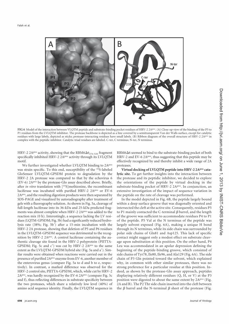
HRV-2 2Apro activity, showing that the RBM66274-520 fragmentspecifically inhibited HRV-2 2Apro activity through its LVLQTMmotif.
We further investigated whether LVLQTM binding to 2Apro
was strain specific. To this end, susceptibility of the 35S-labeledGloSensor LVLQTM-GPSDM protein to degradation by theHRV-2 2A protease was compared to that by the echovirus 6(EV-6) 2Apro by the protease-Glo assay described above. Briefly,after in vitro translation with [35S]methionine, the recombinantluciferase was incubated with purified HRV-2 2Apro or EV-62Apro, and the resulting digestion products were then separated bySDS-PAGE and visualized by autoradiography after treatment ofgels with a fluorography solution. As shown in Fig. 5a, cleavage offull-length luciferase into its 36-kDa and 25-kDa predicted frag-ments was almost complete when HRV-2 2Apro was added to thereaction mix (0 h). Interestingly, a sequence lacking the LV resi-dues (LQTM-GPSDM; Fig. 5b) had a significantly reduced hydro-lysis rate (38%; Fig. 5b=) after a 15-min incubation with theHRV-2 2A protease, showing that deletion of P5 and P6 residuesin the LVLQTM-GPSDM sequence was detrimental to the recog-nition by HRV-2 2Apro. A control luciferase containing the au-thentic cleavage site found in the HRV-2 polyprotein (PIITTA-GPSDM; Fig. 5c and c=) was cut by HRV-2 2Apro to the sameextent as the LVLQTM-GPSDM hybrid site (Fig. 5a and a=). Sim-ilar results were obtained when reactions were carried out in thepresence of purified 2Apro enzyme fromEV-6, anothermember ofthe enterovirus genus (compare Fig. 5a to d and b to e, respec-tively). In contrast, a marked difference was observed for theHRV-2 control site, PIITTA-GPSDM, which, while cut by HRV-22Apro, was hardly recognized by the EV-6 2Apro (compare Fig. 5cand f), thus reflecting differences in substrate specificity betweenthe two proteases, which share a relatively low level (40%) ofamino acid sequence identity. Finally, the LVLQTM sequence in
RBM66 seemed to bind to the substrate-binding pocket of bothHRV-2 and EV-6 2Apro, thus suggesting that this peptide may beeffectively recognized by and thereby inhibit a wide range of 2Aproteases.
Virtual docking of LVLQTM peptide into HRV-2 2Apro cata-lytic site. To get further insights into the interaction betweenthe protease and its peptidic inhibitor, we decided to explorethe orientations of the peptide by virtual docking in thesubstrate-binding pocket of HRV-2 2Apro. In conjunction, anextensive investigation of the impact of sequence variation inthe peptide on the rate of cleavage was performed.
In the model depicted in Fig. 6B, the peptide largely boundwithin a deep surface groove that was diagonally oriented andintersected the cleft at the active site. Consequently, residues P5to P1 mainly contacted the C-terminal � barrel, and the lengthof the groove was sufficient to accommodate residues P4 to P1of the peptide. P5 Val at the N terminus of the peptide waslargely solvent exposed (Fig. 6A), making a unique H bondthrough its N terminus, while its side chain was surrounded bypolar side chains of Gln81 and Asp125. This lack of specificcontact might suggest only a modest effect on substrate cleav-age upon substitution at this position. On the other hand, P4Leu was accommodated in an apolar depression defining thebeginning of the peptide-binding groove, which consisted ofside chains of Tyr78, Ile80, Ile96, and Ala129 (Fig. 6A). The sidechain of P3 Gln pointed toward the solvent, which explainedwhy, in common with other similar proteases, there was nostrong preference for a particular residue at this position. In-deed, as shown by the protease-Glo assay approach, peptidesdisplaying relatively different residues (Q, H, or V) at the P3position were digested to about the same extent by 2Apro (Fig.2A and B). The P2 Thr side chain inserted into the cleft betweenthe � barrel and the N-terminal � sheet of the protease (Fig.
FIG 6 Model of the interaction between VLQTM peptide and substrate-binding pocket residues of HRV-2 2Apro. (A) Close-up view of the binding of the P5 toP1 residues from the LVLQTM inhibitor. The protease backbone is depicted as a line covered by a semitransparent Van der Walls surface, except for catalyticresidues with large labels, depicted as sticks; protease-interacting residues have small labels. (B) Ribbon diagram of the overall structure of HRV-2 2Apro incomplex with the peptidic inhibitor. Catalytic triad residues are labeled. C-ter, C terminus; N-ter, N terminus.
Falah et al.
698 jvi.asm.org Journal of Virology
on J
une 1
, 2013 b
y IN
IST
-CN
RS
Bib
lioV
iehttp
://jvi.a
sm
.org
/D
ow
nlo
aded fro
m

6A), where it made two hydrogen bonds with residues Ser83and Tyr85, both belonging to the same side of the pocket. Inour model and contrary to the one discussed by Petersen et al.(24), binding of P2 Thr did not require a prior rotation ofTyr85.
Finally, the P1Met side chainmade three hydrogen bondswiththe substrate-binding pocket: two between its carbonyl group andboth the carbonyl and side chain of Cys106 (3.1 Å for both Hbonds) and one between its N atom and the hydroxyl group ofTyr85 (2.9 Å) (Fig. 6A). The flat and narrow pocket displayed verygood chemical complementarities to the P1 Met side chain.
In the light of this structural model, we showed that the doublemutation S83A/D125A resulted in an approximately 20-fold de-crease in the initial rate of TRPIITTA-pNA hydrolysis by 2Apro
(data not shown), confirming that these two residues played amajor role in the interaction with the LVLQTM peptide.
The z-LVLQTM-fmk peptide inhibits the replication ofHRV-2 in A549 cells. As LVLQTM was found to inhibit 2Apro
activity, we then investigated whether it also inhibited HRV rep-lication ex vivo. As pseudosubstrates are more potent inhibitorswhen covalently bound to their target proteases, we synthesized amodified LVLQTM peptide containing a fluoromethylketonegroup at its C terminus which forms a persistent, nonlabile cova-lent bond with the catalytic cysteine (Fig. 7A). Moreover, a ben-zyloxycarbonyl group was added at its N terminus to increase itscell permeation. As expected, the z-LVLQTM-fmk molecule gavea sharp decrease in HRV-2 2Apro activity in the TRPIITTA–p-nitroanilide substrate cleavage assay with a 50% inhibitory con-
FIG 7 The z-LVLQTM-fmk peptide inhibits 2Apro activity and HRV replication in A549 cells. (A) The LVLQTM peptide was synthesized and modified (MPBiomedicals Company) by adding a benzyloxycarbonyl (z) at its N terminus and a fluoromethylketone (fmk) group at its C terminus to form z-LVLQTM-fmk.The latter is known to form a covalent link with the catalytic cysteine of the 2Apro (20). (B) Effect of z-LVLQTM-fmk on cleavage in vitro of TRPIITTA–p-nitroanilide by the HRV-2 2Apro. Various concentrations (0 to 25 �M) of z-LVLQTM-fmk or z-FA-fmk (RnD Systems) were added to the TRPIITTA-pNApeptide, and their effects on HRV-2 2Apro catalysis was measured by collecting the absorbance at 405 nm for 10 min at 25°C. The percentage of cleavage activitywas calculated relative to the value obtained with no inhibitor. Data are expressed as means of three independent experiments, and standard deviations areindicated. (C) A549 cells were treated with 100 �M or 200 �M z-LVLQTM-fmk or z-FA-fmk for 1 h and were subsequently transfected with an mRNA codingforHRV-2 2Apro, HRV-14 2Apro, EV-6 2Apro, or GFP for 2 h. Cells were then transfected for 3 hwith a cappedmRNAor an IRES-containingmRNA, both codingfor the Renilla luciferase. The first contained the 5= UTR of the �-globin gene, and the second contained the 5= UTR of the encephalomyocarditis virus (EMCV)RNA. Cells were subsequently lysed, and luciferase activity was measured by luminometry. Error bars denote the standard deviations from the mean valuesobtained from three independent experiments. (D) A549 cells were treated with different concentrations of z-LVLQTM-fmk or z-FA-fmk and infected withHRV-2 or HRV-14 at a multiplicity of infection of 1 for 12 h. The TCID50 in the supernatant of infected cells was determined as described in Materials andMethods.
Inhibition of Human Rhinovirus Replication in Mice
January 2012 Volume 86 Number 2 jvi.asm.org 699
on J
une 1
, 2013 b
y IN
IST
-CN
RS
Bib
lioV
iehttp
://jvi.a
sm
.org
/D
ow
nlo
aded fro
m

centration value of 0.3 �M (Fig. 7B). In contrast, no inhibitoryeffect on 2Apro activity was observed when this assay was con-ducted in the presence of the control peptide z-FA-fmk, whichshared the same chemical changes as z-LVLQTM-fmk. Further-more, inhibition of capped mRNA translation by HRV-2 2Apro
was significantly reduced by the z-LVLQTM-fmk peptide used at100 �Mand 200 �M(Fig. 7C) compared to the z-FA-fmk controlpeptide. These concentrations showed no cytotoxic effect onA549cells (data not shown). The same inhibitory effect was observedusing the same test with HRV-14 2Apro (belonging to the rhinovi-rus species B) andEV-6 2Apro, which again reinforced the idea thatz-LVLQTM-fmk was not strain specific (Fig. 7C). Thus, the z-LVLQTM-fmk-modified peptide strongly and specifically inhibitedseveral 2Apro enzymes.
We next investigated the effect of LVLQTM on virus replica-tion in A549 cells. For this purpose, cells were infected withHRV-2 or HRV-14 at a multiplicity of infection (MOI) of 1 in thepresence of z-LVLQTM-fmk (100 �M or 200 �M) or the unre-lated control peptide z-FA-fmk. At 12 h postinfection, the 50%tissue culture infective dose (TCID50) value in the supernatants ofinfected cells was determined. Results depicted in Fig. 7D indi-cated that the z-LVLQTM-fmk peptide inhibited HRV-2 andHRV-14 replication in a dose-dependentmanner compared to theunrelated control peptide. Altogether, our data demonstrated thatthe observed decrease in virus production correlated directly with2Apro-mediated inhibition by the z-LVLQTM-fmk peptide.
The z-LVLQTM-fmk peptide protects against rhinovirus in-fection in vivo. To assess the role of z-LVLQTM-fmk in vivo, weinvestigated whether z-LVLQTM-fmk could inhibit HRV replica-tion in mice. For this purpose, mice were inoculated with HRV-2(105 PFU/mouse) and, at the same time, treated or not with vari-ous concentrations of z-LVLQTM-fmk. Lungs of infected micewere then harvested at different time points postinfection, andinfectious particles were evaluated by determining the TCID50. Asshown in Fig. 8A, compared to DMSO-treatedmice, z-LVLQTM-fmk-treatedmice had significantly fewer infectious viruses in theirlungs. Without peptide treatment (DMSO) and at 24, 48, and 120h postinfection, infectious virus titers reached 106, 106.75, and106.82 TCID50s/ml, respectively. In contrast, after treatment ofmice with 20, 200, or 500 �M z-LVLQTM-fmk, virus titersdropped to 104.2 to 105 TCID50s/ml for all the conditions tested.To confirm replication, an additional experiment was performedwhere mice were infected and lungs were harvested either imme-diately or at 48 h postinfection and then subjected to viral titra-tions (Fig. 8B). Results showed that the inoculum was totally re-covered in the lungs of infected mice at day 0 postinoculation.When lungs were harvested at 48 h postinfection, the viral titersignificantly increased, thus showing that viral replication indeedoccurred after infection of the mice. In addition, the z-LVLQTM-fmk-mediated inhibition was specific since this peptide also im-paired virus replication compared to the unrelated control pep-tide z-FA-fmk at 48 h postinfection (Fig. 8B). More importantly,when administration of the peptide was performed at 12 h postin-fection, inhibition of viral replication was also readily detectable(Fig. 8C). The viral titers from control-treated animals reached106.53 TCID50s/ml, while the viral titer of z-LVLQTM-fmk-treatedanimals was 104.08 TCID50s/ml at 48 h postinfection. Altogether,these results suggested that LVLQTM inhibited virus replicationin vivo and could be of particular interest for anti-HRV therapies.
DISCUSSION
Rhinoviruses are responsible for a large number of respiratorytract infections which range from the common cold to moreserious complications, such as pneumonia, bronchitis, orbronchiolitis in children as well as in adults. These infectionsconstitute a major public health problem and have a significantsocioeconomic impact due to the fact that there is currently noeffective drug to fight against HRV. In the search for an effec-tive treatment against rhinovirus, we identified a peptide in-hibitor of the viral 2Apro by a yeast two-hybrid screening. Thishigh-throughput screening allowed us to successfully identify10 different peptides that were nonhydrolyzable by the 2A pro-tease. Based on their sequence analysis, we postulated thatthese peptides could bind to the protease in a substrate-likemanner (lock-and-key model). Direct evidence of peptidebinding to the protease was provided by the protease-Glo assay,which elected the sequence motif LVLQTM derived from thevery C terminus of the RBM66 protein to be the best candidatefor subsequent development as an irreversible inhibitor of 2Apro.This peptide had the following main features: (i) it specificallybound in vitro to the HRV-2 2Apro, as demonstrated by pulldownassays. (ii) Its strong similarity to the amino-terminal half of nat-ural cleavage sites of the proteasemeant that this motif behaved invitro as a perfect pseudosubstrate not only ofHRV-2 2Apro but also
FIG 8 z-LVLQTM-fmk specifically inhibits HRV replication in vivo. (A)Micewere inoculated intranasally with 100,000 PFU HRV-2 per mouse (in a 25-�lvolume) and treated with additional 25 �l of a solution containing the indi-cated concentrations of peptide or 1% DMSO. Lungs of infected mice wereharvested at 24, 48, and 120 h postinfection, and virus titers were determined.(B)Micewere infectedwithHRV-2 as described above and treatedwith 20�Mz-LVLQTM-fmk or z-FA-fmk (control peptide). Lungs of infected mice wereharvested immediately (input) or at 48 h postinfection (hpi). Virus titers weredetermined as described in Materials and Methods. (C) Mice were infectedwithHRV-2 (100,000 PFU) and treated with 20 �Mz-LVLQTM-fmk or z-FA-fmk at 12 h postinfection. Lungs of infected mice were harvested at 48 hpostinfection and virus titers were measured. �, P � 0.05.
Falah et al.
700 jvi.asm.org Journal of Virology
on J
une 1
, 2013 b
y IN
IST
-CN
RS
Bib
lioV
iehttp
://jvi.a
sm
.org
/D
ow
nlo
aded fro
m

FIG 9 WebLogo sequence based on alignments of HRV type A, B, and C 2A proteases. These WebLogo sequences were generated using theWebLogo sequencegenerator (4) at http://weblogo.berkeley.edu/logo.cgi and were created using an alignment of 40 HRV-A sequences (A1, A2, A7, A9, A10, A11, A12, A13, A15,A16, A23, A24, A28, A29, A30, A34, A36, A38, A39, A41, A44, A46, A49, A53, A55, A56, A59, A64, A73, A74, A75, A76, A78, A82, A88, A89, A94, A101, A102, andA103) (A), 25 HRV-B sequences (B3, B4, B5, B6, B14, B17, B26, B27, B35, B37, B42, B48, B52, B69, B70, B72, B79, B83, B84, B86, B91, B92, B93, B97, and B99)(B), and 12 HRV-C sequences (C1 to C11 and C15) (C). The letter size is proportional to the degree of amino acid conservation, and arrows indicate residuesinvolved in the binding of the LVLQTM peptide at subsites S5 to S1.
Inhibition of Human Rhinovirus Replication in Mice
January 2012 Volume 86 Number 2 jvi.asm.org 701
on J
une 1
, 2013 b
y IN
IST
-CN
RS
Bib
lioV
iehttp
://jvi.a
sm
.org
/D
ow
nlo
aded fro
m

of EV-6 2Apro, another enteroviral 2A protease. These enzymesshare a relatively low level of amino acid sequence identity (40%),leading to substantially different surface characteristics, and thusrepresent two extremes in the primary sequence diversity of the2Apro family. (iii) It contained a P1 Met which was demonstratedto enhance significantly the binding affinity of the peptide for theenzyme (4, 28).
Therefore, our results support a molecular model wherebythe C terminus of RBM66, acting as a competitive inhibitor of2Apro, docks into the substrate-binding pocket of the enzyme.Notably, in their screening of a HeLa cell cDNA expressionlibrary by the yeast two-hybrid procedure, Ventoso et al.(1999) (33) had previously characterized several four-amino-acid-binding peptides that interfered in vitro with PV-1 2Apro
activity. However, our data indicate that compared to itsshorter version, LQTM, the LVLQTM sequence confers higherbinding affinity to RBM66 for 2Apro. These results thus high-lighted the benefit which may be gained by using 6-mer-basedpeptides instead of 4-mer-based compounds directed againstHRV 2Apro.
To achieve a more potent inhibition of the 2Apro, the LVLQTM peptide was modified so that it contained an electrophilicgroup (fmk) to enable the formation of a covalent bond with theactive-site thiol and a benzyloxycarbonyl group (z) to increase itscell permeation. So modified, the z-LVLQTM-fmk compoundwas an effective inhibitor of purified HRV-2 2Apro activity with aKi value of 0.3 �M, which is about 20 times lower than that of theCBV 2Apro inhibitor z-IETD-fmk (Ki � 7.7 �M) (1) and thecaspase inhibitor z-VAD-fmk, which is also active against HRV2Apro (Ki � 5.6 �M) (5). Then we showed that z-LVLQTM-fmkspecifically inhibited HRV-2 replication in vitro in A549 cells butalso in vivo in BALB/c mice. The mouse model has features verysimilar to those observed in rhinovirus infection in humans, in-cluding augmentation of allergic airway inflammation (2), and toour knowledge, our study is the first one to validate in vivo inmicean antiviral drug directed against HRV. Mouse infection byHRV-2 was made possible by the fact that this virus strain, whichbelongs to the minor HRV group, uses a member of the low-
density-lipoprotein receptor family and can bind themouse coun-terpart. Moreover, direct injection of the peptide by the intranasalroute in mice several hours after infection prefigures the outlinefor administration of a drug that could be used in humans forefficient anti-HRV therapy.
On the basis of HRV-2 2Apro crystallographic data, a virtualdocking model was then proposed to predict the inhibitorbinding mode into the ligand binding pocket of the enzyme.Sequence comparison between different 2Apro enzymes fromHRV-A, -B, and -C species revealed that amino acid residuesinvolved in the interaction with the inhibitor in our model arerelatively well conserved.
The alignment of 40 HRV-A 2Apro sequences (Fig. 9A)shows that for eight serotypes analyzed, Ile96 is replaced by aLeu, which displays the same physicochemical properties andso likely mediates the same hydrophobic interactions with P4Leu. Interestingly, Asp125 found in 10 serotype sequences isreplaced by a Glu, whose longer side chain may enhance amajor interaction with P5 Val. Concerning the 25 HRV-B 2Aproteins analyzed (Fig. 9B), hydrophobic interactions with P4Leu are conserved through hydrophobic residues Ile, Leu, andVal at position 78. Ser83, which appears to be crucial for inter-action with P3 Gln, is poorly conserved in type B sequences, asa Gly and an Asn are found at 50% and 35%, respectively. Thesecond highlighted interaction through Asp125 also seems tobe weakened in type B, as a serine is generally found at thisposition. Basic residues Arg and Lys are found at position 96,probably changing interaction modalities with the hydropho-bic P4 Leu; nevertheless, the peptide LVLQTM seems to wellaccommodate in the catalytic cleft, even though a Lys is found atposition 96 in HRV-B14. On the other hand, the Cys at position129 (A129 in HRV-A2 2Apro) found in all type B sequences mayreinforce the hydrophobic interaction with P4 Leu. Finally, I80V,S83T, I96L, C101A, and D125Emutations occurring in only a fewtype C sequences (Fig. 9C) should have no detrimental effects onthe interaction between the enzyme and its inhibitor, as they rep-resent conservative mutations from a physicochemical point ofview.
FIG 10 Multiple-sequence alignments of enterovirus 2Apro. Sequence alignment was performed using the ClustalW program and plotted with the ESPriptprogram (8). Similar residues are highlighted in gray; identical residues are in black. Secondary structure elements of HRV-2 2Apro are shown above thesequences, while arrows indicate residues involved in the binding of the VLQTM peptide at subsites S5 to S1.
Falah et al.
702 jvi.asm.org Journal of Virology
on J
une 1
, 2013 b
y IN
IST
-CN
RS
Bib
lioV
iehttp
://jvi.a
sm
.org
/D
ow
nlo
aded fro
m

If our peptide inhibitor may be of general use against all HRVserotypes, its use for therapeutic purposes could be extended to otherenterovirus-associateddiseases since it is alsoactiveagainstpoliovirus1 (PV-1; GenBank accession number VO1149) and human entero-virus 71 (HEV-71; GenBank accession number AEF32490) 2A pro-teases (data not shown). Comparison of the sequences of these pro-teases with those of other proteases tested in this study for theirinteractionwithLVLQTMreveals onlyminor differences (Fig. 10).In particular, S5 Gln81 is present in EV-6, while a similar gluta-mate is found in the other viruses. Asp125 properties are con-served through the glutamate or the asparagine in EV-6 and PV-1or HEV-71, respectively. The serine at the same position in HRV-B14 with a side chain shorter than Asp and obviously shorter thanGlu may reflect some flexibility of the substrate at P5, where it islargely solvent exposed (Fig. 5A). The apolar depression definingS4 is conserved, as all residues at positions 78, 80, 96, and 129, eventhough they are not similar, are hydrophobic, except for PV-1 atGln78 andHRV-B14 at Lys96. Glu82 andGly123 at S3 are not wellconserved, but peptide interactions occur through their back-bone; thiswould not have any consequences on affinity. Tyr85 andSer83 are strictly conserved at S2, except PV-1 displays a glu-tamine at position 83. Finally, the flat and narrow pocket display-ing very good chemical complementarities to the P1 Met sidechain is well conserved among the different viruses, as residues101 to 106 and 122 to 124 as well as Tyr85 are homologous. There-fore, this model allows accurate prediction of the interaction ofthe peptide inhibitor with a large spectrum of 2A proteases. Thismodel also suggests affinity differences between enteroviral 2Aproteases for the LVLQTM peptide. In particular, the longer sidechain of S4 I80 and S4 I96 in the HRV-2 2A protease compared toS4V80 and S4 L96 in EV-6 2Apro (Fig. 10)might reinforce the Vander Waals interactions with the P4 L in the peptide inhibitor.Moreover, the presence of a hydrophobic cysteine 101 at positionS1 in HRV-2 2Apro instead of a polar serine in EV-6 2Apro likelystrengthens the interaction with the P1 methionine in the peptideinhibitor. Such predictions could thus account for the better af-finity of HRV-2 2Apro for the LVLQTM peptide compared to theEV-6 2Apro, as confirmed by our experimental data (compare Fig.5a and d).
Fromamore fundamental point of view, this study alsohighlightsseveral clues concerning the exact role, if any, played by theRBM66protein in the context of virus-infected cells. One possibility is thatRBM66 is an antidote molecule which is expressed by the cell inresponse to viral infection and which specifically neutralizes proteinpoison2Apro.Conversely, andmore likely, bindingof theprotease onRBM66 could have a more or less profound impact on normalfunction of this protein in favor of viral infection. Experiments areunder way to try to resolve this new and very exciting enigma.
ACKNOWLEDGMENTS
We thank Monique Ballandras for technical assistance as well as Leslie C.Sutherland and Sophie Bonnal for providing a plasmid encoding the full-length RBM6 protein.
This project was supported by the National Reference Center of En-teroviruses, the Institut de Veille Sanitaire, and the AgenceNationale de laRecherche (ANR; to Béatrice Riteau).
The funders had no role in study design, data collection and analysis,decision to publish, or preparation of the manuscript.
We declare no competing financial interests.
REFERENCES
1. Badorff C, et al. 2000. Enteroviral protease 2A directly cleaves dystrophinand is inhibited by a dystrophin-based substrate analogue. J. Biol. Chem.275:11191–11197.
2. Bartlett NW, et al. 2008. Mouse models of rhinovirus-induced diseaseand exacerbation of allergic airway inflammation. Nat. Med. 14:199–204.
3. Belov GA, et al. 2000. Early alteration of nucleocytoplasmic traffic in-duced by some RNA viruses. Virology 275:244–248.
4. Deszcz L, Cencic R, Sousa C, Kuechler E, Skern T. 2006. An antiviralpeptide inhibitor that is active against picornavirus 2A proteinases but notcellular caspases. J. Virol. 80:9619–9627.
5. Deszcz L, Seipelt J, Vassilieva E, Roetzer A, Kuechler E. 2004. Antiviralactivity of caspase inhibitors: effect on picornaviral 2A proteinase. FEBSLett. 560:51–55.
6. Fromont-Racine M, Rain JC, Legrain P. 1997. Toward a functionalanalysis of the yeast genome through exhaustive two-hybrid screens. Nat.Genet. 16:277–282.
7. Garcia-Calvo M, et al. 1998. Inhibition of human caspases bypeptide-based and macromolecular inhibitors. J. Biol. Chem. 273:32608–32613.
8. Gouet P, Robert X, Courcelle E. 2003. ESPript/ENDscript: extractingand rendering sequence and 3D information from atomic structures ofproteins. Nucleic Acids Res. 31:3320–3323.
9. Gustin KE, Sarnow P. 2002. Inhibition of nuclear import and alterationof nuclear pore complex composition by rhinovirus. J. Virol. 76:8787–8796.
10. Joachims M, Van Breugel PC, Lloyd RE. 1999. Cleavage of poly(A)-binding protein by enterovirus proteases concurrent with inhibition oftranslation in vitro. J. Virol. 73:718–727.
11. Jurgens CK, et al. 2006. 2Apro is a multifunctional protein that regulatesthe stability, translation and replication of poliovirus RNA. Virology 345:346–357.
12. Kempf BJ, Barton DJ. 2008. Poliovirus 2A(Pro) increases viral mRNAand polysome stability coordinately in time with cleavage of eIF4G. J.Virol. 82:5847–5859.
13. Khoufache K, et al. 2009. Protective role for protease-activatedreceptor-2 against influenza virus pathogenesis via an IFN-gamma-dependent pathway. J. Immunol. 182:7795–7802.
14. Kistler A, et al. 2007. Pan-viral screening of respiratory tract infections inadults with and without asthma reveals unexpected human coronavirus andhuman rhinovirus diversity. J. Infect. Dis. 196:817–825.
15. Konig H, Rosenwirth B. 1988. Purification and partial characterization ofpoliovirus protease 2A by means of a functional assay. J. Virol. 62:1243–1250.
16. Laine P, Savolainen C, Blomqvist S, Hovi T. 2005. Phylogenetic analysisof human rhinovirus capsid protein VP1 and 2A protease coding se-quences confirms shared genus-like relationships with human enterovi-ruses. J. Gen. Virol. 86:697–706.
17. Lamphear BJ, et al. 1993. Mapping the cleavage site in protein synthesisinitiation factor eIF-4 gamma of the 2A proteases from human coxsacki-evirus and rhinovirus. J. Biol. Chem. 268:19200–19203.
18. Molla A, Hellen CU, Wimmer E. 1993. Inhibition of proteolytic activityof poliovirus and rhinovirus 2A proteinases by elastase-specific inhibitors.J. Virol. 67:4688–4695.
19. Ohlmann T, Rau M, Pain VM, Morley SJ. 1996. The C-terminal domainof eukaryotic protein synthesis initiation factor (eIF) 4G is sufficient tosupport cap-independent translation in the absence of eIF4E. EMBO J.15:1371–1382.
20. Otto HH, Schirmeister T. 1997. Cysteine proteases and their inhibitors.Chem. Rev. 97:133–172.
21. Palmenberg AC, et al. 2009. Sequencing and analyses of all knownhuman rhinovirus genomes reveal structure and evolution. Science324:55–59.
22. Papi A, et al. 2006. Infections and airway inflammation in chronic ob-structive pulmonary disease severe exacerbations. Am. J. Respir. Crit. CareMed. 173:1114–1121.
23. Park N, Katikaneni P, Skern T, Gustin KE. 2008. Differential targeting ofnuclear pore complex proteins in poliovirus-infected cells. J. Virol. 82:1647–1655.
24. Petersen JF, et al. 1999. The structure of the 2A proteinase from a com-mon cold virus: a proteinase responsible for the shut-off of host-cell pro-tein synthesis. EMBO J. 18:5463–5475.
Inhibition of Human Rhinovirus Replication in Mice
January 2012 Volume 86 Number 2 jvi.asm.org 703
on J
une 1
, 2013 b
y IN
IST
-CN
RS
Bib
lioV
iehttp
://jvi.a
sm
.org
/D
ow
nlo
aded fro
m

25. Prevot D, et al. 2003. Characterization of a novel RNA-binding region ofeIF4GI critical for ribosomal scanning. EMBO J. 22:1909–1921.
26. Reed LJ, Muench H. 1938. A simple method of estimating 50 per centendpoints. Am. J. Hyg. 27:493–497.
27. Sommergruber W, et al. 1994. 2A proteinases of coxsackie- and rhinovi-rus cleave peptides derived from eIF-4 gamma via a common recognitionmotif. Virology 198:741–745.
28. Sommergruber W, et al. 1992. Cleavage specificity on synthetic peptidesubstrates of human rhinovirus 2 proteinase 2A. J. Biol. Chem. 267:22639–22644.
29. Soto Rifo R, Ricci EP, Decimo D, Moncorge O, Ohlmann T. 2007. Backto basics: the untreated rabbit reticulocyte lysate as a competitive system torecapitulate cap/poly(A) synergy and the selective advantage of IRES-driven translation. Nucleic Acids Res. 35:e121.
30. Studier FW. 2005. Protein production by auto-induction in high densityshaking cultures. Protein Expr. Purif. 41:207–234.
31. Timmer T, et al. 1999. A comparison of genomic structures and expres-sion patterns of two closely related flanking genes in a critical lung cancerregion at 3p21.3. Eur. J. Hum. Genet. 7:478–486.
32. van Gunsteren WF. 1996. Biomolecular simulation: the GROMOS96manual and user guide. vdf, Zürich, Switzerland.
33. Ventoso I, Barco A, Carrasco L. 1999. Genetic selection of poliovirus2Apro-binding peptides. J. Virol. 73:814–818.
34. Vojtek AB, Hollenberg SM. 1995. Ras-Raf interaction: two-hybrid anal-ysis. Methods Enzymol. 255:331–342.
35. Zunszain PA, et al. 2010. Insights into cleavage specificity from the crystalstructure of foot-and-mouth disease virus 3C protease complexed with apeptide substrate. J. Mol. Biol. 395:375–389.
Falah et al.
704 jvi.asm.org Journal of Virology
on J
une 1
, 2013 b
y IN
IST
-CN
RS
Bib
lioV
iehttp
://jvi.a
sm
.org
/D
ow
nlo
aded fro
m


Abstract
Influenza is an acute respiratory disease caused by infection with influenza virus and is
a major public health problem. A better understanding of the interaction between influenza
virus and host allow us to better understand the pathophysiology of influenza infection, and
thus, ultimately, to better protect themselves against the disease. Morbidity and mortality
caused by severe influenza infections are associated with dysregulation of the immune response
in the lung. This deleterious inflammation is the cause of lung collateral damage, causing a
decrease in the patient's breathing capacity. Although the mechanisms involved are not fully
understood, recent studies point to a central role of endothelial cells in the deregulation of the
host response to influenza infection. During endothelium aggression, the physiological process
of hemostasis (platelet activation, coagulation and fibrinolysis) is activated in order to allow
wound healing and to maintain the integrity of blood vessels. In many inflammatory diseases,
the only dysregulation of hemostasis is directly linked to a deleterious inflammatory response.
During my thesis, we hypothesized that hemostasis could be the cause of the inflammatory
dysregulation during influenza infections. Our data show the role of two factors strongly
involved in hemostasis: the thrombin activated receptor, PAR-1 (protease activated receptor 1)
and plasminogen, in the deleterious lung inflammation and in the pathogenicity of influenza
virus. Besides the role of hemostasis, we have also been able to show that the influenza virus
incorporates cellular proteins in the viral envelope, allowing it to evade the immune system,
which could also contribute to the deregulation of the host response. All the results obtained
allowed to better understand the mechanisms involved in immune response dysregulation
during influenza infection and suggest new therapeutic targets to fight against the disease.
Related Documents