Hong Kong Baptist University Metabolomics of the Protective Effect of Ampelopsis grossedentata and Its Major Active Compound Dihydromyricetin on the Liver of High-Fat Diet Hamster Fan, Lanlan; Qu, Xiaosheng; YI, Tao; Peng, Yong; Jiang, Manjing; Miao, Jianhua; Xiao, Peigen Published in: Evidence-based Complementary and Alternative Medicine DOI: 10.1155/2020/3472578 Published: 28/01/2020 Link to publication Citation for published version (APA): Fan, L., Qu, X., YI, T., Peng, Y., Jiang, M., Miao, J., & Xiao, P. (2020). Metabolomics of the Protective Effect of Ampelopsis grossedentata and Its Major Active Compound Dihydromyricetin on the Liver of High-Fat Diet Hamster. Evidence-based Complementary and Alternative Medicine, 2020, [3472578]. https://doi.org/10.1155/2020/3472578 General rights Copyright and intellectual property rights for the publications made accessible in HKBU Scholars are retained by the authors and/or other copyright owners. In addition to the restrictions prescribed by the Copyright Ordinance of Hong Kong, all users and readers must also observe the following terms of use: • Users may download and print one copy of any publication from HKBU Scholars for the purpose of private study or research • Users cannot further distribute the material or use it for any profit-making activity or commercial gain • To share publications in HKBU Scholars with others, users are welcome to freely distribute the permanent publication URLs Downloaded on: 20 Mar, 2022

Welcome message from author
This document is posted to help you gain knowledge. Please leave a comment to let me know what you think about it! Share it to your friends and learn new things together.
Transcript
Hong Kong Baptist University
Metabolomics of the Protective Effect of Ampelopsis grossedentata and ItsMajor Active Compound Dihydromyricetin on the Liver of High-Fat DietHamsterFan, Lanlan; Qu, Xiaosheng; YI, Tao; Peng, Yong; Jiang, Manjing; Miao, Jianhua; Xiao,PeigenPublished in:Evidence-based Complementary and Alternative Medicine
DOI:10.1155/2020/3472578
Published: 28/01/2020
Link to publication
Citation for published version (APA):Fan, L., Qu, X., YI, T., Peng, Y., Jiang, M., Miao, J., & Xiao, P. (2020). Metabolomics of the Protective Effect ofAmpelopsis grossedentata and Its Major Active Compound Dihydromyricetin on the Liver of High-Fat DietHamster. Evidence-based Complementary and Alternative Medicine, 2020, [3472578].https://doi.org/10.1155/2020/3472578
General rightsCopyright and intellectual property rights for the publications made accessible in HKBU Scholars are retained by the authors and/or othercopyright owners. In addition to the restrictions prescribed by the Copyright Ordinance of Hong Kong, all users and readers must alsoobserve the following terms of use:
• Users may download and print one copy of any publication from HKBU Scholars for the purpose of private study or research • Users cannot further distribute the material or use it for any profit-making activity or commercial gain • To share publications in HKBU Scholars with others, users are welcome to freely distribute the permanent publication URLs
Downloaded on: 20 Mar, 2022
Authors Authors Lanlan Fan, Xiaosheng Qu, Tao Yi, Yong Peng, Manjing Jiang, Jianhua Miao, and Peigen Xiao
This journal article is available at HKBU Institutional Repository: https://repository.hkbu.edu.hk/hkbu_staff_publication/7056
Research ArticleMetabolomics of the Protective Effect of Ampelopsisgrossedentata and Its Major Active CompoundDihydromyricetin on the Liver of High-Fat Diet Hamster
Lanlan Fan ,1,2 Xiaosheng Qu ,2 Tao Yi ,3 Yong Peng ,4 Manjing Jiang,1
Jianhua Miao ,2 and Peigen Xiao 4
1School of Pharmacy, Guangxi University of Chinese Medicine, Nanning 530001, China2National Engineering Laboratory of Southwest Endangered Medicinal Resources Development,Guangxi Botanical Garden of Medicinal Plants, Nanning 530023, China3School of Chinese Medicine, Hong Kong Baptist University, Kowloon Tong, Hong Kong, China4Institute of Medicinal Plant Development, Chinese Academy of Medical Sciences and Peking Union Medical College,151 Malianwa North Road, Beijing 100193, China
Correspondence should be addressed to Jianhua Miao; [email protected] and Peigen Xiao; [email protected]
Lanlan Fan, Xiaosheng Qu, and Tao Yi contributed equally to this work.
Received 3 July 2019; Revised 30 September 2019; Accepted 5 October 2019; Published 28 January 2020
Academic Editor: Samra Bashir
Copyright © 2020 Lanlan Fan et al. 6is is an open access article distributed under the Creative Commons Attribution License,which permits unrestricted use, distribution, and reproduction in any medium, provided the original work is properly cited.
6e flavonoid dihydromyricetin (DMY) is the main component of Ampelopsis grossedentata (Hand-Mazz) W. T. Wang (AG), a dailybeverage and folkmedicine used in Southern China to treat jaundice hepatitis, cold fever, and sore throat. Recently, DMY andAGwereshown to have a beneficial effect on lipidmetabolism disorder. However, themechanisms of howDMYandAGprotect the liver duringlipid metabolism disorder remain unclear. In this study, we first analyzed the chemical compounds of AG by HPLC-DAD-ESI-IT-TOF-MSn. Of the 31 compounds detected, 29 were identified based on previous results. 6en, the effects of DMY and AG on high-fatdiet hamster livers were studied and the metabolite levels and metabolic pathway activity of the liver were explored by 1H NMRmetabolomics. Compared to the high-fat diet group, supplementation of AG and DMY attenuated the high-fat-induced increase inbody weight, liver lipid deposition, serum triglycerides and total cholesterol levels, and normalized endogenous metabolite con-centrations. PCA and PLS-DA score plots demonstrated that while the metabolic profiles of hamsters fed a high-fat diet supplementedwith DMY or AG were both far from those of hamsters fed a normal diet or a high-fat diet alone, they were similar to each other. Ourdata suggest that the underlying mechanism of the protective effect of DMY and AG might be related to an attenuation of thedeleterious effect of high-fat diet-induced hyperlipidemia on multiple metabolic pathways including amino acid metabolism, ketonebody metabolism, energy metabolism, tricarboxylic acid cycle, and enhanced fatty acid oxidation.
1. Introduction
Ampelopsis grossedentata (Hand-Mazz.) W. T. Wang (AG),also known as Vine tea, is a plant species mainly distributedin central and southern China which is rich in flavonoids,polysaccharides, and polyphenols. First used by the Zhuangand Yao people [1], it became later also widely used in Tujia,Lahu, Dong, Jinuo, and Hakka areas. Besides consumptionas a tea prepared from the tender stems and leaves, AG is alsoused to treat jaundice hepatitis, cold fever, and sore throat.
Next too its antioxidant [2] and anti-inflammatory effects[3], it can also normalize blood lipid [4] and sugar [5] levelsand attenuate liver injuries [6].
Dihydromyricetin (DMY), which is the main flavonoidof AG with a content of up to 35% [7], showed anti-proliferation capacities in lung [8], breast, [9], and ovariancancer [10], improvements on hypertension [11], hyper-lipidemia [12, 13] and abnormal blood sugar levels [14], aswell as neuroprotective activity against Alzheimer’s disease[15], Parkinson’s disease [16], alcohol addiction, depression
HindawiEvidence-Based Complementary and Alternative MedicineVolume 2020, Article ID 3472578, 15 pageshttps://doi.org/10.1155/2020/3472578
[17], and dermatoprotection [18]. In addition, a preventiveeffect on myocardial fibrosis [19], myocardial hypertrophy[20], and cardiac ischemia/reperfusion injury [21] wasreported.
6e liver plays an important role in the digestion, ab-sorption, oxidation, decomposition, and transformation ofnutrients such as lipids, proteins, and sugars [22]. Abnormalaccumulation and distribution of lipids and their metabo-lites in the body result in lipid metabolic disorder which is animportant inducer of diabetes, atherosclerosis, nephroticsyndrome, and cardio- and cerebrovascular disease. Con-sidering the health impact of chronic metabolic diseasessuch as hyperlipidemia, safe and reliable functional com-pounds from natural sources are highly needed. Based on theremarkable blood lipid regulating characteristics describedin the literature [23–26], both AG and DMY might possessgreat health-promoting potentials; however, their capacity toprevent liver injury as well as the underlying mechanisms isunknown.
Metabolomics, reflecting the overall changes of the or-ganism by studying the changes of endogenous metabolitelevels, has recently played an important role in revealing thepharmacodynamics andmechanism of some disease [27–29]or traditional Chinese medicine [30].
Since up to 85% cholesterol is synthesized extrahepatic inmale hamsters, hyperlipidemia can be caused in a relativelyshort period of time. 6us, as the lipid metabolism ofhamsters is similar to that of human, especially malehamsters are widely used as a model organism of lipidmetabolism disorder [31, 32].
In this study, the chemical compounds of AG were firstanalyzed by liquid chromatography-mass spectrometry.6en, the lipid metabolism disorder model of hamster wasinduced by high-fat diet, and the preventive effect of AG andDMY on hyperlipidemia and hyperglycemia was studied byliver metabolomics.
2. Materials and Methods
2.1. Chemicals and Materials. Acetonitrile and formic acid,both LC-MS grade, were purchased from Fisher (Fair Lawn,New Jersey, USA) and Sigma-Aldrich (St. Louis, USA), re-spectively. For sample extraction, analytical grade methanoland ethanol was used while deionized water was obtainedfrom a Milli-Q water purification system (Millipore, Bedford,MA, USA).
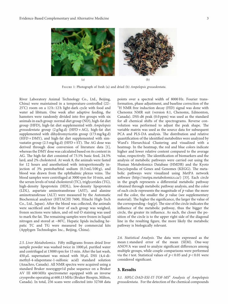
Ampelopsis grossedentata (Hand-Mazz.) W. T. Wang(AG) was purchased from Dayaoshan natural plant devel-opment Co. Ltd. (Guangxi, China) and identified by Pro-fessor Yi Cai from Guangxi University of Chinese Medicine(Figure 1). Dihydromyricetin (DMY) was purchased fromChengdu preferred Biological Technology Co., Ltd.(Chengdu, China) with purity above 98%.
2.2. Sample Preparation. For LC-MS analysis, the AGsample was crushed through an 80-mesh sieve and dried at50°C to constant mass. Next, 0.5 g powder was dissolved in50mL of methanol and ultrasonically extracted for 40min.
6e extract was filtered, the primary filtrate was discarded,and the filtrate was diluted 100 times and filtered through a0.22 μm filter. For the animal experiments, 150 g AG powerwas soaked in 5000mL boiling water for 15min and con-centrated to 0.6 g crude drug/ml.
2.3. HPLC-DAD-ESI-IT-TOF-MSn Analysis. High-performance liquid chromatography with a diode arraydetector and combined with electrospray ionization ion traptime-of-flight multistage mass spectrometry (HPLC–DAD–ESI-IT-TOF-MSn) analyses was performed with aShimadzu LCMS-IT-TOF instrument including two LC-20AD pumps, a SIL-20AC autosampler, a CTO-20A columnoven, a SPD-M20A PDA detector, a CBM-20A systemcontroller, an ESI ion source, and an IT-TOF mass spec-trometer (Shimadzu, Kyoto, Japan).
6e chromatography separations were performed on aMerck Purospher STAR RP-18 column (250mm× 4.6mm,5 μm) with a column temperature of 40°C. 6e mobile phaseconsisted of 0.1% formic acid (v/v) (A) and acetonitrile (B)using a gradient program of 50–58% B in 0–17min, 58–70%B in 17–20min, 70–85% B in 20–26min, and 85–90% B in26–35min. 6e solvent flow rate was 1.0mL/min. 6e PDAdetector wavelength was 350 nm.
For ESI-IT-TOF-MSn analysis, the mass spectrometerwas programmed to execute a full scan over m/z 100–1000(MS1) and m/z 50–1000 (MS2 and MS3) in both positive-ion(PI) and negative-ion (NI) detection modes with the fol-lowing settings: a flow rate of 0.2000mL/min, a heat blockand curved desolvation line temperature of 250°C, a flow rateof the nebulizing nitrogen gas of 1.5 L/min, an interfacevoltage of (+) 4.5 kV and (− ) − 3.5 kV, a detector voltage of1.70 kV, an ion accumulation time of 20ms, and a relativecollision-induced dissociation energy of 50%. Trifluoroaceticacid sodium solution (2.5mM) was used to calibrate themass range from 50 to 1000Da. All data were recorded andanalyzed with Shimadzu LCMS solution Version 3.60,Formula Predictor Version 1.2, and Accurate Mass Calcu-lator (Shimadzu, Kyoto, Japan).
After collecting LC/MS data, the mass spectrum of eachchromatographic peak was extracted based on excimer ions([M+H]+, [M − H]− ) and loading ions ([M+NH4]+,[M+Na]+, [M+Cl]− , [M+HCOO]− , etc.). With PeakView1.2 software, the relative molecular mass of the primary massspectrometry was obtained and the molecular formula wasfitted in the mass deviation range of 5×10− 6. 6e chemicalcompounds of AG and its genus were then collected andsorted out with the Scifinder and Reaxys databases. Next,secondary mass spectrometry information of the chro-matographic peak and the corresponding fragment ions ofthe compound were obtained and the chemical compositionwas compared with the literature based on the cleavage ofthe ions and the reference substance.
2.4. Animal Experiments. Animal care and procedures wereapproved by and conducted according to the standards ofthe Guangxi University of Chinese Medicine (Nanning,China). Male LVG hamsters (110–130 g, 8 weeks old, Vital
2 Evidence-Based Complementary and Alternative Medicine
River Laboratory Animal Technology Co., Ltd., Beijing,China) were maintained in a temperature-controlled (22–25°C) room on a 12 h :12 h light-dark cycle with food andwater ad libitum. One week after adaptive feeding, thehamsters were randomly divided into five groups with sixanimals in each group: normal diet group (ND), high-fat dietgroup (HFD), high-fat diet supplemented with Ampelopsisgrossedentata group (2 g/kg.d) (HFD+AG), high-fat dietsupplemented with dihydromyricetin group (173mg/kg.d)(HFD+DMY), and high-fat diet supplemented with sim-vastatin group (2.5mg/kg.d) (HFD+ ST). 6e AG dose wasderived through dose conversion of literature data [1],whereas the DMY dose was calculated based on its content inAG. 6e high-fat diet consisted of 73.5% basic feed, 24.5%lard, and 2% cholesterol. At week 8, the animals were fastedfor 12 hours and anesthetized with intraperitoneally in-jection of 3% pentobarbital sodium (0.3mL/100 g), andblood was drawn from the ophthalmic plexus veins. 6eblood samples were centrifuged at 3000 rpm for 10min, andthe serum levels of total cholesterol (TC), triglycerides (TG),high-density lipoprotein (HDL), low-density lipoprotein(LDL), aspartate aminotransferase (AST), and alanineaminotransferase (ALT) were measured by the AutomaticBiochemical analyzer (HITACHI 7600, Hitachi High-TechCo., Ltd., Japan). After the blood was collected, the animalswere sacrificed and the liver of each group was weighed,frozen sections were taken, and oil red O staining was usedto mark the fat. 6e remaining samples were frozen in liquidnitrogen and stored at − 80°C. Hepatic lipids including he-patic TC and TG were measured by commercial kits(Applygen Technologies Inc., Beijing, China).
2.5. Liver Metabolomics. Fifty milligrams frozen dried liversample powder was washed twice in 1000 μL purified waterand centrifuged at 13000 rpm for 15min. After the last wash,450 μL supernatant was mixed with 50 μL DSS (4,4-di-methyl-4-silapentane-1-sulfonic acid) standard solution(Anachro, Canada). All NMR spectra were acquired using astandard Bruker noesygppr1d pulse sequence on a BrukerAV III 600MHz spectrometer equipped with an inversecryoprobe operating at 600.13MHz (Bruker Biospin, Milton,Canada). In total, 256 scans were collected into 32768 data
points over a spectral width of 8000Hz. Fourier trans-formation, phase adjustment, and baseline correction of the1H NMR free induction decay (FID) signal was done withChenomx NMR suit (version 8.1, Chenomx, Edmonton,Canada). DSS-d6 peak (0.0 ppm) was used as the standardfor all chemical shifts of the spectrograms. Reverse con-volution was performed to adjust the peak shape. 6evariable matrix was used as the source data for subsequentPCA and PLS-DA analysis. 6e distribution and relativequantification of the identified metabolites were analyzed byWard’s Hierarchical Clustering and visualized with aheatmap. In the heatmap, the red and blue colors indicatehigher and lower relative content compared to the averagevalue, respectively. 6e identification of biomarkers and theanalysis of metabolic pathways were carried out using theHuman Metabolomics Database (HMDB) and the KyotoEncyclopedia of Genes and Genomes (KEGG). 6e meta-bolic pathways were visualized using MetPA networksoftware (http://metpa.metabolomics.ca/) [33]. Each circlein the graph represents a differential metabolic pathwayobtained through metabolic pathway analysis, and the colorof each circle represents the magnitude of p value: the morered the color, the smaller the p value (see supplementarymaterial). 6e higher the significance, the larger the value ofthe corresponding − log(p).6e size of the circle indicates theinfluence of the metabolic pathway, thus the bigger thecircle, the greater its influence. As such, the closer the po-sition of the circle is to the upper right side of the diagonalline in the resulting figure, the more likely the metabolicpathway is biologically relevant.
2.6. Statistical Analysis. 6e data were expressed as themean± standard error of the mean (SEM). One-wayANOVA was used to analyze significant differences amongmultiple groups, while couple comparisons were performedvia the t test. Statistical values of p< 0.05 and p< 0.01 wereconsidered significant.
3. Results
3.1. HPLC-DAD-ESI-IT-TOF-MSn Analysis of Ampelopsisgrossedentata. For the detection of the chemical compounds
(a) (b)
Figure 1: Photograph of fresh (a) and dried (b) Ampelopsis grossedentata.
Evidence-Based Complementary and Alternative Medicine 3
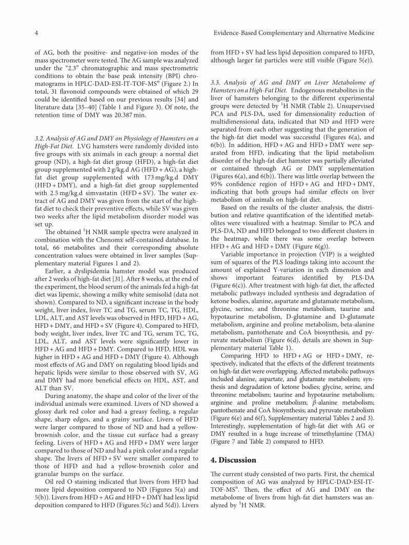
of AG, both the positive- and negative-ion modes of themass spectrometer were tested.6e AG sample was analyzedunder the “2.3” chromatographic and mass spectrometricconditions to obtain the base peak intensity (BPI) chro-matograms in HPLC-DAD-ESI-IT-TOF-MSn (Figure 2.) Intotal, 31 flavonoid compounds were obtained of which 29could be identified based on our previous results [34] andliterature data [35–40] (Table 1 and Figure 3). Of note, theretention time of DMY was 20.387min.
3.2. Analysis of AG and DMY on Physiology of Hamsters on aHigh-Fat Diet. LVG hamsters were randomly divided intofive groups with six animals in each group: a normal dietgroup (ND), a high-fat diet group (HFD), a high-fat dietgroup supplemented with 2 g/kg.d AG (HFD+AG), a high-fat diet group supplemented with 173mg/kg.d DMY(HFD+DMY), and a high-fat diet group supplementedwith 2.5mg/kg.d simvastatin (HFD+ SV). 6e water ex-tract of AG and DMY was given from the start of the high-fat diet to check their preventive effects, while SV was giventwo weeks after the lipid metabolism disorder model wasset up.
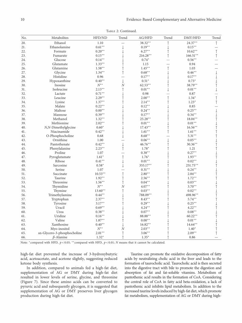
6e obtained 1H NMR sample spectra were analyzed incombination with the Chenomx self-contained database. Intotal, 66 metabolites and their corresponding absoluteconcentration values were obtained in liver samples (Sup-plementary material Figures 1 and 2).
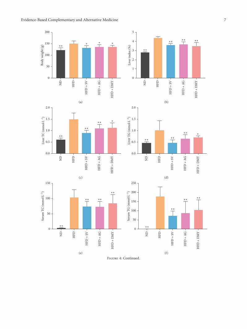
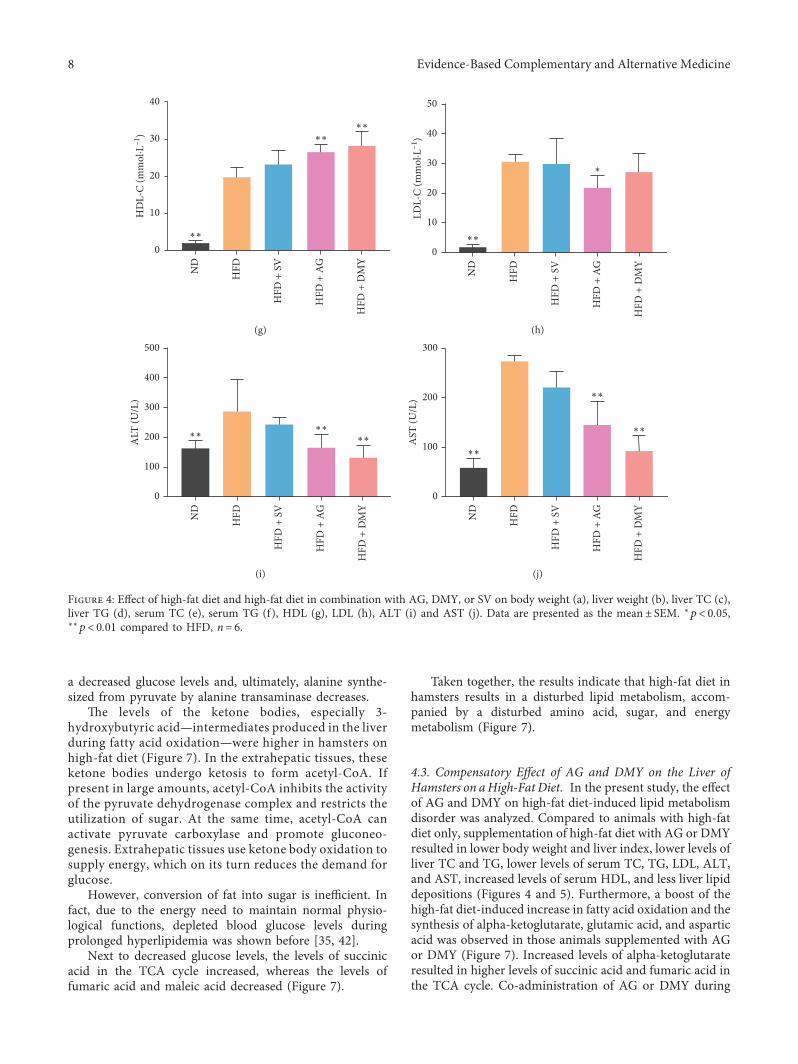
Earlier, a dyslipidemia hamster model was producedafter 2 weeks of high-fat diet [31]. After 8 weeks, at the end ofthe experiment, the blood serum of the animals fed a high-fatdiet was lipemic, showing a milky white semisolid (data notshown). Compared to ND, a significant increase in the bodyweight, liver index, liver TC and TG, serum TC, TG, HDL,LDL, ALT, and AST levels was observed in HFD, HFD+AG,HFD+DMY, and HFD+ SV (Figure 4). Compared to HFD,body weight, liver index, liver TC and TG, serum TC, TG,LDL, ALT, and AST levels were significantly lower inHFD+AG and HFD+DMY. Compared to HFD, HDL washigher in HFD+AG and HFD+DMY (Figure 4). Althoughmost effects of AG and DMY on regulating blood lipids andhepatic lipids were similar to those observed with SV, AGand DMY had more beneficial effects on HDL, AST, andALT than SV.
During anatomy, the shape and color of the liver of theindividual animals were examined. Livers of ND showed aglossy dark red color and had a greasy feeling, a regularshape, sharp edges, and a grainy surface. Livers of HFDwere larger compared to those of ND and had a yellow-brownish color, and the tissue cut surface had a greasyfeeling. Livers of HFD+AG and HFD+DMY were largercompared to those of ND and had a pink color and a regularshape. 6e livers of HFD+ SV were smaller compared tothose of HFD and had a yellow-brownish color andgranular bumps on the surface.
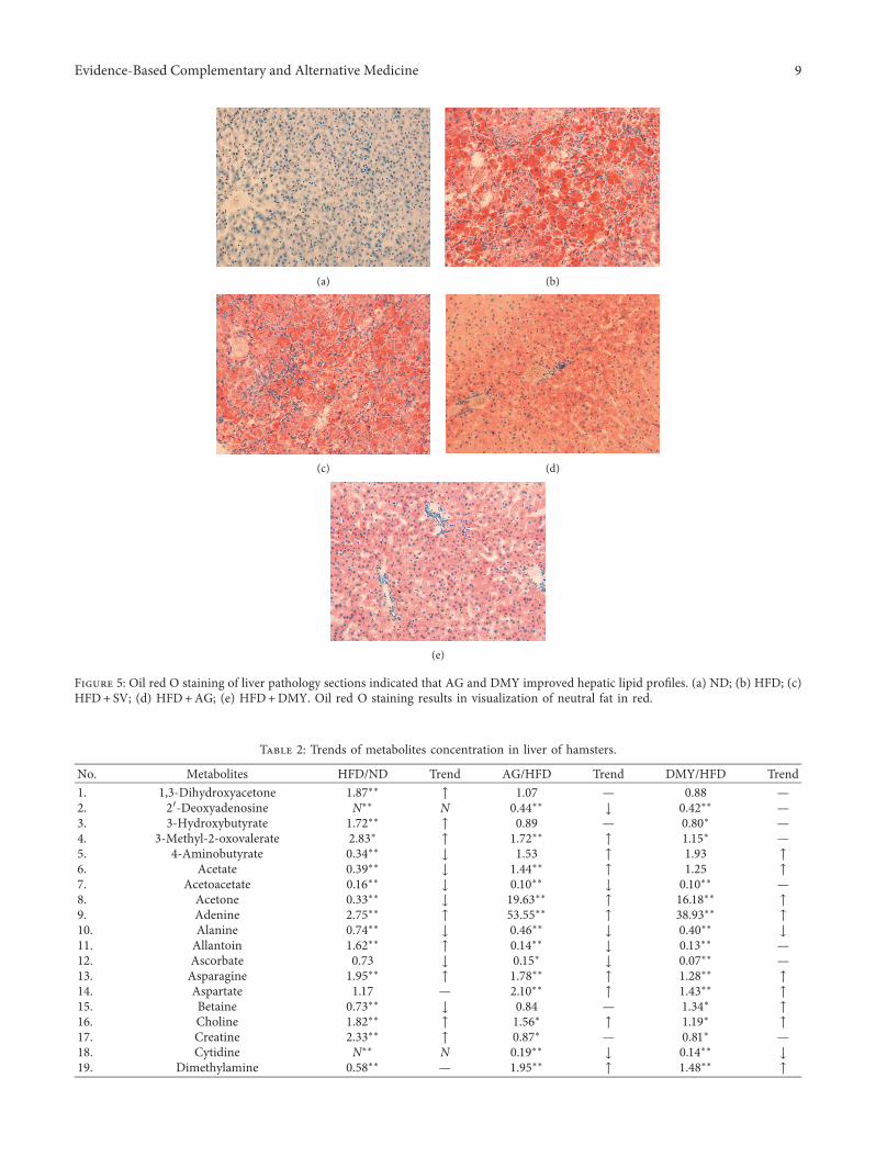
Oil red O staining indicated that livers from HFD hadmore lipid deposition compared to ND (Figures 5(a) and5(b)). Livers fromHFD+AG and HFD+DMY had less lipiddeposition compared to HFD (Figures 5(c) and 5(d)). Livers
from HFD+ SV had less lipid deposition compared to HFD,although larger fat particles were still visible (Figure 5(e)).
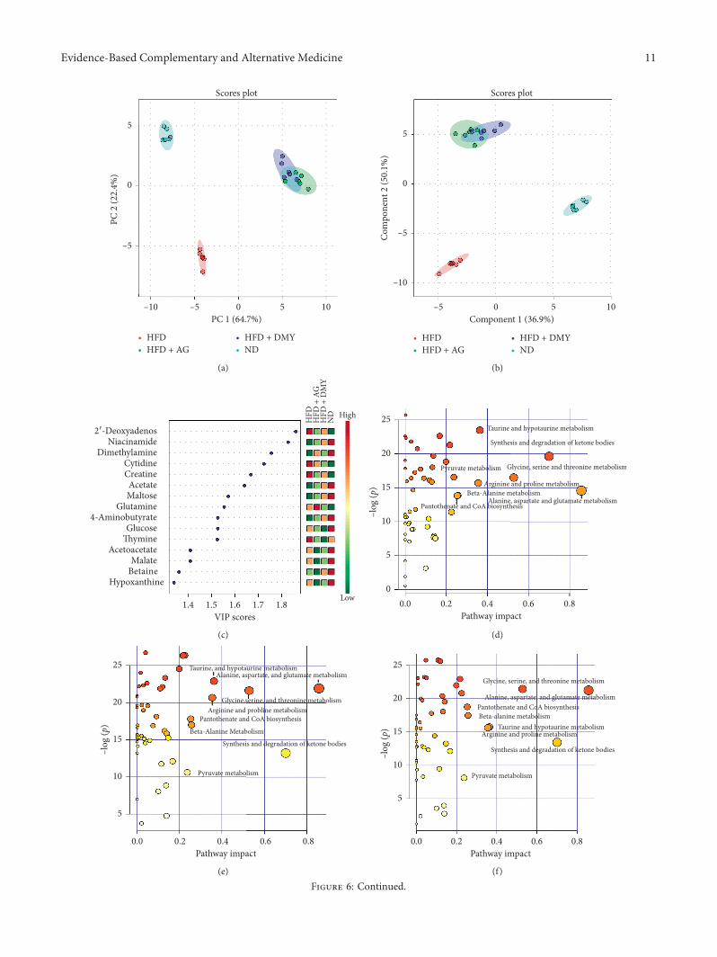
3.3. Analysis of AG and DMY on Liver Metabolome ofHamsters onaHigh-FatDiet. Endogenous metabolites in theliver of hamsters belonging to the different experimentalgroups were detected by 1H NMR (Table 2). UnsupervisedPCA and PLS-DA, used for dimensionality reduction ofmultidimensional data, indicated that ND and HFD wereseparated from each other suggesting that the generation ofthe high-fat diet model was successful (Figures 6(a), and6(b)). In addition, HFD+AG and HFD+DMY were sep-arated from HFD, indicating that the lipid metabolismdisorder of the high-fat diet hamster was partially alleviatedor contained through AG or DMY supplementation(Figures 6(a), and 6(b)). 6ere was little overlap between the95% confidence region of HFD+AG and HFD+DMY,indicating that both groups had similar effects on livermetabolism of animals on high-fat diet.
Based on the results of the cluster analysis, the distri-bution and relative quantification of the identified metab-olites were visualized with a heatmap. Similar to PCA andPLS-DA, ND and HFD belonged to two different clusters inthe heatmap, while there was some overlap betweenHFD+AG and HFD+DMY (Figure 6(g)).
Variable importance in projection (VIP) is a weightedsum of squares of the PLS loadings taking into account theamount of explained Y-variation in each dimension andshows important features identified by PLS-DA(Figure 6(c)). After treatment with high-fat diet, the affectedmetabolic pathways included synthesis and degradation ofketone bodies, alanine, aspartate and glutamate metabolism,glycine, serine, and threonine metabolism, taurine andhypotaurine metabolism, D-glutamine and D-glutamatemetabolism, arginine and proline metabolism, beta-alaninemetabolism, pantothenate and CoA biosynthesis, and py-ruvate metabolism (Figure 6(d), details are shown in Sup-plementary material Table 1).
Comparing HFD to HFD+AG or HFD+DMY, re-spectively, indicated that the effects of the different treatmentson high-fat diet were overlapping. Affectedmetabolic pathwaysincluded alanine, aspartate, and glutamate metabolism; syn-thesis and degradation of ketone bodies; glycine, serine, andthreonine metabolism; taurine and hypotaurine metabolism;arginine and proline metabolism; β-alanine metabolism;pantothenate and CoA biosynthesis; and pyruvate metabolism(Figure 6(e) and 6(f), Supplementary material Tables 2 and 3).Interestingly, supplementation of high-fat diet with AG orDMY resulted in a huge increase of trimethylamine (TMA)(Figure 7 and Table 2) compared to HFD.
4. Discussion
6e current study consisted of two parts. First, the chemicalcomposition of AG was analyzed by HPLC-DAD-ESI-IT-TOF-MSn. 6en, the effect of AG and DMY on themetabolome of livers from high-fat diet hamsters was an-alyzed by 1H NMR.
4 Evidence-Based Complementary and Alternative Medicine
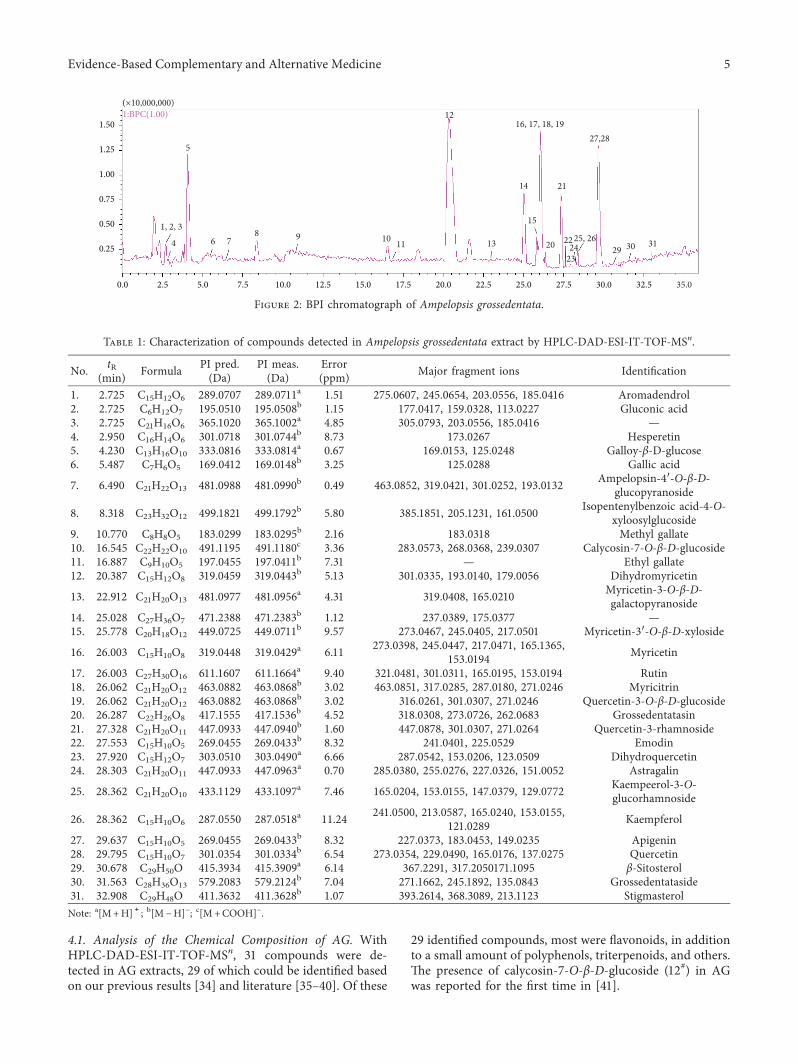
4.1. Analysis of the Chemical Composition of AG. WithHPLC-DAD-ESI-IT-TOF-MSn, 31 compounds were de-tected in AG extracts, 29 of which could be identified basedon our previous results [34] and literature [35–40]. Of these
29 identified compounds, most were flavonoids, in additionto a small amount of polyphenols, triterpenoids, and others.6e presence of calycosin-7-O-β-D-glucoside (12#) in AGwas reported for the first time in [41].
2.5 5.0 7.5 10.0 12.5 17.515.0 20.0 22.5 25.0 27.5 30.0 32.5 35.0
1.50
1.25
1.00
0.75
0.50
0.25
(×10,000,000)1:BPC(1.00)
1, 2, 3
4 6 78 9 10 11
12
13
14
16, 17, 18, 19
15
20
21
22
2324
25, 2629 30 31
27,28
0.0
5
Figure 2: BPI chromatograph of Ampelopsis grossedentata.
Table 1: Characterization of compounds detected in Ampelopsis grossedentata extract by HPLC-DAD-ESI-IT-TOF-MSn.
No. tR(min) Formula PI pred.
(Da)PI meas.(Da)
Error(ppm) Major fragment ions Identification
1. 2.725 C15H12O6 289.0707 289.0711a 1.51 275.0607, 245.0654, 203.0556, 185.0416 Aromadendrol2. 2.725 C6H12O7 195.0510 195.0508b 1.15 177.0417, 159.0328, 113.0227 Gluconic acid3. 2.725 C21H16O6 365.1020 365.1002a 4.85 305.0793, 203.0556, 185.0416 —4. 2.950 C16H14O6 301.0718 301.0744b 8.73 173.0267 Hesperetin5. 4.230 C13H16O10 333.0816 333.0814a 0.67 169.0153, 125.0248 Galloy-β-D-glucose6. 5.487 C7H6O5 169.0412 169.0148b 3.25 125.0288 Gallic acid
7. 6.490 C21H22O13 481.0988 481.0990b 0.49 463.0852, 319.0421, 301.0252, 193.0132 Ampelopsin-4′-O-β-D-glucopyranoside
8. 8.318 C23H32O12 499.1821 499.1792b 5.80 385.1851, 205.1231, 161.0500 Isopentenylbenzoic acid-4-O-xyloosylglucoside
9. 10.770 C8H8O5 183.0299 183.0295b 2.16 183.0318 Methyl gallate10. 16.545 C22H22O10 491.1195 491.1180c 3.36 283.0573, 268.0368, 239.0307 Calycosin-7-O-β-D-glucoside11. 16.887 C9H10O5 197.0455 197.0411b 7.31 — Ethyl gallate12. 20.387 C15H12O8 319.0459 319.0443b 5.13 301.0335, 193.0140, 179.0056 Dihydromyricetin
13. 22.912 C21H20O13 481.0977 481.0956a 4.31 319.0408, 165.0210 Myricetin-3-O-β-D-galactopyranoside
14. 25.028 C27H36O7 471.2388 471.2383b 1.12 237.0389, 175.0377 —15. 25.778 C20H18O12 449.0725 449.0711b 9.57 273.0467, 245.0405, 217.0501 Myricetin-3′-O-β-D-xyloside
16. 26.003 C15H10O8 319.0448 319.0429a 6.11 273.0398, 245.0447, 217.0471, 165.1365,153.0194 Myricetin
17. 26.003 C27H30O16 611.1607 611.1664a 9.40 321.0481, 301.0311, 165.0195, 153.0194 Rutin18. 26.062 C21H20O12 463.0882 463.0868b 3.02 463.0851, 317.0285, 287.0180, 271.0246 Myricitrin19. 26.062 C21H20O12 463.0882 463.0868b 3.02 316.0261, 301.0307, 271.0246 Quercetin-3-O-β-D-glucoside20. 26.287 C22H26O8 417.1555 417.1536b 4.52 318.0308, 273.0726, 262.0683 Grossedentatasin21. 27.328 C21H20O11 447.0933 447.0940b 1.60 447.0878, 301.0307, 271.0264 Quercetin-3-rhamnoside22. 27.553 C15H10O5 269.0455 269.0433b 8.32 241.0401, 225.0529 Emodin23. 27.920 C15H12O7 303.0510 303.0490a 6.66 287.0542, 153.0206, 123.0509 Dihydroquercetin24. 28.303 C21H20O11 447.0933 447.0963a 0.70 285.0380, 255.0276, 227.0326, 151.0052 Astragalin
25. 28.362 C21H20O10 433.1129 433.1097a 7.46 165.0204, 153.0155, 147.0379, 129.0772 Kaempeerol-3-O-glucorhamnoside
26. 28.362 C15H10O6 287.0550 287.0518a 11.24 241.0500, 213.0587, 165.0240, 153.0155,121.0289 Kaempferol
27. 29.637 C15H10O5 269.0455 269.0433b 8.32 227.0373, 183.0453, 149.0235 Apigenin28. 29.795 C15H10O7 301.0354 301.0334b 6.54 273.0354, 229.0490, 165.0176, 137.0275 Quercetin29. 30.678 C29H50O 415.3934 415.3909a 6.14 367.2291, 317.2050171.1095 β-Sitosterol30. 31.563 C28H36O13 579.2083 579.2124b 7.04 271.1662, 245.1892, 135.0843 Grossedentataside31. 32.908 C29H48O 411.3632 411.3628b 1.07 393.2614, 368.3089, 213.1123 StigmasterolNote: a[M+H]+; b[M − H]− ; c[M+COOH]− .
Evidence-Based Complementary and Alternative Medicine 5
4.2. Effect of High-Fat Diet on the Liver of Hamster. In malehamsters, 85% of cholesterol derives from extrahepatic syn-thesis sources meaning that hyperlipidemia can be induced ina relatively short period of time. Moreover, the lipid meta-bolism of hamsters is similar to that of human suggestingmale hamsters are a suitable model organism of lipidmetabolism disorder [31, 32]. In the current study, lipidmetabolism disorder was induced inmale hamsters by feedingthem a high-fat diet consisting of 24.5% lard and 2% cho-lesterol. Because of the high content of fat and cholesterol, theblood serum became lipemic at the end of the treatmentperiod. Animals on a high-fat diet had increased body andliver weight, higher serum levels of TC and TG, and morelipid deposition compared to control suggesting that our lipidmetabolism disorder model was successfully established.
6e effect of high-fat diet on the metabolome of the liverwas analyzed with 1H NMR. 6e results show that thecontent of glutamic acid and aspartic acid increased whilethat of alanine decreased (Figure 7 and Table 2).
As fat intake increases, free fatty acids formed by in-creased lipid hydrolysis undergo beta-oxidation to produceacetyl coenzyme A (CoA) which then condenses withoxaloacetic acid and further converts into alpha-ketoglu-tarate via tricarboxylic acid (TCA) cycling. Alpha-ketoglu-tarate can be aminated or transaminated to produceglutamic acid whereas oxaloacetic acid can be converted intoaspartic acid.
Due to the high fat intake, the utilization of glucose in theanimal is reduced and subsequently the synthesis of hepaticglycogen in the liver is increased. Consequently, this leads to
O
OH R3
R2
R1
HO
OH O
1.Aromadendrol. R1=OH. R2, R3=H.14.Dihydromyricetin. R=R2=R3=OH.26. Dihydroquercetin. R1=R2=OH. R3=H.
O
OOH
HO
R2
R3
R4OR1
16. Rutin. R1=Rutinose. R2=R3=OH. R4=H.17.Myricetin-3-O-β-D-galactopyranoside. R1=Glc. R2=R3=R4=OH.20. Myricetrin. R1=Rha. R2=R3=R4=OH.21.Quercetin-3-O-β-D-glucoside. R1=Glc. R2=R3=OH. R4=H.24.Quercetin. R1=R4=H. R2=R3=OH.29.Kaempferol. R1=R2=R4=H. R3=OH.30.Myricetin. R1=H. R2=R3=R4=OH.
OH
OHO
OH
HO
OH
HO
2.Gluconic acid
R
HO
OH
OH
8.Gallic acid. R=COOH.11.Methyl gallate. R=COOMe.13. Ethyl gallate. R=COOEt.
OGlc
OHO
HO
HO 5.Galloy-β-D-glucose
O
OH
HO OH
O6.Apigenin
O
O
O
OO OCH3
OH
CH3
OH
OH OH
OH
OH
OH
OHO
OH O
O
OH4.Hesperetin
OHO
OH
OH
OGlc
OHOH
O7.Isopentenylbenzoic acid-4-O-xyloosylglucoside
O
OH
HO
HO
OH
O O
OH
OO
12.Calycosin-7-O-β-D-glucoside
15.Grossedentataside
OHO
OH O
OH
OH
O
O
HO
HO
HO19.Myricetin-3′-O-β-D-xyloside
O
OO OCH3
OHCH3
OH
OH OH22.Grossedentatasin
OH
HO
OH
CH3
O
O25.Emodin
O
OOH
O
OH
OOH
OH
OHOH
27.Astragalin
O
O
O
HO
OH
HO
HO
HO
OH
CH3
OH
28.Kaempeerol-3-O-glucorhamnoside
Figure 3: Structures of dihydromyricetin and identified compounds isolated from Ampelopsis grossedentata.
6 Evidence-Based Complementary and Alternative Medicine
ND
HFD
HFD
+ S
V
HFD
+ A
G
HFD
+ D
MY
200
150
100
50
0
Body
wei
ght (
g) ∗∗
∗∗
∗
(a)
ND
HFD
HFD
+ S
V
HFD
+ A
G
HFD
+ D
MY
5
4
3
2
1
0
Live
r ind
ex (%
)
∗∗
∗∗
∗∗∗∗
(b)
ND
HFD
HFD
+ S
V
HFD
+ A
G
HFD
+ D
MY
2.0
1.5
1.0
0.5
0.0
Live
r TC
(mm
ol·L
–1)
∗∗
∗∗
∗∗∗
(c)
ND
HFD
HFD
+ S
V
HFD
+ A
G
HFD
+ D
MY
2.0
1.5
1.0
0.5
0.0
Live
r TG
(mm
ol·L
–1)
∗∗
∗∗
∗∗∗
(d)
ND
HFD
HFD
+ S
V
HFD
+ A
G
HFD
+ D
MY
150
100
50
0
Seru
m T
C (m
mol
·L–1
)
∗∗
∗∗ ∗∗
∗∗
(e)
ND
HFD
HFD
+ S
V
HFD
+ A
G
HFD
+ D
MY
250
200
150
100
50
0
Seru
m T
G (m
mol
·L–1
)
∗∗
∗∗
∗∗
∗∗
(f )
Figure 4: Continued.
Evidence-Based Complementary and Alternative Medicine 7
a decreased glucose levels and, ultimately, alanine synthe-sized from pyruvate by alanine transaminase decreases.
6e levels of the ketone bodies, especially 3-hydroxybutyric acid—intermediates produced in the liverduring fatty acid oxidation—were higher in hamsters onhigh-fat diet (Figure 7). In the extrahepatic tissues, theseketone bodies undergo ketosis to form acetyl-CoA. Ifpresent in large amounts, acetyl-CoA inhibits the activityof the pyruvate dehydrogenase complex and restricts theutilization of sugar. At the same time, acetyl-CoA canactivate pyruvate carboxylase and promote gluconeo-genesis. Extrahepatic tissues use ketone body oxidation tosupply energy, which on its turn reduces the demand forglucose.
However, conversion of fat into sugar is inefficient. Infact, due to the energy need to maintain normal physio-logical functions, depleted blood glucose levels duringprolonged hyperlipidemia was shown before [35, 42].
Next to decreased glucose levels, the levels of succinicacid in the TCA cycle increased, whereas the levels offumaric acid and maleic acid decreased (Figure 7).
Taken together, the results indicate that high-fat diet inhamsters results in a disturbed lipid metabolism, accom-panied by a disturbed amino acid, sugar, and energymetabolism (Figure 7).
4.3. Compensatory Effect of AG and DMY on the Liver ofHamsters on aHigh-Fat Diet. In the present study, the effectof AG and DMY on high-fat diet-induced lipid metabolismdisorder was analyzed. Compared to animals with high-fatdiet only, supplementation of high-fat diet with AG or DMYresulted in lower body weight and liver index, lower levels ofliver TC and TG, lower levels of serum TC, TG, LDL, ALT,and AST, increased levels of serum HDL, and less liver lipiddepositions (Figures 4 and 5). Furthermore, a boost of thehigh-fat diet-induced increase in fatty acid oxidation and thesynthesis of alpha-ketoglutarate, glutamic acid, and asparticacid was observed in those animals supplemented with AGor DMY (Figure 7). Increased levels of alpha-ketoglutarateresulted in higher levels of succinic acid and fumaric acid inthe TCA cycle. Co-administration of AG or DMY during
ND
HFD
HFD
+ S
V
HFD
+ A
G
HFD
+ D
MY
40
30
20
10
0
HD
L-C
(mm
ol·L
–1)
∗∗
∗∗
∗∗
(g)
ND
HFD
HFD
+ S
V
HFD
+ A
G
HFD
+ D
MY
50
40
30
20
10
0
LDL-
C (m
mol
·L–1
)
∗∗
∗
(h)
ND
HFD
HFD
+ S
V
HFD
+ A
G
HFD
+ D
MY
500
400
300
200
100
0
ALT
(U/L
)
∗∗
∗∗
∗∗
(i)
ND
HFD
HFD
+ S
V
HFD
+ A
G
HFD
+ D
MY
300
200
100
0
AST
(U/L
)
∗∗
∗∗
∗∗
(j)
Figure 4: Effect of high-fat diet and high-fat diet in combination with AG, DMY, or SV on body weight (a), liver weight (b), liver TC (c),liver TG (d), serum TC (e), serum TG (f), HDL (g), LDL (h), ALT (i) and AST (j). Data are presented as the mean± SEM. ∗p< 0.05,∗∗p< 0.01 compared to HFD, n� 6.
8 Evidence-Based Complementary and Alternative Medicine
(a) (b)
(c) (d)
(e)
Figure 5: Oil red O staining of liver pathology sections indicated that AG and DMY improved hepatic lipid profiles. (a) ND; (b) HFD; (c)HFD+ SV; (d) HFD+AG; (e) HFD+DMY. Oil red O staining results in visualization of neutral fat in red.
Table 2: Trends of metabolites concentration in liver of hamsters.
No. Metabolites HFD/ND Trend AG/HFD Trend DMY/HFD Trend1. 1,3-Dihydroxyacetone 1.87∗∗ ↑ 1.07 — 0.88 —2. 2′-Deoxyadenosine N∗∗ N 0.44∗∗ ↓ 0.42∗∗ —3. 3-Hydroxybutyrate 1.72∗∗ ↑ 0.89 — 0.80∗ —4. 3-Methyl-2-oxovalerate 2.83∗ ↑ 1.72∗∗ ↑ 1.15∗ —5. 4-Aminobutyrate 0.34∗∗ ↓ 1.53 ↑ 1.93 ↑6. Acetate 0.39∗∗ ↓ 1.44∗∗ ↑ 1.25 ↑7. Acetoacetate 0.16∗∗ ↓ 0.10∗∗ ↓ 0.10∗∗ —8. Acetone 0.33∗∗ ↓ 19.63∗∗ ↑ 16.18∗∗ ↑9. Adenine 2.75∗∗ ↑ 53.55∗∗ ↑ 38.93∗∗ ↑10. Alanine 0.74∗∗ ↓ 0.46∗∗ ↓ 0.40∗∗ ↓11. Allantoin 1.62∗∗ ↑ 0.14∗∗ ↓ 0.13∗∗ —12. Ascorbate 0.73 ↓ 0.15∗ ↓ 0.07∗∗ —13. Asparagine 1.95∗∗ ↑ 1.78∗∗ ↑ 1.28∗∗ ↑14. Aspartate 1.17 — 2.10∗∗ ↑ 1.43∗∗ ↑15. Betaine 0.73∗∗ ↓ 0.84 — 1.34∗ ↑16. Choline 1.82∗∗ ↑ 1.56∗ ↑ 1.19∗ ↑17. Creatine 2.33∗∗ ↑ 0.87∗ — 0.81∗ —18. Cytidine N∗∗ N 0.19∗∗ ↓ 0.14∗∗ ↓19. Dimethylamine 0.58∗∗ — 1.95∗∗ ↑ 1.48∗∗ ↑
Evidence-Based Complementary and Alternative Medicine 9
high-fat diet prevented the increase of 3-hydroxybutyricacid, acetoacetate, and acetone slightly, suggesting reducedketone body synthesis.
In addition, compared to animals fed a high-fat diet,supplementation of AG or DMY during high-fat dietresulted in lower levels of serine, glycine, and threonine(Figure 7). Since these amino acids can be converted topyruvic acid and subsequently glycogen, it is suggested thatsupplementation of AG or DMY preserves liver glycogenproduction during high-fat diet.
Taurine can promote the oxidative decomposition of fattyacids by neutralizing cholic acid in the liver and leads to theformation of taurocholic acid. Taurocholic acid is then secretedinto the digestive tract with bile to promote the digestion andabsorption of fat and fat-soluble vitamins. Metabolism ofpantothenic acid results in the formation of CoA. Consideringthe central role of CoA in fatty acid beta-oxidation, a lack ofpantothenic acid inhibits lipid metabolism. In addition to theincreased taurine levels induced by high-fat diet, which promotefat metabolism, supplementation of AG or DMY during high-
Table 2: Continued.
No. Metabolites HFD/ND Trend AG/HFD Trend DMY/HFD Trend20. Ethanol 1.10 — 38.32∗∗ ↑ 24.37∗∗ ↑21. Ethanolamine 0.61∗∗ ↓ 0.19∗∗ ↓ 0.15∗∗ —22. Formate 0.20∗∗ ↓ 6.27∗∗ ↑ 10.62∗∗ ↑23. Fumarate 0.15∗∗ ↓ 216.28∗∗ ↑ 166.51∗∗ ↑24. Glucose 0.14∗∗ ↓ 0.74∗ — 0.56∗∗ —25. Glutamate 1.33∗∗ ↑ 1.15 — 0.94 —26. Glutamine 1.58∗∗ ↑ 1.45∗∗ ↑ 1.03 —27. Glycine 1.34∗∗ ↑ 0.68∗∗ ↓ 0.46∗∗ ↓28. Histidine 0.96 — 0.17∗∗ ↓ 0.17∗∗ ↓29. Hypoxanthine 0.40∗∗ ↓ 0.51∗ ↓ 0.73∗ ↓30. Inosine N∗∗ N 62.53∗∗ ↑ 38.79∗∗ ↑31. Isoleucine 2.13∗∗ ↑ 0.01∗∗ ↓ 0.01∗∗ ↓32. Lactate 0.71∗∗ ↓ 0.98 — 0.87 —33. Leucine 2.29∗∗ ↑ 2.00∗∗ ↑ 1.34∗ ↑34. Lysine 1.37∗∗ ↑ 2.14∗∗ ↑ 1.23∗ ↑35. Malate 0.22∗∗ ↓ 0.12∗∗ ↓ 0.85 —36. Maltose 0.00∗∗ ↓ 0.24∗∗ ↓ 0.25∗∗ ↓37. Mannose 0.39∗∗ ↓ 0.17∗∗ ↓ 0.34∗∗ ↓38. Methanol 1.32∗∗ ↑ 25.28∗∗ ↑ 18.04∗∗ ↑39. Methionine 2.02∗∗ ↑ 0.01∗∗ ↓ 0.01∗∗ ↓40. N,N-Dimethylglycine 0.60∗ ↓ 17.43∗∗ ↑ 16.56∗∗ ↑41. Niacinamide 0.42∗∗ ↓ 1.61∗∗ ↑ 1.61∗∗ ↑42. O-Phosphocholine 0.68 ↓ 8.68∗∗ — 5.31∗∗ ↑43. Ornithine 1.00 — 0.06∗∗ ↓ 0.05∗∗ ↓44. Pantothenate 0.42∗∗ ↓ 46.76∗∗ ↑ 30.36∗∗ ↑45. Phenylalanine 2.23∗∗ ↑ 1.78∗ ↑ 1.21 ↑46. Proline 1.07 — 0.38∗∗ ↓ 0.27∗∗ ↓47. Pyroglutamate 1.61∗ ↑ 1.76∗ ↑ 1.93∗∗ ↑48. Ribose 0.41∗∗ ↓ 0.01∗∗ ↓ 0.02∗∗ ↓49. Sarcosine 0.58∗ ↓ 355.17∗∗ ↑ 251.75∗∗ ↑50. Serine 1.07 — 0.31∗∗ ↓ 0.24∗∗ ↓51. Succinate 10.53∗∗ ↑ 2.80∗∗ ↑ 2.84∗∗ ↑52. Taurine 1.92∗∗ ↑ 2.56∗∗ ↑ 1.72∗∗ ↑53. 6reonine 1.56∗∗ ↑ 0.04∗∗ ↓ 0.03∗∗ ↓54. 6ymidine N∗∗ N 4.07∗∗ ↑ 3.70∗∗ ↑55. 6ymine 13.60∗∗ ↑ 0.03∗∗ ↓ 0.02∗∗ ↓56. Trimethylamine 0.44∗∗ ↓ 788.09∗∗ ↑ 498.96∗∗ ↑57. Tryptophan 2.37∗∗ ↑ 8.43∗∗ ↑ 5.74∗∗ ↑58. Tyrosine 3.17∗∗ ↑ 0.29∗∗ ↓ 0.25∗∗ ↓59. Uracil 0.69∗∗ ↓ 4.24∗∗ ↑ 4.22∗∗ ↑60. Urea 0.38∗∗ ↓ 0.07∗∗ ↓ 0.08∗∗ ↓61. Uridine 0.16∗∗ ↓ 88.88∗∗ ↑ 60.22∗∗ ↑62. Valine 1.87∗∗ ↑ 0.00∗∗ ↓ 0.01∗∗ ↓63. Xanthosine 0.40∗∗ ↓ 16.82∗∗ ↑ 14.64∗∗ ↑64. Myo-inositol N∗∗ N 2.03∗∗ ↑ 1.40∗ ↑65. sn-Glycero-3-phosphocholine 2.01∗∗ ↑ 3.06∗∗ ↑ 2.09∗∗ ↑66. β-Alanine 1.32∗ ↑ 1.35∗ ↑ 0.86 —Note: ∗compared with HFD, p< 0.05; ∗∗compared with HFD, p< 0.01; N means that it cannot be calculated.
10 Evidence-Based Complementary and Alternative Medicine
Scores plot
HFDHFD + AG
HFD + DMYND
5
0
–5
PC 2
(22.
4%)
–10 –5 0 5 10PC 1 (64.7%)
(a)
Scores plot
HFDHFD + AG
HFD + DMYND
–5 0 5 10Component 1 (36.9%)
5
0
Com
pone
nt 2
(50.
1%)
–5
–10
(b)
1.4 1.5 1.6 1.7 1.8VIP scores
2′-DeoxyadenosNiacinamide
DimethylamineCytidineCreatineAcetateMaltose
Glutamine4-Aminobutyrate
GlucoseThymine
AcetoacetateMalate
BetaineHypoxanthine
HFD
HFD
+ A
GH
FD +
DM
YN
D High
Low
(c)
0.0 0.2 0.4 0.6 0.8Pathway impact
25
20
15
10
0
5
–log
(p)
Taurine and hypotaurine metabolism
Synthesis and degradation of ketone bodies
Pyruvate metabolism Glycine, serine and threonine metabolism
Arginine and proline metabolismBeta-Alanine metabolism
Alanine, aspartate and glutamate metabolismPantothenate and CoA biosynthesis
(d)
0.0 0.2 0.4 0.6 0.8Pathway impact
Taurine, and hypotaurine metabolismAlanine, aspartate, and glutamate metabolism
Glycine,serine, and threonine metabolismArginine and probline metabolism
Pantothenate and CoA biosynthesis
Beta-Alanine Metabolism
Synthesis and degradation of ketone bodies
Pyruvate metabolism
25
20
15
10
5
–log
(p)
(e)
0.0 0.2 0.4 0.6 0.8Pathway impact
25
20
15
10
5
–log
(p)
Glycine, serine, and threonine metabolism
Alanine, aspartate, and glutamate metabolismPantothenate and CoA biosynthesisBeta-alanine metabolism
Taurine and hypotaurine metabolismArginine and proline metabolism
Synthesis and degradation of ketone bodies
Pyruvate metabolism
(f )
Figure 6: Continued.
Evidence-Based Complementary and Alternative Medicine 11
fat diet resulted in a further increase of taurine levels comparedto animals on a high-fat diet only (Figure 7). Moreover, whilehigh-fat diet led to a decrease in pantothenic acid levels, sup-plementation of AG or DMY during high-fat diet couldmaintain the pantothenic acid levels (Figure 7). Combined,these data suggest that supplementation of AG or DMY duringhigh-fat diet can significantly increase fatty acid oxidation.
Supplementation of high-fat diet with AG or DMYresulted in a huge increase of trimethylamine (TMA) (Ta-ble 2) compared to HFD. TMA is synthesized from dietary
sources such as carnitine or choline by the microbiota.Interestingly, higher levels of choline were observed in liverof HFD+Ag or HFD+DMY compared to HFD (Table 2).Next, TMA is converted to trimethylamine N-oxide(TMAO) by hepatic flavin monooxygenases 3 (FMO3) [43].In fact, TMAO was shown earlier to attenuate high-fat high-cholesterol diet-induced steatohepatitis through modulatingthe gut microbiota and as such reducing cholesterol ab-sorption and hepatic cholesterol overload in rats [44]. 6us,our data might suggest that enhanced TMA levels could be
Class
Taurinesn-Glycero-3-phospAspartateLysineCholine3-Methyl-2-oxovaleGlutaminemyo-InositolPhenylalanineAsparagine
O-PhosphocholineTryptophanTrimethylamineEthanolInosineMethanolPantothenateUridineAdenineSarcosineFormateUracilFumarateN,N-DimethylglycinAcetoneXanthosinePyroglutamateSuccinateThymidineAlanineEthanolamine
MannoseRiboseUrea4-AminobutyrateBetaineDimethylamineLactateMalateNiacinamideMaltoseAcetateAcetoacetateGlucoseThreonineValineIsoleucineMethionineAllantoinGlycineAscorbateHistidine
ProlineSerinebeta-Alanine1,3-Dihydroxyaceto3-Hydroxybutyrate2′-DeoxyadenosineCreatineGlutamateTyrosineCytidineThymine
Hypoxantine
Ornithine
Leucine
ND
-5
ND
-4
ND
-6
ND
-1
ND
-3
ND
-2
HFD
-5
HFD
-1
HFD
-4
HFD
-3
HFD
-6
HFD
-2
DM
Y-4
DM
Y-2
DM
Y-5
DM
Y-1
AG-5
AG-6
AG-1
AG-4
AG-3
AG-2
DM
Y-6
DM
Y-3
3
2
1
0
–1
–2
–3
HFD
HFD + AG
HFD + DMY
ND
class
Class
(g)
Figure 6: 1H NMR metabolism analysis of the hamster liver. (a) PCA score (ND vs HFD vs HFD+AG vs HFD+DMY); (b) PLS-DA score(ND vs HFD vs HFD+AG vs HFD+DMY); (c) PLS VIP (ND vs HFD vs HFD+AG vs HFD+DMY); (d) pathway analysis (ND vs HFD);(e) pathway analysis (HFD vs HFD+AG); (f ) pathway analysis (HFD vs HFD+DMY); (g) heatmap analysis (ND vs HFD vs HFD+AG vsHFD+DMY).
12 Evidence-Based Complementary and Alternative Medicine
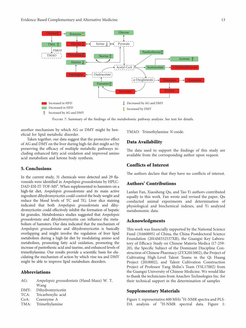
another mechanism by which AG or DMY might be ben-eficial for lipid metabolic disorder.
Taken together, our data suggest that the protective effectof AG and DMY on the liver during high-fat diet might act bypreserving the efficacy of multiple metabolic pathways in-cluding enhanced fatty acid oxidation and improved aminoacid metabolism and ketone body synthesis.
5. Conclusions
In the current study, 31 chemicals were detected and 29 fla-vonoids were identified in Ampelopsis grossedentata by HPLC-DAD-ESI-IT-TOF-MSn. When supplemented to hamsters on ahigh-fat diet, Ampelopsis grossedentata and its main activeingredient dihydromyricetin could control the body weight andreduce the blood levels of TC and TG. Liver slice stainingindicated that both Ampelopsis grossedentata and dihy-dromyricetin could effectively inhibit the formation of hepaticfat granules. Metabolomics studies suggested that Ampelopsisgrossedentata and dihydromyricetin can influence the meta-bolism of hamsters. Our data indicated that the mechanism ofAmpelopsis grossedentata and dihydromyricetin is basicallyoverlapping and might involve the regulation of liver lipidmetabolism during a high-fat diet by modulating amino acidmetabolism, promoting fatty acid oxidation, promoting theincrease of pantothenic acid and taurine, and enhanced levels oftrimethylamine. Our results provide a scientific basis for elu-cidating the mechanism of action by which vine tea and DMYmight be able to improve lipid metabolism disorders.
Abbreviations
AG: Ampelopsis grossedentata (Hand-Mazz) W. T.Wang
DMY: DihydromyricetinTCA: Tricarboxylic acidCoA: Coenzyme ATMA: Trimethylamine
TMAO: Trimethylamine N-oxide.
Data Availability
6e data used to support the findings of this study areavailable from the corresponding author upon request.
Conflicts of Interest
6e authors declare that they have no conflicts of interest.
Authors’ Contributions
Lanlan Fan, Xiaosheng Qu, and Tao Yi authors contributedequally to this work. Fan wrote and revised the paper, Quconducted animal experiments and determination ofphysiological and biochemical indexes, and Yi analyzedmetabonomic data.
Acknowledgments
6is work was financially supported by the National ScienceFund (31660095) of China, the China Postdoctoral ScienceFoundation (2014M552537XB), the Guangxi Key Labora-tory of Efficacy Study on Chinese Materia Medica (17-259-20), the Specific Subject of the Dominant Discipline Con-struction of Chinese Pharmacy (ZYX2015002), the Project ofCultivating High-Level Talent Teams in the Qi HuangProject (2018002), and Talent Cultivation ConstructionProject of Professor Yang Shilin’s Team (YSL17002) fromthe Guangxi University of Chinese Medicine. We would liketo thank the technicians from Anachro Technologies Inc. fortheir technical support in the determination of samples
Supplementary Materials
Figure 1: representative 600MHz 1H-NMR spectra and PLS-DA analysis of 1H-NMR spectral data. Figure 1:
Decreased in HFDIncreased in HFD
Increased by AG and DMY
Decreased by AG and DMY
Increased by DMY
Pyruvate
Acetyl-CoA
α-Oxoglutarate Glutamate Glutamine
ProlineSuccinate
Oxaloacetate
Fumatate
Asparagine
Alanine
Glucose
Panthotherate
Acetoacetate 3-Hydroxybutyrate
AcetoneTaurine
SerineGlycine
Betanine
Threonine
Choline
TMA
TMAOFMO3
Figure 7: Summary of the findings of the metabolomic pathway analysis. See text for details.
Evidence-Based Complementary and Alternative Medicine 13
representative 1H-NMR spectra at 600MHz of the liver (a)and plasma (b) from golden hamster dosed with high-fat dietsupplemented with Ampelopsis grossedentata group (2 g/kg.d). Figure 2: liver 1H NMR spectrum: (a) normal diet(ND); (b) high-fat diet (HFD); (c) high-fat diet supple-mented with Ampelopsis grossedentata (HFD+AG); (d),high-fat diet supplemented with dihydromyricetin group(HFD+DMY). Table 1: metabolic pathway analysis ofgolden hamster (HFD vs ND). Table 2: metabolic pathwayanalysis of golden hamster (HFD vs HFD+AG). Table 3:metabolic pathway analysis of golden hamster (HFD vsHFD+DMY). (Supplementary Materials)
References
[1] Food and Drug Administration of Guangxi Zhuang Auton-omous Region, Quality Standard of Zhuang Medicine inGuangxi Zhuang Autonomous Region, vol. 1, Guangxi Scienceand Technology Press, Nanning, China, 2008.
[2] Q. Gao, R. Ma, L. Chen et al., “Antioxidant profiling of vinetea (Ampelopsis grossedentata): off-line coupling heart-cuttingHSCCC with HPLC-DAD-QTOF-MS/MS,” Food Chemistry,vol. 225, pp. 55–61, 2017.
[3] Y. Chen, Y. Zhang, Y. Dai, and Z. Tang, “Systems pharma-cology approach reveals the antiinflammatory effects ofAmpelopsis grossedentata on dextran sodium sulfate-inducedcolitis,” World Journal of Gastroenterology, vol. 24, no. 13,pp. 1398–1409, 2018.
[4] W. Wan, B. Jiang, L. Sun, L. Xu, and P. Xiao, “Metabolomicsreveals that vine tea (Ampelopsis grossedentata) prevents high-fat-diet-induced metabolism disorder by improving glucosehomeostasis in rats,” PLoS One, vol. 12, no. 8, Article IDe0182830, 2017.
[5] J. Chen, Y. Wu, J. Zou, and K. Gao, “α-glucosidase inhibitionand antihyperglycemic activity of flavonoids from Ampelopsisgrossedentata and the flavonoid derivatives,” Bioorganic &Medicinal Chemistry, vol. 24, no. 7, pp. 1488–1494, 2016.
[6] T. Murakami, M. Miyakoshi, D. Araho et al., “Hep-atoprotective activity of tocha, the stems and leaves ofAmpelopsis grossedentata, and ampelopsin,” BioFactors,vol. 21, no. 1–4, pp. 175–178, 2004.
[7] L. Fan, L. He,W.Wei, F. Cao, Y. Zhang, and J. Miao, “Contentdetermination of dihydromyricetin and myricetin in leaves ofAmpelopsis grossedentata by UPLC and investigation of theirthermal stability,” China Pharmacy, vol. 23, no. 35,pp. 3316–3319, 2012.
[8] K. J. Fan, B. Yang, Y. Liu, X. D. Tian, and B.Wang, “Inhibitionof human lung cancer proliferation through targeting stromalfibroblasts by dihydromyricetin,”Molecular Medicine Reports,vol. 16, no. 6, pp. 9758–9762, 2017.
[9] F. Z. Zhou, “Synergy and attenuation effects of dihy-dromyricetin on tumor-bearing mice affected by breast cancertreated with chemotherapy,” Journal of South China Uni-versity of Technology, vol. 39, no. 9, pp. 147–151, 2011.
[10] Y. Zhou, F. Shu, X. Liang et al., “Ampelopsin induces cellgrowth inhibition and apoptosis in breast cancer cells throughROS generation and endoplasmic reticulum stress pathway,”PLoS One, vol. 9, no. 2, Article ID e89021, 2014.
[11] Q. Li, J. Wang, X. Zhu et al., “Dihydromyricetin preventsmonocrotaline-induced pulmonary arterial hypertension in rats,”Biomedicine & Pharmacotherapy, vol. 96, pp. 825–833, 2017.
[12] T. T. Liu, Y. Zeng, K. Tang, X. M. Chen, W. Zhang, andX. L. Xu, “Dihydromyricetin ameliorates atherosclerosis in
LDL receptor deficient mice,” Atherosclerosis, vol. 262,pp. 39–50, 2017.
[13] Y. Zeng, Y. Peng, K. Tang et al., “Dihydromyricetin ame-liorates foam cell formation via LXRα-ABCA1/ABCG1-de-pendent cholesterol efflux in macrophages,” Biomedicine &Pharmacotherapy, vol. 101, pp. 543–552, 2018.
[14] H. Ling, Z. Zhu, J. Yang et al., “Dihydromyricetin improvestype 2 diabetes-induced cognitive impairment via suppressingoxidative stress and enhancing brain-derived neurotrophicfactor-mediated neuroprotection in mice,” Acta Biochimica etBiophysica Sinica, vol. 50, no. 3, pp. 298–306, 2018.
[15] J. Liang, A. Kerstin Lindemeyer, Y. Shen et al., “Dihy-dromyricetin ameliorates behavioral deficits and reversesneuropathology of transgenic mouse models of Alzheimer’sdisease,” Neurochemical Research, vol. 39, no. 6,pp. 1171–1181, 2014.
[16] Z. X. Ren, Y. F. Zhao, T. Cao, and X. C. Zhen, “Dihy-dromyricetin protects neurons in an MPTP induced model ofParkinson’s disease by suppressing glycogen synthase kinase-3 beta activity,” Acta Pharmacologica Sinica, vol. 37, no. 10,pp. 1315–1324, 2016.
[17] J. Liang, Y. Shen, X. M. Shao et al., “Dihydromyricetin pre-vents fetal alcohol exposure-induced behavioral and physi-ological deficits: the roles of GABAA receptors inadolescence,” Neurochemical Research, vol. 39, no. 6,pp. 1147–1161, 2014.
[18] J. Zhang, Y. Chen, H. Luo et al., “Recent update on thepharmacological effects and mechanisms of dihydromyr-icetin,” Frontiers in Pharmacology, vol. 9, p. 1204, 2018.
[19] Q. Song, L. Liu, J. Yu et al., “Dihydromyricetin attenuated AngII induced cardiac fibroblasts proliferation related to in-hibitory of oxidative stress,” European Journal of Pharma-cology, vol. 807, pp. 159–167, 2017.
[20] Y. Chen, H. Q. Luo, L. L. Sun et al., “Dihydromyricetin at-tenuates myocardial hypertrophy induced by transverse aorticconstriction via oxidative stress inhibition and SIRT3 pathwayenhancement,” International Journal of Molecular Sciences,vol. 19, no. 9, p. 2592, 2018.
[21] L. Wei, X. Sun, X. Qi, Y. Zhang, Y. Li, and Y. Xu, “Dihy-dromyricetin ameliorates cardiac ischemia/reperfusion injurythrough Sirt3 activation,” BioMed Research International,vol. 2019, Article ID 6803943, 9 pages, 2019.
[22] R. Taub, “Liver regeneration: from myth to mechanism,”Nature Reviews Molecular Cell Biology, vol. 5, no. 10,pp. 836–847, 2004.
[23] C. E. Liu, H. Y. Wang, Y. D. Wang, and J. Zhang, “Study onAmpelsis grossedentata to lower plasma lipids level,” FoodScience, vol. 26, no. 11, pp. 237–240, 2005.
[24] Y. Li, Z. Tan, T. Li, B. Xiao, and Q. Dai, “Effects of Ampelopsisgrossedentata on hyperlipidemia and myocardial enzymes inrats,” Acta Nutrimenta Sinica, vol. 6, pp. 506–509, 2006.
[25] X. Zeng, G. Wei, H. Fei et al., “Effect of total flavonoids ofAmpelopsis grossedentata on blood lipid and hemor-rheologyin in atherosclerosis rats,” Chongqing Medicine,vol. 5, pp. 518–520, 2014.
[26] Y. S. Zhang, Z. X. Ning, S. Z. Yang, and H. Wu, “Anti-oxidation properties and mechanism of action of dihy-dromyricetin from Ampelopsis grossedentata,” ActaPharmaceutica Sinica, vol. 38, no. 4, pp. 241–244, 2003.
[27] J. C. Shen, L. Y. Lian, Y. Zhang et al., “Dynamic Analysis ofmetabolic response in gastric ulcer (GU) rats with electro-acupuncture treatment using 1H NMR-based metabolomics,”Evidence-Based Complementary Alternative Medicine,vol. 2019, Article ID 1291427, 12 pages, 2019.
14 Evidence-Based Complementary and Alternative Medicine
[28] E. J. Kim, Y. S. Hong, S. H. Seo, S. E. Park, C. S. Na, andH. S. Son, “Metabolite markers for characterizing Sasangconstitution type through GC-MS and 1H NMR-basedmetabolomics study,” Evidence-Based Complementary Al-ternative Medicine, vol. 2019, Article ID 8783496, 11 pages,2019.
[29] X. Li, H. Chen, W. Jia, and G. Xie, “A metabolomics-basedstrategy for the quality control of traditional Chinese medi-cine: shengmai injection as a case study,” Evidence-BasedComplementary Alternative Medicine, vol. 2013, Article ID836179, 8 pages, 2013.
[30] J. Hao, H. Hu, J. Liu et al., “Integrated metabolomics andnetwork pharmacology study on immunoregulation mecha-nisms of Panax ginseng through macrophages,” Evidence-Based Complementary Alternative Medicine, vol. 2019, ArticleID 3630260, 14 pages, 2019.
[31] Z. X. Miao, L. Yang, C. Y. Jiang, Y. H. Wang, and H. B. Zhu,“Evaluation of dose-related effects of 2′, 3′, 5′-tri-O-acetyl-N6-(3-hydroxylaniline) adenosine using NMR-basedmetabolomics,” Acta Pharmaceutica Sinica, vol. 49, no. 5,pp. 679–685, 2014.
[32] F. Guo, T. Zi, L. Liu, R. Feng, and C. Sun, “A 1H-NMR basedmetabolomics study of the intervention effect of mangiferinon hyperlipidemia hamsters induced by a high-fat diet,” Food& Function, vol. 8, no. 7, pp. 2455–2464, 2017.
[33] J. Chong, M. Yamamoto, and J. Xia, “MetaboAnalystR 2.0:from raw spectra to biological insights,” Metabolites, vol. 9,no. 3, p. 57, 2019.
[34] L. Fan, T. Yi, F. Xu et al., “Characterization of flavonoids inthe ethomedicine fordiae cauliflorae radix and its adulterantmillettiae pulchrae radix by HPLC-DAD-ESI-IT-TOF-MSn,”Molecules, vol. 18, no. 12, pp. 15134–15152, 2013.
[35] D. Wang, J. Liu, J. Zhang, and S. Zheng, “6e chemicalconstituents of Ampelopsis grossedentata,” Subtropical PlantResearch Communications, vol. 27, no. 2, pp. 39–44, 1998.
[36] Y. Zhang, W. Yang, and H. Xiong, “Basic constituent ofAmpelopsis grossedentata,” Natural Product Research Devel-opment, vol. 13, no. 5, pp. 46–48, 2001.
[37] W. Yan, Z. Liling, R. Li, and Y. Wang, “Studies on the chemicalconstituents fromAmpelopsis grossedentata,” Journal of ChineseMedicinal Materials, vol. 25, no. 4, pp. 254–256, 2002.
[38] Y. Cheng, “Research development of Ampelopsis grossedentata,”Economic Forest Researches, vol. 22, no. 3, pp. 51–56, 2004.
[39] Q. Du, P. Chen, G. Jerz, and P. Winterhalter, “Preparativeseparation of flavonoid glycosides in leaves extract ofAmpelopsis grossedentata using high-speed counter-currentchromatography,” Journal of Chromatography A, vol. 1040,no. 1, pp. 147–149, 2004.
[40] G. He, G. Pei, F. Du, Y. Ou, and B. Li, “Studies on the chemicalconstituents of Ampelopsis grossedentata,” Modern ChineseMedicine, vol. 9, no. 12, pp. 11–13, 2007.
[41] X. X. Qi, Y. Dong, C. X. Shan, Y. Y. Xiang, and X. D. Wang,“Analysis of the main components in herbal pairs: astrogaliradix and Salviae miltiorrhizae by UPLC-Q-TOF/MS,”Journal of Nanjing University of Traditional Chinese Medicine,vol. 23, no. 1, pp. 93–96, 2017.
[42] C. Y. Jiang, K. M. Yang, L. Yang, Z. X. Miao, Y. H. Wang, andH. B. Zhu, “A 1H NMR based metabonomics approach toprogression of coronary atherosclerosis in a hamster model,”Acta Pharmaceutica Sinica, vol. 48, no. 4, pp. 495–502, 2013.
[43] M. T. Velasquez, A. Ramezani, A. Manal, and D. S. Raj,“Trimethylamine N-oxide: the good, the bad and the un-known,” Toxins, vol. 8, no. 11, p. 326, 2016.
[44] Z. H. Zhao, F. Z. Xin, D. Zhou et al., “TrimethylamineN-oxideattenuates high-fat high-cholesterol diet-induced steatohe-patitis by reducing hepatic cholesterol overload in rats,”World Journal of Gastroenterology, vol. 25, no. 20,pp. 2450–2462, 2019.
Evidence-Based Complementary and Alternative Medicine 15
Related Documents