Pak. J. Bot., 45(1): 329-340, 2013. ROCK ALGAE OF BATKHELA DISTRICT MALAKAND, PAKISTAN BARKATULLAH 1 , FARRUKH HUSSAIN 2 , NIAZ ALI 1 AND IMTIAZ AHMAD 1 1 Department of Botany, University of Peshawar, Pakistan. 2 Centre of Plant Biodiversity, University of Peshawar, Pakistan. Abstract The present study describes a total of 63 lithophytic algal species belonging to Cyanophyta (11 genera, 30 species), Chlorophyta (3 genera, 7 species) and Bacillariophyta (16 genera, 26 species) from rocks of Batkhela, District Malakand. The collected specimens were identified and described. This is the first ever report on rock algae from this area. Introduction Worldwide, algae are significant for producing half of photosynthetic organic materials that provide bases for food webs of variety of the animals (Valeem & Shameel, 2009). Algae play a significant role in the settlement of environment for human beings (Akhtar & Rehman, 2009). The present study described algae from rocks of Batkhela, District Malakand. Some previous workers also reported algae from rocks (Anjum et al., 1985; Anjum & Faridi, 1985; Hussain et al., 2003; Naz et al., 2004; Zarina et al., 2009; Ali et al., 2010). Recently Hussain et al., (2009, 2010) reported some algae from soil of KPK, Pakistan. The review of literature suggested that no work has been done exclusively on lithophtic algae in this area. This study is the first ever report on rock algae from District Malakand, Pakistan. Materials and Methods Batkhela District Malaknd is located at coordinates of 34°35′N 71°57′E/ 34.583°N 71.95°E in the Khyber Pukhtunkhwah, Pakistan. Algal samples were collected from the different rocks located at Nasir khan dub, Yakh dand, Zwarande oba, Amandara head works, and Gumbat with the help of tooth brush and knife from rock surfaces near water bodies. The collected samples were taken in polythene bags with little quantity of water and was mixed thoroughly till it got homogenised. The materials were then transferred to bottle for preservation in 4% formalin solution (Anon., 1985; Mason, 1967). Identification: The specimens were then identified with the help of available literature (Smith, 1950; Prescott, 1961; Tiffany & Britton, 1971; Desikachary, 1959) using BH-2 Olympus (Japan) microscope. Results and Discussion Algae are reported from variety of fresh water marine and marine habitats. However they also occur in wide variety of terrestrial environments where they form conspicuous growth. These include rocks, urban walls, metals, tree barks, leaves and animal hairs (Nakano, 1997, Lopez-Bautista et al., 2007; Silva et al., 2010). Such algae may not complete their reproductive stages without water (Lewis & McCourt, 2004). The present attempt to explore algal flora from rocks of Batkhela District Malakand indicated a total of 63 lithophytic species (Table 1). These belonged to Cyanophyta (11 genera, 30 species), Chlorophyta (3 genera, 7 species) and Bacillariophyta (16 genera, 26 species) were identified and described. Oscillatoria (11 species), Lyngbya (6 species) Chroococcus (3 species), Aphanocapsa, Gloecapsa (2 species each), Chlorogloea, Microcystis, Microcoleus, Neidium, Phrmidium and Spirolina, (Each with one species) were the blue green algae. Closterium (3 species), Ulothrix (2 species) and Cosmerium (one species) were beloged to Chlorophyta. While Fragilaria, Navicula (4 species each), Cymbella, Epithemia, Gomphonema, Nitzschia (2 species each), Amphipleura, Amphora, Arthrospira, Calonies, Diatoma, Frustule, Meridion, Pinnularia, Rhapalodea and Syenedra (each with one species) belonged to the Bacillariophyta. Aphanocapsa (Nag): Cells spherical or nearly so, many loosely arranged without an order, forming a formless gelatinous mass. Aphanocapsa biformis A. Braun Cells 4-7μ diameter, spherical, loosely arranged 2-4 together in a common mucilaginous envelope (Fig. 1). Aphanocafsa endophytica G. M. Smith Endophytic in the colonial mucilage of microcystis. cells solitary or arrange in small clumps, cell contents homogenous, pale to bright blue green, 2 μ in diameter (Fig. 2). Arthrospira massartii (Kuffareth) Trichomes loosely coiled, cells 5-6μ broad, 2-4μ long end cells rounded conical, cross-walls not granulated (Fig. 3). Chlorogloea fritschii (Mitra) Cells arranged in vertical and horizontal rows, rounded or angular usually 6-8μ diameter, single or in groups of 2 or more cells separating for propagation (Fig. 4). Chroococcus (Nag) Cells spherical or sub spherical, hemispherical, after division in small groups of 2-4 individuals, sometimes 8- 16, rarely single, in gelatinous or mucous matrix.

Welcome message from author
This document is posted to help you gain knowledge. Please leave a comment to let me know what you think about it! Share it to your friends and learn new things together.
Transcript
Pak. J. Bot., 45(1): 329-340, 2013.
ROCK ALGAE OF BATKHELA DISTRICT MALAKAND, PAKISTAN
BARKATULLAH1, FARRUKH HUSSAIN2, NIAZ ALI1 AND IMTIAZ AHMAD1
1Department of Botany, University of Peshawar, Pakistan. 2Centre of Plant Biodiversity, University of Peshawar, Pakistan.
Abstract
The present study describes a total of 63 lithophytic algal species belonging to Cyanophyta (11 genera, 30 species),
Chlorophyta (3 genera, 7 species) and Bacillariophyta (16 genera, 26 species) from rocks of Batkhela, District Malakand. The collected specimens were identified and described. This is the first ever report on rock algae from this area.
Introduction
Worldwide, algae are significant for producing half of photosynthetic organic materials that provide bases for food webs of variety of the animals (Valeem & Shameel, 2009). Algae play a significant role in the settlement of environment for human beings (Akhtar & Rehman, 2009). The present study described algae from rocks of Batkhela, District Malakand. Some previous workers also reported algae from rocks (Anjum et al., 1985; Anjum & Faridi, 1985; Hussain et al., 2003; Naz et al., 2004; Zarina et al., 2009; Ali et al., 2010). Recently Hussain et al., (2009, 2010) reported some algae from soil of KPK, Pakistan. The review of literature suggested that no work has been done exclusively on lithophtic algae in this area. This study is the first ever report on rock algae from District Malakand, Pakistan. Materials and Methods
Batkhela District Malaknd is located at coordinates of 34°35′N 71°57′E/ 34.583°N 71.95°E in the Khyber Pukhtunkhwah, Pakistan. Algal samples were collected from the different rocks located at Nasir khan dub, Yakh dand, Zwarande oba, Amandara head works, and Gumbat with the help of tooth brush and knife from rock surfaces near water bodies. The collected samples were taken in polythene bags with little quantity of water and was mixed thoroughly till it got homogenised. The materials were then transferred to bottle for preservation in 4% formalin solution (Anon., 1985; Mason, 1967). Identification: The specimens were then identified with the help of available literature (Smith, 1950; Prescott, 1961; Tiffany & Britton, 1971; Desikachary, 1959) using BH-2 Olympus (Japan) microscope. Results and Discussion
Algae are reported from variety of fresh water marine and marine habitats. However they also occur in wide variety of terrestrial environments where they form conspicuous growth. These include rocks, urban walls, metals, tree barks, leaves and animal hairs (Nakano, 1997, Lopez-Bautista et al., 2007; Silva et al., 2010). Such algae may not complete their reproductive stages without water (Lewis & McCourt, 2004). The present attempt to explore algal flora from rocks of Batkhela District Malakand indicated a total of 63 lithophytic species
(Table 1). These belonged to Cyanophyta (11 genera, 30 species), Chlorophyta (3 genera, 7 species) and Bacillariophyta (16 genera, 26 species) were identified and described. Oscillatoria (11 species), Lyngbya (6 species) Chroococcus (3 species), Aphanocapsa, Gloecapsa (2 species each), Chlorogloea, Microcystis, Microcoleus, Neidium, Phrmidium and Spirolina, (Each with one species) were the blue green algae. Closterium (3 species), Ulothrix (2 species) and Cosmerium (one species) were beloged to Chlorophyta. While Fragilaria, Navicula (4 species each), Cymbella, Epithemia, Gomphonema, Nitzschia (2 species each), Amphipleura, Amphora, Arthrospira, Calonies, Diatoma, Frustule, Meridion, Pinnularia, Rhapalodea and Syenedra (each with one species) belonged to the Bacillariophyta. Aphanocapsa (Nag): Cells spherical or nearly so, many loosely arranged without an order, forming a formless gelatinous mass. Aphanocapsa biformis A. Braun
Cells 4-7µ diameter, spherical, loosely arranged 2-4 together in a common mucilaginous envelope (Fig. 1). Aphanocafsa endophytica G. M. Smith
Endophytic in the colonial mucilage of microcystis. cells solitary or arrange in small clumps, cell contents homogenous, pale to bright blue green, 2 µ in diameter (Fig. 2). Arthrospira massartii (Kuffareth)
Trichomes loosely coiled, cells 5-6µ broad, 2-4µ
long end cells rounded conical, cross-walls not granulated (Fig. 3). Chlorogloea fritschii (Mitra)
Cells arranged in vertical and horizontal rows, rounded or angular usually 6-8µ diameter, single or in groups of 2 or more cells separating for propagation (Fig. 4). Chroococcus (Nag)
Cells spherical or sub spherical, hemispherical, after division in small groups of 2-4 individuals, sometimes 8-16, rarely single, in gelatinous or mucous matrix.
BARKATULLAH ET AL., 330
Cyanophyceae
Key to genera
1. Cell unicellular or colonial ….………………….......………………..……….……………………..…………........ 2 1. Cell united to form trichome ………………………...……………………………..………………………………. 62. Cells arrange radially in mucilage ……….....………………..……………………..………………….. Chlorogloea2. Cell spherical arrange irregularly in mucilage ……...…………………..………………………………………….. 33. Cells closely packed in colony ……………………………….…….…………………………………… Microcystis3. Cells loosely arranged in colony ……..…………………………………...………..………………………………. 44. Colored mucilage, 2-8 cells ….…………………………………………………………….…………….. Gleocapsa4. Colorless mucilage, spherical cells ….………………………………….………………………………………….. 55. Small spherical cells, many in mucilage …...…………………..…........................................................ Aphanocafsa5. Large spherical cells, 2-4 in mucilage .………...……………………………………………………….. Chroococus6. Trichome without sheath .……………………………..………………..…………...…………….…..……………. 7 6. Trichome with sheath ………………………………….…….................................................................................... 97. Trichome coiled/spiriled, septa not visible ……………………………………………...………………… Spirulina7. Trichome not coiled, septa visible …………………..…………………………………...……………………….... 88. Trichome strait, cell wider than length, many septa …..…………………………………………..……. Oscillatoria8. Trichome isopolar, cells cylindrical, 4-8 septa ……………………………………………...…..……… Arthrospira9. Trichome not in bundle in mucilage ………………………………………...…………….……………….. Lyngbya9. Trichome many, arrange in cluster in mucilage .………………………………………….…………………......... 1010. Trichomes entangled in mucilage, constrictd at cross wall ………...……………………………..…… Phormidium10. Trichome many in mucilage, not constricted at cross wall ...……………………………………...…… Microcoleus
Key to species
1. Cells 4-7µ in diameter …………………………………………………………………………………… A. biformis1. Cells 2 µ in diameter ………………………………………………………………………………… A. endophytica
Key to species
1. Sheath thin hardly visible …...………………….………………………………………………………….. C. minor1. Sheath thick, distinct ……………………………………………………………………………………………….. 22. Cell diameter, 4-8µ with sheath ………………………………………………………………………….... C. varius2. Cell diameter, 8-14µ with sheath ………………………………………………………………...……. C. limneticus
Chroococcus limneticus (Lemm.)
Cells spherical or sub spherical after division, 4-32, free floating, mostly in a tabular gelatinous layer, without sheath cells 6-12µ diameter (Fig. 5). Chroococcus minor (Kutz)
Thallus slimy- gelatinous, cells spherical, 3-4µ in
diam., singly or in pairs, sheath colorless, very thin (Fig. 6). Chroococcus varius A. Br.
Thallus gelatinous, dirty olive green or brownish; cells globular single or 2-4 to gather, irregularly arranged without sheath 2-4 µ, with sheath 4-8 µ (Fig. 7). Gloeocapsa Kutzing
Cells spherical, 2-8 in colonies, seldom many, with a number of concentric special envelopes, colonies single or many forming an expanded mass, individual sheaths
lamellated or unlamellated, cells in large colonies usually with secondary colonies. Gloeocapsa calcarea Tilden
Cell with or without individual sheath, 6-9 µ in diameter, blue-green, sheath colorless often thin, colonies 25-50 µ in diameter, with 4-16 cells (Fig. 8). Gloeocapsa magma (Berb) Kutz
Colonies spherical or irregularly arranged, 30-60 µ in diameter, cells spherical or angular, 3-7 µ in diameter, blue-green, mostly with a thin, 0.5-1.5 µ broad, colored individual sheath (Fig. 9). Lyngbya Ag.
Trichomes single or free in a thin or very massive thick, firm sheath, sheath mostly colorless, seldom colored yellow to brown or red, filaments sometimes coiled or attached, mostly without such attachments.
ROCK ALGAE OF BATKHELA DISTRICT MALAKAND, PAKISTAN 331
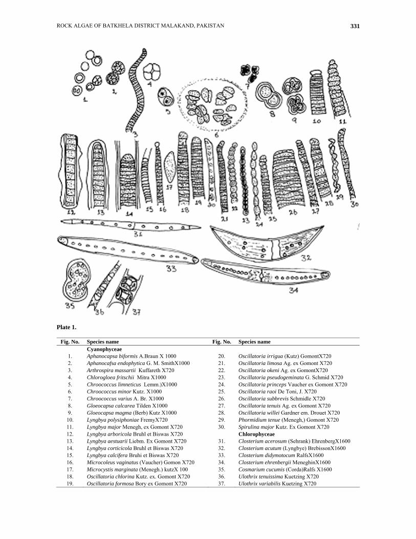
Plate 1.
Fig. No. Species name Fig. No. Species name Cyanophyceae
1. Aphanocapsa biformis A.Braun X 1000 20. Oscillatoria irrigua (Kutz) GomontX720 2. Aphanocafsa endophytica G. M. SmithX1000 21. Oscillatoria limosa Ag. ex Gomont X720 3. Arthrospira massartii Kuffareth X720 22. Oscillatoria okeni Ag. ex GomontX720 4. Chlorogloea fritschii Mitra X1000 23. Oscillatoria pseudogeminata G. Schmid X720 5. Chroococcus limneticus Lemm.)X1000 24. Oscillatoria princeps Vaucher ex Gomont X720 6. Chroococcus minor Kutz. X1000 25. Oscillatoria raoi De Toni, J. X720 7. Chroococcus varius A. Br. X1000 26. Oscillatoria subbrevis Schmidle X720 8. Gloeocapsa calcarea Tilden X1000 27. Oscillatoria tenuis Ag. ex Gomont X720 9. Gloeocapsa magma (Berb) Kutz X1000 28. Oscillatoria willei Gardner em. Drouet X720 10. Lyngbya polysiphoniae FremyX720 29. Phormidium tenue (Menegh,) Gomont X720 11. Lyngbya major Menegh, ex Gomont X720 30. Spirulina major Kutz. Ex Gomont X720 12. Lyngbya arboricola Bruhl et Biswas X720 Chlorophyceae 13. Lyngbya aestuarii Liebm. Ex Gomont X720 31. Closterium acerosum (Sehrank) EhrenbergX1600 14. Lyngbya corticicola Bruhl et Biswas X720 32. Closterium acutum (Lyngbye) BrebissonX1600 15. Lyngbya calcifera Bruhi et Biswas X720 33. Closterium didymotocum RalfsX1600 16. Microcoleus vaginatus (Vaucher) Gomon X720 34. Closterium ehrenbergii MeneghinX1600 17. Microcystis marginata (Menegh.) kutzX 100 35. Cosmarium cucumis (Corda)Ralfs X1600 18. Oscillatoria chlorina Kutz. ex. Gomont X720 36. Ulothrix tenuissima Kuetzing X720 19. Oscillatoria formosa Bory ex Gomont X720 37. Ulothrix variabilis Kuetzing X720
BARKATULLAH ET AL., 332
Key to species
1. Colonies 25-50 µ in diameter, with 4-16 cells …………………………………………….……………. G. calcarea1. Colonies 30-60 µ in diameter, with 4-8 cells ……………………………………………..……………… G. magma
Key to species
1. Cells ½-1/3 as long as broad ……………………………………………………………………………………….. 21. Cells otherwise ……………………………………………………………………………………………………... 32. Filaments flexuous, 12-26 µ broad …………………………………………………...………………. L. corticicola2. Filaments short, 2 µ broad …….…………………...……………………..…………………..……. L. polysiphoniae3. Trichomes constricted ……………………………………………………….……………………………………... 43. Trichomes not constricted ……………………………………………………………..…………………………… 54. Septa not granulated …………………………………………………………..………………………. L. arboricola4. Septa granulated with calcium carbonate ……………………………………………………….………. L. calcifera5. Cells 11-16 µ broad ……………………………………………...........……………………………………. L. major5. Cells 8-24 µ broad ……………………………………………...……………………………………….. L. aestuarii
Lyngbya aestuarii Liebm. Ex Gomont
Filaments single or forming a brown or dull blue-green thallus, nearly straight or coiled, sheath yellow brown, lamellated, cells 8-24 µ broad, 2.7-5.6 µ long, not constricted at the cross walls, cross walls often granulated (Fig. 10). Lyngbya arboricola Bruhl et Biswas
Filaments nearly straight, 18-22 µ broad, sheath reddish brown, firm, 1.5-2 µ thick, homogeneous, trichome distinctly constricted, septa not granulated, cells 5-6 µ long, contents blue green (Fig. 11). Lyngbya calcifera Bruhi et Biswas
Filament 1-2 mm in diameter, filaments are incrusted by calcium carbonate, cells about 4 µ in diameter, 6-10 µ long (Fig. 12). Lyngbya corticicola Bruhi et Biswas
Filaments fragile, moderately flexuous, 12-26 µ thick, sheath at first hyaline, but later becoming brown, 2 µ thick, trichome slightly constructed at the joints, cells about ½ to 1/3 as long as broad, apex rotund, contents green and granular (Fig. 13). Lyngbya major Menegh, ex Gomont
Filaments long, straight, sheath thick, colorless, lamellated, cells 11-16 µ broad, ¼ to 1/8 as long as broad, cell rounded with slightly thickened membrane (Fig. 14). Lyngbya polysiphoniae Fremy
Filaments straight or curved, single or in bundle, up to 200 µ long, sheath very thin, cells ½ to 1/3 as long as broad, cross walls visible, end cells convex not capitate (Fig. 15).
Microcoleus vaginatus (Vaucher) Gomont
Cells 3.5-7µ broad, sub quadrate or 1/2-2 times as long as broad, 3-7µ long. Often granulated at the cross-walls, but not constricted, blue green or green; end cells capitate, with flat conical calyptra (Fig. 16). Microcystis marginata (Menegh.) kutz
Colony round, irregular flattened, simple, mucilage distinct, single colony ellipsoid to ovoid, 140-150µ in length and 60-95µ in width; cells 3-6 µ in diameter (Fig. 17). Oscillatoria Vaucher
Trichome single or forming a flat or spongy free-swimming thallus, sheath absent, motile, mostly by creeping movement, ends of trichome distinctly marked, pointed, bent like sickle or coiled more or less like a screw. Oscillatoria chlorina Kutz. ex. Gomont
Trichome straight or curved, not constricted at the cross walls, 3.4-4 µ broad, Cells sometime longer than broad, cross walls not granulated, calyptra absent (Fig. 18). Oscillatoria formosa Bory ex Gomont
Trichome straight, slightly constricted at the cross walls, 4-6µ wide, attenuated at the ends and bent, ½ as long as broad, 2.5-5µ long, calyptra absent, not capitate (Fig. 19). Oscillatoria irrigua (Kutz) Gomont
Thallus blackish blue-green, trichome straight, 6-11 µ broad, apex slightly attenuated, Cells quadrate, 4-11 µ long, septa not granulated, apical cell convex with an evident thickened wall (Fig. 20).
ROCK ALGAE OF BATKHELA DISTRICT MALAKAND, PAKISTAN 333
Key to species
1. Cells ½ as long as broad …………………………………........................................................................................ 21. Cells otherwise ……………………………………………………………………………………………………... 32. Cells 6-11 µ broad ………………………………..………………………………………….….………... O. irrigua2. Cells 5.5-9 µ broad …………………………………...………………………...…………….……….......... O. okeni3. Trichomes constricted at the cross walls ………………..…………………………………………………………. 43. Trichomes not constricted at cross walls …………………………………………………………………………... 54. End cells hemispherical …………………………………………………………….……………………… O. tenuis4. End cells nearly obtuse ……………………………………………...……………..…………………… O. Formosa5. Cell walls frequently granulated ………………………………………………..…………………………. O. limosa5. Cell walls not granulated ……………………………………………………....………............……………...…… 66. Trichomes strait ………………………………………………………………………………………………......... 76. Trichomes bent\ coiled ……………………………………………………..……….……………………...………. 87. Trichomes, 1.3-2.2 µ broad …………………………….……………………………………….. O. pseudogeminata7. Trichomes bent, 2.4-3.6 µ broad ……………………………………………..…………………………….. O. willei8. Trichome attenuated at the apex, 16-60 µ broad ………………………………………….…………….. O. princeps8. Trichome notattenuated at the apex, less than16µ broad ………………………………………………………….. 99. Cell length less than 2 µ long ………………………………………………………………………..… O. subbrevis9. Cell length more than 2 µ long ……………………………………………….…………………………………... 1010. Trichome, 3-4 µ broad ……………………………………………...…………………………………… O. chlorina10. Trichome, 3-4 µ broad …………………………………………………..…………………………………… O. raoi
Oscillatoria limosa Ag. ex Gomont
Trichomes more or less straight, not constricted at the cross walls, commonly 13-16µ broad, cells 2-5µ long, cross walls frequently granulated, end cell flatly round (Fig. 21). Oscillatoria okeni Ag. ex Gomont
Trichome straight, fragile, distinctly constricted at the cross walls, 5.5-9µ broad, at the ends gradually attenuated, cells 1/3 as long as broad, 2.7-4.5µ long, end cells obtuse, not capitate, without calyptra (Fig. 22). Oscillatoria princeps Vaucher ex Gomont
Trichomes blue-green, mostly straight, not constricted at the cross walls, 16-60 µ broad, slightly attenuated at the apices and bent, cells 3.5-7 µ long, end cells flatly rounded with slightly thickened membrane (Fig. 23). Oscillatoria pseudogeminata G. Schmid
Trichomes coiled, pale blue-green, ends not attenuated, 1.3-2.2 µ broad, cells as long as broad, not constricted at the cross walls, cross walls thick, not granulated, end cell rounded, calyptra absent (Fig. 24). Oscillatoria raoi De Toni, J.
Trichome straight, usually of uniform thickness, without constrictions at the joints, 5.2-6µ broad, and cells 2.5-6 µ long, end cells rounded, not capitate, without any calyptra (Fig. 25).
Oscillatoria subbrevis Schmidle
Trichomes single, 5-6µ broad, nearly straight, not attenuated at the apices, cells 1-2 µ long, not granulated at the side walls, end cells round, calyptra absent (Fig. 26). Oscillatoria tenuis Ag. ex Gomont
Trichome straight, fragile, slightly constricted at the cross walls, 4-10µ broad, not attenuated at the apices, not capitate, 2.6-5µ long, end cells hemispherical with thickened outer membrane (Fig. 27). Oscillatoria willei Gardner em. Drouet
Trichome bent at the ends or screw like, broad unconstricted at the cross walls, ends not attenuated, not capitate, 2.4-3.6 µ broad, and end cell rounded without thickened membrane (Fig. 28). Phormidium tenue (Menegh,) Gomont
Thallus pale blue-green, thin, membranous, expanded; trichome straight or slightly bent, densely entangled, slightly constricted at the cross-walls, attenuated at the ends, 1-2µ broad, pale blue-green; cells up to 3 times longer than broad, 2.5-5µ long, calyptra absent (Fig. 29). Spirulina major Kutz. Ex Gomont
Cells 1.2-1.7 µ broad, regularly spirally coiled, blue green, spirals 2.5-4 µ broad and 2.7-5 µ distant (Fig. 30).
BARKATULLAH ET AL., 334
Chlorophyta
Key to genera
1. Unicellular ………………………………………………………………………………………………………….. 21. Multicellular filamentous ……………………………………………...……………………………………. Ulothrix2. Cells without median constriction ……………………………………………….……………………..... Closterium2. Cells with deeply median constriction …………………………………………………………………... Cosmarium
Closterium Nitzch
Cells elongated, usually markedly alternate, rarely straight, strongly arcuate or lunate, without medium constriction, poles obtuse, truncate, rostrate, attenuate, cells wall smooth, costate or striate, colorless or yellow to brown in color. Closterium acerosum (Sehrank) Ehrenberg
Cells 19-53 × 228-530 µ, 8-16 times longer than wide, almost straight, narrowly fusifrom, pyrenoides 7-12 in a median series (Fig. 31). Closterium acutum (Lyngbye) Brebisson
Cells 3.7-11 × 60-180 µ, slightly curved, gradually attenuated to acute apices, cell wall smooth, colourless, chromatophores without ridges, pyrenoides 4-5 in a median series (Fig. 32). Closterium didymotocum Ralfs
Cells 24-56 × 295-672 µ, slightly curved, median portion with sub-parallel sides, gradually and slightly
attenuated to the broad rounded apices, chromatophores with 5-10 large pyrenoids (Fig. 33). Closterium ehrenbergii Meneghini
Cells 60-145 × 285-720 µ, apices 12-18 µ wide, stout, 5 times longer than wide, pyrenoids numerous, cell wall smooth (Fig. 34). Cosmarium cucumis (Corda)Ralfs
Cells 34-56×59-102 µ and 19-38 µ thick, longer than wide, deeply constricted, sinus linear, the apex rounded, semi-cells semi-elliptic, the apex rounded or slightly truncate., cell wall smooth, chromatophores 6-9 in each semi-cell, pyrenoids several in each chromatophore (Fig. 35). Ulothrix Kutzing
Simple unbranched filaments of cylindrical cells, often showing basal differentiation and arising from a special hold fast cell; chloroplast a parietal band which extends 2/3 to ¾ of the way around the cell and sometime extending the entire length of the cell.
Key to species
1. Pyrenoids, 5-12 …………………………………………………………………………………………………….. 21. Pyrenoids, numerous ………………………………………………..………….................................. C. ehrenbergii2. Cells straight, tapering at the apices ……………………………………………………….………….. C. acerosumi2. Cells slightly curved ……………………………………………………………………………………………….. 33. Cells 24-56 µ broad, rounded apices ………...…….……………………………...……………….. C. didymotocum3. Cells 3.7-11µ broad, acute apices ………………………………………………………..……………….. C. acutum
Key to species
1. Chloroplast broad with two or more pyrenoids ………………………………………………… Ulothrix tenuissima1. Chloroplast folded with one pyrenoid …………………………………..……………………….. Ulothrix variabilis
Ulothrix tenuissima Kuetzing
Filaments long, composed of cylindrical cells shorter than wide, 16-20 μm in diameter, thin walled and not constricted at the cross wall, chloroplast, broad encircling 2/3 of the circumference of the cell with 2 or more pyrenoids (Fig. 36).
Ulothrix variabilis Kuetzing
Long, slender, entangled filaments, forming cottony masses. Cells cylindrical, without constriction of the cross-walls, chloroplast a folded parietal plate, ½ to 2/3 the length of the cell, with one pyrenoid. Cells 4.5-6 μ in diameter and up to 15 μ long, forming bright green slimy masses (Fig. 37).
ROCK ALGAE OF BATKHELA DISTRICT MALAKAND, PAKISTAN 335
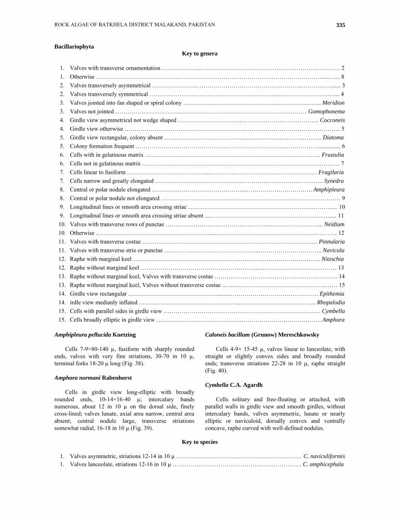
Bacillariophyta Key to genera
1. Valves with transverse ornamentation ………………..………………………………………………….………… 21. Otherwise ……………………………………………………………………………………………………...….... 82. Valves transversely asymmetrical ………………….…………………………………….………..……….……..... 32. Valves transversely symmetrical ……………………………………………………….….…………….…….…... 43. Valves jointed into fan shaped or spiral colony ………………………………………..………………….. Meridion3. Valves not jointed …………………………………………………………………………………… Gomophonema4. Girdle view asymmetrical not wedge shaped …………………………………………………………….. Cocconeis4. Girdle view otherwise …………………………………………………….………………………………………... 55. Girdle view rectangular, colony absent ………………………….………...……………...……………….. Diatoma5. Colony formation frequent ………………………………………………………………….……………................ 66. Cells with in gelatinous matrix ……………………………………………………………………………. Frustulia6. Cells not in gelatinous matrix ……………………………………………………………………………………… 77. Cells linear to fusiform …………………………………...……………………………………………… Fragilaria7. Cells narrow and greatly elongated ……………………………………...…………………………………. Synedra8. Central or polar nodule elongated ………………………………………..……………………………. Amphipleura8. Central or polar nodule not elongated ……………………………………………………………………………… 99. Longitudinal lines or smooth area crossing striae ……………………………………………………………….... 109. Longitudinal lines or smooth area crossing striae absent ...………………………………………………...…...... 1110. Valves with transverse rows of punctae ……………………………………………..……………………... Neidium10. Otherwise …………………………………………………...…………………………………………………….. 1211. Valves with transverse costae ……………………………………………………………………………. Pinnularia11. Valves with transverse strie or punctae ………………………………………...………………………….. Navicula12. Raphe with marginal keel …………………………………………………………………………………. Nitzschia12. Raphe without marginal keel …………………………………………………...………………………………… 1313. Raphe without marginal keel, Valves with transverse costae …………………………………………….………. 1413. Raphe without marginal keel, Valves without transverse costae ..…..……………………………………….…… 1514. Girdle view rectangular ……………………………………….......……………………………………… Epithemia14. irdle view medianly inflated …………………………………………....……………………………….. Rhopalodia15. Cells with parallel sides in girdle view ……………………………………………………………………. Cymbella15. Cells broadly elliptic in girdle view …………………………………….…………………………………. Amphora
Amphipleura pellucida Kuetzing
Cells 7-9×80-140 µ, fusiform with sharply rounded ends, valves with very fine striations, 30-70 in 10 µ, terminal forks 18-20 µ long (Fig. 38). Amphora normani Rabenhorst
Cells in girdle view long-elliptic with broadly rounded ends, 10-14×16-40 µ; intercalary bands numerous, about 12 in 10 µ on the dorsal side, finely cross-lined; valves lunate, axial area narrow, central area absent; central nodule large, transverse striations somewhat radial, 16-18 in 10 µ (Fig. 39).
Caloneis bacillum (Grunow) Mereschkowsky
Cells 4-9× 15-45 µ, valves linear to lanceolate, with straight or slightly convex sides and broadly rounded ends; transverse striations 22-28 in 10 µ, raphe straight (Fig. 40). Cymbella C.A. Agardh
Cells solitary and free-floating or attached, with parallel walls in girdle view and smooth girdles, without intercalary bands, valves asymmetric, lunate or nearly elliptic or naviculoid, dorsally convex and ventrally concave, raphe curved with well-defined nodules.
Key to species
1. Valves asymmetric, striations 12-14 in 10 µ ……………………………………………………… C. naviculiformis1. Valves lanceolate, striations 12-16 in 10 µ ……………………………………..………………… C. amphicephala
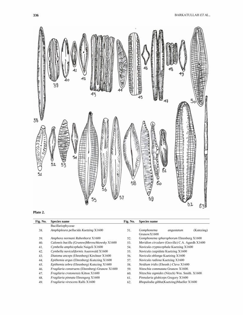
BARKATULLAH ET AL., 336
Plate 2.
Fig. No. Species name Fig. No. Species name Bacillariophyceae
38. Amphipleura pellucida Kuetzing X1600 51. Gomphonema angustatum (Kutezing) GrunowX1600
39. Amphora normani Rabenhorst X1600 52. Gomphonema spharophorum Ehrenberg X1600 40. Caloneis bacillu (Grunow)Mereschkowsky X1600 53. Meridion circulare (Greville) C.A. Agardh X1600 41. Cymbella amphicephala Naigeli X1600 54. Navicula cryptocephala Kuetzing X1600 42. Cymbella naviculiformis Auerswald X1600 55. Navicula cuspidata Kuetzing X1600 43. Diatoma anceps (Ehrenberg) Kirchner X1600 56. Navicula oblonga Kuetzing X1600 44. Epithemia argus (Ehrenberg) Kutezing X1600 57. Navicula radiosa Kuetzing X1600 45. Epithemia zebra (Ehrenberg) Kutezing X1600 58. Neidium iridis (Ehrenb.) Cleve X1600 46. Fragilaria construens (Ehrenberg) Grunow X1600 59. Nitzschia commutata Grunow X1600. 47. Fragilaria crotonensis Kitton X1600 60. Nitzschia sigmides (Nitzch) Wm. Smith. X1600 48. Fragilaria pinnata Ehrengerg X1600 61. Pinnularia globiceps Gregory X1600 49. Fragilaria virescens Ralfs X1600 62. Rhopalodia gibba(Kuetzing)Mueller X1600
ROCK ALGAE OF BATKHELA DISTRICT MALAKAND, PAKISTAN 337
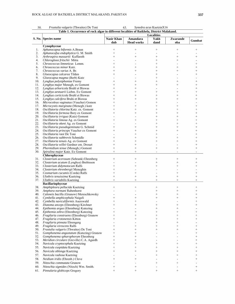
50. Frustulia vulgaris (Thwaites) De Toni 63. Synedra acus KuetzinX16 Table 1. Occurrence of rock algae in different localities of Batkhela, District Malakand.
Localities S. No. Species name Nasir Khan
dub Amandara
Head works Yakh dand
Zwarande oba Gumbat
Cynophyceae 1. Aphanocapsa biformis A.Braun + + + + + 2. Aphanocafsa endophytica G. M. Smith + - - - - 3. Arthrospira massartii Kuffareth + + + + + 4. Chlorogloea fritschii Mitra - - + + - 5. Chroococcus limneticus Lemm. + - + - + 6. Chroococcus minor Kutz. + + + + + 7. Chroococcus varius A. Br. + + + + + 8. Gloeocapsa calcarea Tilden + - + - + 9. Gloeocapsa magma (Berb) Kutz - - + - - 10. Lyngbya polysiphoniae Fremy + + + + + 11. Lyngbya major Menegh, ex Gomont + + + + + 12. Lyngbya arboricola Bruhl et Biswas + + - - + 13. Lyngbya aestuarii Liebm. Ex Gomont + + + + + 14. Lyngbya corticicola Bruhl et Biswas + + + + + 15. Lyngbya calcifera Bruhi et Biswas + + + + + 16. Microcoleus vaginatus (Vaucher) Gomon + - + - - 17. Microcystis marginata (Menegh.) kutz + + + + + 18. Oscillatoria chlorina Kutz. ex. Gomont + + + + + 19. Oscillatoria formosa Bory ex Gomont + + + + + 20. Oscillatoria irrigua (Kutz) Gomont - - + + + 21. Oscillatoria limosa Ag. ex Gomont + + + + + 22. Oscillatoria okeni Ag. ex Gomont - - + + + 23. Oscillatoria pseudogeminata G. Schmid + + + + + 24. Oscillatoria princeps Vaucher ex Gomont + + + + + 25. Oscillatoria raoi De Toni - + + - + 26. Oscillatoria subbrevis Schmidle + + + + + 27. Oscillatoria tenuis Ag. ex Gomont + + + + + 28. Oscillatoria willei Gardner em. Drouet + + - - + 29. Phormidium tenue (Menegh,) Gomont + + + + + 30. Spirulina major Kutz. Ex Gomont + + - - -
Chlorophyceae 31. Closterium acerosum (Sehrank) Ehrenberg + + + + + 32. Closterium acutum (Lyngbye) Brebisson - - + + + 33. Closterium didymotocum Ralfs + + + + + 34. Closterium ehrenbergii Meneghin + - - - - 35. Cosmarium cucumis (Corda) Ralfs + + - + + 36. Ulothrix tenuissima Kuetzing + + + + + 37. Ulothrix variabilis Kuetzing + + + + +
Bacillariophyceae 38. Amphipleura pellucida Kuetzing + + + + + 39. Amphora normani Rabenhorst - - + + + 40. Caloneis bacillu (Grunow) Mereschkowsky - - + + + 41. Cymbella amphicephala Naigeli + + + + + 42. Cymbella naviculiformis Auerswald + + - - + 43. Diatoma anceps (Ehrenberg) Kirchner + + + + + 44. Epithemia argus (Ehrenberg) Kutezing - - + + - 45. Epithemia zebra (Ehrenberg) Kutezing + + + + + 46. Fragilaria construens (Ehrenberg) Grunow + + + + + 47. Fragilaria crotonensis Kitton + + + + + 48. Fragilaria pinnata Ehrengerg - - + + - 49. Fragilaria virescens Ralfs + + + + + 50. Frustulia vulgaris (Thwaites) De Toni + + + + + 51. Gomphonema angustatum (Kutezing) Grunow + + - - + 52. Gomphonema spharophorum Ehrenberg + + - - + 53. Meridion circulare (Greville) C.A. Agardh + + - - + 54. Navicula cryptocephala Kuetzing + + + + + 55. Navicula cuspidata Kuetzing + + + + + 56. Navicula oblonga Kuetzing + + + + + 57. Navicula radiosa Kuetzing - - + + + 58. Neidium iridis (Ehrenb.) Cleve + + - - + 59. Nitzschia commutata Grunow - - + + - 60. Nitzschia sigmides (Nitzch) Wm. Smith. + + - - + 61. Pinnularia globiceps Gregory + + + + +

BARKATULLAH ET AL., 338
62. Rhopalodia gibba (Kuetzing) Mueller - - + + - 63. Synedra acus KuetzinX1600 + + + + +
ROCK ALGAE OF BATKHELA DISTRICT MALAKAND, PAKISTAN 339
Cymbella naviculiformis Auerswald
Cells 9-16 × 30-50 µ, valves asymmetric, raphe slightly curved and excentric, transverse striations radiate 12-14 in 10 µ (Fig. 41). Cymbella amphicephala Naigeli
Cells 9-16 × 30-50 µ, valves lanceolate, with convex sides and evident constrictions, raphe straight, slightly excentric, transverse striations radiate (Fig. 42).
Diatoma anceps (Ehrenberg) Kirchner
Cells 4-8 × 15-100 µ, united into closed chains, with few or no intercalary bands, valves linear, capitate at poles, striations transverse (Fig. 43). Epithemia Brebisson
Cells solitary, rectangular in girdle view, smooth girdles, intercalary bands, valves slightly to strongly curved, dorsally convex, ventrally straight to concave with broadly rounded to capitate and sometimes recurved poles.
Key to species
1. Valves with constricted end, cells 6-15 µ broad …………………………………………………………… E. argus
1. Valves not constricted, cells 7-14 µ broad ……………………………………………...………………….. E. zebra Epithemia argus (Ehrenberg) Kutezing
Cells 6-15 × 30-130 µ, valves ventrally straight to slightly concave, dorsally quite convex, with curved, broadly rounded ends (Fig. 44). Epithemia zebra (Ehrenberg) Kutezing
Cells 7-14 × 30-150 µ, valves lanceolate, gently curved with nearly parallel sides, gradually attenuated to rounded poles (Fig. 45). Fragilaria Lyngbye
Cells rectangular in girdle view, with one or two intercalary band without septa and costae.Valves linear to
fusiform, bilateraly symmetric attenuated with pole, sometime capitated. Ransverse striae usually fine. Pseudo raphe narrow, chromatophores numerous. Fragilaria construens (Ehrenberg) Grunow
Cells 5-12 × 7-25 µ, united into compact chains, cruciform with lace-like pseudoraphe, striation transverse (Fig. 46). Fragilaria crotonensis Kitton Cells 1-3 × 40-150 µ, united medianly into ribbon-like bands, ends often touching, valves narrowly linear, transverse striations, fine, about 15-18 in 10 µ (Fig. 47).
Key to species
1. Cells united in the middle portion only ………………………………………………………………. F. crotonensis
1. Cells united through entire length …………………………………………………………………….……………. 2
2. Radial striations 14-17 in 10 µ …………………………………………………………...…………… F. construens
2. Transverse striations ……………………………………………………………………………….………………. 3
3. Striations 10-12 in 10 µ ………………………………………………….…………..…………………… F. pinnata
3. Striations 12-19 in 10 µ ………………………………………………………………………………… F. virescens Fragilaria pinnata Ehrengerg
Cells 2-6 × 3-30 µ, united into flat chains, valves broadly to narrowly elliptical, evident linear pseudoraphe, rib-like transverse striation (Fig. 48). Fragilaria virescens Ralfs
Cells 5-10 × 12-120 µ, united into long chains, with straight to slightly convex sides, rounded poles and very narrow pseudoraphe, transverse striations moderately fine (Fig. 49).
Frustulia vulgaris (Thwaites)De Toni
Cells 10-13x 40-80 µ, unbranched tube, with eliptic valves having obtuse to rostrate extremities; striae somewhat radiant, about 24 in 10 µ (Fig. 50).
Gomphonema C. A. agarth
Cells usually solitary , free floating, Transversely asymmetric in both girdles and valves view, Cuneat in girdle view, with one pole capitated or broader than the other; strait raphe and conspicuous central and polarnodules; stration strictly transverse or somewhat radial; Chromatophore single.
BARKATULLAH ET AL., 340
Key to species
1. Valves elliptic clavate, cells 7-10 × 30-47 µ ………...………………………………. Gomphonema spharophorum1. Valves slender lanceolate, cells 5-9 × 12-45 µ ………..………………………………… Gomphonema angustatum
Gomphonema angustatum (Kutezing) Grunow
Cells 5-9 × 12-45 µ, valves slender, clavate-lanceolate, nearly symmetric, with slightly constricted and abruptly rounded poles, axial area narrow (Fig. 51). Gomphonema spharophorum Ehrenberg
Cells 7-10 × 30-47 µ, valves elliptic clavate, sharply narrowing towards a rounded and slightly apitate basal pole and with a much wider knob-like apical pole (Fig. 52).
Meridion circulare (Greville) C.A. Agardh
Cells 4-8 × 12-80 µ, wedge-shaped, costae 3-5 in 10 µ and finely punctuate striations (Fig. 53). Navicula Bory
Cells generally solitary and free floating, sometimes aggregated into irregularly radiating clusters, rectangular in girdle view, with smooth girdle without intercalary bands, valves elongate usually attenuated toward capitate.
Key to species
1. Striations transverse ………………………………………………………………………………………………... 21. Striations longitudinal, valve with rohmbo-lanceolate ends …………………………………………… N. cuspidate2. Ends broadly rounded ………………………………………………………………………..………….. N. oblonga2. Ends capitate or pointed ………………………………………………………………………………...………….. 33. Ends pointed, striations 10-12 in 10 µ …………………………………………………………………… N. radiosa3. Ends slightly capitate, striations 16-18 in 10 µ …………………………………………………… N. cryptocephala
Navicula cryptocephala Kuetzing
Cells 5-7 × 40-120 µ, valves lanceolate with slender, somewhat capitate ends, central area elongated, striations medianly radial and polarly convergent, 16-18 in 10 µ (Fig. 54). Navicula cuspidata Kuetzing
Cells 17-37 × 50-170 µ, valves rhombo-lanceolate, tapering sharply to rounded ends, transverse striations, evidently punctat, longitudinal striations about 24 in 10 µ and parallel to axial area (Fig. 55). Navicula oblonga Kuetzing
Cells 13-24 × 70-220 µ, valves linear-lanceolate with broadly rounded ends, transverse striations in polar and sub-polar area, central area large (Fig. 56).
Navicula radiosa Kuetzing
Cells 10-19 × 35-60 µ, valves lanceolate, gradually tapering to more or less pointed ends, transverse striations, 10-12 in 10 µ (Fig. 57). Neidium iridis (Ehrenb.) Cleve
Cells usually solitary, free floating, rectangular in girdle view and without girdle bands. Valve view linear with rounded or oval central area; raphe strait, usually bifurcated at poles, transverse rows of punctae crossed near the valves margins by narrow longitudinal clear spaces; two incised chromatophore (Fig. 58). Nitzschia Hasssall
Cells generally solitary and free floating, sometime band like, elongate, rhombic or rectangular in transverse section, valves longitudinally asymmetric very variable in shape.
Key to species
1. Cells 7-12 µ broad, valves linear ……………………………………………………….…………….. N. commutate1. Cells 5-8 µ broad, valves lanceolate ……………………………………………………………………. N. sigmides
Nitzschia commutata Grunow.
Cells 7-12 × 50-80 µ, in girdle view broadly linear, with rounded poles, valves linear with concave sides and cuneate, ends acute (Fig. 59).
Nitzschia sigmides (Nitzch) Wm. Smith.
Cells 5-8 × 20-50 µ, angular in girdle view, valves lanceolate with undulate margins, striations about 18 in 10 µ (Fig. 60).
ROCK ALGAE OF BATKHELA DISTRICT MALAKAND, PAKISTAN 341
Pinnularia globiceps Gregory
Cells 8-10 × 30-40µ, valves elliptic, with convex sides and broadly capitate poles, striations tranverse (Fig. 61). Rhopalodia gibba (Kuetzing) Mueller
Cells broadly linear with median inflation and broadly rounded poles, 18-30 x 35-300 µ; valves very broadly lunate, with almost strait ventral sites and recurved acute poles. Costae 6-8in 10 µ (Fig. 62). Synedra acus Kuetzing
Cells 5-6×100-300 µ, solitary, valves linear to lanceolate, needle-like towards the poles, transverse striations 12-14 in 10 µ; pseudoraphe narrow, linear with central area rectangular (Fig. 63). References Akhtar, N. and S.R. Rehman. 2009. Some members of
ulotrichales from Jalala, District Mardan, Pakistan. Pak. J. Pl. Sci., 15(1): 19-30.
Ali, A., Z.K. Shinwari and F.M. Sarim. 2010. Contribution to the algal flora (Chlorophyta) of fresh waters of district Swat, K.P.K., Pakistan. Pak. J. Bot., 42(5): 3457-3462.
Anjum, G. and M.A.F. Faridi. 1985. Algae in dry soil of N.W.F.P., Pakistan. Pak. J. Bot., 17: 257-261.
Anjum, G., T. Jabeen, F. Hussain and M.A.F. Faridi. 1982. Some soil-binding algae from Peshawar, Pakistan. Pak. J. Bot., 14: 107-109.
Anonymous. 1985. Standard methods for the examination of water and waste water. Am. Pub. Health. Ass. Washington D.C. 14th ed: 1-1268.
Desikachary, T.V. 1959. Cyanophyta, Indian Council of Agriculture Research, New Dehli. pp. 669.
Hussain, F., G. Anjum and A. Zaman. 2010. Some species of genus Nostoc from soil of Khyber Pukhtunkhwa, Pakistan, Pak. J. pl. Sci., 16(2): 65-77.
Hussain, F., M.I. Zaidi and M.J. Durrani. 2003. A checklist to the algae of Upper Balochistan. Pak. J. Pl. Sci., 9: 1-86.
Hussain, F., Masood, S.M. Shah, F. Hadi, A. Zaman and S.M. Wazir. 2009. Some Blue green algae from rice field of Asota Sharif, District Swabi, Pakistan. Pak. J. Pl. Sci., 15(1): 45-57.
Lewis, L.A. and R.M. Mccourt. 2004. Green algae and the origin of land plants. American Journal of Botany, 91: 1535-1556.
López-Bautista, J.M., F. Rindi and A.D. Casamata. 2007. The systematics of subaerial algae. In Cell origin, life in extreme habitats and Astrobiology. Algae and Cyanobacteria in extreme environments (Ed.): J. Sechbach. Springer, Nether lands. pp. 601-617.
Mason, D.J. 1967. Limnology of Monolake. pu. Zoology Dept: California Univ. California. 83: 102.
Nakano, T., S. Handa and S. Takeshita. 1991. Some corticolous algae from the Taishaku-Kyo Gorge, Western Japan. Nova Hedwigia, 52: 427-451.
Naz, S., Masud-ul-Hasan and M. Shameel. 2004. Taxponomic study of Chroocophyceae (Cyanophyta) from northern areas of Pakistan. Pak. J. Bot., 36(2): 247-28.
Prescott, G.W. 1951. Algae of Western great Lakes area. Cranbrook Institute, Bloomfield Hills, Michigan.
Silva, N.M., L.H.Z. Branco and O. Necchi-Júnior. 2010. Corticolous green algae from tropical forest remnants in the northwest region of São Paulo State, Brazil. Revista Brasil. Bot., 33(2): 215-226.
Smith, G.M. 1950. Fresh Water Algae of United State of America. Mc Graw Hill, New York.
Tiffany, L.H. and M.E. Britton 1952. The algae of Illinois. Hafner Publishing Co., New York. pp. 407.
Valeem, E.E. and M. Shameel. 2009. Influence of aquatic environment on the composition of fatty acids in algae growing in Sindh, Pakistan. Proc. Pakistan Acad. Sci., 46(3): 109-116.
Zarina, A., M. Hasan and M. Shameel. 2009. Diversity of freshwater green macroalgae in the Punjab and neighbouring areas of Pakistan. Pak. J. Bot., 41(1): 277-291.
(Received for publication 25 June 2011)
Related Documents