Robot-based hand motor therapy after stroke Craig D. Takahashi, Lucy Der-Yeghiaian, Vu Le, Rehan R. Motiwala and Steven C. Cramer Department of Neurology and Department of Anatomy & Neurobiology, University of California, Irvine, USA *Present Address: Department of Engineering, Santa Ana College, Santa Ana, CA, USA Correspondence to: Steven C. Cramer, MD, University of California, Irvine Medical Center, 101 The City Drive South, Building 53 Room 203, Orange, CA 92868 - 4280, USA E-mail: [email protected] Robots can improve motor status after stroke with certain advantages, but there has been less emphasis to date on robotic developments for the hand. The goal of this study was to determine whether a hand-wrist robot would improve motor function, and to evaluate the specificity of therapy effects on brain reorganization. Subjects with chronic stroke producing moderate right arm/hand weakness received 3 weeks therapy that emphasized intense active movement repetition as well as attention, speed, force, precision and timing, and included virtual reality games. Subjects initiated hand movements. If necessary, the robot completed move- ments, a feature available at all visits for seven of the subjects and at the latter half of visits for six of the subjects. Significant behavioural gains were found at end of treatment, for example, in Action Research Arm Test (34 ɔ 20 to 38 ɔ 19, P_ 0.0005) and arm motor Fugl-Meyer score (45 ɔ 10 to 52 ɔ 10, P _ 0.0001). Results sug- gest greater gains for subjects receiving robotic assistance in all sessions as compared to those receiving robotic assistance in half of sessions.The grasp task practiced during robotic therapy, when performed during functional MRI, showed increased sensorimotor cortex activation across the period of therapy, while a non-practiced task, supination/pronation, did not. A robot-based therapy showed improvements in hand motor function after chronic stroke. Reorganization of motor maps during the current therapy was task-specific, a finding useful when considering generalization of rehabilitation therapy. Keywords: stroke; motor therapy; functional MRI; generalization Abbreviations: IP = interphalangeal; MCP = metacarpophalangeal Received July 13, 2007 . Revised November 27 , 2007. Accepted November 28, 2007. Advance Access publication December 21, 2007 Stroke remains a leading cause of adult disability in the United States and many other countries. Though stroke can cause deficits in a number of neurological domains, the most commonly affected is the motor system (Gresham et al., 1995; Rathore et al., 2002). Disability due to motor deficits has therefore been a topic of considerable research. Furthermore, given the central role that hand movements normally play in human existence (Porter and Lemon, 1993; Wilson, 1998; Wing et al., 1998; Connolly, 1999; Mountcastle, 2005), much attention in rehabilitation research has been focused on understanding and restoring hand motor function after stroke (Baron et al., 2004; Luft et al., 2004; Nudo, 2007). A major issue in hand motor therapy has been how to best restore function. A recurring theme is that interven- tions emphasizing intense, active repetitive movement are of high value in this regard. These increase strength, accuracy and functional use when applied to subjects with paresis due to stroke (Taub et al., 1993; Butefisch et al., 1995; Carey et al., 2002b; Wolf et al., 2006). One approach to providing such therapy is robotic technology. Robots hold promise for enhancing traditional post- stroke therapy. Specifically, robots can provide therapy for long time periods, in a consistent and precise manner, without fatigue; can be programmed to perform in different functional modes with a single click; can be automated for many functions; can measure and record a range of behaviours in parallel with therapeutic applications; and can be enabled to do the above with only remote human control (Burgar et al., 2000; Dobkin, 2004; Fasoli et al., 2004; Reinkensmeyer et al., 2004; Volpe et al., 2005). The latter extends the promise of telerehabilitation, which might improve access by underserved populations (Reinkensmeyer et al., 2002; Lai et al., 2004; Winters, 2004). In addition, robots can be used to gain insights into the stroke recovery process (Takahashi and Reinkensmeyer, 2003; Krakauer, 2006), for example through their ability to apply novel force assistance patterns (Patton and Mussa-Ivaldi, 2004). doi:10.1093/brain/awm311 Brain (2008), 131 , 425 ^ 437 ß The Author (2007). Published by Oxford University Press on behalf of the Guarantors of Brain. All rights reserved. For Permissions, please email: [email protected]

Welcome message from author
This document is posted to help you gain knowledge. Please leave a comment to let me know what you think about it! Share it to your friends and learn new things together.
Transcript

Robot-based hand motor therapy after strokeCraig D.Takahashi,� Lucy Der-Yeghiaian,Vu Le, Rehan R. Motiwala and Steven C.Cramer
Department of Neurology and Department of Anatomy & Neurobiology, University of California, Irvine, USA
*Present Address: Department of Engineering, Santa Ana College, Santa Ana, CA, USA
Correspondence to: Steven C.Cramer, MD, University of California, Irvine Medical Center, 101 The City Drive South,Building 53 Room 203, Orange, CA 92868-4280, USAE-mail: [email protected]
Robots can improvemotor status after strokewith certain advantages, but there has been less emphasis to dateon robotic developments for the hand. The goal of this study was to determine whether a hand-wrist robotwould improve motor function, and to evaluate the specificity of therapy effects on brain reorganization.Subjects with chronic stroke producing moderate right arm/hand weakness received 3 weeks therapy thatemphasized intense active movement repetition as well as attention, speed, force, precision and timing, andincluded virtual reality games. Subjects initiated hand movements. If necessary, the robot completed move-ments, a feature available at all visits for seven of the subjects and at the latter half of visits for six of thesubjects. Significant behavioural gains were found at end of treatment, for example, in Action Research ArmTest (34� 20 to 38� 19, P_ 0.0005) and arm motor Fugl-Meyer score (45� 10 to 52� 10, P_ 0.0001). Results sug-gest greater gains for subjects receiving robotic assistance in all sessions as compared to those receiving roboticassistance in half of sessions.The grasp task practiced during robotic therapy, when performed during functionalMRI, showed increased sensorimotor cortex activation across the period of therapy, while a non-practicedtask, supination/pronation, did not. A robot-based therapy showed improvements in hand motor functionafter chronic stroke. Reorganization of motor maps during the current therapy was task-specific, a findinguseful when considering generalization of rehabilitation therapy.
Keywords: stroke; motor therapy; functional MRI; generalization
Abbreviations: IP= interphalangeal; MCP=metacarpophalangeal
Received July 13, 2007. Revised November 27, 2007. Accepted November 28, 2007. Advance Access publication December 21, 2007
Stroke remains a leading cause of adult disability in theUnited States and many other countries. Though stroke cancause deficits in a number of neurological domains, themost commonly affected is the motor system (Greshamet al., 1995; Rathore et al., 2002). Disability due to motordeficits has therefore been a topic of considerable research.Furthermore, given the central role that hand movementsnormally play in human existence (Porter and Lemon,1993; Wilson, 1998; Wing et al., 1998; Connolly, 1999;Mountcastle, 2005), much attention in rehabilitationresearch has been focused on understanding and restoringhand motor function after stroke (Baron et al., 2004; Luftet al., 2004; Nudo, 2007).A major issue in hand motor therapy has been how to
best restore function. A recurring theme is that interven-tions emphasizing intense, active repetitive movement areof high value in this regard. These increase strength,accuracy and functional use when applied to subjects withparesis due to stroke (Taub et al., 1993; Butefisch et al.,
1995; Carey et al., 2002b; Wolf et al., 2006). One approachto providing such therapy is robotic technology.
Robots hold promise for enhancing traditional post-stroke therapy. Specifically, robots can provide therapy forlong time periods, in a consistent and precise manner,without fatigue; can be programmed to perform in differentfunctional modes with a single click; can be automatedfor many functions; can measure and record a range ofbehaviours in parallel with therapeutic applications; andcan be enabled to do the above with only remote humancontrol (Burgar et al., 2000; Dobkin, 2004; Fasoli et al.,2004; Reinkensmeyer et al., 2004; Volpe et al., 2005). Thelatter extends the promise of telerehabilitation, which mightimprove access by underserved populations (Reinkensmeyeret al., 2002; Lai et al., 2004; Winters, 2004). In addition,robots can be used to gain insights into the stroke recoveryprocess (Takahashi and Reinkensmeyer, 2003; Krakauer,2006), for example through their ability to apply novelforce assistance patterns (Patton and Mussa-Ivaldi, 2004).
doi:10.1093/brain/awm311 Brain (2008), 131, 425^437
� The Author (2007). Published by Oxford University Press on behalf of the Guarantors of Brain. All rights reserved. For Permissions, please email: [email protected]

Though robot-assisted therapy has been shown to signifi-cantly improve arm motor function after stroke (Aisenet al., 1997; Volpe et al., 1999; Krebs et al., 2002; Lumet al., 2002; Fasoli et al., 2003; Ferraro et al., 2003;Kahn et al., 2006b), with few exceptions (Jack et al., 2001;Hesse et al., 2003b), these efforts have been primarilyfocused on the proximal arm (Krebs et al., 1999;Reinkensmeyer et al., 2000; Lum et al., 2002).The current study aimed to develop, then assess clinical
effects of, a robotic therapy targeting the distal arm. Themain hypothesis was that robotic therapy would improvearm motor function in patients with chronic motor deficitsafter stroke. The content of therapy was built upon severalprinciples of motor learning: (i) intense, active repetitivemovement; (ii) sensorimotor integration, given the keyinfluence that sensory events have on motor learning in thenormal and post-stroke states (Kaelin-Lang et al., 2002;Lewis and Byblow, 2004; Walker-Batson et al., 2004) and(iii) high attentional valence and complexity of the experi-ence given the effects that these have in normal andneurologically impaired brains (Will et al., 1977; Kolband Gibb, 1991; Langhorne et al., 1993; Ottenbacher andJannell, 1993; Kempermann et al., 1997; van Praag et al.,2000). Some studies suggest utility of a virtual realityinterface in this regard (Holden et al., 2001; Jack et al.,2001; Ku et al., 2003; Merians et al., 2006; Fischer et al.,2007), and so this too was incorporated. The use of realobjects in a natural or purposeful context enhances motorperformance of individuals with hemiparesis (Hsieh et al.,1996; Wu et al., 1998, 2000) and might also be useful tomaximize attention to the task, and was therefore alsoincorporated.Study subjects were divided into two groups that differed
according to the dose of active robotic assistance. Roboticsystems are capable of assisting movement in a number ofdifferent modes (Prange et al., 2006), including active non-assist mode, in which the subject does all work and therobot provides no help; and active assist mode, in which thesubject attempts to move and the robot supplements thiseffort. These two active modes, which differ in amount ofrobotic assistance, were directly compared during the firsthalf of the current therapeutic program. These two modeswere chosen, and a passive assist mode in which thesubjects relaxes while the robot performs all limb move-ments excluded, because interventional studies suggestgreater gains are achieved when the subject actively exertsan effort (Lotze et al., 2003; Perez et al., 2004). In these twoactive modes, the subject’s effort, i.e. devotion of attentionand energy to movement generation, is likely similar,though active assist mode might at times require less effortthan non-assist mode because a portion of movements canbe passive. Active assist mode might have advantages. Forexample, active assist mode in subjects with hand paresis islikely to produce a larger range of motion, with superiormultijoint coordination, than is non-assist mode. As such,active assist mode likely generates greater proprioceptive
sensory signals to the brain than does the active non-assistmode. Proprioceptive sensory signals from these move-ments reach motor cortex (Vogt and Pandya, 1978; Brodal,1981; Jones, 1986). The quantity and character of suchsensory signals are known to modulate motor cortex func-tion and excitability (Ridding et al., 2000; Kaelin-Langet al., 2002), and increased afferent feedback has beenconsidered useful for improving motor learning (Poon,2004; Rossini and Dal Forno, 2004). A sub-hypothesis ofthis study, therefore, addressed during the first half oftherapy, was that a higher dose of active assist mode wouldbe associated with greater behavioural gains.
The current study also attempted to gain insight intothe issue of generalization of therapeutic gains. A concernsometimes raised in relation to stroke rehabilitation is thatgains achieved during therapy incompletely generalize tothe range of demands faced in real-world tasks (Stokesand Baer, 1977; Page, 2003; Huxlin and Pasternak, 2004;Krakauer, 2006; Van Peppen et al., 2006). Given thattherapy-related gains are achieved on the basis of brainplasticity (Hodics et al., 2006), this suggests the hypothesisthat a highly standardized therapy such as the currentrobotic intervention will induce motor cortex plasticity forthe task employed in therapy but, in the absence of gen-eralization, not for a separate motor task that was not partof therapy.
The approach employed by Nudo et al. in non-humanprimates was used to address this hypothesis (Nudo et al.,1996). These authors used electrophysiological methods tomap motor cortex representations before and after monkeystrained for 2–7 weeks at either a finger grasping task, or aforearm supination/pronation task. The authors found thattraining in a specific behavioural task differentially alteredmovement representations, with flexion task trainingspecifically associated with expansion of finger movementrepresentations, and supination/pronation task trainingspecifically associated with expansion of forearm movementrepresentations. Studies in healthy human subjects havebeen concordant with this (Pascual-Leone et al., 1995;Karni et al., 1996; Muellbacher et al., 2001; Floyer-Lea andMatthews, 2005). However, this issue has not beenexamined in relation to post-stroke therapeutics, where anon-practiced task has not been simultaneously evaluated.Thus, the specificity of training on cortical plasticity has notbeen previously examined in the post-stroke setting (Hodicset al., 2006). In the current study, functional MRI (fMRI)brain mapping was performed twice, once before and onceafter therapy, each time examining both the task practiced(grasping), and a task not practiced (supination/pronation),during therapy. The hypothesis was that a movementperformed by the stroke-affected distal upper extremityduring therapy would show increased representationarea over time in stroke-affected primary sensorimotorcortex, while a movement not performed during therapywould not.
426 Brain (2008), 131, 425^437 C. D.Takahashi et al.
Materials and MethodsSubjects and overall study designThirteen subjects with chronic stroke (Table 1), ages 37–86 years,
6 males and 7 females, participated. Each subject provided infor-
med consent in accordance with the U.C. Irvine Institutional
Review Board.Entry criteria included age 418 years, right-handed (Oldfield,
1971), stroke at least 3 months prior that caused right-hand
weakness, at least 10 degrees range of motion in the right index
finger metacarpophalangeal (MCP) joint, score of 2–20 on the 24
points of the Fugl-Meyer score related to hand motor function,
and the time to complete the 9-hole pegboard test had to be at
least 25% longer than that measured with the left (non-affected)
hand. Exclusion criteria included apraxia [score 42.5 on
Alexander’s scale (Alexander et al., 1992)], reduced attention
[score 40 on NIH Stroke Scale questions 1a–c], substantial
sensory loss [right hand Nottingham sensory score (Lincoln et al.,
1998a) 575% of normal], severe increase in tone (Ashworth
spasticity score 54 at right elbow, wrist or MCP), severe aphasia
(score 52 on NIH Stroke Scale question 9), major depression
(Geriatric Depression scale score48) or another diagnosis having
a major effect on hand function.Prior to treatment, stroke subjects underwent two assessments
of hand motor function ability separated by �2 weeks, to establish
motor function stability (Fig. 1A). The latter baseline assessment
included an MRI scan and was performed �1 week prior to treat-
ment initiation. Subjects then underwent 15 consecutive weekdays
of treatment. A third set of assessments was performed halfway
through treatment. A fourth immediately followed completionof therapy and was accompanied by repeat MRI scanning. Subjectsreturned for a fifth assessment 1 month after completing treat-
ment. The two study primary endpoints were change from base-line to end of therapy in the Action Research Arm Test (ARAT)and the Box and Blocks test scores, and the main secondaryendpoint was change in the arm motor Fugl-Meyer score.Subjects were assigned to one of two therapy groups. In the first
group, the robot was in active non-assist mode for the first 7.5days of treatment and then was switched to active assist mode forthe latter 7.5 days of treatment (‘ANA-A group’). For the secondgroup of patients, the robot was in active assist mode for all15 days (‘A-A group’). The first eight patients were randomly
assigned; an interval assessment found differences in baselinemeasures, and so for the last five patients, treatment groupassignment attempted to balance this.
Description of the robotThe Hand Wrist Assistive Rehabilitation Device (‘HWARD’) isa 3 degrees-of-freedom, pneumatically actuated device that assiststhe hand in grasp and in release movements. The three degrees are
flexion/extension of the four fingers together about the MCP joint,flexion/extension of the thumb at the MCP joint and flexion/extension of the wrist (Fig. 1B–D).The subject is seated, facing a computer monitor. The hand
is secured to the robot mechanism via three soft straps, and the
forearm is secured inside of a padded splint that is mountedto the surface of a platform (Fig. 1C). The palmar hand is left
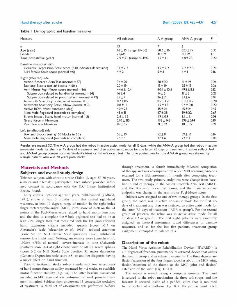
Table 1 Demographic and baseline measures
Measure All subjects A-A group ANA-A group P
n 13 7 6Age (year) 63�16 (range 37^86) 58.6�16 67.3�15 0.35Gender 7F/6M 4F/3M 3F/3M 1.0Time post-stroke (year) 2.9�5.1 (range 4^19.6) 1.2�1.1 4.8�7.3 0.22
Baseline characteristicsGeriatric Depression Scale score (410 indicates depression) 3.1�2.3 3.9�2.2 2.2�2.3 0.20NIH Stroke Scale score (normal=0) 4�2 5�3 4�1 0.16
Right (affected) sideAction Research ArmTest (normal=57) 34�20 28�20 41�19 0.26Box and Blocks test # blocks in 60 s 20�19 15�19 25�19 0.36Arm Motor Fugl-Meyer score (normal=66) 44.6�10.4 40.4�10.5 49.5�8.6 0.12Subportion related to hand/wrist (normal=24) 16�4 14�5 17�3 0.29Subportion related to proximal arm (normal=42) 29�7 26�7 33�6 0.09
Ashworth Spasticity Scale, wrist (normal=0) 0.7�0.9 0.9�1.2 0.3�0.5 0.28Ashworth Spasticity Scale, elbow (normal=0) 0.8�1.1 1.2�1.3 0.4�0.8 0.22Active ROM, wrist extension (deg) 40�25 35�26 45�24 0.49Nine Hole Pegboard (seconds to complete) 43�31 47�38 39�22 0.67Stroke Impact Scale, hand motor (normal=5) 2.4�1.2 1.9�0.9 3.1�1.1 0.06Grasp force in Newtons 290�215 198�148 396�244 0.10Pinch force in Newtons 89�55 71�52 111�55 0.21
Left (unaffected) sideBox and Blocks test # 60 blocks in 60 s 55�10 52�8 59�10 0.16Nine Hole Pegboard (seconds to complete) 25�5 27�6 22�3 0.08
Results are mean�SD.The A-A group had the robot in active assist mode for all 15 days, while the ANA-A group had the robot in activenon-assist mode for the first 7.5 days of treatment and then active assist mode for the latter 7.5 days of treatment. P values reflect A-Aand ANA-A group comparisons via Student’s t-test or Fisher’s exact test. The time post-stroke for the ANA-A group was skewed bya single patient who was 20 years post-stroke.
Hand therapy after stroke Brain (2008), 131, 425^437 427
unobstructed, permitting the placement of real objects into agrasping hand.A Windows-based software interface facilitates the use and
control of the robot via a second computer monitor that is onlyvisible to the examiner. Joint angle sensors in the robot are usedto measure the movement of the robot’s joints, and hence,movement of the subject’s limbs when attached to the robot. Thisfeature enabled real-time virtual reality hand movements, wherebythe subject’s hand controlled a computer screen virtual hand.The robot is backdriveable, enabling subjects to freely drivemovements when the robot is not engaged in active assistance.A more detailed description of this device has been publishedpreviously (Takahashi et al., 2005).
Treatment protocolEach subject received 15 daily sessions, on weekdays, over 3 weeks.Each session was �1.5 h long, with a brief break in the middle.Several adjustments were made for each subject. Prior to placing
the subject’s hand into the robot, the subject’s right hand waspositioned into HWARD so that both the MCP joint and the wristcentre of rotation were aligned with the robot’s finger and wristjoint axes, respectively. To do this, the examiner measured thedistance between the centre of wrist rotation to finger MCP joints,
the distance between wrist centre of rotation to thumb inter-phalangeal (IP) joint, and the distance between the MCP joints tothe midpoint of finger IP joints, and then adjusted the robotinterface to accommodate. These measures were restored for eachsubject at the start of each session. Every day, the experimenterdetermined a subject’s comfortable passive range of motion foreach degree-of-freedom and adjusted the robot’s hard stopsaccordingly. When in active assist mode, airflow limiters wereadjusted at the beginning of each session to insure robot-generated movements were brisk but not excessively forceful. Toavoid ceiling and floor effects, the duration of each grasp-releaserepetition was adjusted, based on hand function, to be shorter forthose with better hand function and longer for those with poorerhand function. Note that subjects completed the same number ofcycles regardless of hand function of robot mode.During treatment, subjects sat in an upright position, with the
knees flexed at about 90�, trunk supported and maintained againstthe back of the chair with a shoulder harness, shoulder abductedabout 30�, and elbow flexed about 90�. To avoid pain or dis-comfort during robot treatment, the position of the shoulderwas intermittently changed via small movements of the robotplatform. The subject’s arm was secured to the robotic device.In the first half of each day’s session, subjects performed
9 cycles of 10 repetitions of simple grasp-release exercises.
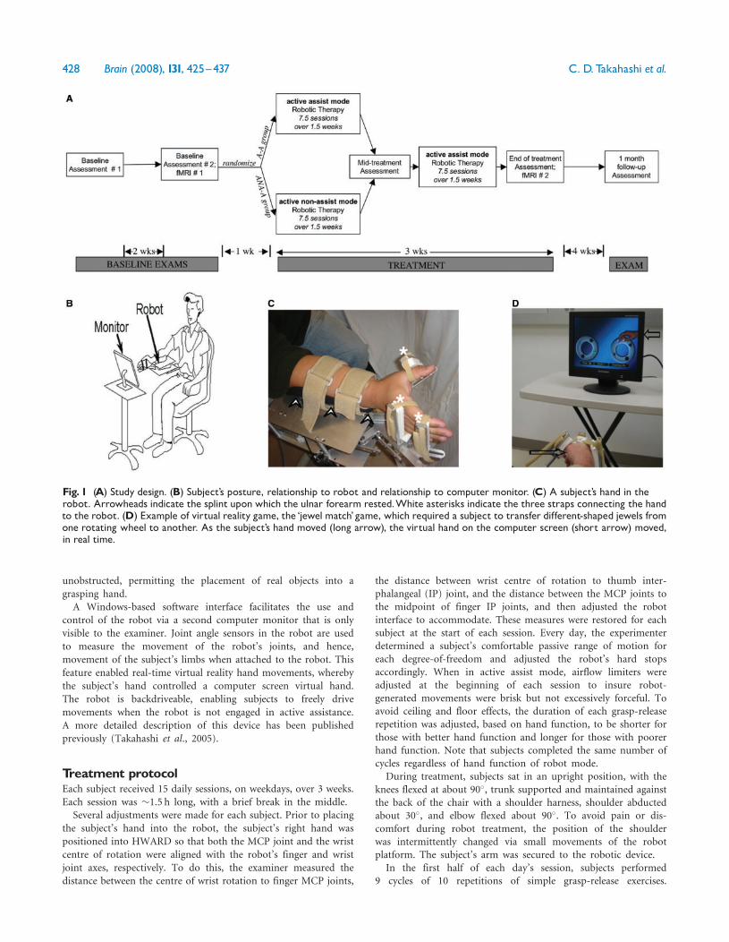
Fig. 1 (A) Study design. (B) Subject’s posture, relationship to robot and relationship to computer monitor. (C) A subject’s hand in therobot. Arrowheads indicate the splint upon which the ulnar forearm rested.White asterisks indicate the three straps connecting the handto the robot. (D) Example of virtual reality game, the ‘jewel match’ game, which required a subject to transfer different-shaped jewels fromone rotating wheel to another. As the subject’s hand moved (long arrow), the virtual hand on the computer screen (short arrow) moved,in real time.
428 Brain (2008), 131, 425^437 C. D.Takahashi et al.
Each grasp-release exercise was 11–15 s long and involved graspingand releasing while the patient viewed an LCD monitor (Fig. 1B)that provided instructions for each step: ‘Get Ready,’ ‘Close,’‘Open’ or ‘Rest’.When performing the ‘Close’ and the ‘Open’ steps in active
assist mode, the subjects were provided 1–3 s to attempt to openor close their hand, after which the robot provided assistance for1–3 s. In this way, if the patient did not complete the handmovement, the robot moved the hand to complete the range ofmotion; if the patient did complete the movement, the robotmerely applied pressure against the hard stops with no effect onthe hand. The 3 degrees-of-freedom were yoked such that activeassistance by the robot was done in a power grip pattern, withflexion of fingers and thumb accompanied by wrist extension.Assisted opening therefore extended fingers and thumb and flexedwrist. When performing the ‘Close’ and the ‘Open’ steps in activenon-assist mode, the subjects were provided 3–5 s to attempt toopen or close their hand, with no subsequent robotic assistance.Other efforts further increased a subject’s attention to move-
ments. During 75% of the cycles, an examiner placed into the areain front of the subject’s palm one of several objects, each havingrich and varied sensory characteristics. Subjects were asked tolook at the hand and to answer questions about the object askedby the experimenter regarding object temperature, texture,stiffness, shape, familiarity and functionality.The second half of each day’s session followed a 15min break
and focused on playing a set of interactive virtual reality computergames (Fig. 1D). Whichever form of robotic assistance wasassigned to the exercises was subsequently maintained during thesegames. Information on joint angle at each of the three degrees offreedom was passed to the computer game program so that asubject’s hand controlled a virtual hand in real time. Ninedifferent computer games were played, each requiring a subject toperform the same hand opening and closing movements as duringthe first half of the session. Game rules emphasized control ofhand movement range, speed and timing. The therapist adjustedgame difficulty to avoid ceiling and floor performances.
AssessmentsSubjects underwent functional motor testing five times as earlier(Fig. 1A). All assessments were done by a single person (LD) andincluded the Action Research Arm Test (ARAT) (Lyle, 1981),Box and Blocks test (Mathiowetz et al., 1985), Fugl-Meyer armmotor scale (Fugl-Meyer et al., 1975), the NIH Stroke Scale (Brottet al., 1989), Geriatric Depression Scale (short version) (Burkeet al., 1991), Nottingham Sensory Assessment (Lincoln et al.,1998b), an assessment of apraxia (Alexander et al., 1992),dynamometer recording of grip and pinch strength, a goniometermeasure of wrist active range of motion, the 9-hole Peg test(Oxford Grice et al., 2003), Stroke Impact Scale hand motorsubscale (Duncan et al., 1999) and the modified Ashworth scale(Katz et al., 1992). During assessments, the subject was seatedwith a standardized posture, using a chair with a firm back butno armrests and a table that approximated the subject’smid-abdomen.
ElectromyographyImmediately prior to each of the two MRI visits, in a roomoutside of the MRI scanner, surface electromyography (EMG) wasacquired while subjects rehearsed the motor tasks to be performed
during fMRI. This rehearsal employed equipment, guidance videoand posture that closely mimicked those subsequently employedduring fMRI scanning. EMG lead pairs were placed over threemuscles (right and left wrist extensor, plus right biceps), signalswere amplified (2000�) and bandpass filtered (30–1000Hz)(ICP511, Grass Technologies), digitally converted (Powerlab 8SP,ADInstruments) and stored for offline analysis.
MRI acquisitionScanning was all on the same 1.5 Tesla Philips scanner andincluded acquisition of high-resolution anatomical images,followed by fMRI of two right-sided motor tasks. The first task,‘grasp’, contrasted rest with opening/closing of the right hand,using a plastic non-actuated exoskeleton identical to the roboticinterface. The second task, ‘supination’, contrasted rest withsupinating/pronating the right forearm. The range of motion foreach task was not controlled, but instead was self-determined.Two fMRI series were acquired for each task. Each seriescontained 30 s epochs that alternated rest with 0.125Hz move-ment. Scanning parameters included 25 axial 5mm thick sliceswith no gap, 50 volumes/series, TR= 2500ms and TE= 40ms.During the fMRI scan, subjects viewed a guidance video thatdisplayed the desired movement in the form of a stick-figurehand. The video ran continuously with 0.125Hz movement cycle,being red during rest epochs and green during movement epochs.An investigator observed subject movements during scanning toverify task compliance.
Data analysisUsing SPM2 (www.fil.ion.ucl.ac.uk/spm/), the fMRI images wererealigned, normalized to MNI space and then spatially smoothed(FWHM=8mm). The first two volumes from each series werediscarded due to tissue non-saturation, and then images at restwere contrasted with images during task performance, with thetwo fMRI series for each task combined, using the standardhemodynamic response function. The fMRI data were analysed intwo ways: voxelwise using group maps, and also using region ofinterest methods in individual maps. For the voxelwise analysis,images were analysed at P50.001 without correction for multiplecomparisons. A one-sample t-test was performed on scans for eachof the two fMRI tasks, at each of the two visits, from each ofwhich the activation volume was determined for the largest clusterwithin left (stroke-affected) primary sensorimotor cortex. A pairedt-test was used to evaluate within subject changes in left primarysensorimotor cortex activation over time, for each task. Inindividual maps, task-related fMRI signal change was calculated(Brett et al., 2002) within a left sensorimotor cortex region ofinterest derived from prior hand motor imaging studies (‘handarea’ from http://hendrix.imm.dtu.dk/services/jerne/ninf/voi.html).For EMG data, the root mean square (RMS) was extracted from
each muscle’s EMG trace for the first representative 1 s period, foreach of task performance and of rest. Results were expressed, foreach muscle, as a ratio of (RMS during task)/(RMS at rest).Statistical analyses were done using JMP (SAS, Cary, NC) and
two-tailed, parametric methods, at alpha 50.05. Data withinsubject over time were analysed using a paired t-test. Whencomparing the two patient subgroups at any one time-point,Student’s t-test (for continuous data) and Fisher’s exact test(for categorical data) were used. When comparing the two patientsubgroups over time, a repeated measures ANOVA was performed
Hand therapy after stroke Brain (2008), 131, 425^437 429

to examine the time� treatment group interaction, with time asthe within-group variable and treatment group as the between-group variable.
ResultsSubjectsThere were 13 subjects enrolled. Of these, 11 were right-handed and 2 were ambidextrous, 8 were diagnosed withhypertension, 3 were diabetic, 7 were hyperlipidemic and5 had a history of coronary artery disease. Other baselinemeasures are presented in the Table 1. Baseline assessmentswere stable, as paired t-tests of the two baseline measure-ments demonstrated no significant differences for anybehavioural measure (P= 0.67–1.0). No baseline measureor demographic differed significantly between the twotreatment groups. One subject, in the ANA-A group, wasnot available for the exam at 1 month post-treatment.There were no safety issues or adverse events related tostudy participation. Among the 10/13 subjects able toundergo MRI, stroke location was pons in two, cortical intwo and subcortical in six.
Clinical findingsAt baseline, subjects had substantial deficits, for example,the Box and Blocks score on the affected side averaged lessthan half of the score on the non-affected side (Table 1).Across all subjects, significant gains were found from themean baseline to end of treatment for the main studyendpoints (Table 2). The ARAT showed a 4.2 ± 2.3(mean ± SD) point increase from baseline to end oftreatment (P50.0001, paired t-test). The Box and Blockstest overall showed a 4.2 ± 3.5 gain in the number of blocksover 60 s from baseline to end of treatment (P= 0.0009).The main secondary endpoint, arm motor Fugl-Meyerscore, showed a 7.6 ± 2.5 point increase from baseline to
end of treatment (P50.0001). All subjects showed a gain,for example, 13/13 subjects had an increase in Fugl-Meyerscore to end of treatment, and note that in 11/13 subjectsthis gain was 45 points. Gains were relatively stable overthe month following end of therapy: for each scale, changefrom baseline to 1 month post-therapy remained signifi-cant, and in no instance was a significant change presentfound from end of therapy to exam 1 month later (Fig. 2).
Most other clinical measures also showed significantimprovement with robotic therapy, including modest gainsin spasticity (Ashworth scale at wrist and elbow), wristactive range of motion, difficulty of hand usage (SIS score)and force of hand grasping as well as of finger pinching(Table 2). Changes in the 9-hole pegboard test did notreach significance. Note that significant change over timewas present for both the proximal arm and the hand/wristsubportions of the arm motor Fugl-Meyer scale.
Subjects in the A-A group received active assist mode forboth halves of the study and had significantly greater gainsthan subjects in the ANA-A group, who received activeassist mode for only the latter half of the study. Thissuggests two points. First, this suggests a dose-dependentbenefit for the active assist robotic therapy mode, based onresults at end of treatment and at 1 month after treatment:A-A group subjects had greater gains than ANA-A groupsubjects for ARAT score [repeated measures ANOVA,time� treatment group interaction, F(2,10) = 5.2, P50.03to end of treatment and F(3,8) = 5.0, P50.04 to 1 monthpost-treatment, using exact F-values, see Fig. 2] and for thearm motor Fugl-Meyer score [F(2,10) = 4.8, P50.04 andF(3,8) = 8.4, P50.008, respectively]. Second, this suggestsgreater benefit with active assist, as compared to activenon-assist, robotic therapy mode, based on results frombaseline to mid-treatment, i.e. based on the time whenrobot mode differed: during this period, subjects in the A-Agroup showed greater gains as compared to subjects in the
Table 2 Effects of therapy on behavioral measures
Test Change at end of therapy in
All Subjects (n=13) A-A group (n=7) ANA-A group (n=6)
Action Research ArmTest (normal=57) 4.2�2.3��� (12%) 5.3�2.1��^ (19%) 2.8�1.8� (7%)Box and Blocks test # blocks in 60 s 4.2�3.5�� (21%) 5.0�4.2� (33%) 3.3�2.4� (13%)Arm Motor Fugl-Meyer Scale (normal=66) 7.6�2.5��� (17%) 9.1�2.1���^ (23%) 5.8�1.6�� (12%)subportion related to hand/wrist (normal=24) 4.0�1.7��� (25%) 3.8�1.5�� (27%) 4.2�2.1� (25%)subportion related to proximal arm (normal=42) 3.7�3.0�� (13%) 5.4�1.3���^ (21%) 1.7�3.2 (5%)
Ashworth Spasticity Scale, wrist (normal=0) �0.5�0.8� (�71%) �0.8�1.0 (�89%) �0.2�0.4 (67%)Ashworth Spasticity Scale, elbow (normal=0) �0.4�0.5� (�50%) �0.6�0.6� (�50%) �0.1�0.2 (�25%)Active ROM, wrist extension (deg) 4.2�5.5� (11%) 3.9�4.0� (11%) 4.6�7.3 (10%)Nine Hole Pegboard seconds to complete �6.6�12.5 (�15%) �9.6�16.6 (20%) �3.0�3.8 (8%)Stroke Impact Scale, hand motor (normal=5) 0.6�0.5�� (25%) 0.9�0.4��^ (47%) 0.3�0.3 (10%)Grasp force in Newtons 45�56� (16%) 68�67� (34%) 19�23 (5%)Pinch force in Newtons 16�24� (18%) 28�22�^ (39%) 1�11 (1%)
Values are mean�SD (and percent) change from baseline to end of therapy for the right (affected) side. ���P50.0001, ��P50.001, �P50.05change within each group from baseline to end of therapy as assessed using a two-tailed paired t-test. ^P50.05, comparing change frombaseline to end of therapy between the A-A versus ANA-A groups via Student’s t-test. ROM=range of motion.
430 Brain (2008), 131, 425^437 C. D.Takahashi et al.

ANA-A group for ARAT score [F(1,11) = 10.9, P50.008]and arm motor Fugl-Meyer score [F(1,11) = 5.1, P50.05].Inspection of Fig. 2 provides a graphic demonstration ofthe latter point, with the A-A group having a steeper slopethan the ANA-A group for the ARAT and Fugl-Meyerscores from baseline to mid-treatment. Post-hoc analysiscomparison of the two groups at each separate time pointfound that, for ARAT and Fugl-Meyer score, change inscore from baseline was significantly different between thetwo treatment groups at mid-treatment and at end oftreatment but not at 1 month-post treatment; when therespective baseline score was added as a covariate to thesepost-hoc analyses, it was not significant in any instance,ARAT findings and end of treatment Fugl-Meyer findingswere unchanged, and Fugl-Meyer findings at mid-treatmentbecame a trend. Also, note too that, while the aboveanalyses of change from baseline to mid-treatment for thesetwo scales used actual score values, findings remainedsignificantly different between groups when expressed aspercentage change (for ARAT, 22 ± 19% versus 0.9 ± 1.9%,A-A versus ANA-A group, P50.02; for arm motor Fugl-Meyer, 14 ± 8% versus 6 ± 6%, P50.05). Furthermore, theANA-A group did derive some benefit from baseline tomid-treatment: while lesser gains were present for thisgroup during this interval in comparison to the A-A group,paired testing from baseline to mid-treatment for theANA-A group was nevertheless significant (P50.05), forthe Fugl-Meyer score (Fig. 2c). The Box and Blocks test wasnot significant in any of these analyses.
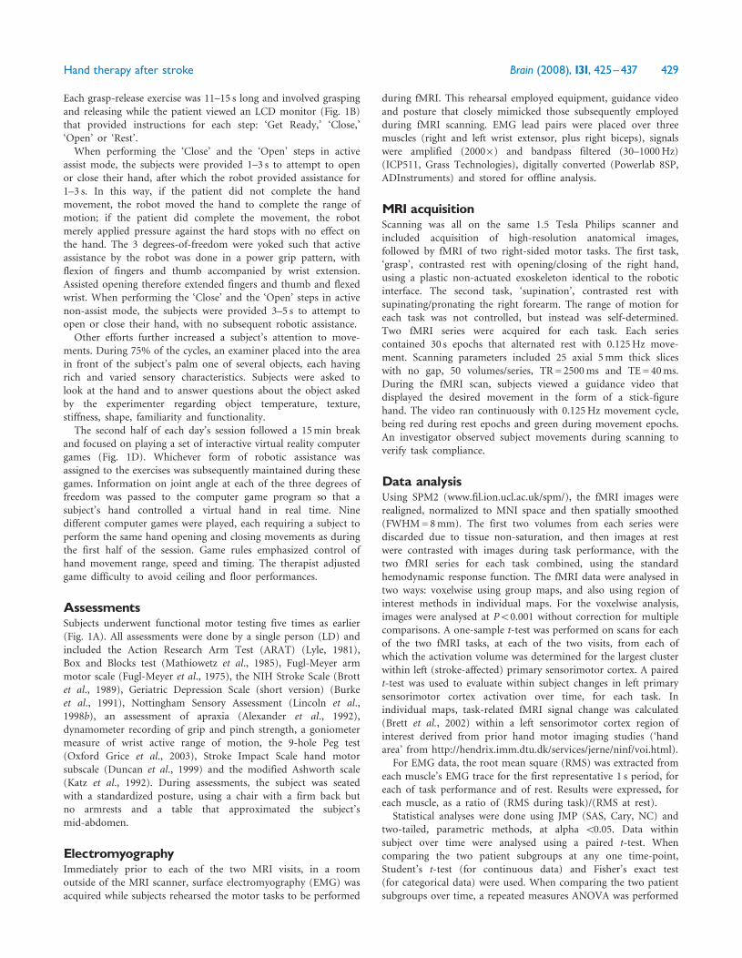
EMG findingsEMG could not be obtained in one subject at the secondpre-fMRI session due to technical reasons. Motor taskperformance was stable over time for each task, as from thefirst pre-fMRI EMG to the second pre-fMRI EMG, for eachof the three muscles, there was no significant within subjectEMG change over time (Fig. 3). Furthermore, the twomotor tasks had a similar pattern of muscle recruitment, asthere was no significant EMG difference between the twotasks for any of the three muscles, at either time point.
Functional MRI findingsMRI scanning could not be attempted in three subjects dueto presence of ferrous metal or claustrophobia. One subjectshowed excess head movement during the supination/pronation task at each fMRI visit, leaving 10 interpretablescans for the grasp task and 9 interpretable scans forthe supination/pronation task, at each of the two fMRIsessions. During the pre-therapy fMRI scan, one subject
Fig. 2 Results are presented for the two primary endpoints(A and B) and the main secondary endpoint (C), with scores onthe y-axis presented in relation to the five testing times.Valuesdisplayed for all subjects (mean�SEM) are shown in black.Valuesfor the A-A (active assist mode throughout the study) and ANA-A(active non-assist mode for initial study visits, followed by activeassist mode for latter study visits) subject groups are shown ingray. Note that baseline differences between these two groups inthese endpoints were not significant (P40.1). Across all subjects,for all three endpoints, significant gains were found across theperiod of time from baseline to end of treatment. These gainswere stable, as no significant changes from end of treatment to 1month later were present. Results from baseline to end oftreatment suggest a dose-dependent benefit for active assist modeof robotic therapy. During this interval, the A-A group had twicethe dose of active assist therapy and showed greater gains inARAT and Fugl-Meyer scores, as compared to the ANA-A group.
Also, results from baseline to mid-treatment suggest greaterbenefit with active assist mode of robotic therapy as compared toactive non-assist. During this interval, the A-A group had activeassist therapy and showed greater gains in ARATand Fugl-Meyerscores.On the other hand, the ANA-A group had active non-assisttherapy during this period and showed lesser behavioural gains.
Hand therapy after stroke Brain (2008), 131, 425^437 431
had incomplete movements of the right hand, one hadsmall visible mirror movements in the left hand and six hadvisible movement in the right foot or elbow at least onetime. During the post-therapy scan, one subject hadincomplete movement of the right hand, three had smallvisible mirror movements in the left hand and the same sixsubjects had visible movement in the foot or elbow at leastone time.The task that was part of therapy (grasp) showed
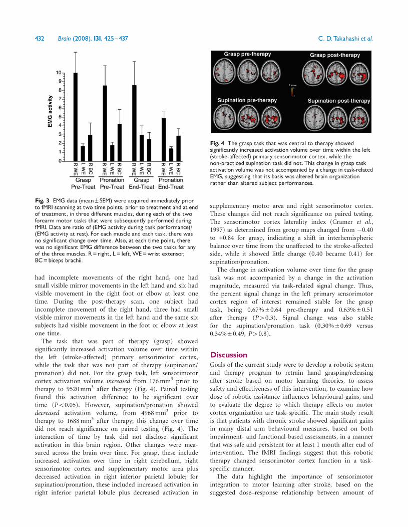
significantly increased activation volume over time withinthe left (stroke-affected) primary sensorimotor cortex,while the task that was not part of therapy (supination/pronation) did not. For the grasp task, left sensorimotorcortex activation volume increased from 176mm3 prior totherapy to 9520mm3 after therapy (Fig. 4). Paired testingfound this activation difference to be significant overtime (P50.05). However, supination/pronation showeddecreased activation volume, from 4968mm3 prior totherapy to 1688mm3 after therapy; this change over timedid not reach significance on paired testing (Fig. 4). Theinteraction of time by task did not disclose significantactivation in this brain region. Other changes were mea-sured across the brain over time. For grasp, these includeincreased activation over time in right cerebellum, rightsensorimotor cortex and supplementary motor area plusdecreased activation in right inferior parietal lobule; forsupination/pronation, these included increased activation inright inferior parietal lobule plus decreased activation in
supplementary motor area and right sensorimotor cortex.These changes did not reach significance on paired testing.The sensorimotor cortex laterality index (Cramer et al.,1997) as determined from group maps changed from �0.40to +0.84 for grasp, indicating a shift in interhemisphericbalance over time from the unaffected to the stroke-affectedside, while it showed little change (0.40 became 0.41) forsupination/pronation.
The change in activation volume over time for the grasptask was not accompanied by a change in the activationmagnitude, measured via task-related signal change. Thus,the percent signal change in the left primary sensorimotorcortex region of interest remained stable for the grasptask, being 0.67%±0.64 pre-therapy and 0.63%±0.51after therapy (P40.3). Signal change was also stablefor the supination/pronation task (0.30%±0.69 versus0.34%±0.49, P40.8).
DiscussionGoals of the current study were to develop a robotic systemand therapy program to retrain hand grasping/releasingafter stroke based on motor learning theories, to assesssafety and effectiveness of this intervention, to examine howdose of robotic assistance influences behavioural gains, andto evaluate the degree to which therapy effects on motorcortex organization are task-specific. The main study resultis that patients with chronic stroke showed significant gainsin many distal arm behavioural measures, based on bothimpairment- and functional-based assessments, in a mannerthat was safe and persistent for at least 1 month after end ofintervention. The fMRI findings suggest that this robotictherapy changed sensorimotor cortex function in a task-specific manner.
The data highlight the importance of sensorimotorintegration to motor learning after stroke, based on thesuggested dose–response relationship between amount of
Fig. 3 EMG data (mean�SEM) were acquired immediately priorto fMRI scanning at two time points, prior to treatment and at endof treatment, in three different muscles, during each of the twoforearm motor tasks that were subsequently performed duringfMRI. Data are ratio of (EMG activity during task performance)/(EMG activity at rest). For each muscle and each task, there wasno significant change over time. Also, at each time point, therewas no significant EMG difference between the two tasks for anyof the three muscles. R=right, L= left,WE=wrist extensor,BC=biceps brachii.
Fig. 4 The grasp task that was central to therapy showedsignificantly increased activation volume over time within the left(stroke-affected) primary sensorimotor cortex, while thenon-practiced supination task did not. This change in grasp taskactivation volume was not accompanied by a change in task-relatedEMG, suggesting that its basis was altered brain organizationrather than altered subject performances.
432 Brain (2008), 131, 425^437 C. D.Takahashi et al.
active assist mode robot therapy and resultant behaviouralgains. The dose of active robot assistance was experimen-tally manipulated by varying robot assist mode during theinitial 7.5 of the 15 therapy sessions. Two sets of findingssuggest greater behavioural gains associated with the activeassist mode. First, active assist mode appeared to be moreeffective than active non-assist mode: subjects who receivedthe active assist mode of robotic therapy for completinghand/wrist movements from baseline to mid-treatment(A-A group) showed significantly greater gains than thosesubjects who were in active non-assist mode (ANA-Agroup) during this interval. Second, a larger dose of activeassist mode appeared to be associated with greaterbehavioural gains: subjects who received active assistmode for both halves of the study (A-A group) hadsignificantly greater gains at end of treatment than subjectswho received active assist mode for only the latter half ofthe study (ANA-A group). The comparison of resultsbetween treatment groups must be interpreted with somecaution given the small sample sizes as well as the trend forbaseline imbalances in clinical status (Fig. 2). Regarding thelatter concern, however, note that some (Feys et al., 2000;Shelton et al., 2001; Lin et al., 2003; Cramer et al., 2007;Stinear et al., 2007), though not all (Stinear et al., 2007),prior analyses of stroke patient cohorts mitigate this issuebecause they suggest that the direction of the current trend(towards better baseline motor scores in the ANA-A group)would predict greater, not the observed lesser, gains amongsubjects in the ANA-A group.One key effect that active assist mode likely has, as
compared to active non-assist mode, is to produce a widerrange of motion for hand/wrist joints, that is more nor-mally coordinated, and thus a larger and more organizedafferent signal to brain sensorimotor areas (Humphreyet al., 1970; Brooks and Stoney, 1971; Miles and Evarts,1979; Waldvogel et al., 1999). Though active assist modemight also generate clinical benefit via other mechanisms,such as by increasing strength or by decreasing spasticity,these findings regarding dose of active robot assistancesubstantiate the assertion that proprioceptive feedback andsensorimotor integration are important to the effectivenessof motor-based therapies (Ridding et al., 2000; Kaelin-Langet al., 2002; Poon, 2004; Rossini and Dal Forno, 2004), aconclusion that is underscored by the rich structural andfunctional connections between primary sensory and motorcortices (Vogt and Pandya, 1978; Brodal, 1981; Jones,1986). Future designs might vary any of several aspects ofthis proprioceptive feedback, for example, by changing thetiming at which it is provided in relation to active subjectmovements, such as to a time much closer to the point ofmovement onset.The current findings, suggesting that active robotic
assistance provides greater benefits than dose-matched butunassisted practice, contrast with those of Kahn et al.(2006b), who found that in subjects with chronic stroke,reaching exercises supported by an active assist mode robot
provided the same, rather than greater, behavioural gains ascompared to matched unassisted reaching. There are severalpossible reasons for their observations, discussed in theirreport. Most notably was that for subjects in the activeassist group of Kahn et al (2006b), the robotic device wasprogrammed to assist the moving arm in real time in orderto specifically minimize errors of movement trajectory, anapproach that might have diminished the motor system’sown learning processes related to correcting these errors.Given the potential importance of sensorimotor processingto achieving behavioural gains from active assist moderobotic therapy, therefore, a key factor to consider whenimplementing active assistance is how sensorimotor proces-sing and learning are affected.
Some gains in the ANA-A group did achieve significanceduring the period when the robot was in the active non-assist mode. This emphasizes that active robotic assistance(i.e. active assist mode) per se is not a requirement formotor gains in this setting (Kahn et al., 2006a). Indeed, byincreasing the dose of active non-assist therapy beyond thatprovided herein, it might be possible to achieve outcomessimilar to those obtained in the current study with 3 weeksof active assist therapy. If equally effective, such anapproach would vastly reduce the complexity and cost ofa robotic therapy device.
Robot-assisted therapy has been shown to significantlyimprove arm motor function after stroke (Aisen et al.,1997; Volpe et al., 1999; Krebs et al., 2002; Lum et al., 2002;Fasoli et al., 2003; Ferraro et al., 2003; Reinkensmeyer et al.,2004; Kahn et al., 2006b). With few exceptions, however,these efforts have been primarily focused on the proximalrather than distal upper extremity (Hesse et al., 2003a).HWARD represents only one of a number of new roboticdevices designed for distal upper extremity stroke rehabi-litation. Other recently introduced examples that focus onthe hand and/or wrist include a recent extension of theMIT-MANUS (Charles et al., 2005), a pneumatically orcable-controlled glove (Kline et al., 2005; Merians et al.,2006; Fischer et al., 2007), an EMG-controlled handexoskeleton (Mulas et al., 2005), an augmented-realitybody-powered finger orthosis (Luo et al., 2005), an MR-compatible exerciser (Khanicheh et al., 2005), the 3By6Finger Device (Kurillo et al., 2005) and the electrical musclestimulation-based Bi-Manu-Trak robot arm trainer (Hesseet al., 2005). Each of these devices has its own uniqueapproach to stroke treatment, but it is not yet clear howeach design will improve function after stroke, andultimately a combination of approaches might prove best.
Functional MRI brain mapping performed before andafter therapy provided insights of possible importance tothis therapy as well as to stroke rehabilitation in general.The primary sensorimotor cortex representational map forthe grasp task that was the content of therapy increasedsubstantially over time. However, the size of the map forsupination/pronation, a task not practiced over time, didnot change. These results therefore characterize specificity
Hand therapy after stroke Brain (2008), 131, 425^437 433

of treatment-induced cortical reorganization, particularlygiven that muscle activity was similar across the two tasksat both time points. Furthermore, the fMRI changes for thegrasp task reflect altered brain organization rather thanaltered task performance given the stability of grasp taskEMG measures over time. The finding of increasedactivation volume but not signal change over time suggeststhat behavioural gains were more supported by recruitmentof cortical areas neighboring the original ipsilesionalsensorimotor cortex activation site rather than by achange in cortical function at this original site. As withall motor fMRI studies in stroke patients with significantdeficits, the results need to be interpreted with cautiongiven the occurrence of occasional and small adventitialmovements. The fMRI findings over time raise the questionas to whether gains from the robot therapy, which wasfocused on hand/wrist, generalized to the proximal arm.Generalization can be said to be present when therapy-
induced changes ‘occur over time, persons and settings, andthe effects of the change sometimes should spread to avariety of related behaviours’ (Stokes and Baer, 1977). Thisis a potentially important topic in post-stroke rehabilita-tion, as the intent of therapy is to improve function acrossreal-world demands, beyond the tasks rehearsed duringtherapy. Reduced generalization might therefore limit theimpact of certain rehabilitation interventions (Stokes andBaer, 1977; Page, 2003; Huxlin and Pasternak, 2004;Krakauer, 2006; Van Peppen et al., 2006). The currentintervention repeated a very highly stereotyped, singlehand/wrist motor task but was associated with significantmotor gains in both proximal arm and hand/wrist(Table 2). This is similar to the findings of Butefischet al. and others (Taub et al., 1993; Butefisch et al., 1995;Carey et al., 2002b; Wolf et al., 2006), who found thatpatients undergoing repetitive hand flexion/extension train-ing showed gains not only in hand movements, but also inoverall arm function. Such results might be interpreted asdemonstrating presence or absence of generalization.Interpreting the current behavioural results as demon-
strating generalization suggests that a 3-week program ofhighly controlled exercises restricted to the distal armdirectly produced motor gains in the proximal arm. Anextension of this interpretation would be that behaviouralchanges are dissociated from fMRI changes over time; thatis, while all tasks showed behavioural gains over time, therehearsed task showed expanded cortical representation buta non-rehearsed arm task did not.Interpreting the current behavioural results as demon-
strating lack of generalization suggests that, while measuredmotor gains in proximal arm are real, they are not directlydue to generalization. Instead, other processes must havecontributed to these findings, secondarily, such as reducedtone throughout the arm (suggested by change in elbowtone, Table 2), increased social activity (such as providedacross the 15 study visits to the university), and/orincreased proximal arm use secondary to therapy-induced
gains in distal arm function. In this interpretation, theeffects of treatment do correspond to the change in fMRImaps over time, with the change in fMRI maps represent-ing experience-driven alterations in cortical representations.This interpretation is directly concordant with the primateintracortical microstimulation mapping results of Nudoet al. (1996), though the cortical infarct model employed bythese authors varied from the subcortical stroke locationpresent in the majority of the current subjects. The resultsare also consistent with prior fMRI (Karni et al., 1996;Floyer-Lea and Matthews, 2005) and transcranial magneticstimulation (Pascual-Leone et al., 1995; Muellbacher et al.,2001) studies in healthy human subjects, which found taskspracticed over weeks show increased size of representationalmaps in primary sensorimotor cortex, while non-practicedtasks do not. The results for the laterality index, i.e. anincrease indicating a shift in interhemispheric balance overtime towards sensorimotor cortex in the stroke-affectedhemisphere, are also consistent with prior fMRI studies onthe effects of therapy in chronic stroke (Pariente et al.,2001; Carey et al., 2002a; You et al., 2005). This view wouldsuggest that the issue of behavioural result generalizationwas more untested than disproved, and that highly precisemeasures, such as multijoint kinematics (Cirstea and Levin,2007), would have identified greater gains in distal thanproximal arm. In retrospect, the current study would havebeen improved by serially collecting careful measures ofa supination/pronation behaviour.
Two points arise from this consideration of general-ization of behavioural gains with rehabilitation therapy.First, precise behavioural measures might be needed tomost accurately address the issue of generalization. Second,for long-term multi-visit interventional studies, the activ-ities in which research subjects engage outside of thelaboratory might impact results. Indeed, the current cohortof subjects described numerous gains during study inter-vention, such as with dressing or meal preparation, andsuch non-study activities likely contributed to the finalbehavioural scores.
The current therapy, like other activity-based approaches,significantly improved upper extremity motor function insubjects with chronic stroke and mild-moderate handmotor deficits. The extent to which such gains differ fromthose achieved by traditional physiotherapy remains to beclarified (Dromerick et al., 2006). Robotic therapy, how-ever, offers certain potential advantages over traditionaltherapies, such as consistency, precision, non-fatigability,programmability and ability to measure and record inparallel with therapy delivery. Robotic approaches, capableof functioning at sites remote from human therapists, alsoenable telerehabilitation (Burgar et al., 2000; Reinkensmeyeret al., 2002, 2004; Dobkin, 2004; Fasoli et al., 2004; Lai et al.,2004; Winters, 2004; Volpe et al., 2005). In some cases,these properties might allow patients to achieve accessto rehabilitation therapy, and in other cases robot-based
434 Brain (2008), 131, 425^437 C. D.Takahashi et al.

therapy might be used to enhance traditional post-stroketherapies.The results of this study suggest that the current robot-
based therapy produced significant behavioural gains inpatients with moderate motor deficits chronically afterstroke. The therapy emphasized several motor learningtheories: active repetitive movement practice, maximumsensory input via tactile (grasping sensory-rich objects) andproprioceptive pathways, maximum attention via multiplestudy features, plus use of interactive virtual reality com-puter games. The specific factors that contributed most tothe measured gains remain unclear, but the results of thisfeasibility study are promising. Future directions mightinclude adapting the approach to patients with a widerrange of motor deficits after stroke, and implementationof technology for home use given increasing emphasis ontelerehabilitation (Reinkensmeyer et al., 2002; Lai et al.,2004; Lum et al., 2006).
AcknowledgementsThis work was funded by an institutional training grantAR47752-01A1 T32AMS and carried out in the GeneralClinical Research Center, University of California, Irvine,with funds provided by the National Center of ResearchResources, 5M011 RR-00827-29, US Public Health Service.
ReferencesAisen ML, Krebs HI, Hogan N, McDowell F, Volpe BT. The effect of
robot-assisted therapy and rehabilitative training on motor recovery
following stroke. Arch Neurol 1997; 54: 443–6.
Alexander MP, Baker E, Naeser MA, Kaplan E, Palumbo C.
Neuropsychological and neuroanatomical dimensions of ideomotor
apraxia. Brain 1992; 115 (Pt 1): 87–107.
Baron J, Cohen L, Cramer S, Dobkin B, Johansen-Berg H, Loubinoux I,
et al. Neuroimaging in stroke recovery: a position paper from the First
International Workshop on Neuroimaging and Stroke Recovery.
Cerebrovasc Dis 2004; 18: 260–7.
Brett M, Anton J-L, Valabregue R, Poline J-B. Region of interest analysis
using an SPM toolbox. In: 8th International Conference on Functional
Mapping of the Human Brain. Japan: Sendai; 2002.
Brodal A. Descending supraspinal pathways. Neurological anatomy in
relation to clinical medicine. New York: Oxford University Press; 1981.
p. 180–293.
Brooks V, Stoney S. Motor mechanisms: the role of the pyramidal system
in motor control. Ann Rev Physiol 1971; 33: 337–92.
Brott T, Adams HP Jr, Olinger CP, Marler JR, Barsan WG, Biller J, et al.
Measurements of acute cerebral infarction: a clinical examination scale.
Stroke 1989; 20: 864–70.
Burgar CG, Lum PS, Shor PC, Machiel Van der Loos HF. Development of
robots for rehabilitation therapy: the Palo Alto VA/Stanford experience.
J Rehabil Res Dev 2000; 37: 663–73.
Burke WJ, Roccaforte WH, Wengel SP. The short form of the Geriatric
Depression Scale: a comparison with the 30-item form. J Geriatr
Psychiatry Neurol 1991; 4: 173–8.
Butefisch C, Hummelsheim H, Denzler P, Mauritz KH. Repetitive training
of isolated movements improves the outcome of motor rehabilitation of
the centrally paretic hand. J Neurol Sci 1995; 130: 59–68.
Carey J, Kimberley T, Lewis S, Auerbach E, Dorsey L, Rundquist P, et al.
Analysis of fMRI and finger tracking training in subjects with chronic
stroke. Brain 2002a; 125: 773–88.
Carey JR, Kimberley TJ, Lewis SM, Auerbach EJ, Dorsey L, Rundquist P,
et al. Analysis of fMRI and finger tracking training in subjects with
chronic stroke. Brain 2002b; 125: 773–88.
Charles SK, Krebs HI, Volpe BT, Lynch D, Hogan N. Wrist rehabilitation
following stroke: initial clinical results. In: IEEE Proceedings of the
9th International Conference on Rehabilitation Robotics. Chicago, IL,
Madison (WI): Omnipress; 2005. p. 13–6.
Cirstea MC, Levin MF. Improvement of arm movement patterns and
endpoint control depends on type of feedback during practice in stroke
survivors. Neurorehabil Neural Repair 2007; 21: 398–411.
Connolly K. The psychobiology of the hand. London: MacKeith Press;
1999.
Cramer S, Nelles G, Benson R, Kaplan J, Parker R, Kwong K, et al.
A functional MRI study of subjects recovered from hemiparetic stroke.
Stroke 1997; 28: 2518–27.
Cramer SC, Parrish TB, Levy RM, Stebbins GT, Ruland SD, Lowry DW,
et al. Predicting functional gains in a stroke trial. Stroke 2007; 38:
2108–14.
Dobkin BH. Strategies for stroke rehabilitation. Lancet Neurol 2004; 3:
528–36.
Dromerick AW, Lum PS, Hidler J. Activity-based therapies. NeuroRx 2006;
3: 428–38.
Duncan PW, Wallace D, Lai SM, Johnson D, Embretson S, Laster LJ. The
stroke impact scale version 2.0. Evaluation of reliability, validity, and
sensitivity to change. Stroke 1999; 30: 2131–40.
Fasoli SE, Krebs HI, Hogan N. Robotic technology and stroke rehabilita-
tion: translating research into practice. Top Stroke Rehabil 2004; 11:
11–9.
Fasoli SE, Krebs HI, Stein J, Frontera WR, Hogan N, Jang SH, et al. Effects
of robotic therapy on motor impairment and recovery in chronic stroke.
Arch Phys Med Rehabil 2003; 84: 477–82.
Ferraro M, Palazzolo JJ, Krol J, Krebs HI, Hogan N, Volpe BT. Robot-
aided sensorimotor arm training improves outcome in patients with
chronic stroke. Neurology 2003; 61: 1604–7.
Feys H, De Weerdt W, Nuyens G, van de Winckel A, Selz B, Kiekens C.
Predicting motor recovery of the upper limb after stroke rehabilitation:
value of a clinical examination. Physiother Res Int 2000; 5: 1–18.
Fischer HC, Stubblefield K, Kline T, Luo X, Kenyon RV, Kamper DG. Hand
rehabilitation following stroke: a pilot study of assisted finger extension
training in a virtual environment. Top Stroke Rehabil 2007; 14: 1–12.
Floyer-Lea A, Matthews PM. Distinguishable brain activation networks for
short- and long-term motor skill learning. J Neurophysiol 2005; 94:
512–8.
Fugl-Meyer AR, Jaasko L, Leyman I, Olsson S, Steglind S. The post-stroke
hemiplegic patient. 1. a method for evaluation of physical performance.
Scand J Rehabil Med 1975; 7: 13–31.
Gresham G, Duncan P, Stason W, Adams H, Adelman A, Alexander D,
et al. Post-Stroke Rehabilitation. Rockville, MD: U.S. Department of
Health and Human Services. Public Health Service, Agency for Health
Care Policy and Research, 1995.
Hesse S, Schmidt H, Werner C, Bardeleben A. Upper and lower extremity
robotic devices for rehabilitation and for studying motor control. Curr
Opin Neurol 2003a; 16: 705–10.
Hesse S, Schulte-Tigges G, Konrad M, Bardeleben A, Werner C. Robot-
assisted arm trainer for the passive and active practice of bilateral
forearm and wrist movements in hemiparetic subjects. Arch Phys Med
Rehabil 2003b; 84: 915–20.
Hesse S, Werner C, Pohl M, Rueckriem S, Mehrholz J, Lingnau ML.
Computerized arm training improves the motor control of the severely
affected arm after stroke: a single-blinded randomized trial in two
centers. Stroke 2005; 36: 1960–6.
Hodics T, Cohen LG, Cramer SC. Functional imaging of intervention
effects in stroke motor rehabilitation. Arch Phys Med Rehabil 2006; 87:
36–42.
Holden MK, Dettwiler A, Dyar T, Niemann G, Bizzi E. Retraining
movement in patients with acquired brain injury using a virtual
environment. Stud Health Technol Inform 2001; 81: 192–8.
Hand therapy after stroke Brain (2008), 131, 425^437 435
Hsieh CL, Nelson DL, Smith DA, Peterson CQ. A comparison of perfor-
mance in added-purpose occupations and rote exercise for dynamic
standing balance in persons with hemiplegia. Am J Occup Ther 1996;
50: 10–6.
Humphrey D, Schmidt E, Thompson W. Predicting measures of motor
performance from multiple cortical spike trains. Science 1970; 170:
758–62.
Huxlin KR, Pasternak T. Training-induced recovery of visual motion
perception after extrastriate cortical damage in the adult cat. Cereb
Cortex 2004; 14: 81–90.
Jack D, Boian R, Merians AS, Tremaine M, Burdea GC, Adamovich SV,
et al. Virtual reality-enhanced stroke rehabilitation. IEEE Trans Neural
Syst Rehabil Eng 2001; 9: 308–18.
Jones E. Connectivity of the primate sensory-motor cortex. In: Peters A,
Jones E, editors. Cerebral cortex. New York: Plenum Press; 1986.
p. 113–84.
Kaelin-Lang A, Luft A, Sawaki L, Burstein A, Sohn Y, Cohen L.
Modulation of human corticomotor excitability by somatosensory
input. J Physiol 2002; 540: 623–33.
Kahn LE, Lum PS, Rymer WZ, Reinkensmeyer DJ. Robot-assisted
movement training for the stroke-impaired arm: does it matter what
the robot does? J Rehabil Res Dev 2006a; 43: 619–30.
Kahn LE, Zygman ML, Rymer WZ, Reinkensmeyer DJ. Robot-assisted
reaching exercise promotes arm movement recovery in chronic hemi-
paretic stroke: a randomized controlled pilot study. J Neuroengineering
Rehabil 2006b; 3: 12.
Karni A, Meyer G, Jezzard P, Adams M, Turner R, Ungerleider L.
Functional MRI evidence for adult motor cortex plasticity during motor
skill learning. Nature 1996; 377: 155–8.
Katz RT, Rovai GP, Brait C, Rymer WZ. Objective quantification of spastic
hypertonia: correlation with clinical findings. Arch Phys Med Rehabil
1992; 73: 339–47.
Kempermann G, Kuhn HG, Gage FH. More hippocampal neurons in adult
mice living in an enriched environment. Nature 1997; 386: 493–5.
Khanicheh A, Triantafyllou C, Weinberg B, Astrakas L, Tzika A,
Mavroidis C. MR compatible ERF driven hand rehabilitation device.
In: IEEE Proceedings of the 9th International Conference on
Rehabilitation Robotics. Chicago, IL, Madison (WI): Omnipress; 2005.
p. 7–12.
Kline T, Kamper DG, Schmit BD. Control system for pneumatically
controlled glove to assist in grasp activities. In: IEEE Proceedings of the
9th International Conference on Rehabilitation Robotics. Chicago, IL,
Madison (WI): Omnipress; 2005. p. 78–81.
Kolb B, Gibb R. Environmental enrichment and cortical injury: behavioral
and anatomical consequences of frontal cortex lesions. Cereb Cortex
1991; 1: 189–98.
Krakauer JW. Motor learning: its relevance to stroke recovery and
neurorehabilitation. Curr Opin Neurol 2006; 19: 84–90.
Krebs HI, Hogan N, Volpe BT, Aisen ML, Edelstein L, Diels C. Overview
of clinical trials with MIT-MANUS: a robot-aided neuro-rehabilitation
facility. Technol Health Care 1999; 7: 419–23.
Krebs HI, Volpe BT, Ferraro M, Fasoli S, Palazzolo J, Rohrer B, et al.
Robot-aided neurorehabilitation: from evidence-based to science-based
rehabilitation. Top Stroke Rehabil 2002; 8: 54–70.
Ku J, Mraz R, Baker N, Zakzanis KK, Lee JH, Kim IY, et al. A data glove
with tactile feedback for FMRI of virtual reality experiments.
Cyberpsychol Behav 2003; 6: 497–508.
Kurillo G, Mihelj M, Munih M, Bajd T. Grasping and manipulation in
virtual environment using 3By6 finger device. In: IEEE Proceedings of
the 9th International Conference on Rehabilitation Robotics. Chicago,
IL, Madison (WI): Omnipress; 2005. p. 131–4.
Lai JC, Woo J, Hui E, Chan WM. Telerehabilitation – a new model for
community-based stroke rehabilitation. J Telemed Telecare 2004; 10:
199–205.
Langhorne P, Williams BO, Gilchrist W, Howie K. Do stroke units save
lives? Lancet 1993; 342: 395–8.
Lewis GN, Byblow WD. The effects of repetitive proprioceptive stimulation
on corticomotor representation in intact and hemiplegic individuals.
Clin Neurophysiol 2004; 115: 765–73.
Lin JH, Hsieh CL, Lo SK, Hsiao SF, Huang MH. Prediction of functional
outcomes in stroke inpatients receiving rehabilitation. J Formos Med
Assoc 2003; 102: 695–700.
Lincoln N, Jackson J, Adams S. Reliability and revision of the Nottingham
Sensory Assessment for stroke patients. Physiotherapy 1998a; 84:
358–65.
Lincoln NB, Jackson JM, Adams SA. Reliability and revision of the
Nottingham sensory assessment for stroke patients. Physiotherapy
1998b; 84: 358–65.
Lotze M, Braun C, Birbaumer N, Anders S, Cohen L. Motor learning
elicited by voluntary drive. Brain 2003; 126: 866–72.
Luft A, McCombe-Waller S, Whitall J, Forrester L, Macko R, Sorkin J,
et al. Repetitive bilateral arm training and motor cortex activation in
chronic stroke: a randomized controlled trial. JAMA 2004; 292: 1853–61.
Lum PS, Burgar CG, Shor PC, Majmundar M, Van der Loos M. Robot-
assisted movement training compared with conventional therapy
techniques for the rehabilitation of upper-limb motor function after
stroke. Arch Phys Med Rehabil 2002; 83: 952–9.
Lum PS, Uswatte G, Taub E, Hardin P, Mark VW. A telerehabilitation
approach to delivery of constraint-induced movement therapy. J Rehabil
Res Dev 2006; 43: 391–400.
Luo X, Kenyon RV, Kline T, Waldinger HC, Kamper DG. An augmented
reality training environment for post-stroke finger extension rehabilita-
tion. In: IEEE Proceedings of the 9th International Conference on
Rehabilitation Robotics. Chicago, IL, Madison (WI): Omnipress; 2005.
p. 329–32.
Lyle RC. A performance test for assessment of upper limb function in
physical rehabilitation treatment and research. Int J Rehabil Res 1981; 4:
483–92.
Mathiowetz V, Volland G, Kashman N, Weber K. Adult norms for the
Box and Block Test of manual dexterity. Am J Occup Ther 1985; 39:
386–91.
Merians AS, Poizner H, Boian R, Burdea G, Adamovich S. Sensorimotor
training in a virtual reality environment: does it improve functional
recovery poststroke? Neurorehabil Neural Repair 2006; 20: 252–67.
Miles F, Evarts E. Concepts of motor organization. Ann Rev Psychol 1979;
30: 327–62.
Mountcastle V. The sensory hand: neural mechanisms of somatic
sensation. Boston, MA: Harvard University Press; 2005.
Muellbacher W, Ziemann U, Boroojerdi B, Cohen L, Hallett M. Role of
the human motor cortex in rapid motor learning. Exp Brain Res 2001;
136: 431–8.
Mulas M, Folgheraiter M, Gini G. An EMG-controlled exoskeleton for
hand rehabilitation. In: IEEE Proceedings of the 9th International
Conference on Rehabilitation Robotics. Chicago, IL, Madison (WI):
Omnipress; 2005. p. 371–4.
Nudo R, Milliken G, Jenkins W, Merzenich M. Use-dependent alterations
of movement representations in primary motor cortex of adult squirrel
monkeys. J Neurosci 1996; 16: 785–807.
Nudo RJ. Postinfarct cortical plasticity and behavioral recovery. Stroke
2007; 38: 840–5.
Oldfield R. The assessment and analysis of handedness: The Edinburgh
Inventory. Neuropsychologia 1971; 9: 97–113.
Ottenbacher KJ, Jannell S. The results of clinical trials in stroke
rehabilitation research. Arch Neurol 1993; 50: 37–44.
Oxford Grice K, Vogel KA, Le V, Mitchell A, Muniz S, Vollmer MA. Adult
norms for a commercially available Nine Hole Peg Test for finger
dexterity. Am J Occup Ther 2003; 57: 570–3.
Page SJ. Intensity versus task-specificity after stroke: how important is
intensity? Am J Phys Med Rehabil 2003; 82: 730–2.
Pariente J, Loubinoux I, Carel C, Albucher J, Leger A, Manelfe C, et al.
Fluoxetine modulates motor performance and cerebral activation of
patients recovering from stroke. Ann Neurol 2001; 50: 718–29.
436 Brain (2008), 131, 425^437 C. D.Takahashi et al.
Pascual-Leone A, Nguyet D, Cohen L, Brasil-Neto J, Cammarota A,
Hallett M. Modulation of muscle responses evoked by transcranial
magnetic stimulation during the acquisition of new fine motor skills.
J Neurophysiol 1995; 74: 1037–45.
Patton JL, Mussa-Ivaldi FA. Robot-assisted adaptive training: custom force
fields for teaching movement patterns. IEEE Trans Biomed Eng 2004;
51: 636–46.
Perez MA, Lungholt BK, Nyborg K, Nielsen JB. Motor skill training
induces changes in the excitability of the leg cortical area in healthy
humans. Exp Brain Res 2004; 159: 197–205.
Poon CS. Sensorimotor learning and information processing by Bayesian
internal models. Conf Proc IEEE Eng Med Biol Soc 2004; 6: 4481–2.
Porter R, Lemon R. Corticospinal function & voluntary movement.
Oxford: Clarendon Press; 1993.
Prange GB, Jannink MJ, Groothuis-Oudshoorn CG, Hermens HJ,
Ijzerman MJ. Systematic review of the effect of robot-aided therapy
on recovery of the hemiparetic arm after stroke. J Rehabil Res Dev 2006;
43: 171–84.
Rathore S, Hinn A, Cooper L, Tyroler H, Rosamond W. Characterization
of incident stroke signs and symptoms: findings from the atherosclerosis
risk in communities study. Stroke 2002; 33: 2718–21.
Reinkensmeyer D, Emken J, Cramer S. Robotics, motor learning, and
neurologic recovery. Annu Rev Biomed Eng 2004; 6: 497–525.
Reinkensmeyer DJ, Kahn LE, Averbuch M, McKenna-Cole A, Schmit BD,
Rymer WZ. Understanding and treating arm movement impairment
after chronic brain injury: progress with the ARM guide. J Rehabil Res
Dev 2000; 37: 653–62.
Reinkensmeyer DJ, Pang CT, Nessler JA, Painter CC. Web-based
telerehabilitation for the upper extremity after stroke. IEEE Trans
Neural Syst Rehabil Eng 2002; 10: 102–8.
Ridding MC, Brouwer B, Miles TS, Pitcher JB, Thompson PD. Changes in
muscle responses to stimulation of the motor cortex induced by
peripheral nerve stimulation in human subjects. Exp Brain Res 2000;
131: 135–43.
Rossini P, Dal Forno G. Integrated technology for evaluation of brain
function and neural plasticity. Phys Med Rehabil Clin N Am 2004; 15:
263–306.
Shelton FD, Volpe BT, Reding M. Motor impairment as a predictor of
functional recovery and guide to rehabilitation treatment after stroke.
Neurorehabil Neural Repair 2001; 15: 229–37.
Stinear CM, Barber PA, Smale PR, Coxon JP, Fleming MK, Byblow WD.
Functional potential in chronic stroke patients depends on corticospinal
tract integrity. Brain 2007; 130: 170–80.
Stokes TF, Baer DM. An implicit technology of generalization. J Appl
Behav Anal 1977; 10: 349–67.
Takahashi C, Der-Yeghiaian L, Le V, Cramer SC. A robotic device
for hand motor therapy after stroke. In: Proceedings of IEEE 9th
International Conference on Rehabilitation Robotics: Frontiers of the
Human-Machine Interface. Chicago, Illinois; 2005. p. 17–20.
Takahashi CD, Reinkensmeyer DJ. Hemiparetic stroke impairs anticipatory
control of arm movement. Exp Brain Res 2003; 149: 131–40.
Taub E, Miller NE, Novack TA, Cook EW 3rd, Fleming WC,
Nepomuceno CS, et al. Technique to improve chronic motor deficit
after stroke. Arch Phys Med Rehabil 1993; 74: 347–54.
Van Peppen RP, Kortsmit M, Lindeman E, Kwakkel G. Effects of visual
feedback therapy on postural control in bilateral standing after stroke:
a systematic review. J Rehabil Med 2006; 38: 3–9.
van Praag H, Kempermann G, Gage FH. Neural consequences of
environmental enrichment. Nat Rev Neurosci 2000; 1: 191–8.
Vogt BA, Pandya DN. Cortico-cortical connections of somatic sensory
cortex (areas 3, 1 and 2) in the rhesus monkey. J Comp Neurol 1978;
177: 179–91.
Volpe BT, Ferraro M, Lynch D, Christos P, Krol J, Trudell C, et al.
Robotics and other devices in the treatment of patients recovering from
stroke. Curr Neurol Neurosci Rep 2005; 5: 465–70.
Volpe BT, Krebs HI, Hogan N, Edelsteinn L, Diels CM, Aisen ML. Robot
training enhanced motor outcome in patients with stroke maintained
over 3 years. Neurology 1999; 53: 1874–6.
Waldvogel D, van Gelderen P, Ishii K, Hallett M. The effect of movement
amplitude on activation in functional magnetic resonance imaging
studies. J Cereb Blood Flow Metab 1999; 19: 1209–12.
Walker-Batson D, Smith P, Curtis S, Unwin DH. Neuromodulation paired
with learning dependent practice to enhance post stroke recovery?
Restor Neurol Neurosci 2004; 22: 387–92.
Will BE, Rosenzweig MR, Bennett EL, Hebert M, Morimoto H. Relatively
brief environmental enrichment aids recovery of learning capacity and
alters brain measures after postweaning brain lesions in rats. J Comp
Physiol Psychol 1977; 91: 33–50.
Wilson F. The hand: how its use shapes the brain, language, and human
culture. New York: Pantheon; 1998.
Wing A, Haggard P, Flanagan J. Hand and brain: the neurophysiology and
psychology of hand movements. New York: Academic Press; 1998.
Winters JM. A telehomecare model for optimizing rehabilitation out-
comes. Telemed J E Health 2004; 10: 200–12.
Wolf SL, Winstein CJ, Miller JP, Taub E, Uswatte G, Morris D, et al. Effect
of constraint-induced movement therapy on upper extremity function
3 to 9 months after stroke: the EXCITE randomized clinical trial. JAMA
2006; 296: 2095–104.
Wu C, Trombly CA, Lin K, Tickle-Degnen L. Effects of object affordances
on reaching performance in persons with and without cerebrovascular
accident. Am J Occup Ther 1998; 52: 447–56.
Wu C, Trombly CA, Lin K, Tickle-Degnen L. A kinematic study of
contextual effects on reaching performance in persons with and without
stroke: influences of object availability. Arch Phys Med Rehabil 2000; 81:
95–101.
You SH, Jang SH, Kim YH, Hallett M, Ahn SH, Kwon YH, et al. Virtual
reality-induced cortical reorganization and associated locomotor recov-
ery in chronic stroke: an experimenter-blind randomized study. Stroke
2005; 36: 1166–71.
Hand therapy after stroke Brain (2008), 131, 425^437 437
Related Documents


