TECHNICAL NOTE RNAscope ® 2.5 LS Assay- BROWN Combined with IHC MK 50-019-TN/Rev A/Date 03022018 1 RNAscope ® 2.5 LS Assay- BROWN Combined with Immunohistochemistry (IHC) Introduction This Technical Note provides guidelines for performing automated in situ hybridization (ISH) using an RNAscope ® 2.5 LS Reagent Kit-BROWN (Cat. No. 322100) combined with immunohistochemistry (IHC) on the Leica BOND RX System. This procedure is based on the standard RNAscope ® 2.5 LS BROWN Assay and requires the Leica BOND Refine Detection Kit or Leica BOND Red Refine Detection Kit for immunohistochemistry. Before starting the procedure, create a protocol for the RNAscope ® assay combined with IHC on the RX controller with the help of your ACD FAS. For every chemical, read the Material Safety Data Sheet (MSDS) and follow handling instructions. Wear appropriate protective eyewear, clothing, and gloves. For the latest service and support information, go to www.acdbio.com/support. Consult www.leicabiosystems.com/ihc-ish-fish/immunohistochemistry-ihc-antibodies-novocastra-reagents/primary- antibodies/ for Ready-To-Use (RTU) compatible antibodies with the BOND RX. Note: RNAscope ® uses a proprietary protease that may not be compatible with all antibodies. Please validate your antibody for use with the RNAscope ® Assay. ISH - IHC Chromogen Combinations For optimal results using ISH – IHC chromogen combinations, see the following table: RNAscope ® ISH Sequential IHC Automated RNAscope ® Detection Kit IHC Detection System/Reagents Brown (DAB) Red RNAscope ® 2.5 LS Reagent Kit - BROWN or RNAscope ® LSx Reagent Kit Leica BOND Red Refine Detection Kit Brown (DAB) Green RNAscope ® 2.5 LS Reagent Kit - BROWN or RNAscope ® LSx Reagent Kit Leica BOND Refine Detection Kit, RNAscope ® 2.5 LS Green Accessory Pack Red* Green RNAscope ® 2.5 LS Reagent Kit - RED Leica BOND Refine Detection Kit, RNAscope ® 2.5 LS Green Accessory Pack * To perform RED ISH - GREEN IHC, refer to the RNAscope ® 2.5 LS Assay – RED Combined with Immunohistochemistry (IHC) Technical Note at www.acdbio.com/support. Note: We do not recommend the RED ISH - BROWN IHC combination because of low contrast. Note: We do not recommend using the green chromogen to perform ISH because of its instability. Materials Required RNAscope ® 2.5 LS Reagent Kit- BROWN The RNAscope ® 2.5 LS Reagent kit - BROWN (Cat. No. 322100) provides enough reagents to stain ~60 standard slides on the Leica Biosystems’ BOND RX System. The RNAscope ® 2.5 LS Probes are available separately. The reagents are Ready-To-Use (RTU) and are stored as indicated in the following table:

Welcome message from author
This document is posted to help you gain knowledge. Please leave a comment to let me know what you think about it! Share it to your friends and learn new things together.
Transcript
TECHNICAL NOTE
RNAscope® 2.5 LS Assay- BROWN Combined with IHC MK 50-019-TN/Rev A/Date 03022018 1
RNAscope® 2.5 LS Assay- BROWN Combined with Immunohistochemistry (IHC)
Introduction This Technical Note provides guidelines for performing automated in situ hybridization (ISH) using an RNAscope® 2.5 LS Reagent Kit-BROWN (Cat. No. 322100) combined with immunohistochemistry (IHC) on the Leica BOND RX System. This procedure is based on the standard RNAscope® 2.5 LS BROWN Assay and requires the Leica BOND Refine Detection Kit or Leica BOND Red Refine Detection Kit for immunohistochemistry. Before starting the procedure, create a protocol for the RNAscope® assay combined with IHC on the RX controller with the help of your ACD FAS. For every chemical, read the Material Safety Data Sheet (MSDS) and follow handling instructions. Wear appropriate protective eyewear, clothing, and gloves. For the latest service and support information, go to www.acdbio.com/support.
Consult www.leicabiosystems.com/ihc-ish-fish/immunohistochemistry-ihc-antibodies-novocastra-reagents/primary-antibodies/ for Ready-To-Use (RTU) compatible antibodies with the BOND RX.
Note: RNAscope® uses a proprietary protease that may not be compatible with all antibodies. Please validate your antibody for use with the RNAscope® Assay.
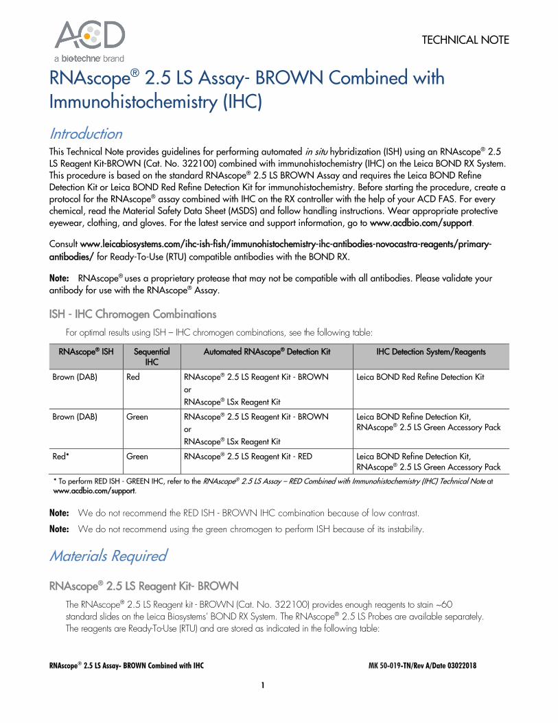
ISH - IHC Chromogen Combinations For optimal results using ISH – IHC chromogen combinations, see the following table:
RNAscope® ISH Sequential IHC
Automated RNAscope® Detection Kit IHC Detection System/Reagents
Brown (DAB) Red RNAscope® 2.5 LS Reagent Kit - BROWN or RNAscope® LSx Reagent Kit
Leica BOND Red Refine Detection Kit
Brown (DAB) Green RNAscope® 2.5 LS Reagent Kit - BROWN or RNAscope® LSx Reagent Kit
Leica BOND Refine Detection Kit, RNAscope® 2.5 LS Green Accessory Pack
Red* Green RNAscope® 2.5 LS Reagent Kit - RED Leica BOND Refine Detection Kit, RNAscope® 2.5 LS Green Accessory Pack
* To perform RED ISH - GREEN IHC, refer to the RNAscope® 2.5 LS Assay – RED Combined with Immunohistochemistry (IHC) Technical Note at www.acdbio.com/support.
Note: We do not recommend the RED ISH - BROWN IHC combination because of low contrast.
Note: We do not recommend using the green chromogen to perform ISH because of its instability.
Materials Required
RNAscope® 2.5 LS Reagent Kit- BROWN
The RNAscope® 2.5 LS Reagent kit - BROWN (Cat. No. 322100) provides enough reagents to stain ~60 standard slides on the Leica Biosystems’ BOND RX System. The RNAscope® 2.5 LS Probes are available separately. The reagents are Ready-To-Use (RTU) and are stored as indicated in the following table:
RNAscope® 2.5 LS Assay- BROWN Combined with IHC MK 50-019-TN/Rev A/Date 03022018 2
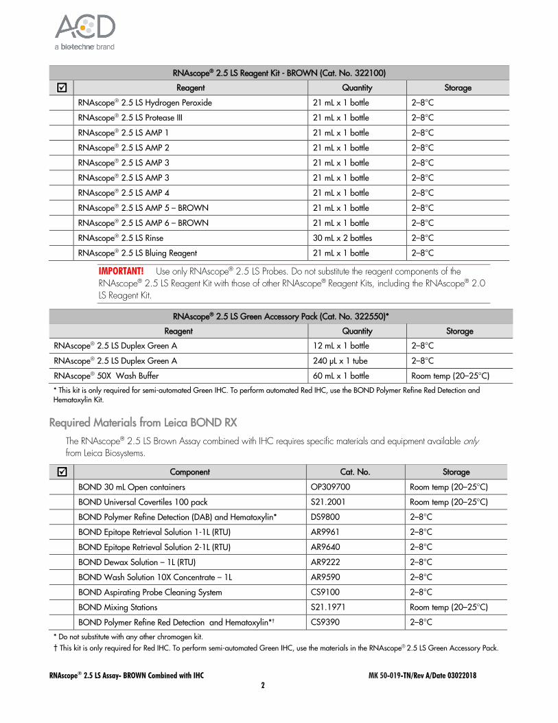
RNAscope® 2.5 LS Reagent Kit - BROWN (Cat. No. 322100)
Reagent Quantity Storage
RNAscope® 2.5 LS Hydrogen Peroxide 21 mL x 1 bottle 2–8°C
RNAscope® 2.5 LS Protease III 21 mL x 1 bottle 2–8°C
RNAscope® 2.5 LS AMP 1 21 mL x 1 bottle 2–8°C
RNAscope® 2.5 LS AMP 2 21 mL x 1 bottle 2–8°C
RNAscope® 2.5 LS AMP 3 21 mL x 1 bottle 2–8°C
RNAscope® 2.5 LS AMP 3 21 mL x 1 bottle 2–8°C
RNAscope® 2.5 LS AMP 4 21 mL x 1 bottle 2–8°C
RNAscope® 2.5 LS AMP 5 – BROWN 21 mL x 1 bottle 2–8°C
RNAscope® 2.5 LS AMP 6 – BROWN 21 mL x 1 bottle 2–8°C
RNAscope® 2.5 LS Rinse 30 mL x 2 bottles 2–8°C
RNAscope® 2.5 LS Bluing Reagent 21 mL x 1 bottle 2–8°C
IMPORTANT! Use only RNAscope® 2.5 LS Probes. Do not substitute the reagent components of the RNAscope® 2.5 LS Reagent Kit with those of other RNAscope® Reagent Kits, including the RNAscope® 2.0 LS Reagent Kit.
RNAscope® 2.5 LS Green Accessory Pack (Cat. No. 322550)*
Reagent Quantity Storage
RNAscope® 2.5 LS Duplex Green A 12 mL x 1 bottle 2–8°C
RNAscope® 2.5 LS Duplex Green A 240 µL x 1 tube 2–8°C
RNAscope® 50X Wash Buffer 60 mL x 1 bottle Room temp (20–25°C)
* This kit is only required for semi-automated Green IHC. To perform automated Red IHC, use the BOND Polymer Refine Red Detection and Hematoxylin Kit.
Required Materials from Leica BOND RX
The RNAscope® 2.5 LS Brown Assay combined with IHC requires specific materials and equipment available only from Leica Biosystems.
Component Cat. No. Storage
BOND 30 mL Open containers OP309700 Room temp (20–25°C)
BOND Universal Covertiles 100 pack S21.2001 Room temp (20–25°C)
BOND Polymer Refine Detection (DAB) and Hematoxylin* DS9800 2–8°C
BOND Epitope Retrieval Solution 1-1L (RTU) AR9961 2–8°C
BOND Epitope Retrieval Solution 2-1L (RTU) AR9640 2–8°C
BOND Dewax Solution – 1L (RTU) AR9222 2–8°C
BOND Wash Solution 10X Concentrate – 1L AR9590 2–8°C
BOND Aspirating Probe Cleaning System CS9100 2–8°C
BOND Mixing Stations S21.1971 Room temp (20–25°C)
BOND Polymer Refine Red Detection and Hematoxylin*† CS9390 2–8°C
* Do not substitute with any other chromogen kit. † This kit is only required for Red IHC. To perform semi-automated Green IHC, use the materials in the RNAscope® 2.5 LS Green Accessory Pack.
RNAscope® 2.5 LS Assay- BROWN Combined with IHC MK 50-019-TN/Rev A/Date 03022018 3
Workflow
Part 1: Create software protocols to perform in situ hybridization (ISH) This section provides instructions for creating in situ hybridization (ISH) software protocols compatible with sequential immunohistochemistry (IHC) on the Leica BOND RX System.
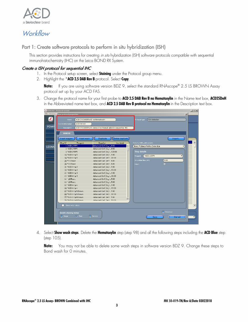
Create a ISH protocol for sequential IHC 1. In the Protocol setup screen, select Staining under the Protocol group menu. 2. Highlight the *ACD 2.5 DAB Rev B protocol. Select Copy.
Note: If you are using software version BDZ 9, select the standard RNAscope® 2.5 LS BROWN Assay protocol set up by your ACD FAS.
3. Change the protocol name for your first probe to ACD 2.5 DAB Rev B no Hematoxylin in the Name text box, ACD25DnH in the Abbreviated name text box, and ACD 2.5 DAB Rev B protocol no Hematoxylin in the Description text box.
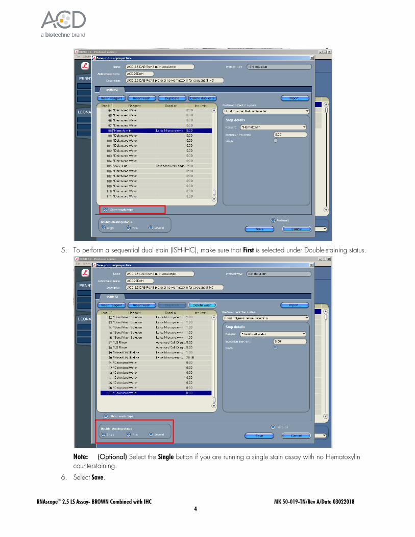
4. Select Show wash steps. Delete the Hematoxylin step (step 98) and all the following steps including the ACD Blue step (step 105).
Note: You may not be able to delete some wash steps in software version BDZ 9. Change these steps to Bond wash for 0 minutes.
RNAscope® 2.5 LS Assay- BROWN Combined with IHC MK 50-019-TN/Rev A/Date 03022018 4
5. To perform a sequential dual stain (ISH-IHC), make sure that First is selected under Double-staining status.
Note: (Optional) Select the Single button if you are running a single stain assay with no Hematoxylin counterstaining.
6. Select Save.
RNAscope® 2.5 LS Assay- BROWN Combined with IHC MK 50-019-TN/Rev A/Date 03022018 5
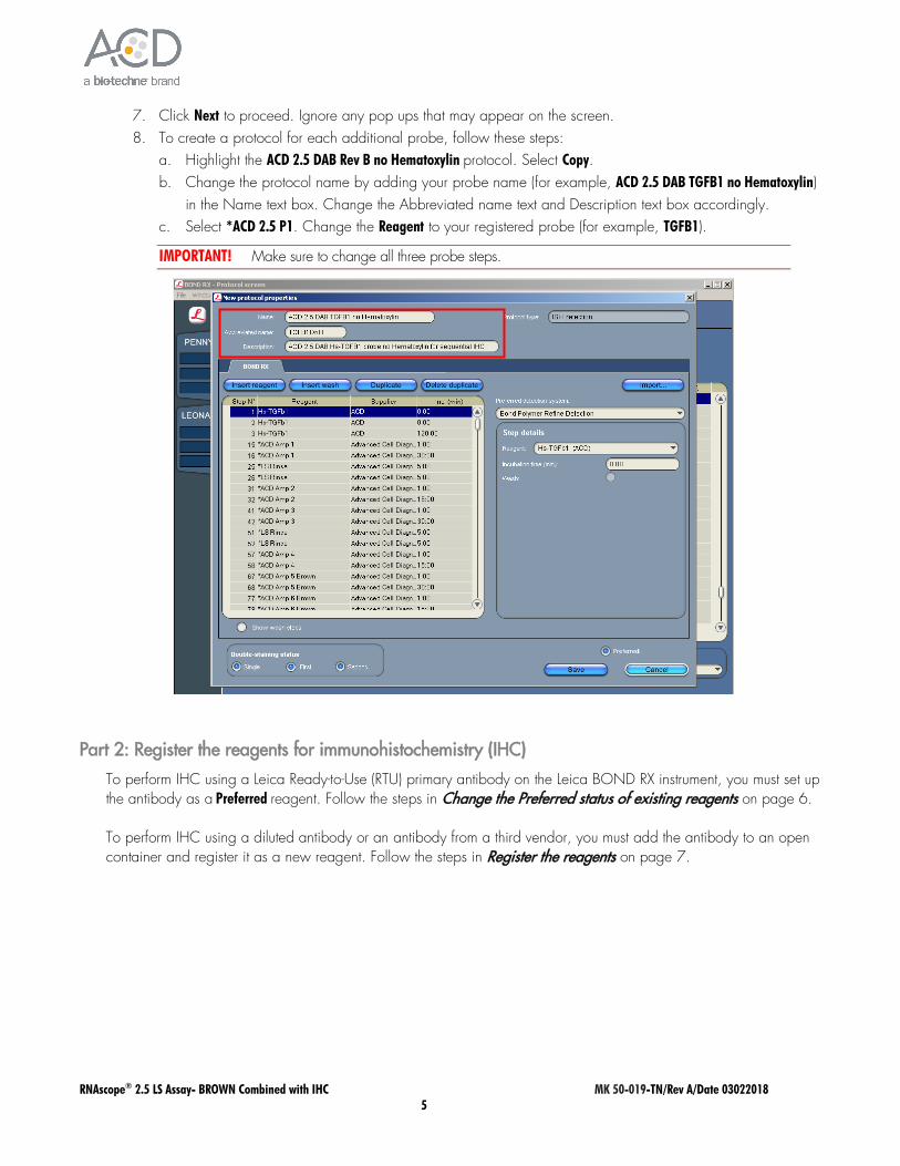
7. Click Next to proceed. Ignore any pop ups that may appear on the screen. 8. To create a protocol for each additional probe, follow these steps:
a. Highlight the ACD 2.5 DAB Rev B no Hematoxylin protocol. Select Copy. b. Change the protocol name by adding your probe name (for example, ACD 2.5 DAB TGFB1 no Hematoxylin)
in the Name text box. Change the Abbreviated name text and Description text box accordingly. c. Select *ACD 2.5 P1. Change the Reagent to your registered probe (for example, TGFB1).
IMPORTANT! Make sure to change all three probe steps.
Part 2: Register the reagents for immunohistochemistry (IHC)
To perform IHC using a Leica Ready-to-Use (RTU) primary antibody on the Leica BOND RX instrument, you must set up the antibody as a Preferred reagent. Follow the steps in Change the Preferred status of existing reagents on page 6. To perform IHC using a diluted antibody or an antibody from a third vendor, you must add the antibody to an open container and register it as a new reagent. Follow the steps in Register the reagents on page 7.
RNAscope® 2.5 LS Assay- BROWN Combined with IHC MK 50-019-TN/Rev A/Date 03022018 6
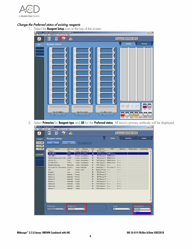
Change the Preferred status of existing reagents 1. Select the Reagent Setup icon at the top of the screen.
2. Select Primaries for Reagent type and All for the Preferred status. All Leica’s primary antibody will be displayed.

RNAscope® 2.5 LS Assay- BROWN Combined with IHC MK 50-019-TN/Rev A/Date 03022018 7
3. Select the antibody to be used (for example, *CD4). Double click to edit reagent properties 4. Select Preferred then Save.
Register the reagents 1. Select the Reagent Setup icon at the top of the screen.
RNAscope® 2.5 LS Assay- BROWN Combined with IHC MK 50-019-TN/Rev A/Date 03022018 8
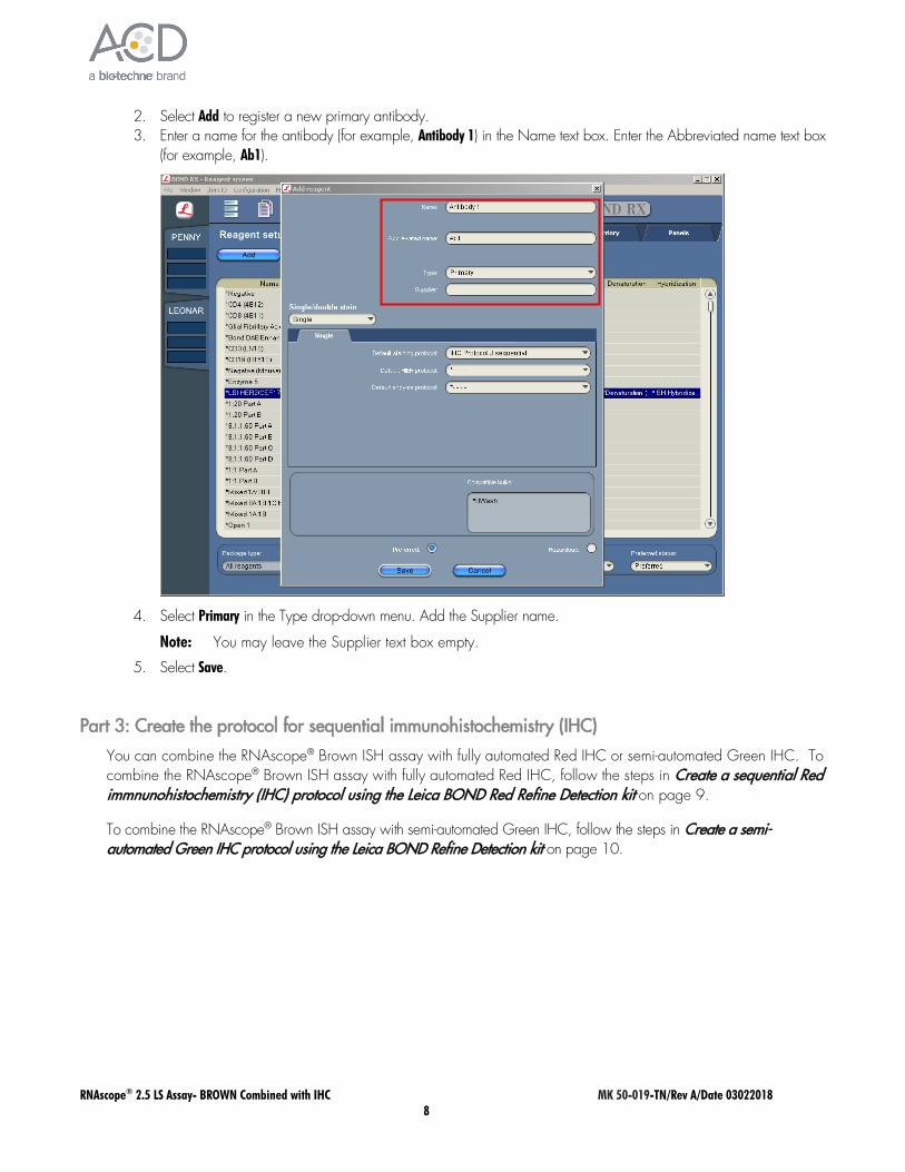
2. Select Add to register a new primary antibody. 3. Enter a name for the antibody (for example, Antibody 1) in the Name text box. Enter the Abbreviated name text box
(for example, Ab1).
4. Select Primary in the Type drop-down menu. Add the Supplier name.
Note: You may leave the Supplier text box empty.
5. Select Save.
Part 3: Create the protocol for sequential immunohistochemistry (IHC)
You can combine the RNAscope® Brown ISH assay with fully automated Red IHC or semi-automated Green IHC. To combine the RNAscope® Brown ISH assay with fully automated Red IHC, follow the steps in Create a sequential Red immnunohistochemistry (IHC) protocol using the Leica BOND Red Refine Detection kit on page 9.
To combine the RNAscope® Brown ISH assay with semi-automated Green IHC, follow the steps in Create a semi-automated Green IHC protocol using the Leica BOND Refine Detection kit on page 10.
RNAscope® 2.5 LS Assay- BROWN Combined with IHC MK 50-019-TN/Rev A/Date 03022018 9
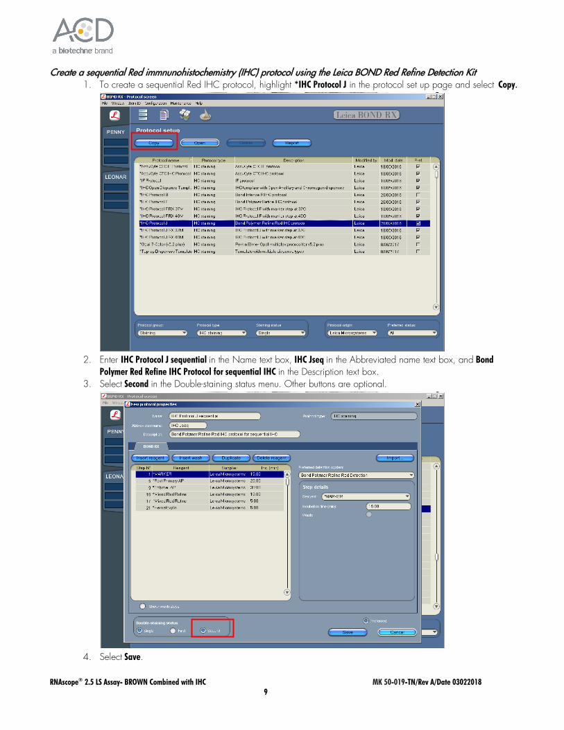
Create a sequential Red immnunohistochemistry (IHC) protocol using the Leica BOND Red Refine Detection Kit 1. To create a sequential Red IHC protocol, highlight *IHC Protocol J in the protocol set up page and select Copy.
2. Enter IHC Protocol J sequential in the Name text box, IHC Jseq in the Abbreviated name text box, and Bond
Polymer Red Refine IHC Protocol for sequential IHC in the Description text box. 3. Select Second in the Double-staining status menu. Other buttons are optional.
4. Select Save.

RNAscope® 2.5 LS Assay- BROWN Combined with IHC MK 50-019-TN/Rev A/Date 03022018 10
Create a semi-automated Green IHC protocol using the Leica BOND Refine Detection Kit 1. To create a semi-automated IHC protocol using Green Chromogen, highlight the*IHC Protocol F protocol in
the protocol set up page and select Copy.
2. Enter IHC Protocol Offline Green in the Name text box, IHCgreen in the Abbreviated name text box, and Bond
Polymer Refine IHC Protocol for offline Green in the Description text box. 3. Select Second in the Double-staining status menu. Other buttons are optional.
RNAscope® 2.5 LS Assay- BROWN Combined with IHC MK 50-019-TN/Rev A/Date 03022018 11
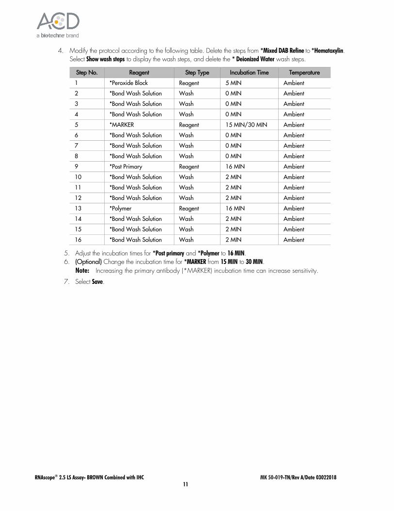
4. Modify the protocol according to the following table. Delete the steps from *Mixed DAB Refine to *Hematoxylin. Select Show wash steps to display the wash steps, and delete the * Deionized Water wash steps.
Step No. Reagent Step Type Incubation Time Temperature
1 *Peroxide Block Reagent 5 MIN Ambient
2 *Bond Wash Solution Wash 0 MIN Ambient
3 *Bond Wash Solution Wash 0 MIN Ambient
4 *Bond Wash Solution Wash 0 MIN Ambient
5 *MARKER Reagent 15 MIN/30 MIN Ambient
6 *Bond Wash Solution Wash 0 MIN Ambient
7 *Bond Wash Solution Wash 0 MIN Ambient
8 *Bond Wash Solution Wash 0 MIN Ambient
9 *Post Primary Reagent 16 MIN Ambient
10 *Bond Wash Solution Wash 2 MIN Ambient
11 *Bond Wash Solution Wash 2 MIN Ambient
12 *Bond Wash Solution Wash 2 MIN Ambient
13 *Polymer Reagent 16 MIN Ambient
14 *Bond Wash Solution Wash 2 MIN Ambient
15 *Bond Wash Solution Wash 2 MIN Ambient
16 *Bond Wash Solution Wash 2 MIN Ambient
5. Adjust the incubation times for *Post primary and *Polymer to 16 MIN. 6. (Optional) Change the incubation time for *MARKER from 15 MIN to 30 MIN.
Note: Increasing the primary antibody (*MARKER) incubation time can increase sensitivity.
7. Select Save.
RNAscope® 2.5 LS Assay- BROWN Combined with IHC MK 50-019-TN/Rev A/Date 03022018 12
Part 4: Set up a study for sequential ISH-IHC
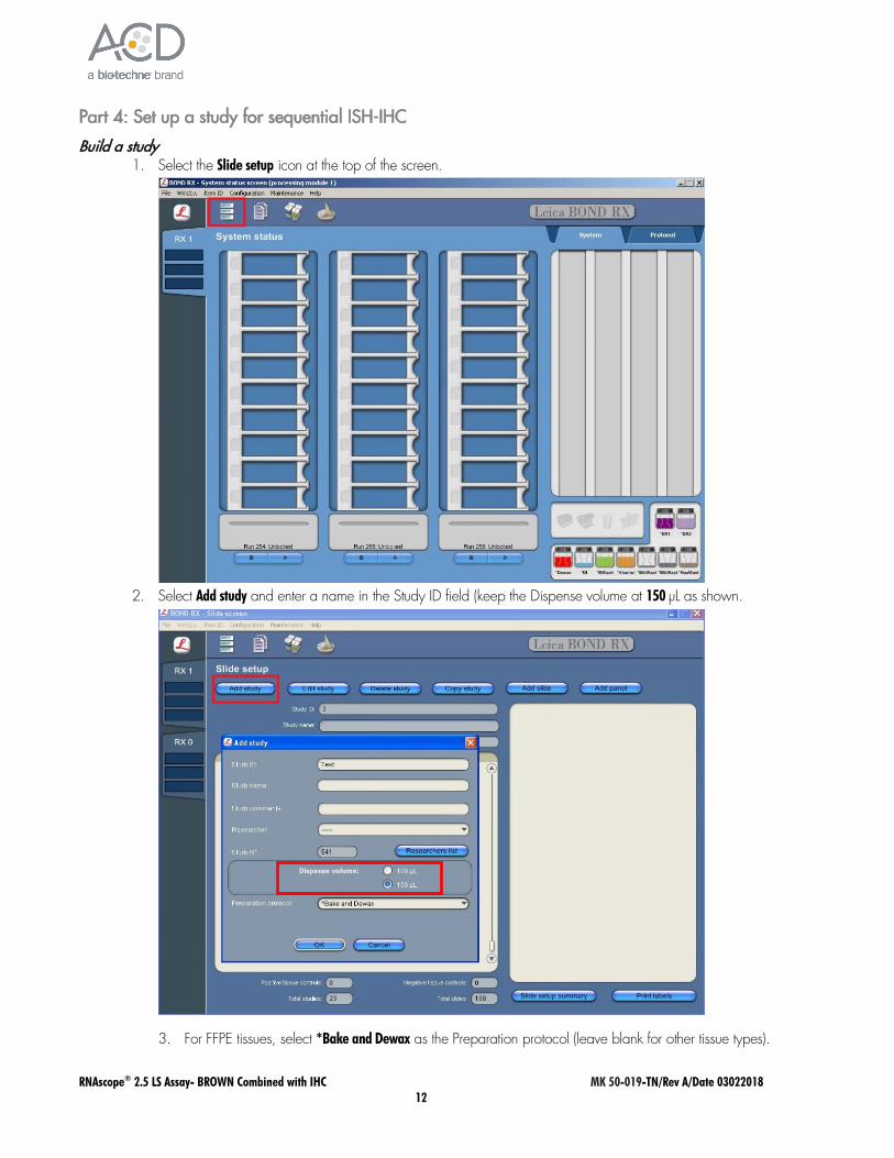
Build a study 1. Select the Slide setup icon at the top of the screen.
2. Select Add study and enter a name in the Study ID field (keep the Dispense volume at 150 µL as shown.
3. For FFPE tissues, select *Bake and Dewax as the Preparation protocol (leave blank for other tissue types).
RNAscope® 2.5 LS Assay- BROWN Combined with IHC MK 50-019-TN/Rev A/Date 03022018 13
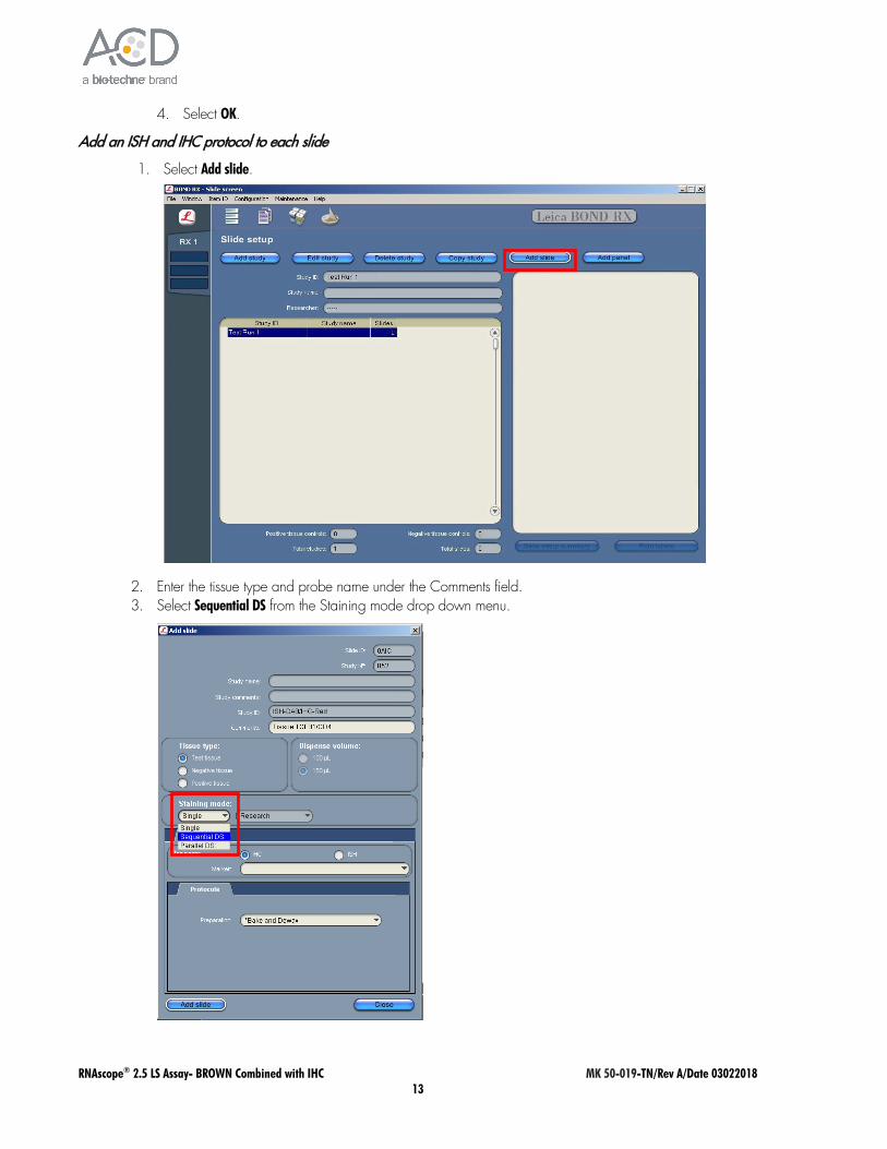
4. Select OK.
Add an ISH and IHC protocol to each slide
1. Select Add slide.
2. Enter the tissue type and probe name under the Comments field. 3. Select Sequential DS from the Staining mode drop down menu.
RNAscope® 2.5 LS Assay- BROWN Combined with IHC MK 50-019-TN/Rev A/Date 03022018 14
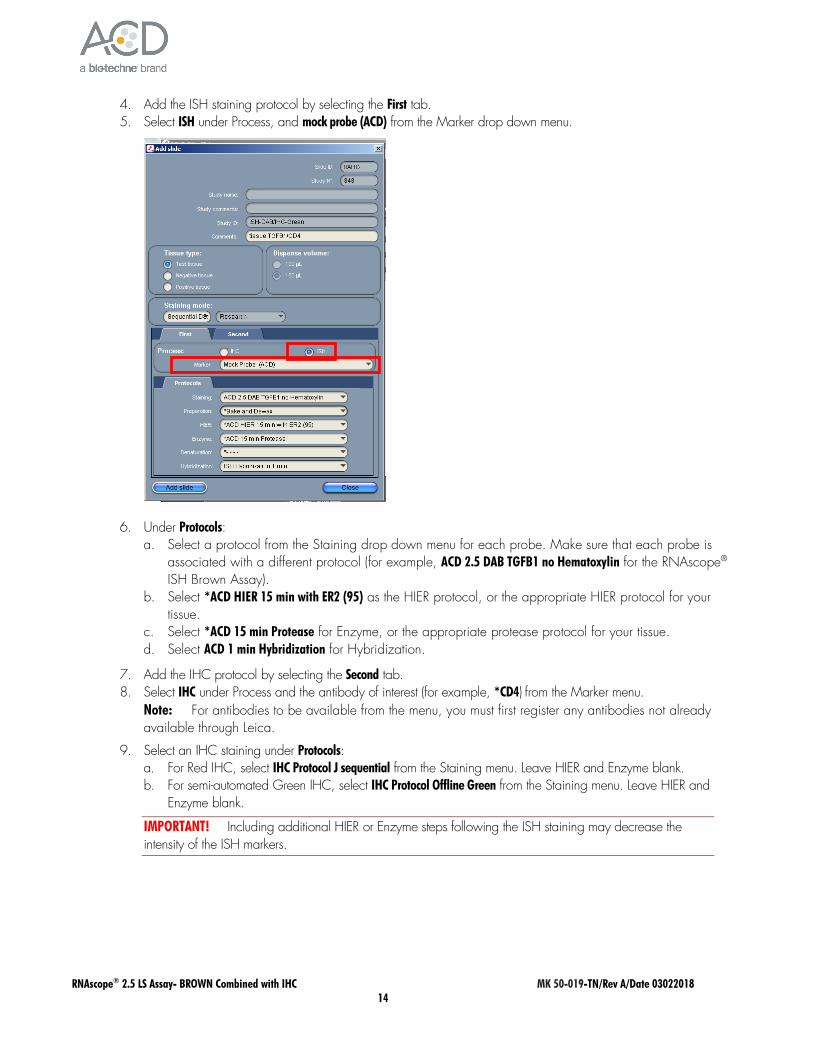
4. Add the ISH staining protocol by selecting the First tab. 5. Select ISH under Process, and mock probe (ACD) from the Marker drop down menu.
6. Under Protocols: a. Select a protocol from the Staining drop down menu for each probe. Make sure that each probe is
associated with a different protocol (for example, ACD 2.5 DAB TGFB1 no Hematoxylin for the RNAscope® ISH Brown Assay).
b. Select *ACD HIER 15 min with ER2 (95) as the HIER protocol, or the appropriate HIER protocol for your tissue.
c. Select *ACD 15 min Protease for Enzyme, or the appropriate protease protocol for your tissue. d. Select ACD 1 min Hybridization for Hybridization.
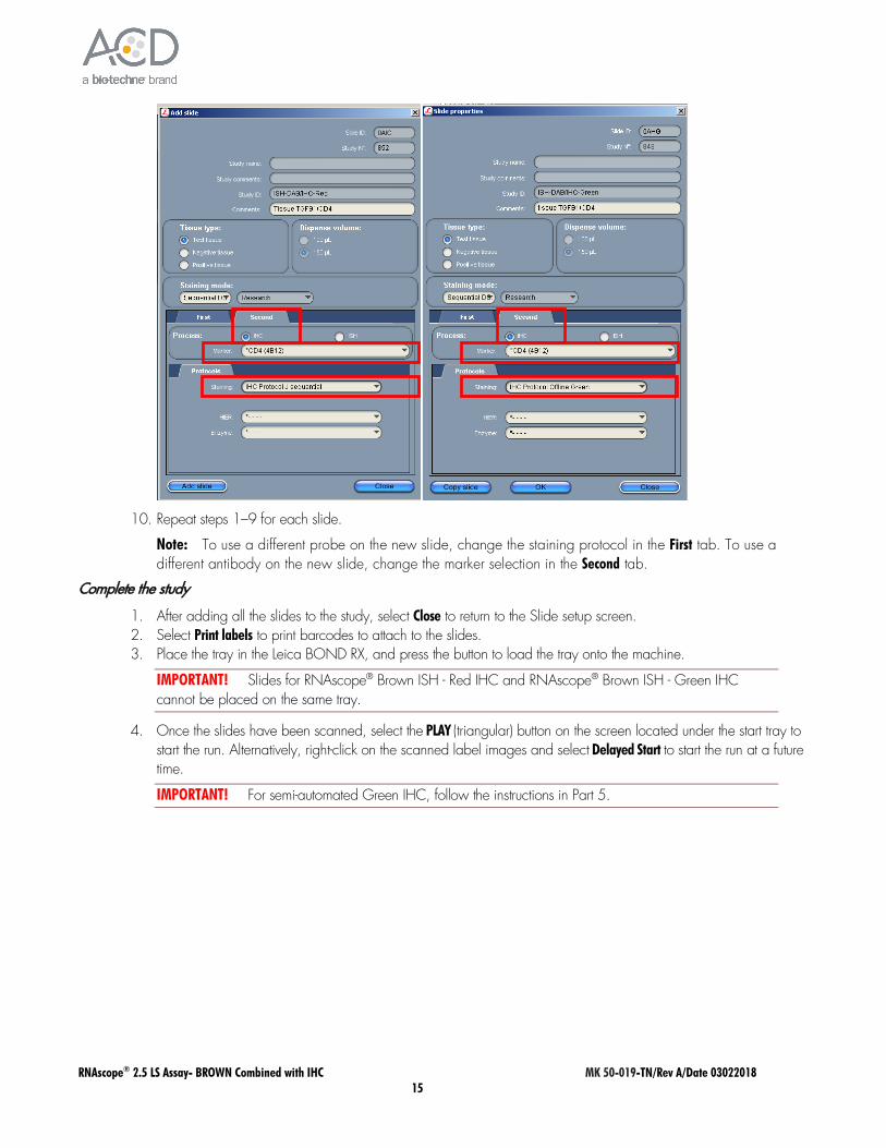
7. Add the IHC protocol by selecting the Second tab. 8. Select IHC under Process and the antibody of interest (for example, *CD4) from the Marker menu.
Note: For antibodies to be available from the menu, you must first register any antibodies not already available through Leica.
9. Select an IHC staining under Protocols: a. For Red IHC, select IHC Protocol J sequential from the Staining menu. Leave HIER and Enzyme blank. b. For semi-automated Green IHC, select IHC Protocol Offline Green from the Staining menu. Leave HIER and
Enzyme blank.
IMPORTANT! Including additional HIER or Enzyme steps following the ISH staining may decrease the intensity of the ISH markers.
RNAscope® 2.5 LS Assay- BROWN Combined with IHC MK 50-019-TN/Rev A/Date 03022018 15
10. Repeat steps 1–9 for each slide.
Note: To use a different probe on the new slide, change the staining protocol in the First tab. To use a different antibody on the new slide, change the marker selection in the Second tab.
Complete the study
1. After adding all the slides to the study, select Close to return to the Slide setup screen. 2. Select Print labels to print barcodes to attach to the slides. 3. Place the tray in the Leica BOND RX, and press the button to load the tray onto the machine.
IMPORTANT! Slides for RNAscope® Brown ISH - Red IHC and RNAscope® Brown ISH - Green IHC cannot be placed on the same tray.
4. Once the slides have been scanned, select the PLAY (triangular) button on the screen located under the start tray to start the run. Alternatively, right-click on the scanned label images and select Delayed Start to start the run at a future time.
IMPORTANT! For semi-automated Green IHC, follow the instructions in Part 5.
RNAscope® 2.5 LS Assay- BROWN Combined with IHC MK 50-019-TN/Rev A/Date 03022018 16
Part 5: Detect green IHC staining off the instrument
Prepare reagents and equipment
1. Before the run completes, remove the Green A and Green B reagents from the refrigerator and warm to ambient temperature.
IMPORTANT! View the wash step video at www.acdbio.com/technical-support/learn-more before proceeding.
Detect Green staining off the instrument
1. As soon as the run is complete, press the button on the front of the instrument and unload the slides immediately.
IMPORTANT! If you do not perform the Green assay immediately, store the slides in 4X SSC at 4˚C. After the run is completed, do not leave the slides loaded on the instrument. The slides are automatically re-hydrated by the instrument. Do not store the slides in water or wash buffer.
IMPORTANT! Do not let sections dry out between incubation steps. Work quickly and make sure the sections are hydrated at all times.
2. Wash slides in 1X Wash Buffer for 2 MIN at RT. Agitate slides by moving the slide rack up and down in the staining dish.
3. Repeat Step 2 with fresh 1X Wash Buffer. 4. Briefly spin down the contents of the Green B tube to be sure content is at the bottom of the tube before opening
the cap. 5. Prepare 200 µL of Green working solution per slide using a 1:50 ratio of Green B to Green A. Mix well.
IMPORTANT! Use the Green solution within 5 MIN. Do not expose to direct sunlight or UV light.
6. Take each slide one at a time from the Tissue-Tek® Slide Rack and tap and/or flick to remove the excess liquid.
7. Pipette ~200 µL Green solution onto each tissue section. Ensure sections are covered. 8. Incubate the slides for 15 MIN at RT. 9. To remove the Green working solution from the slides, tilt each slide one at a time over a waste container and tap
the corner on the edge of the container. Immediately insert the slide into a Tissue-Tek®Slide Rack submerged in a Tissue-Tek®
Staining Dish filled with distilled water. 10. Quickly rinse the slides with fresh distilled water for less than 30 seconds.
IMPORTANT! Proceed quickly to the next step. Green substrate may fade if stored in water for too long.
Counterstain the slides
1. Move the Tissue-Tek® Slide Rack into the staining dish containing 50% Hematoxylin I staining solution for 30 SEC at
RT. Slides will be purple.
IMPORTANT! Proceed quickly to the next step. Green substrate may fade if in Hematoxylin for longer than 30 seconds.
2. Immediately transfer the slide rack into a staining dish filled with tap water. Do not let the slides remain in the water for more than 30 seconds.
3. Repeat Step 2 once or twice.

RNAscope® 2.5 LS Assay- BROWN Combined with IHC MK 50-019-TN/Rev A/Date 03022018 17
Dry and mount the samples
1. Remove the slide rack from the staining dish and dry slides in a 60°C dry oven for 30 MIN.
IMPORTANT! The Red and Green substrates are alcohol sensitive. Do not dehydrate the slides in alcohol.
2. Cool the slides for 5 MIN at RT. 3. Briefly dip one slide into fresh pure xylene and immediately place 1–2 drops of VectaMount™ Mounting Medium
on the slide before the xylene dries. 4. Carefully place a 24 mm x 50 mm coverslip over the tissue section. Avoid trapping air bubbles. 5. Repeat steps 2 and 3 for each slide. 6. Air dry the slides for 5 MIN.
For Research Use Only. Not For Diagnostic Use.
Advanced Cell Diagnostics, Inc. and/or its affiliate(s) warrant their products as set forth in the ACD General Terms and Conditions of Sale found on the ACD website. Advanced Cell Diagnostics, Inc. reserves the right to change its products and services at any time to incorporate technological developments. This document is subject to change without notice. Although this document has been prepared with every precaution to ensure accuracy, Advanced Cell Diagnostics, Inc. assumes no liability for any errors, omissions, or for any damages resulting from the use of this information. © 2018 Advanced Cell Diagnostics. All rights reserved. RNAscope is a registered trademark of Advanced Cell Diagnostics, Inc. All other trademarks belong to their respective owners.
Headquarters 7707 Gateway Blvd Suite 200, Newark, CA 94545 Phone 1-510-576-8800 Toll Free 1-877-576-3636 For support, email [email protected] www.acdbio.com
Related Documents