RNA Tertiary Structure Analysis by 2′-Hydroxyl Molecular Interference Philip J. Homan, † Arpit Tandon, ‡ Greggory M. Rice, † Feng Ding, § Nikolay V. Dokholyan, ‡ and Kevin M. Weeks* ,† † Departments of Chemistry and ‡ Biochemistry and Biophysics, University of North Carolina, Chapel Hill, North Carolina 27599-3290, United States § Department of Physics and Astronomy, Clemson University, Clemson, South Carolina 29631, United States * S Supporting Information ABSTRACT: We introduce a melded chemical and computational approach for probing and modeling higher-order intramolecular tertiary interactions in RNA. 2′-Hydroxyl molecular interference (HMX) identifies nucleotides in highly packed regions of an RNA by exploiting the ability of bulky adducts at the 2′-hydroxyl position to disrupt overall RNA structure. HMX was found to be exceptionally selective for quantitative detection of higher-order and tertiary interactions. When incorporated as experimental constraints in discrete molecular dynamics simulations, HMX information yielded accurate three-dimensional models, emphasizing the power of molecular interference to guide RNA tertiary structure analysis and fold refinement. In the case of a large, multidomain RNA, the Tetrahymena group I intron, HMX identified multiple distinct sets of tertiary structure interaction groups in a single, concise experiment. RNA plays diverse and central roles in the regulation of gene expression. 1 Information is encoded in RNA at several levels: the primary sequence, the specific base pairing pattern that defines the secondary structure, and higher-order RNA structures composed of tightly packed secondary structure elements stabilized by a few key tertiary interactions. 2 The precise formation of higher-order tertiary structures is critical to the function of many RNAs. 3,4 RNA secondary and tertiary interactions can be interrogated by modifying an RNA with chemical probes or by incorporating nucleotide substitutions that disrupt native structure. In modification interference, an RNA is treated to introduce chemical modifications, usually at the nucleobases, and then the RNA is subjected to a partitioning experiment to distinguish functional from non- functional molecules. 5−7 For the nucleotide analogue interfer- ence mapping (NAIM) 8,9 strategy, nucleotide analogues are incorporated into an RNA transcript, and active RNAs are partitioned from those that are inactivated because of the nucleotide analogue. Both modification interference and NAIM can interrogate most nucleotides in an RNA to identify single- nucleotide and single-atom interactions, respectively, critical to the tertiary structure. 5,6,8,9 These approaches generally require multiple distinct experiments to interrogate the tertiary environment of every nucleotide in an RNA. Chemical probes are also widely used to examine both solvent accessibility and dynamics in the RNA backbone. The solvent accessibility of the RNA backbone can be monitored by hydroxyl radical footprinting (HRP). 10,11 Backbone dynamics can be monitored in the selective 2′-hydroxyl acylation analyzed by primer extension (SHAPE) strategy that uses reagents sensitive to the nucleophilicity of the 2′-OH group, which is dependent on the underlying flexibility of the nucleotide. 12−14 Reactivities of these and other chemical probes, like DMS, CMCT, and kethoxal, are modulated by both secondary and tertiary structure interactions, and it is usually difficult to deconvolute the relative influence of each type of interaction. Here, we describe a strategy in which 2′-hydroxyl-selective reagents are used in a modification interference experiment to simply, directly, and specifically interrogate RNA tertiary structure. In this approach, which we call 2′-hydroxyl molecular interference, or HMX, a hydroxyl-selective reagent is used to create a pool of RNAs with evenly distributed 2′-O-ester adducts. Next, a structure-selective pressure, such as RNA folding, is placed on the pool of modified RNAs. A subset of 2′- O-ester groups will interfere with molecular interactions and prevent native structure formation. By partitioning the sample into folded and unfolded states, nucleotides whose modification disrupts tertiary interactions are identified. Here we use this information to characterize the internal packing interactions that define higher-order RNA structure, to refine three- dimensional structure models, and to detect multiple sets of tertiary interactions in a large complexly folded RNA. Received: September 26, 2014 Published: October 23, 2014 Article pubs.acs.org/biochemistry © 2014 American Chemical Society 6825 dx.doi.org/10.1021/bi501218g | Biochemistry 2014, 53, 6825−6833

Welcome message from author
This document is posted to help you gain knowledge. Please leave a comment to let me know what you think about it! Share it to your friends and learn new things together.
Transcript

RNA Tertiary Structure Analysis by 2′-Hydroxyl MolecularInterferencePhilip J. Homan,† Arpit Tandon,‡ Greggory M. Rice,† Feng Ding,§ Nikolay V. Dokholyan,‡
and Kevin M. Weeks*,†
†Departments of Chemistry and ‡Biochemistry and Biophysics, University of North Carolina, Chapel Hill, North Carolina27599-3290, United States§Department of Physics and Astronomy, Clemson University, Clemson, South Carolina 29631, United States
*S Supporting Information
ABSTRACT: We introduce a melded chemical and computationalapproach for probing and modeling higher-order intramolecular tertiaryinteractions in RNA. 2′-Hydroxyl molecular interference (HMX)identifies nucleotides in highly packed regions of an RNA by exploitingthe ability of bulky adducts at the 2′-hydroxyl position to disrupt overallRNA structure. HMX was found to be exceptionally selective forquantitative detection of higher-order and tertiary interactions. Whenincorporated as experimental constraints in discrete molecular dynamicssimulations, HMX information yielded accurate three-dimensionalmodels, emphasizing the power of molecular interference to guideRNA tertiary structure analysis and fold refinement. In the case of alarge, multidomain RNA, the Tetrahymena group I intron, HMXidentified multiple distinct sets of tertiary structure interaction groups ina single, concise experiment.
RNA plays diverse and central roles in the regulation of geneexpression.1 Information is encoded in RNA at several levels:the primary sequence, the specific base pairing pattern thatdefines the secondary structure, and higher-order RNAstructures composed of tightly packed secondary structureelements stabilized by a few key tertiary interactions.2 Theprecise formation of higher-order tertiary structures is critical tothe function of many RNAs.3,4 RNA secondary and tertiaryinteractions can be interrogated by modifying an RNA withchemical probes or by incorporating nucleotide substitutionsthat disrupt native structure. In modification interference, anRNA is treated to introduce chemical modifications, usually atthe nucleobases, and then the RNA is subjected to apartitioning experiment to distinguish functional from non-functional molecules.5−7 For the nucleotide analogue interfer-ence mapping (NAIM)8,9 strategy, nucleotide analogues areincorporated into an RNA transcript, and active RNAs arepartitioned from those that are inactivated because of thenucleotide analogue. Both modification interference and NAIMcan interrogate most nucleotides in an RNA to identify single-nucleotide and single-atom interactions, respectively, critical tothe tertiary structure.5,6,8,9 These approaches generally requiremultiple distinct experiments to interrogate the tertiaryenvironment of every nucleotide in an RNA.Chemical probes are also widely used to examine both
solvent accessibility and dynamics in the RNA backbone. Thesolvent accessibility of the RNA backbone can be monitored byhydroxyl radical footprinting (HRP).10,11 Backbone dynamics
can be monitored in the selective 2′-hydroxyl acylationanalyzed by primer extension (SHAPE) strategy that usesreagents sensitive to the nucleophilicity of the 2′-OH group,which is dependent on the underlying flexibility of thenucleotide.12−14 Reactivities of these and other chemicalprobes, like DMS, CMCT, and kethoxal, are modulated byboth secondary and tertiary structure interactions, and it isusually difficult to deconvolute the relative influence of eachtype of interaction.Here, we describe a strategy in which 2′-hydroxyl-selective
reagents are used in a modification interference experiment tosimply, directly, and specifically interrogate RNA tertiarystructure. In this approach, which we call 2′-hydroxyl molecularinterference, or HMX, a hydroxyl-selective reagent is used tocreate a pool of RNAs with evenly distributed 2′-O-esteradducts. Next, a structure-selective pressure, such as RNAfolding, is placed on the pool of modified RNAs. A subset of 2′-O-ester groups will interfere with molecular interactions andprevent native structure formation. By partitioning the sampleinto folded and unfolded states, nucleotides whose modificationdisrupts tertiary interactions are identified. Here we use thisinformation to characterize the internal packing interactionsthat define higher-order RNA structure, to refine three-dimensional structure models, and to detect multiple sets oftertiary interactions in a large complexly folded RNA.
Received: September 26, 2014Published: October 23, 2014
Article
pubs.acs.org/biochemistry
© 2014 American Chemical Society 6825 dx.doi.org/10.1021/bi501218g | Biochemistry 2014, 53, 6825−6833
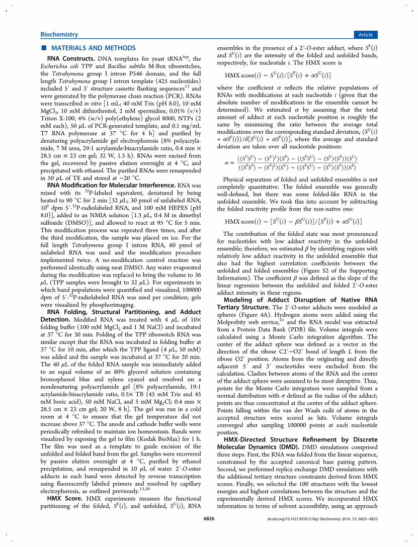
■ MATERIALS AND METHODS
RNA Constructs. DNA templates for yeast tRNAAsp, theEscherichia coli TPP and Bacillus subtilis M-Box riboswitches,the Tetrahymena group I intron P546 domain, and the fulllength Tetrahymena group I intron template (425 nucleotides)included 5′ and 3′ structure cassette flanking sequences13 andwere generated by the polymerase chain reaction (PCR). RNAswere transcribed in vitro [1 mL; 40 mM Tris (pH 8.0), 10 mMMgCl2, 10 mM dithiothreitol, 2 mM spermidine, 0.01% (v/v)Triton X-100, 4% (w/v) poly(ethylene) glycol 8000, NTPs (2mM each), 50 μL of PCR-generated template, and 0.1 mg/mLT7 RNA polymerase at 37 °C for 4 h] and purified bydenaturing polyacrylamide gel electrophoresis (8% polyacryla-mide, 7 M urea, 29:1 acrylamide:bisacrylamide ratio, 0.4 mm ×28.5 cm × 23 cm gel; 32 W, 1.5 h). RNAs were excised fromthe gel, recovered by passive elution overnight at 4 °C, andprecipitated with ethanol. The purified RNAs were resuspendedin 50 μL of TE and stored at −20 °C.RNA Modification for Molecular Interference. RNA was
mixed with its 32P-labeled equivalent, denatured by beingheated to 90 °C for 2 min [32 μL; 30 pmol of unlabeled RNA,106 dpm 5′-32P-radiolabeled RNA, and 100 mM HEPES (pH8.0)], added to an NMIA solution [1.3 μL, 0.4 M in dimethylsulfoxide (DMSO)], and allowed to react at 95 °C for 5 min.This modification process was repeated three times, and afterthe third modification, the sample was placed on ice. For thefull length Tetrahymena group I intron RNA, 60 pmol ofunlabeled RNA was used and the modification procedureimplemented twice. A no-modification control reaction wasperformed identically using neat DMSO. Any water evaporatedduring the modification was replaced to bring the volume to 36μL (TPP samples were brought to 32 μL). For experiments inwhich band populations were quantified and visualized, 100000dpm of 5′-32P-radiolabeled RNA was used per condition; gelswere visualized by phosphorimaging.RNA Folding, Structural Partitioning, and Adduct
Detection. Modified RNA was treated with 4 μL of 10×folding buffer (100 mM MgCl2 and 1 M NaCl) and incubatedat 37 °C for 30 min. Folding of the TPP riboswitch RNA wassimilar except that the RNA was incubated in folding buffer at37 °C for 10 min, after which the TPP ligand (4 μL, 50 mM)was added and the sample was incubated at 37 °C for 20 min.The 40 μL of the folded RNA sample was immediately addedto an equal volume of an 80% glycerol solution containingbromophenol blue and xylene cyanol and resolved on anondenaturing polyacrylamide gel [8% polyacrylamide, 19:1acrylamide:bisacrylamide ratio, 0.5× TB (45 mM Tris and 45mM boric acid), 50 mM NaCl, and 5 mM Mg2Cl; 0.4 mm ×28.5 cm × 23 cm gel; 20 W, 8 h]. The gel was run in a coldroom at 4 °C to ensure that the gel temperature did notincrease above 37 °C. The anode and cathode buffer wells wereperiodically refreshed to maintain ion homeostasis. Bands werevisualized by exposing the gel to film (Kodak BioMax) for 1 h.The film was used as a template to guide excision of theunfolded and folded band from the gel. Samples were recoveredby passive elution overnight at 4 °C, purified by ethanolprecipitation, and resuspended in 10 μL of water. 2′-O-esteradducts in each band were detected by reverse transcriptionusing fluorescently labeled primers and resolved by capillaryelectrophoresis, as outlined previously.13,30
HMX Score. HMX experiments measure the functionalpartitioning of the folded, SF(i), and unfolded, SU(i), RNA
ensembles in the presence of a 2′-O-ester adduct, where SF(i)and SU(i) are the intensity of the folded and unfolded bands,respectively, for nucleotide i. The HMX score is
α∼ +i S i S i S iHMX score( ) ( )/[ ( ) ( )]U F U
where the coefficient α reflects the relative populations ofRNAs with modifications at each nucleotide i (given that theabsolute number of modifications in the ensemble cannot bedetermined). We estimated α by assuming that the totalamount of adduct at each nucleotide position is roughly thesame by minimizing the ratio between the average totalmodifications over the corresponding standard deviation, ⟨SU(i)+ αSF(i)⟩/δ[SU(i) + αSF(i)], where the average and standarddeviation are taken over all nucleotide positions:
α = ⟨ ⟩ − ⟨ ⟩ ⟨ ⟩ − ⟨ ⟩ − ⟨ ⟩⟨ ⟩ ⟨ ⟩⟨ ⟩ − ⟨ ⟩ ⟨ ⟩ − ⟨ ⟩ − ⟨ ⟩⟨ ⟩ ⟨ ⟩S S S S S S S S SS S S S S S S S S
( ) ( )( ) ( )
U U U 2 F F U U F U
F F F 2 U F U U F F
Physical separation of folded and unfolded ensembles is notcompletely quantitative. The folded ensemble was generallywell-defined, but there was some folded-like RNA in theunfolded ensemble. We took this into account by subtractingthe folded reactivity profile from the non-native one:
β α∼ − +i S i S i S i S iHMX score( ) [ ( ) ( )]/[ ( ) ( )]U U F U
The contribution of the folded state was most pronouncedfor nucleotides with low adduct reactivity in the unfoldedensemble; therefore, we estimated β by identifying regions withrelatively low adduct reactivity in the unfolded ensemble thatalso had the highest correlation coefficients between theunfolded and folded ensembles (Figure S2 of the SupportingInformation). The coefficient β was defined as the slope of thelinear regression between the unfolded and folded 2′-O-esteradduct intensity in these regions.
Modeling of Adduct Disruption of Native RNATertiary Structure. The 2′-O-ester adducts were modeled asspheres (Figure 4A). Hydrogen atoms were added using theMolprobity web service,31 and the RNA model was extractedfrom a Protein Data Bank (PDB) file. Volume integrals werecalculated using a Monte Carlo integration algorithm. Thecenter of the adduct sphere was defined as a vector in thedirection of the ribose C2′−O2′ bond of length L from theribose O2′ position. Atoms from the originating and directlyadjacent 5′ and 3′ nucleotides were excluded from thecalculation. Clashes between atoms of the RNA and the centerof the adduct sphere were assumed to be most disruptive. Thus,points for the Monte Carlo integration were sampled from anormal distribution with σ defined as the radius of the adduct;points are thus concentrated at the center of the adduct sphere.Points falling within the van der Waals radii of atoms in theaccepted structure were scored as hits. Volume integralsconverged after sampling 100000 points at each nucleotideposition.
HMX-Directed Structure Refinement by DiscreteMolecular Dynamics (DMD). DMD simulations comprisedthree steps. First, the RNA was folded from the linear sequence,constrained by the accepted canonical base pairing pattern.Second, we performed replica exchange DMD simulations withthe additional tertiary structure constraints derived from HMXscores. Finally, we selected the 100 structures with the lowestenergies and highest correlations between the structure and theexperimentally derived HMX scores. We incorporated HMXinformation in terms of solvent accessibility, using an approach
Biochemistry Article
dx.doi.org/10.1021/bi501218g | Biochemistry 2014, 53, 6825−68336826
previously developed to model hydroxyl radical probing.24 Therelative solvent accessibility was interpreted as the number ofcontacts that sugar pseudoatoms have within a predetermineddistance from the center of each pseudoatom. Positions withhigh HMX scores were allowed more tertiary contacts thanthose with low scores.24 We assigned two biasing potentialsbased on the number of tertiary contacts as calculated from theHMX score. The first term is an attractive potential thatcollapses the RNA to achieve a compact structure. The secondterm is a repulsion potential to prevent overburial of anucleotide when it exceeds the assigned number of thresholdcontacts. The minimal number and maximal number ofthreshold contacts were calculated from a structure databaseto be 0.5 and 11, respectively.24 For any nucleotide exceedingthe HMX scores below or above the threshold, we assignedvalues of 0.5 and 11.DMD Simulations and Consensus Structure Modeling.
We performed replica exchange DMD simulations for eachRNA system using 12 replicas with temperatures of 0.200,0.215, 0.230, 0.246, 0.262, 0.277, 0.293, 0.311, 0.330, 0.350,0.375, and 0.400 kcal mol−1 kB
−1. We set each exchange eventto occur at 1000 DMD time steps according to a Metropolis-based Monte Carlo algorithm. For each replica, we performed a5 × 105 DMD time step simulation. We then generatedsnapshots every 100 DMD time units; from these snapshots, weselected 1000 snapshots with the lowest energies, calculatedcorrelations between the number of tertiary contacts and therelative solvent accessibilities derived from the HMX scores,and selected the 100 structures with the lowest (negative)correlations. We then performed the same selection procedurein reverse order, first selecting the 1000 structures with thelowest correlations based on the experimentally determinedHMX scores and then selecting the 100 structures with thelowest energies. These 200 structures were then ranked byenergy and structure reactivity correlation coefficient, and 100structures were used to represent the final structuralensemble.24 These final ensembles were clustered byhierarchical clustering. For each RNA, the clustering cutoffwas three-quarters of the average root-mean-square deviation(rmsd) as a function of RNA length; that is, the clusteringcutoff was smaller for smaller RNAs with the maximum forlarger RNAs equal to 4 Å.26
■ RESULTSHMX Overview. In the first step of the HMX strategy, an
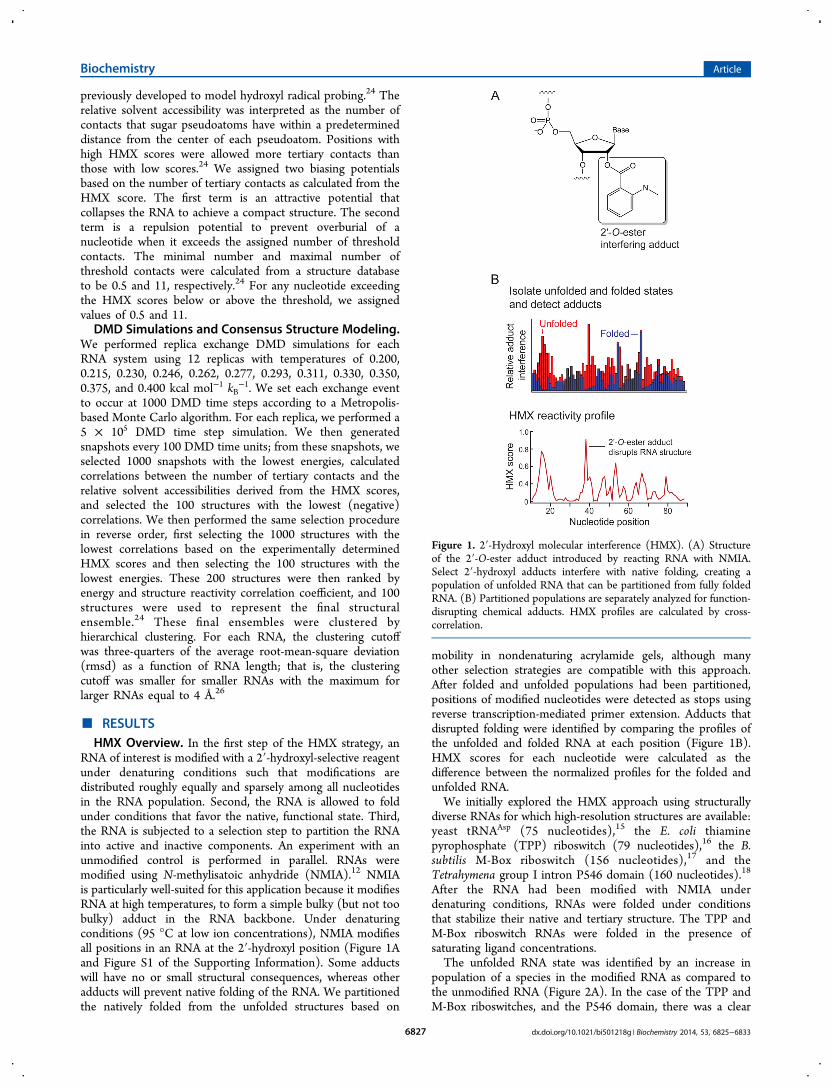
RNA of interest is modified with a 2′-hydroxyl-selective reagentunder denaturing conditions such that modifications aredistributed roughly equally and sparsely among all nucleotidesin the RNA population. Second, the RNA is allowed to foldunder conditions that favor the native, functional state. Third,the RNA is subjected to a selection step to partition the RNAinto active and inactive components. An experiment with anunmodified control is performed in parallel. RNAs weremodified using N-methylisatoic anhydride (NMIA).12 NMIAis particularly well-suited for this application because it modifiesRNA at high temperatures, to form a simple bulky (but not toobulky) adduct in the RNA backbone. Under denaturingconditions (95 °C at low ion concentrations), NMIA modifiesall positions in an RNA at the 2′-hydroxyl position (Figure 1Aand Figure S1 of the Supporting Information). Some adductswill have no or small structural consequences, whereas otheradducts will prevent native folding of the RNA. We partitionedthe natively folded from the unfolded structures based on
mobility in nondenaturing acrylamide gels, although manyother selection strategies are compatible with this approach.After folded and unfolded populations had been partitioned,positions of modified nucleotides were detected as stops usingreverse transcription-mediated primer extension. Adducts thatdisrupted folding were identified by comparing the profiles ofthe unfolded and folded RNA at each position (Figure 1B).HMX scores for each nucleotide were calculated as thedifference between the normalized profiles for the folded andunfolded RNA.We initially explored the HMX approach using structurally
diverse RNAs for which high-resolution structures are available:yeast tRNAAsp (75 nucleotides),15 the E. coli thiaminepyrophosphate (TPP) riboswitch (79 nucleotides),16 the B.subtilis M-Box riboswitch (156 nucleotides),17 and theTetrahymena group I intron P546 domain (160 nucleotides).18
After the RNA had been modified with NMIA underdenaturing conditions, RNAs were folded under conditionsthat stabilize their native and tertiary structure. The TPP andM-Box riboswitch RNAs were folded in the presence ofsaturating ligand concentrations.The unfolded RNA state was identified by an increase in
population of a species in the modified RNA as compared tothe unmodified RNA (Figure 2A). In the case of the TPP andM-Box riboswitches, and the P546 domain, there was a clear
Figure 1. 2′-Hydroxyl molecular interference (HMX). (A) Structureof the 2′-O-ester adduct introduced by reacting RNA with NMIA.Select 2′-hydroxyl adducts interfere with native folding, creating apopulation of unfolded RNA that can be partitioned from fully foldedRNA. (B) Partitioned populations are separately analyzed for function-disrupting chemical adducts. HMX profiles are calculated by cross-correlation.
Biochemistry Article
dx.doi.org/10.1021/bi501218g | Biochemistry 2014, 53, 6825−68336827

shift to a second (unfolded and less compact) state in themodified RNA, relative to the unmodified control RNA (Figure2B). In the case of tRNAAsp, folded and unfolded statesseparated, but just barely. The unfolded state for tRNAAsp
migrated slightly more rapidly than the folded state, implyingthat the L-shaped tertiary structure causes the folded RNA tomigrate more slowly than the unfolded structure, consistentwith the known behavior of bent RNAs.19
After partitioning, sites of modification were identified in thefolded and unfolded populations by reverse transcription-mediated primer extension. 2′-O-Ester adducts that preventfolding were over-represented in the unfolded band and under-represented in the folded band (Figure S2A of the SupportingInformation). The resulting modified RNA data werenormalized using a cross-correlation approach to create an
HMX score that allowed identification of nucleotidespreferentially modified in the unfolded population relative tothe folded population. The HMX score takes into account thatthe separation of unfolded and folded populations using 2′-O-adduct molecular interference is imperfect and that there issome noise in the separated signals (Materials and Methodsand Figure S2 of the Supporting Information). Positions withintermediate and high interference scores were visualized onthe known three-dimensional structures15−18 of each RNA(Figure 3). Nucleotides with high HMX scores correspondedto nucleotides directly involved in tertiary interactions and tonucleotides within densely packed regions of the RNA. Becausethe 2′-O-ribose modification occurs in the RNA backbone andlikely does not significantly destabilize helix formation,20,21
interfering positions corresponded almost exclusively to higher-order interactions and not to canonically base-pairednucleotides (Figure 3).
Molecular Overlap Model for HMX Intensities. Becausemolecular interference appeared to correlate so strongly withRNA tertiary interactions, we sought to understand themolecular basis of this correlation. To do so, we first defineda pseudoatom, representing the 2′-O-ester adduct, described bytwo parameters: L, the length of the pseudoatom vectorextended from the 2′-carbon−2′-oxygen bond, and r, the radiusof the pseudoatom. Using the accepted three-dimensional RNAstructures, we calculated the degree to which surroundingnucleotide atoms intersected the defined pseudoatom shell,based on their van der Waals radii (Figure 4A). Thepseudoatom bond length and atomic radius were determinedby calculating a correlation coefficient between the simulatedand experimental interference scores (Figure 4B). Thepseudoatom parameters that best fit the experimental data forall RNAs were an L of 2 Å and an r of 5 Å. A pseudoatom withthese parameters tightly, and fully, encapsulates the NMIAadduct ester at a ribose ring (Figure 4C).The correlations between the experimental interference
scores and the molecular overlap calculations for each RNA
Figure 2. Partitioning of RNA populations. (A) Folded and unfoldedpopulations for modified and unmodified RNAs separated bynondenaturing gel electrophoresis in the presence of 50 mM NaCland 5 mM MgCl2. For clarity, gel images were straightened and scaledto show similar representations for each RNA; band intensity profilesare unaltered. (B) Band intensities as a function of gel migrationdistance.
Figure 3. Visualization of HMX interference information on accepted three-dimensional structures.15−18 The 2′-OH group for each nucleotide isshown as a sphere and the phosphoribose backbone as a tube. Nucleotides are colored by HMX score; the TPP ligand is colored green.
Biochemistry Article
dx.doi.org/10.1021/bi501218g | Biochemistry 2014, 53, 6825−68336828
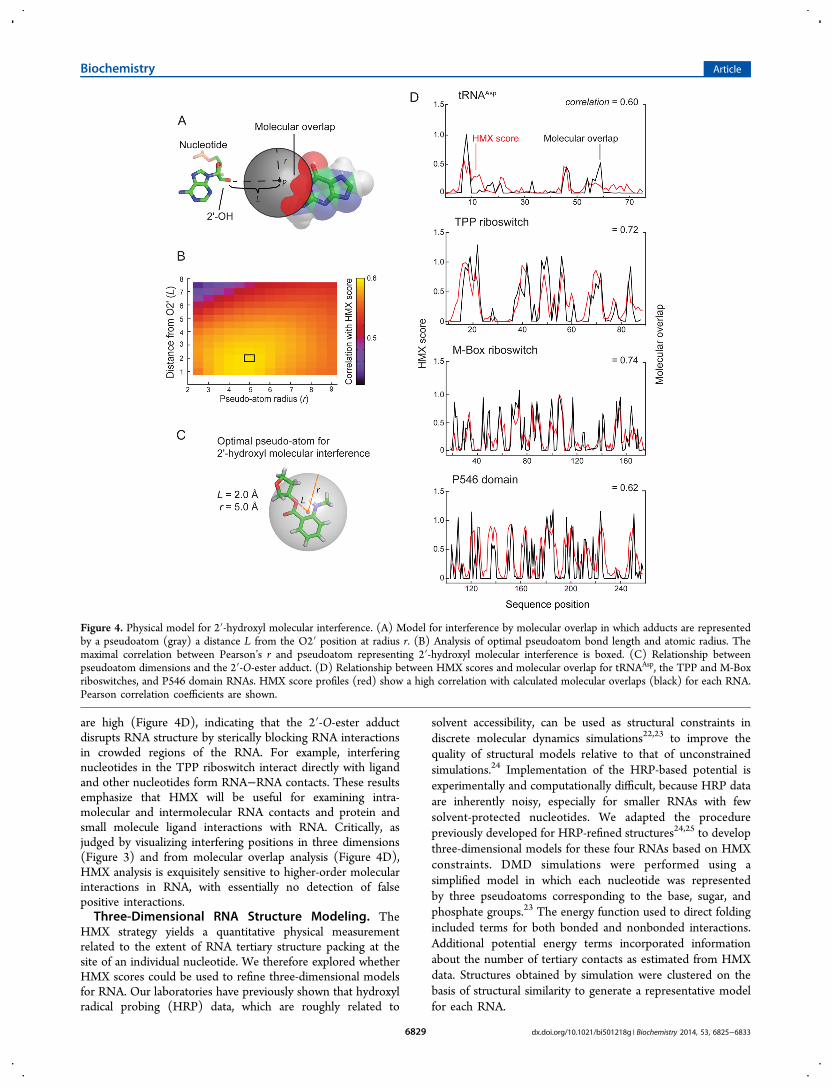
are high (Figure 4D), indicating that the 2′-O-ester adductdisrupts RNA structure by sterically blocking RNA interactionsin crowded regions of the RNA. For example, interferingnucleotides in the TPP riboswitch interact directly with ligandand other nucleotides form RNA−RNA contacts. These resultsemphasize that HMX will be useful for examining intra-molecular and intermolecular RNA contacts and protein andsmall molecule ligand interactions with RNA. Critically, asjudged by visualizing interfering positions in three dimensions(Figure 3) and from molecular overlap analysis (Figure 4D),HMX analysis is exquisitely sensitive to higher-order molecularinteractions in RNA, with essentially no detection of falsepositive interactions.Three-Dimensional RNA Structure Modeling. The
HMX strategy yields a quantitative physical measurementrelated to the extent of RNA tertiary structure packing at thesite of an individual nucleotide. We therefore explored whetherHMX scores could be used to refine three-dimensional modelsfor RNA. Our laboratories have previously shown that hydroxylradical probing (HRP) data, which are roughly related to
solvent accessibility, can be used as structural constraints indiscrete molecular dynamics simulations22,23 to improve thequality of structural models relative to that of unconstrainedsimulations.24 Implementation of the HRP-based potential isexperimentally and computationally difficult, because HRP dataare inherently noisy, especially for smaller RNAs with fewsolvent-protected nucleotides. We adapted the procedurepreviously developed for HRP-refined structures24,25 to developthree-dimensional models for these four RNAs based on HMXconstraints. DMD simulations were performed using asimplified model in which each nucleotide was representedby three pseudoatoms corresponding to the base, sugar, andphosphate groups.23 The energy function used to direct foldingincluded terms for both bonded and nonbonded interactions.Additional potential energy terms incorporated informationabout the number of tertiary contacts as estimated from HMXdata. Structures obtained by simulation were clustered on thebasis of structural similarity to generate a representative modelfor each RNA.
Figure 4. Physical model for 2′-hydroxyl molecular interference. (A) Model for interference by molecular overlap in which adducts are representedby a pseudoatom (gray) a distance L from the O2′ position at radius r. (B) Analysis of optimal pseudoatom bond length and atomic radius. Themaximal correlation between Pearson’s r and pseudoatom representing 2′-hydroxyl molecular interference is boxed. (C) Relationship betweenpseudoatom dimensions and the 2′-O-ester adduct. (D) Relationship between HMX scores and molecular overlap for tRNAAsp, the TPP and M-Boxriboswitches, and P546 domain RNAs. HMX score profiles (red) show a high correlation with calculated molecular overlaps (black) for each RNA.Pearson correlation coefficients are shown.
Biochemistry Article
dx.doi.org/10.1021/bi501218g | Biochemistry 2014, 53, 6825−68336829

The predicted structural models for each RNA wereevaluated in terms of the number and population of clustersas well as the calculated rmsd from the accepted crystalstructures. HMX-directed simulations produced structurallyand statistically significant26 nativelike RNA structured modelsfor the TPP and M-Box riboswitches and for the P546 domain(Figure 5). For each RNA, the central structure in the highest-
population cluster contained the structure with the lowest rmsdrelative to the accepted structure. In contrast, HMX-directedsimulations for tRNAAsp did not produce a single structure andthe model with the lowest rmsd corresponded to the lowest-population cluster (Figure 5, top). Critically, the tRNAAsp
simulation itself revealed that no well-determined modelemerged, as no single cluster included a majority of structures
consistent with the molecular interference information (Figure5 and Table S1 of the Supporting Information). Thus, theHMX-directed simulations proved to be highly robust, did notreturn a false positive model when data were insufficient (as inthe case of tRNAAsp), and yielded highly significant models forRNAs as large as 160 nucleotides.
HMX Reveals Individual Structural Domains in aLarge Group I Intron RNA. The Tetrahymena group I intronis formed from a set of conserved RNA domains. Thesedomains include the P5−P4−P6 and P9−P7−P3−P8 domains,which form the active site of the RNA, and a P1−P2 helix thatcontains the 5′ splice site.27,28 When the Tetrahymena group Iintron RNA construct (∼420 nucleotides), designed torecapitulate the intron structure visualized by crystallography,29
was subjected to the HMX experiment, partitioning immedi-ately revealed multiple unfolded states (Figure 6A). The fastestmigrating band is the most nativelike structure, and eachsubsequent lower-mobility band represents an RNA state withdecreasing levels of tertiary structure, with the slowestmigrating band reflecting an RNA with no tertiary structure.HMX scores for each unfolded band were calculated bycomparing each state to the fully folded RNA.The HMX profile for the first structural intermediate, I1,
reveals eight regions with high HMX scores (Figure 6B). Thesepositions fall primarily in the P1−P2 helices and in the P9−P7−P3−P8 domain around the intron active site (Figure 6B,red circles). The first intermediate band thus corresponds to astructure in which 2′-hydroxyl interference disrupts docking ofthe P1−P2 helix via interactions with the P9−P7−P3−P8domain (Figure 7, red).To visualize the specific nucleotides whose modification
induced formation of the I2 and fully unfolded states, wecalculated difference HMX scores for these two transitions(Figure 6C,D and Figure S3 of the Supporting Information).Analysis of differences between I1 and the second intermediate,I2, reveals a large number of newly interfering nucleotides,involving many of the long-range tertiary contacts that stabilizethe interaction between the P9−P7−P3−P8 and P5−P4−P6domains (Figures 6C and 7A, blue). Changes in interferenceprofiles between I2 and the fully unfolded RNA fall almostexclusively in the P5−P4−P6 domain (Figures 6D and 7A,green). Taken together, these data indicate that the I2intermediate corresponds to a state in which most or alltertiary interactions have been disrupted except those in theP5−P4−P6 domain. The transition from the I2 intermediate tothe fully unfolded state then reports unfolding of this latterdomain and loss of all remaining tertiary interactions.This single HMX experiment (Figure 6) reveals a hierarchy
of tertiary interactions fully consistent with extensive priorcrystallographic and biochemical analyses.27,28 Moreover, HMXalso emphasizes the high degrees of cooperativity in theseinteractions, as 2′-O-ester interference at any nucleotide in oneof the three structural networks of interfering nucleotides yieldssimilar folding intermediate states (Figure 7).
■ DISCUSSIONHMX measures the effect of introducing a molecularperturbation at the ribose 2′-OH position on RNA folding.Modifications at the 2′-ribose position, which lie on the exteriorof an RNA duplex, generally do not substantially destabilizesimple RNA secondary structures.20,21 Thus, the 2′-O-estermolecular interference measurement is exquisitely andspecifically sensitive to interactions that govern RNA tertiary
Figure 5. HMX-directed RNA fold refinements. RNAs are shown asbackbone traces. Accepted structures15−18 and HMX-directed refine-ments for each RNA are colored gray and blue, respectively. Thecluster populations (n, out of 100), mean rmsd, and p values26 areshown. For tRNAAsp, both the largest cluster (large image) and thelowest-rmsd structures (inset) are shown.
Biochemistry Article
dx.doi.org/10.1021/bi501218g | Biochemistry 2014, 53, 6825−68336830

folding. For the five RNAs evaluated in this work (tRNAAsp, theTPP and M-Box riboswitch aptamer domains, the P546 domainRNA, and the intact Tetrahymena group I intron), theinterfering nucleotides identified by HMX correspond closelyto the densely packed interior of these structures (Figures 3 and
7B). This relationship is quantitative. Molecular interference bythe 2′-O-ester group was highly correlated with a sphere ofdefined location relative to the RNA ribose group (Figure 4).We anticipate that 2′-O-ester-mediated molecular interferencewill prove to be broadly useful in evaluating higher-order RNApacking in the context of large RNAs and RNA−proteincomplexes.The gel electrophoresis partitioning approach used here
revealed the high level of cooperativity and structuralinterdependency in RNA tertiary folding. For the TPP andM-Box riboswitch aptamer domains and the P546 domainRNA, the introduction of distinct molecular adducts resulted ina single, well-defined, predominant unfolded structuralpopulation (Figure 2), rather than multiple populations thatwould be expected because of the absence of individual tertiaryinteractions. There thus exists a high degree of cooperativity inthe folding of these RNAs of up to 160 nucleotides.Use of HMX information to direct a DMD-based three-
dimensional structure refinement yielded highly significantstructure models, relative to accepted RNA structures.Refinement was based on a recently developed approach thatallows solvent accessibility to be incorporated into thesimulation as an energy restraint.24 In general, use of molecularinterference information required fewer assumptions to beintroduced and resulted in final clustered structures that werebetter defined than those obtained with hydroxyl radicalrestraints. For the TPP and M-Box riboswitches and the P546domain RNAs, HMX-directed refinement yielded a singlepredominant cluster with highly statistically significant agree-ment with the accepted structure (Figure 5). tRNAAsp waspoorly modeled, likely reflecting both the inability tocompletely separate folded and unfolded states (Figure 2)and the resulting low-magnitude molecular interference data (inFigure 3, compare tRNA with other RNAs). Critically, thesimulation itself clearly reported that this RNA was not a goodtarget for refinement because a relatively large number ofclusters were recovered and no single cluster dominated thesimulation. The overall success of HMX-directed refinement(Figure 5) suggests that de novo RNA structure refinement,based on easily obtained high-quality biochemical constraints,holds substantial promise for understanding structure−functioninterrelationships for RNA.Experiments with the intact Tetrahymena group I intron
illustrated how HMX analysis can characterize multiple stablestructural intermediates within a single RNA population atsingle-nucleotide resolution. HMX revealed that this RNA isstabilized by three sets of highly cooperative tertiaryinteractions. The first set corresponds to interactions betweenthe P1−P2 helix and the rest of the folded RNA. The secondcorresponds to an extensive set of interactions that formbetween the P9−P7−P3−P8 domain and the P5−P4−P6domain. The final set of interactions corresponds tointermolecular interactions that stabilize the P5−P4−P6domain. This higher-order domain structure was easily definedin a single set of experiments (Figures 6 and 7) and is fullyconsistent with extensive prior characterization of thisRNA.27,28
HMX is a simple, information-rich, and highly quantitativeapproach for analysis of the tertiary structure architecture offunctionally important RNAs and provides a unique view ofinternal and closely packed RNA tertiary structure. Here, RNAswere partitioned on the basis of size using gel electrophoresis;however, any strategy that separates functional from nonfunc-
Figure 6. HMX analysis of the Tetrahymena group I intron. (A)Partitioning of the RNA reveals folded and unfolded states, and twointermediate states, labeled I1 and I2. Gel images (left) werestraightened; gel lane intensity profiles (right) are unmanipulated.(B) HMX profile for the first (I1) intermediate, relative to the fullyfolded band. Sites of strongest interference are indicated with spheres.Difference HMX profiles calculated from the HMX profiles for the I1,I2, and unfolded bands (Figure S3 of the Supporting Information),correspond to transitions (C) from the first (I1) to the second (I2)intermediates and (D) from I2 to the fully unfolded RNA. Differenceslarger than 0.2 HMX unit are emphasized in color.
Biochemistry Article
dx.doi.org/10.1021/bi501218g | Biochemistry 2014, 53, 6825−68336831

tional RNAs could be used, allowing HMX analysis to beimplemented on the basis of the ability of an RNA to interactwith proteins or with other RNAs, or to perform catalysis andother functions.
■ ASSOCIATED CONTENT*S Supporting InformationAdditional supplemental table and figures. This material isavailable free of charge via the Internet at http://pubs.acs.org.
■ AUTHOR INFORMATIONCorresponding Author*E-mail: [email protected] work was supported by the National Science Foundation(Grant MCB-1121024 to K.M.W.).NotesThe authors declare no competing financial interest.
■ REFERENCES(1) Sharp, P. A. (2009) The centrality of RNA. Cell 136, 577−580.(2) Leontis, N. B., Lescoute, A., and Westhof, E. (2006) The buildingblocks and motifs of RNA architecture. Curr. Opin. Struct. Biol. 16,279−287.(3) Montange, R. K., and Batey, R. T. (2008) Riboswitches:Emerging themes in RNA structure and function. Annu. Rev. Biophys.37, 117−133.(4) Dethoff, E. A., Chugh, J., Mustoe, A. M., and Al-Hashimi, H. M.(2012) Functional complexity and regulation through RNA dynamics.Nature 482, 322−330.
(5) Conway, L., and Wickens, M. (1989) Modification interferenceanalysis of reactions using RNA substrates. In Methods in Enzymology,pp 369−379, Academic Press, Inc., New York.(6) Clarke, P. A. (1999) RNA footprinting and modificationinterference analysis. In Methods in Molecular Biology (Haynes, S.,Ed.) pp 73−91, Humana Press, Totowa, NJ.(7) Merryman, C., and Noller, H. F. (1998) Footprinting andmodification-interference analysis of binding sites on RNA. InRNA:protein interactions, a practical approach, pp 237−253, OxfordUniversity Press, Oxford, U.K.(8) Ryder, S. P., and Strobel, S. A. (1999) Nucleotide analoginterference mapping. Methods 18, 38−50.(9) Strobel, S. A. (1999) A chemogenetic approach to RNAfunction/structure analysis. Curr. Opin. Struct. Biol. 9, 346−352.(10) Tullius, T. D., and Greenbaum, J. A. (2005) Mapping nucleicacid structure by hydroxyl radical cleavage. Curr. Opin. Chem. Biol. 9,127−134.(11) Pastor, N., Weinstein, H., Jamison, E., and Brenowitz, M. (2000)A detailed interpretation of OH radical footprints in a TBP-DNAcomplex reveals the role of dynamics in the mechanism of sequence-specific binding. J. Mol. Biol. 304, 55−68.(12) Merino, E. J., Wilkinson, K. A., Coughlan, J. L., and Weeks, K.M. (2005) RNA structure analysis at single nucleotide resolution byselective 2′-hydroxyl acylation and primer extension (SHAPE). J. Am.Chem. Soc. 127, 4223−4231.(13) Wilkinson, K. A., Merino, E. J., and Weeks, K. M. (2006)Selective 2′-hydroxyl acylation analyzed by primer extension(SHAPE): Quantitative RNA structure analysis at single nucleotideresolution. Nat. Protoc. 1, 1610−1616.(14) McGinnis, J. L., Dunkle, J. A., Cate, J. H. D., and Weeks, K. M.(2012) The mechanisms of RNA SHAPE chemistry. J. Am. Chem. Soc.134, 6617−6624.
Figure 7. Visualization of 2′-O-ester interferences on the (A) secondary and (B) tertiary structure of the Tetrahymena group I intron RNA. Uniqueinterferences corresponding to the I1, I2, and fully unfolded RNA states (see Figure 6) are colored red, blue, and green, respectively. In panel B, 2′-OH groups are shown as spheres and the phosphoribose backbone is shown as a tube. The three-dimensional structure corresponds to an extendedmodel32 based on the crystal structure of core regions.29
Biochemistry Article
dx.doi.org/10.1021/bi501218g | Biochemistry 2014, 53, 6825−68336832

(15) Westhof, E., Dumas, P., and Moras, D. (1988) Restrainedrefinement of two crystalline forms of yeast aspartic acid andphenylalaine transfer RNA. Acta Crystallogr. A44, 112−123.(16) Serganov, A., Polonskaia, A., Phan, A. T., Breaker, R. R., andPatel, D. J. (2006) Structural basis for gene regulation by a thiaminepyrophosphate-sensing riboswitch. Nature 441, 1167−1171.(17) Dann, C. E., III, Wakeman, C. A., Sieling, C. L., Baker, S. C.,Irnov, I., and Winkler, W. C. (2007) Structure and mechanism of ametal-sensing regulatory RNA. Cell 130, 878−892.(18) Cate, J. H., Gooding, A. R., Podell, E., Zhou, K., Golden, B. L.,Kundrot, C. E., Cech, T. R., and Doudna, J. A. (1996) Crystal structureof a group I ribozyme domain: Principles of RNA packing. Science2733, 1678−1685.(19) Bhattacharyya, A., Murchie, A. I. H., and Lilley, D. M. J. (1990)RNA bulges and the helical periodicity of double-stranded RNA.Nature 343, 484−487.(20) Lesnik, E. A., Guinosso, C. J., Kawasaki, A. M., Sasmor, H.,Zounes, M., Cummins, L. L., Ecker, D. J., Cook, P. D., and Freier, S.M. (1993) Oligodeoxynucleotides containing 2′-O-modified adeno-sine: synthesis and effects on stability of DNA:RNA duplexes.Biochemistry 32, 7832−7838.(21) Lesnik, E. A., and Freier, S. M. (1998) What affects the effect of2′-alkoxy modifications? 1. Stabilization effect of 2′-methoxysubstitutions in uniformly modified DNA oligonucleotides. Biochem-istry 37, 6991−6997.(22) Gherghe, C. M., Leonard, C. W., Ding, F., Dokholyan, N. V.,and Weeks, K. M. (2009) Native-like RNA tertiary structures using asequence-encoded cleavage agent and refinement by discretemolecular dynamics. J. Am. Chem. Soc. 131, 2541−2546.(23) Ding, F., Sharma, S., Chalasani, P., Demidov, V. V., Broude, N.E., and Dokholyan, N. V. (2008) Ab initio RNA folding by discretemolecular dynamics: From structure prediction to folding mechanisms.RNA 14, 1164−1173.(24) Ding, F., Lavender, C. A., Weeks, K. M., and Dokholyan, N. V.(2012) Three-dimensional RNA structure refinement by hydroxylradical probing. Nat. Methods 9, 603−608.(25) Lavender, C. A., Ding, F., Dokholyan, N. V., and Weeks, K. M.(2010) Robust and generic RNA modeling using inferred constraints:A structure for the hepatitis C virus IRES pseudoknot domain.Biochemistry 49, 4931−4933.(26) Hajdin, C. E., Ding, F., Dokholyan, N. V., and Weeks, K. M.(2010) On the significance of an RNA tertiary structure prediction.RNA 16, 1340−1349.(27) Vicens, Q., and Cech, T. R. (2006) Atomic level architecture ofgroup I introns revealed. Trends Biochem. Sci. 31, 41−51.(28) Hougland, J. L., Piccirilli, J. A., Forconi, M., Lee, J., andHerschlag, D. (2005) How the Group I intron works: a case study ofRNA structure and function. In The RNA World (Cech, T. R.,Gesteland, R. F., and Atkins, J. F., Eds.) 3rd ed., pp 133−205, ColdSpring Harbor Laboratory Press, Plainview, NY.(29) Golden, B. L., Gooding, A. R., Podell, E. R., and Cech, T. R.(1998) A preorganized active site in the crystal structure of theTetrahymena ribozyme. Science 282, 259−264.(30) Karabiber, F., McGinnis, J. L., Favorov, O. V., and Weeks, K. M.(2013) QuShape: Rapid, accurate, and best-practices quantification ofnucleic acid probing information, resolved by capillary electrophoresis.RNA 19, 63−73.(31) Davis, I. W., Murray, L. W., Richardson, J. S., and Richardson, D.C. (2004) MOLPROBITY: Structure validation and all-atom contactanalysis for nucleic acids and their complexes. Nucleic Acids Res. 32,W615−W619.(32) Lescoute, A., and Westhof, E. (2006) The interaction networksof structured RNAs. Nucleic Acids Res. 34, 6587−6604.
Biochemistry Article
dx.doi.org/10.1021/bi501218g | Biochemistry 2014, 53, 6825−68336833
Related Documents