Welcome message from author
This document is posted to help you gain knowledge. Please leave a comment to let me know what you think about it! Share it to your friends and learn new things together.
Transcript

RNA In SilicoThe Computational Biology of RNASecondary StructuresChristoph FlammInstitut f�ur Theoretische Chemie, Universit�at WienW�ahringerstra�e 17, A-1090 Wien, [email protected] L. HofackerInstitut f�ur Theoretische Chemie, Universit�at WienW�ahringerstra�e 17, A-1090 Wien, [email protected] F. Stadler�Institut f�ur Theoretische Chemie, Universit�at WienW�ahringerstra�e 17, A-1090 Wien, Austria; andSanta Fe Institute1399 Hyde Park Road, Santa Fe, NM 87501, [email protected] or [email protected]�Address for correspondence(Version Time-stamp: "1999-04-28 22:05:33 ivo")ABSTRACT . RNA secondary structures provide a unique computer modelfor investigating the most important aspects of structural and evolutionarybiology. The existence of e�cient algorithms for solving the folding problem,i.e., for predicting the secondary structure given only the sequence, allows theconstruction of realistic computer simulations. The notion of a \landscape"

2 C. Flamm, I.L. Hofacker, P.F. Stadlerunderlies both the structure formation (folding) and the (in vitro) evolutionof RNA.Evolutionary adaptation may be seen as hill climbing process on a �tnesslandscape which is determined by the phenotype of the RNA molecule (withinthe model this is its secondary structure) and the selection constraints actingon the molecules. We �nd that a substantial fraction of point mutations do notchange an RNA secondary structure. On the other hand, a comparable fractionof mutations leads to very di�erent structures. This interplay of smoothnessand ruggedness (or robustness and sensitivity) is a generic feature of both RNAand protein sequence-structure maps. Its consequences, \shape space covering"and \neutral networks" are inherited by the �tness landscapes and determinethe dynamics of RNA evolution. Punctuated equilibria at phenotype level and adi�usion like evolution of the underlying genotypes are a characteristic featureof such models. As a practical application of these theoretical �ndings wehave designed an algorithm that �nds conserved (and therefore potentiallyfunctional) substructures of RNA virus genomes from sparse data sets.The folding dynamics of particular RNA molecule can also be studied suc-cessfully based on secondary structures. Given an RNA sequence, we con-sider the energy landscape formed by all possible conformations (secondarystructures). A straight forward implementation of the Metropolis algorithmis su�cient to produce a quite realistic folding kinetics, allowing to identifymeta-stable states and folding pathways. Just as in the protein case there aregood and bad folders which can be distinguished by the properties of theirenergy landscapes.KEYWORDS : RNA Secondary Structures, Fitness Landscapes, Energy Land-scapes, Molecular Evolution, Punctuated Equilibria, Folding Kinetics, FoldingPathways.1. IntroductionThe relationships between the sequence and the (three-dimensional) structureof a biopolymer is a core issue in biochemistry and molecular biology. Whilemost of the research on biopolymer folding is concerned with protein folding,the same questions can be posed for RNA molecules (Draper, 1996). An impor-tant advantage of RNA is that, on the level of secondary structure, the structureprediction problem can be solved with reasonable accuracy. Based on this obser-vation, it is possible to construct detailed computer models of di�erent aspectsof the sequence-structure-function relationships ranging all the way from in vitroevolution to folding kinetics.RNA secondary structures provide a discrete, coarse grained concept of struc-ture similar in complexity to lattice models of proteins. In contrast to the latter,

RNA in silico 3RNA secondary structures are a faithful coarse graining of the 3D structures. Itshould be noted, however, that there are examples of RNA molecules with sig-ni�cantly di�erent secondary structure which exhibit similar 3D structures andthe same function (Uhlenbeck, 1998). Secondary structures are routinely used todisplay, organize, and interpret experimental �ndings, they are oftentimes con-served over evolutionary times scales, and in vitro selection experiments withRNA more often than not yield families of selected sequences that share distinc-tive secondary structure features.In this contribution we (brie y) review three aspects of the \computationalbiology of RNA secondary structures": (1) the solutions to the folding problemand its variants, (2) the generic properties of the sequence structure relationsand their implications for the dynamics of RNA evolution, (3) the properties ofthe conformational energy function and its implications for the kinetics of RNAfolding. The notion of a landscape plays a key role in our investigations.2. RNA Secondary Structures and Their PredictionWe begin our discussion with the formal de�nition of a secondary structure:A secondary structure on a sequence is a list of base pairs [i; j] with i < j suchthat for any two base pairs [i; j] and [k; l] with i � k holds:(i) i = k if and only if j = l, and(ii) k < j implies i < k < l < j.The �rst condition simply means that each nucleotide can take part in at mostone base pair. The second condition forbids knots and pseudo-knots. Secondarystructures form a special type of graphs. In particular, a secondary structuregraph is outer-planar, which means that it can be drawn in the plane in such away that all vertices (which represent the nucleotides) are arranged on a circle,and all edges (which represent the bases pairs) lie inside the circle and do notintersect. While pseudo-knots are important in many natural RNAs (Westhofand Jaeger, 1992), they can be considered part of the tertiary structure for ourpurposes. The restriction to knot-free structures is necessary for e�cient compu-tation by dynamic programming algorithms. The recent algorithm by Rivas andEddy (1999) is able to deal with a large class of pseudo-knotted structures, but isextremely costly. Moreover, the information about the energetics of pseudo-knotsis still very limited (Gultyaev et al., 1999).Regarding secondary structures as special types of outer-planar graphs pavesthe way for a mathematical investigation of the structures. For instance, onecan count the number of possible distinct secondary structures for a given chain
4 C. Flamm, I.L. Hofacker, P.F. StadlerGC
U
A
C
G
closing base pair
A
5
3
3
5
A
5
3
closing base pair
interior base pairsclosing base pair
3
C
U
A
A
U G
C
closing base pair
multi-loop
interior base pair
A U
interior base pair
interior base pair
hairpin loop
bulge
C
G
C U
A
closing base pair
stacking pair
interior loop
G
G
G
3
5A
G
C
CA
5
Figure 1. RNA secondary structure elements. Any secondary structure can be uniquelydecomposed into these types of loops.length n or the number of all secondary structures that can be formed by a partic-ular sequence (Waterman, 1995; Hofacker et al., 1998). For instance, the numberof secondary structures with minimal stack length s = 2 grows like 1:86n. If themost common types of pseudo-knots are included, there are about 2:35n di�er-ent structures (Haslinger and Stadler, 1999). Consequently, there are many moresequences, 4n, than secondary structures. Before we explore the consequences ofthis observations, however, we consider the structure prediction problem.Secondary structures can be uniquely decomposed into loops as shown in Fig-ure 1 (note that a stacked base pair is considered a loop of size zero). The energyof an RNA secondary structure is assumed to be the sum of the energy contribu-tions of all loops. Energy parameters for the contribution of individual loops havebeen determined experimentally, see, e.g., (Freier et al., 1986; Jaeger et al., 1989;Walter et al., 1994) and depend on the loop type, loop size, and partly on itssequence. Usually, only Watson-Crick (AU, UA, CG and GC) and GU andUG pairs are allowed in computational approaches since non-standard base-pairshave in general context-dependent energy contributions that do not �t into the\nearest-neighbor model". It turns out that this standard energy model has asolid graph theoretical foundation (Leydold and Stadler, 1998): the loops formthe unique minimal cycle basis of the secondary structure graph.

RNA in silico 5N(E)
MFE 0.0 25.0 50.0 75.0 100.0Energy [kcal/mol]
0
2.1014
4.1014
6.1014
8.1014
1.1015
−20.0 −15.0 −10.0Energy [kcal/mol]
0
5
N(m
in)
0
20
40
60
80
100
N(e
)
Figure 2. Density of states of the yeast tRNAphe. Top: Complete Density of Statescomputed with an energy resolution of 0:1 kcal/mol, computed using the Density of statealgorithm. The total number of structures is 14; 995; 224; 405; 213; 184. Less than 2 millionstructures have negative energy, the reference state being the open structure. The lower�gure shows the density of states and the density of local minima in the region above thenative state at higher resolution. For this plot all structures within 15kcal/mol the groundstate were generated by suboptimal folding and tested for being local minima. The tRNAsequence with modi�ed bases used here displays only a few suboptimal structures within afew kT above the native state.The additive form of the energy model allows for an elegant solution of the min-imum energy folding problem by means of a dynamic programming scheme thatis similar to sequence alignment. This similarity was �rst realized and exploitedby Waterman 1978, see also (Waterman and Smith, 1978), the �rst dynamic pro-gramming solution was proposed by Nussinov and Jacobson (1980), originally forthe \maximum matching" problem of �nding the structure with the maximumnumber of base pairs (Nussinov et al., 1978). Zuker and coworkers (1981; 1984)formulated the algorithm for the minimum energy problem using the now stan-dard energy model. Since then several variations have been developed: MichaelZuker (1989) devised a modi�ed algorithm that can generate a subset of subop-timal structures within a prescribed increment of the minimum energy, see also(Schmitz and Steger, 1992). The algorithm will �nd any structure that is opti-mal in the sense that there is no other structure 0 with lower energy containingall base pairs that are present in . John McCaskill (1990) noted that the par-tition function over all secondary structures Q =P exp(��G( )=kT ) can becalculated by dynamic programming as well. In addition his algorithm can calcu-late the frequency with which each base pair occurs in the Boltzmann weightedensemble of all possible structures, which can be conveniently represented in a\dot-plot", see Figure 7.The memory and CPU requirements of these algorithms scale with sequencelength n as O(n2) and O(n3), respectively, making structure prediction feasible

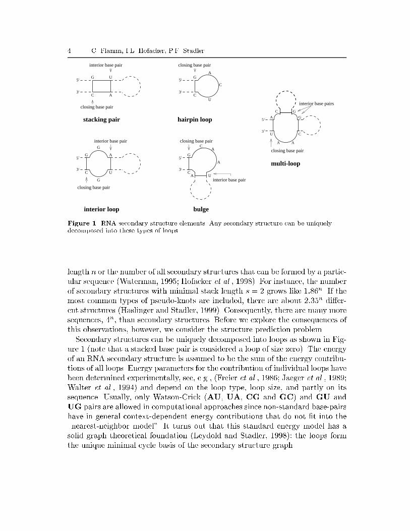
6 C. Flamm, I.L. Hofacker, P.F. Stadlereven for large RNAs of about 10000 nucleotides, such as the genomes of RNAviruses (Hofacker et al., 1996; Huynen et al., 1996a).McCaskill's work was extended in our group to yield an algorithm that com-putes the complete density of states of an RNA sequence at prede�ned energyresolution (Cupal et al., 1996; Cupal, 1997). Another method for calculating thedensity of states, based on enumeration of structures, was proposed earlier byHiggs (1993). However, his algorithm is restricted to subset of structures contain-ing no helices shorter than three and uses a simpli�ed energy model. Still, ouralgorithm is rather demanding as it needs to store O(n2m) entries and O(n3m2)operations to compute them, where m is the number of energy bins used. Thusit is applicable only to sequences up to some 100 nucleotides.Most recently, a program has been designed by the Vienna group that cangenerate all secondary structures within some interval of the minimum energybased on dynamic programming and multiple backtracking (Wuchty et al., 1999;Wuchty, 1998). The performance of the algorithm depends mainly on the numberof structures found. Since the number of possible structures grows exponentiallywith chain length, the energy range that can be considered shrinks with increas-ing chain length. In practice, suboptimal folding can handle about a few millionstructures, corresponding, e.g., to an energy range of, say, 12 kcal/mol at a chainlength of 100 bases. An example application is shown in Figure 2.Most of these algorithms are part of the Vienna RNA Package (Hofacker et al.,1994), which is freely available from http://www.tbi.univie.ac.at/.Another approach to RNA structure prediction is to take into account the dy-namics of the folding process. Such kinetic folding algorithms are the topic of sec-tion 5. In the case of functional RNAs, and provided a su�cient number of relatedsequences is available, the structure can be inferred from co-variations. This phy-logenetic approach is beyond the scope of this review, but see e.g. (Gutell, 1993).3. The Sequence-Structure MapWe have already mentioned above that there are many more sequences thanstructures. Hence, many sequences must fold into the same secondary structure.Moreover, extensive computer simulations have shown that there are only fewcommon secondary structures and many rare ones, see Figure 3.A structure is common if it is formed by more sequences than the averagestructure. Data from both large samples of long sequences (n � 30) (Schus-ter et al., 1994; Schuster, 1995) and from exhaustive folding of all short se-quences (Gr�uner et al., 1996a; 1996b) support two important observations: (i)the common structures represent only a small fraction of all structures and thisfraction decreases with increasing chain length; (ii) the fraction of sequences
RNA in silico 7
1 10 100 1000 10000Rank
10-4
10-3
10-2
10-1
Fre
quen
cy
1 10 100 1000 10000Rank
10-4
10-3
10-2
10-1
Fre
quen
cyFigure 3. Zipf's law for coarse grained RNA secondary structures. The structures of 100,000random sequences are ranked according to their frequencies. The ranking yields a distributionwhich follows a generalized Zipf's law f(r) = a(r + b)�c, where r and f(r) are the rank andthe frequency of the corresponding structure, respectively. The constant a is a normalizationfactor, b can be interpreted as the number of \very frequent" structures. The constant cdetermines the slope of the tail of the distribution. We found distributions following this formof generalized Zipf's law for all algorithms, parameter sets, and alphabets. Full line:Minimum energy structures computed with the an up-to-date parameter set. Dash-dottedline: Minimum energy structures computed with an older parameter set. Dashed line:Deterministic kinetic folding algorithm. L.h.s.: AUGC alphabet, r.h.s.: GC alphabet. Forthe details of the coarse graining procedure and the parameter sets see (Tacker et al., 1996).folding into common structures increases with chain length and approaches100% in the limit of long chains. Thus, for su�ciently long chains almost allRNA sequences fold into a small fraction of the secondary structures. The ef-fective ratio of sequences to structures is therefore even larger than the com-binatorial estimate. Furthermore, only common structures are likely to playa role in natural evolution and in evolutionary biotechnology (Schuster, 1995;Bacher and Ellington, 1998). RNA and proteins, despite their di�erent chem-istry, apparently share fundamental properties of their sequence-structure maps:the repertoire of stable native folds seems to be highly restricted or even vanish-ingly small (Chothia, 1992).Naturally, we ask how sequences folding into the same (common) secondarystructure are distributed in sequence space. In the following we review the resultsfor minimum energy folding, i.e., we assume that the folding map assigns to eachsequence the most stable secondary structure. The results reported below, how-

8 C. Flamm, I.L. Hofacker, P.F. Stadlerever, have been shown to depend very little on the choice of algorithm (includingvarious approaches to kinetic folding) and parameter sets (Tacker et al., 1996).Some notation is in order here: We call the set S( ) of all sequences (geno-types) folding into phenotype the neutral set of . (For a mathematician S isthe pre-image of w.r.t. the folding map f).Inverse folding can be used to determine S( ). Naturally, a sequence x canfold into a given secondary structure only if each pair of sequence positionsthat is paired in is realized by one of the six possible base pairs. The set ofall such sequences forms C( ), the set of compatible sequences. Clearly, we haveS( ) � C( ). Note that many sequences in C( ) will not have as their moststable or kinetically most accessible structure. Thus the neutral set of (for aparticular folding map) will in general be only a small subset of the compatibleset.For RNA secondary structures an e�cient inverse folding algorithm is available(Hofacker et al., 1994). It was used to show that sequences folding into the samestructure are (almost) randomly distributed within the set C( ) of compatiblesequences. A similar result was obtained for \protein space" (Babajide et al.,1997) using so-called potentials of mean force (Sippl, 1990; 1993a;1993b).The shape or topology of neutral sets has important implications for theevolution of both nucleic acids and proteins and for de novo design: For ex-ample, it has frequently been observed that seemingly unrelated protein se-quences have essentially the same fold (Holm and Sander, 1997; Murzin, 1994;Murzin, 1996). Similarly, the genomic sequences of closely related RNA virusesshow a large degree of sequence variation while sharing many conserved fea-tures in their secondary structures (Hofacker et al., 1996; Rauscher et al., 1997;Mandl et al., 1998; Hofacker et al., 1998).Another well known example is the clover leaf secondary structure of tRNAs:The sequences of di�erent tRNAs have little sequence homology (Eigen et al.,1988) but nevertheless fold into the same secondary structure motif. Whethersimilar structures with distant sequences may have originated from a commonancestor, or whether they must be the result of convergent evolution, dependson the geometry of the neutral sets S( ) in sequence space.The local properties of the sequence-structure map can be investigated byconsidering pairs of RNA sequences that di�er only by a single point mutation.A variety of methods is available to compare secondary structures and to quantifytheir di�erences by a (metric) distance, from counting the number of di�eringbase pairs, to sophisticated alignment-like procedure such as tree editing.It was noticed already in early work on RNA secondary structures (Fontanaand Schuster, 1987) that a substantial fraction of point mutations are neutral,i.e., that many sequences di�ering only in a single position fold into the same sec-
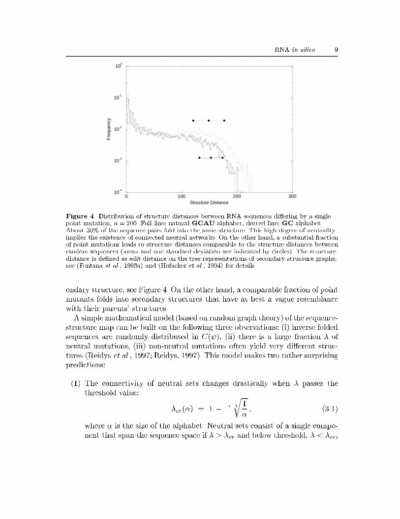
RNA in silico 9
0 100 200 300Structure Distance
10-4
10-3
10-2
10-1
100
Fre
quen
cy
Figure 4. Distribution of structure distances between RNA sequences di�ering by a singlepoint mutation, n = 200. Full line: natural GCAU alphabet, dotted line: GC alphabet.About 30% of the sequence pairs fold into the same structure. This high degree of neutralityimplies the existence of connected neutral networks. On the other hand, a substantial fractionof point mutations leads to structure distances comparable to the structure distances betweenrandom sequences (mean and one standard deviation are indicated by circles). The structuredistance is de�ned as edit distance on the tree representations of secondary structure graphs,see (Fontana et al., 1993a) and (Hofacker et al., 1994) for details.ondary structure, see Figure 4. On the other hand, a comparable fraction of pointmutants folds into secondary structures that have at best a vague resemblancewith their parents' structures.A simple mathematical model (based on random graph theory) of the sequence-structure map can be built on the following three observations: (i) inverse foldedsequences are randomly distributed in C( ), (ii) there is a large fraction � ofneutral mutations, (iii) non-neutral mutations often yield very di�erent struc-tures (Reidys et al., 1997; Reidys, 1997). This model makes two rather surprisingpredictions:(1) The connectivity of neutral sets changes drastically when � passes thethreshold value: �cr(�) = 1 � ��1r 1� ; (3.1)where � is the size of the alphabet. Neutral sets consist of a single compo-nent that span the sequence space if � > �cr and below threshold, � < �cr,

10 C. Flamm, I.L. Hofacker, P.F. Stadlerthe network is partitioned into a large number of components, in general, agiant component and many small ones. In the �rst case we refer to S( ) asthe neutral network of . For RNA it is necessary to split the random graphinto two factors corresponding to unpaired bases and base pairs and to usea di�erent value of � for each factor. For � = 2 we �nd �cr = 0:5. For nat-ural RNA sequences we have � = 4 for the unpaired regions and � = 6 forthe paired regions. The critical values are �cr(4) � 0:37 and �cr(6) � 0:301,respectively. The fraction of neutral neighbors is much larger than thesecritical values for common RNA secondary structures, hence the neutralsets S( ) form connected neutral networks within the sets C( ) of com-patible sequences (Reidys et al., 1997). The situation appears to be similarfor proteins (Babajide et al., 1997).(2) There is shape space covering, that is, in a moderate size ball centered atany position in sequence space there is a sequence x that folds into anyprescribed secondary structure . The radius of such a sphere, called thecovering radius rcov, can be estimated from simple probability arguments(Schuster, 1995) rcov � min�h �� B(h) � Sn ; (3.2)with B(h) being the number of sequences contained in a ball of radiush. The covering radius is approximately 10-15% of the diameter of the se-quence space. The covering sphere represents only a small connected subsetof all sequences but contains, nevertheless, all common structures and formsan evolutionary representative part of shape space.Extensive sample statistics (Schuster et al., 1994) and exhaustive folding of allGC-sequences with given chain length n � 30 (Gr�uner et al., 1996b) have so farbeen in excellent agreement with the random graph theory.The existence of extensive neutral networks meets a claim raised by Maynard-Smith (Maynard-Smith, 1970) for protein spaces that are suitable for e�cientevolution. The evolutionary implications of neutral networks are explored indetail in (Huynen et al., 1996b; Huynen, 1996) and will be reviewed in thefollowing section. Empirical evidence for a large degree of functional neutrality inprotein space was presented recently by Wain-Hobson and co-workers (Martinezet al., 1996).4. Fitness Landscapes and Evolutionary DynamicsSince Sewall Wright's seminal paper (Wright, 1932) the notion of a �tnesslandscape underlying the dynamics of evolutionary optimization has proved to

RNA in silico 11be one of the most powerful concepts in evolutionary theory. Implicit in thisidea is a collection of genotypes arranged in an abstract metric space, witheach genotype next to those other genotypes which can be reached by a singlemutation, as well as a �tness value assigned to each genotype.It has been known since Eigen's pioneering work on the molecular quasi-species(Eigen, 1971; Eigen and Schuster, 1977; Eigen et al., 1989) that the dynamicsof evolutionary adaptation (optimization) on a landscape depends crucially onthe detailed structure of the landscapes itself. Extensive computer simulations(Fontana and Schuster, 1987; Fontana et al., 1989) have made it very clear that acomplete understanding of the dynamics is impossible without a thorough inves-tigation of the underlying landscape. Landscapes derived from well-known com-binatorial optimization problems such as the Traveling Salesman Problem TSP(Lawler et al., 1985), the Graph Bipartitioning Problem GBP (Fu and Anderson,1986), or the Graph Matching Problem GMP have been investigated in some detail,see (Stadler, 1996) and the references therein. A detailed survey of a variety ofmodel landscapes obtained by folding RNA molecules into their secondary struc-tures has been performed during the last decade, see (Schuster and Stadler, 1994;Schuster et al., 1997; Schuster, 1997a) and the references therein. While the useof (computationally simple) landscapes derived from spin-glasses or combinato-rial optimization problems, or of the closely related Nk model (Kau�man, 1993)is certainly appealing, it is by no means clear that these models will capturethe salient features of biochemically relevant landscapes. Indeed, we know nowthat landscapes derived from folding biopolymers into their spatial structuresare quite di�erent from spin-glass-like landscapes (Hordijk and Stadler, 1998).One of the most important characteristics of a landscape is its ruggedness, a no-tion that is closely related to the hardness of the optimization problem for heuris-tic algorithms (Manderick et al., 1991). Three distinct approaches haven beenproposed to measure and quantify ruggedness and to subsequently compare dif-ferent landscapes. Sorkin (1988), Eigen et al. (1989) and Weinberger (1990) usedpair correlation functions. Kau�man and Levin (1987) proposed adaptive walks,and Palmer (1991) based his discussion on the number of meta-stable states(local optima). The relationship between correlation measures and local op-tima is discussed in detail by Garc��a-Pelayo and Stadler (1997). A mathematicalframework for studying landscapes is developed in (Stadler, 1995; Stadler, 1996;Stadler and Happel, 1999).Not surprisingly, landscapes based on sequence-structure maps (Figure 5) in-herit their ruggedness even if the map from structures to �tness values is smoothor even linear, since shape space covering implies that a substantial fraction ofpoint mutations lead to unrelated structures. On the other hand, a completely
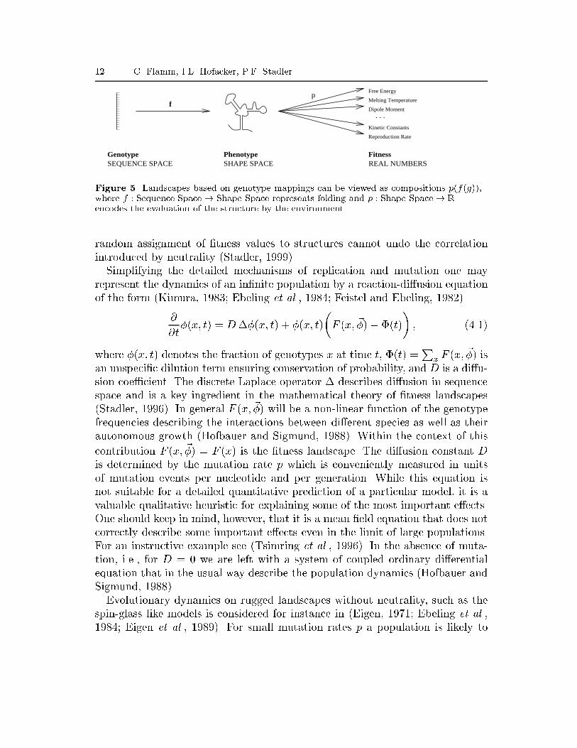
12 C. Flamm, I.L. Hofacker, P.F. StadlerFree Energy
Melting Temperature
Dipole Moment
Kinetic Constants
Reproduction Rate
SEQUENCE SPACE SHAPE SPACE REAL NUMBERSGenotype Phenotype Fitness
fp
. . .
Figure 5. Landscapes based on genotype mappings can be viewed as compositions p(f(g)),where f : Sequence Space! Shape Space represents folding and p : Shape Space! Rencodes the evaluation of the structure by the environment.random assignment of �tness values to structures cannot undo the correlationintroduced by neutrality (Stadler, 1999).Simplifying the detailed mechanisms of replication and mutation one mayrepresent the dynamics of an in�nite population by a reaction-di�usion equationof the form (Kimura, 1983; Ebeling et al., 1984; Feistel and Ebeling, 1982)@@t�(x; t) = D��(x; t) + �(x; t)�F (x; ~�)� �(t)� ; (4.1)where �(x; t) denotes the fraction of genotypes x at time t, �(t) =Px F (x; ~�) isan unspeci�c dilution term ensuring conservation of probability, and D is a di�u-sion coe�cient. The discrete Laplace operator � describes di�usion in sequencespace and is a key ingredient in the mathematical theory of �tness landscapes(Stadler, 1996). In general F (x; ~�) will be a non-linear function of the genotypefrequencies describing the interactions between di�erent species as well as theirautonomous growth (Hofbauer and Sigmund, 1988). Within the context of thiscontribution F (x; ~�) = F (x) is the �tness landscape. The di�usion constant Dis determined by the mutation rate p which is conveniently measured in unitsof mutation events per nucleotide and per generation. While this equation isnot suitable for a detailed quantitative prediction of a particular model, it is avaluable qualitative heuristic for explaining some of the most important e�ects.One should keep in mind, however, that it is a mean �eld equation that does notcorrectly describe some important e�ects even in the limit of large populations.For an instructive example see (Tsimring et al., 1996). In the absence of muta-tion, i.e., for D = 0 we are left with a system of coupled ordinary di�erentialequation that in the usual way describe the population dynamics (Hofbauer andSigmund, 1988).Evolutionary dynamics on rugged landscapes without neutrality, such as thespin-glass like models is considered for instance in (Eigen, 1971; Ebeling et al.,1984; Eigen et al., 1989). For small mutation rates p a population is likely to
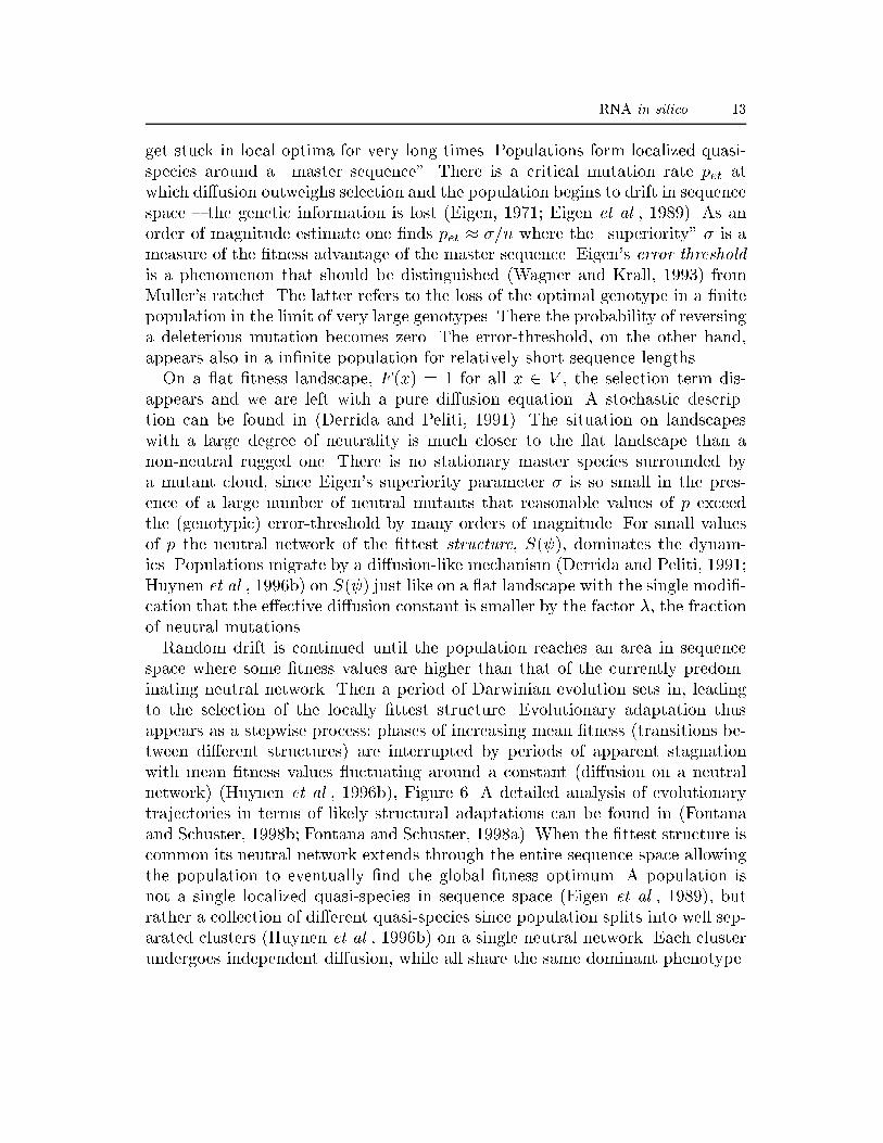
RNA in silico 13get stuck in local optima for very long times. Populations form localized quasi-species around a \master sequence". There is a critical mutation rate pet atwhich di�usion outweighs selection and the population begins to drift in sequencespace { the genetic information is lost (Eigen, 1971; Eigen et al., 1989). As anorder of magnitude estimate one �nds pet � �=n where the \superiority" � is ameasure of the �tness advantage of the master sequence. Eigen's error thresholdis a phenomenon that should be distinguished (Wagner and Krall, 1993) fromMuller's ratchet. The latter refers to the loss of the optimal genotype in a �nitepopulation in the limit of very large genotypes. There the probability of reversinga deleterious mutation becomes zero. The error-threshold, on the other hand,appears also in a in�nite population for relatively short sequence lengths.On a at �tness landscape, F (x) = 1 for all x 2 V , the selection term dis-appears and we are left with a pure di�usion equation. A stochastic descrip-tion can be found in (Derrida and Peliti, 1991). The situation on landscapeswith a large degree of neutrality is much closer to the at landscape than anon-neutral rugged one. There is no stationary master species surrounded bya mutant cloud, since Eigen's superiority parameter � is so small in the pres-ence of a large number of neutral mutants that reasonable values of p exceedthe (genotypic) error-threshold by many orders of magnitude. For small valuesof p the neutral network of the �ttest structure, S( ), dominates the dynam-ics. Populations migrate by a di�usion-like mechanism (Derrida and Peliti, 1991;Huynen et al., 1996b) on S( ) just like on a at landscape with the single modi�-cation that the e�ective di�usion constant is smaller by the factor �, the fractionof neutral mutations.Random drift is continued until the population reaches an area in sequencespace where some �tness values are higher than that of the currently predom-inating neutral network. Then a period of Darwinian evolution sets in, leadingto the selection of the locally �ttest structure. Evolutionary adaptation thusappears as a stepwise process: phases of increasing mean �tness (transitions be-tween di�erent structures) are interrupted by periods of apparent stagnationwith mean �tness values uctuating around a constant (di�usion on a neutralnetwork) (Huynen et al., 1996b), Figure 6. A detailed analysis of evolutionarytrajectories in terms of likely structural adaptations can be found in (Fontanaand Schuster, 1998b; Fontana and Schuster, 1998a). When the �ttest structure iscommon its neutral network extends through the entire sequence space allowingthe population to eventually �nd the global �tness optimum. A population isnot a single localized quasi-species in sequence space (Eigen et al., 1989), butrather a collection of di�erent quasi-species since population splits into well sep-arated clusters (Huynen et al., 1996b) on a single neutral network. Each clusterundergoes independent di�usion, while all share the same dominant phenotype.
14 C. Flamm, I.L. Hofacker, P.F. Stadler
Sequence Space
Sequence Space
Fitn
ess
Fitn
ess
Adaptive Walks without Selective Neutrality
Adaptive Walk on Neutral Networks
Start of Walk
Start of Walk
Start of Walk
Start of Walk
End of Walk
End of Walk
End of Walk
End of WalkRandom Drift
Figure 6. The role of neutral networks in evolution (Schuster, 1997b). Optimization occursthrough adaptive walks and random drift. Adaptive walks allow to choose the next steparbitrarily from all directions where �tness is (locally) non-decreasing. Populations can bridgeover narrow valleys with widths of a few point mutations. In the absence of selectiveneutrality (spin-glass-like landscape, above) they are, however, unable to span largerHamming distances and thus will approach only the next major �tness peak. Populations onrugged landscapes with extended neutral networks evolve along the networks by acombination of adaptive walks and random drift at constant �tness (below). In this manner,populations bridge over large valleys and may eventually reach the global maximum of the�tness landscape.
RNA in silico 15It is not surprising hence that there are abundant examples of both RNA andprotein structures that have been conserved over evolutionary time scales whilethe underlying sequences have lost (almost) all homology.For larger mutation rates p the di�usion term dominates the dynamics andthe population is not con�ned to the neutral network any more. The phenotypicerror threshold (Forst et al., 1995; Huynen et al., 1996b; Reidys et al., 1999) isthe mutation rate at which the dominating phenotype is lost.Di�usion in sequence space, the existence of phenotypic error threshold, and aclose connection (Huynen et al., 1996b) with Kimura's neutral theory (Kimura,1983) which we have not discussed here, are consequences of the existence ofneutral networks. Shape space covering implies a constant rate of innovation(Huynen, 1996): While di�using along a neutral network, a population constantlyproduces non-neutral mutants folding into di�erent structures. Shape space cov-ering implies that almost all structures can be found somewhere near the currentneutral network.Hence the population keeps discovering structures that it has never encoun-tered. When a superior structure is produced, Darwinian selection becomes thedominating e�ect and the population \jumps" onto the neutral network of thenovel structure while the old network is abandoned. Figure 6 sketches the di�er-ence between evolutionary adaptation on spin-glass-like landscapes and on thehighly neutral landscapes arising from biopolymer structures.Neutral evolution, arising as a consequence of the high degree of neutralityobserved in genotype-phenotype mappings of biopolymers, therefore, is not adispensable addendum to evolutionary theory (as it has often been suggested).On the contrary, neutral networks, provide a powerful mechanism through whichevolution can become truly e�cient.The evolution of sequences on neutral networks can be observed very clearly inRNA viruses. Our simulations show that sequence di�erences of as little as 10%lead almost surely to unrelated structures if the mutated sequence positions arechosen randomly (Fontana et al., 1993b). The presence of conserved secondarystructure elements such as the TAR or RRE region in HIV, the IRES region of pi-corna viruses, or the stem loop structure at the 3' terminus of avivirus genomes,which show a signi�cant sequence variation between di�erent virus strains (onlyabout 80% average pairwise sequence identity), must therefore be regarded asthe result of stabilizing selection acting on the secondary structure. This e�ectcan be used to design an algorithm that reliably detects conserved, and there-fore most likely functional, RNA secondary structure elements in viral genomesbased on a combination of secondary structure prediction and comparative se-quence analysis (Hofacker et al., 1998; Hofacker and Stadler, 1999). Evolutionon neutral networks leads to an increased level of robustness against mutation
16 C. Flamm, I.L. Hofacker, P.F. Stadlersince a di�using population prefers the denser regions of the neutral network(van Nimwegen et al., 1999). The e�ect can be observed by comparing conservedand non-conserved sub-structures in the rapidly evolving genomic sequences ofRNA and retroviruses (Wagner and Stadler, 1999).5. Energy Landscapes and Folding KineticsThe energy landscape of an RNA molecule is, for our purposes, de�ned on theset of all secondary structures that are compatible with its sequence. Concep-tually, this energy landscape is closely related to the potential energy surfaces(PES) which constitute one of the most important issues of theoretical chemistry(Mezey, 1987; Heidrich et al., 1991). As a consequence of the validity of the Born-Oppenheimer approximation, the PES provides the potential energy U(~R) of amolecule as a function of its nuclear geometry ~R. PES are therefore de�ned on ahigh-dimensional continuous space and they are assumed to be smooth (usuallytwice continuously di�erentiable almost everywhere). The (global) analysis ofPES thus makes extensive use of di�erential topology. In contrast, our notion ofenergy landscapes is discrete. Their analysis is therefore similar to the analysisof �tness landscapes.A crucial ingredient for the simulation of RNA folding kinetics is the choiceof a \move set" for inter-converting secondary structures. This move-set de�nesthe topology of the energy landscape by de�ning which secondary structures areneighbors of each other and encodes the set of structural changes that RNAs canundergo with moderate activation energies. It is the basis of all kinetic algorithmsfor RNA folding.The assumptions that an RNA molecule folds into its thermodynamic groundstate may well be wrong even for moderately long sequences (Morgan and Higgs,1996). Consequently, several groups have designed kinetic folding algorithms forRNA secondary structures, mostly in an attempt to get more accurate predictionsor in order to include pseudo-knots, see e.g. (Martinez, 1984; Mironov et al., 1985;Abrahams et al., 1990; Gultyaev, 1991; Tacker et al., 1994). Only a few papershave attempted to reconstruct folding pathways (Higgs, 1995; Gultyaev et al.,1995; Suvernev and Frantsuzov, 1995). These algorithms generally operate on alist of all possible helices and consequently use move-sets that destroy or formentire helices in a single move. Such a move-set can introduce large structuralchanges in a single move and furthermore, ad hoc assumptions have to madeabout the rates of helix formation and disruption. A more local move-set is,therefore, preferable if one hopes to observe realistic folding trajectories.The most elementary move-set, on the level of secondary structures, consistsof removal and insertion of single base pairs (while making sure that one does

RNA in silico 17
G C G G A U U U A G C U C A G U U G G G A G A G C G C C A G A C U G A A G A U C U G G A G G U C C U G U G U U C G A U C C A C A G A A U U C G C A C C A
G C G G A U U U A G C U C A G U U G G G A G A G C G C C A G A C U G A A G A U C U G G A G G U C C U G U G U U C G A U C C A C A G A A U U C G C A C C A
AC
CA
CG
CU
UA
AG
AC
AC
CU
AG
CU
UG
UG
UC
CU
GG
AG
GU
CU
AG
AA
GU
CA
GA
CC
GC
GA
GA
GG
GU
UG
AC
UC
GA
UU
UA
GG
CG
GC
GG
AU
UU
AG
CU
CA
GU
UG
GG
AG
AG
CG
CC
AG
AC
UG
AA
GA
UC
UG
GA
GG
UC
CU
GU
GU
UC
GA
UC
CA
CA
GA
AU
UC
GC
AC
CAFigure 7. Base pair probabilities for an phenylalanine tRNA with and without modi�edbases. The equilibrium frequency p of a pair [i; j] is represented by a square of area p inposition i; j and j; i of the matrix. Lower left: only base pairs contained in the ground stateoccur with signi�cant frequency for the sequence with modi�ed bases. Upper right: Theunmodi�ed sequence displays a large number of base pairs from suboptimal structures,although the ground state remains unchanged.not introduce knots or pseudo-knots). The simulations reported below are de-scribed in detail in Christoph Flamm's PhD thesis (Flamm, 1998) and (Flammet al., 1999). Either this simple move-set or, as in the data shown below, base pairinsertion and deletions together with base pair \shifts" (in which a base pair [i; j]is converted into a new pair [i; k]) are used. These shift moves facilitate sliding ofthe two strands of helix, which is assumed to be an important e�ect in dynamicsof RNA molecules. The dynamics itself is simulated by an algorithm designedfor stochastic chemical reactions by Gillespie (1976). The time scale is �xed us-ing the measured hairpin formation of the oligonucleotide AAAAAACCCCCCUUUUUU(P�orschke, 1974). For the rates constants a symmetrical rule k � exp(��G=2kT )independent of the sign of �G has been assumed (Kawasaki, 1966) instead of theusual Metropolis rule. Additional simulations using the Metropolis rule showedqualitatively similar results.Local minima are of particular importance for the folding dynamics, since theycan trap the molecule in a misfolded state. For a given sequence the low-energylocal minima can be constructed with reasonable e�ort: Structures within someinterval of the ground state are generated through complete suboptimal folding(Wuchty et al., 1999), for each structure all neighboring structures are generated
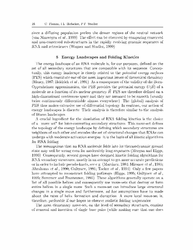
18 C. Flamm, I.L. Hofacker, P.F. Stadler
0.0 100 200 300Time [µs]
-20.0
-15.0
-10.0
-5.0
0.0
5.0
10.0
Ene
rgy
[kca
l/mol
]G C
GG
A
UU U
A
GC
UC
A GN
N
GGG
A
GA
GC
GCC
A
G A C U
GA A N
A
U
CUGG
AGN
UCC
U
G
U
G NUC
G
N
UC
CACA
G
A
A
UU
C
G
C
ACC
A
G C GGAU
U
U
A
GC
UC
A GN
N
GGG
A
GA
GC
GCCAGA C
U
GAA
N
A
UC
UG
GAGNUC
CU
GU
G
NU
CGNU
CC
A
C
AGA
A U UCG
CA
C
C
A
G C GGA
U
U
UA G
C
U
CA
G
NN G
G
G
AG
A
G
CGC C A G A
C UG
A
ANA
UCUGG
AGNU
CCU
GU
G
NU C
G
NUC
CA
CA
G
A
AU
UCGCA
C
C
A
G C G G A U U U
AG
C
U
CAG
NN
G GG
AG
A
G
C GCCAGA
CU G
A
A
NAU
CU
GG
A
G
NU
CCU
GU
G
NUC
G
N
UC C
ACA
GAAUUCGCACC
A
Figure 8. Energy as a function of time for a representative simulation of the modi�edtRNA. A few intermediate structures are shown at the top, the last one being the nativeclover-leaf structure. The stem closing the multi-loop forms last in most simulations.and their energies compared to the reference structure. Figure 2 shows the densityof local minima within 15 kcal/mol of the ground state for the tRNAphe.By an extension of the above procedure one can even determine the heightof the energy barriers and the structures of the saddle points (transition states)connecting local minima. This analysis yields a tree with the local minima asleaves and the transition states as internal nodes; the branch lengths representthe height of the energy barriers. An example of such a tree is shown Figure 11.Transfer RNA molecules from most organisms contain several modi�ed bases,particularly methylations. These modi�ed bases occur mostly in unpaired regionsand often the modi�cations are such that base pairing is made impossible. Hence,one might speculate that the modi�ed bases help to stabilize the correct fold.The phenylalanine tRNA from yeast used in the following contains six modi�-cations which prohibit base pairing its 76 nucleotides. As can be seen in Figure 7the modi�cations have a strong e�ect on the equilibrium ensemble of structures.The frequency of the correct fold in the thermodynamic ensemble rises from 4.4%to 28% and suboptimal folding shows that the lowest six suboptimal structureare prohibited by the modi�cations and consequently the energy gap from theground state to the next possible structures increases from 0:4 to 0:9 kcal/mol.
RNA in silico 19
0.0 200.0 400.0 600.0Time [µs]
0.0
0.2
0.4
0.6
0.8
1.0
Fra
ctio
n of
Fol
ded
Mol
ecul
es
tRNA_inv
G C U U U C C A G C G C G A G A A U U A A C G C G A G G U A U C G A A U U U A U A C U A A A C G C C U G G A A C A U G G U U A G G G G G G G G U A G U U
G C U U U C C A G C G C G A G A A U U A A C G C G A G G U A U C G A A U U U A U A C U A A A C G C C U G G A A C A U G G U U A G G G G G G G G U A G U U
UU
GA
UG
GG
GG
GG
GA
UU
GG
UA
CA
AG
GU
CC
GC
AA
AU
CA
UA
UU
UA
AG
CU
AU
GG
AG
CG
CA
AU
UA
AG
AG
CG
CG
AC
CU
UU
CG
GC
UU
UC
CA
GC
GC
GA
GA
AU
UA
AC
GC
GA
GG
UA
UC
GA
AU
UU
AU
AC
UA
AA
CG
CC
UG
GA
AC
AU
GG
UU
AG
GG
GG
GG
GU
AG
UU
0.0 200.0 400.0 600.0Time [µs]
0.0
0.2
0.4
0.6
0.8
1.0
Fra
ctio
n of
Fol
ded
Mol
ecul
es
tRNA_opt
G C C G G G C A A U C G C A C C A A C C C G C G A A G C G G C U C C C U U C G C C G C A A C C A G C C G U U U C C C C U G C G G C G C C C G G C A A A U
G C C G G G C A A U C G C A C C A A C C C G C G A A G C G G C U C C C U U C G C C G C A A C C A G C C G U U U C C C C U G C G G C G C C C G G C A A A U
UA
AA
CG
GC
CC
GC
GG
CG
UC
CC
CU
UU
GC
CG
AC
CA
AC
GC
CG
CU
UC
CC
UC
GG
CG
AA
GC
GC
CC
AA
CC
AC
GC
UA
AC
GG
GC
CG
GC
CG
GG
CA
AU
CG
CA
CC
AA
CC
CG
CG
AA
GC
GG
CU
CC
CU
UC
GC
CG
CA
AC
CA
GC
CG
UU
UC
CC
CU
GC
GG
CG
CC
CG
GC
AA
AU
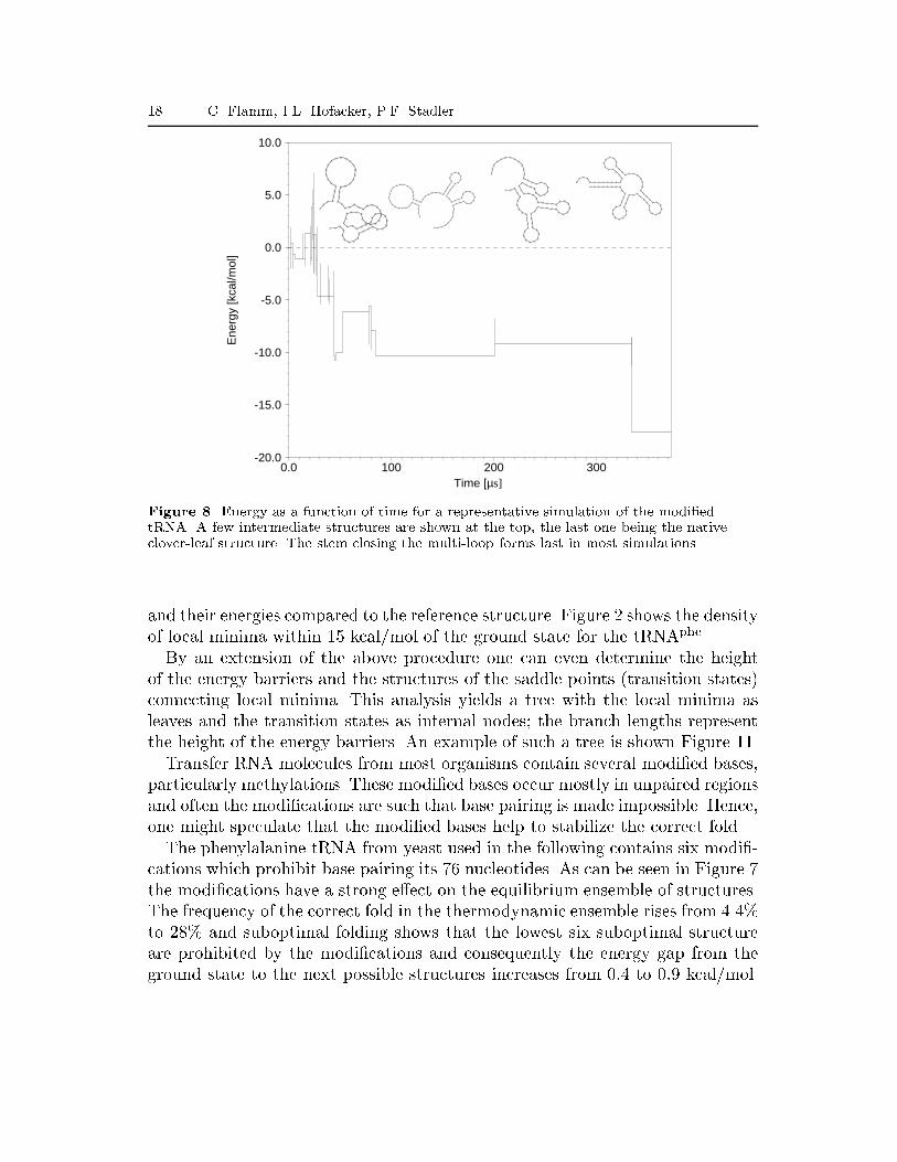
Figure 9. Thermodynamic stability and \foldability". The fraction of folded sequences p(t)(thick lines) as a function of time and folding times (thin lines) for two arti�cial sequencesdesigned to fold into the tRNA clover-leaf structure are derived from 1000 independent runs.Inset: dot plots showing the equilibrium base pair probabilities (upper right) as obtainedfrom McCaskill's algorithm and the contact map of the tRNA structure (lower left). L.h.s: arandomly chosen sequence with tRNA structure shows many alternative foldings in the dotplot but nevertheless folds e�ciently. R.h.s: A sequence designed to be thermodynamicallystable (see inset) folds only in less than 50% of the simulations.The modi�ed sequence exhibits very few local minima in the low energy region,there are only 10 local minima within 5 kcal/mol of the ground state comparedto 173 for the unmodi�ed sequence (Hofacker, 1998).To study the kinetic e�ect of the modi�cations, the folding of modi�ed andunmodi�ed tRNA sequence has been simulated (Flamm, 1998). The resultingtrajectories were then analyzed for the existence of typical folding pathways,Figure 8. In this particular run the RNA folds somewhat slower than average, butnevertheless shows features common to all trajectories. A rapid collapse leads toa structure with almost as many base pairs as the native state but little overlap.Folding then proceeds through a series of local minima that have more and morestructural elements in common with the ground state. The waiting times in thelocal minima increase with decreasing energy. Many trajectories visit the samelow energy intermediates, in particular, the stem closing the multi-loop forms lastin almost all simulations. Interestingly, the correct hairpins closest to the 5'-endare often formed �rst, which might support e�cient folding during transcription.As a measure of foldability we recorded the folding times, i.e., the time afterwhich the ground state appears in the simulation for the �rst time. We considerin particular the fraction p(t) of molecules that have reached the ground stateat time t. The resulting distribution can be seen in Figure 9. For the modi-

20 C. Flamm, I.L. Hofacker, P.F. Stadler
10−1
100
101
102
103
104
105
Folding Time t
10−4
10−3
10−2
10−1
100
t*p’
(t)/
p(t)
100
101
102
103
104
105
Folding Time t
10−2
10−1
100
t*p’
(t)/
p(t)
Figure 10. Folding kinetics of two di�erent RNA molecules. L.h.s.: The (arti�cial) moleculewhose pathway is described in detail in �gure 11. The curve shows two distinct peakscorresponding to two di�erent dominating folding pathways. A less prominent foldingpathway is indicated by the shoulder on the right hand side of the �rst peak (indicated by anarrow). R.h.s.: The kinetic signature of the modi�ed tRNA shows only a single peak. Thetime scale of folding is set by the closing of the multi-loop, see Figure 8.�ed sequence the ground state was found in all simulations. This is consistentwith recent analysis by Thirumalai and Woodson (1996) of experimental data,suggesting a directed pathway to the native state for tRNAs. The unmodi�edsequence folds much more slowly and only 46% of runs reach the ground statewithin the simulation time. The fraction of folded sequences is still rising atthat point and longer simulations will be needed to decide whether the curvesaturates at less than unity.In the case of phenylalanine tRNA the modi�ed bases improved both thermo-dynamic stability, conferred by a large energy gap between native and misfoldedstates, and foldability. The same link has been claimed for lattice protein modelsby �Sali et al. (1994b). To test this hypothesis we have designed two arti�cialsequences with the tRNA structure as ground state using the RNAinverse pro-gram from the Vienna RNA Package. The thermodynamic properties of the �rstsequence are typical for sequences of this size: the frequency of the ground statein the ensemble is about 7% and several alternative foldings can be seen in thebase pair probability matrix, see the inset on the l.h.s. of Figure 9. The othersequence was designed to be particularly stable: its ground state dominates theensemble with a frequency of 96% and no alternative foldings are discernible inthe dot plot. We than ran 1000 folding simulations for each sequence; the resultsare summarized in Figure 9. Surprisingly, the thermodynamically more stablesequence folds poorly in this example.

RNA in silico 21
Steps [arbitrary]
−8
−6
−4
−2
0
2E
nerg
y [k
cal/m
ol]
1
2
3
4
5
6
7
8
9
10
11
2.45[5]
[9]3.80 1 [11]
1.60 3 [7]
13
141.30 9
1.40 192.10 11
3.80 2 [1][3] 12
1.20 8 [4]
173.60 4
2.20 73.61 5
1.40 161.90 10
1.50 152.00 205.02 6
3.90 18
G C U A U U A
GC
GC
G
UG
A
CG
UG
CG
U
UU
A
G C U A U UCG
UG
CG
U
UU
A
C
C
G
G
G
UG
A
A G C U A U UCG
UG
CG
U
UU
A
C C G G A
G
UG
A
G C U A U UCG
UG
CG
U
UU
A
C
C
A
G
UGGGA
G C U A G UCG
UG
CG
U
UU
A
G G G
AC
CU
U
A
G C G U G GCG
UG
CG
U
UU
A
G A
AU
C
CU
U
A
G U G G G ACG
UG
CG
U
UU
A
GC
AU
C
CU
U
A
G U
UG
CG
U
UU
A GC
AU
C
CU
U
A
C G G GG
G U
CG
U
UU
A
GC
AU
C
CU
U
A
C G
G GUGG
G U
GC
AU
C
CU
U
A
C G
GU
C GUUUAGGG
8 9 10
1 2 3
4567
A A
G U
GC
AU
C
CU
U
A
C G
GU
C G
UUUAGGG
11
U AAFigure 11. Trapping and escape from a local minimum. An arti�cial RNA molecule wasdesigned with a low-energy mis-folded state formed by two hairpins at the 5' and 3' ends. The3' hairpin of the misfolded state blocks the formation of the ground state, which consists of asingle, much longer, 3' hairpin. The upper left plot shows the energy pro�le of the two mostprominent folding pathways. The upper right plot shows the energy barriers between the 20lowest local minima. The fast pathway begins with the formation of the correct hairpin at the3' end and the rapid elongation of the stem. The only an energy barrier occurs at the �rststep with a height of about 2.7 kcal. The second pathway begins with the fast formation ofthe meta-stable structure (frame 1 in the lower plot). In order to escape from the mis-foldedstate the wrong stem at the 3' end has to be unfolded (steps 2 to 4). Then the correct 3' stemcan be initiated (step 5). However, this stem cannot be elongated rapidly, since it is stillblocked by the 5' stem. A series of shift moves (steps 8 to 10) leads to the ground state 11.Structures along this trajectory are indicated by square brackets in the tree diagram. Thedashed lines in the upper part indicate the energy barriers in the absence of shifts.
22 C. Flamm, I.L. Hofacker, P.F. StadlerEven an isolated example such as this one shows that it is easy to constructcases where the kinetics cannot be predicted from thermodynamic properties.More test cases will be needed in order to decide if and how strongly thermody-namic stability and foldability correlate on average.Some information about folding pathways can be inferred directly from thekinetics of the folding process, in particular from p(t) curves. Thirumalai andWoodson (1996) noted that meta-stable states cause dents in log-log plots ofp(t) versus t. We found that plotting tp0(t)=p(t) as a function of log t yields amore detailed picture, see Figure 10.Some molecules have folding pathways with very di�erent time scales. In gen-eral, these are determined by local minima with large basins of attraction onthe energy landscapes. Such local minima act as traps for the folding process.In some cases these meta-stable states are long-lived enough for experimentaldetection (Loss et al., 1991; Biebricher and Luce, 1992; Rosenbaum et al., 1993).Here we consider an arti�cial RNA of 25 nucleotides that was designed in such away, that it can form either of two overlapping hairpins. This molecule is smallenough that we can readily see the escape from a meta-stable state within thesimulation time. Figure 11 shows a detailed analysis of the two most prominentfolding pathways. The left peak in Figure 10 corresponds to \direct folding",i.e., running down the correct \funnel" of the energy landscape. Once the hair-pin loop is formed, a smooth \zipper" closes the base pairs of the stack andthe ground-state is reached very rapidly. The right peak corresponds to trappingin a meta-stable state. An intricate pathway, detailed in Figure 11, allows themolecule to escape by partially unfolding the meta-stable structure. The shoulderin Figure 10, �nally, corresponds to shallow meta-stable states that are unfoldedcompletely before the folding process follows the \funnel".6. DiscussionBoth folding and evolution of biopolymers can be formulated in terms of land-scapes, that is, mappings from a con�guration space (sequence space or shapespace) into the real numbers (energy or �tness). Fitness landscapes inherit theirproperties from the underlying sequence-structure map. The latter is well un-derstood in the case of RNA secondary structures because the folding problemis easily solved within this model.The dynamics of evolutionary adaptation is determined by the interplay ofthe large fraction of neutral mutations and the high degree of ruggedness. Theseproperties imply a di�usive motion along neutral network of a dominating struc-ture punctuated by fast transitions to di�erent structures. It seems that RNAand proteins behave very similar in this respect.
RNA in silico 23RNA secondary structures provide an ideal model system to study both struc-ture formation and evolution. The secondary structure model is simple enoughto allow e�cient algorithms to compute (almost) any thermodynamic quantityof interest, yet it is still close enough to reality to address problems of practicalinterest. Furthermore, it is relatively easy to explore the energy landscape ofa particular sequence. RNA secondary structures are thus an elegant model toaddress questions about foldability. In the following we very brie y point outthe main di�erences between RNA and protein models, emphasizing the ways inwhich RNA presents itself as the more tractable system.Protein folding has remained (almost) intractable for a good biophysical reasondespite the e�orts of many groups. Protein structure is stabilized by hydrophobicinteractions and hydrogen bonds that depend on a meticulous packing of aminoacid side chains. Hence the contribution of an amino acid to the overall structureis determined by the details of its entire spatial neighborhood rather than thesimple speci�c interaction with a single pairing partner that is characteristic fornucleic acids. As a consequence, protein secondary structure is neither a partic-ularly good description of the spatial structure nor the single most importantfolding intermediate.The crucial dependence on side chain packing, which is not an important is-sue in RNA, has far-reaching consequences on protein folding: Not all aminoacid sequences even reach a stable \native-like" structure. Instead they arestuck in a exible, partially folded molten globule state. It is worth notingthat not even the fraction of amino acid sequences that fold into a native-likeground state is known with any certainty. As a many-point interaction, side-chain packing is also very hard to incorporate into knowledge based potentialsof mean force (Bauer and Beyer, 1994; Bowie et al., 1991; Godzik et al., 1992;Goldstein et al., 1992; Grossman et al., 1995; Hendlich et al., 1990; Sippl, 1993a;Sippl, 1993b). Such potentials describe the e�ective interactions between aminoacid residues and can be regarded as a natural analog of the standard energymodel for nucleic acids. While such potential functions are very e�ective foridentifying sequences that fold into a given native protein structure (the inversefolding problem) or to identify incorrectly folded proteins (or sections of pro-teins), they cannot be used for folding a particular sequence into its groundstate structure. Inverse folding based on knowledge based potentials can be usedto partially explore the sequence-structure relationships. We found neutral net-works and strong indications of shape space covering (Babajide et al., 1997;Babajide et al., 1999), suggesting that the global properties of protein space donot di�er very much from the RNA case.The overwhelming part of theoretical investigations into protein folding areaimed at understanding the principles of the folding process rather than fold-
24 C. Flamm, I.L. Hofacker, P.F. Stadlering individual sequences. We may distinguish two main approaches: computersimulations based on simpli�ed lattice models, and statistical mechanics papers.Lattice models (Lau and Dill, 1990; Chan and Dill, 1991; Crippen, 1991;Lipman and Wilbur, 1991; Camacho and Thirumalai, 1993; �Sali et al., 1994b;Dill et al., 1995; Chan and Dill, 1996; Li et al., 1996; Bornberg-Bauer, 1997;Hart and Istrail, 1997a) provide a coarse grained view on protein structure notunlike the approximation of RNA structure by secondary structures. Unfortu-nately, the lattice protein folding problem is NP hard (Ngo and Marks, 1992;Unger and Moult, 1993; Hart and Istrail, 1997b). Thus most computationalstudies are limited to fairly short molecules (n � 30 in most work on theHP model), or strongly constrained sets of structures (such as 27-mers that�ll a 3 � 3 � 3 cube). These models allow to study the hydrophobic collapse.Furthermore they admit an intrinsic distinction between folding and non-foldingsequences (a sequences folds into a native structure if the lowest-energy structureis unique); it is not clear how well this approach will generalize to more com-plex potential functions and larger alphabets which will lead to non-degenerateground states for most sequences (Buchler and Goldstein, 1999). In addition,some results, such as the clustering of S( ) and the relatively small extent ofneutral networks observed in some lattice models (Bornberg-Bauer, 1997) arenot very well compatible with simulations based on knowledge based poten-tials. This discrepancy might be explained by the short chains n < 30 and thetwo-letter HP alphabet used in these models. While native-like proteins canbe designed from reduced alphabets, recent experiments (Davidson et al., 1995;Plaxco et al., 1998) as well as computer simulations (Babajide et al., 1997) sug-gest that two letters are not su�cient.The concept of a folding funnel was introduced based on an analysis of therandom energy model (Bryngelson and Wolynes, 1987) and has since inspiredmany studies of protein folding, e.g. (�Sali et al., 1994a; Shrivastava et al., 1995;Dill and Chan, 1997; Onuchic et al., 1997). In this description the folding pro-cess is determined entirely by the density of states while the topology of thefolding landscape is disregarded. The foldability of a sequence is then related tothe energy gap between the ground state and the �rst excited state or an en-semble of mis-folded states. In the case of RNA, however, one can easily designcounterexamples of sequences that fold poorly in spite of high thermodynamicstability, see Figure 9. Similar results for the protein case were presented recentlyby Crippen and Ohkubo (1998).The RNA secondary structure model does not su�er from all the shortcomingsand/or technical di�culties of the various protein folding models. On the otherhand it deals only with a coarse grained description, which disregards both theoverall three-dimensional shape and the detailed arrangement of the chemical

RNA in silico 25groups that are oftentimes essential for the molecules functionality. Despite theseshortcomings and the limit accuracy of the standard energy model, it is the onlycase that allows a complete treatment of all the various aspects, from the foldingkinetics of a single molecule to the long term evolution of a population of RNAmolecules in vitro, within a single consistent computational framework.AcknowledgmentsThe research on RNA folding and evolution is an on-going joint e�ort withPeter Schuster and Walter Fontana at the Department of Theoretical Chem-istry of the University of Vienna. Partial �nancial support by the AustrianFonds zur F�orderung der Wissenschaftlichen Forschung Proj. 12591-INF, the Ju-bil�aumsfond der �Osterreichischen Nationalbank Proj. 6792, and by the EuropeanCommission in the framework of the Biotechnology Program (BIO-4-98-0189) isgratefully acknowledged. ReferencesAbrahams, J. P., van den Berg, M., van Batenburg, E. and Pleij, C. Prediction of RNAsecondary structure, including pseudoknotting, by computer simulation. Nucl. Acids Res.18, 3035{3044 (1990).Babajide, A., Farber, R., Hofacker, I. L., Inman, J., Lapedes, A. S. and Stadler, P. F. Ex-ploring protein sequence space using knowledge based potentials. J. Comp. Biol. (1999).Submitted, Santa Fe Institute preprint 98-11-103.Babajide, A., Hofacker, I. L., Sippl, M. J. and Stadler, P. F. Neutral networks in protein space:A computational study based on knowledge-based potentials of mean force. Folding &Design 2, 261{269 (1997).Bacher, J. M. and Ellington, A. D. Nucleic acid selection as a tool for drug discovery. DrugDiscovery Today 3, 265{273 (1998).Bauer, A. and Beyer, A. An improved pair potential to recognize native protein folds. Proteins18, 254{261 (1994).Biebricher, C. K. and Luce, R. In vitro recombination and terminal elongation of RNA by Q�replicase. EMBO J. 11, 5129{5135 (1992).Bornberg-Bauer, E. G. How are model protein structures distributed in sequence space? Bio-phys. J. 73, 2393{2403 (1997).Bowie, J. U., Luthy, R. and Eisenberg, D. A method to identify protein sequences that foldinto a known three-dimensional structure. Science 253, 164{170 (1991).Bryngelson, J. D. and Wolynes, P. G. Spin glasses and the statistical mechanics of proteinfolding. Proc. Natl. Acad. Sci. USA 84, 7524{7528 (1987).Buchler, N. E. G. and Goldstein, R. A. The e�ect of alphabet size and foldability requirementson protein structure designability. Proteins (1999). In press.Camacho, C. J. and Thirumalai, D. Minimum energy compact structures of random sequencesof heteropolymers. Phys. Lett. 71, 2505{2508 (1993).Chan, H. S. and Dill, K. A. Sequence space soup. J. Chem. Phys. 95, 3775{3787 (1991).Chan, H. S. and Dill, K. A. Comparing folding codes for proteins and polymers. Proteins 24,335{344 (1996).Chothia, C. Proteins. One thousand families for the molecular biologist. Nature 357, 543{544(1992).
26 C. Flamm, I.L. Hofacker, P.F. StadlerCrippen, G. M. Prediction of protein folding from amino acid sequences of discrete conforma-tion spaces. Biochemistry 30, 4232{4237 (1991).Crippen, G. M. and Ohkubo, Y. Z. Statistical mechanics of protein folding by exhaustiveenumeration. Proteins 32, 425{437 (1998).Cupal, J. The Density of States of RNA Secondary Structures. Master's thesis, University ofVienna (1997).Cupal, J., Hofacker, I. L. and Stadler, P. F. Dynamic programming algorithm for the density ofstates of RNA secondary structures. In: Computer Science and Biology 96 (Proceedingsof the German Conference on Bioinformatics) (Hofst�adt, R., Lengauer, T., L�o�er, M.and Schomburg, D., eds.), pp. 184{186. Leipzig (Germany): Univerist�at Leipzig (1996).Davidson, A. R., Lumb, K. J. and Sauer, R. T. Cooperatively folded proteins in randomsequence libraries. Nat. Struc. Biol. 2, 856{863 (1995).Derrida, B. and Peliti, L. Evolution in a at �tness landscape. Bull. Math. Biol. 53, 355{382(1991).Dill, K. A., Bromberg, S., Yue, K., Fiebig, K. M., Yeo, D. P., Thomas, P. D. and Chan, H. S.Principles of protein folding: a perspective from simple exact models. Prot. Sci. 4, 561{602(1995).Dill, K. A. and Chan, H. S. From Levinthal to pathways to funnels. Nature Struct. Biol. 4,10{19 (1997).Draper, D. E. Parallel worlds. Nature Struct. Biol. 3, 397{400 (1996).Ebeling, W., Engel, A., Esser, B. and Feistel, R. Di�usion and reaction in random media andmodels of evolution processes. J. Stat. Phys. 37, 369{384 (1984).Eigen, M. Selforganization of matter and the evolution of biological macromolecules. DieNaturwissenschaften 10, 465{523 (1971).Eigen, M., McCaskill, J. and Schuster, P. The molecular Quasispecies. Adv. Chem. Phys. 75,149{263 (1989).Eigen, M., Oswatitsch-Winkler, R. and Dress, A. Statistical geometry in sequecne space: Amethod of quantitative comparative sequence analysis. Proc. Natl. Acad. Sci. USA 85,5913{5917 (1988).Eigen, M. and Schuster, P. The hypercycle A: A principle of natural self-organization : Emer-gence of the hypercycle. Naturwissenschaften 64, 541{565 (1977).Feistel, R. and Ebeling, W. Models of Darwinian processes and evolutionary principles. Biosys-tems 15, 291{299 (1982).Flamm, C. Kinetic Folding of RNA. Ph.D. thesis, University of Vienna (1998).Flamm, C., Fontana, W., Hofacker, I. and Schuster, P. RNA folding kinetics at elementarystep resolution. Tech. rep., Inst. f. Theor. Chemie, Univ. Vienna (1999). In preparation.Fontana, W., Konings, D. A. M., Stadler, P. F. and Schuster, P. Statistics of RNA secondarystructures. Biopolymers 33, 1389{1404 (1993a).Fontana, W., Schnabl, W. and Schuster, P. Physical aspects of evolutionary optimization andadaption. Phys. Rev. A 40, 3301{3321 (1989).Fontana, W. and Schuster, P. A computer model of evolutionary optimization. Biophys. Chem.26, 123{147 (1987).Fontana, W. and Schuster, P. Continuity in evolution: On the nature of transitions. Science280, 1451{1455 (1998a).Fontana, W. and Schuster, P. Shaping space. The possible and the attainable in RNA genotype-phenotype mapping. J. Theor. Biol. 194, 491{515 (1998b).Fontana, W., Stadler, P. F., Bornberg-Bauer, E. G., Griesmacher, T., Hofacker, I. L., Tacker,M., Taranzona, P., Weinberger, E. D. and Schuster, P. RNA folding and combinatorylandscapes. Phys. Rev. E 47 (3), 2083{2099 (1993b).Forst, C. V., Reidys, C. M. and Weber, J. Evolutionary dynamics and optimization: NeutralNetworks as model-landscape for RNA secondary-structure folding-landscapes. In: Ad-vances in Arti�cial Life (Mor�an, F., Moreno, A., Merelo, J. and Chac�on, P., eds.), vol.929 of Lecture Notes in Arti�cial Intelligence, pp. 128{147. ECAL '95, Berlin, Heidelberg,New York: Springer (1995).

RNA in silico 27Freier, S. M., Kierzek, R., Jaeger, J. A., Sugimoto, N., Caruthers, M. H., Neilson, T. andTurner, D. H. Improved free-energy parameters for prediction of RNA duplex stability.Proc. Natl. Acad. Sci. USA 83, 9373{9377 (1986).Fu, Y. and Anderson, P. W. Application of statistical mechanics to NP-complete problems incombinatorial optimization. J. Phys. A 19, 1605{1620 (1986).Garc��a-Pelayo, R. and Stadler, P. F. Correlation length, isotropy, and meta-stable states.Physica D 107, 240{254 (1997).Gillespie, D. T. A general method for numerically simulating the stochastic time evolution ofcoupled chemical reactions. J. Comput. Phys. 22, 403 (1976).Godzik, A., Kolzinski, A. and Skolnik, J. A topology �ngerprint approach to the inverse proteinfolding problem. J. Mol. Biol. 227, 227{238 (1992).Goldstein, R., Luthey-Schulten, Z. and Wolynes, P. Protein tertiary structure recognition usingoptimized hamiltonians with local interaction. Proc. Natl. Acad. Sci. USA 89, 9029{9033(1992).Grossman, T., Farber, R. and Lapedes, A. Neural net representations of empirical proteinpotentials. Ismb 3, 154{61 (1995).Gr�uner, W., Giegerich, R., Strothmann, D., Reidys, C. M., Weber, J., Hofacker, I. L., Stadler,P. F. and Schuster, P. Analysis of RNA sequence structure maps by exhaustive enumer-ation. I. Neutral networks. Monatsh. Chem. 127, 355{374 (1996a).Gr�uner, W., Giegerich, R., Strothmann, D., Reidys, C. M., Weber, J., Hofacker, I. L., Stadler,P. F. and Schuster, P. Analysis of RNA sequence structure maps by exhaustive enumera-tion. II. Structures of neutral networks and shape space covering. Monatsh. Chem. 127,375{389 (1996b).Gultyaev, A. P. The computer simulation of RNA folding involving pseudoknot formation.Nucl. Acids Res. 19, 2489{2493 (1991).Gultyaev, A. P., van Batenburg and Pleij, C. W. A. The computer simulation of RNA foldingpathways using an genetic algorithm. J. Mol. Biol. 250, 37{51 (1995).Gultyaev, A. P., van Batenburg, F. H. D. and Pleij, C. W. A. An Approximation of Loop FreeEnergy Values of RNA H-Pseudoknots. Tech. rep., Gorlaeus Laboratories, Univ. Leiden(1999). Submitted.Gutell, R. R. Evolutionary characteristics of RNA: Inferring higher-order structure from pat-terns of sequence variation. Curr. Opin. Struct. Biol 3, 313{322 (1993).Hart, W. E. and Istrail, S. Lattice and o�-lattice side chain models of protein folding: linear timestructure prediction better than 86% of optimal. J. Comput. Biol. 4, 241{259 (1997a).Hart, W. E. and Istrail, S. Robust proofs of np-hardness for protein folding: general latticesand energy potentials. J. Comput. Biol. 4, 1{22 (1997b).Haslinger, C. and Stadler, P. F. RNA structures with pseudo-knots: Graph-theoretical, combi-natorial, and statistical properties. Bull. Math. Biol. (1999). In press, Santa Fe InstitutePreprint 97-03-030.Heidrich, D., Kliesch, W. and Quapp, W. Properties of Chemically Interesting Potential EnergySurfaces, vol. 56 of Lecture Notes in Chemistry. Berlin: Springer-Verlag (1991).Hendlich, M., Lackner, P., Weitckus, S., Floeckner, H., Froschauer, R., Gottsbacher, K., Casari,G. and Sippl, M. J. Identi�cation of native protein folds amongst a large number ofincorrect models | the calculation of low energy conformations from potentials of meanforce. J. Mol. Biol. 216, 167{180 (1990).Higgs, P. G. RNA secondary structure: a comparison of real and random sequences. J. Phys.I (France) 3, 43 (1993).Higgs, P. G. Thermodynamic properties of transfer RNA: A computational study. J. Chem.Soc. Faraday Trans. 91 (16), 2531{2540 (1995).Hofacker, I. L. RNA secondary structures: A tractable model of biopolymer folding. In:Proceedings of \Monte Carlo Approach to Biopolymers an Protein Folding" (Grassberger,P., Barkema, G. and Nadler, W., eds.). J�ulich (1998).

28 C. Flamm, I.L. Hofacker, P.F. StadlerHofacker, I. L., Fekete, M., Flamm, C., Huynen, M. A., Rauscher, S., Stolorz, P. E. and Stadler,P. F. Automatic detection of conserved RNA structure elements in complete RNA virusgenomes. Nucl. Acids Res. 26, 3825{3836 (1998).Hofacker, I. L., Fontana, W., Stadler, P. F., Bonhoe�er, S., Tacker, M. and Schuster, P. Fastfolding and comparison of RNA secondary structures. Monatsh. Chemie 125, 167{188(1994).Hofacker, I. L., Huynen, M. A., Stadler, P. F. and Stolorz, P. E. Knowledge discovery in RNAsequence families of HIV using scalable computers. In: Proceedings of the 2nd Interna-tional Conference on Knowledge Discovery and Data Mining, Portland, OR (Simoudis,E., Han, J. and Fayyad, U., eds.), pp. 20{25. Menlo Park, CA: AAAI Press (1996).Hofacker, I. L. and Stadler, P. F. Automatic detection of conserved base pairing patterns inRNA virus genomes. Comp. & Chem. 23, 401{414 (1999).Hofbauer, J. and Sigmund, K. Dynamical Systems and the Theory of Evolution. CambridgeU.K.: Cambridge University Press (1988).Holm, L. and Sander, C. Dali/FSSP classi�cation of three-dimensional protein folds. Nucl.Acids Res. 25, 231{234 (1997).Hordijk, W. and Stadler, P. F. Amplitude spectra of �tness landscapes. J. Complex Systems1, 39{66 (1998).Huynen, M. A. Exploring phenotype space through neutral evolution. J. Mol. Evol. 43,165{169 (1996).Huynen, M. A., Perelson, A. S., Vieira, W. A. and Stadler, P. F. Base pairing probabilities ina complete HIV-1 RNA. J. Comp. Biol. 3, 253{274 (1996a).Huynen, M. A., Stadler, P. F. and Fontana, W. Smoothness within ruggedness: the role ofneutrality in adaptation. Proc. Natl. Acad. Sci. USA 93, 397{401 (1996b).Jaeger, J. A., Turner, D. H. and Zuker, M. Improved predictions of secondary structures forRNA. Proc. Natl. Acad. Sci. USA 86, 7706{7710 (1989).Kau�man, S. The Origin of Order . New York, Oxford: Oxford University Press (1993).Kau�man, S. A. and Levin, S. Towards a general theory of adaptive walks on rugged landscapes.J. Theor. Biol. 128, 11 (1987).Kawasaki, K. Di�usion constants near the critical point for time-dependent Ising models. Phys.Rev. 145, 224{230 (1966).Kimura, M. The Neutral Theory of Molecular Evolution. Cambridge, UK: Cambridge Univer-sity Press (1983).Lau, K. F. and Dill, K. A. Theory for protein mutability and biogenesis. Proc. Natl. Acad.Sci. USA 87, 638{642 (1990).Lawler, E. L., Lenstra, J. K., Kan, A. H. G. R. and Shmoys, D. B. The Traveling SalesmanProblem. A Guided Tour of Combinatorial Optimization. John Wiley & Sons (1985).Leydold, J. and Stadler, P. F. Minimal cycle basis, outerplanar graphs. Elec. J. Comb. 5, R16(1998). See http://www.combinatorics.org.Li, H., Helling, R., Tang, C. and Wingreen, N. Emergence of preferred structures in a simplemodel of protein folding. Science 273, 666{669 (1996).Lipman, D. J. and Wilbur, W. J. Modelling neutral and selective evolution of protein folding.Proc. R. Soc. London B 245, 7{11 (1991).Loss, P., Schmitz, M., Steger, G. and Riesner, D. Formation of a thermodynamically metastablestructure containing hairpin II is critical for infectivity of potato spindle tuber viroid RNA.EMBO J. 10, 719{727 (1991).Manderick, B., de Weger, M. and Spiessen, P. The genetic algorithm and the structure ofthe �tness landscape. In: Proceedings of the 4th International Conference on GeneticAlgorithms (Belew, R. K. and Booker, L. B., eds.). Morgan Kaufmann Inc. (1991).Mandl, C. W., Holzmann, H., Meixner, T., Rauscher, S., Stadler, P. F., Allison, S. L. andHeinz, F. X. Spontaneous and engineered deletions in the 3'-noncoding region of tick-borneencephalitis virus: Construction of highly attenuated mutants of avivirus. J. Virology72, 2132{2140 (1998).

RNA in silico 29Martinez, H. M. An RNA folding rule. Nucl. Acid Res. 12, 323{335 (1984).Martinez, M. A., Pezo, V., Marli�ere, P. and Wain-Hobson, S. Exploring the functional robust-ness of an enzyme by in vitro evolution. EMBO J. 15, 1203{1210 (1996).Maynard-Smith, J. Natural selection and the concept of a protein space. Nature 225, 563{564(1970).McCaskill, J. S. The equilibrium partition function and base pair binding probabilities forRNA secondary structure. Biopolymers 29, 1105{1119 (1990).Mezey, P. G. Potential Energy Hypersurfaces. Amsterdam: Elsevier (1987).Mironov, A. A., Dyakonova, L. P. and Kister, A. E. A kinetic approach to the predictionof RNA secondary structures. Journal of Biomolecular Structure and Dynamics 2, 953(1985).Morgan, S. R. and Higgs, P. G. Evidence for kinetic e�ects in the folding of large RNAmolecules. J. Chem. Phys. 105, 7152{7157 (1996).Murzin, A. G. New protein folds. Curr. Opin. Struct. Biol. 4, 441{449 (1994).Murzin, A. G. Structural classi�cation of proteins: new superfamilies. Curr. Opin. Struct.Biol. 6, 386{394 (1996).Ngo, J. T. and Marks, J. Computational complexity of a problem in molecular structureprediction. Protein Engineering 5, 313{321 (1992).Nussinov, R. and Jacobson, A. B. Fast algorithm for predicting the secondary structure ofsingle-stranded RNA. Proc. Natl. Acad. Sci. USA 77 (11), 6309{6313 (1980).Nussinov, R., Piecznik, G., Griggs, J. R. and Kleitman, D. J. Algorithms for loop matching.SIAM J. Appl. Math. 35 (1), 68{82 (1978).Onuchic, J. N., Luthey-Schulten, Z. and Wolynes, P. G. Theory of protein folding: The land-scape perspective. Annu. Rev. Phys. Chem. 48, 539{594 (1997).Palmer, R. Optimization on rugged landscapes. In:Molecular Evolution on Rugged Landscapes:Proteins, RNA, and the Immune System (Perelson, A. S. and Kau�man, S. A., eds.), pp.3{25. Redwood City, CA: Addison Wesley (1991).Plaxco, K., Riddle, D., Grantcharova, V. and Baker, D. Simpli�ed proteins: minimalist solu-tions to the \protein folding problem". Curr. Opin. Struct. Biol. 8, 80{85 (1998).P�orschke, D. Thermodynamic and kinetic parameters of an oligonucleotide hairpin helix.Biopys. Chem. 1, 381{386 (1974).Rauscher, S., Flamm, C., Mandl, C., Heinz, F. X. and Stadler, P. F. Secondary structureof the 3'-non-coding region of avivirus genomes: Comparative analysis of base pairingprobabilities. RNA 3, 779{791 (1997).Reidys, C., Forst, C. and Schuster, P. Replication and mutation on neutral networks. Bull.Math. Biol. (1999). In press.Reidys, C. M. Random induced subgraphs of generalized n-cubes. Adv. Appl. Math. 19,360{377 (1997).Reidys, C. M., Stadler, P. F. and Schuster, P. Generic properties of combinatory maps: Neuralnetworks of RNA secondary structures. Bull. Math. Biol. 59, 339{397 (1997).Rivas, E. and Eddy, S. R. A dynamic programming algorithm for RNA structure predictionincluding pseudoknots. J. Mol. Biol. 285, 2053{2068 (1999).Rosenbaum, V., Klahn, T. U., Lundberg, E., Holmgren, E., von Gabain, A. and Riesner, D.Co-existing structures of an mRNA stability determinant. the 5' region of the escherichiacoli and serratia marcescens ompA mRNA. J. Mol. Biol. 229 (3), 656{670 (1993).�Sali, A., Shakhnovich, E. and Karplus, M. How does a protein fold? Nature 369, 248{251(1994a).�Sali, A., Shakhnovich, E. and Karplus, M. Kinetics of protein folding. A lattice model studyon the requirements for folding of native states. J. Mol. Biol. 253, 1614{1636 (1994b).Schmitz, M. and Steger, G. Base-pair probability pro�les of RNA secondary structures. Com-put. Appl. Biosci. 8, 389{399 (1992).Schuster, P. How to search for RNA structures. Theoretical concepts in evolutionary biotech-nology. J. Biotechnology 41, 239{257 (1995).

30 C. Flamm, I.L. Hofacker, P.F. StadlerSchuster, P. Genotypes with phenotypes: Adventures in an RNA toy world. Biophys. Chem.66, 75{110 (1997a).Schuster, P. Landscapes and molecular evolution. Physica D 107, 351{365 (1997b).Schuster, P., Fontana, W., Stadler, P. F. and Hofacker, I. L. From sequences to shapes andback: A case study in RNA secondary structures. Proc. Roy. Soc. Lond. B 255, 279{284(1994).Schuster, P. and Stadler, P. F. Landscapes: Complex optimization problems and biopolymerstructures. Computers Chem. 18, 295{314 (1994).Schuster, P., Stadler, P. F. and Renner, A. RNA Structure and folding. From conventional tonew issues in structure predictions. Curr. Opinion Struct. Biol. 7 (1997). 229-235.Shrivastava, I., Vishveshwara, S., Cieplak, M., Maritan, A. and Banavar, J. R. Lattice modelfor rapidly folding protein-like heteropolymers. Proc. Natl. Acad. Sci. USA 92, 9206{9209(1995).Sippl, M. J. Calculation of conformational ensembles from potentials of mean force | anapproach to the knowledge-based prediction of local structures in globular proteins. J.Mol. Biol. 213, 859{883 (1990).Sippl, M. J. Boltzmann's principle, knowledge-based mean �elds and protein folding. an ap-proach to the computational determination of protein structures. J. Computer-AidedMolec. Design 7, 473{501 (1993a).Sippl, M. J. Recognition of errors in three-dimensional structures of proteins. Proteins 17,355{362 (1993b). URL:http://lore.came.sbg.ac.at/Extern/software/Prosa/prosa.html.Sorkin, G. B. Combinatorial optimization, simulated annealing, and fractals. Tech. Rep.RC13674 (No.61253), IBM Research Report (1988).Stadler, P. F. Towards a theory of landscapes. In: Complex Systems and Binary Networks(L�opez Pe~na, R., Capovilla, R., Garc��a-Pelayo, R., Waelbroeck, H. and Zertuche, F., eds.),vol. 461 of Lecture Notes in Physics. New York: Springer-Verlag (1995).Stadler, P. F. Landscapes and their correlation functions. J. Math. Chem. 20, 1{45 (1996).Stadler, P. F. Fitness landscapes arising from the sequence-structure maps of biopolymers. J.Mol. Struct. (THEOCHEM) 463, 7{19 (1999).Stadler, P. F. and Happel, R. Random �eld models for �tness landscapes. J. Math. Biol.(1999). In press, Santa Fe Institute preprint 95-07-069.Suvernev, A. and Frantsuzov, P. Statistical description of nucleic acid secondary structurefolding. J. Biomolec. Struct. Dyn. 13, 135{144 (1995).Tacker, M., Fontana, W., Stadler, P. F. and Schuster, P. Statistics of RNA melting kinetics.Eur. Biophys. J. 23, 29{38 (1994).Tacker, M., Stadler, P. F., Bornberg-Bauer, E. G., Hofacker, I. L. and Schuster, P. Algorithmindependent properties of RNA secondary structure prediction. Eur. Biophys. J. 25,115{130 (1996).Thirumalai, D. and Woodson, S. Kinetics of folding of proteins and RNA. Acc. Chem. Res.29, 433{439 (1996).Tsimring, L. S., Levine, H. and Kessler, D. A. RNA virus evolution via a �tness-space model.Phys. Rev. Letters 76, 4440{4443 (1996).Uhlenbeck, O. C. A coat for all sequences. Nature Struct. Biol. 5, 174{176 (1998).Unger, R. and Moult, J. Finding the lowest free energy conformation of a protein is an NP-hardproblem: Proof and implications. Bull. Math. Biol. 55, 1183 { 1198 (1993).van Nimwegen, E., Crutch�eld, J. P. and Huynen, M. Neutral evolution of mutational ro-bustness. Proc. Natl. Acad. Sci. USA (1999). Submitted, Santa Fe Institute preprint99-03-021.Wagner, A. and Stadler, P. F. Viral rna and evolved mutational robustness. J. Exp. Zool./MDE (1999). In press, Santa Fe Institute preprint 99-02-010.Wagner, G. P. and Krall, P. What is the di�erence between models of error thresholds andmuller's ratchet? J. Math. Biol. 32, 33{44 (1993).
RNA in silico 31Walter, A. E., Turner, D. H., Kim, J., Lyttle, M. H., M�uller, P., Mathews, D. H. and Zuker,M. Co-axial stacking of helixes enhances binding of oligoribonucleotides and improvespredicions of rna folding. Proc. Natl. Acad. Sci. USA 91, 9218{9222 (1994).Waterman, M. S. Secondary structure of single - stranded nucleic acids. Studies on foun-dations and combinatorics, Advances in mathematics supplementary studies, AcademicPress N.Y. 1, 167 { 212 (1978).Waterman, M. S. Introduction to Computational Biology: Maps, Sequences, and Genomes.London: Chapman & Hall (1995).Waterman, M. S. and Smith, T. F. RNA secondary structure: A complete mathematicalanalysis. Mathematical Biosciences 42, 257{266 (1978).Weinberger, E. D. Correlated and uncorrelated �tness landscapes and how to tell the di�erence.Biol. Cybern. 63, 325{336 (1990).Westhof, E. and Jaeger, L. RNA pseudoknots. Current Opinion Struct. Biol. 2, 327{333(1992).Wright, S. The roles of mutation, inbreeding, crossbreeeding and selection in evolution. In: Int.Proceedings of the Sixth International Congress on Genetics (Jones, D. F., ed.), vol. 1,pp. 356{366 (1932).Wuchty, S. Suboptimal secondary structures of RNA. Master's thesis, Univiersity of Vienna(1998).Wuchty, S., Fontana, W., Hofacker, I. L. and Schuster, P. Complete suboptimal folding of RNAand the stability of secondary structures. Biopolymers 49, 145{165 (1999).Zuker, M. On �nding all suboptimal foldings of an RNA molecule. Science 244, 48{52 (1989).Zuker, M. and Sanko�, D. RNA secondary structures and their prediction. Bull. Math. Biol.46, 591{621 (1984).Zuker, M. and Stiegler, P. Optimal computer folding of larger RNA sequences using thermo-dynamics and auxiliary information. Nucleic Acids Research 9, 133{148 (1981).
Related Documents