doi:10.1182/blood-2004-12-4885 Prepublished online April 12, 2005; Pogliani and Giuseppe Remuzzi Erica Daina, Paolo Perseghin, Friedrich Scheiflinger, Fadi Fakhouri, Jean-Pierre Grunfeld, Enrico Miriam Galbusera, Elena Bresin, Marina Noris, Sara Gastoldi, Daniela Belotti, Cristina Capoferri, purpura: a case report Rituximab prevents recurrence of thrombotic thrombocytopenic (577 articles) Immunotherapy (2497 articles) Hemostasis, Thrombosis, and Vascular Biology (3716 articles) Clinical Trials and Observations (1653 articles) Brief Reports Articles on similar topics can be found in the following Blood collections http://bloodjournal.hematologylibrary.org/site/misc/rights.xhtml#repub_requests Information about reproducing this article in parts or in its entirety may be found online at: http://bloodjournal.hematologylibrary.org/site/misc/rights.xhtml#reprints Information about ordering reprints may be found online at: http://bloodjournal.hematologylibrary.org/site/subscriptions/index.xhtml Information about subscriptions and ASH membership may be found online at: articles must include the digital object identifier (DOIs) and date of initial publication. priority; they are indexed by PubMed from initial publication. Citations to Advance online prior to final publication). Advance online articles are citable and establish publication yet appeared in the paper journal (edited, typeset versions may be posted when available Advance online articles have been peer reviewed and accepted for publication but have not Copyright 2011 by The American Society of Hematology; all rights reserved. Washington DC 20036. by the American Society of Hematology, 2021 L St, NW, Suite 900, Blood (print ISSN 0006-4971, online ISSN 1528-0020), is published weekly For personal use only. by guest on June 4, 2013. bloodjournal.hematologylibrary.org From

Welcome message from author
This document is posted to help you gain knowledge. Please leave a comment to let me know what you think about it! Share it to your friends and learn new things together.
Transcript
doi:10.1182/blood-2004-12-4885Prepublished online April 12, 2005;
Pogliani and Giuseppe RemuzziErica Daina, Paolo Perseghin, Friedrich Scheiflinger, Fadi Fakhouri, Jean-Pierre Grunfeld, Enrico Miriam Galbusera, Elena Bresin, Marina Noris, Sara Gastoldi, Daniela Belotti, Cristina Capoferri, purpura: a case reportRituximab prevents recurrence of thrombotic thrombocytopenic
(577 articles)Immunotherapy � (2497 articles)Hemostasis, Thrombosis, and Vascular Biology �
(3716 articles)Clinical Trials and Observations � (1653 articles)Brief Reports �
Articles on similar topics can be found in the following Blood collections
http://bloodjournal.hematologylibrary.org/site/misc/rights.xhtml#repub_requestsInformation about reproducing this article in parts or in its entirety may be found online at:
http://bloodjournal.hematologylibrary.org/site/misc/rights.xhtml#reprintsInformation about ordering reprints may be found online at:
http://bloodjournal.hematologylibrary.org/site/subscriptions/index.xhtmlInformation about subscriptions and ASH membership may be found online at:
articles must include the digital object identifier (DOIs) and date of initial publication. priority; they are indexed by PubMed from initial publication. Citations to Advance online prior to final publication). Advance online articles are citable and establish publicationyet appeared in the paper journal (edited, typeset versions may be posted when available Advance online articles have been peer reviewed and accepted for publication but have not
Copyright 2011 by The American Society of Hematology; all rights reserved.Washington DC 20036.by the American Society of Hematology, 2021 L St, NW, Suite 900, Blood (print ISSN 0006-4971, online ISSN 1528-0020), is published weekly
For personal use only. by guest on June 4, 2013. bloodjournal.hematologylibrary.orgFrom
RITUXIMAB PREVENTS RECURRENCE OF THROMBOTIC
THROMBOCYTOPENIC PURPURA: A CASE REPORT.
Miriam Galbusera*, Elena Bresin*, Marina Noris*, Sara Gastoldi*, Daniela Belotti§,
Cristina Capoferri*, Erica Daina*, Paolo Perseghin§§, Friedrich Scheiflinger**, Fadi
Fakhouri+, Jean-Pierre Grunfeld+, Enrico Pogliani§, and Giuseppe Remuzzi*#
For the International Registry of Recurrent and Familial HUS/TTP
*Mario Negri Institute for Pharmacological Research, Clinical Research Center for
Rare Diseases, Aldo e Cele Daccò, Villa Camozzi-Ranica,
§ Unit of Hematology and §§ Immunohematology and Blood Trasfusion Center,
Ospedale San Gerardo, Monza, Italy
**Baxter BioScience, Biomedical Research Center, Uferstrasse 15, A-2304
Orth/Donau, Austria
+ Unit of Nephrology, Hospital Necker, Paris, France
#Unit of Nephrology and Dialysis, Azienda Ospedaliera, Ospedali Riuniti di
Bergamo, Italy
Running Title: Rituximab for the treatment of recurrent TTP
Key words: Rituximab, ADAMTS-13, TTP
Word count abstract: 153
Word count text: 1882
Correspondence to:Giuseppe Remuzzi, MDMario Negri Institute for Pharmacological ResearchVia Gavazzeni, 1124125 Bergamo, ItalyPhone:+39-035-319888Fax: +39-035-319331E-mail: [email protected]
Blood First Edition Paper, prepublished online April 12, 2005; DOI 10.1182/blood-2004-12-4885
Copyright © 2005 American Society of Hematology
For personal use only. by guest on June 4, 2013. bloodjournal.hematologylibrary.orgFrom
2
List of contributions.
M. Galbusera was responsible for biochemical studies on ADAMTS13 and VWF,
partecipated to the study design, interpretation of the data and in writing the report.
E. Bresin partecipated to the study design, interpretation of the data and in writing the
report.
M. Noris partecipated to the study design, interpretation of the data and in writing the
report.
S. Gastoldi performed biochemical studies on VWF and inhibitor titer analysis.
D. Belotti partecipated to patient care and treatment and discussion of the data.
C. Capoferri partecipated to ADAMTS13 studies and performed western blot
experiments.
E. Daina partecipated to the study design and interpretation of the data.
P. Perseghin partecipated to patient care and treatment.
F. Scheiflinger was responsible for the characterization of anti-ADAMTS13
antibodies.
F. Fakhouri partecipated to the study design.
J-P. Grunfeld partecipated to the study design.
E. Pogliani was responsible for patient care and treatment.
G. Remuzzi partecipated to the study design, discussion and interpretation of the data
and in writing the report.
For personal use only. by guest on June 4, 2013. bloodjournal.hematologylibrary.orgFrom

3
Abstract
Thrombotic thrombocytopenic purpura (TTP) is a rare disorder of small vessels that is
associated with deficiency of the von Willebrand factor-cleaving protease, ADAMTS-
13. The presence of anti-ADAMTS-13 autoantibodies is considered a factor
predisposing to relapses. Despite close monitoring and intensive plasma treatment, in
these patients acute episodes are still associated with substantial morbidity and
mortality rates and the optimal therapeutic option should rather be prevention of
relapses. This study was conducted in a patient with recurrent TTP due to high titers
of ADAMTS-13 inhibitors, who used to have 2 relapses of TTP a year and compared
the standard treatment plasma exchange with rituximab. Results documented that
plasma exchange had only a small transient effect on ADAMTS-13 activity and
inhibitors, on the contrary prophylaxis with rituximab was associated with
disappearance of anti-ADAMTS-13 antibodies, a progressive recovery of protease
activity, and allowed the patient to maintain a disease-free state for more than 2 year
follow-up.
For personal use only. by guest on June 4, 2013. bloodjournal.hematologylibrary.orgFrom
4
Introduction
Thrombotic thrombocytopenic purpura (TTP) is a potential life-threatening
disease characterized by microangiopathic hemolytic anemia, thrombocytopenia and
formation of microthrombi in different organs.1,2 The disease may manifest once in
the lifetime or may relapse even after complete recovery of the initial episode; in
these recurrent cases death and neurological sequelae are common final outcomes.
Deficiency of the von Willebrand factor (VWF) cleaving protease, ADAMTS-13,
has been reported in 60-70% of patients with TTP;3-6 the defect may be constitutive or
acquired due to the presence of circulating inhibitory antibodies that may predispose
to disease recurrence. Several therapies have been used to limit the production of
these autoantibodies, including non selective immunosuppression with
corticosteroids, cyclophosphamide, vincristine, cyclosporine A, and azathioprine, with
variable results.7
Rituximab, a humanized monoclonal antibody directed against the B cell antigen
CD20,8 is widely used in the treatment of B cell lymphoproliferative disorders and
accumulating data suggest that it may prove an optimal treatment in various diseases
related to autoantibodies production. Recently, some patients with chronic TTP
refractory to any therapy achieved complete remission of the disease after rituximab
administration,7,9-13 thus providing the rationale for the use of this drug in ADAMTS-
13 autoantibody-associated TTP. In the above reports patients also received
treatments other than rituximab, so that it is hard to dissect the specific effect of each
drug on ADAMTS-13 autoantibodies and activity. In addition no data are available on
the possible effect of rituximab when given in remission to prevent disease
recurrencies.
For personal use only. by guest on June 4, 2013. bloodjournal.hematologylibrary.orgFrom
5
This study was undertaken in a patient with chronic relapsing TTP and
ADAMTS-13 autoantibodies to address a number of unsolved issues on the efficacy
of rituximab in these patients. First we formally compared the effect of plasma
exchange with that of rituximab on ADAMTS-13 inhibitors and activity. A
characterization and quantification of ADAMTS-13 autoantibodies and VWF
multimers was also done before and after each treatment. Second we evaluated
whether prophylactic rituximab administration during remission prevented disease
recurrence. Finally we tested the possibility that re-treatment with rituximab was safe
and effective in maintaining long-term remission.
For personal use only. by guest on June 4, 2013. bloodjournal.hematologylibrary.orgFrom
6
Methods
ADAMTS-13 activity, inhibitors assays and ADAMTS-13 antigen.
ADAMTS-13 activity was assessed using the residual VWF-collagen binding assay.6
The inhibitor titer was measured using a procedure based on the Bethesda method.14
The characterization of anti-ADAMTS-13 antibodies as well as the IgG and IgM titer
was done with an ELISA-based assay as described.15 The presence of IgG was
confirmed by immunoblots with recombinant ADAMTS-13 (kindly provided by Prof.
PM. Mannucci and Dr F. Peyvandi, Milan, Italy) reacted with serum samples and
detected with goat anti-human IgG antibodies (Sigma, Immuno Chemicals, St. Louis,
MO). Plasmatic ADAMTS-13 antigenic levels were evaluated with an ELISA using
recombinant human ADAMTS-13 as standard. 16
Analysis of VWF antigen and multimers
Plasma VWF antigen was measured by ELISA as previously described.17 The VWF
multimeric pattern was analyzed by discontinuous SDS-agarose gel
electrophoresis.18,19
CD20 cell count.
The absolute number of CD20 cells was determined by FACs analysis as described. 20
For personal use only. by guest on June 4, 2013. bloodjournal.hematologylibrary.orgFrom

7
Results and Discussion
The study was approved by the Institutional Review Board and is in accordance
with the Helsinki Declaration. Informed written consent was obtained from the patient
before treatment. The patient, a 60-year-old male, developed his first episode of TTP
in 1992 at the age of 48 years, presenting with thrombocytopenia, hemolytic anemia,
headache, aphasia and seizures. Remission was achieved with plasma exchange and
intravenous immunoglobulins. Between 1994 and 2000, the patient experienced eight
relapses treated with plasma exchanges, vincristine, corticosteroids, antiplatelets
agents, cyclophosphamide and cyclosporine A (figure 1, panel A). During a relapse in
1997, a severe deficiency of ADAMTS-13 activity, due to high levels of ADAMTS-
13 inhibitor, was firstly detected6 and the defect was not corrected by any of the above
treatments. In 2000, the patient underwent splenectomy, reaching a temporary
stabilization of his platelet count (figure 1, panel A). However, during the next two
years he continued to have relapses (approximately twice a year), preceded by viral
infections, ADAMTS-13 activity was undetectable and inhibitors present (figure 1,
panel A). During the last relapse the ADAMTS-13 inhibitor titer was 12 BU/mL.
Characterization and quantification of anti-ADAMTS13 autoantibodies by ELISA
revealed high titer of anti-ADAMTS-13 IgG (1:1600), but no IgM. Western blot
analysis (figure 2, panel A) showed that patient serum reacted with a band of about
170 kDa corresponding to rADAMTS-13, confirming the presence of anti-ADAMTS-
13 IgG. ADAMTS-13 antigen plasma levels were below the detection limit (<100
ng/mL).
After achieving remission ADAMTS-13 activity remained undetectable due to
persistent anti-ADAMTS-13 inhibitors and ADAMTS-13 antigen levels (351 ng/mL)
were below the normal range (680-1350 ng/mL). In September 2002, a course of
For personal use only. by guest on June 4, 2013. bloodjournal.hematologylibrary.orgFrom

8
plasma exchange (2 volume exchange/daily for three consecutive days) was started
(day –23) with the aim to remove ADAMTS-13 inhibitors and prevent relapse.
ADAMTS-13 activity, which was undetectable before plasma (figure 1, panel B),
increased during plasma exchange and the inhibitors became undetectable, however
one week after the end of treatment the protease activity fell below 6% and the
inhibitors were again detectable (figure 1, panel B) and antigen levels (226 ng/mL)
were below normal range. At day -3 a second cycle of plasma exchange was started,
but during this course the protease activity remained undetectable and the inhibitor
did not disappear. At day 0 ADAMTS-13 was undetectable, inhibitors and IgG titers
were 2.3 BU/mL and 1:400 respectively (figure 1, panel B). Rituximab (MabTheraR,
Roche, Italy) treatment was started, at the dose of 375 mg/m2 once weekly for a total
of four i.v. infusions at days 0, 7, 14, and 21. Before each dose, the patient received
paracetamol and diphenhydramine. The initial infusion rate was 50 mg/h, and was
progressively increased up to 100 mg/h according to drug tolerability.
ADAMTS-13 inhibitors disappeared starting from one month follow-up (day 50)
after rituximab and remained undetectable thereafter. ELISA assay for anti-
ADAMTS-13 IgG was negative at 3, 6, 9 and 12 month follow-up (figure 1, panel B),
a result confirmed by immunoblot (figure 2, panel A) at day 123. Inhibitors became
again detectable at 16 month follow-up (figure 1, panel B). Protease activity was
measurable (15%) from the second month follow-up (day 88), reached a peak at the
sixth month (day 214), then decreased slowly and at 16 months it was undetectable.
From 6 to 12 month follow-up ADAMTS-13 antigen levels were in the lower normal
range (day 214: 695, day 315: 624, day 399: 683 ng/mL). A second course rituximab
was started (day 587), which was followed by a faster recovery of ADAMTS-13
activity as compared with the first course. Indeed 25 days after the last rituximab
For personal use only. by guest on June 4, 2013. bloodjournal.hematologylibrary.orgFrom
9
infusion protease activity was again detectable and inhibitors disappeared, a finding
confirmed at 661 days follow-up (figure 1, panel B).
VWF antigen levels were always in the normal range. Unusually large (UL)
VWF multimers were found in plasma either before any treatment (day –23) or after
plasma exchange (day 0). By contrast at day 88 the VWF multimeric pattern was
normal and no UL VWF forms were detected (figure 2, panel B).
Before rituximab (day 0) platelet count, LDH, haptoglobin (figure 1, panel C),
hemoglobin, and red and white cell count were normal. No adverse reaction, but a
transient increase in LDH (peak at day 7: 704 IU/L) and a transient decrease in
haptoglobin (nadir at day 14: 19 mg/100 mL) and platelet count (nadir at day 7: 126 x
109/L), were observed in association with the first, but not with the second course of
rituximab. By day 21 all the above parameters completely normalized (figure 1, panel
C). The patient had no signs nor symptoms of TTP with a normal platelet count,
stable hematocrit, normal LDH and normal haptoglobin during the entire (748 days)
follow-up (figure 1, panel C). Total lymphocyte count did not change during
treatment and follow-up; as expected B lymphocytes (CD20) decreased during
rituximab treatment, slightly increased from 6 month follow-up but remained lower
than normal for the entire follow-up (figure 1, panel D).
Anti-ADAMTS-13 inhibitory antibodies during remission have been reported in
30 to 40% of patients with recurrent TTP, 21 although no data are available on their
long-term persistence. However, the relapsing clinical course of our patient would
indicate that the presence of anti-ADAMTS-13 antibodies may be a risk of relapse.
Here we present one case in whom we characterized the nature and titer of ADAMTS-
13 inhibitor and searched for the best prophylactic strategy to prevent recurrences. In
agreement with a recent study,21 we found that plasma exchange alone had a small
For personal use only. by guest on June 4, 2013. bloodjournal.hematologylibrary.orgFrom

10
transient effect on ADAMTS-13 inhibitor. We also found that this procedure did not
modify the abnormal VWF multimeric pattern, thus suggesting that plasma is not the
ideal treatment for preventing relapses in these patients. By contrast, rituximab
treatment was followed by a progressive disappearance of inhibitors with a
subsequent increase of protease activity, which was associated with normalization of
VWF pattern. The partial recovery of B cells was followed by a decline in ADAMTS-
13 activity and reappearance of the inhibitor after 16 months, which is in line with a
previous report showing a gradual decrease in ADAMTS-13 activity from 14 months
after rituximab.7 However, here we document that a second course of rituximab - as
sole therapy - is very effective in inducing a prompt recovery of ADAMTS-13
activity and disappearance of the inhibitor. Of note the peak value of ADAMTS-13
activity recorded after rituximab was 32%, which is below the normal range, however
it is known that ADAMTS-13 activity values over 5-10% are sufficient to protect
from disease recurrences.22 This possibility is confirmed by the fact that the patient,
who used to have two disease relapses a year, experienced no recurrence during the 2
year follow-up after rituximab. The partial recovery of ADAMTS-13 activity could be
possibly due to persistence of very low titer of antibodies that were not detected by
our assays. Alternatively, some kind of hereditary partially ADAMTS-13 deficiency
could be suspected, although normal antigen levels after rituximab would exclude the
presence of common disease-associated mutations that usually cause an impaired
secretion.23
Chronic relapsing TTP is a debilitating and life threatening disease.
Current management of these patients essentially relies on treatment of the relapse as
soon as hematological and neurological symptoms manifest. However acute episodes
are still associated with high morbidity and mortality rates with coma and death being
For personal use only. by guest on June 4, 2013. bloodjournal.hematologylibrary.orgFrom

11
recorded in 20-30% of cases carrying anti-ADAMTS-13 antibodies.21 The ideal
treatment should be the one capable of retarding or even preventing relapses, which
occur so frequently in these patients. Splenectomy has been shown to induce
sustained remission in some patients with anti-ADAMTS-13 antibodies,24 but was of
no benefit in other similar cases,7,25 as it was the case in our present patient. Here we
provide the evidence that prophylaxis with rituximab may be effective in maintaining
a long-term relapse free condition in patients with a diagnosis of relapsing TTP and
anti-ADAMTS-13 antibodies in which other treatments failed to maintain a sustained
remission. Re-treatment with rituximab should be considered when ADAMTS-13
inhibitors reappear into the circulation, to avoid a new relapse.
A prospective clinical trial is needed to prove the efficacy of rituximab prophylaxis
in these patients as compared with other currently used treatments such as steroids,
and to define clinical parameters (such as the rise of CD20 population) or laboratory
test (ADAMTS-13 activity and inhibitors), that may predict a relapses and provide the
indication for re-treatment.
Acknowledgments
C. Capoferri is a recipient of Helsinn Healthcare Lugano fellowship through the
Fondazione ARMR.
For personal use only. by guest on June 4, 2013. bloodjournal.hematologylibrary.orgFrom
12
References
1) Remuzzi G, Ruggenenti P, Bertani T. Thrombotic microangiopathies. In: Tisher
CC, Brenner BM, eds. Renal Pathology. With Clinical and Functional Correlations,
Philadelphia: J.B. Lippincott Company; 1994:1154-1184
2) George JN. How I treat patients with thrombotic thrombocytopenic purpura-
hemolytic uremic syndrome. Blood. 2000;96:1223-1229.
3) Furlan M, Robles R, Galbusera M, et al. von Willebrand factor-cleaving protease in
thrombotic thrombocytopenic purpura and the hemolytic-uremic syndrome. N Engl J
Med. 1998;339:1578-1584.
4) Tsai HM, Lian EC. Antibodies to von Willebrand factor-cleaving protease in acute
thrombotic thrombocytopenic purpura. N Engl J Med. 1998;339:1585-1594.
5) Veyradier A, Obert B, Houllier A, Meyer D, Girma JP. Specific von Willebrand
factor-cleaving protease in thrombotic microangiopathies: a study of 111 cases.
Blood. 2001;98:1765-1772.
6) Remuzzi G, Galbusera M, Noris M, et al. von Willebrand factor cleaving protease
(ADAMTS13) is deficient in recurrent and familial thrombotic thrombocytopenic
purpura and haemolytic uremic syndrome. Blood. 2002;100:778-785.
7) Yomtovian R, Niklinski W, Silver B, Sarode R, Tsai HM. Rituximab for chronic
recurring thrombotic thrombocytopenic purpura: a case report and review of the
literature. Br J Haematol. 2004;124:787-795.
8) Johnson PW, Glennie MJ. Rituximab: mechanisms and applications. Br J Cancer.
2001;85:1619-1623.
9) Gutterman LA, Kloster B, Tsai HM. Rituximab therapy for refractory thrombotic
thrombocytopenic purpura. Blood Cells Mol Dis. 2002;28:385-391.
For personal use only. by guest on June 4, 2013. bloodjournal.hematologylibrary.orgFrom

13
10) Chemnitz J, Draube A, Scheid C, et al. Successful treatment of severe thrombotic
thrombocytopenic purpura with the monoclonal antibody rituximab. Am J Hematol.
2002;71:105-108.
11) Tsai HM, Shulman K. Rituximab induces remission of cerebral ischemia caused
by thrombotic thrombocytopenic purpura. Eur J Haematol. 2003;70:183-185.
12) Sallah S, Husain A, Wan JY, Nguyen NP. Rituximab in patients with refractory
thrombotic thrombocytopenic purpura. J Thromb Haemost. 2004;2:834-836.
13) Fakhouri F, Teixeira L, Delarue R, Grunfeld JP, Veyradier A. Responsiveness of
thrombotic thrombocytopenic purpura to rituximab and cyclophosphamide. Ann
Intern Med. 2004;140:314-315.
14) Lattuada A, Rossi E, Calzarossa C, Candolfi R, Mannucci PM. Mild to moderate
reduction of a von Willebrand factor cleaving protease (ADAMTS-13) in pregnant
women with HELLP microangiopathic syndrome. Haematologica. 2003;88:1029-
1034.
15) Scheiflinger F, Knobl P, Trattner B, et al. Nonneutralizing IgM and IgG
antibodies to von Willebrand factor-cleaving protease (ADAMTS-13) in a patient
with thrombotic thrombocytopenic purpura. Blood. 2003;102:3241-3243.
16) Rieger M, Ferrari S, Herzog A, et al. Quantification of ADAMTS13 antigen
levels in healty donors and patients with thrombotic microangiopathies by a newly
developed sandwich ELISA. Blood 2004;104:72b.
17) Galbusera M, Benigni A, Paris S, et al. Unrecognized pattern of von Willebrand
factor abnormalities in hemolytic uremic syndrome and thrombotic thrombocytopenic
purpura. J Am Soc Nephrol. 1999;10:1234-1241.
18) Ruggeri ZM, Zimmerman TS. The complex multimeric composition of factor
VIII/von Willebrand factor. Blood. 1981;57:1140-1143.
For personal use only. by guest on June 4, 2013. bloodjournal.hematologylibrary.orgFrom
14
19) Ciavarella G, Ciavarella N, Antoncecchi S, et al. High-resolution analysis of von
Willebrand factor multimeric composition defines a new variant of type I von
Willebrand disease with aberrant structure but presence of all size multimers (type
IC). Blood. 1985;66:1423-1429.
20) Loken MR, Shah VO, Dattilio KL, Civin CI. Flow cytometric analysis of human
bone marrow. II. Normal B lymphocyte development. Blood 1987;70:1316-1324.
21) Zheng XL, Kaufman RM, Goodnough LT, Sadler JE. Effect of plasma exchange
on plasma ADAMTS13 metalloprotease activity, inhibitor level, and clinical outcome
in patients with idiopathic and nonidiopathic thrombotic thrombocytopenic purpura.
Blood. 2004;103:4043-4049.
22) Barbot J, Costa E, Guerra M, et al. Ten years of prophylactic treatment with fresh
frozen plasma in a child with chronic relapsing thrombotic thrombocytopenic purpura
as a result of a congenital deficiency of von Willebrand factor-cleaving protease. Br J
Haematol. 2001;113:649-651.
23) Kokame K, Miyata T. Genetic defects leading to hereditary thrombotic
thrombocytopenic purpura. Semin Hematol 2004;41:34-40.
24) Kremer Hovinga JA, Studt JD, Demarmels Biasiutti F, et al. Splenectomy in
relapsing and plasma-refractory acquired thrombotic thrombocytopenic purpura.
Haematologica 2004; 89:320-324.
25) Reddy PS, Deauna-Limayo D, Cook JD, et al. Rituximab in the treatment of
relapsed thrombotic thrombocytopenic purpura. Ann Hematol 2005; 84:232-235.
For personal use only. by guest on June 4, 2013. bloodjournal.hematologylibrary.orgFrom

15
Figure legend
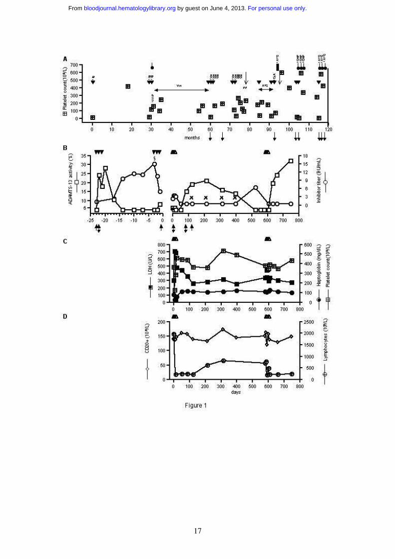
Figure 1. History of the patient, ADAMTS-13 activity and inhibitors and
laboratory parameters.
Panel A: Time-line history of the patients. Treatments: plasma exchange ( );
immunoglobulins (Ig); corticosteroids ( ); fresh frozen plasma (FFP); vincristine
(Vin); antiplatelet agents (ATG); cyclosporine A (CyA); cyclophosphamide (CHP);
plasma exchange, vincristine, antiplatelet agents and corticosteroids (#). The symbol
indicate splenectomy. : TIA. : ADAMTS-13 activity <6% (normal range:
50-150%), ADAMTS-13 inhibitors present.
Panel B: ADAMTS-13 activity and anti-ADAMTS-13 inhibitor titer in the two
courses of plasma exchange ( ), during rituximab ( ) treatment and in the follow-
up. §: IgG titer: 1:1600; IgM: negative. x: IgG titer: negative; IgM: negative. : time
of sampling for immunoblot (figure 2, panel A). : time of sampling for VWF
multimer analysis (figure 2, panel B).
Panel C: Platelet count (normal range: 140-440 x 109/L), LDH (normal range: 240-
460 U/L) and haptoglobin (normal range: 49-246 mg/dL).
Panel D: Lymphocytes count (normal range: 1.000-5.000 x 10⁶/L) and CD20 count
(normal range: 5-15% of lymphocytes count).
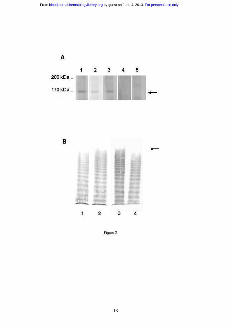
Figure 2. Immuniblot of anti-ADAMTS-13 IgG and VWF multimeric pattern.
Panel A: Immunoblot showing the presence of IgG reacting with recombinant (r)
ADAMTS-13. Serum samples were reacted with human rADAMTS-13 previously
transblotted to a nitrocellulose membrane. The presence of IgG were detected by a
peroxidase conjugate goat anti-human IgG antibody. Lane 1: patient serum collected
For personal use only. by guest on June 4, 2013. bloodjournal.hematologylibrary.orgFrom
16
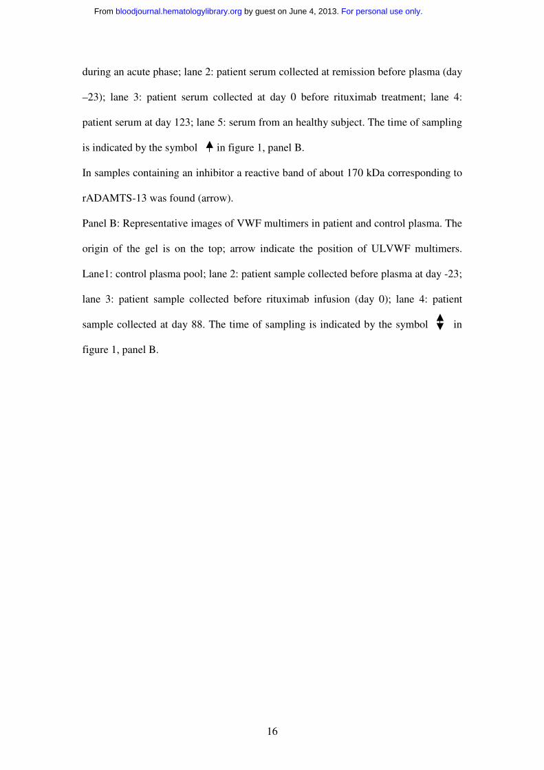
during an acute phase; lane 2: patient serum collected at remission before plasma (day
–23); lane 3: patient serum collected at day 0 before rituximab treatment; lane 4:
patient serum at day 123; lane 5: serum from an healthy subject. The time of sampling
is indicated by the symbol in figure 1, panel B.
In samples containing an inhibitor a reactive band of about 170 kDa corresponding to
rADAMTS-13 was found (arrow).
Panel B: Representative images of VWF multimers in patient and control plasma. The
origin of the gel is on the top; arrow indicate the position of ULVWF multimers.
Lane1: control plasma pool; lane 2: patient sample collected before plasma at day -23;
lane 3: patient sample collected before rituximab infusion (day 0); lane 4: patient
sample collected at day 88. The time of sampling is indicated by the symbol in
figure 1, panel B.
For personal use only. by guest on June 4, 2013. bloodjournal.hematologylibrary.orgFrom
17
For personal use only. by guest on June 4, 2013. bloodjournal.hematologylibrary.orgFrom
18
For personal use only. by guest on June 4, 2013. bloodjournal.hematologylibrary.orgFrom
Related Documents