Journal of Biomechanics 41 (2008) 1274–1280 Rigid-body analysis of a lizard skull: Modelling the skull of Uromastyx hardwickii M. Moazen a, , N. Curtis a , S.E. Evans c , P. O’Higgins b , M.J. Fagan a a Centre for Medical Engineering and Technology, University of Hull, Hull, East Yorkshire HU6 7RX, UK b Department of Biology and Hull York Medical School, University of York, York YO10 5DD, UK c Department of Anatomy and Developmental Biology, UCL, London WC1E 6BT, UK Accepted 17 January 2008 Abstract Lizard skulls vary greatly in their detailed morphology. Theoretical models and practical studies have posited a definite relationship between skull morphology and bite performance, but this can be difficult to demonstrate in vivo. Computer modelling provides an alternative approach, as long as hard and soft tissue components can be integrated and the model can be validated. An anatomically accurate three-dimensional computer model of an Uromastyx hardwickii skull was developed for rigid-body dynamic analysis. The Uromastyx jaw was first opened under motion control, and then muscle forces were applied to produce biting simulations where bite forces and joint forces were calculated. Bite forces comparable to those reported in the literature were predicted, and detailed muscular force information was produced along with additional information on the stabilizing role of temporal ligaments in late jaw closing. r 2008 Elsevier Ltd. All rights reserved. Keywords: Bite force; Muscle force; Temporal ligament; Multibody dynamics analysis; Lizards 1. Introduction Lizards, snakes and their relatives (Squamata) comprise the largest and most successful group of living reptiles. They are distinguished from crocodiles and turtles by a variable degree of intracranial mobility (e.g. Metzger, 2002), most notably streptostyly. In the latter movement, the quadrate bone is able to pivot fore-and-aft due to a relaxation of its bony contacts with the rest of the skull, allowing the mandible to move forwards during mouth opening. These movements are constrained dorsally and ventrally by ligamentous attachments, the precise role of which is debated (Iordansky, 1996; Herrel et al., 1998a, b). Furthermore, although squamate skulls show a number of common characteristics (e.g. Estes et al., 1988; Evans, 2003), they differ markedly in overall shape, in the details of their joint morphology, in bone density, and in the structure and implantation of the teeth. These structural and shape differences must be linked to differences in feeding strategy (e.g. Herrel et al., 2001; Erickson et al., 2003; Metzger and Herrel, 2005). However, developing an understanding of the evolutionary selective factors influen- cing craniofacial geometry is a daunting task, as it requires knowledge of the interactions between bone form and function, ligament arrangement, muscle forces, joint forces and bite forces. Unravelling the effects of these different components in vivo is difficult, precisely because they are closely integrated, but computer modelling techniques provide an opportunity to explore in detail the biomechanical char- acteristics of different skulls and to examine the develop- ment, adaptation and influence of specific features. The relatively new technique of multibody dynamics analysis (MDA) allows us to simulate rigid-body motion and to model the external forces and internal musculature responsible for skull loading (Langenbach et al., 2002; Sellers and Crompton, 2004). This, in turn, enables biting simulations to be performed, and the resulting information ARTICLE IN PRESS www.elsevier.com/locate/jbiomech www.JBiomech.com 0021-9290/$ - see front matter r 2008 Elsevier Ltd. All rights reserved. doi:10.1016/j.jbiomech.2008.01.012 Corresponding author. Tel.: +44 1482 466036; fax: +44 1482 466664. E-mail addresses: [email protected] (M. Moazen), [email protected] (N. Curtis), [email protected] (S.E. Evans), [email protected] (P. O’Higgins), [email protected] (M.J. Fagan).

Welcome message from author
This document is posted to help you gain knowledge. Please leave a comment to let me know what you think about it! Share it to your friends and learn new things together.
Transcript
ARTICLE IN PRESS
0021-9290/$ - se
doi:10.1016/j.jb
�CorrespondE-mail addr
paul.ohiggins@
(M.J. Fagan).
Journal of Biomechanics 41 (2008) 1274–1280
www.elsevier.com/locate/jbiomech
www.JBiomech.com
Rigid-body analysis of a lizard skull: Modelling the skullof Uromastyx hardwickii
M. Moazena,�, N. Curtisa, S.E. Evansc, P. O’Higginsb, M.J. Fagana
aCentre for Medical Engineering and Technology, University of Hull, Hull, East Yorkshire HU6 7RX, UKbDepartment of Biology and Hull York Medical School, University of York, York YO10 5DD, UK
cDepartment of Anatomy and Developmental Biology, UCL, London WC1E 6BT, UK
Accepted 17 January 2008
Abstract
Lizard skulls vary greatly in their detailed morphology. Theoretical models and practical studies have posited a definite relationship
between skull morphology and bite performance, but this can be difficult to demonstrate in vivo. Computer modelling provides an
alternative approach, as long as hard and soft tissue components can be integrated and the model can be validated. An anatomically
accurate three-dimensional computer model of an Uromastyx hardwickii skull was developed for rigid-body dynamic analysis. The
Uromastyx jaw was first opened under motion control, and then muscle forces were applied to produce biting simulations where bite
forces and joint forces were calculated. Bite forces comparable to those reported in the literature were predicted, and detailed muscular
force information was produced along with additional information on the stabilizing role of temporal ligaments in late jaw closing.
r 2008 Elsevier Ltd. All rights reserved.
Keywords: Bite force; Muscle force; Temporal ligament; Multibody dynamics analysis; Lizards
1. Introduction
Lizards, snakes and their relatives (Squamata) comprisethe largest and most successful group of living reptiles.They are distinguished from crocodiles and turtles by avariable degree of intracranial mobility (e.g. Metzger,2002), most notably streptostyly. In the latter movement,the quadrate bone is able to pivot fore-and-aft due to arelaxation of its bony contacts with the rest of the skull,allowing the mandible to move forwards during mouthopening. These movements are constrained dorsally andventrally by ligamentous attachments, the precise role ofwhich is debated (Iordansky, 1996; Herrel et al., 1998a, b).Furthermore, although squamate skulls show a number ofcommon characteristics (e.g. Estes et al., 1988; Evans,2003), they differ markedly in overall shape, in the details
e front matter r 2008 Elsevier Ltd. All rights reserved.
iomech.2008.01.012
ing author. Tel.: +441482 466036; fax: +44 1482 466664.
esses: [email protected] (M. Moazen),
c.uk (N. Curtis), [email protected] (S.E. Evans),
hyms.ac.uk (P. O’Higgins), [email protected]
of their joint morphology, in bone density, and in thestructure and implantation of the teeth. These structuraland shape differences must be linked to differences infeeding strategy (e.g. Herrel et al., 2001; Erickson et al.,2003; Metzger and Herrel, 2005). However, developing anunderstanding of the evolutionary selective factors influen-cing craniofacial geometry is a daunting task, as it requiresknowledge of the interactions between bone form andfunction, ligament arrangement, muscle forces, joint forcesand bite forces.Unravelling the effects of these different components
in vivo is difficult, precisely because they are closelyintegrated, but computer modelling techniques provide anopportunity to explore in detail the biomechanical char-acteristics of different skulls and to examine the develop-ment, adaptation and influence of specific features. Therelatively new technique of multibody dynamics analysis(MDA) allows us to simulate rigid-body motion and tomodel the external forces and internal musculatureresponsible for skull loading (Langenbach et al., 2002;Sellers and Crompton, 2004). This, in turn, enables bitingsimulations to be performed, and the resulting information
ARTICLE IN PRESSM. Moazen et al. / Journal of Biomechanics 41 (2008) 1274–1280 1275
can then be applied to finite element analysis (FEA) studiesto calculate stresses and strains, where the internal andexternal bone structure is widely believed to be influencedby the loading.
Although computational simulation is a relatively newapproach in the study of cranial functional morphology, agrowing body of work demonstrates both its potential andits validity. Validation is always an area of concern, butthere is suitable bite force data available in the literaturefor comparison (e.g. Hylander, 1979; Dumont and Herrel,2003). In a preliminary study, Langenbach and Hannam(1999) developed a three-dimensional (3D) dynamic modelof the human jaw to assess the role of passive muscletension. More recently, Langenbach et al. (2002, 2006)were able to demonstrate the accuracy of computationalsimulations with a computer model of a pig jaw that agreedwell with recorded in vivo data. Sellers and Crompton(2004) used MDA in a study of the sensitivity of muscleforce application, and this helped validate their computa-tional model into the prediction of bite force in the humanskull. Koolstra and Van Eijden (2005, 2006) were amongthe first to use a combined rigid-body and finite elementmethod to study the musculoskeletal system, developing amodel of the human jaw joint with the help of MADYMO(TNO Automotive, The Netherlands). Most recently, Peckand Hannam (2007) modelled the human jaw, and de Zeeet al. (2007) performed a validation study that comparedthe results of in vivo experiments on the human mandiblewith those from musculoskeletal models. The potentialapplications to extinct taxa were illustrated by Konakanchi(2005) who used AnyBody musculoskeletal modellingsoftware to predict the bite force of a saber-toothed cat(Smilodon fatalis), and by Del Signor (2005) who integrateda Graphical-User-Interface (GUI) of MATLAB into MSCVisualNastran4D, again to estimate the bite force of anextinct cat.
The present work is part of a larger study into theevolution of reptilian skull architecture. The goal was todevelop an anatomically accurate computational model ofa lizard skull (Uromastyx hardwickii), in which bitingsimulations are conducted by applying varying muscleloads to predict ligament, joint and bite reaction forces.The extensive data produced from this research willimprove the accuracy of future FEA studies, in whichquestions directed towards mechanical loading and facialmorphology can begin to be answered.
2. Materials and methods
2.1. Model construction
Micro-CT data of an Uromastyx hardwickii skull (skull length �44mm;
skull width �41mm) were supplied by the University of Texas, Austin, in
the form of a tiff image data set. The data set was imported into AMIRA
image segmentation software (Berlin, Germany), which was used to
extract the bony geometries through a segmentation process and to create
accurate surface models of the upper skull (cranium), lower jaws
(mandibles), and the quadrates. Separation of the quadrates allowed
them to move independently. The mandible was divided into left and right
sections. The surface models were exported from AMIRA in a wavefront
format (.obj) and imported into MSC ADAMS motion simulation
software (Santa Ana, US) as rigid-bodies for MDA.
Definition of the mass properties of all moving parts, constraints on
their motion, and the forces or motions applied to them must be defined
when carrying out multibody dynamic modelling. The mass of the jaw and
quadrates was calculated directly from ADAMS using the geometry of the
rigid-bodies and assuming a standard tissue density of 1050 kg/m3 (Sellers
and Crompton, 2004). A mass of 1.54 and 0.27 g was produced for the jaw
and quadrates, respectively. Gravity was also applied in the model, with a
constant value of 9.81m/s2. The cranium was fixed throughout all testing
and the quadrates were attached to the cranium at one end (quadrato-
squamosal joint—specified as joint 1) and the mandible at the other
(quadrato-mandibular joint—specified as joint 2) via hinge joints. The
mandible was imported as two separate parts that were rigidly connected
via the coupler tool in ADAMS.
2.2. Muscle, ligament and food bolus development
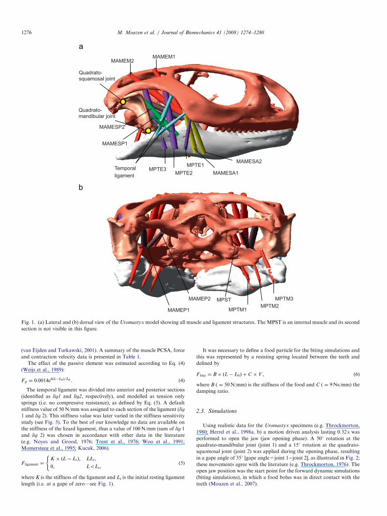
The attachment locations of the masticatory muscles and the temporal
ligament were defined in accordance with the anatomical data described by
Haas (1973) and Throckmorton (1976). Since we were interested in jaw
closing/biting only the adductor muscles were represented: these were the
adductor mandibulae externus superficialis anterior (MAMESA), adduc-
tor mandibulae externus superficialis posterior (MAMESP), adductor
mandibulae externus medialis (MAMEM), adductor mandibulae externus
profundus (MAMEP), pterygoideus externus (MPTE), pterygoideus
medialis (MPTM) and the pseudotemporalis superficialis (MPST). To
cover the broad attachment areas the MPTE and MPTM muscle groups
were divided into 3 sections and fanned out accordingly. All other muscle
groups were divided into two sections (see Fig. 1). Muscle pennation
could not be taken into account as fibre lengths and orientations vary
throughout each muscle.
The muscles were modelled according to van Ruijven and Weijs (1990)
(see Eq. (1)), which in turn are based on the Hill-type muscle model
(Hill, 1938). An additional feature was included into the muscle model
such that if the working length of the muscle became less than its resting
length then that muscle would not exert a force:
Fmuscle ¼ Fmax � ðFA � FV � FQ þ FPÞ, (1)
where Fmax is the maximal tetanic force, i.e. physiological cross-sectional
area (PCSA)� 30N/cm2 (van Ruijven and Weijs, 1990), FA is a force/
length factor, FV is a force/velocity factor, FQ is an activation factor and
FP a passive muscle element. The PCSA values used to calculate Fmax were
based on scaled PCSA data reported by Herrel et al. (1998a, b).
The force length factor (FA) was estimated by a second order
polynomial, as defined in Eq. (2) (Epstein and Herzog, 1998):
FA ¼ �6:25ðL=L0Þ2þ 12:5ðL=L0Þ � 5:25, (2)
where L is the current muscle length and L0 is optimal muscle length at
which the muscle can apply its maximum force. For the purpose of this
study an optimum gape of 151 was chosen (Herrel, personal communica-
tion; Turkawski and van Eijden, 2001) and then the corresponding muscle
length (i.e. L0) for each individual muscle was calculated. The force
velocity relation (FV) was specified separately for shortening and
lengthening according to the model of Otten (1987) which again was
based on Hill’s equations (Hill, 1938) (Eq. (3)).
FV ¼ð1� V=VmaxÞ=ð1þ V=k �VmaxÞ; V0;
1:8� 0:8ð1þ V=VmaxÞ=ð1� 7:56V=k � VmaxÞ; Vo0;
((3)
where Vmax is the maximum contraction velocity, which is dependent on
the length of muscle. Here we scaled muscle fibre lengths published by
Herrel et al. (1998a, b) according to the skull length of our model, and
multiplied this by a value of 14.6�muscle length/s, as reported by Marsh
and Bennett (1985) (see Table 1). k ( ¼ 0.25) is a constant calculated
by Close (1964) assuming that jaw muscles consist mainly of fast fibres
ARTICLE IN PRESS
MPTM2 MPTM3
MPTM1
MAMEP2
MAMEP1
MPST
MPTE1 MPTE2
MPTE3 MAMESA1
MAMESA2
MAMESP1
MAMEM2
MAMESP2
MAMEM1
Temporal ligament
Quadrato-squamosal joint
Quadrato-mandibular joint
Fig. 1. (a) Lateral and (b) dorsal view of the Uromastyx model showing all muscle and ligament structures. The MPST is an internal muscle and its second
section is not visible in this figure.
M. Moazen et al. / Journal of Biomechanics 41 (2008) 1274–12801276
(van Eijden and Turkawski, 2001). A summary of the muscle PCSA, force
and contraction velocity data is presented in Table 1.
The effect of the passive element was estimated according to Eq. (4)
(Weijs et al., 1989):
Fp ¼ 0:0014e6ðL�L0Þ=L0 . (4)
The temporal ligament was divided into anterior and posterior sections
(identified as lig1 and lig2, respectively), and modelled as tension only
springs (i.e. no compressive resistance), as defined by Eq. (5). A default
stiffness value of 50N/mm was assigned to each section of the ligament (lig
1 and lig 2). This stiffness value was later varied in the stiffness sensitivity
study (see Fig. 5). To the best of our knowledge no data are available on
the stiffness of the lizard ligament, thus a value of 100N/mm (sum of lig 1
and lig 2) was chosen in accordance with other data in the literature
(e.g. Noyes and Grood, 1976; Trent et al., 1976; Woo et al., 1991;
Momersteeg et al., 1995; Kucuk, 2006).
F ligament ¼K � ðL� LiÞ; LLi;
0; LoLi;
((5)
where K is the stiffness of the ligament and Li is the initial resting ligament
length (i.e. at a gape of zero—see Fig. 1).
It was necessary to define a food particle for the biting simulations and
this was represented by a resisting spring located between the teeth and
defined by
Fbite ¼ B� ðL� L0Þ þC �V , (6)
where B ( ¼ 50N/mm) is the stiffness of the food and C ( ¼ 9Ns/mm) the
damping ratio.
2.3. Simulations
Using realistic data for the Uromastyx specimens (e.g. Throckmorton,
1980; Herrel et al., 1998a, b) a motion driven analysis lasting 0.32 s was
performed to open the jaw (jaw opening phase). A 501 rotation at the
quadrato-mandibular joint (joint 1) and a 151 rotation at the quadrato-
squomosal joint (joint 2) was applied during the opening phase, resulting
in a gape angle of 351 [gape angle=joint 1�joint 2], as illustrated in Fig. 2;
these movements agree with the literature (e.g. Throckmorton, 1976). The
open jaw position was the start point for the forward dynamic simulations
(biting simulations), in which a food bolus was in direct contact with the
teeth (Moazen et al., 2007).
ARTICLE IN PRESSM. Moazen et al. / Journal of Biomechanics 41 (2008) 1274–1280 1277
Muscle lengths [L] (i.e. the distance between the muscle attachment on
the cranium and the muscle attachment of the mandible) directly influence
muscle forces as defined in Eq. (2). These muscle lengths along with their
rate of change, or velocity (V) are measured by ADAMS and used to solve
Table 1
Muscle data used in the MDA modelling
Muscle group Scaled
PCSA (cm2)
Fmax
(N/cm2)
Scaled fibre
length (cm)
Vmax
(mm/s)
MAMEM 0.353 0.120 17.520
MAMEM1 0.176 5.295
MAMEM2 0.176 5.295
MAMESP 0.274 0.144 21.024
MAMESP1 0.137 4.110
MAMESP2 0.137 4.110
MAMESA 0.230 0.064 9.344
MAMESA1 0.115 3.450
MAMESA2 0.115 3.450
MAMEP 0.187 0.096 14.016
MAMEP1 0.093 2.805
MAMEP2 0.093 2.805
MPTE 0.531 0.351 51.246
MPTE1 0.177 5.310
MPTE2 0.177 5.310
MPTE3 0.177 5.310
MPTM 0.442 0.175 25.550
MPTM1 0.147 4.410
MPTM2 0.147 4.410
MPTM3 0.147 4.410
MPST 0.051 0.032 4.672
MPST1 0.025 0.765
MPST2 0.025 0.765
0
10
20
30
40
50
60
0Time (sec)
Ang
le (d
egre
e)
Gape angle quadrato-squamosal rotationquadrato-mandibular rotation
Jaw
opening
Jaw
closing
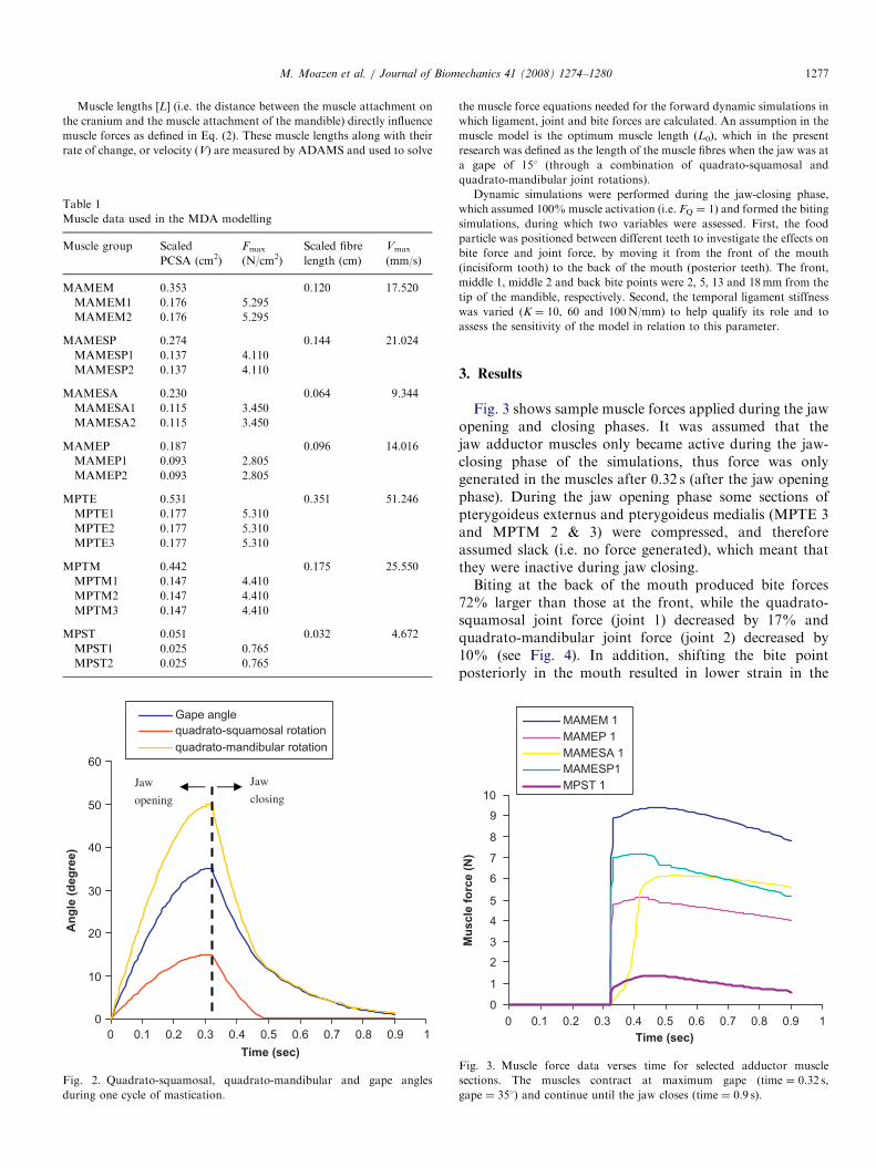
10.90.80.70.60.50.40.30.20.1
Fig. 2. Quadrato-squamosal, quadrato-mandibular and gape angles
during one cycle of mastication.
the muscle force equations needed for the forward dynamic simulations in
which ligament, joint and bite forces are calculated. An assumption in the
muscle model is the optimum muscle length (L0), which in the present
research was defined as the length of the muscle fibres when the jaw was at
a gape of 151 (through a combination of quadrato-squamosal and
quadrato-mandibular joint rotations).
Dynamic simulations were performed during the jaw-closing phase,
which assumed 100%muscle activation (i.e. FQ ¼ 1) and formed the biting
simulations, during which two variables were assessed. First, the food
particle was positioned between different teeth to investigate the effects on
bite force and joint force, by moving it from the front of the mouth
(incisiform tooth) to the back of the mouth (posterior teeth). The front,
middle 1, middle 2 and back bite points were 2, 5, 13 and 18mm from the
tip of the mandible, respectively. Second, the temporal ligament stiffness
was varied (K ¼ 10, 60 and 100N/mm) to help qualify its role and to
assess the sensitivity of the model in relation to this parameter.
3. Results
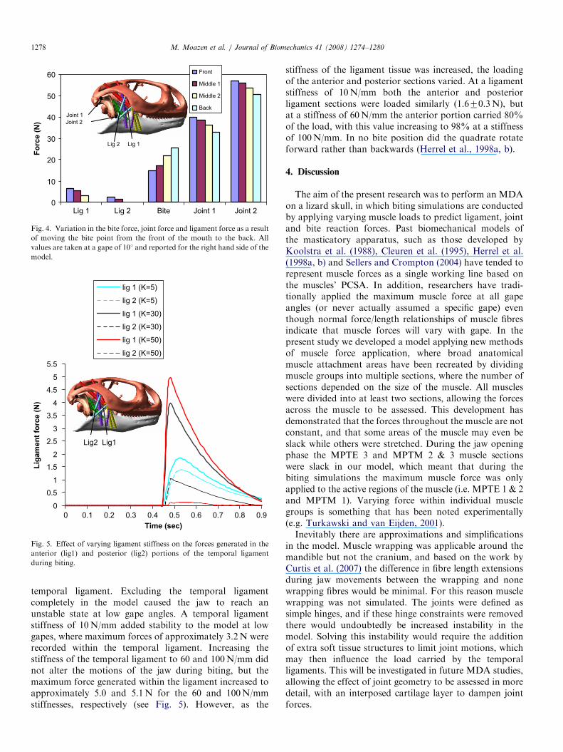
Fig. 3 shows sample muscle forces applied during the jawopening and closing phases. It was assumed that thejaw adductor muscles only became active during the jaw-closing phase of the simulations, thus force was onlygenerated in the muscles after 0.32 s (after the jaw openingphase). During the jaw opening phase some sections ofpterygoideus externus and pterygoideus medialis (MPTE 3and MPTM 2 & 3) were compressed, and thereforeassumed slack (i.e. no force generated), which meant thatthey were inactive during jaw closing.Biting at the back of the mouth produced bite forces
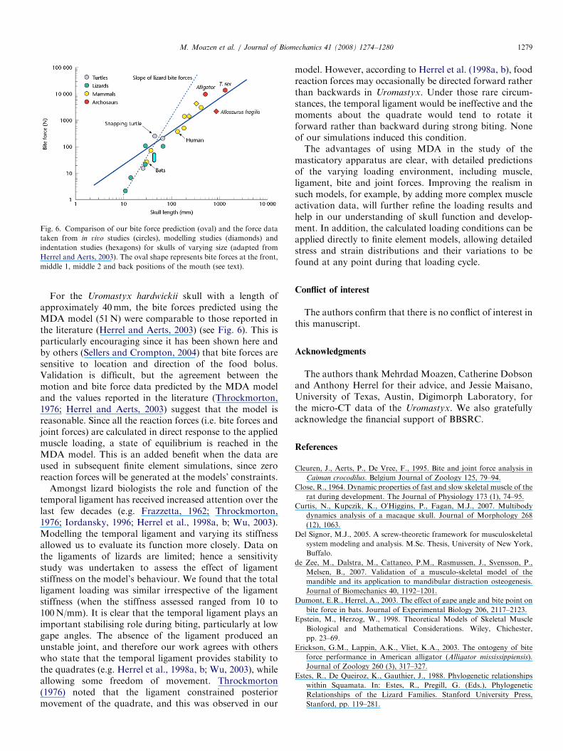
72% larger than those at the front, while the quadrato-squamosal joint force (joint 1) decreased by 17% andquadrato-mandibular joint force (joint 2) decreased by10% (see Fig. 4). In addition, shifting the bite pointposteriorly in the mouth resulted in lower strain in the
0
1
2
3
4
5
6
7
8
9
10
0Time (sec)
Mus
cle
forc
e (N
)
MAMEM 1MAMEP 1MAMESA 1MAMESP1MPST 1
10.90.80.70.60.50.40.30.20.1
Fig. 3. Muscle force data verses time for selected adductor muscle
sections. The muscles contract at maximum gape (time ¼ 0.32 s,
gape ¼ 351) and continue until the jaw closes (time ¼ 0.9 s).
ARTICLE IN PRESS
0
10
20
30
40
50
60
Lig 1
Forc
e (N
)
Front
Middle 1
Middle 2
BackJoint 1
Joint 2
Lig 2 Lig 1
Joint 2Joint 1BiteLig 2
Fig. 4. Variation in the bite force, joint force and ligament force as a result
of moving the bite point from the front of the mouth to the back. All
values are taken at a gape of 101 and reported for the right hand side of the
model.
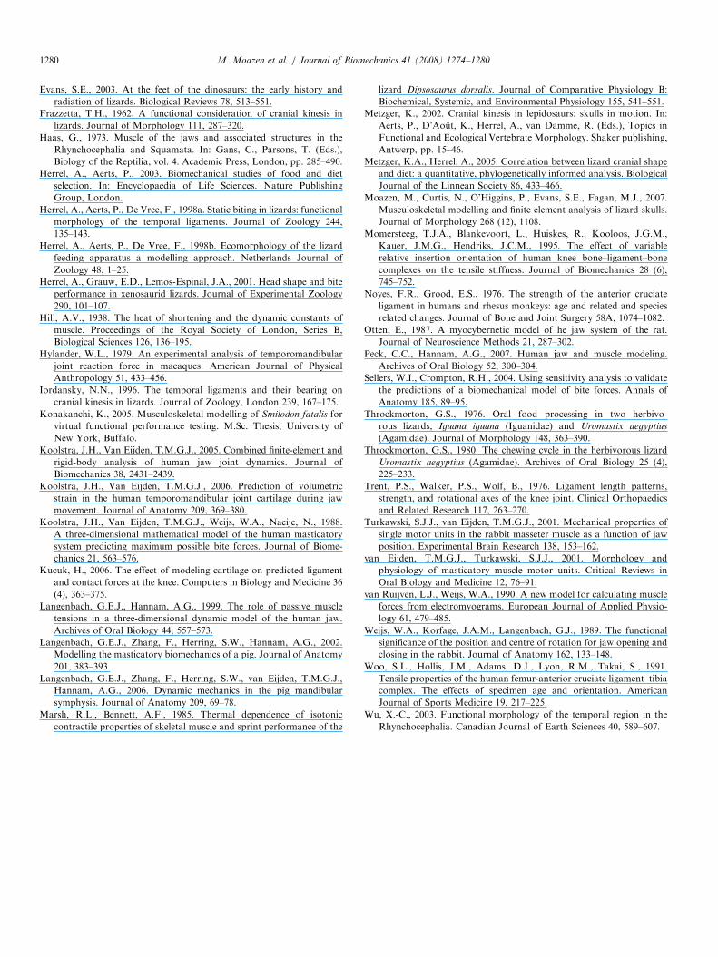
0
0.51
1.52
2.53
3.5
4
4.5
5
5.5
0Time (sec)
Liga
men
t for
ce (N
)
lig 1 (K=5)
lig 2 (K=5)
lig 1 (K=30)
lig 2 (K=30)
lig 1 (K=50)
lig 2 (K=50)
Lig2 Lig1
0.90.80.70.60.50.40.30.20.1
Fig. 5. Effect of varying ligament stiffness on the forces generated in the
anterior (lig1) and posterior (lig2) portions of the temporal ligament
during biting.
M. Moazen et al. / Journal of Biomechanics 41 (2008) 1274–12801278
temporal ligament. Excluding the temporal ligamentcompletely in the model caused the jaw to reach anunstable state at low gape angles. A temporal ligamentstiffness of 10N/mm added stability to the model at lowgapes, where maximum forces of approximately 3.2N wererecorded within the temporal ligament. Increasing thestiffness of the temporal ligament to 60 and 100N/mm didnot alter the motions of the jaw during biting, but themaximum force generated within the ligament increased toapproximately 5.0 and 5.1N for the 60 and 100N/mmstiffnesses, respectively (see Fig. 5). However, as the
stiffness of the ligament tissue was increased, the loadingof the anterior and posterior sections varied. At a ligamentstiffness of 10N/mm both the anterior and posteriorligament sections were loaded similarly (1.670.3N), butat a stiffness of 60N/mm the anterior portion carried 80%of the load, with this value increasing to 98% at a stiffnessof 100N/mm. In no bite position did the quadrate rotateforward rather than backwards (Herrel et al., 1998a, b).
4. Discussion
The aim of the present research was to perform an MDAon a lizard skull, in which biting simulations are conductedby applying varying muscle loads to predict ligament, jointand bite reaction forces. Past biomechanical models ofthe masticatory apparatus, such as those developed byKoolstra et al. (1988), Cleuren et al. (1995), Herrel et al.(1998a, b) and Sellers and Crompton (2004) have tended torepresent muscle forces as a single working line based onthe muscles’ PCSA. In addition, researchers have tradi-tionally applied the maximum muscle force at all gapeangles (or never actually assumed a specific gape) eventhough normal force/length relationships of muscle fibresindicate that muscle forces will vary with gape. In thepresent study we developed a model applying new methodsof muscle force application, where broad anatomicalmuscle attachment areas have been recreated by dividingmuscle groups into multiple sections, where the number ofsections depended on the size of the muscle. All muscleswere divided into at least two sections, allowing the forcesacross the muscle to be assessed. This development hasdemonstrated that the forces throughout the muscle are notconstant, and that some areas of the muscle may even beslack while others were stretched. During the jaw openingphase the MPTE 3 and MPTM 2 & 3 muscle sectionswere slack in our model, which meant that during thebiting simulations the maximum muscle force was onlyapplied to the active regions of the muscle (i.e. MPTE 1 & 2and MPTM 1). Varying force within individual musclegroups is something that has been noted experimentally(e.g. Turkawski and van Eijden, 2001).Inevitably there are approximations and simplifications
in the model. Muscle wrapping was applicable around themandible but not the cranium, and based on the work byCurtis et al. (2007) the difference in fibre length extensionsduring jaw movements between the wrapping and nonewrapping fibres would be minimal. For this reason musclewrapping was not simulated. The joints were defined assimple hinges, and if these hinge constraints were removedthere would undoubtedly be increased instability in themodel. Solving this instability would require the additionof extra soft tissue structures to limit joint motions, whichmay then influence the load carried by the temporalligaments. This will be investigated in future MDA studies,allowing the effect of joint geometry to be assessed in moredetail, with an interposed cartilage layer to dampen jointforces.
ARTICLE IN PRESS
Fig. 6. Comparison of our bite force prediction (oval) and the force data
taken from in vivo studies (circles), modelling studies (diamonds) and
indentation studies (hexagons) for skulls of varying size (adapted from
Herrel and Aerts, 2003). The oval shape represents bite forces at the front,
middle 1, middle 2 and back positions of the mouth (see text).
M. Moazen et al. / Journal of Biomechanics 41 (2008) 1274–1280 1279
For the Uromastyx hardwickii skull with a length ofapproximately 40mm, the bite forces predicted using theMDA model (51N) were comparable to those reported inthe literature (Herrel and Aerts, 2003) (see Fig. 6). This isparticularly encouraging since it has been shown here andby others (Sellers and Crompton, 2004) that bite forces aresensitive to location and direction of the food bolus.Validation is difficult, but the agreement between themotion and bite force data predicted by the MDA modeland the values reported in the literature (Throckmorton,1976; Herrel and Aerts, 2003) suggest that the model isreasonable. Since all the reaction forces (i.e. bite forces andjoint forces) are calculated in direct response to the appliedmuscle loading, a state of equilibrium is reached in theMDA model. This is an added benefit when the data areused in subsequent finite element simulations, since zeroreaction forces will be generated at the models’ constraints.
Amongst lizard biologists the role and function of thetemporal ligament has received increased attention over thelast few decades (e.g. Frazzetta, 1962; Throckmorton,1976; Iordansky, 1996; Herrel et al., 1998a, b; Wu, 2003).Modelling the temporal ligament and varying its stiffnessallowed us to evaluate its function more closely. Data onthe ligaments of lizards are limited; hence a sensitivitystudy was undertaken to assess the effect of ligamentstiffness on the model’s behaviour. We found that the totalligament loading was similar irrespective of the ligamentstiffness (when the stiffness assessed ranged from 10 to100N/mm). It is clear that the temporal ligament plays animportant stabilising role during biting, particularly at lowgape angles. The absence of the ligament produced anunstable joint, and therefore our work agrees with otherswho state that the temporal ligament provides stability tothe quadrates (e.g. Herrel et al., 1998a, b; Wu, 2003), whileallowing some freedom of movement. Throckmorton(1976) noted that the ligament constrained posteriormovement of the quadrate, and this was observed in our
model. However, according to Herrel et al. (1998a, b), foodreaction forces may occasionally be directed forward ratherthan backwards in Uromastyx. Under those rare circum-stances, the temporal ligament would be ineffective and themoments about the quadrate would tend to rotate itforward rather than backward during strong biting. Noneof our simulations induced this condition.The advantages of using MDA in the study of the
masticatory apparatus are clear, with detailed predictionsof the varying loading environment, including muscle,ligament, bite and joint forces. Improving the realism insuch models, for example, by adding more complex muscleactivation data, will further refine the loading results andhelp in our understanding of skull function and develop-ment. In addition, the calculated loading conditions can beapplied directly to finite element models, allowing detailedstress and strain distributions and their variations to befound at any point during that loading cycle.
Conflict of interest
The authors confirm that there is no conflict of interest inthis manuscript.
Acknowledgments
The authors thank Mehrdad Moazen, Catherine Dobsonand Anthony Herrel for their advice, and Jessie Maisano,University of Texas, Austin, Digimorph Laboratory, forthe micro-CT data of the Uromastyx. We also gratefullyacknowledge the financial support of BBSRC.
References
Cleuren, J., Aerts, P., De Vree, F., 1995. Bite and joint force analysis in
Caiman crocodilus. Belgium Journal of Zoology 125, 79–94.
Close, R., 1964. Dynamic properties of fast and slow skeletal muscle of the
rat during development. The Journal of Physiology 173 (1), 74–95.
Curtis, N., Kupczik, K., O0Higgins, P., Fagan, M.J., 2007. Multibody
dynamics analysis of a macaque skull. Journal of Morphology 268
(12), 1063.
Del Signor, M.J., 2005. A screw-theoretic framework for musculoskeletal
system modeling and analysis. M.Sc. Thesis, University of New York,
Buffalo.
de Zee, M., Dalstra, M., Cattaneo, P.M., Rasmussen, J., Svensson, P.,
Melsen, B., 2007. Validation of a musculo-skeletal model of the
mandible and its application to mandibular distraction osteogenesis.
Journal of Biomechanics 40, 1192–1201.
Dumont, E.R., Herrel, A., 2003. The effect of gape angle and bite point on
bite force in bats. Journal of Experimental Biology 206, 2117–2123.
Epstein, M., Herzog, W., 1998. Theoretical Models of Skeletal Muscle
Biological and Mathematical Considerations. Wiley, Chichester,
pp. 23–69.
Erickson, G.M., Lappin, A.K., Vliet, K.A., 2003. The ontogeny of bite
force performance in American alligator (Alligator mississippiensis).
Journal of Zoology 260 (3), 317–327.
Estes, R., De Queiroz, K., Gauthier, J., 1988. Phylogenetic relationships
within Squamata. In: Estes, R., Pregill, G. (Eds.), Phylogenetic
Relationships of the Lizard Families. Stanford University Press,
Stanford, pp. 119–281.
ARTICLE IN PRESSM. Moazen et al. / Journal of Biomechanics 41 (2008) 1274–12801280
Evans, S.E., 2003. At the feet of the dinosaurs: the early history and
radiation of lizards. Biological Reviews 78, 513–551.
Frazzetta, T.H., 1962. A functional consideration of cranial kinesis in
lizards. Journal of Morphology 111, 287–320.
Haas, G., 1973. Muscle of the jaws and associated structures in the
Rhynchocephalia and Squamata. In: Gans, C., Parsons, T. (Eds.),
Biology of the Reptilia, vol. 4. Academic Press, London, pp. 285–490.
Herrel, A., Aerts, P., 2003. Biomechanical studies of food and diet
selection. In: Encyclopaedia of Life Sciences. Nature Publishing
Group, London.
Herrel, A., Aerts, P., De Vree, F., 1998a. Static biting in lizards: functional
morphology of the temporal ligaments. Journal of Zoology 244,
135–143.
Herrel, A., Aerts, P., De Vree, F., 1998b. Ecomorphology of the lizard
feeding apparatus a modelling approach. Netherlands Journal of
Zoology 48, 1–25.
Herrel, A., Grauw, E.D., Lemos-Espinal, J.A., 2001. Head shape and bite
performance in xenosaurid lizards. Journal of Experimental Zoology
290, 101–107.
Hill, A.V., 1938. The heat of shortening and the dynamic constants of
muscle. Proceedings of the Royal Society of London, Series B,
Biological Sciences 126, 136–195.
Hylander, W.L., 1979. An experimental analysis of temporomandibular
joint reaction force in macaques. American Journal of Physical
Anthropology 51, 433–456.
Iordansky, N.N., 1996. The temporal ligaments and their bearing on
cranial kinesis in lizards. Journal of Zoology, London 239, 167–175.
Konakanchi, K., 2005. Musculoskeletal modelling of Smilodon fatalis for
virtual functional performance testing. M.Sc. Thesis, University of
New York, Buffalo.
Koolstra, J.H., Van Eijden, T.M.G.J., 2005. Combined finite-element and
rigid-body analysis of human jaw joint dynamics. Journal of
Biomechanics 38, 2431–2439.
Koolstra, J.H., Van Eijden, T.M.G.J., 2006. Prediction of volumetric
strain in the human temporomandibular joint cartilage during jaw
movement. Journal of Anatomy 209, 369–380.
Koolstra, J.H., Van Eijden, T.M.G.J., Weijs, W.A., Naeije, N., 1988.
A three-dimensional mathematical model of the human masticatory
system predicting maximum possible bite forces. Journal of Biome-
chanics 21, 563–576.
Kucuk, H., 2006. The effect of modeling cartilage on predicted ligament
and contact forces at the knee. Computers in Biology and Medicine 36
(4), 363–375.
Langenbach, G.E.J., Hannam, A.G., 1999. The role of passive muscle
tensions in a three-dimensional dynamic model of the human jaw.
Archives of Oral Biology 44, 557–573.
Langenbach, G.E.J., Zhang, F., Herring, S.W., Hannam, A.G., 2002.
Modelling the masticatory biomechanics of a pig. Journal of Anatomy
201, 383–393.
Langenbach, G.E.J., Zhang, F., Herring, S.W., van Eijden, T.M.G.J.,
Hannam, A.G., 2006. Dynamic mechanics in the pig mandibular
symphysis. Journal of Anatomy 209, 69–78.
Marsh, R.L., Bennett, A.F., 1985. Thermal dependence of isotonic
contractile properties of skeletal muscle and sprint performance of the
lizard Dipsosaurus dorsalis. Journal of Comparative Physiology B:
Biochemical, Systemic, and Environmental Physiology 155, 541–551.
Metzger, K., 2002. Cranial kinesis in lepidosaurs: skulls in motion. In:
Aerts, P., D’Aout, K., Herrel, A., van Damme, R. (Eds.), Topics in
Functional and Ecological Vertebrate Morphology. Shaker publishing,
Antwerp, pp. 15–46.
Metzger, K.A., Herrel, A., 2005. Correlation between lizard cranial shape
and diet: a quantitative, phylogenetically informed analysis. Biological
Journal of the Linnean Society 86, 433–466.
Moazen, M., Curtis, N., O’Higgins, P., Evans, S.E., Fagan, M.J., 2007.
Musculoskeletal modelling and finite element analysis of lizard skulls.
Journal of Morphology 268 (12), 1108.
Momersteeg, T.J.A., Blankevoort, L., Huiskes, R., Kooloos, J.G.M.,
Kauer, J.M.G., Hendriks, J.C.M., 1995. The effect of variable
relative insertion orientation of human knee bone–ligament–bone
complexes on the tensile stiffness. Journal of Biomechanics 28 (6),
745–752.
Noyes, F.R., Grood, E.S., 1976. The strength of the anterior cruciate
ligament in humans and rhesus monkeys: age and related and species
related changes. Journal of Bone and Joint Surgery 58A, 1074–1082.
Otten, E., 1987. A myocybernetic model of he jaw system of the rat.
Journal of Neuroscience Methods 21, 287–302.
Peck, C.C., Hannam, A.G., 2007. Human jaw and muscle modeling.
Archives of Oral Biology 52, 300–304.
Sellers, W.I., Crompton, R.H., 2004. Using sensitivity analysis to validate
the predictions of a biomechanical model of bite forces. Annals of
Anatomy 185, 89–95.
Throckmorton, G.S., 1976. Oral food processing in two herbivo-
rous lizards, Iguana iguana (Iguanidae) and Uromastix aegyptius
(Agamidae). Journal of Morphology 148, 363–390.
Throckmorton, G.S., 1980. The chewing cycle in the herbivorous lizard
Uromastix aegyptius (Agamidae). Archives of Oral Biology 25 (4),
225–233.
Trent, P.S., Walker, P.S., Wolf, B., 1976. Ligament length patterns,
strength, and rotational axes of the knee joint. Clinical Orthopaedics
and Related Research 117, 263–270.
Turkawski, S.J.J., van Eijden, T.M.G.J., 2001. Mechanical properties of
single motor units in the rabbit masseter muscle as a function of jaw
position. Experimental Brain Research 138, 153–162.
van Eijden, T.M.G.J., Turkawski, S.J.J., 2001. Morphology and
physiology of masticatory muscle motor units. Critical Reviews in
Oral Biology and Medicine 12, 76–91.
van Ruijven, L.J., Weijs, W.A., 1990. A new model for calculating muscle
forces from electromyograms. European Journal of Applied Physio-
logy 61, 479–485.
Weijs, W.A., Korfage, J.A.M., Langenbach, G.J., 1989. The functional
significance of the position and centre of rotation for jaw opening and
closing in the rabbit. Journal of Anatomy 162, 133–148.
Woo, S.L., Hollis, J.M., Adams, D.J., Lyon, R.M., Takai, S., 1991.
Tensile properties of the human femur-anterior cruciate ligament–tibia
complex. The effects of specimen age and orientation. American
Journal of Sports Medicine 19, 217–225.
Wu, X.-C., 2003. Functional morphology of the temporal region in the
Rhynchocephalia. Canadian Journal of Earth Sciences 40, 589–607.
Related Documents