Rifampicin Protects PC12 Cells from Rotenone-Induced Cytotoxicity by Activating GRP78 via PERK-eIF2a-ATF4 Pathway Xiuna Jing 1. , Qiaoyun Shi 2. , Wei Bi 1,3. , Zhifen Zeng 1 , Yanran Liang 1 , Xia Wu 1 , Songhua Xiao 1 , Jun Liu 1 , Lianhong Yang 1 , Enxiang Tao 1 * 1 Department of Neurology, Sun Yat-sen Memorial Hospital of Sun Yat-sen University, Guangzhou, People’s Republic of China, 2 Department of Radiology, School of Medicine, Stanford University, Stanford, California, United States of America, 3 Department of Neurology, the First Affiliated Hospital of Jinan University, Guangzhou, People’s Republic of China Abstract Rifampicin has been proposed as a therapeutic candidate for Parkinson’s disease (PD). We previously showed that rifampicin was neuroprotective in PD models in vivo and in vitro. However, the molecular mechanisms underlying are not fully elucidated. In this study, using the comprehensive proteomic analysis, we identified that the 78 kDa glucose-regulated protein (GRP78), a hallmark of the unfolded protein response (UPR), was upregulated in rifampicin-treated PC12 cells. Western blot analysis confirmed GRP78 activation. GRP78 functions cytoprotectively in stressed cells, therefore, we hypothesized that GRP78 mediated rifampicin-induced neuroprotection. Using RNA interference, we found that GRP78 gene knockdown significantly attenuated the neuroprotective effects of rifampicin. Next, we examined three UPR transducers, namely, protein kinase RNA-like endoplasmic reticulum kinase (PERK), inositol requiring kinase a (IREa) and activating transcription factor 6 (ATF 6), and how they regulated rifampicin-stimulated GRP78 expression. Our results showed that PERK, eukaryotic initiation factor 2a (eIF2a), and activating transcription factor 4 (ATF4) were activated in rifampicin-treated PC12 cells. Silencing the ATF4 gene using RNAi inhibited GRP78 stimulation. Interestingly, we did not detect significant IREa activation, X-box binding protein 1 mRNA splicing, or ATF6 cleavage up to 24 h after rifampicin treatment. Taken together, our data suggested that rifampicin induced GRP78 via the PERK-eIF2a-ATF4 pathway to protect neurons against rotenone-induced cell damage. Targeting molecules in this pathway could be a novel therapeutic approach for PD treatment. Citation: Jing X, Shi Q, Bi W, Zeng Z, Liang Y, et al. (2014) Rifampicin Protects PC12 Cells from Rotenone-Induced Cytotoxicity by Activating GRP78 via PERK- eIF2a-ATF4 Pathway. PLoS ONE 9(3): e92110. doi:10.1371/journal.pone.0092110 Editor: Wenhui Hu, Temple University School of Medicine, United States of America Received November 27, 2013; Accepted February 17, 2014; Published March 17, 2014 Copyright: ß 2014 Jing et al. This is an open-access article distributed under the terms of the Creative Commons Attribution License, which permits unrestricted use, distribution, and reproduction in any medium, provided the original author and source are credited. Funding: This work was funded by the the National Natural Science Foundation of China (NSFC, 81371391), and the Guangdong Provincial Department of Science and Technology (7001599; S2012010010731) and the Ph.D. Programs Foundation of Ministry of Education of China (No. 20070558257). The funders had no role in study design, data collection and analysis, decision to publish, or preparation of the manuscript. Competing Interests: The authors have declared that no competing interests exist. * E-mail: [email protected] . These authors contributed equally to this work. Introduction Parkinson’s disease (PD) is the second most common neurode- generative disorder after Alzheimer’s disease. Neuropathologically, it is characterized by the progressive loss of dopaminergic neurons within the substantia nigra pars compacta of the midbrain [1]. Current PD treatments are focused on symptomatic relief, which have risks of causing severe side effects and fail to prevent or delay the progression of the disease [2]. Therefore, searching for novel therapies to reduce the loss of dopaminergic neurons will shed new light on PD treatments. Rifampicin is an antibiotic that is widely used for tuberculosis and leprosy. It has been proposed to treat Parkinson’s disease [3]. Reports using PD models have demonstrated that it is neuropro- tective in vivo [4] and in vitro [5]. In line with this, our previous study showed that rifampicin protected PC12 cells against 1- methyl-4-phenylpyridinium (MPP+)-induced apoptosis [6]. Pre- treatment with rifampicin decreased rotenone-induced neurotox- icity in rats [7]. However, the molecular mechanisms underlying the neuroprotection of rifampicin remain unknown. In the present study, we performed a comprehensive proteomic analysis to explore the mechanisms by which rifampicin elicited protective cellular responses. The expression of the glucose- regulated protein 78 (GRP78) was significantly increased in rifampicin-treated PC12 cells. This result was confirmed by Western blot analysis. Gene silencing using RNA interference verified the mediation of GRP78 in rifampicin-induced neuro- protection. GRP78, also known as Bip, is a chaperone protein localized in the endoplasmic reticulum (ER) and plays an important role in cytoprotection and cell survival [8,9]. GRP78 is the hallmark of unfolded protein response (UPR) [10]. UPR is a cellular defense system in response to the accumulation of misfolded proteins under ER stress [11]. UPR induces the expression of GRP78 by activating ER-resident transmembrane proteins, including the activated pancreatic ER kinase-like ER kinase (PERK), inositol PLOS ONE | www.plosone.org 1 March 2014 | Volume 9 | Issue 3 | e92110

Welcome message from author
This document is posted to help you gain knowledge. Please leave a comment to let me know what you think about it! Share it to your friends and learn new things together.
Transcript
Rifampicin Protects PC12 Cells from Rotenone-InducedCytotoxicity by Activating GRP78 via PERK-eIF2a-ATF4PathwayXiuna Jing1., Qiaoyun Shi2., Wei Bi1,3., Zhifen Zeng1, Yanran Liang1, Xia Wu1, Songhua Xiao1, Jun Liu1,
Lianhong Yang1, Enxiang Tao1*
1 Department of Neurology, Sun Yat-sen Memorial Hospital of Sun Yat-sen University, Guangzhou, People’s Republic of China, 2 Department of Radiology, School of
Medicine, Stanford University, Stanford, California, United States of America, 3 Department of Neurology, the First Affiliated Hospital of Jinan University, Guangzhou,
People’s Republic of China
Abstract
Rifampicin has been proposed as a therapeutic candidate for Parkinson’s disease (PD). We previously showed that rifampicinwas neuroprotective in PD models in vivo and in vitro. However, the molecular mechanisms underlying are not fullyelucidated. In this study, using the comprehensive proteomic analysis, we identified that the 78 kDa glucose-regulatedprotein (GRP78), a hallmark of the unfolded protein response (UPR), was upregulated in rifampicin-treated PC12 cells.Western blot analysis confirmed GRP78 activation. GRP78 functions cytoprotectively in stressed cells, therefore, wehypothesized that GRP78 mediated rifampicin-induced neuroprotection. Using RNA interference, we found that GRP78gene knockdown significantly attenuated the neuroprotective effects of rifampicin. Next, we examined three UPRtransducers, namely, protein kinase RNA-like endoplasmic reticulum kinase (PERK), inositol requiring kinase a (IREa) andactivating transcription factor 6 (ATF 6), and how they regulated rifampicin-stimulated GRP78 expression. Our resultsshowed that PERK, eukaryotic initiation factor 2a (eIF2a), and activating transcription factor 4 (ATF4) were activated inrifampicin-treated PC12 cells. Silencing the ATF4 gene using RNAi inhibited GRP78 stimulation. Interestingly, we did notdetect significant IREa activation, X-box binding protein 1 mRNA splicing, or ATF6 cleavage up to 24 h after rifampicintreatment. Taken together, our data suggested that rifampicin induced GRP78 via the PERK-eIF2a-ATF4 pathway to protectneurons against rotenone-induced cell damage. Targeting molecules in this pathway could be a novel therapeutic approachfor PD treatment.
Citation: Jing X, Shi Q, Bi W, Zeng Z, Liang Y, et al. (2014) Rifampicin Protects PC12 Cells from Rotenone-Induced Cytotoxicity by Activating GRP78 via PERK-eIF2a-ATF4 Pathway. PLoS ONE 9(3): e92110. doi:10.1371/journal.pone.0092110
Editor: Wenhui Hu, Temple University School of Medicine, United States of America
Received November 27, 2013; Accepted February 17, 2014; Published March 17, 2014
Copyright: � 2014 Jing et al. This is an open-access article distributed under the terms of the Creative Commons Attribution License, which permits unrestricteduse, distribution, and reproduction in any medium, provided the original author and source are credited.
Funding: This work was funded by the the National Natural Science Foundation of China (NSFC, 81371391), and the Guangdong Provincial Department ofScience and Technology (7001599; S2012010010731) and the Ph.D. Programs Foundation of Ministry of Education of China (No. 20070558257). The funders hadno role in study design, data collection and analysis, decision to publish, or preparation of the manuscript.
Competing Interests: The authors have declared that no competing interests exist.
* E-mail: [email protected]
. These authors contributed equally to this work.
Introduction
Parkinson’s disease (PD) is the second most common neurode-
generative disorder after Alzheimer’s disease. Neuropathologically,
it is characterized by the progressive loss of dopaminergic neurons
within the substantia nigra pars compacta of the midbrain [1].
Current PD treatments are focused on symptomatic relief, which
have risks of causing severe side effects and fail to prevent or delay
the progression of the disease [2]. Therefore, searching for novel
therapies to reduce the loss of dopaminergic neurons will shed new
light on PD treatments.
Rifampicin is an antibiotic that is widely used for tuberculosis
and leprosy. It has been proposed to treat Parkinson’s disease [3].
Reports using PD models have demonstrated that it is neuropro-
tective in vivo [4] and in vitro [5]. In line with this, our previous
study showed that rifampicin protected PC12 cells against 1-
methyl-4-phenylpyridinium (MPP+)-induced apoptosis [6]. Pre-
treatment with rifampicin decreased rotenone-induced neurotox-
icity in rats [7]. However, the molecular mechanisms underlying
the neuroprotection of rifampicin remain unknown.
In the present study, we performed a comprehensive proteomic
analysis to explore the mechanisms by which rifampicin elicited
protective cellular responses. The expression of the glucose-
regulated protein 78 (GRP78) was significantly increased in
rifampicin-treated PC12 cells. This result was confirmed by
Western blot analysis. Gene silencing using RNA interference
verified the mediation of GRP78 in rifampicin-induced neuro-
protection.
GRP78, also known as Bip, is a chaperone protein localized in
the endoplasmic reticulum (ER) and plays an important role in
cytoprotection and cell survival [8,9]. GRP78 is the hallmark of
unfolded protein response (UPR) [10]. UPR is a cellular defense
system in response to the accumulation of misfolded proteins
under ER stress [11]. UPR induces the expression of GRP78 by
activating ER-resident transmembrane proteins, including the
activated pancreatic ER kinase-like ER kinase (PERK), inositol
PLOS ONE | www.plosone.org 1 March 2014 | Volume 9 | Issue 3 | e92110
requiring kinase a (IREa) and activating transcription factor 6
(ATF 6) [12]. Increasing evidence has suggested that GRP78
activation prevents neurons from apoptosis [13,14,15]. Therefore,
we hypothesized that rifampicin protected PC12 cells against
rotenone-induced cytotoxicity by regulating the GRP78 gene
expression. We also investigated the signaling pathways through
which rifampicin stimulated GRP78. Our study was aimed to
explore potential novel therapeutic targets for PD treatment.
Methods
MaterialsRifampicin, Rotenone, dimethyl sulfoxide (DMSO), 3-(4,5-
Dimethylthiazol-2-yl)-2,5-diphenyltetrazolium bromide (MTT),
49,6-diamidino-2-phenylindole (DAPI) and thapsigargin (Tg) were
purchased from Sigma (St. Louis, MO, USA). Rifampicin was
dissolved in less than 0.1% of DMSO solution. RPMI medium
1640, fetal horse serum (FCS), fetal bovine serum (FBS), penicillin,
streptomycin, and other tissue culture reagents were purchased
from Gibco (Grand Island, NY, USA). Antibodies against
PERK(sc-13073), p-PERK(sc-32577), ATF6, and beta-actin were
from Santa Cruz Biotechnology (Santa Cruz, CA, USA).
Antibodies against GRP78, p-eIF2a, eIF2a and ATF4 were from
Cell Signaling (Beverly, MA, USA). Antibodies against p-IREawere from Abcam (Hong Kong, China).
Cell CulturePC12 cells were purchased from the Cell Center of the Institute
of Basic Medical Science Research (Chinese Academy of Medical
Sciences, China). Cells were cultivated in RPMI medium 1640
supplemented with 10% heat-inactivated fetal horse serum, 5%
heat-inactivated fetal bovine serum, 100 U/mL penicillin, and
100 mg/mL streptomycin. Cells were kept at 37 uC in a humidified
atmosphere with 5% CO2. Growth medium was changed three
times a week. Unless indicated otherwise, prior to the experimen-
tal investigation, PC12 cells were differentiated by adding nerve
growth factor (NGF) at 50 ng/mL every other day for 6 days,
followed by rifampicin treatment at 150 mM for 24 h. In GRP78
gene silencing study, after differentiation and siRNAs treatment,
PC12 cells were incubated with 150 mM rifampicin for 2 h
followed by 1 mM rotenone for 24 h.
Cell Viability AssayPC12 cells were seeded at a density of 16104 cells/well in 96
well plates, and the cell viability was determined by the
conventional MTT assay. Briefly, cells were treated with the
MTT solution for 4 h at 37 uC. Then, the medium was removed
and 150 mL of DMSO was added to each well. The formazan dye
crystals were solubilized on the shaker for 15 min, and the
absorbance at 595 nm was measured using a microplate reader
(Bio-Rad, Hercules, CA, USA). Cell viability was determined by
comparing the number of viable cells to that of untreated controls,
in which the viability was defined as 100%.
DAPI stainingAfter treatment, cells were washed with PBS and fixed with 4%
formaldehyde for 30 min at room temperature. Following
incubation with Triton X-100 0.2% in PBS for 5 min, PC12 cells
were incubated with 49,6-diamidino-2-phenylindole (DAPI; 1 mg/
ml) for 5 min at room temperature.
2-DE and Image AnalysisPC12 cells were treated with or without rifampicin (150 mM) for
24 h. After treatment, cells were washed three times with ice-cold
washing buffer (10 mM Tris-HCl, 250 mM sucrose, pH 7.0),
collected in clean 1.5 ml eppendorf tubes. Lysis buffer [7 Murea,
2 M thiourea, 4% CHAPS (w/v), 1% dithiothreitol (DTT), 2%
immobilized pH gradients (IPG) (v/v), pH 3–10 NL] was added,
and samples were centrifuged at 13,200 g for 30 min at 4 uC. The
supernatant was subjected to 2-DE using an Amersham Biosci-
ences IPGphor IEF System and Hoefer SE 600 (GE healthcare,
Uppsala, Sweden) electrophoresis units (13 cm), according to
manufacturer’s instructions and a previously described protocol
[16]. Protein lysates and 2-DE gels were processed in parallel.
Protein concentrations were determined using the Bradford assay.
After 2-DE, the gels underwent silver nitrate staining according to
a previously described protocol [17], followed by scanning using
an Image Scanner (GE Healthcare). The images were analyzed
using the Image Master 2D Platinum (GE Healthcare).
MALDI-TOF-MS and Database SearchOnly protein spots that were differentially expressed in at least
three independent experiments were analyzed by MALDI-TOF-
MS. Protein spots were excised from the silver-stained gels and
transferred into the siliconized 1.5 ml eppendorf tubes. Tryptic in-
gel digestion was performed as previously reported with slight
modifications [16]. Molecular mass analysis of the tryptic peptides
was performed using an ABI 4800 plus a MALDI-TOF-MS mass
spectrometer (Applied Biosystems, Foster City, CA, USA). Spectra
were interpreted and processed using the Global Protein Server
Workstation (V3.6, Applied Biosystems) via the internal MAS-
COT search engine (V2.1, Matrix Science, London, UK) to
analyze MALDI-TOF-MS and MS/MS data. Based on combined
MALDI-TOF-MS and MS/MS spectra, MASCOT protein scores
of greater than 65 were considered statistically significant
(p,0.05). It was also accepted when the individual MS/MS
spectrum had the best ion score that was statistically significant
(p,0.05). Searching was performed against the IPI mouse
database (V3.36) with the following parameters: the enzyme
trypsin with one missed cleavage was allowed; variable modifica-
tions included acetamidation of cysteine and oxidation of
methionine; peptide mass tolerance was set to 50 ppm and
fragment ion mass tolerance was set to 0.2 Da; only monoisotopic
masses were included in the search.
Western Blot AnalysisAfter treatment, PC12 cells were harvested for western blot
analysis. Cell pellets were briefly lysed in RIPA buffer [1 mM
ethylenediaminetetraacetic acid (EDTA), 150 mM NaCl, 1%
igepal, 0.1% SDS, 0.5% sodium deoxycholate, 50 mM Tris–
HCl, pH 8.0]. Equal amounts of proteins (50 mg) were separated
by 10% sodium dodecyl sulfate polyacrylamide gel electrophoresis
(SDS-PAGE), transferred to polyvinylidene fluoride (PVDF)
membranes (Millipore Corp, MA, USA), blocked with 5% nonfat
milk for 2 h, and incubated overnight at 4 uC with primary
antibodies at a dilution of 1:1000 in blocking buffer. The next day,
the membrane was washed by TBST three times, 10 min each,
and incubated with the corresponding secondary antibodies
(1:4000) that were horseradish peroxidase-conjugated for 1 h at
room temperature.
Analysis was detected using the Syngene G:BOX Chemi XT4
fluorescence and chemiluminescence gel imaging system (Cam-
bridge, UK).
RNA extraction and RT-PCRPC12 cells were treated with rifampicin for various periods of
time (3 to 24 h). In positive controls, PC12 cells were incubated
with Tg at 1 mM for 6 h. Total RNA was isolated using Trizol
Rifampicin Protects PC12 Cells by Elevating GRP78
PLOS ONE | www.plosone.org 2 March 2014 | Volume 9 | Issue 3 | e92110
reagent (Invitrogen, Groningen, NL). cDNA was synthesized using
the Superscript III First strand synthesis Kit (Invitrogen). To
evaluate relative expression levels of XBP1u/XBP1s, RT-PCR
analysis was performed using PCR SuperMix (Invitrogen). XBP1
primer sequences were as follows: 59-
GGCGGCCCCCAAAGTGCTAC-39 (Forward) and 59-
CCCGGAACCATGAGCGGCAG-39 (Reverse). b-actin was used
as a loading control, with primers as follows: 59-
GCGTCCACCCGCGAGTACAA-39 (Forward) and 59-CGAC-
GACGAGCGCAGCGATA-39 (Reverse). PCR products were
analyzed on a 3% agarose gel. Gene expression was quantified
using ImageJ 1.45 s.
RNA Interference and TransfectionGene silencing was performed using siRNAs. GRP78-specific
siRNAs were synthesized by Shanghai GenePharma (Shanghai,
China) with the sense strand sequence of 59-GAGGC-
GUAUUUGGGAAAGATT-39.
The scrambled siRNA has the sense sequence of 59-UUCUCC-
GAACGUGUCACGUTT-39. To efficiently knock down ATF4,
we used a pool of four different siRNAs (ON-TARGET plus
SMART pool Rat ATF4; Thermo Scientific Dharmacon) to target
rat ATF4 mRNA. Control cultures were incubated with non-
targeting siRNAs (ON-TARGET plus non-targeting siRNA#1;
Thermo Scientific Dharmacon).
Transfection of siRNAs was carried out using LipofectamineTM
2000 (Invitrogen, Grand Island, NY, USA), according to the
manufacturer’s instructions. Briefly, siRNA and LipofectamineTM
2000 reagent were mixed in Opti-MEM medium (Invitrogen) and
incubated for 30 min at room temperature to allow the complex
formation. Cells were washed with Opti-MEM medium, and the
transfection mixture was added. 6 h after transfection, cells were
washed and cultured for 24 h in complete medium containing
10% FCS and 5% FBS. The silencing efficacy was evaluated by
western blotting.
Statistical AnalysisAll data were presented as the mean 6 standard error of the
mean (SEM) derived from three or more independent experi-
ments. Comparison between groups was made by one-way
analysis of variance (ANOVA) followed by an appropriate post-
hoc test to analyze the difference. A value of p,0.05 was deemed
to be statistically significant.
Results
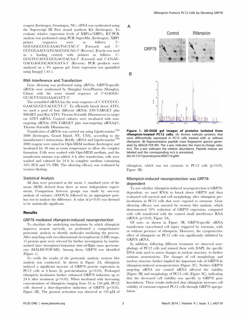
GRP78 mediated rifampicin-induced neuroprotectionTo elucidate the underlying mechanisms by which rifampicin
improves neuron survivals, we performed a comprehensive
proteomic analysis to identify molecules mediating the process.
After matching with two-dimensional electrophoresis (2-DE) maps,
15 protein spots were selected for further investigation by matrix-
assisted laser desorption/ionization time-of-flight mass spectrom-
etry (MALDI-TOF-MS). Among them, GRP78 was identified
(Figure 1).
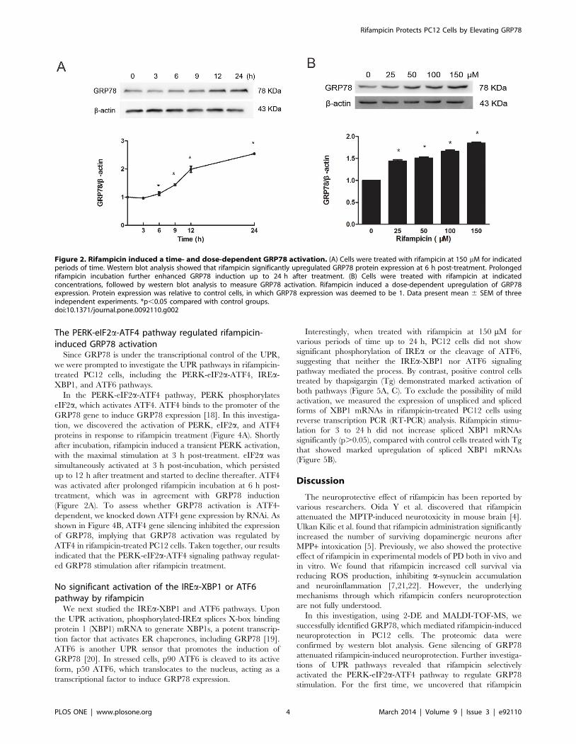
To verify the results of the proteomic analysis, western blot
analysis was conducted. As shown in Figure 2A, rifampicin
induced a significant increase of GRP78 protein expression in
PC12 cells at 6 hours (h) post-incubation (p,0.05). Prolonged
rifampicin incubation further enhanced GRP78 induction up to
24 h after treatment (p,0.05). When incubated with increasing
concentrations of rifampicin ranging from 25 to 150 mM, PC12
cells showed a dose-dependent induction of GRP78 (p,0.05,
Figure 2B). The greatest activation was observed at 150 mM of
rifampicin, which was not cytotoxic to PC12 cells (p.0.05,
Figure 3B).
Rifampicin-induced neuroprotection was GRP78-dependent
To test whether rifampicin-induced neuroprotection is GRP78-
dependent, we used RNAi to knock down GRP78 and then
evaluated cell survival and cell morphology after rifampicin pre-
incubation in PC12 cells that were exposed to rotenone. Gene
silencing efficacy was assessed by western blot analysis, which
demonstrated 70% reduction of GRP78 expression, compared
with cells transfected with the control small interference RNA
(siRNA) (p,0.05, Figure 3A).
Of note, as shown in Figure 3B, GRP78-specific siRNA
transfection exacerbated cell injury triggered by rotenone, with
or without presence of rifampicin. Moreover, the cytoprotective
effect of rifampicin on PC12 cells was significantly inhibited by
GRP78 siRNA.
In addition, following different treatment we observed mor-
phology of PC12 cells and stained them with DAPI, the specific
DNA stain used to assess changes in nuclear structure, to further
estimate neurotoxicity. The changes of cell morphology and
nuclear structure further implied the important role of GRP78 in
rifampicin-induced neuroprotection (Figure 3C). Neither GRP78
targeting siRNA nor control siRNA affected the viability
(Figure 3B) and morphology of PC12 cells (Figure 3C), indicating
that the decreased cell viability was specific to GRP78 gene
knockdown. These results indicated that rifampicin increases cell
viability of rotenone-exposed PC12 cells through GRP78 upregu-
lation.
Figure 1. 2D-DIGE gel images of proteins isolated fromrifampicin-treated PC12 cells. (A) Arrows indicate proteins thatwere differentially expressed in PC12 cells treated with or withoutrifampicin. (B) Representative peptide mass fingerprint spectra gener-ated by MALDI-TOF-MS. The x-axis indicates the mass-to-charge ratio,m/z. The y-axis indicates the relative abundance. Peptide masses arelabeled and the corresponding m/z is annotated.doi:10.1371/journal.pone.0092110.g001
Rifampicin Protects PC12 Cells by Elevating GRP78
PLOS ONE | www.plosone.org 3 March 2014 | Volume 9 | Issue 3 | e92110
The PERK-eIF2a-ATF4 pathway regulated rifampicin-induced GRP78 activation
Since GRP78 is under the transcriptional control of the UPR,
we were prompted to investigate the UPR pathways in rifampicin-
treated PC12 cells, including the PERK-eIF2a-ATF4, IREa-
XBP1, and ATF6 pathways.
In the PERK-eIF2a-ATF4 pathway, PERK phosphorylates
eIF2a, which activates ATF4. ATF4 binds to the promoter of the
GRP78 gene to induce GRP78 expression [18]. In this investiga-
tion, we discovered the activation of PERK, eIF2a, and ATF4
proteins in response to rifampicin treatment (Figure 4A). Shortly
after incubation, rifampicin induced a transient PERK activation,
with the maximal stimulation at 3 h post-treatment. eIF2a was
simultaneously activated at 3 h post-incubation, which persisted
up to 12 h after treatment and started to decline thereafter. ATF4
was activated after prolonged rifampicin incubation at 6 h post-
treatment, which was in agreement with GRP78 induction
(Figure 2A). To assess whether GRP78 activation is ATF4-
dependent, we knocked down ATF4 gene expression by RNAi. As
shown in Figure 4B, ATF4 gene silencing inhibited the expression
of GRP78, implying that GRP78 activation was regulated by
ATF4 in rifampicin-treated PC12 cells. Taken together, our results
indicated that the PERK-eIF2a-ATF4 signaling pathway regulat-
ed GRP78 stimulation after rifampicin treatment.
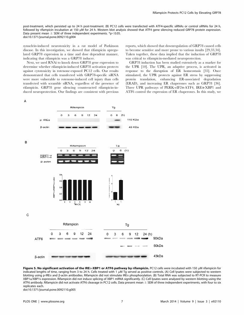
No significant activation of the IREa-XBP1 or ATF6pathway by rifampicin
We next studied the IREa-XBP1 and ATF6 pathways. Upon
the UPR activation, phosphorylated-IREa splices X-box binding
protein 1 (XBP1) mRNA to generate XBP1s, a potent transcrip-
tion factor that activates ER chaperones, including GRP78 [19].
ATF6 is another UPR sensor that promotes the induction of
GRP78 [20]. In stressed cells, p90 ATF6 is cleaved to its active
form, p50 ATF6, which translocates to the nucleus, acting as a
transcriptional factor to induce GRP78 expression.
Interestingly, when treated with rifampicin at 150 mM for
various periods of time up to 24 h, PC12 cells did not show
significant phosphorylation of IREa or the cleavage of ATF6,
suggesting that neither the IREa-XBP1 nor ATF6 signaling
pathway mediated the process. By contrast, positive control cells
treated by thapsigargin (Tg) demonstrated marked activation of
both pathways (Figure 5A, C). To exclude the possibility of mild
activation, we measured the expression of unspliced and spliced
forms of XBP1 mRNAs in rifampicin-treated PC12 cells using
reverse transcription PCR (RT-PCR) analysis. Rifampicin stimu-
lation for 3 to 24 h did not increase spliced XBP1 mRNAs
significantly (p.0.05), compared with control cells treated with Tg
that showed marked upregulation of spliced XBP1 mRNAs
(Figure 5B).
Discussion
The neuroprotective effect of rifampicin has been reported by
various researchers. Oida Y et al. discovered that rifampicin
attenuated the MPTP-induced neurotoxicity in mouse brain [4].
Ulkan Kilic et al. found that rifampicin administration significantly
increased the number of surviving dopaminergic neurons after
MPP+ intoxication [5]. Previously, we also showed the protective
effect of rifampicin in experimental models of PD both in vivo and
in vitro. We found that rifampicin increased cell survival via
reducing ROS production, inhibiting a-synuclein accumulation
and neuroinflammation [7,21,22]. However, the underlying
mechanisms through which rifampicin confers neuroprotection
are not fully understood.
In this investigation, using 2-DE and MALDI-TOF-MS, we
successfully identified GRP78, which mediated rifampicin-induced
neuroprotection in PC12 cells. The proteomic data were
confirmed by western blot analysis. Gene silencing of GRP78
attenuated rifampicin-induced neuroprotection. Further investiga-
tions of UPR pathways revealed that rifampicin selectively
activated the PERK-eIF2a-ATF4 pathway to regulate GRP78
stimulation. For the first time, we uncovered that rifampicin
Figure 2. Rifampicin induced a time- and dose-dependent GRP78 activation. (A) Cells were treated with rifampicin at 150 mM for indicatedperiods of time. Western blot analysis showed that rifampicin significantly upregulated GRP78 protein expression at 6 h post-treatment. Prolongedrifampicin incubation further enhanced GRP78 induction up to 24 h after treatment. (B) Cells were treated with rifampicin at indicatedconcentrations, followed by western blot analysis to measure GRP78 activation. Rifampicin induced a dose-dependent upregulation of GRP78expression. Protein expression was relative to control cells, in which GRP78 expression was deemed to be 1. Data present mean 6 SEM of threeindependent experiments. *p,0.05 compared with control groups.doi:10.1371/journal.pone.0092110.g002
Rifampicin Protects PC12 Cells by Elevating GRP78
PLOS ONE | www.plosone.org 4 March 2014 | Volume 9 | Issue 3 | e92110

activated GRP78 via the PERK-eIF2a-ATF4 pathway to protect
PC12 cells against rotenone-induced cytotoxicity.
Increasing evidence has suggested that heat shock proteins
(HSPs) play a pivotal role in neurodegenerative diseases. HSPs
provide a therapeutic target for neurodegenerative disorders due
to the finding that upregulation of HSPs decreases the protein
misfolding and aggregation in cells [23,24,25]. GRP78 is a
member of the 70-kDa HSP family and acts as a molecular
chaperone in the folding and assembly of newly synthesized
proteins within the ER [26]. It is reported that GRP78 suppressed
caspase activation and caspase-mediated cell death [27], suggest-
ing it is cytoprotective. Several groups have demonstrated that
GRP78 improves cell survival in vivo and in vitro [28,29,30,31].
Gorbatyuk, MS et al. [32] revealed that GRP78 diminished a-
Figure 3. Rifampicin-induced neuroprotection was GRP78-dependent. PC12 cells were transfected with GRP78-specific siRNAs or controlsiRNAs for 24 h. After that, cells were treated with or without 150 mM rifampicin for 2 h, followed by 1 mM rotenone for 24 h. (A) Western blot analysisverified the efficient gene silencing of GRP78. (B) After the above treatment, cell viability was measured and presented as the relative viability (%control). (C) Morphological evaluation of PC12 cells under the above-mentioned treatment by light microscopic observation and DAPI staining. Theapoptotic cells were marked with arrows. Scale bar = 25 mm. (B–C) GRP78 gene silencing significantly exacerbated rotenone-triggered neuron injury,with or without the presence of rifampicin. Neither GRP78-specific nor control siRNAs decreased cell viability. Data present mean 6 SEM of threeindependent experiments. *p,0.05.doi:10.1371/journal.pone.0092110.g003
Rifampicin Protects PC12 Cells by Elevating GRP78
PLOS ONE | www.plosone.org 5 March 2014 | Volume 9 | Issue 3 | e92110
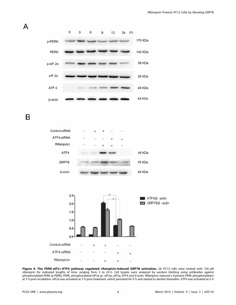
Figure 4. The PERK-eIF2a-ATF4 pathway regulated rifampicin-induced GRP78 activation. (A) PC12 cells were treated with 150 mMrifampicin for indicated lengths of time, ranging from 3 to 24 h. Cell lysates were analyzed by western blotting using antibodies againstphosphorylated PERK (p-PERK), PERK, phosphorylated eIF2a (p- eIF2a), eIF2a, ATF4 and b-actin. Rifampicin induced a transient PERK phosphorylationat 3 h post-incubation. eIF2a was activated at 3 h post-treatment, which persisted for 9 h and started to decline thereafter. ATF4 was activated at 6 h
Rifampicin Protects PC12 Cells by Elevating GRP78
PLOS ONE | www.plosone.org 6 March 2014 | Volume 9 | Issue 3 | e92110
synuclein-induced neurotoxicity in a rat model of Parkinson
disease. In this investigation, we showed that rifampicin upregu-
lated GRP78 expression in a time and dose dependent manner,
indicating that rifampicin was a GRP78 inducer.
Next, we used RNAi to knock down GRP78 gene expression to
determine whether rifampicin-induced GRP78 activation protects
against cytotoxicity in rotenone-exposed PC12 cells. Our results
demonstrated that cells transfected with GRP78-specific siRNA
were more vulnerable to rotenone-induced cell injury than cells
transfected with scramble siRNA, regardless of the presence of
rifampicin. GRP78 gene silencing counteracted rifampicin-in-
duced neuroprotection. Our findings are consistent with previous
reports, which showed that downregulation of GRP78 caused cells
to become sensitive and more prone to various insults [29,33,34].
Taken together, these data implied that the induction of GRP78
was critical to rifampicin-mediated neuroprotection.
GRP78 induction has been studied extensively as a marker for
the UPR [10]. The UPR, an adaptive process, is activated in
response to the disruption of ER homeostasis [35]. Once
stimulated, the UPR protects against ER stress by suppressing
protein translation, enhancing ER-associated degradation
(ERAD), and increasing ER chaperones such as GRP78 [36].
Three UPR pathways of PERK-eIF2a-ATF4, IREa-XBP1 and
ATF6 control the expression of ER chaperones. In this study, we
post-treatment, which persisted up to 24 h post-treatment. (B) PC12 cells were transfected with ATF4-specific siRNAs or control siRNAs for 24 h,followed by rifampicin incubation at 150 mM for 24 h. Western blot analysis showed that ATF4 gene silencing reduced GRP78 protein expression.Data present mean 6 SEM of three independent experiments. *p,0.05.doi:10.1371/journal.pone.0092110.g004
Figure 5. No significant activation of the IREa-XBP1 or ATF6 pathway by rifampicin. PC12 cells were incubated with 150 mM rifampicin forindicated lengths of time, ranging from 3 to 24 h. Cells treated with 1 mM Tg served as positive controls. (A) Cell lysates were subjected to westernblotting using p-IREa and b-actin antibodies. Rifampicin did not stimulate IREa phosphorylation. (B) Total RNA was subjected to RT-PCR to measureXBP1u/XBP1s expression. Rifampicin did not induce splicing of XBP1 mRNA significantly. (C) Cell lysates were analyzed by western blotting using theATF6 antibody. Rifampicin did not activate ATF6 cleavage in PC12 cells. Data present mean 6 SEM of three independent experiments, with four to sixreplicates each.doi:10.1371/journal.pone.0092110.g005
Rifampicin Protects PC12 Cells by Elevating GRP78
PLOS ONE | www.plosone.org 7 March 2014 | Volume 9 | Issue 3 | e92110
showed that rifampicin triggered an early phosphorylation of
PERK and eIF2a proteins in PC12 cells, which subsequently
stimulated ATF4. ATF4 gene knockdown decreased rifampicin-
induced GRP78 activation. By contrast, we did not detect IREaphosphorylation, XBP1 mRNAs splicing, or ATF6 cleavage up to
24 h after rifampicin administration. Our findings suggested that
rifampicin selectively activated the PERK-eIF2a-ATF4 signaling
pathway to regulate GRP78 induction.
If a GRP78 inducer is just an ER stressor such as Tg, its
application as a therapeutic strategy is unlikely to be realized
because it may activate several pathways of the UPR, including
ER stress-induced apoptotic pathways[28]. The present study
showed that the PERK-eIF2a-ATF4 pathway was activated by
rifampicin to induce GRP78, neither IREa pathway or ATF6
pathway. The selective activation of one UPR pathway by
rifampicin may account for its protection.
Our investigation showed that with the increase of rifampicin
dosages, there was an enhancement of GRP78 activation, with the
highest induction observed at 150 mM of rifampicin, which did not
affect cell viability. Since GRP78 induction is a hallmark of ER
stress [37], rifampicin might enhance cellular defense systems by
exposing PC12 cells to a mild stress state. This phenomenon is
known as preconditioning. Several lines of evidence have
suggested that cells pretreated with sublethal stress could adapt
to stress and increase their defense capacities to resist more severe
stress [30,38]. Here, we showed that induction of GRP78 by
rifampicin ‘‘preconditioned’’ PC12 cells and protected them
against cell injury triggered by rotenone. Although we tested
rotenone-induced cytotoxicity in this study, it is possible that
rifampicin and GRP78 might protect cells against other parkin-
sonian neurotoxins, thus, function as potential treatments for
neurodegeneration because oxidative stress and ER stress have
been implicated in the disease development after exposure to these
toxins [39]. Nevertheless, further investigations are needed to
clarify it.
To our best knowledge, we for the first time identified that
rifampicin activated GRP78 to protect PC12 cells from rotenone-
induced cytotoxicity. Our results provided added information to
the mechanism underlying rifampicin-induced neuroprotection,
particularly, signaling pathways involved in this process. Future
directions include, but are not limited to, in vivo verification of
GRP78 and UPR pathways in rifampicin-treated PD animal
models, investigation of the functions of GRP78 proteins in ER
stress and how they are regulated, identification of other potential
therapeutic targets. Based on our findings, we concluded that
GRP78 and PERK-eIF2a-ATF4 pathway mediated rifampicin-
induced neuroprotection and their targeting could serve as a novel
potential therapy for PD treatment.
Author Contributions
Conceived and designed the experiments: ET XJ. Performed the
experiments: XJ WB YL ZZ XW SX ET. Analyzed the data: ET XJ
WB QS. Contributed reagents/materials/analysis tools: SX JL LY. Wrote
the paper: ET XJ QS.
References
1. Ozansoy M, Basak AN (2013) The Central Theme of Parkinson’s Disease:
alpha-Synuclein. Mol Neurobiol 47: 460–465.
2. Jankovic J, Aguilar LG (2008) Current approaches to the treatment of
Parkinson’s disease. Neuropsychiatr Dis Treat 4: 743–757.
3. Bradbury J (2005) New hope for mechanism-based treatment of Parkinson’s
disease. Drug Discov Today 10: 80–81.
4. Oida Y, Kitaichi K, Nakayama H, Ito Y, Fujimoto Y, et al. (2006) Rifampicin
attenuates the MPTP-induced neurotoxicity in mouse brain. Brain Res 1082:
196–204.
5. Kilic U, Kilic E, Lingor P, Yulug B, Bahr M (2004) Rifampicin inhibits
neurodegeneration in the optic nerve transection model in vivo and after 1-
methyl-4-phenylpyridinium intoxication in vitro. Acta Neuropathol 108: 65–68.
6. Xu J, Wei C, Xu C, Bennett MC, Zhang G, et al. (2007) Rifampicin protects
PC12 cells against MPP+-induced apoptosis and inhibits the expression of an
alpha-Synuclein multimer. Brain Res 1139: 220–225.
7. Sun Y, Zhang G, Xu J, Chen S, Tao E, et al. (2010) Effect of rifampicin pre- and
post-treatment on rotenone-induced dopaminergic neuronal apoptosis and
alpha-synuclein expression. Neural Regen Res 5: 85–91.
8. Liu H, Miller E, van de Water B, Stevens JL (1998) Endoplasmic reticulum stress
proteins block oxidant-induced Ca2+ increases and cell death. J Biol Chem 273:
12858–12862.
9. Hung CC, Ichimura T, Stevens JL, Bonventre JV (2003) Protection of renal
epithelial cells against oxidative injury by endoplasmic reticulum stress
preconditioning is mediated by ERK1/2 activation. J Biol Chem 278: 29317–
29326.
10. Dong W, Li X, Feng Y, Fan C, Chen Z, et al. (2009) The differential expressions
of 78-kDa glucose-regulated protein of infiltrating plasma cells in peripheral
joints with the histopathological variants of rheumatoid synovitis. Arthritis Res
Ther 11: R4.
11. Lee DY, Lee KS, Lee HJ, Kim DH, Noh YH, et al. (2010) Activation of PERK
signaling attenuates Abeta-mediated ER stress. PLoS One 5: e10489.
12. Rutkowski DT, Hegde RS (2010) Regulation of basal cellular physiology by the
homeostatic unfolded protein response. J Cell Biol 189: 783–794.
13. Goldenberg-Cohen N, Raiter A, Gaydar V, Dratviman-Storobinsky O,
Goldstein T, et al. (2012) Peptide-binding GRP78 protects neurons from
hypoxia-induced apoptosis. Apoptosis 17: 278–288.
14. Jiang Y, Lv H, Liao M, Xu X, Huang S, et al. (2012) GRP78 counteracts cell
death and protein aggregation caused by mutant huntingtin proteins. Neurosci
Lett 516: 182–187.
15. Oida Y, Izuta H, Oyagi A, Shimazawa M, Kudo T, et al. (2008) Induction of
BiP, an ER-resident protein, prevents the neuronal death induced by transient
forebrain ischemia in gerbil. Brain Res 1208: 217–224.
16. Wang Y, Cheung YH, Yang Z, Chiu JF, Che CM, et al. (2006) Proteomic
approach to study the cytotoxicity of dioscin (saponin). Proteomics 6: 2422–2432.
17. Jessie K, Pang WW, Haji Z, Rahim A, Hashim OH (2010) Proteomic analysis of
whole human saliva detects enhanced expression of interleukin-1 receptorantagonist, thioredoxin and lipocalin-1 in cigarette smokers compared to non-
smokers. Int J Mol Sci 11: 4488–4505.
18. Luo S, Baumeister P, Yang S, Abcouwer SF, Lee AS (2003) Induction of Grp78/BiP by translational block: activation of the Grp78 promoter by ATF4 through
and upstream ATF/CRE site independent of the endoplasmic reticulum stresselements. J Biol Chem 278: 37375–37385.
19. Yoshida H, Matsui T, Yamamoto A, Okada T, Mori K (2001) XBP1 mRNA is
induced by ATF6 and spliced by IRE1 in response to ER stress to produce ahighly active transcription factor. Cell 107: 881–891.
20. Doroudgar S, Thuerauf DJ, Marcinko MC, Belmont PJ, Glembotski CC (2009)
Ischemia activates the ATF6 branch of the endoplasmic reticulum stressresponse. J Biol Chem 284: 29735–29745.
21. Chen S, Sun Y, Zeng Z, Tao E (2010) Rifampicin inhibits apoptosis in rotenone-
induced differentiated PC12 cells by ameliorating mitochondrial oxidative stress.Neural Regen Res 5: 251–256.
22. Bi W, Zhu L, Wang C, Liang Y, Liu J, et al. (2011) Rifampicin inhibits
microglial inflammation and improves neuron survival against inflammation.Brain Res 1395: 12–20.
23. Kong XC, Zhang D, Qian C, Liu GT, Bao XQ (2011) FLZ, a novel HSP27 and
HSP70 inducer, protects SH-SY5Y cells from apoptosis caused by MPP(+).Brain Res 1383: 99–107.
24. Brown IR (2007) Heat shock proteins and protection of the nervous system.
Ann N Y Acad Sci 1113: 147–158.
25. Selkoe DJ (2004) Alzheimer disease: mechanistic understanding predicts noveltherapies. Ann Intern Med 140: 627–638.
26. Lehotsky J, Urban P, Pavlikova M, Tatarkova Z, Kaminska B, et al. (2009)
Molecular mechanisms leading to neuroprotection/ischemic tolerance: effect ofpreconditioning on the stress reaction of endoplasmic reticulum. Cell Mol
Neurobiol 29: 917–925.
27. Rao RV, Peel A, Logvinova A, Del RG, Hermel E, et al. (2002) Couplingendoplasmic reticulum stress to the cell death program: role of the ER
chaperone GRP78. FEBS Lett 514: 122–128.
28. Kudo T, Kanemoto S, Hara H, Morimoto N, Morihara T, et al. (2008) Amolecular chaperone inducer protects neurons from ER stress. Cell Death Differ
15: 364–375.
29. Wu HL, Li YH, Lin YH, Wang R, Li YB, et al. (2009) Salvianolic acid Bprotects human endothelial cells from oxidative stress damage: a possible
protective role of glucose-regulated protein 78 induction. Cardiovasc Res 81:
148–158.
Rifampicin Protects PC12 Cells by Elevating GRP78
PLOS ONE | www.plosone.org 8 March 2014 | Volume 9 | Issue 3 | e92110
30. Hayashi T, Saito A, Okuno S, Ferrand-Drake M, Chan PH (2003) Induction of
GRP78 by ischemic preconditioning reduces endoplasmic reticulum stress and
prevents delayed neuronal cell death. J Cereb Blood Flow Metab 23: 949–961.
31. Yu Z, Luo H, Fu W, Mattson MP (1999) The endoplasmic reticulum stress-
responsive protein GRP78 protects neurons against excitotoxicity and apoptosis:
suppression of oxidative stress and stabilization of calcium homeostasis. Exp
Neurol 155: 302–314.
32. Gorbatyuk MS, Shabashvili A, Chen W, Meyers C, Sullivan LF, et al. (2012)
Glucose regulated protein 78 diminishes alpha-synuclein neurotoxicity in a rat
model of Parkinson disease. Mol Ther 20: 1327–1337.
33. Yu Z, Luo H, Fu W, Mattson MP (1999) The endoplasmic reticulum stress-
responsive protein GRP78 protects neurons against excitotoxicity and apoptosis:
suppression of oxidative stress and stabilization of calcium homeostasis. Exp
Neurol 155: 302–314.
34. Jia Z, Person MD, Dong J, Shen J, Hensley SC, et al. (2004) Grp78 is essential
for 11-deoxy-16,16-dimethyl PGE2-mediated cytoprotection in renal epithelialcells. Am J Physiol Renal Physiol 287: F1113–F1122.
35. Lee AS (2001) The glucose-regulated proteins: stress induction and clinical
applications. Trends Biochem Sci 26: 504–510.36. Zhang K, Kaufman RJ (2006) The unfolded protein response: a stress signaling
pathway critical for health and disease. Neurology 66: S102–S109.37. Shen C, Li Z, Yang X, Wang K (2008) La3+ binds to BiP/GRP78 and induces
unfolded protein response in HepG2 cells. Chem Biol Interact 176: 196–203.
38. Hara H, Kamiya T, Adachi T (2011) Endoplasmic reticulum stress inducersprovide protection against 6-hydroxydopamine-induced cytotoxicity. Neuro-
chem Int 58: 35–43.39. Holtz WA, O’Malley KL (2003) Parkinsonian mimetics induce aspects of
unfolded protein response in death of dopaminergic neurons. J Biol Chem 278:19367–19377.
Rifampicin Protects PC12 Cells by Elevating GRP78
PLOS ONE | www.plosone.org 9 March 2014 | Volume 9 | Issue 3 | e92110
Related Documents


