British Journal of Ophthalmology, 1979, 63, 40 44 Rieger's syndrome with pericentric inversion of chromosome 6 MURK-HEIN HEINEMANN, ROY BREG, AND EDWARD COTLIER From the Department of Ophthalmology and Visual Science and Institute of Human Genetics, Yale University School of Medicine, New Haven, Connecticut, USA SUMMARY Pericentric inversion of chromosome 6 (6p+q-) was found in a girl with Rieger's syndrome and in her father. The only ocular signs in the father were prominent iris mounds and Schwalbe's line. The association of chromosomal anomalies with Rieger's syndrome indicates the need for a chromosome banding test in familial or sporadic patients with the syndrome and in patients with mild anomalies of the anterior chamber angle. Rieger's syndrome is characterised by mesodermal anomalies of the anterior chamber angle and facial, dental, and musculoskeletal malformations. Major ocular abnormalities include hypoplasia of the anterior iris stroma, a prominent Schwalbe's line (posterior embryotoxon), and large peripheral an- terior synechiae stretching from the peripheral and mid portions of the iris (Waring et al., 1975). Abnormalities of the iris such as polycoria and dyscoria and secondary corneal anomalies such as microcornea and macrocornea occur frequently (Waring et al., 1975). On the basis of an extensive review of 175 patients Alkemade (1969) concluded that Rieger's anomaly is inherited in an autosomal dominant pattern in 70% of cases, while 30% are thought to arise as isolated events (Alkemade, 1969). Thus, spontaneous or transmitted mutations associated with chromosomal anomalies may occur in Rieger's syndrome. Tabbara et al. (1973) reported the case of a young man with Rieger's syndrome associated with an extra metacentric chromosome. A case of Rieger's syndrome with associated inverted chromosome 6 in a young girl is presented here. Similar chromo- somal anomalies were found in the father, who presented minimal mesodermal anomalies of the anterior chamber angle. Case report CASE 1 The proposita is a 10-year-old white girl who is the product of a 38-week gestation and uncomplicated Address for reprints: Edward Cotlier, MD, Yale University School of Medicine, Department of Ophthalmology and Visual Science, 333 Cedar Street, New Haven, Connecticut 06510, USA vaginal delivery. The birth weight was 3 335 kg. The only physical abnormality noted during the initial physical examination was epithelisation of the umbilicus. Soon after her birth hypoplastic upper gums were noted as well as a short lingual frenulum. She underwent frenulectomy at the age of 4 months. The patient sat at 7 months of age and walked at 14 months despite bilateral tibial distortion (genu varus) which necessitated the wearing of a night brace for a brief period. She began to talk at 18 months. The child's height and weight were con- sistently within the 5th and 15th percentiles, and she was evaluated for the possible presence of a growth hormone disturbance. Post-exercise levels of human growth hormone were 9 ng/mI, near the lower limit of normal. Radiological appearances of the sella turcica were within normal limits. Bone age measure- ments using the standards of Greulick and Pyle showed no evidence for retarded skeletal maturation. Her dentition has been markedly abnormal. At agc 6 months she was found to have hypoplasia of the maxilla. Mandibular incisors appeared at 7 months of age. Recent dental films disclosed no permanent teeth except for 6-year molars. At 5 months of age unusually shaped pupils were noted. Eye examination revealed microcornea and diagonal pupils bilaterally. Phenylephrine 2-5 % eye drops were prescribed for pupillary dilatation, but therapy was unsuccessful. An examination at 16 months of age showed intraocular pressures of 16 mmHg bilaterally, and corneal diameters of 9*5 mm (horizontal) by 9 5 mm (vertical) in the right eye and 9 0 mm (horizontal) by 9-5 mm (vertical) in the left. When the child was 2j years of age she was found to have an abnormally thickened and centrally displaced ridge of Schwalbe's line as well as numerous iris processes attached anteriorly to 40

Welcome message from author
This document is posted to help you gain knowledge. Please leave a comment to let me know what you think about it! Share it to your friends and learn new things together.
Transcript
British Journal of Ophthalmology, 1979, 63, 40 44
Rieger's syndrome with pericentric inversionof chromosome 6MURK-HEIN HEINEMANN, ROY BREG, AND EDWARD COTLIERFrom the Department of Ophthalmology and Visual Science and Institute ofHuman Genetics,Yale University School of Medicine, New Haven, Connecticut, USA
SUMMARY Pericentric inversion of chromosome 6 (6p+q-) was found in a girl with Rieger's syndromeand in her father. The only ocular signs in the father were prominent iris mounds and Schwalbe'sline. The association of chromosomal anomalies with Rieger's syndrome indicates the need for achromosome banding test in familial or sporadic patients with the syndrome and in patients withmild anomalies of the anterior chamber angle.
Rieger's syndrome is characterised by mesodermalanomalies of the anterior chamber angle and facial,dental, and musculoskeletal malformations. Majorocular abnormalities include hypoplasia of theanterior iris stroma, a prominent Schwalbe's line(posterior embryotoxon), and large peripheral an-terior synechiae stretching from the peripheral andmid portions of the iris (Waring et al., 1975).Abnormalities of the iris such as polycoria anddyscoria and secondary corneal anomalies such asmicrocornea and macrocornea occur frequently(Waring et al., 1975). On the basis of an extensivereview of 175 patients Alkemade (1969) concludedthat Rieger's anomaly is inherited in an autosomaldominant pattern in 70% of cases, while 30% arethought to arise as isolated events (Alkemade,1969). Thus, spontaneous or transmitted mutationsassociated with chromosomal anomalies may occurin Rieger's syndrome.Tabbara et al. (1973) reported the case of a young
man with Rieger's syndrome associated with anextra metacentric chromosome. A case of Rieger'ssyndrome with associated inverted chromosome 6in a young girl is presented here. Similar chromo-somal anomalies were found in the father, whopresented minimal mesodermal anomalies of theanterior chamber angle.
Case report
CASE 1The proposita is a 10-year-old white girl who is theproduct of a 38-week gestation and uncomplicated
Address for reprints: Edward Cotlier, MD, Yale UniversitySchool of Medicine, Department of Ophthalmology andVisual Science, 333 Cedar Street, New Haven, Connecticut06510, USA
vaginal delivery. The birth weight was 3 335 kg.The only physical abnormality noted during theinitial physical examination was epithelisation of theumbilicus. Soon after her birth hypoplastic uppergums were noted as well as a short lingual frenulum.She underwent frenulectomy at the age of 4 months.The patient sat at 7 months of age and walked at
14 months despite bilateral tibial distortion (genuvarus) which necessitated the wearing of a nightbrace for a brief period. She began to talk at 18months. The child's height and weight were con-sistently within the 5th and 15th percentiles, and shewas evaluated for the possible presence of a growthhormone disturbance. Post-exercise levels of humangrowth hormone were 9 ng/mI, near the lower limitof normal. Radiological appearances of the sellaturcica were within normal limits. Bone age measure-ments using the standards of Greulick and Pyleshowed no evidence for retarded skeletal maturation.Her dentition has been markedly abnormal. At
agc 6 months she was found to have hypoplasia ofthe maxilla. Mandibular incisors appeared at 7months of age. Recent dental films disclosed nopermanent teeth except for 6-year molars.At 5 months of age unusually shaped pupils
were noted. Eye examination revealed microcorneaand diagonal pupils bilaterally. Phenylephrine 2-5 %eye drops were prescribed for pupillary dilatation,but therapy was unsuccessful. An examination at 16months of age showed intraocular pressures of16 mmHg bilaterally, and corneal diameters of9*5 mm (horizontal) by 9 5 mm (vertical) in the righteye and 9 0 mm (horizontal) by 9-5 mm (vertical)in the left. When the child was 2j years of age shewas found to have an abnormally thickened andcentrally displaced ridge of Schwalbe's line as well asnumerous iris processes attached anteriorly to
40
Rieger's syndrome withl pericentric inversion of chromosome 6
following weeks gave normal pressures both off and........0on medication and further drug therapy was dis-
continued. Dental examination showed maxillary... ......... .
..
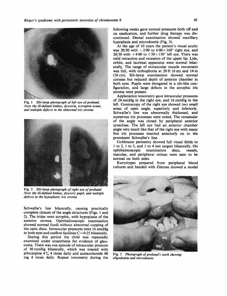
hypoplasia and microdontia (Fig. 3).' : 4g m |..............................At the age of 10 years the patient's visual acuity|...was20/50 with ±2-00 to 6 00 x 1600 right eye, and
20/30 with +4-00 to 1-50x 1500 left eye. There wasAa 0 mild retraction and recession of the upper lip. Lids,
orbits, and lacrimal apparatus were normal bilat-erally. The range of extraocular muscle movementwas full, with orthophoria at 20 ft (6 m) and 14 in(36cm). Slit-lamp examination showed normal
; corneas but reduced depth of anterior chamber in~~~~~~~~~~~both eyes. Pupils were elongated in a slit-like con-......~~~~~~~~~~~~~~~~~~~~~~~~~~~~~~~~~~~~~~~~~~~~~~~~........
;i ~~~~~~~~~Applanation tonometry gave intraocular pressures
Fig. 1 Slit-lamp photograph of left eye ofproband. of 24 mmHg in the right eye, and 16 mmHg in theNote the ill-defined limbus, dysc.oria, ectropion uveae, left. Gonioscopy of the right eye showed two smalland multiple defects in the abnormal iris stroma areas of open angle, superiorly and inferiorly.
Schwalbe's line was abnormally thickened, andnumerous iris processes were noted. The remainder
_ ~~~~~~~~~~~ofthe angle was closed by peripheral anterior_
- X s ~~~~~~synechiae. The left eye had an anterior chambert _ L angle very much like that of the right eye with many
_ ~~~~~~~~~~~fineiris processes inserted anteriorly on to the_ ~~~~~~~~~~~~prominent Schwalbe's line._ ~~~~~~~~~~~~~Goldmann perimetry showed full visual fields to
_ _ E~~~~~~~~~; i 1to 2, i to 3, and i to 4 test targets bilaterally. On__ _ ~~~~~~~~~ophthalmoscopic examination discs, vessels,
_ ~~~~~~~~~~maculae,and peripheral retinas were seen to bes _ ~~~~~~normal on both sides.
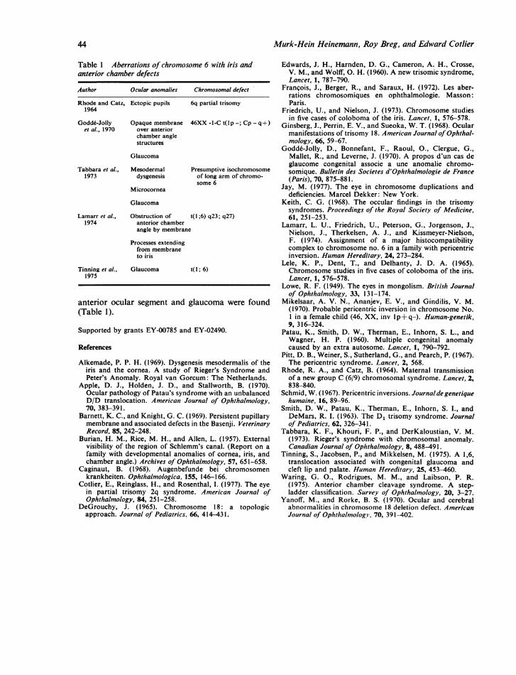
_ ~~~~~~~~~~~Karyotypesprepared from peripheral blood.cultures and banded with Giemsa showed a modal
Fig.2 Slit-lamp photograph oright eye ofproband.Notetheill-definedlimbus, dyscoric pupil, and multiple a........ke cdefects in the hypoplastic iris stroma
~~~~~~~~~~~C...Schwalbe's line bilaterally, causing practically;;0,-.complete closure of the angle structures (Figs. 1 and __
_\ ~~~~~~~~~~~~~~~~~~~~~~~~~~~~~~.
2). The irides were atrophic, withl hypoplasia of theanterior stroma. Ophthalmoscopic examination- -showed normal fundi without abnormal cupping of vthe optic discs. Intraocular pressures were 16 mmHg
examined under anaesthesia for evidence of glau-coma. There was one episode of intraocular pressure __of 50 mmHg bilaterally, which was treated withpilocarpine 4% 4 times daily and acetazolamide 60 Fig. 3 Photograph ofJproband's teeth showingmg 4 times daily. Repeat tonometry during the oligodontia and microdontia
41

Mairk-Hein Heinemann, Roy Breg, and Edward Cotlier
1 2 3
Fig. 4 Karyotype of probanid
19 20
16 17 18
21 22 x x
count of 46 with pericentric inversion of chromo-some 6 (6p +q-) (Figs. 4 and 5).
CASE 2The father of the proband is 36 years old. Visualacuity was 20/20 right eye, 20/25 left eye, withoutcorrection. External examination showed the lids,orbits, and lacrimal apparatus to be within normallimits. Slit-lamp examination revealed normal con-junctivae and corneas. Gonioscopy was remarkablefor prominent Schwalbe's line and iris moundsbilaterally. On ophthalmoscopic examination discs,vessels, maculae, and peripheral retinas were seen tobe normal on both sides. Intraocular pressures were17 mmHg right eye and 15 mmHg left eye.Karyotypes prepared from peripheral blood
cultures of the proband's father showed a pericentricinversion of chromosome 6 (6p+ q-).
Fig. 5 To right (between lines) chromosome 6 pairfrom proband showing pericentric inversion.To left, chromosome 6 pair offather with pericentricinversion
CASE 3
The proband's mother is 33 years old. Visual acuitywas 20/20 bilaterally without correction. Externalappearances of the lids, orbits, and lacrimal appara-tus were within normal limits. Slit-lamp examinationrevealed normal conjunctivae, corneas, anteriorchambers, irides, and lenses. Gonioscopy revealednormal angle structures bilaterally. On ophthalmo-scopic examination optic discs, vessels, maculae, andperipheral retinas were seen to be normal on bothsides. Intraocular pressures were 14 mmHg righteye and 14 mmHg left eye.
Karyotypes prepared from peripheral bloodcultures of the proband's mother were normal(46 XX).
Discussion
Pericentric inversions of autosomal chromosomesare rare events. Reviews of chromosomal analysesof large series of consecutive births have shown anincidence ranging from 016 to 0-56 per 1000 livebirths (Friedrich and Nielson, 1973). Ocular anoma-lies associated with pericentric inversions have beensummarised by Jay (1977). Simple and typical colo-bomata of the iris have been found in 4 cases ofpericentric inversions (Lele et al., 1965; Schmid,1967; Mikelsaar et al., 1970). Pitt (1967) presented acase of an inversion of a C-group chromosome withassociated exotropia, epicanthal folds, and hyper-telorism; however, no anomalies of the iris oranterior chamber angle were noted.
42
am :i:: im
-ii
4 5
10 12
13 :14 15

Rieger's syndrome with pericentric inversion of chromosome 6
The highly variable constellation of ocular andsystemic defects which have been found in cases ofRieger's syndrome underscores the variance ofphenotypic expression of this disorder (Alkemade,1969). Microcornea, found bilaterally in the pro-band, is a relatively uncommon finding in Rieger'ssyndrome. Only 9% of eyes in Alkemade's series(1969) were found to have corneal diameters under9 5 mm. Actual corneal dimensions are often hard todetermine accurately, as most cases of the disorderhave indistinct limbi owing to persistent corneo-scleral membranes. The proband had multiple irisabnormalities including stromal defects, hypoplasiaof the anterior stroma, and ectropion uveae. Thedyscoric, slit-like pupils are seen in most cases ofRieger's syndrome, though the large iridal defects,pseudopolycoria, such as those found in the probandare less common (Alkemade, 1969).Most pedigrees of families with primary meso-
dermal dysgenesis show considerable interfamilialvariability of phenotypic expression. The minimalocular anomalies found in the proband's father,prominent iris mounds bilaterally, may representa mild clinical manifestation of the disease. Burian(1957) reported a similar pedigree in which the fatherwas found to have anterior chamber angle structuresand irides which were interpreted as being abnormalonly after stigmata of primary mesodermal dys-genesis were discovered in 4 of his 8 children.
Defects of the anterior chamber angle and irissuch as colobomata and stromal hypoplasia arefound in many of the autosomal syndromes. Hypo-plasia of the iris is often seen in trisomy 21 in addi-tion to the characteristic Brushfield spots (Lowe,1949; Caginaut, 1968). Trisomy 13-15 is notablefor the extent of ocular involvement (Patau et al.,1960). These patients had the signs of generalisedmesodermal dysplasia. Facial cleft defects and poordifferentiation of the anterior chamber anglestructures are common (Apple et al., 1970). Theirides and choroid are usually very abnormal, oftencontaining intraocular cartilage thought to beformed from mesodermal remnants within the ciliarybody (Smith et al., 1963).The ocular anomalies associated with trisomy 18
syndrome are largely external, but intraoculardefects have been found (Edwards et al., 1960). Keith(1968) reviewed 5 cases and found microcornea,shallow anterior chambers, and abnormal ciliaryprocesses in 1 case. Ginsberg et al. (1968) reported acase with eccentric pupils and another with irissphincter atrophy, anomalous ciliary processes, andabnormal iris stroma.
Clinical syndromes involving chromosomal dele-tions also affect the iris and anterior chamber angle.Iris colobomata, sclerocornea, anterior chamber
angle malformations, and ectopic pupils have beenfound in cases of 4p- (Wolf-Hirschorn) syndrome(Frangois et al., 1972). Glaucoma is a prominentfeature of 18q- (De Grouchy) syndrome and hasbeen reported in association with anomalies of theanterior chamber angle (De Grouchy, 1965). Ringdefects of chromosome 18 (18r syndrome) are foundin patients with malformations of the anteriorchamber angle and iris hypoplasia (Yanoff andRorke, 1970).One of us (Cotlier et al., 1977) presented a pedigree
of partial trisomy 2q syndrome of which 2 membershad prominent, anteriorly displaced Schwalbe's lineand iridocorneal adhesions. Although these patientslacked the iridal stromal hypoplasia and pupillaryanomalies found in Rieger's syndrome, the abnor-malities of the anterior chamber angle and thepresence of glaucoma suggest a similar pathogenicmechanism.The pathogenesis of Rieger's syndrome and
similar ocular malformations has yet to be estab-lished. There are few animal models for the disease,though pedigrees of basenji dogs have been studiedand found to have microcornea, persistent pupillarymembranes, typical colobomata involving the iris,choroid, and retina, and sclerocorneal membranes(Barnett and Knight, 1969). One dog was also notedto have missing upper premolar teeth unilaterally.The mode of inheritance was autosomal dominant.Chromosomal analyses have not been reported.The well-documented hereditary patterns of in-
heritance of Rieger's syndrome and the associationof chromosomal defects with aberrant developmentof the anterior chamber angle and iris suggest agenetically-induced defect during early morpho-genesis involving the iridogenic mesoderm, probablyduring the 14 to 20 mm state of development. Theabnormalities of the iris, pupil, and anterior chamberangle are the result of imperfect progression of thecleavage process which forms the anterior chamber.No specific chromosome can be implicated, but thegenetic imbalance resulting from a chromosomaldefect may be enough to upset the delicate morpho-genetic process.
This case represents only the second time that achromosomal anomaly has been associated withRieger's syndrome. The case of Tabbara et al. (1973)showed ocular and systemic defects in associationwith an extra metacentric chromosome, similar to aG-chromosome, and a missing group C-chromo-some. Clinical findings included many defects alsonoted in the proposita such as bilateral defects of theanterior chamber angle structures, microcornea,dyscoria, pseudopolycoria, hypoplasia of the maxilla,and anodontia. Defects of chromosome 6 have beenassociated with 5 cases in which defects of the
43
Murk-Hein Heinemann, Roy Breg, and Edward Cotlier
Table I Aberrations of chromosome 6 with iris andanterior chamber defects
Author Ocular anomalies Chromosomal defect
Rhode and Catz, Ectopic pupils 6q partial trisomy1964
Godd6-Jolly Opaque membrane 46XX -1-C t(lp -; Cp - q+)et al., 1970 over anterior
chamber anglestructures
Glaucoma
Tabbara et al., Mesodermal Presumptive isochromosome1973 dysgenesis of long arm of chromo-
some 6Microcornea
Glaucoma
Lamarr et al., Obstruction of t(1 ;6) q23; q27)1974 anterior chamber
angle by membrane
Processes extendingfrom membraneto iris
Tinning et al., Glaucoma t(l; 6)1975
anterior ocular segment and glaucoma were found(Table 1).
Supported by grants EY-00785 and EY-02490.
References
Alkemade, P. P. H. (1969). Dysgenesis mesodermalis of theiris and the cornea. A study of Rieger's Syndrome andPeter's Anomaly. Royal van Gorcum: The Netherlands.
Apple, D. J., Holden, J. D., and Stallworth, B. (1970).Ocular pathology of Patau's syndrome with an unbalancedD/D translocation. American Journal of Ophthalmology,70, 383-391.
Barnett, K. C., and Knight, G. C. (1969). Persistent pupillarymembrane and associated defects in the Basenji. VeterinaryRecord, 85, 242-248.
Burian, H. M., Rice, M. H., and Allen, L. (1957). Externalvisibility of the region of Schlemm's canal. (Report on afamily with developmental anomalies of cornea, iris, andchamber angle.) Archives of Ophthalmology, 57, 651-658.
Caginaut, B. (1968). Augenbefunde bei chromosomenkrankheiten. Ophthalmologica, 155, 146-166.
Cotlier, E., Reinglass, H., and Rosenthal, 1. (1977). The eyein partial trisomy 2q syndrome. American Journal ofOphthalmology, 84, 251-258.
DeGrouchy, J. (1965). Chromosome 18: a topologicapproach. Journal of Pediatrics, 66, 414-431.
Edwards, J. H., Harnden, D. G., Cameron, A. H., Crosse,V. M., and Wolff, 0. H. (1960). A new trisomic syndrome,Lancet, 1, 787-790.
Franqois, J., Berger, R., and Saraux, H. (1972). Les aber-rations chromosomiques en ophthalmologie. Masson:Paris.
Friedrich, U., and Nielson, J. (1973). Chromosome studiesin five cases of coloboma of the iris. Lancet, 1, 576-578.
Ginsberg, J., Perrin, E. V., and Sueoka, W. T. (1968). Ocularmanifestations of trisomy 18. American Journal of Ophthal-mology, 66, 59-67.
Godde-Jolly, D., Bonnefant, F., Raoul, O., Clergue, G.,Mallet, R., and Leverne, J. (1970). A propos d'un cas deglaucome congenital associe a une anomalie chromo-somique. Bulletin des Societes d'Ophthalmologie de France(Paris), 70, 875-881.
Jay, M. (1977). The eye in chromosome duplications anddeficiencies. Marcel Dekker: New York.
Keith, C. G. (1968). The occular findings in the trisomysyndromes. Proceedings of the Royal Society of Medicine,61, 251-253.
Lamarr, L. U., Friedrich, U., Peterson, G., Jorgenson, J.,Nielson, J., Therkelsen, A. J., and Kissmeyer-Nielson,F. (1974). Assignment of a major histocompatibilitycomplex to chromosome no. 6 in a family with pericentricinversion. Human Hereditary, 24, 273-284.
Lele, K. P., Dent, T., and Delhanty, J. D. A. (1965).Chromosome studies in five cases of coloboma of the iris.Lancet, 1, 576-578.
Lowe, R. F. (1949). The eyes in mongolism. British Journalof Ophthalmology, 33, 131-174.
Mikelsaar, A. V. N., Ananjev, E. V., and Gindilis, V. M.(1970). Probable pericentric inversion in chromosome No.1 in a female child (46, XX, inv lp+q-). Human-genetik,9, 316-324.
Patau, K., Smith, D. W., Therman, E., Inhorn, S. L., andWagner, H. P. (1960). Multiple congenital anomalycaused by an extra autosome. Lancet, 1, 790-792.
Pitt, D. B., Weiner, S., Sutherland, G., and Pearch, P. (1967).The pericentric syndrome. Lancet, 2, 568.
Rhode, R. A., and Catz, B. (1964). Maternal transmissionof a new group C (6/9) chromosomal syndrome. Lancet, 2,838-840.
Schmid, W. (1967). Pericentric inversions. Journalde genetiquehumaine, 16, 89-96.
Smith, D. W., Patau, K., Therman, E., Inhorn, S. I., andDeMars, R. I. (1963). The D1 trisomy syndrome. Journalof Pediatrics, 62, 326-341.
Tabbara, K. F., Khouri, F. P., and DerKaloustian, V. M.(1973). Rieger's syndrome with chromosomal anomaly.Canadian Journal of Ophthalmology, 8, 488-491.
Tinning, S., Jacobsen, P., and Mikkelsen, M. (1975). A 1,6,translocation associated with congenital glaucoma andcleft lip and palate. Human Hereditary, 25, 453-460.
Waring, G. O., Rodrigues, M. M., and Laibson, P. R.(1975). Anterior chamber cleavage syndrome. A step-ladder classification. Survey of Ophthalmology, 20, 3-27.
Yanoff, M., and Rorke, B. S. (1970). Ocular and cerebralabnormalities in chromosome 18 deletion defect. AmericanJournal of Ophthalmology, 70, 391-402.
44
Related Documents