MOLECULAR AND CELLULAR BIOLOGY, June 2007, p. 4070–4081 Vol. 27, No. 11 0270-7306/07/$08.000 doi:10.1128/MCB.02011-06 Copyright © 2007, American Society for Microbiology. All Rights Reserved. RIAM Links the ADAP/SKAP-55 Signaling Module to Rap1, Facilitating T-Cell-Receptor-Mediated Integrin Activation Gae ¨l Me ´nasche ´, 1 † Stefanie Kliche, 2 † Emily J. H. Chen, 1 Theresia E. B. Stradal, 3 Burkhart Schraven, 2 and Gary Koretzky 1,4 * Signal of Transduction Program, Abramson Family Cancer Research Institute, University of Pennsylvania School of Medicine, Philadelphia, Pennsylvania 19104 1 ; Institute of Immunology, Otto von Guericke University, 39120 Magdeburg, Germany 2 ; Signaling and Motility Group, Helmholtz Center for Infection Research (HZI), 38124 Braunschweig, Germany 3 ; and Department of Pathology and Laboratory Medicine, Department of Medicine, University of Pennsylvania, Philadelphia, Pennsylvania 19104 4 Received 26 October 2006/Returned for modification 8 December 2006/Accepted 20 March 2007 One outcome of T-cell receptor (TCR) signaling is increased affinity and avidity of integrins for their ligands. This occurs through a process known as inside-out signaling, which has been shown to require several molecular components including the adapter proteins ADAP (adhesion and degranulation-promoting adapter protein) and SKAP-55 (55-kDa src kinase-associated phosphoprotein) and the small GTPase Rap1. Herein, we provide evidence linking ADAP and SKAP-55 to RIAM, a recently described adapter protein that binds selectively to active Rap1. We identified RIAM as a key component linking the ADAP/SKAP-55 module to the small GTPase Rap1, facilitating TCR-mediated integrin activation. We show that RIAM constitutively inter- acts with SKAP-55 in both a heterologous transfection system and primary T cells and map the region essential for this interaction. Additionally, we find that the SKAP-55/RIAM complex is essential both for TCR-mediated adhesion and for efficient conjugate formation between T cells and antigen-presenting cells. Mechanistic studies revealed that the ADAP/SKAP-55 module relocalized RIAM and Rap1 to the plasma membrane following TCR activation to facilitate integrin activation. These results describe for the first time a link between ADAP/SKAP-55 and the Rap1/RIAM complex and provide a potential new mechanism for TCR-mediated integrin activation. Integrins are cell surface receptors that mediate cell-cell interactions and cell-matrix interactions. These receptors are critical for T-cell migration to peripheral lymph nodes and inflammatory sites and are necessary for productive interac- tions between T cells and antigen-presenting cells (APCs) (22). The major integrins expressed on T cells are the 2 integrin LFA-1 (lymphocyte function-associated antigen 1; L2) and the 1 family of integrins (very late antigen [VLA]; 41, 51, and 61). LFA-1 binds to intercellular adhesion mol- ecule 1 (ICAM-1), ICAM-2, and ICAM-3, whereas the 1 family binds to vascular cell adhesion molecule 1 or the extra- cellular matrix protein fibronectin (12, 36). On resting T cells, integrins are maintained in an inactive state and adopt a low- affinity conformation for their ligands. T-cell receptor (TCR) engagement by a peptide-major histocompatibility complex (MHC) complex induces integrin clustering as well as a con- formational change leading to a higher-affinity state, thereby markedly enhancing the avidity of integrins for their ligands (1, 22, 32). This activation-induced modulation of integrin activa- tion via antigen receptors (or other cell surface receptors) occurs through a process known as inside-out signaling (9, 22). The molecular basis for inside-out signaling remains poorly understood; however, several proteins in the TCR signaling pathway are known to be involved (22, 39). Following engage- ment of the TCR and activation of src and syk family protein tyrosine kinases, the transmembrane adapter LAT (linker for activation of T cells) and the cytosolic adapter protein SLP-76 (76-kDa src homology 2 domain-containing leukocyte phos- phoprotein) form a scaffold to create a critical signaling complex (25, 42). Elements of this complex include ADAP (adhesion- and degranulation-promoting adapter protein) and SKAP-55 (55-kDa src kinase-associated phosphoprotein), two adapter proteins constitutively associated through the interac- tion of the SH3 (src homology 3) domain of SKAP-55 with the proline-rich domain of ADAP (29, 30). The phosphorylation of ADAP through the src kinase Fyn (3), following TCR stimu- lation, allows ADAP to interact with the SH2 domain of SLP-76 and recruits the ADAP/SKAP-55 complex to the plasma membrane via the SLP-76/GADS/LAT module (8, 33). T cells from ADAP-deficient mice show impaired LFA-1- and VLA-4-mediated adhesion, whereas the knockdown of SKAP-55 by small interfering RNA (siRNA) decreases T-cell– APC conjugation and TCR-mediated activation of LFA-1 and VLA-4 (10, 15, 23, 35). Another molecule essential for inside-out signaling from the TCR to integrins is the GTPase Rap1 (4). Like other low- molecular-weight GTP-binding proteins, Rap1 cycles between an inactive GDP-bound form and an active form associated with GTP. Activation of Rap1 has been shown to regulate T-cell adhesion and spreading. In T cells, constitutively active Rap1 increases adhesion to integrin ligands, including ICAM-1 * Corresponding author. Mailing address: Abramson Family Cancer Research Institute, University of Pennsylvania School of Medicine, 415 BRBII/III, 421 Curie Blvd., Philadelphia, PA 19104-6160. Phone: (215) 746-5524. Fax: (215) 746-5525. E-mail: [email protected] .upenn.edu. † These authors contributed equally to this work. Published ahead of print on 2 April 2007. 4070

Welcome message from author
This document is posted to help you gain knowledge. Please leave a comment to let me know what you think about it! Share it to your friends and learn new things together.
Transcript

MOLECULAR AND CELLULAR BIOLOGY, June 2007, p. 4070–4081 Vol. 27, No. 110270-7306/07/$08.00�0 doi:10.1128/MCB.02011-06Copyright © 2007, American Society for Microbiology. All Rights Reserved.
RIAM Links the ADAP/SKAP-55 Signaling Module to Rap1,Facilitating T-Cell-Receptor-Mediated Integrin Activation�
Gael Menasche,1† Stefanie Kliche,2† Emily J. H. Chen,1 Theresia E. B. Stradal,3Burkhart Schraven,2 and Gary Koretzky1,4*
Signal of Transduction Program, Abramson Family Cancer Research Institute, University of Pennsylvania School of Medicine,Philadelphia, Pennsylvania 191041; Institute of Immunology, Otto von Guericke University, 39120 Magdeburg, Germany2;
Signaling and Motility Group, Helmholtz Center for Infection Research (HZI), 38124 Braunschweig, Germany3; andDepartment of Pathology and Laboratory Medicine, Department of Medicine, University of Pennsylvania,
Philadelphia, Pennsylvania 191044
Received 26 October 2006/Returned for modification 8 December 2006/Accepted 20 March 2007
One outcome of T-cell receptor (TCR) signaling is increased affinity and avidity of integrins for their ligands.This occurs through a process known as inside-out signaling, which has been shown to require severalmolecular components including the adapter proteins ADAP (adhesion and degranulation-promoting adapterprotein) and SKAP-55 (55-kDa src kinase-associated phosphoprotein) and the small GTPase Rap1. Herein, weprovide evidence linking ADAP and SKAP-55 to RIAM, a recently described adapter protein that bindsselectively to active Rap1. We identified RIAM as a key component linking the ADAP/SKAP-55 module to thesmall GTPase Rap1, facilitating TCR-mediated integrin activation. We show that RIAM constitutively inter-acts with SKAP-55 in both a heterologous transfection system and primary T cells and map the region essentialfor this interaction. Additionally, we find that the SKAP-55/RIAM complex is essential both for TCR-mediatedadhesion and for efficient conjugate formation between T cells and antigen-presenting cells. Mechanisticstudies revealed that the ADAP/SKAP-55 module relocalized RIAM and Rap1 to the plasma membranefollowing TCR activation to facilitate integrin activation. These results describe for the first time a link betweenADAP/SKAP-55 and the Rap1/RIAM complex and provide a potential new mechanism for TCR-mediatedintegrin activation.
Integrins are cell surface receptors that mediate cell-cellinteractions and cell-matrix interactions. These receptors arecritical for T-cell migration to peripheral lymph nodes andinflammatory sites and are necessary for productive interac-tions between T cells and antigen-presenting cells (APCs) (22).The major integrins expressed on T cells are the �2 integrinLFA-1 (lymphocyte function-associated antigen 1; �L�2) andthe �1 family of integrins (very late antigen [VLA]; �4�1,�5�1, and �6�1). LFA-1 binds to intercellular adhesion mol-ecule 1 (ICAM-1), ICAM-2, and ICAM-3, whereas the �1family binds to vascular cell adhesion molecule 1 or the extra-cellular matrix protein fibronectin (12, 36). On resting T cells,integrins are maintained in an inactive state and adopt a low-affinity conformation for their ligands. T-cell receptor (TCR)engagement by a peptide-major histocompatibility complex(MHC) complex induces integrin clustering as well as a con-formational change leading to a higher-affinity state, therebymarkedly enhancing the avidity of integrins for their ligands (1,22, 32). This activation-induced modulation of integrin activa-tion via antigen receptors (or other cell surface receptors)occurs through a process known as inside-out signaling (9, 22).
The molecular basis for inside-out signaling remains poorly
understood; however, several proteins in the TCR signalingpathway are known to be involved (22, 39). Following engage-ment of the TCR and activation of src and syk family proteintyrosine kinases, the transmembrane adapter LAT (linker foractivation of T cells) and the cytosolic adapter protein SLP-76(76-kDa src homology 2 domain-containing leukocyte phos-phoprotein) form a scaffold to create a critical signalingcomplex (25, 42). Elements of this complex include ADAP(adhesion- and degranulation-promoting adapter protein) andSKAP-55 (55-kDa src kinase-associated phosphoprotein), twoadapter proteins constitutively associated through the interac-tion of the SH3 (src homology 3) domain of SKAP-55 with theproline-rich domain of ADAP (29, 30). The phosphorylation ofADAP through the src kinase Fyn (3), following TCR stimu-lation, allows ADAP to interact with the SH2 domain ofSLP-76 and recruits the ADAP/SKAP-55 complex to theplasma membrane via the SLP-76/GADS/LAT module (8, 33).T cells from ADAP-deficient mice show impaired LFA-1- andVLA-4-mediated adhesion, whereas the knockdown ofSKAP-55 by small interfering RNA (siRNA) decreases T-cell–APC conjugation and TCR-mediated activation of LFA-1 andVLA-4 (10, 15, 23, 35).
Another molecule essential for inside-out signaling from theTCR to integrins is the GTPase Rap1 (4). Like other low-molecular-weight GTP-binding proteins, Rap1 cycles betweenan inactive GDP-bound form and an active form associatedwith GTP. Activation of Rap1 has been shown to regulateT-cell adhesion and spreading. In T cells, constitutively activeRap1 increases adhesion to integrin ligands, including ICAM-1
* Corresponding author. Mailing address: Abramson Family CancerResearch Institute, University of Pennsylvania School of Medicine, 415BRBII/III, 421 Curie Blvd., Philadelphia, PA 19104-6160. Phone:(215) 746-5524. Fax: (215) 746-5525. E-mail: [email protected].
† These authors contributed equally to this work.� Published ahead of print on 2 April 2007.
4070
and fibronectin, whereas a dominant-negative Rap1 mutantblocks TCR-induced integrin activation (16, 17, 37). The mech-anism for how Rap1 regulates integrin activation is not com-pletely understood; however, recent studies have identifiedseveral effectors that bind active Rap1, including RAPL (reg-ulator for cell adhesion and polarization enriched in lymphoidtissues), PKD1 (protein kinase D1), and RIAM (Rap1-GTPinteracting adapter molecule), which appear essential for link-ing Rap1 to the integrins. RAPL and PKD1 are known toregulate the subcellular localization of Rap1 at the plasmamembrane via their associations with the �L-integrin subunitof LFA-1 (RAPL) and the �1-integrin subunit of VLA-4(PKD1) (19, 31). RIAM has been shown to regulate VLA-4integrin (�4�1) and LFA-1 integrin (�L�2) activation in Jur-kat T cells through its association with Rap1 upon TCR liga-tion (27). Additionally, RIAM has been linked to the regula-tion of actin dynamics in T cells through its interaction withEna/VASP (vasodilator-stimulated phosphoprotein) familyproteins and profilin (14, 27). Recently, RIAM has also beenshown to regulate one of the major platelet integrins, �IIb�3,through talin-mediated integrin activation (11).
Although the critical role of Rap1 in TCR-induced integrinactivation is now appreciated, exactly how TCR stimulationlinks to Rap1 activation remains unclear. One clue to thisprocess followed from the observation that the interactionbetween ADAP and SKAP-55 is critical for optimal TCR-mediated integrin activation. We have recently found that theADAP/SKAP-55 module is required for the recruitment ofRap1 to the plasma membrane (23). How the ADAP/SKAP-55complex is linked to Rap1 is the subject of this study. In thisreport we describe, for the first time, an interaction amongADAP, SKAP-55, and RIAM and demonstrate that this largercomplex is critical for efficient translocation of active Rap1 tothe membrane, facilitating TCR-mediated adhesion and con-jugate formation between T cells and APCs.
MATERIALS AND METHODS
Constructs. (i) RIAM cloning. A cDNA encoding a full-length open readingframe of RIAM was obtained by reverse transcription-PCR (RT-PCR), using theSuperScript One-Step RT-PCR System with Platinum Taq (Invitrogen) fromtotal RNA of murine T-cell blasts. The following primers were used: EcoRV-EcoRI-RIAM-Met-sense, 5�-CCGGATATCGCGAATTCTGGTCGACGTCAAGATGGGTGAATCAAATGAA-3�, and BamHI-RIAM-stop-antisense, 5�-CGCGGATCCAGAGCATCCCAAATACTAAC-3�. The digested PCR productwas cloned into pFlag-CMV-4 (Sigma) and pEGFP-C3 (Clontech).
(ii) RIAM fragment cloning. The murine RIAM domains were describedpreviously (14). We designed our RIAM fragments to include the entirety of theappropriate domains and therefore included a small number of amino acids bothamino and carboxyl terminal to the regions defined by Jenzora et al. (14). Forease of description, however, we designated these domains as the “RIAM Rasassociation (RA) domain” and “RIAM pleckstrin homology (PH) domain,”recognizing that each domain is extended slightly. These RIAM fragment do-mains were amplified by PCR using full-length RIAM as a template. The fol-lowing primers were used: EcoRI-ProI-sense, 5�-TTGAATTCTATGGGTGAATCAAATGAAGA-3�; BamHI-ProI-antisense, 5�-GGATCCCTAGGCTTCTTCTTCTTCTTTA-3�; EcoRI-RA-PH-sense, 5�-TTGAATTCTAAAGAAGAAGAAGAAGCCAAAG-3�; BamHI-RA-PH-antisense, 5�-GGATCCCTATCCAGCTCTTGCCACAG-3�; EcoRI-RA-antisense, 5�-GGATCCCTATAAGTATTCTTTGTTTTTAGCGTTC-3�; EcoRI-PH-sense, 5�-TTGAATTCTATGGAAAGCTTCTGCGGCACATCCA-3�; EcoRI-ProII-sense, 5�-TTGAATTCTGCTGTGGCAAGAGCTGG-3�; and BamHI-ProII-antisense, 5�-GGATCCCTAGGGTATATTGCCTCTTTTC-3�. The PCR products were digested with EcoRI andBamHI and cloned into pFlag-CMV-4 and pEGFP-C1 vectors (Sigma and Clon-tech), respectively.
(iii) ADAP cloning. A cDNA encoding a full-length open reading frame ofADAP (120 kDa) was obtained by RT-PCR from total RNA of murine T-cellblasts. The following primers were used: KpnI-ADAP-Met-sense, 5�-CGGTACCCCTCATGGCGAAGTTCAACACG-3�, and XhoI-ADAP-stop-antisense, 5�-CTCGAGCAGTGGAATGAACAGAGCAG-3�. The digested PCR productwas cloned into pcDNA3.1 His (Invitrogen). Human cloning of Flag-taggedADAP cDNA into pEF-BOS expression vector has been described previously(23).
(iv) SKAP-55 cloning. Cloning of full-length SKAP-55 fused with either ahemagglutinin (HA) or a Flag tag into the pEF-BOS vector has been reportedpreviously (30).
(v) SLP-76 cloning. Cloning of full-length Slp-76 fused with a Flag tag into thepEF-BOS vector has been reported previously (43).
(vi) Rap1 mutant. The cDNA of the Rap1Q63E mutant was kindly providedby Alfred Wittinghofer (Max-Planck-Institut fur Molekulare Physiologie, Dort-mund, Germany) and was used as a template to be reamplified with the followingprimers: EcoRI-Q63E(Rap1)-sense, 5�-GTAATCTTGGAATTCTGATGCGTGAGTACAAGCTAGTGGTC-3�, and BamHI-Q63E(Rap1)-antisense, 5�-GTGGATCCCTCGAGAGCTGCTGCTGACTATGGG-3�. The cDNA was subse-quently cloned into the pEGFP-C3 vector (Clontech). pGEX RalGDS-RBD waskindly provided by Johannes L. Bos (Utrecht Medical Center, Utrecht, TheNetherlands). The constructs for pDsRedC1-tagged Rap1G12V were kindlyprovided by Ignacio Rubio (University of Jena, Jena, Germany).
(vii) siRNA of SKAP-55. For siRNA of SKAP-55, the oligonucleotides SK4(5�-GAAAGAATCCTGCTTTGAA-3�) and SK1 (5�-GCGATTAGAGATCATACTA-3�) cloned into the pSuper vector (OligoEngine) were used (23). Thesame oligonucleotides, SK4 and SK1, were also cloned into pCMS3-EGFP vector(23).
Cell culture and transfection. The Jurkat (JE6.1; ATCC) T-cell line and theRaji B-cell line were cultured in RPMI 1640 (Gibco BRL) supplemented with10% fetal calf serum (FCS) at 37°C with 5% CO2. For immunofluorescence,Jurkat T cells were electroporated with 20 �g of plasmid DNA using the BTXelectroporator (310 V and 10 ms). For adhesion assays, Jurkat cells were elec-troporated using 30 �g of plasmid DNA, as previously described (23). For siRNAof SKAP-55, Jurkat T cells were transfected with 40 �g of the indicated vectorand cultured for 48 h before use. The 293T cell line was cultured in Dulbeccomodified Eagle medium (Gibco BRL) supplemented with 10% FCS and trans-fected using the liposomal transfection reagent Lipofectamine (Invitrogen). Pri-mary human T cells were prepared from a healthy donor by standard separationtechniques using AutoMACS (Miltenyi Biotech). Total mouse splenocytes werestimulated with phorbol myristate acetate (10�7 M)-ionomycin (10�6 M)(Sigma) and cultured in RPMI supplemented with 10% FCS and 0.01 M �-mer-captoethanol for 6 days in the presence of interleukin-2 (40 U/ml) to generateT-cell blasts.
Genetically altered mice. Mice made deficient in ADAP by gene targetinghave been described previously (35). Splenic T cells were prepared from wild-type or ADAP-deficient animals by standard separation techniques using Auto-MACS (Miltenyi Biotech) per the manufacturer’s instructions.
Immunoprecipitation and Western blot analysis. Flag-RIAM or fragmentsand HA/Flag-SKAP-55, His/Flag-ADAP, or GFP (green fluorescent protein)/HA/myc-Rap1 and its mutants were coexpressed in 293T cells. One day aftertransfection, cells were lysed in IPP150 lysis buffer (10 mM Tris-HCl, pH 8.0, 150mM NaCl, 0.1% NP-40 containing 2.5 mM MgCl2, when Rap1 or mutants werecoexpressed) supplemented with one protease inhibitor cocktail tablet (EDTAfree; Roche). The lysate was centrifuged at 25,000 � g for 10 min, and thesupernatant was rotated for 2 h at 4°C with anti-Flag M2 affinity gel (Sigma) oranti-HA (Roche). For the HA immunoprecipitation, protein G-agarose beads(Santa Cruz) were added for 1 h. The beads were then washed and recovered insample buffer and loaded onto a 4 to 15% gradient sodium dodecyl sulfate(SDS)-polyacrylamide gel (Bio-Rad). Anti-M2 (Sigma), anti-GFP (full length;Clontech), and anti-HA (Roche) were used to develop the Western blots. Forimmunoprecipitation of endogenous proteins, murine T-cell blasts (1.5 � 107)were rested in phosphate-buffered saline (PBS) or stimulated for 10 min with500A2 (anti-CD3; BD Pharmingen) or with 2.5 mM pervanadate (PV) for 3 min.Cells were lysed in IPP150 containing 2.5 mM MgCl2, supplemented with aprotease inhibitor cocktail tablet (EDTA free; Roche). Jurkat T cells (1.0 � 107)or primary human T cells (2.0 � 107) were left untreated or stimulated for 5 minwith monoclonal antibody (MAb) C305 (41) (provided by Arthur Weiss, Uni-versity of California, San Francisco, San Francisco, CA) or MEM92 (provided byVaclav Horejsi, Academy of Sciences of the Czech Republic, Prague, CzechRepublic) and lysed as described above. The protein concentration of the lysateswas determined using the Bradford assay (Roth), and a total of 500 �g proteinwas used for immunoprecipitation studies. Either the polyclonal rabbit antiserum
VOL. 27, 2007 ADAP/SKAP-55 RECRUIT RIAM/Rap1 FOR INTEGRIN ACTIVATION 4071
to RIAM (14) or the sheep antiserum to ADAP (33) was used for immunopre-cipitation. Immune complexes were analyzed as described above using anti-SKAP-55 (BD Pharmingen), anti-SKAP-55 MAb (23), anti-RIAM (14), anti-ADAP (Upstate), anti-ADAP (33), or anti-Rap1 (Santa Cruz) for Western blotanalysis. For proper stimulation of T cells, lysates were also analyzed for phos-pho-extracellular signal-regulated kinase 1/2 (phospho-ERK1/2) and ERK1/2(Cell Signaling).
Immunofluorescence, confocal microscopy, and quantification. TransfectedJurkat T cells were plated onto glass coverslips coated with poly-L-lysine (Sigma)for 45 min at room temperature or for 20 min at 37°C with Raji cells preincu-bated with 0.5 �g/ml of staphylococcal enterotoxin E (SEE; Toxin Technologies)as well as loaded with the cell tracker Blue CMAC to generate conjugates. Cellswere fixed in 3.7% paraformaldehyde, washed in 50 mM NH4Cl, and stained forphalloidin or HA-SKAP-55 and mounted in a medium containing N-propylgallate (Sigma). Phalloidin-Alexa Fluor 594 and 488, rabbit anti-rat immuno-globulin G (IgG)–Alexa Fluor 594, and the cell tracker Blue CMAC wereobtained from Molecular Probes. Cells were analyzed using a Nikon EclipseE800 microscope. Image analysis was performed using IPLab version 3.9.3 r4 andAdobe Photoshop 6.0 software. For each study, more than 30 transfected cellswere analyzed, and each experiment was repeated at least three times. Toexamine the effect of SKAP-55 on Rap1 localization, Jurkat T cells were trans-fected with either pCMS3E-C (scrambled) or pCMS3E-SK (an siRNA againstSKAP-55) in combination with pDsRed-tagged RapG12V. After 48 h, resting orstimulated (5 min with anti-TCR MAb C305) Jurkat T cells were seeded ontopoly-L-lysine-coated slides, fixed with 3.5% paraformaldehyde in PBS, andmounted with Mowiol (Calbiochem). Cells were imaged with a Leica TCS SP2laser scanning confocal system and analyzed using Leica software. Corel Pho-topaint histogram tools were used to obtain the number of red pixels per cell ofRap1G12V (pDsRedC1 Rap1G12V) colocalized at the plasma membrane areacompared to the total amount of pDsRedC1 Rap1G12V (Rap1 pixels) within thecell.
Purified T cells (1 � 107 cells) from wild-type or ADAP-knockout mice werepreincubated with biotinylated CD3 MAb (2C11; BD Pharmingen) for 30 min at4°C and cross-linked with streptavidin (Dianova) for 3 min at 37°C. T cells wereseeded onto poly-L-lysine-coated slides, fixed with 3.5% paraformaldehyde inPBS, permeabilized with 0.3% Triton X-100, and blocked with 5% horse serumin PBS. Endogenous RIAM was detected using a polyclonal rabbit antiserum toRIAM (14) and visualized with goat anti-rabbit antibody–fluorescein isothiocya-nate and tetramethyl rhodamine isocyanate-phalloidin (Sigma). Cells were im-aged with a Leica TCS SP2 laser scanning confocal system and analyzed usingLeica software.
Flow cytometry, TCR-mediated CD69 expression, and calcium mobilization.For flow cytometry analysis of the �2 subunit of LFA-1 (MEM48), the �1 subunitof VLA-4 (MEM101A), CD3 (OKT3; ATCC), or MHC class I (W6/32; ATCC),MAbs in combination with Cy5-conjugated anti-mouse IgG (Dianova) wereused, and samples were analyzed using a FACSCalibur flow cytometer andCellquestPro software (BD Bioscience). MEM48 and MEM101A were providedby Vaclav Horejsi (Academy of Sciences of the Czech Republic, Prague, CzechRepublic). The measurement of CD69 upregulation and calcium mobilizationhave been described elsewhere (38). For TCR-induced CD69 expression, cellswere cultured for 18 h in wells containing RPMI supplemented with 10% FCSalone (medium) or medium containing 20-ng/ml phorbol myristate acetate or inwells that had earlier been coated with the anti-TCR MAb C305. After overnightculture, cells were stained with an anti-CD69 antibody (BD Pharmingen) incombination with Cy5-conjugated anti-mouse IgG, and surface expression wasanalyzed by fluorescence-activated cell sorting (FACS). For calcium mobiliza-tion, cells were loaded with Indo-1 AM (Molecular Probes) at 37°C for 45 minin RPMI 1640 without phenol red (Gibco BRL) supplemented with 10% FCS.Cells were washed and reincubated for an additional 30 min at 37°C in the sameRPMI medium. Baseline calcium levels were measured for 30 s; cells werestimulated with the C305 antibody followed by ionomycin (1 �g/ml) treatment.Data for calcium mobilization were acquired on a FACSort LSR flow cytometer(BD Bioscience), and ratiometric analysis was performed with Flow Jo software.
Adhesion assay. Adhesion assays were performed as previously described (23,24, 34). Briefly, Jurkat T cells (1 � 106 cells per dish) were incubated with OKT3for 30 min at 37°C prior to the adhesion on fibronectin- or ICAM-1-coateddishes. The bound cell fraction was determined by counting four independentfields by microscopy using an ocular counting reticule. The induction (n-fold) ofcell binding to fibronectin was calculated as a ratio of the mean of stimulated tothe mean of unstimulated vector-transfected cells for each experiment. Theexpression of GFP or individual GFP fusion proteins of RIAM was analyzed byWestern blotting or flow cytometric analysis.
Rap1 activity assay. Rap1 activity was assessed as previously described (23, 24,34). Briefly, Jurkat T cells (5 � 106 cells) were either left untreated or stimulatedfor 5 min with the anti-TCR MAb C305 and then lysed. Activated Rap1 wasisolated using a glutathione S-transferase–RalGDS-Rap1 binding fusion protein(5), and bound Rap1 was quantified by Western blotting using anti-Rap1. Theexpression of GFP or individual GFP fusion proteins of RIAM was analyzed byWestern blot analysis.
Conjugate assay. Raji B cells were pulsed for 16 h with 2-�g/ml SEE or leftuntreated and stained with 0.5 �M of the red cell-labeling agent DDAO-SE(Molecular Probes). Jurkat T cells were incubated with an equal number of Bcells for 30 min at 37°C. Nonspecific aggregates were disrupted by vortexing, andthen cells were fixed by adding 3.5% paraformaldehyde and analyzed using aFACSCalibur flow cytometer (BD Bioscience). The percent conjugation wasdefined as the number of double-positive events in the upper right quadrantdivided by the total number of GFP-expressing cells multiplied by 100.
Isolation of plasma membrane fractions. Isolation of cytosolic and plasmamembrane fractions has been described previously (23, 24, 34). Briefly, eitherresting or stimulated (MAb C305 for 5 min) Jurkat T cells (2 � 107) or purifiedsplenic T cells (6 � 107) from mice (which for stimulation were preincubatedwith biotinylated CD3 MAb [2C11; BD Pharmingen] for 30 min at 4°C andcross-linked with streptavidin [Dianova] for 3 min at 37°C) were washed in RPMIand resuspended on ice in a hypotonic buffer. Cells were sheared; nuclei andunbroken cells were removed by low-speed centrifugation. The remaining super-natant was recentrifuged, and the cytosolic fraction (supernatant) was collected.The remaining pellet (membrane fraction) was washed twice with hypotonicbuffer (washing fraction) and finally resuspended on ice in lysis buffer as de-scribed above. To ensure equal loading, the protein concentrations of the cyto-solic and membrane fractions were determined by the Bradford assay. Westernblot analysis revealed that the membrane fraction was strongly enriched for theplasma membrane markers Na�/K� ATPase and LAT (an integral membraneprotein) and contained only minor amounts of EEA1-positive membranes(EEA1 is an endosomal marker), whereas most of the EEA1-positive mem-branes were found in the washing fraction (data not shown).
RESULTS
RIAM interacts and colocalizes with SKAP-55. Both ADAPand RIAM have been shown to interact with the actin-bindingproteins VASP and profilin, and both play critical roles inTCR-mediated integrin activation. Therefore, we speculatedthat the two adapter proteins may function in overlappingpathways to regulate integrin function in T cells. To addressthis notion, we tested if RIAM associates directly with ADAP.293T cells were cotransfected with His-tagged ADAP andFlag-tagged RIAM; however, anti-Flag immunoprecipitatesfrom the transfectants failed to show an association betweenthe two proteins (Fig. 1a, left). To confirm this finding, wecotransfected 293T cells with Flag-ADAP and GFP-RIAM andagain performed anti-Flag immunoprecipitation. As shown (Fig.1a, right), we again found no evidence of association between thetwo proteins.
Because ADAP is known to function via its association withSKAP-55, we questioned if SKAP-55 may potentially serve asa link to RIAM. When 293T cells were transfected with cDNAencoding Flag-RIAM along with a construct encoding HA-SKAP-55, anti-Flag immunoprecipitation revealed a strong in-teraction between the two proteins (Fig. 1b, left). Similarly,Flag-SKAP-55 associates with GFP-RIAM after anti-Flag im-munoprecipitation in transfected 293T cells (Fig. 1b, right). Asa specificity control, we expressed Flag-SLP-76 along withGFP-RIAM and found no interaction (Fig. 1b, right).
To extend these findings to T cells and to determine if theinteraction between SKAP-55 and RIAM is inducible, we stud-ied colocalization in transfected Jurkat cells. For these exper-iments, we expressed RIAM fused to a GFP along with HA-tagged SKAP-55. Both proteins were found diffusely in the
4072 MENASCHE ET AL. MOL. CELL. BIOL.
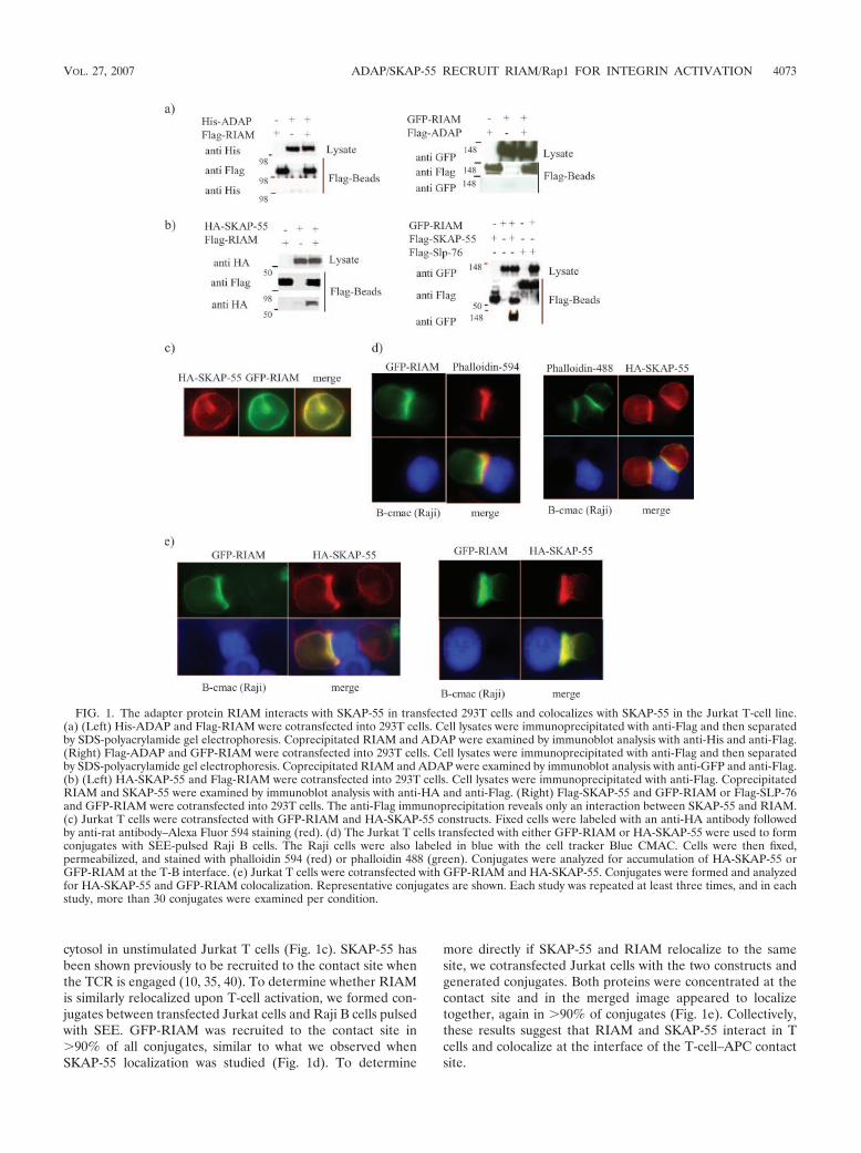
cytosol in unstimulated Jurkat T cells (Fig. 1c). SKAP-55 hasbeen shown previously to be recruited to the contact site whenthe TCR is engaged (10, 35, 40). To determine whether RIAMis similarly relocalized upon T-cell activation, we formed con-jugates between transfected Jurkat cells and Raji B cells pulsedwith SEE. GFP-RIAM was recruited to the contact site in�90% of all conjugates, similar to what we observed whenSKAP-55 localization was studied (Fig. 1d). To determine
more directly if SKAP-55 and RIAM relocalize to the samesite, we cotransfected Jurkat cells with the two constructs andgenerated conjugates. Both proteins were concentrated at thecontact site and in the merged image appeared to localizetogether, again in �90% of conjugates (Fig. 1e). Collectively,these results suggest that RIAM and SKAP-55 interact in Tcells and colocalize at the interface of the T-cell–APC contactsite.
FIG. 1. The adapter protein RIAM interacts with SKAP-55 in transfected 293T cells and colocalizes with SKAP-55 in the Jurkat T-cell line.(a) (Left) His-ADAP and Flag-RIAM were cotransfected into 293T cells. Cell lysates were immunoprecipitated with anti-Flag and then separatedby SDS-polyacrylamide gel electrophoresis. Coprecipitated RIAM and ADAP were examined by immunoblot analysis with anti-His and anti-Flag.(Right) Flag-ADAP and GFP-RIAM were cotransfected into 293T cells. Cell lysates were immunoprecipitated with anti-Flag and then separatedby SDS-polyacrylamide gel electrophoresis. Coprecipitated RIAM and ADAP were examined by immunoblot analysis with anti-GFP and anti-Flag.(b) (Left) HA-SKAP-55 and Flag-RIAM were cotransfected into 293T cells. Cell lysates were immunoprecipitated with anti-Flag. CoprecipitatedRIAM and SKAP-55 were examined by immunoblot analysis with anti-HA and anti-Flag. (Right) Flag-SKAP-55 and GFP-RIAM or Flag-SLP-76and GFP-RIAM were cotransfected into 293T cells. The anti-Flag immunoprecipitation reveals only an interaction between SKAP-55 and RIAM.(c) Jurkat T cells were cotransfected with GFP-RIAM and HA-SKAP-55 constructs. Fixed cells were labeled with an anti-HA antibody followedby anti-rat antibody–Alexa Fluor 594 staining (red). (d) The Jurkat T cells transfected with either GFP-RIAM or HA-SKAP-55 were used to formconjugates with SEE-pulsed Raji B cells. The Raji cells were also labeled in blue with the cell tracker Blue CMAC. Cells were then fixed,permeabilized, and stained with phalloidin 594 (red) or phalloidin 488 (green). Conjugates were analyzed for accumulation of HA-SKAP-55 orGFP-RIAM at the T-B interface. (e) Jurkat T cells were cotransfected with GFP-RIAM and HA-SKAP-55. Conjugates were formed and analyzedfor HA-SKAP-55 and GFP-RIAM colocalization. Representative conjugates are shown. Each study was repeated at least three times, and in eachstudy, more than 30 conjugates were examined per condition.
VOL. 27, 2007 ADAP/SKAP-55 RECRUIT RIAM/Rap1 FOR INTEGRIN ACTIVATION 4073

Mapping the region of RIAM responsible for SKAP-55 bind-ing. RIAM contains a central region with an RA domain anda PH domain flanked by two proline-rich regions (Fig. 2a). Todetermine which region of RIAM is involved in SKAP-55 bind-ing, we generated cDNAs encoding Flag-tagged fusion pro-teins of the various RIAM fragments that include each domain(Fig. 2a). These cDNAs were cotransfected into 293T cellsalong with cDNA encoding HA-SKAP-55. Cell lysates wereprepared and subjected to anti-Flag immunoprecipitation. Thecentral region of RIAM (containing the RA and PH domains)interacted with SKAP-55 (Fig. 2b). In contrast, the two pro-line-rich regions failed to demonstrate an association (Fig. 2b).Further analysis of the central region of RIAM revealed that
the RIAM RA as well as the RIAM PH domain bound inde-pendently to SKAP-55 (Fig. 2c). Consistently, we found thatthe SKAP-55/RIAM interaction could be blocked by either theisolated RA or PH domain of RIAM (Fig. 2d), indicating thatthe central region of RIAM is necessary and sufficient forSKAP-55 binding, and yet the interaction likely involves mul-tiple contact sites.
The RIAM/SKAP-55 complex associates with ADAP and theactive form of Rap1. As SKAP-55 and ADAP are known tointeract constitutively, we examined whether RIAM overex-pression impacts the binding of SKAP-55 to ADAP by express-ing GFP-RIAM, HA-SKAP-55, and Flag-ADAP in 293T cellsand then coimmunoprecipitating them with anti-Flag. Even
FIG. 2. Mapping the RIAM domains involved in SKAP-55 binding. (a) Schematic representation of the RIAM constructs used in theseexperiments. (b) Each domain of RIAM fused to a Flag tag was cotransfected with HA-SKAP-55 in 293T cells. Cell lysates were immunopre-cipitated with anti-Flag and then separated by SDS-polyacrylamide gel electrophoresis. Coprecipitated RIAM domains and SKAP-55 wereexamined by immunoblot analysis with anti-HA and anti-Flag. (c) Flag-RIAM RA or Flag-RIAM PH was cotransfected along with HA-SKAP-55.Cell lysates were immunoprecipitated with anti-Flag. Coprecipitated domains of RIAM and SKAP-55 were examined by immunoblot analysis withanti-HA and anti-Flag. (d) The HA-SKAP-55 and Flag-RIAM were cotransfected with the GFP-RIAM RA or GFP-RIAM PH domain in 293Tcells.
4074 MENASCHE ET AL. MOL. CELL. BIOL.

when highly expressed, RIAM did not interfere with the inter-action between SKAP-55 and ADAP (Fig. 3a). In fact, wefound RIAM present in the ADAP immunoprecipitates, indi-cating that although ADAP and RIAM do not associate di-rectly (Fig. 1), a trimolecular complex including ADAP,SKAP-55, and RIAM exists within the cell. We then addressedwhether active Rap1, which has been shown to associate withRIAM, still binds RIAM when RIAM is associated withSKAP-55. For these experiments, we cotransfected HA-SKAP-55, Flag-RIAM, and GFP-tagged active Rap1 (the Q63E mu-tant) into 293T cells and then immunoprecipitated them withanti-Flag. We found that active Rap1 binds RIAM to the sameextent in the presence and in the absence of SKAP-55. Addi-tionally, we found that SKAP-55 was present in the Rap1/RIAM complex (Fig. 3b). To ensure that RIAM was the onlymolecular link between ADAP/SKAP-55 and Rap1, we ad-dressed whether ADAP or SKAP-55 was able to interact indi-vidually with Rap1 and found no such association (data notshown). Since it has been shown previously that Rap1 bindsRIAM only when Rap1 is in its active conformation (27), wespeculate that one consequence of TCR engagement is theformation of a multimolecular complex including ADAP/SKAP-55/RIAM and active Rap1.
Work by others has shown that the central region of RIAM(consisting of the RA and PH domains) is involved in therecruitment of active Rap1 (27). To determine if the isolatedRA or PH domain of RIAM (either of which interferes withRIAM/SKAP-55 binding [Fig. 2d]) is also able to block forma-tion of the RIAM/Rap1 complex, 293T cells were cotrans-
fected with RIAM and active Rap1 in the absence or presenceof the RIAM RA or RIAM PH domain. The RA domain ofRIAM failed to diminish the interaction between RIAM andactive Rap1 (Fig. 3c). In contrast, however, the RIAM PHdomain did interfere with complex formation between activeRap1 and RIAM (Fig. 3d).
The endogenous ADAP/SKAP-55 complex interacts consti-tutively with RIAM in primary T cells. We next asked if theRIAM/SKAP-55 association occurs in primary T cells. Forthese studies, T-cell blasts were left resting or were activatedby cross-linking the TCR or stimulated with the phosphataseinhibitor PV. To ensure adequate T-cell activation, mitogen-associated protein kinase ERK1/2 phosphorylation was moni-tored (Fig. 4a). When cellular lysates were subjected to immu-noprecipitation with anti-RIAM, we found that SKAP-55associated with RIAM in both resting and activated cells (Fig.4a). Additionally, we found ADAP in the RIAM immunopre-cipitations, suggesting the presence of the trimolecular com-plex in both resting and activated T cells. To distinguish be-tween an ADAP/SKAP-55/RIAM module and the presence oftwo different pools of SKAP-55, one associated with ADAPand the other with RIAM, we cleared ADAP from the celllysate by immunoprecipitation and then asked if there re-mained a residual SKAP-55/RIAM association. ADAP immu-noprecipitation followed by a RIAM immunoprecipitation,while still bringing down substantial amounts of RIAM, failedto coprecipitate SKAP-55 (Fig. 4b). These results suggest thatRIAM is constitutively associated with SKAP-55 but onlywithin a trimolecular complex including ADAP.
FIG. 3. The SKAP-55/RIAM complex interacts with ADAP and the active form of Rap1. (a) Flag-ADAP, HA-SKAP-55, and GFP-RIAM weretransfected in 293T cells. Cell lysates were immunoprecipitated with anti-Flag and then examined by immunoblot analysis using anti-GFP, anti-HA,and anti-Flag. (b) GFP-Rap1Q63E, HA-SKAP-55, and Flag-RIAM were transfected in 293T cells. Cell lysates were immunoprecipitated withanti-Flag. RIAM coprecipitates were examined by immunoblot analysis with anti-GFP, anti-HA, and anti-Flag. (c) Cell lysates containingGFP-Rap1Q63E, Flag-RIAM, and GFP-RIAM RA domain were immunoprecipitated with anti-Flag. The immunoblots were revealed usinganti-GFP and anti-Flag. (d) Cell lysates containing GFP-Rap1Q63E, Flag-RIAM, and GFP-RIAM PH domain were immunoprecipitated withanti-Flag. The immunoblots were analyzed using anti-GFP and anti-Flag.
VOL. 27, 2007 ADAP/SKAP-55 RECRUIT RIAM/Rap1 FOR INTEGRIN ACTIVATION 4075
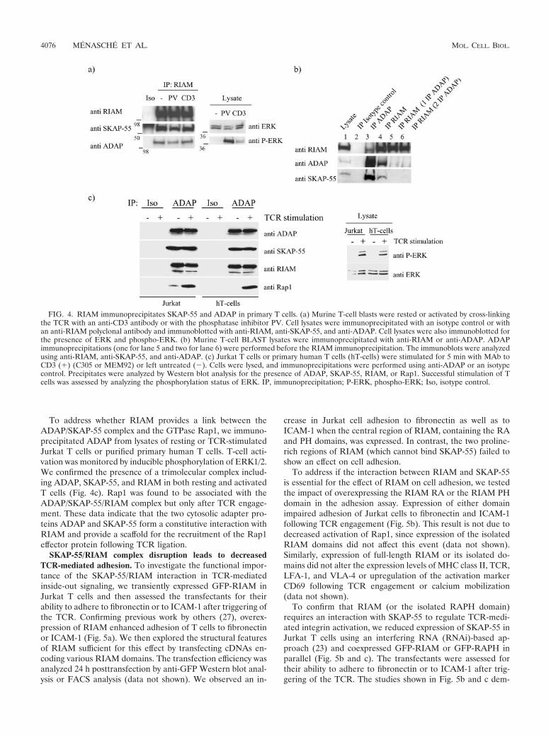
To address whether RIAM provides a link between theADAP/SKAP-55 complex and the GTPase Rap1, we immuno-precipitated ADAP from lysates of resting or TCR-stimulatedJurkat T cells or purified primary human T cells. T-cell acti-vation was monitored by inducible phosphorylation of ERK1/2.We confirmed the presence of a trimolecular complex includ-ing ADAP, SKAP-55, and RIAM in both resting and activatedT cells (Fig. 4c). Rap1 was found to be associated with theADAP/SKAP-55/RIAM complex but only after TCR engage-ment. These data indicate that the two cytosolic adapter pro-teins ADAP and SKAP-55 form a constitutive interaction withRIAM and provide a scaffold for the recruitment of the Rap1effector protein following TCR ligation.
SKAP-55/RIAM complex disruption leads to decreasedTCR-mediated adhesion. To investigate the functional impor-tance of the SKAP-55/RIAM interaction in TCR-mediatedinside-out signaling, we transiently expressed GFP-RIAM inJurkat T cells and then assessed the transfectants for theirability to adhere to fibronectin or to ICAM-1 after triggering ofthe TCR. Confirming previous work by others (27), overex-pression of RIAM enhanced adhesion of T cells to fibronectinor ICAM-1 (Fig. 5a). We then explored the structural featuresof RIAM sufficient for this effect by transfecting cDNAs en-coding various RIAM domains. The transfection efficiency wasanalyzed 24 h posttransfection by anti-GFP Western blot anal-ysis or FACS analysis (data not shown). We observed an in-
crease in Jurkat cell adhesion to fibronectin as well as toICAM-1 when the central region of RIAM, containing the RAand PH domains, was expressed. In contrast, the two proline-rich regions of RIAM (which cannot bind SKAP-55) failed toshow an effect on cell adhesion.
To address if the interaction between RIAM and SKAP-55is essential for the effect of RIAM on cell adhesion, we testedthe impact of overexpressing the RIAM RA or the RIAM PHdomain in the adhesion assay. Expression of either domainimpaired adhesion of Jurkat cells to fibronectin and ICAM-1following TCR engagement (Fig. 5b). This result is not due todecreased activation of Rap1, since expression of the isolatedRIAM domains did not affect this event (data not shown).Similarly, expression of full-length RIAM or its isolated do-mains did not alter the expression levels of MHC class II, TCR,LFA-1, and VLA-4 or upregulation of the activation markerCD69 following TCR engagement or calcium mobilization(data not shown).
To confirm that RIAM (or the isolated RAPH domain)requires an interaction with SKAP-55 to regulate TCR-medi-ated integrin activation, we reduced expression of SKAP-55 inJurkat T cells using an interfering RNA (RNAi)-based ap-proach (23) and coexpressed GFP-RIAM or GFP-RAPH inparallel (Fig. 5b and c). The transfectants were assessed fortheir ability to adhere to fibronectin or to ICAM-1 after trig-gering of the TCR. The studies shown in Fig. 5b and c dem-
FIG. 4. RIAM immunoprecipitates SKAP-55 and ADAP in primary T cells. (a) Murine T-cell blasts were rested or activated by cross-linkingthe TCR with an anti-CD3 antibody or with the phosphatase inhibitor PV. Cell lysates were immunoprecipitated with an isotype control or withan anti-RIAM polyclonal antibody and immunoblotted with anti-RIAM, anti-SKAP-55, and anti-ADAP. Cell lysates were also immunoblotted forthe presence of ERK and phospho-ERK. (b) Murine T-cell BLAST lysates were immunoprecipitated with anti-RIAM or anti-ADAP. ADAPimmunoprecipitations (one for lane 5 and two for lane 6) were performed before the RIAM immunoprecipitation. The immunoblots were analyzedusing anti-RIAM, anti-SKAP-55, and anti-ADAP. (c) Jurkat T cells or primary human T cells (hT-cells) were stimulated for 5 min with MAb toCD3 (�) (C305 or MEM92) or left untreated (�). Cells were lysed, and immunoprecipitations were performed using anti-ADAP or an isotypecontrol. Precipitates were analyzed by Western blot analysis for the presence of ADAP, SKAP-55, RIAM, or Rap1. Successful stimulation of Tcells was assessed by analyzing the phosphorylation status of ERK. IP, immunoprecipitation; P-ERK, phospho-ERK; Iso, isotype control.
4076 MENASCHE ET AL. MOL. CELL. BIOL.
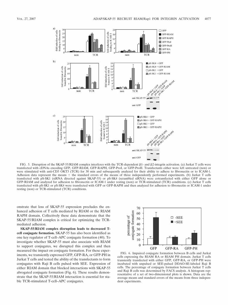
onstrate that loss of SKAP-55 expression precludes the en-hanced adhesion of T cells mediated by RIAM or the RIAMRAPH domain. Collectively these data demonstrate that theSKAP-55/RIAM complex is critical for optimizing the TCR-mediated adhesion.
SKAP-55/RIAM complex disruption leads to decreased T-cell conjugate formation. SKAP-55 has also been identified asone key regulator of T-cell–APC conjugate formation (40). Toinvestigate whether SKAP-55 must also associate with RIAMto support conjugates, we disrupted this complex and thenmeasured the impact on conjugate formation. For these exper-iments, we transiently expressed GFP, GFP-RA, or GFP-PH inJurkat T cells and tested the ability of the transfectants to formconjugates with Raji B cells pulsed with SEE. Expression ofeither RIAM domain that blocked interactions with SKAP-55abrogated conjugate formation (Fig. 6). These results demon-strate that the SKAP-55/RIAM interaction is essential for sta-ble TCR-stimulated T-cell–APC conjugates.
FIG. 5. Disruption of the SKAP-55/RIAM complex interferes with the TCR-dependent �1- and �2-integrin activation. (a) Jurkat T cells weretransfected with cDNAs encoding GFP, GFP-RIAM, GFP-RAPH, GFP-ProI, or GFP-ProII. Transfectants either were left untreated (non) orwere stimulated with anti-CD3 OKT3 (TCR) for 30 min and subsequently analyzed for their ability to adhere to fibronectin or to ICAM-1.Adhesion data represent the means the standard errors of the means of three independently performed experiments. (b) Jurkat T cellstransfected with pS-SK1 (siRNA directed against SKAP-55) or pS-SK4 (scrambled siRNA) were cotransfected with either GFP alone orGFP-RIAM and analyzed for adhesion to fibronectin or ICAM-1 under resting (non) or TCR-stimulated (TCR) conditions. (c) Jurkat T cellstransfected with pS-SK1 or pS-SK4 were transfected with GFP or GFP-RAPH and then analyzed for adhesion to fibronectin or ICAM-1 underresting (non) or TCR-stimulated (TCR) conditions.
FIG. 6. Impaired conjugate formation between B cells and Jurkatcells expressing the RIAM RA or RIAM PH domain. Jurkat T cellstransiently transfected with either GFP, GFP-RA, or GFP-PH wereincubated with unpulsed or SEE-pulsed DDAO-SE-labeled Raji Bcells. The percentage of conjugate formation between Jurkat T cellsand Raji B cells was determined by FACS analysis. A histogram rep-resentative of a set of two-dimensional plots is shown. Data are theaverage means and standard errors of the means from three indepen-dent experiments.
VOL. 27, 2007 ADAP/SKAP-55 RECRUIT RIAM/Rap1 FOR INTEGRIN ACTIVATION 4077
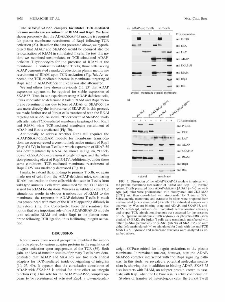
The ADAP/SKAP-55 complex facilitates TCR-mediatedplasma membrane recruitment of RIAM and Rap1. We haveshown previously that the ADAP/SKAP-55 module is requiredfor plasma membrane recruitment of Rap1 following TCRactivation (23). Based on the data presented above, we hypoth-esized that ADAP and SKAP-55 would be required also forrelocalization of RIAM in stimulated T cells. To test this no-tion, we examined unstimulated or TCR-stimulated ADAP-deficient T lymphocytes for the presence of RIAM at themembrane. In contrast to wild-type T cells, those cells lackingADAP demonstrated a marked reduction in plasma membranerecruitment of RIAM upon TCR activation (Fig. 7a). As ex-pected, the TCR-mediated increase in membrane targeting ofRap1 seen in ADAP-deficient T cells was also attenuated.
We and others have shown previously (13, 23) that ADAPexpression appears to be required for stable expression ofSKAP-55. Thus, in our experiment using ADAP-deficient cells,it was impossible to determine if failed RIAM and Rap1 mem-brane recruitment was due to loss of ADAP or SKAP-55. Totest more directly the importance of SKAP-55 in this process,we made further use of Jurkat cells transfected with the RNAitargeting SKAP-55. As shown, “knockdown” of SKAP-55 mark-edly attenuates TCR-mediated membrane targeting of both Rap1and RIAM, while TCR-mediated membrane recruitment ofADAP and Ras is unaffected (Fig. 7b).
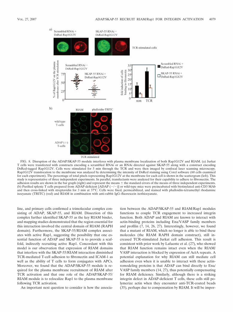
Additionally, to address whether Rap1 still requires theADAP/SKAP-55/RIAM module for membrane transloca-tion, we overexpressed a constitutively active mutant of Rap1(Rap1G12V) in Jurkat T cells in which expression of SKAP-55was downregulated by RNAi. As shown in Fig. 8a, “knock-down” of SKAP-55 expression strongly antagonized the adhe-sion-promoting effect of Rap1G12V. Additionally, under thesesame conditions, TCR-mediated membrane recruitment ofRap1G12V was markedly decreased (Fig. 8a).
Finally, to extend these findings to primary T cells, we againmade use of cells from the ADAP-deficient mice, comparingRIAM localization in these cells with that seen in T cells fromwild-type animals. Cells were stimulated via the TCR and as-sessed for RIAM localization. Whereas in wild-type cells TCRstimulation results in obvious translocation of RIAM to themembrane, the response in ADAP-deficient T cells is muchless pronounced, with most of the RIAM appearing diffusely inthe cytosol (Fig. 8b). Collectively, these data reinforce thenotion that one important role of the ADAP/SKAP-55 moduleis to relocalize RIAM and active Rap1 to the plasma mem-brane following TCR ligation, thus facilitating integrin activa-tion.
DISCUSSION
Recent work from several groups has identified the impor-tant role played by various adapter proteins in the regulation ofintegrin activation upon engagement of the TCR (39). Bothgain- and loss-of-function studies of primary T cells have dem-onstrated that ADAP and SKAP-55 are two such criticaladapters for TCR-mediated inside-out-signaling of integrins(10, 35, 40). It appears that the constitutive association ofADAP with SKAP-55 is critical for their effect on integrinfunction (23). One role for the ADAP/SKAP-55 complex ap-pears to be recruitment of activated Rap1, a low-molecular-
weight GTPase critical for integrin activation, to the plasmamembrane. It remained unclear, however, how the ADAP/SKAP-55 complex intersected with the Rap1 signaling path-way. In this study, we revealed a potential molecular mecha-nism by showing that in addition to binding ADAP, SKAP-55also interacts with RIAM, an adapter protein known to asso-ciate with Rap1 when the GTPase is in its active conformation.
Studies of transfected heterologous cells, the Jurkat T-cell
FIG. 7. Disruption of the ADAP/SKAP-55 module interferes withthe plasma membrane localization of RIAM and Rap1. (a) Purifiedsplenic T cells prepared from ADAP-deficient [ADAP (�/�)] or wild-type (wt) mice were preincubated with biotinylated anti-CD3 MAb(2C11) and then cross-linked with streptavidin for 3 min at 37°C.Subsequently, membrane and cytosolic fractions were prepared fromunstimulated (�) or stimulated (�) cells. The individual samples wereanalyzed by Western blotting using anti-ADAP, anti-SKAP-55, anti-RIAM, anti-Rap1, and anti-Ras. To control the fractionation efficiencyand proper TCR stimulation, fractions were assessed for the presenceof LAT (plasma membrane), ERK (cytosol), or phospho-ERK (stim-ulation) (P-ERK). (b) Jurkat T cells were transiently transfected witheither pS-SK4 (scrambled) or pS-SK1 (siRNA of SKAP-55) or wereeither left unstimulated (�) or stimulated for 5 min with the anti-TCRMAb C305. Cytosolic and membrane fractions were analyzed as de-scribed above.
4078 MENASCHE ET AL. MOL. CELL. BIOL.
line, and primary cells confirmed a trimolecular complex con-sisting of ADAP, SKAP-55, and RIAM. Dissection of thiscomplex further identified SKAP-55 as the key RIAM binder,and mapping studies demonstrated that the region essential forthis interaction involved the central domain of RIAM (RAPHdomain). Furthermore, the SKAP-55/RIAM complex associ-ates with active Rap1, suggesting the possibility that one es-sential function of ADAP and SKAP-55 is to provide a scaf-fold, indirectly recruiting active Rap1. Concordant with thismodel is our observation that expression of RIAM domainsthat interfere with the SKAP-55/RIAM interaction diminishedTCR-mediated T-cell adhesion to fibronectin and ICAM-1 aswell as the ability of T cells to form conjugates with APCs.Moreover, we found that the ADAP/SKAP-55 module is re-quired for the plasma membrane recruitment of RIAM afterTCR activation and that one role of the ADAP/SKAP-55/RIAM module is to relocalize Rap1 to the plasma membranefollowing TCR activation.
An important next question to consider is how the associa-
tion between the ADAP/SKAP-55 and RIAM/Rap1 modulesfunctions to couple TCR engagement to increased integrinfunction. Both ADAP and RIAM are known to interact withactin-binding proteins including Ena/VASP family membersand profilin (7, 14, 26, 27). Interestingly, however, we foundthat a mutant of RIAM, which no longer is able to bind thesemolecules (the RIAM RAPH domain construct), still in-creased TCR-stimulated Jurkat cell adhesion. This result isconsistent with prior work by Lafuente et al. (27), who showedthat RIAM function remains intact even when the RIAM/VASP interaction is blocked by expression of ActA repeats. Apotential explanation for why RIAM can still mediate celladhesion even when it is unable to interact with these actin-remodeling proteins is that ADAP can bind directly to Ena/VASP family members (14, 27), thus potentially compensatingfor RIAM deficiency. Similarly, although there is a strikingintegrin defect in ADAP-deficient T cells, these cells still po-lymerize actin when they encounter anti-TCR-coated beads(35), perhaps due to compensation by RIAM. It will be impor-
FIG. 8. Disruption of the ADAP/SKAP-55 module interferes with plasma membrane localization of both Rap1G12V and RIAM. (a) JurkatT cells were transfected with constructs encoding a scrambled RNAi or an RNAi directed against SKAP-55 along with a construct encodingDsRed-tagged Rap1G12V. Cells were stimulated for 5 min through the TCR and were then imaged by confocal laser scanning microscopy.Rap1G12V translocation to the membrane was analyzed by determining the intensity of DsRed staining using Corel software (60 cells examinedfor each experiment). The percentage of total pixels representing Rap1G12V at the membrane for each cell is shown in the scattergram (left). Thisstudy is representative of three independent experiments. In parallel, transfectants were analyzed for their capability to adhere to fibronectin. Theadhesion results are shown in the bar graph (right) and represent the means the standard errors of the means of three independent experiments.(b) Purified splenic T cells prepared from ADAP-deficient [ADAP (�/�)] or wild-type mice were preincubated with biotinylated anti-CD3 MAband then cross-linked with streptavidin for 3 min at 37°C. Cells were fixed, permeabilized, and stained with phalloidin-tetramethyl rhodamineisocyanate (TRITC) (red) and RIAM in combination with anti-rabbit IgG–fluorescein isothiocyanate.
VOL. 27, 2007 ADAP/SKAP-55 RECRUIT RIAM/Rap1 FOR INTEGRIN ACTIVATION 4079

tant to generate cells in which both ADAP and RIAM areabsent to determine the effect of this combined deficiency onactin dynamics.
We were intrigued by the finding that the PH domain ofRIAM contributes to such a great extent to SKAP-55 binding.Although PH domains are best known for phosphoinositideinteractions, recent studies have indicated that the specificityof membrane targeting by PH domains may be conferred byprotein interactions, either alone or together with phospholip-ids (21, 28). In vitro binding studies have revealed that the PHdomain of RIAM displays highest affinity for phosphatidylino-sitol monophosphates (14). However, we found that the tan-dem RA and PH domains of RIAM are not sufficient formembrane targeting, as the deficiency of SKAP-55 results inretention of RIAM in the cytosol following TCR engagement(Fig. 7). Thus, a GTP-Rap1/RIAM module alone is not suffi-cient for membrane targeting after TCR stimulation, as a re-quirement for both ADAP and SKAP-55 remains.
Although apparently necessary for the recruitment of activeRap1 to the plasma membrane, the ADAP/SKAP-55/RIAMmodule is not needed for activation of the GTPase followingTCR engagement. This finding was shown in our previousstudy using primary T cells obtained from ADAP-deficientmice and in Jurkat T cells in which SKAP-55 was knockeddown using siRNAs (23). Previous work by others has shownthat RIAM is required for localization of Rap1-GTP at theplasma membrane (27). In our current study, expression of theisolated RA or PH domain of RIAM (either of which abro-gates the SKAP-55/RIAM interaction) had no effect on TCR-mediated activation of Rap1. However, “knockdown” of SKAP-55 in Jurkat cells markedly reduced the inducible relocalizationof active Rap1 to the plasma membrane from the cytosol.Collectively, these results demonstrate that the role of theADAP/SKAP-55/RIAM module is not to activate Rap1 butrather to promote its translocation to the plasma membraneupon TCR engagement.
An important question to consider is how Rap1 regulatesintegrin function in T cells. One attractive candidate is RAPL,a Rap1-binding effector protein that mediates Rap1-depen-dent integrin activation (18–20). Although the mechanism bywhich RAPL functions in this pathway is not completely clear,RAPL has been shown to coimmunoprecipitate with LFA-1 inthe presence of active Rap1, providing an indirect link betweenRap1 and an important T-cell integrin (19). It remains unclear,however, whether RIAM works synergistically with RAPL tobring active Rap1 to the plasma membrane or whether thesetwo proteins function in independent pathways. Another po-tential effector of active Rap1 is PKD1, which has recentlybeen implicated in integrin function in T cells as having anadapter-like role linking active Rap1 to �1 integrins (31). Fur-ther studies are required to determine how RAPL and PKD1intersect with the ADAP/SKAP-55/RIAM signaling cascade tofacilitate integrin activation.
In conclusion, our work suggests one means by which TCRsignaling may utilize a network of adapter molecules to bringactive Rap1 to its site of action. Our model suggests that TCRengagement activates Rap1 in an ADAP/SKAP-55/RIAM-in-dependent fashion. Previous work by others suggests a numberof possible links between the TCR and Rap1 activation, in-cluding several guanine nucleotide exchange factors such as
C3G and CalDAG-GEFI, which are activated downstream ofFyn and phospholipase C, respectively (2, 6). However, ourdata indicate that, although not required for its activation,Rap1 cannot be recruited to its site of action without theADAP/SKAP-55/RIAM complex. Additional studies are nowrequired to determine the mechanism(s) by which other keyeffector molecules, for example, RAPL and PKD1, function inthis pathway to optimize TCR-mediated integrin function.
ACKNOWLEDGMENTS
We thank Arthur Weiss, Alfred Wittinghofer, Johannes L. Bos,Ignacio Rubio, and Vaclav Horejsi for providing reagents and MarthaJordan and Marisa Juntilla for critical reading of the manuscript. Wethank A. Nehring and J. Hoppe for excellent technical assistance.
This work was supported by long-term postdoctoral fellowships fromthe European Molecular Biology Organization (G.M.), by DeutscheForschungsgemeinschaft grants KL1292/5-1 and GRK1167 (S.K. andB.S.), and by the U.S. National Institutes of Health (G.K.).
REFERENCES
1. Bazzoni, G., and M. E. Hemler. 1998. Are changes in integrin affinity andconformation overemphasized? Trends Biochem. Sci. 23:30–34.
2. Bivona, T. G., H. H. Wiener, I. M. Ahearn, J. Silletti, V. K. Chiu, and M. R.Philips. 2004. Rap1 up-regulation and activation on plasma membrane reg-ulates T cell adhesion. J. Cell Biol. 164:461–470.
3. Boerth, N. J., B. A. Judd, and G. A. Koretzky. 2000. Functional associationbetween SLAP-130 and SLP-76 in Jurkat T cells. J. Biol. Chem. 275:5143–51452.
4. Bos, J. L. 2005. Linking Rap to cell adhesion. Curr. Opin. Cell Biol. 17:123–128.
5. Bos, J. L., B. Franke, L. M’Rabet, K. Reedquist, and F. Zwartkruis. 1997. Insearch of a function for the Ras-like GTPase Rap1. FEBS Lett. 410:59–62.
6. Boussiotis, V. A., G. J. Freeman, A. Berezovskaya, D. L. Barber, and L. M.Nadler. 1997. Maintenance of human T cell anergy: blocking of IL-2 genetranscription by activated Rap1. Science 278:124–128.
7. Coppolino, M. G., M. Krause, P. Hagendorff, D. A. Monner, W. Trimble, S.Grinstein, J. Wehland, and A. S. Sechi. 2001. Evidence for a molecularcomplex consisting of Fyb/SLAP, SLP-76, Nck, VASP and WASP that linksthe actin cytoskeleton to Fcgamma receptor signalling during phagocytosis.J. Cell Sci. 114:4307–4318.
8. da Silva, A. J., Z. Li, C. de Vera, E. Canto, P. Findell, and C. E. Rudd. 1997.Cloning of a novel T-cell protein FYB that binds FYN and SH2-domain-containing leukocyte protein 76 and modulates interleukin 2 production.Proc. Natl. Acad. Sci. USA 94:7493–7498.
9. Dustin, M. L., and T. A. Springer. 1989. T-cell receptor cross-linking tran-siently stimulates adhesiveness through LFA-1. Nature 341:619–624.
10. Griffiths, E. K., C. Krawczyk, Y. Y. Kong, M. Raab, S. J. Hyduk, D. Bouchard,V. S. Chan, I. Kozieradzki, A. J. Oliveira-Dos-Santos, A. Wakeham, P. S.Ohashi, M. I. Cybulsky, C. E. Rudd, and J. M. Penninger. 2001. Positiveregulation of T cell activation and integrin adhesion by the adapter Fyb/Slap.Science 293:2260–2263.
11. Han, J., C. J. Lim, N. Watanabe, A. Soriani, B. Ratnikov, D. A. Calderwood,W. Puzon-McLaughlin, E. M. Lafuente, V. A. Boussiotis, S. J. Shattil, andM. H. Ginsberg. 2006. Reconstructing and deconstructing agonist-inducedactivation of integrin alphaIIbbeta3. Curr. Biol. 16:1796–1806.
12. Hogg, N., M. Laschinger, K. Giles, and A. McDowall. 2003. T-cell integrins:more than just sticking points. J. Cell Sci. 116:4695–4705.
13. Huang, Y., D. D. Norton, P. Precht, J. L. Martindale, J. K. Burkhardt, andR. L. Wange. 2005. Deficiency of ADAP/Fyb/SLAP-130 destabilizes SKAP55in Jurkat T cells. J. Biol. Chem. 280:23576–23583.
14. Jenzora, A., B. Behrendt, J. V. Small, J. Wehland, and T. E. Stradal. 2005.PREL1 provides a link from Ras signalling to the actin cytoskeleton viaEna/VASP proteins. FEBS Lett. 579:455–463.
15. Jo, E. K., H. Wang, and C. E. Rudd. 2005. An essential role for SKAP-55 inLFA-1 clustering on T cells that cannot be substituted by SKAP-55R. J. Exp.Med. 201:1733–1739.
16. Katagiri, K., M. Hattori, N. Minato, S. Irie, K. Takatsu, and T. Kinashi.2000. Rap1 is a potent activation signal for leukocyte function-associatedantigen 1 distinct from protein kinase C and phosphatidylinositol-3-OHkinase. Mol. Cell. Biol. 20:1956–1969.
17. Katagiri, K., M. Hattori, N. Minato, and T. Kinashi. 2002. Rap1 functions asa key regulator of T-cell and antigen-presenting cell interactions and mod-ulates T-cell responses. Mol. Cell. Biol. 22:1001–1015.
18. Katagiri, K., M. Imamura, and T. Kinashi. 2006. Spatiotemporal regulationof the kinase Mst1 by binding protein RAPL is critical for lymphocytepolarity and adhesion. Nat. Immunol. 7:919–928.
4080 MENASCHE ET AL. MOL. CELL. BIOL.
19. Katagiri, K., A. Maeda, M. Shimonaka, and T. Kinashi. 2003. RAPL, aRap1-binding molecule that mediates Rap1-induced adhesion through spa-tial regulation of LFA-1. Nat. Immunol. 4:741–748.
20. Katagiri, K., N. Ohnishi, K. Kabashima, T. Iyoda, N. Takeda, Y. Shinkai, K.Inaba, and T. Kinashi. 2004. Crucial functions of the Rap1 effector moleculeRAPL in lymphocyte and dendritic cell trafficking. Nat. Immunol. 5:1045–1051.
21. Kavran, J. M., D. E. Klein, A. Lee, M. Falasca, S. J. Isakoff, E. Y. Skolnik,and M. A. Lemmon. 1998. Specificity and promiscuity in phosphoinositidebinding by pleckstrin homology domains. J. Biol. Chem. 273:30497–30508.
22. Kinashi, T. 2005. Intracellular signalling controlling integrin activation inlymphocytes. Nat. Rev. Immunol. 5:546–559.
23. Kliche, S., D. Breitling, M. Togni, R. Pusch, K. Heuer, X. Wang, C. Freund,A. Kasirer-Friede, G. Menasche, G. A. Koretzky, and B. Schraven. 2006. TheADAP/SKAP55 signaling module regulates T-cell receptor-mediated inte-grin activation through plasma membrane targeting of Rap1. Mol. Cell. Biol.26:7130–7144.
24. Kolanus, W., W. Nagel, B. Schiller, L. Zeitlmann, S. Godar, H. Stockinger,and B. Seed. 1996. Alpha L beta 2 integrin/LFA-1 binding to ICAM-1induced by cytohesin-1, a cytoplasmic regulatory molecule. Cell 86:233–242.
25. Koretzky, G. A., and P. S. Myung. 2001. Positive and negative regulation ofT-cell activation by adaptor proteins. Nat. Rev. Immunol. 1:95–107.
26. Krause, M., A. S. Sechi, M. Konradt, D. Monner, F. B. Gertler, and J. Wehland.2000. Fyn-binding protein (Fyb)/SLP-76-associated protein (SLAP), Ena/vasodilator-stimulated phosphoprotein (VASP) proteins and the Arp2/3complex link T cell receptor (TCR) signaling to the actin cytoskeleton.J. Cell Biol. 149:181–194.
27. Lafuente, E. M., A. A. van Puijenbroek, M. Krause, C. V. Carman, G. J.Freeman, A. Berezovskaya, E. Constantine, T. A. Springer, F. B. Gertler, andV. A. Boussiotis. 2004. RIAM, an Ena/VASP and profilin ligand, interactswith Rap1-GTP and mediates Rap1-induced adhesion. Dev. Cell 7:585–595.
28. Lemmon, M. A. 2004. Pleckstrin homology domains: not just for phospho-inositides. Biochem. Soc. Trans. 32:707–711.
29. Liu, J., H. Kang, M. Raab, A. J. da Silva, S. K. Kraeft, and C. E. Rudd. 1998.FYB (FYN binding protein) serves as a binding partner for lymphoid proteinand FYN kinase substrate SKAP55 and a SKAP55-related protein in T cells.Proc. Natl. Acad. Sci. USA 95:8779–8784.
30. Marie-Cardine, A., L. R. Hendricks-Taylor, N. J. Boerth, H. Zhao, B.Schraven, and G. A. Koretzky. 1998. Molecular interaction between theFyn-associated protein SKAP55 and the SLP-76-associated phosphoproteinSLAP-130. J. Biol. Chem. 273:25789–25795.
31. Medeiros, R. B., D. M. Dickey, H. Chung, A. C. Quale, L. R. Nagarajan, D. D.
Billadeau, and Y. Shimizu. 2005. Protein kinase D1 and the beta 1 integrincytoplasmic domain control beta 1 integrin function via regulation of Rap1activation. Immunity 23:213–226.
32. Mueller, K. L., M. A. Daniels, A. Felthauser, C. Kao, S. C. Jameson, and Y.Shimizu. 2004. Cutting edge: LFA-1 integrin-dependent T cell adhesion isregulated by both ag specificity and sensitivity. J. Immunol. 173:2222–2226.
33. Musci, M. A., L. R. Hendricks-Taylor, D. G. Motto, M. Paskind, J. Kamens,C. W. Turck, and G. A. Koretzky. 1997. Molecular cloning of SLAP-130, anSLP-76-associated substrate of the T cell antigen receptor-stimulated proteintyrosine kinases. J. Biol. Chem. 272:11674–11677.
34. Nagel, W., L. Zeitlmann, P. Schilcher, C. Geiger, J. Kolanus, and W. Kolanus.1998. Phosphoinositide 3-OH kinase activates the beta2 integrin adhesionpathway and induces membrane recruitment of cytohesin-1. J. Biol. Chem.273:14853–14861.
35. Peterson, E. J., M. L. Woods, S. A. Dmowski, G. Derimanov, M. S. Jordan,J. N. Wu, P. S. Myung, Q. H. Liu, J. T. Pribila, B. D. Freedman, Y. Shimizu,and G. A. Koretzky. 2001. Coupling of the TCR to integrin activation bySlap-130/Fyb. Science 293:2263–2265.
36. Pribila, J. T., A. C. Quale, K. L. Mueller, and Y. Shimizu. 2004. Integrins andT cell-mediated immunity. Annu. Rev. Immunol. 22:157–180.
37. Sebzda, E., M. Bracke, T. Tugal, N. Hogg, and D. A. Cantrell. 2002. Rap1Apositively regulates T cells via integrin activation rather than inhibitinglymphocyte signaling. Nat. Immunol. 3:251–258.
38. Singer, A. L., S. C. Bunnell, A. E. Obstfeld, M. S. Jordan, J. N. Wu, P. S.Myung, L. E. Samelson, and G. A. Koretzky. 2004. Roles of the proline-richdomain in SLP-76 subcellular localization and T cell function. J. Biol. Chem.279:15481–15490.
39. Togni, M., J. Lindquist, A. Gerber, U. Kolsch, A. Hamm-Baarke, S. Kliche,and B. Schraven. 2004. The role of adaptor proteins in lymphocyte activa-tion. Mol. Immunol. 41:615–630.
40. Wang, H., E. Y. Moon, A. Azouz, X. Wu, A. Smith, H. Schneider, N. Hogg,and C. E. Rudd. 2003. SKAP-55 regulates integrin adhesion and formationof T cell-APC conjugates. Nat. Immunol. 4:366–374.
41. Weiss, A., and J. D. Stobo. 1984. Requirement for the coexpression of T3 andthe T cell antigen receptor on a malignant human T cell line. J. Exp. Med.160:1284–1299.
42. Wu, J. N., and G. A. Koretzky. 2004. The SLP-76 family of adapter proteins.Semin. Immunol. 16:379–393.
43. Zhong, X. P., E. A. Hainey, B. A. Olenchock, H. Zhao, M. K. Topham, andG. A. Koretzky. 2002. Regulation of T cell receptor-induced activation of theRas-ERK pathway by diacylglycerol kinase zeta. J. Biol. Chem. 277:31089–31098.
VOL. 27, 2007 ADAP/SKAP-55 RECRUIT RIAM/Rap1 FOR INTEGRIN ACTIVATION 4081
Related Documents