Plant and Soil 194: 81–98, 1997. 81 c 1997 Kluwer Academic Publishers. Printed in the Netherlands. Rhizobial communication with rice roots: Induction of phenotypic changes, mode of invasion and extent of colonization P.M. Reddy 1 , J.K. Ladha 14 , R.B. So 1 , R.J. Hernandez 1 , M.C. Ramos 1 , O.R. Angeles 1 , F.B. Dazzo and Frans J. de Bruijn 23 1 International Rice Research Institute, P.O. Box 933, Manila 1099, Philippines, 2 Department of Microbiology and 3 DOE Plant Research Laboratory, Michigan State University, East Lansing, MI 48824, USA. 4 Corresponding author Key words: colonization, indole-3-acetic acid, invasion, Nod factors, nod gene induction, rhizobia, rhizobial attachment, rice, thick short lateral roots, trans-zeatin Abstract Legume-rhizobial interactions culminate in the formation of structures known as nodules. In this specialized niche, rhizobia are insulated from microbial competition and fix nitrogen which becomes directly available to the legume plant. It has been a long-standing goal in the field of biological nitrogen fixation to extend the nitrogen- fixing symbiosis to non-nodulated cereal plants, such as rice. To achieve this goal, extensive knowledge of the legume-rhizobia symbioses should help in formulating strategies for developing potential rice-rhizobia symbioses or endophytic interactions. As a first step to assess opportunities for developing a rice-rhizobia symbiosis, we evaluated certain aspects of rice-rhizobia associations to determine the extent of predisposition of rice roots for forming an intimate association with rhizobia. Our studies indicate that: a. Rice root exudates do not activate the expression of nodulation genes such as nodY of Bradyrhizobium japonicum USDA110, nodA of R. leguminosarum bv. trifolii, or nodSU of Rhizobium. sp. NGR234; b. Neither viable wild-type rhizobia, nor purified chitolipooligosaccharide (CLOS) Nod factors elicit root hair deformation or true nodule formation in rice; c. Rhizobia-produced indole-3- acetic acid, but neither trans-zeatin nor CLOS Nod factors, seem to promote the formation of thick, short lateral roots in rice; d. Rhizobia develop neither the symbiont-specific pattern of root hair attachment nor extensive cellulose microfibril production on the rice root epidermis; e. A primary mode of rhizobial invasion of rice roots is through cracks in the epidermis and fissures created during emergence of lateral roots; f. This infection process is nod-gene independent, nonspecific, and does not involve the formation of infection threads; g. Endophytic colonization observed so far is restricted to intercellular spaces or within host cells undergoing lysis. h. The cortical sclerenchymatous layer containing tightly packed, thick walled fibers appears to be a significant barrier that restricts rhizobial invasion into deeper layers of the root cortex. Therefore, we conclude that the molecular and cell biology of the Rhizobium-rice association differs in many respects from the biology underlying the development of root nodules in the Rhizobium-legume symbiosis. Introduction Nitrogen supply is critical for attaining yield potential in any crop. In rice, it takes 1 kg of nitrogen to produce 15-20 kg of grain. Lowland rice in the tropics can utilize the nitrogen that is naturally available in the soil through continuous biological nitrogen fixation (BNF) to produce 2-3 tons of grain per hectare. However, FAX No: +6328911292. E-mail: [email protected] additional nitrogen must be applied to obtain higher yields. Rice suffers from a mismatch of its nitrogen demand and its nitrogen supplied as chemical fertilizer, resulting in a 50-70% loss of the fertilizer applied. As pointed out in the introductory chapter, two approach- es may be used to try to solve this problem. One is to regulate the timing of nitrogen application based on the plant’s needs, thus increasing the efficiency of the plant’s use of applied nitrogen (Cassman et al., ICPC: PIPS No.: 142992 BIO2KAP

Welcome message from author
This document is posted to help you gain knowledge. Please leave a comment to let me know what you think about it! Share it to your friends and learn new things together.
Transcript
Plant and Soil 194: 81–98, 1997. 81c 1997 Kluwer Academic Publishers. Printed in the Netherlands.
Rhizobial communication with rice roots: Induction of phenotypic changes,mode of invasion and extent of colonization
P.M. Reddy1, J.K. Ladha1;4, R.B. So1, R.J. Hernandez1, M.C. Ramos1, O.R. Angeles1,F.B. Dazzo and Frans J. de Bruijn2;3
1International Rice Research Institute, P.O. Box 933, Manila 1099, Philippines, 2Department of Microbiology and3DOE Plant Research Laboratory, Michigan State University, East Lansing, MI 48824, USA. 4Correspondingauthor�
Key words: colonization, indole-3-acetic acid, invasion, Nod factors, nod gene induction, rhizobia, rhizobialattachment, rice, thick short lateral roots, trans-zeatin
Abstract
Legume-rhizobial interactions culminate in the formation of structures known as nodules. In this specializedniche, rhizobia are insulated from microbial competition and fix nitrogen which becomes directly available to thelegume plant. It has been a long-standing goal in the field of biological nitrogen fixation to extend the nitrogen-fixing symbiosis to non-nodulated cereal plants, such as rice. To achieve this goal, extensive knowledge of thelegume-rhizobia symbioses should help in formulating strategies for developing potential rice-rhizobia symbioses orendophytic interactions. As a first step to assess opportunities for developing a rice-rhizobia symbiosis, we evaluatedcertain aspects of rice-rhizobia associations to determine the extent of predisposition of rice roots for forming anintimate association with rhizobia. Our studies indicate that: a. Rice root exudates do not activate the expressionof nodulation genes such as nodY of Bradyrhizobium japonicum USDA110, nodA of R. leguminosarum bv. trifolii,or nodSU of Rhizobium. sp. NGR234; b. Neither viable wild-type rhizobia, nor purified chitolipooligosaccharide(CLOS) Nod factors elicit root hair deformation or true nodule formation in rice; c. Rhizobia-produced indole-3-acetic acid, but neither trans-zeatin nor CLOS Nod factors, seem to promote the formation of thick, short lateralroots in rice; d. Rhizobia develop neither the symbiont-specific pattern of root hair attachment nor extensivecellulose microfibril production on the rice root epidermis; e. A primary mode of rhizobial invasion of rice rootsis through cracks in the epidermis and fissures created during emergence of lateral roots; f. This infection processis nod-gene independent, nonspecific, and does not involve the formation of infection threads; g. Endophyticcolonization observed so far is restricted to intercellular spaces or within host cells undergoing lysis. h. The corticalsclerenchymatous layer containing tightly packed, thick walled fibers appears to be a significant barrier that restrictsrhizobial invasion into deeper layers of the root cortex. Therefore, we conclude that the molecular and cell biologyof the Rhizobium-rice association differs in many respects from the biology underlying the development of rootnodules in the Rhizobium-legume symbiosis.
Introduction
Nitrogen supply is critical for attaining yield potentialin any crop. In rice, it takes 1 kg of nitrogen to produce15-20 kg of grain. Lowland rice in the tropics canutilize the nitrogen that is naturally available in the soilthrough continuous biological nitrogen fixation (BNF)to produce 2-3 tons of grain per hectare. However,
� FAX No: +6328911292. E-mail: [email protected]
additional nitrogen must be applied to obtain higheryields.
Rice suffers from a mismatch of its nitrogendemand and its nitrogen supplied as chemical fertilizer,resulting in a 50-70% loss of the fertilizer applied. Aspointed out in the introductory chapter, two approach-es may be used to try to solve this problem. One isto regulate the timing of nitrogen application basedon the plant’s needs, thus increasing the efficiency ofthe plant’s use of applied nitrogen (Cassman et al.,
ICPC: PIPS No.: 142992 BIO2KAPplso18la.tex; 11/12/1997; 12:15; v.7; p.1
82
1997). The other is to increase the efficiency of theuse of available soil nitrogen, and meet the additionalN-demand by making rice capable of “fixing its ownnitrogen” either directly, or via a close interaction withdiazotrophic bacteria (Ladha et al., 1997). Achievingthe latter goal is a long-term strategy, but it potentiallyhas a considerable payoff in terms of increasing riceproduction, helping resource-poor farmers and reduc-ing environmental pollutants. If half of the nitrogenfertilizer applied to the 120 million hectares of lowlandrice could be replaced with biologically fixed nitrogen,the equivalent of about 7.6 million tons of oil could beconserved annually.
Some free-living diazotrophic bacteria form natu-ral associations with roots and submerged portions ofthe stem of rice plants. The amounts of N2 fixed byassociative diazotrophs are low and inefficiently uti-lized by the rice plant compared to the biologicallyfixed nitrogen provided by rhizobia to legumes undersuitable conditions (see De Bruijn et al., 1995; Ladhaand Reddy, 1995). Hence, associative BNF has onlya limited capacity to render rice cultivation indepen-dent of external sources of N. If a BNF system couldbe assembled in the rice plant itself, then the fixed Nwould be directly available to the plant with little orno loss. This long-term goal could be achieved in avariety of ways, including the transfer to and expres-sion of nitrogen fixation (nif; fix) genes in rice itself(Dixon et al., 1997). Alternatively, naturally occurringdiazotrophic endophytic bacteria could be isolated andgenetically modified to efficiently provide the plantwith fixed nitrogen (Barraquio et al., 1997; Colnaghiet al., 1997; Kennedy et al., 1997; Kirchof et al., 1997;Stoltzfus et al., 1997). Yet another possibility is to tryto establish a functional symbiotic interaction of rhizo-bia with rice via genetic manipulation of both plant andmicrobe. Extensive knowledge of the legume-rhizobiasymbioses would need to be obtained to design strate-gies for extending this symbiosis to rice or other cereals(see De Bruijn et al., 1995). A first essential step in thisprocess would be to critically evaluate and unders andthe responses of rice plants to rhizobia, and vice versa,in comparison with the Rhizobium-legume symbiosis(De Bruijn et al., 1995; Gough et al., 1997; Kennedy etal., 1997; Reddy and Ladha, 1995; Stacey and Shibuya,1997; Webster et al., 1997; Yanni et al., 1997). More-over, a detailed assessmen of root morphogenesis inlegumes versus cereals, and the distinct response ofthese systems to microbial infection, would need to becompleted (Rolfe et al., 1997).
Parasponia is the only known non-legume nodu-lated by rhizobia (Trinick, 1973). Although the essen-tial steps toward establishing a functional symbiosisin most legumes and Parasponia are remarkably sim-ilar, the latter involves a different mode of rhizobialinfection. In Parasponia, rhizobia infect roots throughcracks between epidermal cells or at the point of emer-gence of lateral roots. This mode of entry of rhizobiais interestingly shared by many legumes that live inan aquatic habitat similar to that where rice is typi-cally grown (De Bruijn, 1995; Dreyfus et al., 1984;Subba-Rao et al., 1995; Tsien et al., 1983).
In the past, several researchers have examined rice-rhizobia interactions and reported a variety of respons-es, such as the ability of rhizobia to attach to rice roots(Terouchi and Syono, 1990), elicit the deformation ofroot hairs (Plazinski et al., 1985), and to form nodule-like structures/hypertrophies (Al-Mallah et al., 1989;Bender et al., 1990; De Bruijn et al., 1995; Jing et al.,1990, 1992; Li et al., 1991; Rolfe and Bender, 1990)or thick short lateral roots on rice plants (Cocking etal., 1993). Moreover, recently a report has appearedthat examined a range of diverse rice and rhizobialgenotypes to determine the variability of responses andinteractions between the two partners. In this study,natural endophytic associations between rhizobia andrice were found and inoculation with certain endo-phytic rhizobial isolates was shown to promote ricegrowth under laboratory and field conditions (Yanniet al., 1997). Although some studies have reportedthe entry of rhizobia through the cracks at the pointof emergence of lateral roots, microscopical detailsregarding the extent of invasion and patterns of colo-nization in relation to anatomical peculiarities of therice root and the status of the cells of the interactingorganisms during this interplay have been mostly lack-ing.
The present study forms a part of the internationalfrontier project on assessing opportunities for nitrogenfixation in rice (see Introductory chapter of this vol-ume). In order to create a foundation of information togenetically manipulate rice and/or nitrogen-fixing rhi-zobia so that a functional symbiosis could be achieved,we have conducted investigations on the following top-ics: a) Cellular and molecular aspects of the interac-tions between rice and rhizobia, b) Evaluation of ricegenes similar to nodulin genes of legumes and elu-cidation of their functions, and c) Assessment of theexpression of legume nodulin genes in a rice back-ground and in response to rhizobial inoculation or Nodfactors. Here we present recent results on the first topic,
plso18la.tex; 11/12/1997; 12:15; v.7; p.2
83
with the following specific objectives: to compare theability of diverse rhizobia to interact with different ricegenotypes; to determine if nodulation genes, CLOSNod factors, and/or phytohormones are involved inthe Rhizobium-rice association; and to document thepatterns of rhizobial colonization and invasion of riceroots. Our findings on the other two topics will bepublished elsewhere.
Materials and methods
Rhizobial strains, culture methods and inoculumpreparation
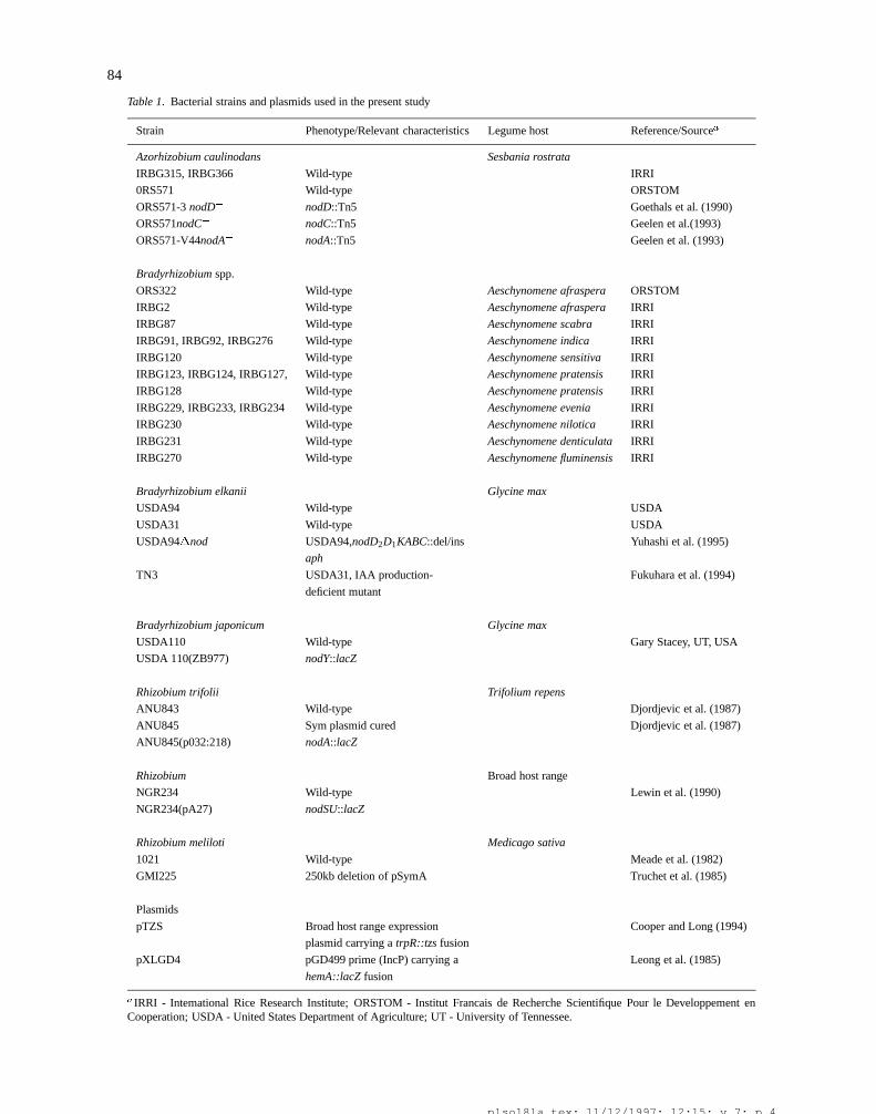
The rhizobial strains used are shown in Table 1. Thebacteria were grown routinely on yeast extract manni-tol agar medium (Vincent, 1970) at 30 �C under aerobicconditions in the dark. Single rhizobial colonies fromagar plates were transferred to 50 mL of liquid mediacontaining the appropriate antibiotics and incubated at30 �C on a shaker. After 24 h, 5 mL of the culturewas transferred to a 500 mL flask containing 95 mL offresh liquid medium and allowed to grow for another6-8 h. Subsequently, the bacterial cells were pelletedby centrifugation, washed with and resuspended in N-free Fahraeus (1957) or Jensen (1942) medium and theresuspension was adjusted to an OD650 of 0.25.
Surface sterilization of rice seeds
All operations for surface sterilization of seeds wereperformed at room temperature. Rice seeds, obtainedfrom the International Rice Germplasm Center at IRRI,were gently dehulled, washed with sterile distilledwater and immersed in either 70% ethanol for 4 min or95% ethanol for 20 sec. Subsequently, the seeds wereimmediately washed with sterile distilled water (3�10min) and incubated in 0.1% mercuric chloride solutionfor 4 min. Following this treatment, the seeds werewashed repeatedly in excess amounts of sterile dis-tilled water for 5-6 h on a shaker before seeding themin petri dishes containing either tryptone glucose yeastextract agar (Difco, USA) or potato dextrose agar (Dif-co, USA) medium, an incubated in the dark at 30 �Cto test for possible contamination. Using this method,more than 98% of the seeds germinated, and 95% ofthese seedlings were found to be contamination-free.The seedlings devoid of any contamination were usedin inoculation experiments.
Induction of nod gene expression
In vivo assays of nod gene expression were per-formed according to Redmond et al. (1986) and Petersand Long (1988) using Bradyrhizobium japonicumUSDA110(ZB977) containing a nodY::lacZ reportergene fusion, Rhizobium leguminosarum bv. tri-folii ANU845(pRt032:218) harboring a nodA::lacZfusion, or Rhizobium NGR234(pA27) harboring a nod-SU::lacZ fusion and rice seedlings germinated fromsurface sterilized seeds. The induction of nod geneexpression was determined by examining the blue col-or production due to ß-galactosidase activity using 5-bromo-4-chloro-3-indolyl-�-D-galactoside (X-gal, 40�m L�1) as indicator substrate.
Rice culture and inoculation
Two-three day old rice seedlings germinated from sur-face sterilized seeds were aseptically transferred to cul-ture tubes (200�20 mm) containing 25–30 mL N-freeFahraeus or Jensen medium solidified with 0.3% agarand incubated for 2 days in the plant growth room(maintained at a 14 h light/10 h dark cycle, at temper-atures of 27 �C/25 �C, respectively). One mL aliquotsof mid-log rhizobial cultures (see above) were inocu-lated on rice seedlings on the 3rd day and the culturetubes were returned to the growth room for incubation.Rice seedlings inoculated with equal amounts of heat-killed bacteria served as controls. For the assessmentof bacterial colonization, roots were sampled on the15th and 30th day after inoculation (DAI).
Histochemical localization of �-galactoside activity
The histochemical staining method used to measure�-Gal activity is essentially the same as described byBoivin et al. (1990). Briefly, the entire root systemof uninoculated rice plants or plants inoculated withAzorhizobium caulinodans strain ORS571 harboringplasmid pXLGD4 (a broad host-range plasmid harbor-ing the reporter gene lacZ fused to the constitutivelyexpressed hemA promoter of R. meliloti (Leong et al.,1985) was washed in 0.1 M Na-phosphate buffer (pH7.2; 3�5 min) and fixed using 1.25% glutaraldehydein 0.2 M Na-cacodylate buffer (pH 7.2) for 30 minunder vacuum followed by 1 h at atmospheric pressure.The fixed roots were rinsed with 0.2 M Na-cacodylatebuffer (pH 7.2; 2�15 min), transferred to a stainingsolution containing 800 mL of 0.2 M Na-cacodylate(pH 7.2), 50 mL of 100 mM K3 [Fe(CN)6], 50 mL
plso18la.tex; 11/12/1997; 12:15; v.7; p.3
84
Table 1. Bacterial strains and plasmids used in the present study
Strain Phenotype/Relevant characteristics Legume host Reference/Sourcea
Azorhizobium caulinodans Sesbania rostrata
IRBG315, IRBG366 Wild-type IRRI
0RS571 Wild-type ORSTOM
ORS571-3 nodD� nodD::Tn5 Goethals et al. (1990)
ORS571nodC� nodC::Tn5 Geelen et al.(1993)
ORS571-V44nodA� nodA::Tn5 Geelen et al. (1993)
Bradyrhizobium spp.
ORS322 Wild-type Aeschynomene afraspera ORSTOM
IRBG2 Wild-type Aeschynomene afraspera IRRI
IRBG87 Wild-type Aeschynomene scabra IRRI
IRBG91, IRBG92, IRBG276 Wild-type Aeschynomene indica IRRI
IRBG120 Wild-type Aeschynomene sensitiva IRRI
IRBG123, IRBG124, IRBG127, Wild-type Aeschynomene pratensis IRRI
IRBG128 Wild-type Aeschynomene pratensis IRRI
IRBG229, IRBG233, IRBG234 Wild-type Aeschynomene evenia IRRI
IRBG230 Wild-type Aeschynomene nilotica IRRI
IRBG231 Wild-type Aeschynomene denticulata IRRI
IRBG270 Wild-type Aeschynomene fluminensis IRRI
Bradyrhizobium elkanii Glycine max
USDA94 Wild-type USDA
USDA31 Wild-type USDA
USDA94�nod USDA94,nodD2D1KABC::del/ins Yuhashi et al. (1995)
aph
TN3 USDA31, IAA production- Fukuhara et al. (1994)
deficient mutant
Bradyrhizobium japonicum Glycine max
USDA110 Wild-type Gary Stacey, UT, USA
USDA 110(ZB977) nodY::lacZ
Rhizobium trifolii Trifolium repens
ANU843 Wild-type Djordjevic et al. (1987)
ANU845 Sym plasmid cured Djordjevic et al. (1987)
ANU845(p032:218) nodA::lacZ
Rhizobium Broad host range
NGR234 Wild-type Lewin et al. (1990)
NGR234(pA27) nodSU::lacZ
Rhizobium meliloti Medicago sativa
1021 Wild-type Meade et al. (1982)
GMI225 250kb deletion of pSymA Truchet et al. (1985)
Plasmids
pTZS Broad host range expression Cooper and Long (1994)
plasmid carrying a trpR::tzs fusion
pXLGD4 pGD499 prime (IncP) carrying a Leong et al. (1985)
hemA::lacZ fusion
aIRRI - Intemational Rice Research Institute; ORSTOM - Institut Francais de Recherche Scientifique Pour le Developpement enCooperation; USDA - United States Department of Agriculture; UT - University of Tennessee.
plso18la.tex; 11/12/1997; 12:15; v.7; p.4
85
of 100 mM K4 [Fe(CN)6], 40 mL of 2% X-gal in N,N-dimethyl formamide and incubated overnight in thedark at 30 �C. Subsequently, the roots were rinsed withdistilled water (5�10 min), and cleared using commer-cial chlorox solution for 40 sec, followed by a finalrinse with distilled water. Stained whole roots and 30mm transverse sections were examined by brightfieldlight microscopy.
Other microscopical methods
Rice roots colonized by bacteria, as visualized byX-gal staining, were further processed for detailedexamination by brightfield light microscopy, scan-ning electron microscopy (SEM), laser scanning con-focal microscopy (LSCM), and transmission electronmicroscopy (TEM). To prepare embedded sections, thestained roots were fixed again in a solution contain-ing 4% glutaraldehyde, 1% paraformaldehyde and 1mM CaCl2 in 50 mM Na-cacodylate buffer (pH 6.8)at room temperature under vacuum for 2 h, washedwith the same buffer (4�15 min), and post-fixed in1% osmium tetroxide in 100 mM Na-cacodylate buffer(pH 6.8) for 2 h. Fixed roots were rinsed with dis-tilled water (3�15 min) and suspended in 1% uranylacetate for 2 h. Subsequently, the roots were rinsedagain with distilled water (3�15 min), dehydratedthrough an acetone series, embedded in Spurr’s firmepoxy medium. Three �m sections were stained with0.1% (w/v) toluidine blue in 0.1% sodium tetraboratefor about 1 min at 60 �C, or 15 min at room tem-perature, and examined by brightfield and phase con-trast light microscopy. Ultrathin sections (70 nm) wererestained with 2% uranyl acetate in 0.5% aqueous leadcitrate solution and examined by TEM. For SEM, theroot samples were fixed in glutaraldehyde and post-fixed in osmium tetroxide as described above, dehy-drated through an ethanol series, critical point dried,mounted on stubs, and sputter-coated with 21 nm goldbefore examination. For LSCM, root segments werepreserved in 1% Na-azide during transit to MichiganState University, stained with an aqueous solution ofacridine orange (0.1 mg mL�1), rinsed and mountedin 1% Na-pyrophosphate, and examined using the epi-fluorescence confocal mode (Subba-Rao et al., 1995).
Results and discussion
Nodule development in legumes results from a seriesof complex interactions between the host plant and
rhizobia; including recognition and attachment of thebacteria, root hair curling, infection thread formationand the induction of cortical cell divisions (see Denar-ie and Cullimore, 1993; Hirsch, 1992; Nap and Bis-seling, 1990; Vijn et al., 1993). The initial steps inthis interplay are invoked by specific signal molecules,flavonoids and Nod factors, produced by legume plantand rhizobial symbionts, respectively. The processbegins with the secretion of flavonoids from roots, andconsequent flavonoid-triggered nod gene expression inthe microsymbiont leading to the production of variousNod factors. Nod factors in turn induce nodule ontoge-ny, and activate nodulin genes governing the processesinvolved in root hair deformation/curling, initiation ofinfection threads and cortical cell divisions. To achievethe goal of establishing a symbiosis between rhizobiaand rice, it is essential to systematically analyze theinteractions of rice with rhizobia to identify similar-ities and differences in rice-rhizobial interplay vis-a-vis those occurring in legume-rhizobial interactions.Another important aspect to consider while comparingsuch interactions is the inherent differences betweenthe root anatomy of rice and legumes (also Rolfe et al.,1997).
We examined the effects of diverse rhizobia on riceroots at 1–2 and 15–30 days after incubation. Exami-nation at the first time point would reveal the extentto which rhizobia attach to the root epidermis andtheir ability to elicit root hair induction, deformationand/or curling. Examination at the second time pointwould reveal if rhizobia induce any other morphologi-cal changes in roots, including unique root-derived andnodule-like structures.
Morphological responses of rice root hairs torhizobia and Nod factors
Most rhizobia were found to stimulate the formationof root hairs in the rice cultivars tested. However, rhi-zobia failed to induce deformation or curling of riceroot hairs. This was the case for 70 different rice geno-types inoculated with 24 of the 25 different broad andnarrow host-range strains of rhizobia (data not shown).The only exception was a marginal ability of Bradyrhi-zobium ORS322 to induce root hair deformation on ricevariety Milyang 54 (Reddy et al., 1995). These resultssuggest that rhizobia may not produce Nod factors inthe rice root environment and/or their Nod factors maybe unable to promote root hair deformation in rice.We tested these hypotheses by two approaches. First,we used various nod::lacZ fusion reporter strains to
plso18la.tex; 11/12/1997; 12:15; v.7; p.5
86
examine whether axenically generated rice root exu-date could activate expression of nodY in B. japon-icum USDA110, nodSU in Rhizobium NGR234, andnodA in R. leguminosarum bv. trifolii ANU843. Nosuch activation was observed (data not shown). Con-trol experiments using the reporter strains and root exu-dates of the corresponding host legumes were found tobe positive. These results suggest that rice root exu-date either lacks the appropriate activators of nod geneexpression and/or contains antagonistic substance(s)that inhibit(s) activation of the rhizobial nod genes.
In order to ascertain whether Nod factors were ableto induce root hair deformation in rice at all, we exam-ined more than 25 rice varieties for changes in roothair morphology at regular intervals during a 2-dayincubation in the presence of a mixture of purifiedsulfated and nonsulfated CLOS Nod factors from Rhi-zobium NGR234 (at concentrations of 10�6 to 10�9
M). The results obtained showed that these Nod fac-tors were unable to induce root hair deformation inrice under the experimental conditions tested (data notshown), and contrast with the induction of rice roothair deformation observed when using a recombinantstrain of R. leguminosarum containing multiple copiesof pSym-borne nodDABC genes from bv. trifolii strainANU843 (Plazinski et al., 1985).
The apparent inability of rice root hairs to respondto CLOSs from NGR234 may be due to the absenceof the appropriate CLOS structures required for induc-tion, the putative plant receptor(s) that perceive them,or some other element(s) in the Nod factor signal trans-duction pathway required for induction of root hairdeformation. It is also possible that the induction ofthese responses requires a critical concentration of oth-er non-CLOS classes of Nod factors made by rhizobia(Orgambide et al., 1994; Philip Hollingsworth et al.,1991).
Induction of abnormal lateral root development inrice by rhizobia and IAA
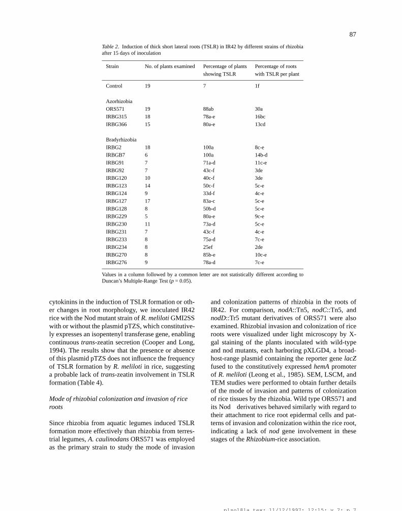
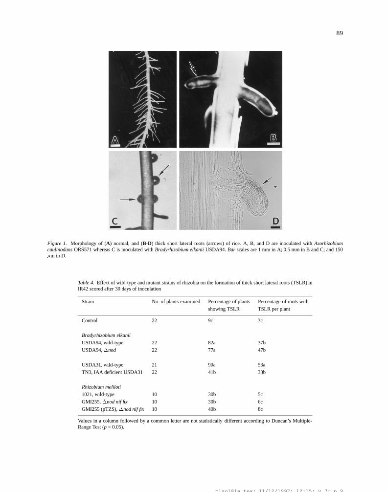
None of the rhizobia tested induced genuine root nod-ules on any of the rice varieties examined. However,all rice varieties exhibited abnormal lateral root devel-opment within two weeks after inoculation with rhi-zobia, particularly those that nodulate aquatic legumes(Tables 2 and 3). The growth of newly emerging lat-erals in some main roots was highly retarded resultingin shorter and thicker modified lateral root structures(Figure 1A-D). More than 75% of the plants of allvarieties tested exhibited this Thick Short Lateral Root
(TSLR) phenotype by 30 DAI (Table 3). Only rice vari-ety IR42 produced TSLRs without inoculation. Never-theless, the frequency of TSLR formation in IR42 wassignificantly increased when inoculated with rhizobia.Although rhizobial inoculation induced TSLR forma-tion in a very high percentage of plants, not all mainroots in a plant developed TSLRs. Their formationwas restricted to about 16-50% of the roots dependingon the rice genotype and rhizobial strain combinationused.
Nod factors (see Denarie and Cullimore, 1993; Napand Bisseling, 1990; Vijn et al., 1993) produced byrhizobia are clearly involved in promoting nodule for-mation in legumes. Moreover, rhizobially producedindole acetic acid (IAA; Fukuhara et al., 1994; Yuhashiet al., 1995) has been shown to be involved in pro-moting nodule formation. Because wild-type strains ofrhizobia that normally induce TSLR formation in ricehave the ability under certain conditions to produceNod factors as well as IAA, we examined whether rhi-zobia defective in the production of these bioactivemolecules were capable of inducing any phenotyp-ic changes in rice roots. Microscopical investigationsshowed that R. meliloti GMI255�nod and B. elkaniiUSDA94�nod induced TSLRs on IR42 with the sameefficiency as their parent wild-type strains, R. meliloti1021 and B. elkanii USDA94, respectively (Table 4).These results, indicating that nod genes required forCLOS production do not play a key role in the induc-tion of TSLR in rice, were further confirmed by studiesshowing that addition of a 10�9 M mixture of sulfat-ed and non-sulfated CLOSs from Rhizobium NGR234,supplied to the growth medium every alternate day for15 days, did not induce TSLRs (data not shown). Con-trary to the above results, B. elkanii TN3, a mutant ofUSDA31 deficient in IAA production but not in Nodfactor synthesis, induced TSLRs with a lower frequen-cy than the parent wild-type strain, suggesting a rolefor IAA in the promotion of TSLR formation in rice.
Along with IAA, rhizobia also produce cytokinins(see Taller and Sturtevant, 1991; Torrey, 1986). Inalfalfa roots, localized trans-zeatin production by R.meliloti GMI2SS(pTZS) was found to induce the for-mation of nodule-like structures (Cooper and Long,1994). In addition, certain early nodulin genes inlegumes have been found to be induced by cytokinins(Dehio and de Bruijn, 1992; Hirsch and Fang, 1994;Silver et al., 1996), and the signal transduction path-way responsible for this phenomenon has been shownto be conserved in non-legumes, such as Arabidopsis(Silver et al., 1997). To explore the potential role of
plso18la.tex; 11/12/1997; 12:15; v.7; p.6
87
Table 2. Induction of thick short lateral roots (TSLR) in IR42 by different strains of rhizobiaafter 15 days of inoculation
Strain No. of plants examined Percentage of plants Percentage of roots
showing TSLR with TSLR per plant
Control 19 7 1f
Azorhizobia
ORS571 19 88ab 30a
IRBG315 18 78a-e 16bc
IRBG366 15 80a-e 13cd
Bradyrhizobia
IRBG2 18 100a 8c-e
IRBGB7 6 100a 14b-d
IRBG91 7 71a-d 11c-e
IRBG92 7 43c-f 3de
IRBG120 10 40c-f 3de
IRBG123 14 50c-f 5c-e
IRBG124 9 33d-f 4c-e
IRBG127 17 83a-c 5c-e
IRBG128 8 50b-d 5c-e
IRBG229 5 80a-e 9c-e
IRBG230 11 73a-d 5c-e
IRBG231 7 43c-f 4c-e
IRBG233 8 75a-d 7c-e
IRBG234 8 25ef 2de
IRBG270 8 85b-e 10c-e
IRBG276 9 78a-d 7c-e
Values in a column followed by a common letter are not statistically different according toDuncan’s Multiple-Range Test (p = 0.05).
cytokinins in the induction of TSLR formation or oth-er changes in root morphology, we inoculated IR42rice with the Nod mutant strain of R. meliloti GMI2SSwith or without the plasmid pTZS, which constitutive-ly expresses an isopentenyl transferase gene, enablingcontinuous trans-zeatin secretion (Cooper and Long,1994). The results show that the presence or absenceof this plasmid pTZS does not influence the frequencyof TSLR formation by R. meliloti in rice, suggestinga probable lack of trans-zeatin involvement in TSLRformation (Table 4).
Mode of rhizobial colonization and invasion of riceroots
Since rhizobia from aquatic legumes induced TSLRformation more effectively than rhizobia from terres-trial legumes, A. caulinodans ORS571 was employedas the primary strain to study the mode of invasion
and colonization patterns of rhizobia in the roots ofIR42. For comparison, nodA::Tn5, nodC::Tn5, andnodD::Tr5 mutant derivatives of ORS571 were alsoexamined. Rhizobial invasion and colonization of riceroots were visualized under light microscopy by X-gal staining of the plants inoculated with wild-typeand nod mutants, each harboring pXLGD4, a broad-host-range plasmid containing the reporter gene lacZfused to the constitutively expressed hemA promoterof R. meliloti (Leong et al., 1985). SEM, LSCM, andTEM studies were performed to obtain further detailsof the mode of invasion and patterns of colonizationof rice tissues by the rhizobia. Wild type ORS571 andits Nod� derivatives behaved similarly with regard totheir attachment to rice root epidermal cells and pat-terns of invasion and colonization within the rice root,indicating a lack of nod gene involvement in thesestages of the Rhizobium-rice association.
plso18la.tex; 11/12/1997; 12:15; v.7; p.7
88
Table 3. Induction of thick short lateral roots (TSLR) in different rice varieties 15 and 30 days after inoculation(DAI) with rhizobial isolates from Sesbania rostrata
15 DAI 30 DAI
Percentage Percentage Percentage Percentage
No. of of plants of roots with of plants of roots with
plants showing TSLR showing TSLR
Strain Variety examined TSLR per plant TSLR per plant
Control
Azucena 16 0 0 0 0
IR42 58 15 0 15 4
IRAT 110 16 0 0 0 0
Kinandang Patong 16 0 0 0 0
Moroberekan 16 0 0 0 0
OS 4 16 0 0 0 0
ORS 571
Azucena 15 33c 7b 100a 36a
IR 42 130 78a 9b 100a 54a
IRAT 110 16 44bc 7bc 100a 28b
Kinandang Patong 14 7d 2c 100a 27b
Moroberekan 16 13cd 11ab 100a 35a
OS4 14 71ab 13ab 100a 35a
IRBG 315
Azucena 12 42c 8b 100a 36a
IR 42 124 60bc 11b 100a 33a
IRAT 110 13 84ab 10b 93ab 32a
Kinandang Patong 16 31c 7b 81b 16b
Moroberekan 16 100a 21a 93ab 31a
OS 4 14 64bc 11b 100a 30a
IRBG 366
Azucena 17 41b 8b 100a 37a
IR42 132 81a 13a 100a 39a
IRAT 110 17 83a 14a 100a 41a
Kinandang Patong 16 6c 1b 75b 21b
Moroberekan 16 88a 14a 100a 37ab
OS 4 17 23bc 3b 100a 34ab
Values in a column followed by a common letter are not statistically different according to Duncan’s Multiple-RangeTest (p=0.05).
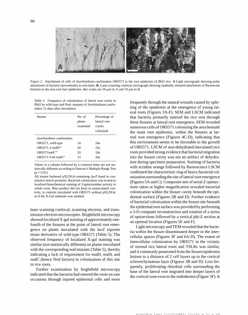
During the first 2 days of incubation, cells ofORS571 attached frequently in a polar orientation torice root hairs (Figure 2A), in a fashion similar to thatobserved during rhizobial inoculant studies on rice byTerouchi and Syono 1990. No infection thread forma-tion was observed in rice root hairs. Within 4-5 daysafter incubation, further colonization of the root epi-dermis occurred without a preferential cellular orienta-tion of attachment or extensive elaboration of cellulosemicrofibrils (Figure 2B, 4A-D). Similar results were
observed with the nod mutant derivatives of ORS571.These early events in the Rhizobium-rice associationcontrast with the discrete nodD-dependent cell aggre-gation at root hair tips and extensive elaboration of cel-lulose microfibrils characteristic of Phase 1 attachmentand Phase 2 adhesion in the Rhizobium-legume sym-biosis, respectively (Dazzo et al., 1984, 1988; Mateoset al., 1995).
Rhizobial invasion of rice roots and further internalcolonization were visualized by a combination of light,
plso18la.tex; 11/12/1997; 12:15; v.7; p.8
89
Figure 1. Morphology of (A) normal, and (B-D) thick short lateral roots (arrows) of rice. A, B, and D are inoculated with Azorhizobiumcaulinodans ORS571 whereas C is inoculated with Bradyrhizobium elkanii USDA94. Bar scales are 1 mm in A; 0.5 mm in B and C; and 150�m in D.
Table 4. Effect of wild-type and mutant strains of rhizobia on the formation of thick short lateral roots (TSLR) inIR42 scored after 30 days of inoculation
Strain No. of plants examined Percentage of plants Percentage of roots with
showing TSLR TSLR per plant
Control 22 9c 3c
Bradyrhizobium elkanii
USDA94, wild-type 22 82a 37b
USDA94,�nod 22 77a 47b
USDA31, wild-type 21 90a 53a
TN3, IAA deficient USDA31 22 41b 33b
Rhizobium meliloti
1021, wild-type 10 30b 5c
GMI255,�nod nif fix 10 30b 6c
GMI255 (pTZS),�nod nif fix 10 40b 8c
Values in a column followed by a common letter are not statistically different according to Duncan’s Multiple-Range Test (p = 0.05).
plso18la.tex; 11/12/1997; 12:15; v.7; p.9
90
Figure 2. Attachment of cells of Azorhizobium caulinodans ORS571 to the root epidermis of IR42 rice. A Light micrograph showing polarattachment of bacteria (arrowheads) to root hairs. B. Laser scanning confocal micrograph showing randomly oriented attachment of fluorescentbacteria to the non-root hair epidermis. Bar scales are 50 �m in A and 10 �m in B.
Table 5. Frequency of colonization of lateral root cracks inIR42 by wild-type and Nod- mutants of Azorhizobium caulin-odans 15 days after inoculation
Strains No. of Percentage of
plants lateral root
examined cracks
colonized
Azorhizobium caulinodans
ORS571, wild-type 14 24a
ORS571-3 nodD� 20 25a
ORS571nodC� 20 18a
ORS571-V44 nodA� 15 34a
Values in a column followed by a common letter are not sta-tistically different according to Duncan’s Multiple-Range Test(p = 0.01).All strains harbored pXLDG4 containing lacZ fused to con-stitutive hemA promoter. Bacterial colonization was scored aslocalized histochemical staining of �-galactosidase activity inwhole roots. Blue product did not form in uninoculated con-trols, in controls inoculated with ORS571 without pXLDG4,or if the X-Gal substrate was omitted.
laser scanning confocal, scanning electron, and trans-mission electron microscopies. Brightfield microscopyshowed localized X-gal staining of approximately one-fourth of the fissures at the point of lateral root emer-gence on plants inoculated with the lacZ reporterstrain derivative of wild-type ORS571 (Table 5). Theobserved frequency of localized X-gal staining wassimilar (not statistically different) on plants inoculatedwith the corresponding nod mutants (Table 5), therebyindicating a lack of requirement for nodD, nodA, andnodC (hence Nod factors) in colonization of this sitein rice roots.
Further examination by brightfield microscopyindicated that the bacteria had entered the roots on rareoccasions through injured epidermal cells and more
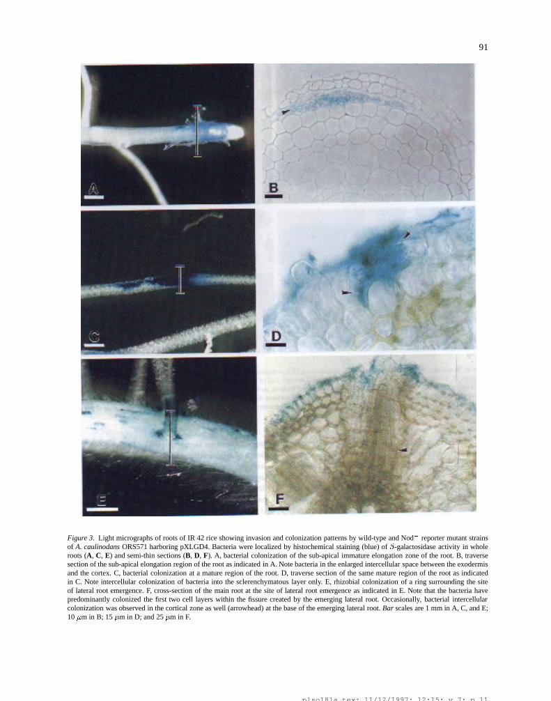
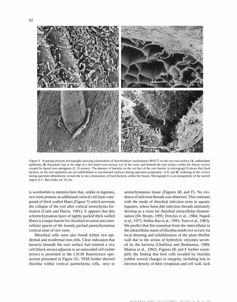
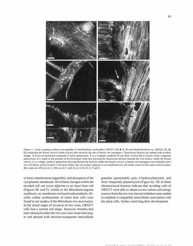
frequently through the natural wounds caused by split-ting of the epidermis at the emergence of young lat-eral roots (Figures 3A-F). SEM and LSCM indicatedthat bacteria primarily entered the rice root throughthese fissures at lateral root emergence. SEM revealednumerous cells of ORS571 colonizing the area beneaththe main root epidermis, within the fissures at lat-eral root emergence (Figures 4C-D), indicating thatthis environment seems to be favorable to the growthof ORS571. LSCM of non-dehydrated inoculated riceroots provided strong evidence that bacterial migrationinto the fissure cavity was not an artifact of dehydra-tion during specimen preparation. Staining of bacteriawith acridine orange followed by fluorescence LSCMconfirmed the characteristic ring of heavy bacterial col-onization surrounding the site of lateral root emergence(Figures 5A and C). Composite sets of serial Z optisec-tions taken at higher magnification revealed bacterialcolonization within the fissure cavity beneath the epi-dermal surface (Figures 5B and D). Further evidenceof bacterial colonization within the fissure site beneaththe epidermal root surface was provided by performinga 3-D computer reconstruction and rotation of a seriesof optisections followed by a vertical phi-Z section atan optimal location (Figures 5E and F).
Light microscopy and TEM revealed that the bacte-ria within the fissure disseminated deeper in the inter-cellular spaces (Figures 3F and 6A-D). The extent ofintercellular colonization by ORS571 in the vicinityof normal rice lateral roots and TSLRs was similar,and it commonly penetrated from the fissure/epidermislesions to a distance of 2 cell layers up to the corticalsclerenchymatous layer (Figures 3B and D). Less fre-quently, proliferating rhizobial cells surrounding thebase of the lateral root migrated into deeper layers ofthe cortical zone even to the endodermis (Figure 3F). It
plso18la.tex; 11/12/1997; 12:15; v.7; p.10
91
Figure 3. Light micrographs of roots of IR 42 rice showing invasion and colonization patterns by wild-type and Nod� reporter mutant strainsof A. caulinodans ORS571 harboring pXLGD4. Bacteria were localized by histochemical staining (blue) of �-galactosidase activity in wholeroots (A, C, E) and semi-thin sections (B, D, F). A, bacterial colonization of the sub-apical immature elongation zone of the root. B, traversesection of the sub-apical elongation region of the root as indicated in A. Note bacteria in the enlarged intercellular space between the exodermisand the cortex. C, bacterial colonization at a mature region of the root. D, traverse section of the same mature region of the root as indicatedin C. Note intercellular colonization of bacteria into the sclerenchymatous layer only. E, rhizobial colonization of a ring surrounding the siteof lateral root emergence. F, cross-section of the main root at the site of lateral root emergence as indicated in E. Note that the bacteria havepredominantly colonized the first two cell layers within the fissure created by the emerging lateral root. Occasionally, bacterial intercellularcolonization was observed in the cortical zone as well (arrowhead) at the base of the emerging lateral root. Bar scales are 1 mm in A, C, and E;10 �m in B; 15 �m in D; and 25 �m in F.
plso18la.tex; 11/12/1997; 12:15; v.7; p.11
92
Figure 4. Scanning electron micrographs showing colonization of Azorhizobium caulinodans ORS571 on the rice root surface (A, undisturbedepidermis; B, rhizoplane [rp] at the edge of a free-hand cross-section [cx] of the root), and beneath the root surface within the fissure crevicecreated by lateral root emergence (C, D arrows). The absence of bacteria on the cut face of the root interior in micrograph B shows that fixedbacteria on the root epidermis are not redistributed to uncolonized surfaces during specimen preparation. In C and D, widening of the creviceduring specimen dehydration reveals the in situ colonization of fixed bacteria within the fissure. Micrograph D is an enlargement of the starredregion in C. Bar scales are 10 �m.
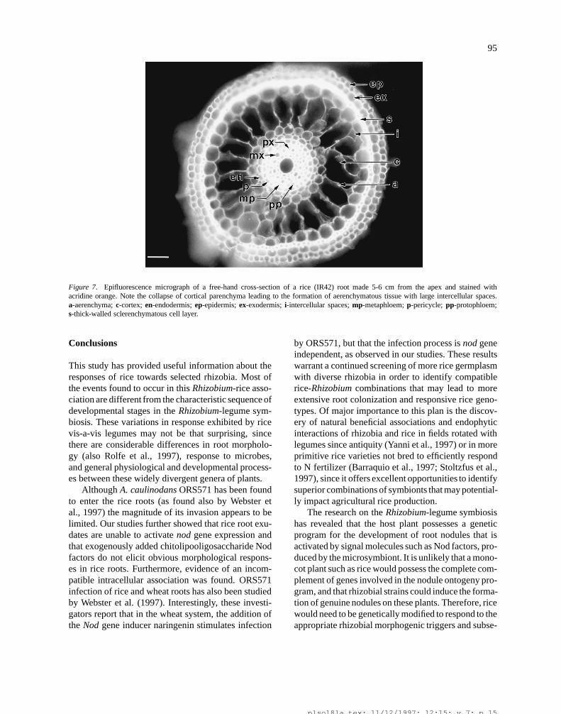
is worthwhile to mention here that, unlike in legumes,rice roots possess an additional cortical cell layer com-posed of thick walled fibers (Figure 7) which preventsthe collapse of the root after cortical aerenchyma for-mation (Clark and Harris, 1981). It appears that thissclerenchymatous layer of tightly packed thick walledfibers is a major barrier for rhizobial invasion into inter-cellular spaces of the loosely packed parenchymatouscortical zone of rice roots.
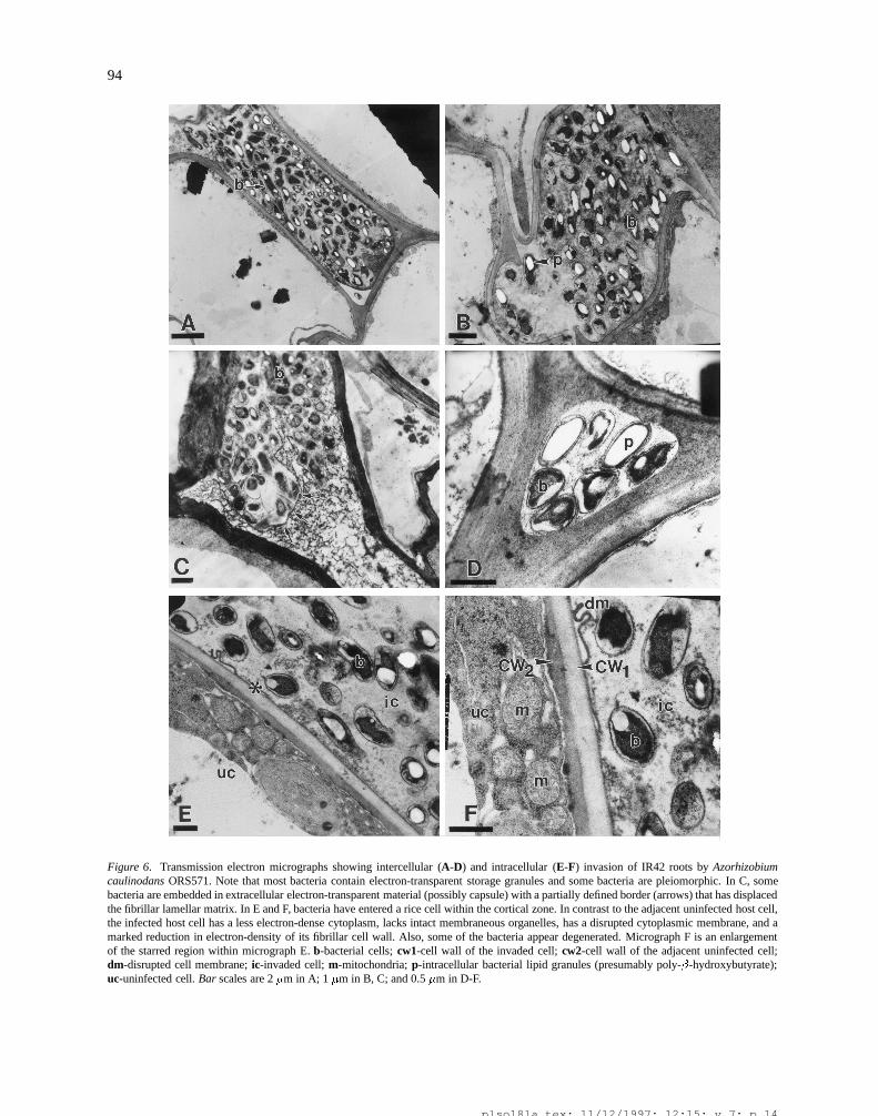
Rhizobial cells were also found within rice epi-dermal and exodermal root cells. Clear indication thatbacteria beneath the root surface had entered a ricecell (black arrow) adjacent to an uninvaded cell (whitearrow) is presented in the LSCM fluorescence opti-section presented in Figure 5G. TEM further showedrhizobia within cortical parenchyma cells, next to
aerenchymatous tissue (Figures 6E and F). No evi-dence of infection threads was observed. This contrastswith the mode of rhizobial infection even in aquaticlegumes, where bona fide infection threads ultimatelydevelop as a route for rhizobial intracellular dissemi-nation (De Bruijn, 1995; Dreyfus et al., 1984; Napoliet al., 1975; Subba-Rao et al., 1995; Tsien et al., 1983).We predict that this transition from the intercellular tothe intracellular status of rhizobia inside rice occurs vialocal thinning and solubilization of the plant fibrillarwall due to the action of hydrolytic enzymes secret-ed by the bacteria (Chalifour and Benhamou, 1989;Mateos et al., 1992). Figures 6E and F further exem-plify the finding that host cells invaded by rhizobiaexhibit several changes in integrity, including loss inelectron density of their cytoplasm and cell wall, lack
plso18la.tex; 11/12/1997; 12:15; v.7; p.12
93
Figure 5. Laser scanning confocal micrographs of Azorhizobium caulinodans ORS571 (A, B, E, F) and Bradyrhizobium sp. ORS322 (C, D,G) colonizing the fissure crevice (white arrows) that encircles the site of lateral root emergence. Fluorescent bacteria are stained with acridineorange. A-D are reconstructed composite Z serial optisections. E is a computer rendered 50 �m thick vertical phi-Z section of the compositeoptisections in F, made at the position of the horizontal white line showing the fluorescent bacteria beneath the root surface within the fissurecrevice. G is a single confocal optisection showing fluorescent bacteria within the fissure crevice at lateral root emergence (arrowheads) and arice cell (black arrow) located 2 cell layers below the root surface adjacent to an uninfected rice cell (white arrow) in the same confocal plane.Bar scales are 250 �m in A; 500 �m in C; and 10 �m in B, D, E, F and G.
of intact membranous organelles, and disruption of thecytoplasmic membrane. All of these changes within theinvaded cell can occur adjacent to an intact host cell(Figures 6E and F). Unlike in the Rhizobium-legumesymbiosis, no membrane-enclosed endosymbiotic rhi-zobia within symbiosomes of intact host cells werefound in our studies of the Rhizobium-rice association.In the initial stages of invasion of rice roots, ORS571cells had a normal rod shape. However rhizobia thathad colonized within the rice root were somewhat larg-er and abound with electron-transparent intracellular
granules (presumably poly-�-hydroxybutyrate), andmore frequently plasmolyzed (Figure 6). All of theseultrastructural features indicate that invading cells ofORS571 were able to obtain excess carbon and energysources from the rice root, but nevertheless were unableto establish a compatible intracellular association withthe plant cells, further restricting their development.
plso18la.tex; 11/12/1997; 12:15; v.7; p.13
94
Figure 6. Transmission electron micrographs showing intercellular (A-D) and intracellular (E-F) invasion of IR42 roots by Azorhizobiumcaulinodans ORS571. Note that most bacteria contain electron-transparent storage granules and some bacteria are pleiomorphic. In C, somebacteria are embedded in extracellular electron-transparent material (possibly capsule) with a partially defined border (arrows) that has displacedthe fibrillar lamellar matrix. In E and F, bacteria have entered a rice cell within the cortical zone. In contrast to the adjacent uninfected host cell,the infected host cell has a less electron-dense cytoplasm, lacks intact membraneous organelles, has a disrupted cytoplasmic membrane, and amarked reduction in electron-density of its fibrillar cell wall. Also, some of the bacteria appear degenerated. Micrograph F is an enlargementof the starred region within micrograph E. b-bacterial cells; cw1-cell wall of the invaded cell; cw2-cell wall of the adjacent uninfected cell;dm-disrupted cell membrane; ic-invaded cell; m-mitochondria; p-intracellular bacterial lipid granules (presumably poly-�-hydroxybutyrate);uc-uninfected cell. Bar scales are 2 �m in A; 1 �m in B, C; and 0.5 �m in D-F.
plso18la.tex; 11/12/1997; 12:15; v.7; p.14
95
Figure 7. Epifluorescence micrograph of a free-hand cross-section of a rice (IR42) root made 5-6 cm from the apex and stained withacridine orange. Note the collapse of cortical parenchyma leading to the formation of aerenchymatous tissue with large intercellular spaces.a-aerenchyma; c-cortex; en-endodermis; ep-epidermis; ex-exodermis; i-intercellular spaces; mp-metaphloem; p-pericycle; pp-protophloem;s-thick-walled sclerenchymatous cell layer.
Conclusions
This study has provided useful information about theresponses of rice towards selected rhizobia. Most ofthe events found to occur in this Rhizobium-rice asso-ciation are different from the characteristic sequence ofdevelopmental stages in the Rhizobium-legume sym-biosis. These variations in response exhibited by ricevis-a-vis legumes may not be that surprising, sincethere are considerable differences in root morpholo-gy (also Rolfe et al., 1997), response to microbes,and general physiological and developmental process-es between these widely divergent genera of plants.
Although A. caulinodans ORS571 has been foundto enter the rice roots (as found also by Webster etal., 1997) the magnitude of its invasion appears to belimited. Our studies further showed that rice root exu-dates are unable to activate nod gene expression andthat exogenously added chitolipooligosaccharide Nodfactors do not elicit obvious morphological respons-es in rice roots. Furthermore, evidence of an incom-patible intracellular association was found. ORS571infection of rice and wheat roots has also been studiedby Webster et al. (1997). Interestingly, these investi-gators report that in the wheat system, the addition ofthe Nod gene inducer naringenin stimulates infection
by ORS571, but that the infection process is nod geneindependent, as observed in our studies. These resultswarrant a continued screening of more rice germplasmwith diverse rhizobia in order to identify compatiblerice-Rhizobium combinations that may lead to moreextensive root colonization and responsive rice geno-types. Of major importance to this plan is the discov-ery of natural beneficial associations and endophyticinteractions of rhizobia and rice in fields rotated withlegumes since antiquity (Yanni et al., 1997) or in moreprimitive rice varieties not bred to efficiently respondto N fertilizer (Barraquio et al., 1997; Stoltzfus et al.,1997), since it offers excellent opportunities to identifysuperior combinations of symbionts that may potential-ly impact agricultural rice production.
The research on the Rhizobium-legume symbiosishas revealed that the host plant possesses a geneticprogram for the development of root nodules that isactivated by signal molecules such as Nod factors, pro-duced by the microsymbiont. It is unlikely that a mono-cot plant such as rice would possess the complete com-plement of genes involved in the nodule ontogeny pro-gram, and that rhizobial strains could induce the forma-tion of genuine nodules on these plants. Therefore, ricewould need to be genetically modified to respond to theappropriate rhizobial morphogenic triggers and subse-
plso18la.tex; 11/12/1997; 12:15; v.7; p.15
96
quent Rhizobium-modulated nodule ontogeny require-ments. Recent studies have shown that rice pos-sesses homologues of some of the (nodulin) genesspecifically expressed in legume nodules during ear-ly events in infection and nodule formation (Reddy etal., 1996a,1996b, unpublished). These results suggestthat rice may have at least part of the genetic pro-gram involved in a functional symbiosis with Rhizobi-um. Other studies have shown that CLOS Nod factorsenhance the promoter activity of MtENOD12 withinrice roots, indicating that a portion of the signal trans-duction machinery important for legume nodulationmay also already exist in rice (Reddy et al., 1996c).
Another reason for optimism may be that rice isable to enter into symbiotic associations with myc-orrhizal fungi (Secilia and Bagyaraj, 1992). Genet-ic links between the processes involved in nodulationand arbuscular mycorrhiza have been found in legumes(Cook et al., 1997; Gianinazzi-Pearson, 1996). Thus,rice may possess part of the genetic program necessaryfor entering into mutually beneficial, endosymbioticassociations with other soil microorganisms.
In conclusion, many differences exist between theassociation of rice and rhizobia relative to the root- orstem-nodule symbiosis with legumes. However, someof the molecular interactions that occur in these plant-microbe associations may be similar. It is thereforeessential that studies be further extended at the cellularand molecular levels to identify why such responses donot fully occur in rice, in order to contemplate geneti-cally engineering this major cereal crop to form a moreintimate endosymbiotic association with rhizobia.
Acknowledgements
We are thankful to the following scientists for kindlyproviding us plasmids, chitolipooligosaccharide Nodfactors, and/or rhizobial strains: J Denarie for pXL-GD4 and purified Nod factors; W J Broughton forRhizobium NGR234 and NGR234(pA27); C Goughfor A. caulinodans ORS571-3 nodD� (pXLDG4),ORS571 nodC� (pXLGD4) and ORS571-V44 nodA�;S R Long for R. meliloti 1021, GM1225 ad GMI225(pTZS); K Minamisawa for B. elkanii USDA94,USDA31, USDA94Dnod and TN3; B G Rolfe forR. leguminosarum bv. trifolii ANU843, ANU845 andANU845(pRt032:218); G Stacey for B. japonicumUSDA11O and USDA11O(ZB977). This work wassupported by grants from the Danish InternationalDevelopment Agency (DANIDA) to IRRI and MSU,
the MSU-NSF Center for Microbial Ecology (NSFGrant No DEB 9120006) and the DOE (De-FGO2-91ER20021) to MSU.
References
Al-Mallah M K, Davey M R and Cocking E C 1989 Formation ofnodular structures on rice seedlings by rhizobia. J. Exp. Bot. 40,473–478.
Barraquio W L, Revilla L and Ladha J K 1997 Isolation of endophyticdiazotrophic bacteria from wetland rice. Plant Soil 194, 15–24.
Bender G L, Preston L, Barnard D and Rolfe B G 1990 Formationof nodule-like structures on the roots of the non-legumes rice andwheat. In Nitrogen Fixation: Achievements and Objectives. Eds.P M Gresshoff, L E Roth, G Stacey and W E Newton. p 825.Chapman and Hall, London.
Boivin C, Camut S, Malpica C A, Truchet G and Rosenberg C 1990Rhizobium meliloti genes encoding catabolism of trigonellin areinduced under symbiotic conditions. Plant Cell 2, 1157–1170.
Cassman K G, Peng S, Olk D C, Ladha J K, Reichardt W, DobermannA and Singh U 1997 Opportunities for increased nitrogen useefficiency from improved resource management in irrigated ricesystems. Field Crops Res. (In press).
Chalifour F-P and Benhamou N 1989 Indirect evidence for cellulaseproduction by Rhizobium in pea root nodules during bacteroiddifferentiation: Cytochemical aspects of cellulose breakdown inrhizobial droplets. Can. J. Microbiol. 35, 821–829.
Clark L H and Harris W H 1981 Observations on the root anatomyof rice (Oryza sativa L.). Am. J. Bot. 68, 154–161.
Cocking E C, Srivastava J S, Cook J M, Kothari S L and Davey M R1993 Studies on nodulation of maize, wheat, rice and oilseed rape:interactions of rhizobia with emerging lateral roots. In BiologicalNitrogen Fixation - Novel Associations with Non-legume Crops.Eds. N Yanfu, I R Kennedy and C Tingwei. pp 53–58. QingdaoOcean University Press, Qingdao.
Colnaghi R, Green A, He L, Rudnick P and Kennedy C 1997 Strate-gies for increased ammonium production in free-living or plantassociated nitrogen fixing bacteria. Plant Soil 194, 145–154.
Cook D R, VandenBosch K, de Bruijn F J and Huguet T 1997 Modellegumes get the nod. Plant Cell 3, 275–281.
Cooper J B and Long S R 1994 Morphogenetic rescue of Rhizobiummeliloti nodulation mutants by trans-zeatin secretion. Plant Cell6, 215–225.
Dazzo F B, Hollingsworth R, Philip-Hollingsworth S, Robeles M,Olen T, Salzwedel J, Djordjevic M and Rolfe B 1988 Recogni-tion process in the Rhizobium trifolii-white clover symbiosis. InNitrogen Fixation: Hundred Years After. Eds. H Bothe and F J deBruijn. pp 431–435. Gustav Fischer, Stuttgart.
Dazzo F B, Truchet G L, Sherwood J, Hrabak E M, Abe M andPankratz H S 1984 Specific phases of root hair attachment in theRhizobium trifolii-clover symbiosis. Appl. Environ. Microbiol.48, 1140–1150.
De Bruijn F J, Jing Y and Dazzo F B 1995 Potential and pitfalls oftrying to extend symbiotic interactions of nitrogen-fixing organ-isms to presently non-nodulated plants, such as rice. Plant Soil172, 207–219.
Dehio C and de Bruijn F J 1992 The early nodulin gene SrEnod2 fromSesbania rostrata is inducible by cytokinin. Plant J. 2, 117–128
Denarie J and Cullimore J 1993 Lipo-oligosaccharide nodulationfactors: A minireview. New class of signaling molecules mediat-ing recognition and morphogenesis. Cell 74, 951–954.
plso18la.tex; 11/12/1997; 12:15; v.7; p.16
97
Dixon R, Cheng Q, Shen G-F, Day A and Dowson-Day M 1997Nif gene transfer and expression in chloroplasts: prospects andproblems. Plant Soil 194, 193–203.
Djordjevic M A, Redmond J W, Batley M and Rolfe B G 1987Clovers secrete specific phenolic compounds which either stimu-late or repress nod gene expression in Rhizobium trifolii. EMBOJ. 6, 1173–1179.
Dreyfus B L, Alazard D and Dommergues Y R 1984 New andunusual microorganisms and their niches. In Current Perspectivesin microbial Ecology. Eds. M J Klug and C A Reddy. pp 161–169.American Society of Microbiology, Washington DC.
Fahraeus G 1957 The infection of clover root hairs by nodule bacteriastudied by a simple glass slide technique. J. Gen. Microbiol. 16,374–381.
Fukuhara H, Minakawa Y, Akao S and Minarnisawa K 1994 Theinvolvement of indole-3-acetic acid produced by Bradyrhizobiumelkanii in nodule formation. Plant Cell Physiol. 35, 1261–1265.
Geelen D, Mergaert P, Geremia R A, Goormachtig S, Van Mon-tagu M and Holsters M 1993 Identification of nodSUIJ genes inNod locus 1 of Azorhizobium caulinodans.: evidence that nodSencodes a methyltransferase involved in Nod factor modification.Mol. Microbiol. 9, 145–154.
Gianinazzi-Pearson V 1996 Plant cell responses to arbuscular myc-orrhizal fungi: Getting to the roots of the symbiosis. Plant Cell 8,1871–1883.
Goethals K, Van den Eede G, Van Montague M and Holsters M 1990Identification and characterization of a nodD gene in Azorhizobi-um caulinodans ORS571. J. Bacteriol. 172, 2658–2666.
Gough C, Vasse J, Galera C, Webster G, Cocking E and Denarie J1997 Interactions between bacterial diazotrophs and non-legumedicots: Arabidopsis thaliana as a model plant. Plant Soil 194,123–130.
Hirsch A M 1992 Developmental biology of legume nodulation.New Phytol. 122, 211–237.
Hirsch A M and Fang Y 1994 Plant hormones and nodulation: what’sthe connection? Plant Mol. Biol. 26, 5–9.
Jensen H L 1942 Nitrogen fixation in leguminous plants. I. Gen-eral characters of root nodule bacteria isolated from species ofMedicago and Trifolium in Australia. Proc. Linn. Soc. N.S.W. 66,98–108.
Jing Y, Li G, Jin G, Shan X, Zhang, B Guan C and Li J 1990 Rice rootnodules with acetylene reduction activity. In Nitrogen FixationAchievements and Objectives. Eds. P M Gresshoff, L E Roth, GStacey and W E Newton. p 829. Chapman and Hall, London.
Jing Y, Li G and Shan X 1992 Development of nodule-like structureon rice roots. In Nodulation and Nitrogen Fixation in Rice. Eds. GS Khush and J Bennett. pp 123–126. International Rice ResearchInstitute, Manila.
Kennedy I R, Pereg-Gerk L, Wood C, Deaker R, Gilchrist K andKatupitiya S 1997 Biological nitrogen fixation in non-leguminousfield crops: Facilitating the evolution of an effective associationbetween Azospirillum and wheat. Plant Soil 194, 65–79.
Kirchhof G, Reis V M, Baldani J I, Eckert B, Dobereiner J andHartmann A 1997 Occurence, physiological and and molecu-lar analysis of endophytic diazotrophic bacteria in gramineousenergy plants. Plant Soil 194, 45–55.
Ladha J K, Kirk G, Bennett J, Peng S, Reddy C K, Reddy P M andSingh U 1997 Opportunities for increased nitrogen use efficiencyfrom improved lowland rice germplasm. Field Crops Res. (Inpress).
Ladha J K and Reddy P M 1995 Extension of nitrogen fixation torice-necessity and possibilities. Geojournal 35, 363–372.
Leong S A, Williams P H and Ditta G S 1985 Analysis of the 50 reg-ulatory region of the gene for d-amino levulinic acid synthetaseof Rhizobium meliloti. Nucl. Acids Res. 13, 5965–5976.
Lewin A, Cervantes E, Chee-Hoong W and Broughton W J 1990nodSU, two new nod genes of the broad host range Rhizobi-um strain NGR234 encode host-specific nodulation of the tropi-cal tree Leucaena leucocephala. Mol. Plant-Microbe Interact. 3,317–326.
Li G, Jing Y, Shan X, Wang H and Guan C 1991 Identification of ricenodules that contain Rhizobium bacteria. Chin. J. Bot. 3, 8–17.
Mateos P, Baker D, Philip-Hollingsworth S, Squartini A, PeruffoA, Nuti M and Dazzo F B 1995 Direct in situ identification ofcellulose microfibrils associated with Rhizobium leguminosarumbv. trifolii attached to the root epidermis of white clover. Can. J.Microbiol. 41, 202–207.
Mateos P, Jiminez-Zurdo, J Chen, A Squartini, S Haack, E Martinez-Molina, D Hubbell and Dazzo F B 1992 Cell-associated pecti-nolytic and cellulolytic enzymes in Rhizobium leguminosarumbv. trifolii. Appl. Environ. Microbiol. 58, 1816–1822.
Meade H, Long S R, Ruvkun G B, Brown S E and Ausubel FM 1982 Physical and genetic characterization of symbiotic andauxotrophic mutants of Rhizobium meliloti. J. Bacteriol. 149,114–122.
Nap J-P and Bisseling T 1990 Developmental biology of a plant-prokaryote symbiosis: The legume root nodule. Science 250,948–954.
Napoli C A, Dazzo F B and Hubbell D H 1975 Ultrastucture ofinfection and common antigen relationships in the Rhizobium-Aeschynomene symbiosis. In Proc. 5th Australian Legume Nodu-lation Conference. Ed. J Vincent. pp 35–37. Brisbane.
Orgambide G G, Philip Hollingsworth S, Hollingsworth R T andDazzo F B 1994 Flavone-enhanced accumulation and symbiosis-related activity of a diglycosyl diacylglycerol membrane glycol-ipid from Rhizobium leguminosarum bv. trifolii. J. Bacteriol. 176,4338–4347.
Peters N K and Long S R 1988 Alfalfa root exudates and com-pounds which promote or inhibit induction of Rhizobium melilotinodulation genes. Plant Physiol. 88, 396–400.
Philip-Hollingsworth S, Hollingsworth R I and Dazzo F B 1991N-acetylglutamic acid: an extracellular Nod signal of Rhizobiumtrifolii ANU843 which induces root hair deformation and nodule-like primordia in white clover roots. J. Biol. Chem. 266, 16854–16858.
Plazinski J, Innes R W and Rolfe B G 1985 Expression of Rhizo-bium trifolii early nodulation genes on maize and rice plants. JBacteriol. 163, 812–815.
Reddy P M, Kouchi H, Hata S and Ladha J K 1996a Homologs ofGmENOD93 from rice. 8th International Congress on MolecularPlant-Microbe Interactions, Knoxville, TN.
Reddy P M, Kouchi H, Hata S and Ladha J K 1996b Identification,cloning and expression of rice homologs of GmN93 7th Interna-tional Symposium on BNF with Non-Legumes, Faisalabad.
Reddy P M and Ladha J K 1995 Can symbiotic nitrogen fixationbe extended to rice? In Nitrogen fixation: Fundamentals andApplications. Eds. I A Tikhonovich, N A Provorov, V I Romanovand W E Newton. pp 629–633. Kluwer Academic Publishers,Dordrecht.
Reddy P M, Ramos M C, Hernandez R J and Ladha J K 1995Rice-rhizobial interactions. 15th North American Conference onSymbiotic Nitrogen Fixation, Raleigh, NC.
Reddy P M, Torrizo L, Ramos M C, Datta S K and Ladha J K 1996cExpression of MtENOD12 promoter driven GUS in transformedrice. 8th International Congress on Molecular Plant-MicrobeInteractions, Knoxville, TN.
plso18la.tex; 11/12/1997; 12:15; v.7; p.17
98
Redmond J W, Batley M, Djordjevic M A, Innes R W, Kuempel PL and Rolfe B G 1986 Flavones induce expression of nodulationgenes in Rhizobium. Nature 323, 632–635.
Rolfe B G and Bender G L 1990 Evolving a Rhizobium fornon-legume nodulation. In Nitrogen Fixation:Achievements andObjectives. Eds. P M Gresshoff, L E Roth, G Stacey and W ENewton. pp 779–786. Chapman and Hall, London.
Rolfe B G, Djordjevic M A, Weinman J J, Mathesius U, PittockC, Gartner E, Dong Z, McCully M and Mclver J 1997 Rootmorphogenesis in legumes and cereals and the effect of bacterialinoculation on root development. Plant Soil 194, 131–144.
Secilia j and Bagyaraj D J 1992 Selection of efficient vesicular-arbuscular mycorrhizal fungi for wetland rice (Oryza sativa L.).Biol. Fert. Soils 13, 108–111.
Silver D L, Pinaev A, Chen R and de Bruijn F J 1996 Post-transcriptional regulation of the Sesbania rostrata early nodulingene SrEnod2 by cytokinin. Plant Physiol. 112, 559–567.
Silver D L, Deikman J, Chen R and de Bruijn F J 1997 The SrEnod2gene is contnolled by a conserved cytokinin signal transductionpathway. Plant Physiol. (In press).
Stacey G and Shibuya N 1997 Chitin recognition in rice and legumes.Plant Soil 194, 161–169.
Stoltzfus J R, So R, Malarvizhi P P, Ladha J K and de Bruijn F J1997 Isolation of endophytic bacteria from rice and assessment oftheir potential for supplying rice with biologically fixed nitrogen.Plant Soil. 194, 25–36.
Subba-Rao N S, Mateos P F, Baker D, Pankratz H S, Palma J,Dazzo F B and Sprent J I 1995 The unique root nodule symbiosisbetween Rhizobium and the aquatic legume, Neptunia natans.(L.F.) Druce. Planta 196, 311–320.
Taller B J and Sturtevant D B 1991 Cytokinin production by rhizo-bia. In Advances in Molecular Genetics of Plant-Microbe Inter-actions. Vol. 1. Eds. H Hennecke and D P S Verma. pp 215–221.Kluwer Academic Publishers, Dordrecht.
Terouchi N and Syono K 1990 Rhizobium attachment and curling inasparagus, rice and oat plants. Plant Cell Physiol. 31, 119–127.
Torrey J G 1986 Endogenous and exogenous influences on the regu-lation of lateral root formation. In New Root Formation in Plantsand Cuttings. Eds. M B Jackson. pp 31–66. Martinus NijhoffPublishers, Dordrecht.
Trinick M J 1973 Symbiosis between Rhizobium and the non-legume, Trema aspera. Nature 244, 459–460.
Truchet G, Debelle F, Vasse J, Terzaghi B, Garnerone A M, Rosen-berg C, Batut J, Maillet F and Denarie J 1985 Identification of Rhi-zobium meliloti Sym2011 region controlling the host specificityof root hair curling and nodulation. J. Bacteriol. 164, 1200–1210.
Tsien H C, Dreyfus B L and Schmidt E L 1983 Initial stages inthe morphogenesis of nitrogen-fixing stem nodules of Sesbaniarostrata. J. Bacteriol. 156, 888–897.
Vijn I, das Neves L, van Kammen A, Franssen H and Bisseling T1993 Nod factors and nodulation in plants. Science 260, 1764–1765.
Vincent J M 1970 A manual for the study of root nodule bacteria.IBP Handbook 15, Blackwell Scientific Publ. London. 164 p.
Webster G, Gough C, Vasse J, Batchelor C A, O’Callaghan K J,Kothari S L, Davey M R, Dinarii J and Cocking E C 1997 lnter-actions of rhizobia with rice and wheat. Plant Soil 194, 115–122.
Yanni Y G, Rizk E Y, Corich V, Squartini A, Ninke K, Philip-Hollingsworth S, Orgambide G G, de Bruijn F J, Stoltzfus J,Buckley D, Schmidt T M, Mateos P F, Ladha J K and DazzoF B 1997 Natural endophytic association between Rhizobiumleguminosarum bv. trifolii and rice roots and assessment of itspotential to promote rice growth. Plant Soil 194, 99–114.
Yuhashi, K-I, Akao S, Fukuhara H, Tateno E, Chun J-Y, Stacey G,Hara H, Kubota M, Asami T and Minamisawa K 1995 Bradyrhi-zobium elkanii induces outer cortical root swelling in soybean.Plant Cell Physiol. 36, 1571–1577.
Guest editors: J K Ladha, F J de Bruijn and K A Malik
plso18la.tex; 11/12/1997; 12:15; v.7; p.18
Related Documents


