Clinical and Experimental Rheumatology 2009; 27: 594-602. Rheumatoid arthritis, Klippel-Feil syndrome and Pott’s disease in Cardinal Carlo de’ Medici (1595-1666) V. Giuffra, A. Vitiello, S. Giusiani, A. Fornaciari 1 , D. Caramella 2 , N. Villari 3 , G. Fornaciari Department of Oncology, Transplants and Advanced Technologies in Medicine, Division of Paleopathology, History of Medicine and Bioethics, University of Pisa; 1 Department of Archaeology and History of Art, Section of Medieval Archaeology, University of Siena; 2 Department of Oncology, Transplants and Advanced Technologies in Medicine, Division of Diagnostic and Interventional Radiology, University of Pisa; 3 Department of Clinical Physiopathology, Section of Clinical Radiology, University of Florence, Italy. Abstract Objective A paleopathological study was carried out on the she skeletal remains of Cardinal Carlo de’ Medici (1595-1666), son of the Grand Duke Ferdinando I (1549-1609) and Cristina from Lorraine (1565-1636), to investigate the articular pathology described in the archival sources. Methods The skeletal remains of Carlo, buried in the Basilica of San Lorenzo in Florence, have been exhumed and submitted to macroscopic and radiological examination. Results The skeleton of Carlo revealed a concentration of different severe pathologies. Ankylosis of the cervical column, associated with other facial and spine anomalies suggests a diagnosis of congenital disease: the Klippel-Feil syndrome. In addition, the cervical segment presents the results of the tuberculosis (Pott’s disease) from which the Cardinal suffered in his infancy. The post-cranial skeleton shows an ankylosing disease, mainly symmetrical and extremely severe, involving the large as well as small articulations, and characterized by massive joint fusion, that totally disabled the Cardinal in his last years of life. Conclusions The final diagnosis suggests an advanced, ankylosing stage of rheumatoid arthritis. Key words Rheumatoid arthritis, ankylosis, tuberculosis, Medici, Florence, Renaissance.

Welcome message from author
This document is posted to help you gain knowledge. Please leave a comment to let me know what you think about it! Share it to your friends and learn new things together.
Transcript

Clinical and Experimental Rheumatology 2009; 27: 594-602.
Rheumatoid arthritis, Klippel-Feil syndrome and Pott’s disease in Cardinal Carlo de’ Medici (1595-1666)
V. Giuffra, A. Vitiello, S. Giusiani, A. Fornaciari1, D. Caramella2, N. Villari3, G. Fornaciari
Department of Oncology, Transplants and Advanced Technologies in Medicine, Division of Paleopathology, History of Medicine and Bioethics, University of Pisa; 1Department of Archaeology and History of Art, Section of Medieval Archaeology, University of Siena; 2Department of Oncology,
Transplants and Advanced Technologies in Medicine, Division of Diagnostic and Interventional Radiology, University of Pisa; 3Department of Clinical Physiopathology, Section of Clinical
Radiology, University of Florence, Italy.
AbstractObjective
A paleopathological study was carried out on the she skeletal remains of Cardinal Carlo de’ Medici (1595-1666), son of the Grand Duke Ferdinando I (1549-1609) and Cristina from Lorraine (1565-1636), to investigate the articular pathology
described in the archival sources.
MethodsThe skeletal remains of Carlo, buried in the Basilica of San Lorenzo in Florence, have been exhumed and submitted to
macroscopic and radiological examination.
ResultsThe skeleton of Carlo revealed a concentration of different severe pathologies. Ankylosis of the cervical column,
associated with other facial and spine anomalies suggests a diagnosis of congenital disease: the Klippel-Feil syndrome. In addition, the cervical segment presents the results of the tuberculosis (Pott’s disease) from which the Cardinal suffered in his infancy. The post-cranial skeleton shows an ankylosing disease, mainly symmetrical and extremely severe, involving the large as well as small articulations, and characterized by massive joint fusion, that totally disabled the Cardinal in his
last years of life.
ConclusionsThe final diagnosis suggests an advanced, ankylosing stage of rheumatoid arthritis.
Key wordsRheumatoid arthritis, ankylosis, tuberculosis, Medici, Florence, Renaissance.

595
Polyarthropathy of Carlo de’ Medici / V. Giuffra et al.
Valentina Giuffra, PhDAngelica Vitiello Sara Giusiani, PhD student Antonio Fornaciari, PhD student Davide Caramella, MD Natale Villari, MD Gino Fornaciari, MDPlease address correspondence to:Dr Valentina Giuffra, Department of Oncology, Transplants and Advanced Technologies in Medicine, Division of Paleopathology, History of Medicine and Bioethics, University of Pisa, Via Roma 57, 56126 Pisa, Italy. E-mail: [email protected] on February 9, 2009; accepted in revised form on March 20, 2009.©Copyright CLINICAL AND EXPERIMENTAL RHEUMATOLOGY 2009.
Competing interests: none declared.
IntroductionThe well-preserved skeleton of Cardi-nal Carlo de’ Medici (1595-1666), the younger son of Grand Duke Ferdinan-do I (1549-1609) and Cristina of Lor-raine (1565-1636), has been exhumed and subjected to careful examination. The cardinal was buried beneath the floor of the Basilica of San Lorenzo in Florence, together with the other mem-bers of the junior branch of the family of the grand dukes of Tuscany, which began with Giovanni delle Bande Nere (1498-1526), a famous captain of mer-cenary troops, and ended with Gian Gastone (1671-1737), the last Medici grand duke.The Medici were one of the most pow-erful and influential families of the Ital-ian Renaissance. On the foundations of a successful commercial and banking business, they accumulated long-last-ing social power and political promi-nence, initially in Florence and later in the entire region of Tuscany. With the generous permission of Dr. Antonio Paolucci, Superintendent of the Museums of Florence, in 2004 the “Medici Project” was officially launched. This is a multidisciplinary re-search project devoted to the study of the 49 members of the Medici family buried in the Basilica of San Lorenzo, involv-ing research groups from the University of Pisa, the University of Florence and the Superintendency for the Museums of Florence. So far the skeletal remains in twenty tombs, including those of nine children, have been studied (1, 2).Carlo (Fig. 1) was the third son of Fer-dinando I and Cristina of Lorraine and was early in his life directed toward an ecclesiastic career, which lasted more than fifty years, during which he was appointed to various prestigious posi-tions. In 1615, at the age of 20, he be-came a cardinal under Pope Paolo V and in 1652 he was nominated Dean of the Sacred College of Cardinals. Carlo loved the pleasures of life, however; his favourite pastimes were hunting, feasting, gambling, and gallant con-versation. He was passionately fond of music and the theatre, he commis-sioned and collected works of art, and he had several of the Medici residences restored and redecorated.
Archival documents attest to the fact that Carlo suffered from many diseases during his life. At the age of 8 he was infected with tuberculosis, resulting in the deformation of his cervical column (Pott’s disease). In addition, documents inform us that he suffered from an acute joint disorder involving the feet, hands and knees, which began at the age of 24 and was identified by the court physi-cians as ‘gout’; Carlo suffered 18 severe attacks between the age of 35 and 59, and his condition markedly worsened between the age of 60 and 65. Indeed, it appears that the cardinal was totally disabled during the last years of his life; from 1658, when he was 63, he was no longer able to sign letters or documents. In a letter to a nephew dated 4th Decem-ber 1658 he writes: “Y(our) H(ighness) please excuse me for not being able to sign in my own hand (writing), because my hand does not function” (3). From the age of 50 to 70 the cardinal was af-fected by recurrent bronchitis and he finally died of bronchopneumonia at 71 years of age (3). The present study discusses the joint lesions observed in the skeleton of this important member of the Medici family.
Materials and methodsThe remains of Carlo de’ Medici were buried, together with those of his par-ents Ferdinando I and Cristina of Lor-raine and his brother Francesco, in a side chapel of the Basilica of San Lorenzo. His skeleton, as well as those of other family members in San Loren-zo, had already been studied during the Second World War (4), so the remains of Carlo were not located in their origi-nal burial place. The remains are well preserved, with the exception of some missing middle and terminal phalanges of the hands and feet. The bones were first examined macroscopically and then by x-ray at the Careggi Hospital in Florence. The patho-logical segments were also scanned by CT.
ResultsAnthropological study showed the skel-eton of a elderly male (more than 60 years of age) with several anomalies and pathologies.

596
Polyarthropathy of Carlo de’ Medici / V. Giuffra et al.
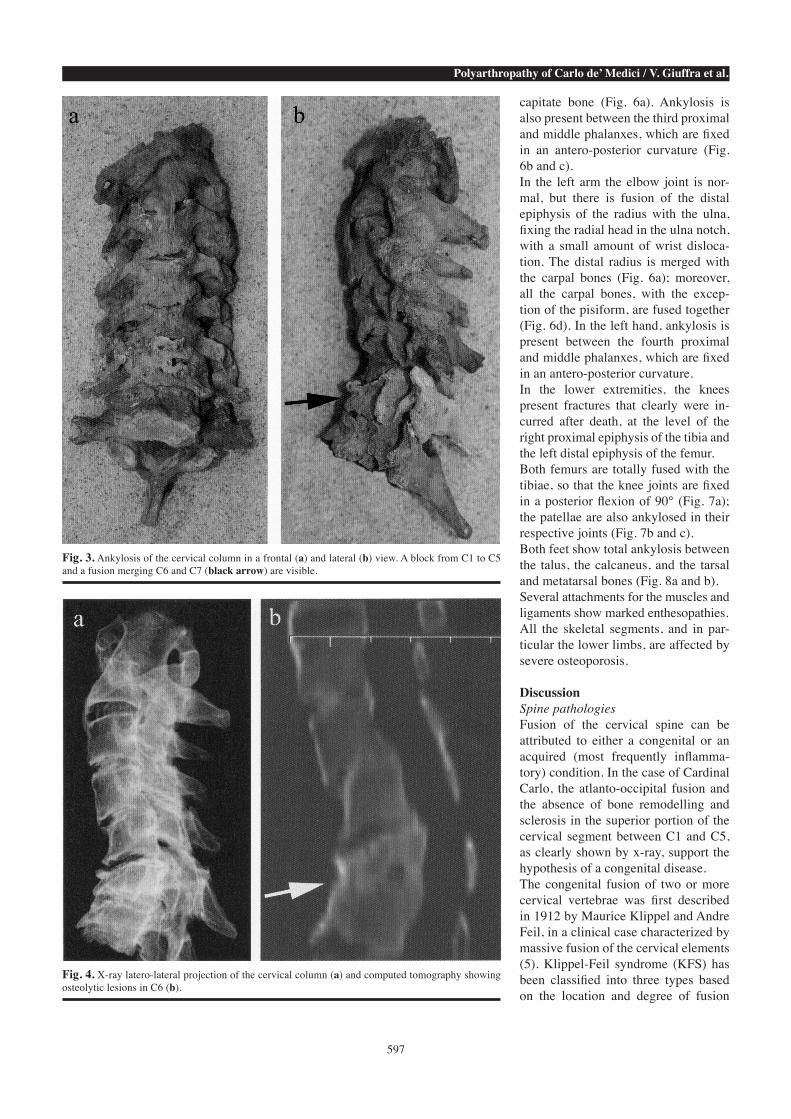
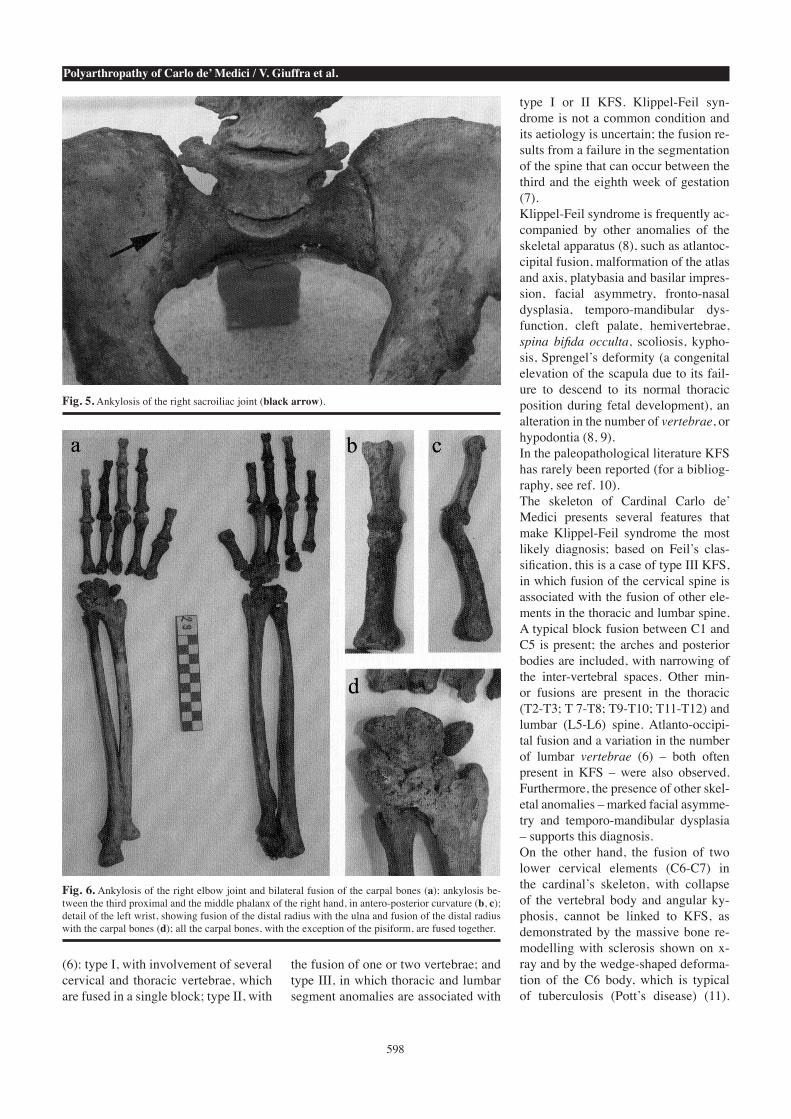
The facial skeleton presents asymme-try of the nasal and maxillary bones, which appear larger on the left side (Fig. 2a). There is marked hypoplasia of the right hemi-mandible corpus and the right ramus (Fig. 2b). The atlas is totally fused with the occip-ital bone. The cervical column is fused in a single block from the first to the fifth vertebrae, involving the articular facets, pillars and posterior portions of the vertebral bodies, with narrow disc spaces. The axis is fixed in an abnormal oblique right-directed position (Figs. 3 and 4). This anomaly probably resulted in a chronic torticollis in vita.A second block at the level of C6-C7 and involving the entire vertebral bod-ies also shows a wedge-shaped col-lapse, with the formation of an angular kyphosis (Fig. 3b, black arrow). CT demonstrated some osteolytic lesions with no sclerotic margins (Fig. 4b, white arrow).Several other column fusions involving the articular facets, pillars and posterior vertebral bodies are present: T2 is merged with T3, T7 with T8, T9 with T10, and T11 with T12. Ossification of the ante-rior right vertebral ligament at the level of the T9 and T10 vertebral bodies led to the formation of a bony bridge. The remaining vertebrae are normal. With regard to the lumbar spine, L5 is fused with a supernumerary L6. Fi-nally, the right innominate bone of the pelvis is fused with the sacrum, while the left sacro-iliac joint is normal (Fig. 5, black arrow).In the thoracic area, the manubrium of the sternum presents marked ossifica-tion of the sterno-costal cartilage on both sides, which is more severe on the left. The internal surfaces of most of the ribs show a slight diffuse periostitis. The right humerus and ulna are totally fused, with the elbow fixed in a flex-ion of 110°. In addition, the head of the radius is partially fused with the radial notch of the ulna. The right wrist shows total ankylosis of the distal epiphysis of the radius with the carpal bones, which are also fused together, with the exception of the hamate and the pisi-form. In the right hand, the second met-acarpal is merged with the trapezoid bone and the third metacarpal with the
Fig. 1. A portrait of Cardinal Carlo de’ Medici by Justus Sus-termans (Galleria Pa-latina).
Fig. 2. Antero-posterior projection of the skull showing evident asymmetry of the facial skeleton at the level of the nasal and maxillary bones, which appear larger on the left side: (a); marked hypoplasia of the right hemi-mandible corpus of the mandible, visible on x-ray (b).
597
Polyarthropathy of Carlo de’ Medici / V. Giuffra et al.
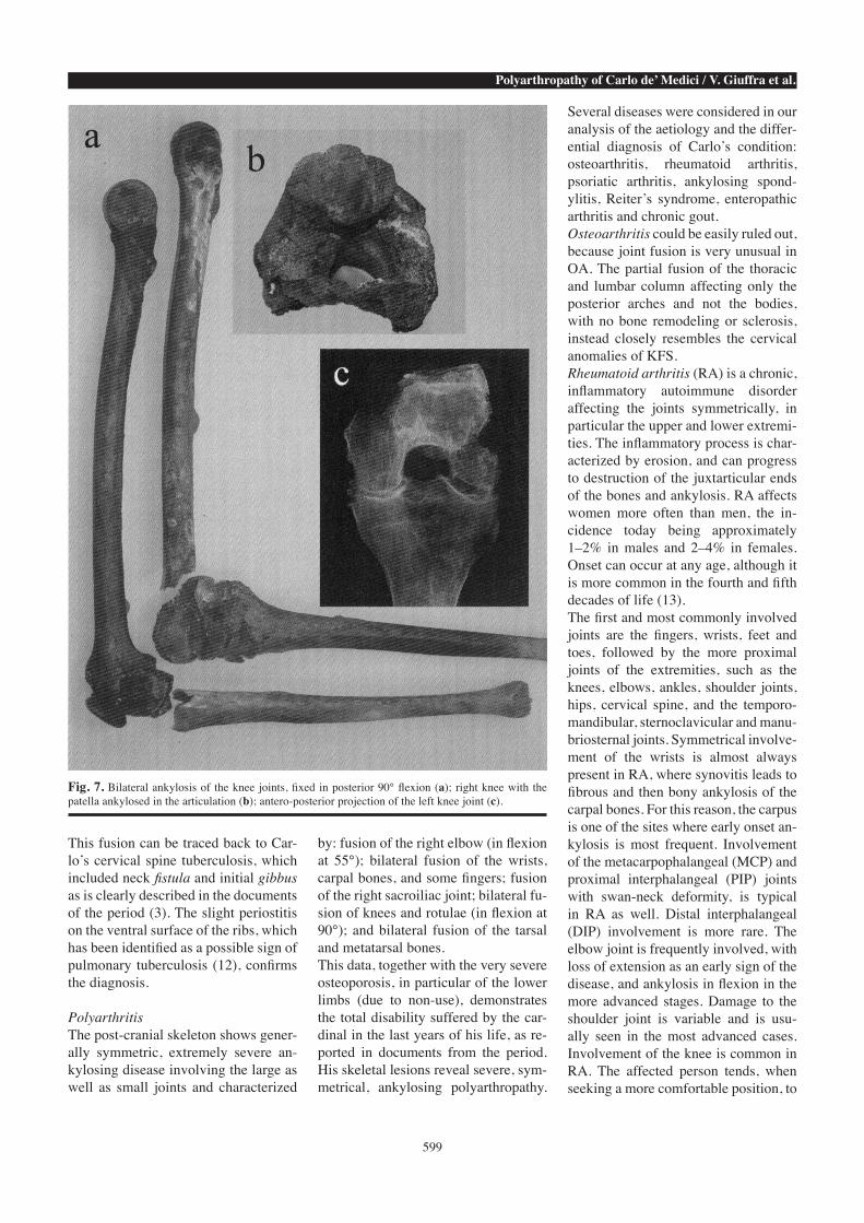
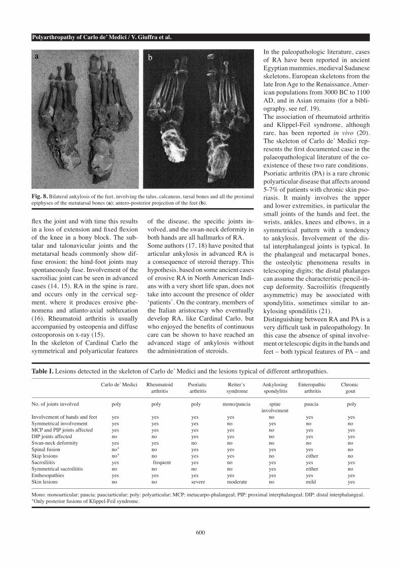
capitate bone (Fig. 6a). Ankylosis is also present between the third proximal and middle phalanxes, which are fixed in an antero-posterior curvature (Fig. 6b and c). In the left arm the elbow joint is nor-mal, but there is fusion of the distal epiphysis of the radius with the ulna, fixing the radial head in the ulna notch, with a small amount of wrist disloca-tion. The distal radius is merged with the carpal bones (Fig. 6a); moreover, all the carpal bones, with the excep-tion of the pisiform, are fused together (Fig. 6d). In the left hand, ankylosis is present between the fourth proximal and middle phalanxes, which are fixed in an antero-posterior curvature.In the lower extremities, the knees present fractures that clearly were in-curred after death, at the level of the right proximal epiphysis of the tibia and the left distal epiphysis of the femur. Both femurs are totally fused with the tibiae, so that the knee joints are fixed in a posterior flexion of 90° (Fig. 7a); the patellae are also ankylosed in their respective joints (Fig. 7b and c).Both feet show total ankylosis between the talus, the calcaneus, and the tarsal and metatarsal bones (Fig. 8a and b). Several attachments for the muscles and ligaments show marked enthesopathies. All the skeletal segments, and in par-ticular the lower limbs, are affected by severe osteoporosis.
DiscussionSpine pathologiesFusion of the cervical spine can be attributed to either a congenital or an acquired (most frequently inflamma-tory) condition. In the case of Cardinal Carlo, the atlanto-occipital fusion and the absence of bone remodelling and sclerosis in the superior portion of the cervical segment between C1 and C5, as clearly shown by x-ray, support the hypothesis of a congenital disease. The congenital fusion of two or more cervical vertebrae was first described in 1912 by Maurice Klippel and Andre Feil, in a clinical case characterized by massive fusion of the cervical elements (5). Klippel-Feil syndrome (KFS) has been classified into three types based on the location and degree of fusion
Fig. 3. Ankylosis of the cervical column in a frontal (a) and lateral (b) view. A block from C1 to C5 and a fusion merging C6 and C7 (black arrow) are visible.
Fig. 4. X-ray latero-lateral projection of the cervical column (a) and computed tomography showing osteolytic lesions in C6 (b).
598
Polyarthropathy of Carlo de’ Medici / V. Giuffra et al.
(6): type I, with involvement of several cervical and thoracic vertebrae, which are fused in a single block; type II, with
the fusion of one or two vertebrae; and type III, in which thoracic and lumbar segment anomalies are associated with
type I or II KFS. Klippel-Feil syn-drome is not a common condition and its aetiology is uncertain; the fusion re-sults from a failure in the segmentation of the spine that can occur between the third and the eighth week of gestation (7). Klippel-Feil syndrome is frequently ac-companied by other anomalies of the skeletal apparatus (8), such as atlantoc-cipital fusion, malformation of the atlas and axis, platybasia and basilar impres-sion, facial asymmetry, fronto-nasal dysplasia, temporo-mandibular dys-function, cleft palate, hemivertebrae, spina bifida occulta, scoliosis, kypho-sis, Sprengel’s deformity (a congenital elevation of the scapula due to its fail-ure to descend to its normal thoracic position during fetal development), an alteration in the number of vertebrae, or hypodontia (8, 9). In the paleopathological literature KFS has rarely been reported (for a bibliog-raphy, see ref. 10). The skeleton of Cardinal Carlo de’ Medici presents several features that make Klippel-Feil syndrome the most likely diagnosis; based on Feil’s clas-sification, this is a case of type III KFS, in which fusion of the cervical spine is associated with the fusion of other ele-ments in the thoracic and lumbar spine. A typical block fusion between C1 and C5 is present; the arches and posterior bodies are included, with narrowing of the inter-vertebral spaces. Other min-or fusions are present in the thoracic (T2-T3; T 7-T8; T9-T10; T11-T12) and lumbar (L5-L6) spine. Atlanto-occipi-tal fusion and a variation in the number of lumbar vertebrae (6) – both often present in KFS – were also observed. Furthermore, the presence of other skel-etal anomalies – marked facial asymme-try and temporo-mandibular dysplasia – supports this diagnosis.On the other hand, the fusion of two lower cervical elements (C6-C7) in the cardinal’s skeleton, with collapse of the vertebral body and angular ky-phosis, cannot be linked to KFS, as demonstrated by the massive bone re-modelling with sclerosis shown on x-ray and by the wedge-shaped deforma-tion of the C6 body, which is typical of tuberculosis (Pott’s disease) (11).
Fig. 5. Ankylosis of the right sacroiliac joint (black arrow).
Fig. 6. Ankylosis of the right elbow joint and bilateral fusion of the carpal bones (a); ankylosis be-tween the third proximal and the middle phalanx of the right hand, in antero-posterior curvature (b, c); detail of the left wrist, showing fusion of the distal radius with the ulna and fusion of the distal radius with the carpal bones (d); all the carpal bones, with the exception of the pisiform, are fused together.
599
Polyarthropathy of Carlo de’ Medici / V. Giuffra et al.
This fusion can be traced back to Car-lo’s cervical spine tuberculosis, which included neck fistula and initial gibbus as is clearly described in the documents of the period (3). The slight periostitis on the ventral surface of the ribs, which has been identified as a possible sign of pulmonary tuberculosis (12), confirms the diagnosis.
PolyarthritisThe post-cranial skeleton shows gener-ally symmetric, extremely severe an-kylosing disease involving the large as well as small joints and characterized
by: fusion of the right elbow (in flexion at 55°); bilateral fusion of the wrists, carpal bones, and some fingers; fusion of the right sacroiliac joint; bilateral fu-sion of knees and rotulae (in flexion at 90°); and bilateral fusion of the tarsal and metatarsal bones.This data, together with the very severe osteoporosis, in particular of the lower limbs (due to non-use), demonstrates the total disability suffered by the car-dinal in the last years of his life, as re-ported in documents from the period. His skeletal lesions reveal severe, sym-metrical, ankylosing polyarthropathy.
Several diseases were considered in our analysis of the aetiology and the differ-ential diagnosis of Carlo’s condition: osteoarthritis, rheumatoid arthritis, psoriatic arthritis, ankylosing spond-ylitis, Reiter’s syndrome, enteropathic arthritis and chronic gout.Osteoarthritis could be easily ruled out, because joint fusion is very unusual in OA. The partial fusion of the thoracic and lumbar column affecting only the posterior arches and not the bodies, with no bone remodeling or sclerosis, instead closely resembles the cervical anomalies of KFS.Rheumatoid arthritis (RA) is a chronic, inflammatory autoimmune disorder affecting the joints symmetrically, in particular the upper and lower extremi-ties. The inflammatory process is char-acterized by erosion, and can progress to destruction of the juxtarticular ends of the bones and ankylosis. RA affects women more often than men, the in-cidence today being approximately 1–2% in males and 2–4% in females. Onset can occur at any age, although it is more common in the fourth and fifth decades of life (13). The first and most commonly involved joints are the fingers, wrists, feet and toes, followed by the more proximal joints of the extremities, such as the knees, elbows, ankles, shoulder joints, hips, cervical spine, and the temporo-mandibular, sternoclavicular and manu-briosternal joints. Symmetrical involve-ment of the wrists is almost always present in RA, where synovitis leads to fibrous and then bony ankylosis of the carpal bones. For this reason, the carpus is one of the sites where early onset an-kylosis is most frequent. Involvement of the metacarpophalangeal (MCP) and proximal interphalangeal (PIP) joints with swan-neck deformity, is typical in RA as well. Distal interphalangeal (DIP) involvement is more rare. The elbow joint is frequently involved, with loss of extension as an early sign of the disease, and ankylosis in flexion in the more advanced stages. Damage to the shoulder joint is variable and is usu-ally seen in the most advanced cases. Involvement of the knee is common in RA. The affected person tends, when seeking a more comfortable position, to
Fig. 7. Bilateral ankylosis of the knee joints, fixed in posterior 90° flexion (a); right knee with the patella ankylosed in the articulation (b); antero-posterior projection of the left knee joint (c).
600
Polyarthropathy of Carlo de’ Medici / V. Giuffra et al.
flex the joint and with time this results in a loss of extension and fixed flexion of the knee in a bony block. The sub-talar and talonavicular joints and the metatarsal heads commonly show dif-fuse erosion; the hind-foot joints may spontaneously fuse. Involvement of the sacroiliac joint can be seen in advanced cases (14, 15). RA in the spine is rare, and occurs only in the cervical seg-ment, where it produces erosive phe-nomena and atlanto-axial subluxation (16). Rheumatoid arthritis is usually accompanied by osteopenia and diffuse osteoporosis on x-ray (15). In the skeleton of Cardinal Carlo the symmetrical and polyarticular features
of the disease, the specific joints in-volved, and the swan-neck deformity in both hands are all hallmarks of RA. Some authors (17, 18) have posited that articular ankylosis in advanced RA is a consequence of steroid therapy. This hypothesis, based on some ancient cases of erosive RA in North American Indi-ans with a very short life span, does not take into account the presence of older ‘patients’. On the contrary, members of the Italian aristocracy who eventually develop RA, like Cardinal Carlo, but who enjoyed the benefits of continuous care can be shown to have reached an advanced stage of ankylosis without the administration of steroids.
In the paleopathologic literature, cases of RA have been reported in ancient Egyptian mummies, medieval Sudanese skeletons, European skeletons from the late Iron Age to the Renaissance, Amer-ican populations from 3000 BC to 1100 AD, and in Asian remains (for a bibli-ography, see ref. 19).The association of rheumatoid arthritis and Klippel-Feil syndrome, although rare, has been reported in vivo (20). The skeleton of Carlo de’ Medici rep-resents the first documented case in the palaeopathological literature of the co-existence of these two rare conditions.Psoriatic arthritis (PA) is a rare chronic polyarticular disease that affects around 5-7% of patients with chronic skin pso-riasis. It mainly involves the upper and lower extremities, in particular the small joints of the hands and feet, the wrists, ankles, knees and elbows, in a symmetrical pattern with a tendency to ankylosis. Involvement of the dis-tal interphalangeal joints is typical. In the phalangeal and metacarpal bones, the osteolytic phenomena results in telescoping digits; the distal phalanges can assume the characteristic pencil-in-cup deformity. Sacroiliitis (frequently asymmetric) may be associated with spondylitis, sometimes similar to an-kylosing spondilitis (21).Distinguishing between RA and PA is a very difficult task in paleopathology. In this case the absence of spinal involve-ment or telescopic digits in the hands and feet – both typical features of PA – and
Fig. 8. Bilateral ankylosis of the feet, involving the talus, calcaneus, tarsal bones and all the proximal epiphyses of the metatarsal bones (a); antero-posterior projection of the feet (b).
Table I. Lesions detected in the skeleton of Carlo de’ Medici and the lesions typical of different arthropathies.
Carlo de’ Medici Rheumatoid Psoriatic Reiter’s Ankylosing Enteropathic Chronic arthritis arthritis syndrome spondylitis arthritis gout
No. of joints involved poly poly poly mono/paucia spine paucia poly involvement Involvement of hands and feet yes yes yes yes no yes yesSymmetrical involvement yes yes yes no yes no noMCP and PIP joints affected yes yes yes yes no yes yesDIP joints affected no no yes yes no yes yesSwan-neck deformity yes yes no no no no noSpinal fusion no* no yes yes yes yes noSkip lesions no* no yes yes no either noSacroiliitis yes frequent yes no yes yes yesSymmetrical sacroiliitis no no no no yes either noEnthesopathies yes yes yes yes yes yes yesSkin lesions no no severe moderate no mild yes
Mono: monoarticular; paucia: pauciarticular; poly: polyarticular; MCP: metacarpo-phalangeal; PIP: proximal interphalangeal; DIP: distal interphalangeal.*Only posterior fusions of Klippel-Feil syndrome.
601
Polyarthropathy of Carlo de’ Medici / V. Giuffra et al.
the presence of diffuse ankylosis, which is uncommon (except in the carpus) in PA, allow us to rule out the diagnosis of PA. Finally, the distal epiphyses of the middle phalanxes of the hands and feet do not show any joint lesions.Alongside these bone features, a further important observation must be kept in mind. If Carlo de’ Medici suffered from psoriatic arthritis, he would also prob-ably have experienced some cutaneous manifestations. A rare form of psoriatic arthritis sine psoriasis, in which the onset of rheumatic manifestations pre-cedes by several years the development of skin lesions, has been described (22), but Carlo was suffering from advanced stage arthritis that should necessarily have been accompanied by skin psoria-sis. It is unlikely that the highly visible dermatologic manifestations of pso-riasis would not have been described, if present, in the reports of the Medici court physicians and ambassadors, which were generally quite detailed. On the contrary, in the many chronicles of the cardinal’s life conserved in the Florentine archives, no mention of any dermatologic problems can be found (3).Ankylosing spondylitis (AS) is a chron-ic systemic inflammatory disease that primarily involves the axial skeleton. The most obvious manifestation of AS is axial arthritis, such as sacroiliitis or spondylitis. Involvement of the periph-eral limb joints is uncommon (23). In the case of Cardinal Carlo, the ab-sence of axial arthritis and a complete ossification of the spine, which are the main features of AS, and the presence of severe involvement of the hands and feet, which are on the contrary uncom-mon in this arthropathy, rule out the di-agnosis of AS.Reiter’s syndrome (RS) is a rare joint disorder that develops in response to an infection in another part of the body, in particular in the bowels or the urogeni-tal tract. The syndrome is characterized by asymmetrical monoarthritis or oli-goarthritis involving the lower as well as the upper extremities. Sacroiliitis and spondylitis are common findings (24). The symmetrical and polyarticu-lar involvement seen in the cardinal’s skeleton, however, allows us to exclude a diagnosis of RS.
Enteropathic arthritis is associated with some chronic diseases of the intestinal tract (Crohn’s disease, Whipple’s disease, ulcerative colitis) and manifests primari-ly as spondylitis and sacroiliitis. Both the large and the small joints may be affect-ed, in particular those of the lower limbs, but the involvement is pauciarticular and asymmetrical (25). The principal features of enteropathic arthritis – i.e. spondylitis and asymmetrical and pauciarticular in-volvement – were not observed in the skeleton of Carlo de’ Medici.Chronic gout is a crystal-induced ar-thropathy associated with tophus for-mation and bone and joint destruction, and is most commonly seen in middle-aged males (26). The chronic poly-ar-ticular form of tophaceous gout can mimic polyarticular diseases, including RA (27). However, in chronic gout the joint involvement is asymmetric and fusion is only occasional, features that allow us to rule out this diagnosis. In conclusion, Cardinal Carlo de’ Medi-ci was affected by Klippel-Feil syn-drome type III, a congenital anomaly of the cervical spine in which ankylosis of the cervical column is associated with the fusion of other elements in the tho-racic and lumbar segments. There are also clear signs of tuberculosis of the cervical column (Pott’s disease), thus confirming the information conveyed in historical documents. The cardinal was also affected by severe ankylosing polyarthritis involving the large and small joints of the post-cra-nial skeleton. The differential diagnosis demonstrates the presence of rheuma-toid arthritis (RA), as in another 16th century Italian case (19) with positivity for the DRB1*0101 allele that confers a genetic predisposition to European RA (28, 29). These important findings clearly demon-strate the existence in the 16th century of an Old World variant of RA that follows the pattern of the treponematoses (11). We can presume that this disease was present in Europe before the discovery of America by Columbus in 1492.
AcknowledgementsWe wish to thank Dr. Laura Cignoni of the National Research Council in Pisa for her help in the translation of this manuscript.
References 1. FORNACIARI G, VITIELLO A, GIUSIANI S,
GIUFFRA V, FORNACIARI A: The “Medici Project”: first results of the explorations of the Medici tombs in Florence (15th-18th cen-turies). Paleopathol Newsl 2006; 133: 15-22.
2. FORNACIARI G, VITIELLO A, GIUSIANI S, GIUFFRA V, FORNACIARI A, VILLARI N: The “Medici Project”: first anthropological and paleopathological results of the explo-ration of the Medici tombs in Florence (15th -18th centuries). Med Secoli 2007; 19: 521-44.
3. PIERACCINI G: La Stirpe dei Medici di Ca-faggiolo, vol. 2, Florence, Nardini Editore, 1986.
4. GENNA G: Ricerche antropologiche sulla famiglia dei Medici. Atti Accad Naz Lincei, Classe di Scienze Fisiche, Matematiche e Naturali, Serie VIII 1948; 15: 589-93.
5. KLIPPEL M, FEIL A: un cas d’absence des vertèbres cervicales, cage thoracique remon-tant jusqu’à la base du crâne. Nouvelle Icon-ographie de la Salpêtrière 1912; 25: 223-50.
6. BARNES E: Developmental Defects of the Axial Skeleton in Paleopathology, Colorado, University Press Colorado, 1994.
7. AUFDERHEIDE AC, RODRIGUEZ-MARTIN CR: The Cambridge Encyclopedia of Human Paleopathology, Cambridge, Cambridge University Press, 1998.
8. HENSINGER RN, LANG JE, MACEWEN GD: Klippel-Feil syndrome: a constellation of as-sociated anomalies. J Bone Joint Surg 1974; 56: 1246-53.
9. OZDILER E, AKCAM MO, SAYIN MO: Cranio-facial characteristics of Klippel-Feil syn-drome in an eight-year-old female. J Clin Pediatr Dent 2000; 24: 249-54.
10. PANY D, TESCHLER-NICOLA M: Klippel-Feil syndrome in an early Hungarian period juve-nile skeleton from Austria. Int J Osteoarch 2007; 17: 403-15.
11. ORTNER DJ: Identification of Pathological Conditions in Human Skeletal Remains. New York, Smithsonian Institution Press, 2003.
12. KELLEY MA, MICOZZI M: Rib lesions in pul-monary tuberculosis. Am J Phys Anthropol 1984; 65: 381-6.
13. MACGREGOR AJ, SILMAN AJ: Rheumatoid arthritis. Classification and epidemiology. In KLIPPEL JH and DIEPPE PA (Eds): Rheuma-tology, London, Mosby 1998: 5.2.
14. DIXON AJ, LIENCE E: Sacro-iliac joint in adult rheumatoid arthritis and psoriatic arthropathy. Ann Rheum Dis 1961; 20: 247-57.
15. BROWER AC: Rheumatoid arthritis. Imaging. In KLIPPEL JH and DIEPPE PA (Eds): Rheu-matology, London, Mosby 1998: 5.5.
16. GORDON DA, HASTINGS DE: Rheumatoid ar-thritis. Clinical features of early, progressive and late disease. In KLIPPEL JH and DIEPPE PA (Eds): Rheumatology, London, Mosby 1998: 5.3.
17. ROTHSCHILD BM, WOODS RJ: Symmetric- al erosive arthritis in Archaic Indians: The origin of rheumatoid arthritis in the New World? Semin Arthritis Rheum 1990; 19: 278.
18. ROTHSCHILD BM, WOODS RJ, ROTHSCHILD

602
Polyarthropathy of Carlo de’ Medici / V. Giuffra et al.
C, SEBES JI: Geographic distribution of rheu-matoid arthritis in ancient North America: implication for pathogenesis. Semin Arthritis Rheum 1992; 22: 181-7.
19. CIRANNI R, GARBINI F, NERI E, MELAI L, GIUSTI L, FORNACIARI G: The “Braids Lady” of Arezzo: a case of rheumatoid arthritis in a 16th century mummy. Clin Exp Rheumatol 2002; 20: 745-52.
20. LUNA E, NANNINI G: Sindrome di Klip-pel-Feil associata ad artrite reumatoide. Descrizione clinico-radiografica di un caso. Chir Ital 1981; 33 (Suppl. 1): 68-75.
21. HELLIWELL PS, WRIGHT V: Spondyloar-thropathies. Psoriatic arthritis: Clinical fea-tures. In KLIPPEL JH and DIEPPE PA (Eds): Rheumatology, London, Mosby 1998: 6.21.
22. BARTH WF: Arthritis sine psoriasis. In KLIP-PEL JH and DIEPPE PA (Eds): Rheumatology, London, Mosby 1998: 6.25.
23. GRAN JT, HUSBY G: Ankylosing spondylitis: Prevalence and demography. In KLIPPEL JH and DIEPPE PA (Eds): Rheumatology, Lon-don, Mosby 1998: 6.15.
24. TOIVANEN A: Spondyloarthropathies. Re-active arthritis and Reiter’s syndrome: his-tory and clinical features. In KLIPPEL JH and DIEPPE PA (Eds): Rheumatology, London, Mosby, 1998: 6.11.
25. VEYS EM, MIELANTS H: Spondyloarthropa-thies. Enteropathic arthropathies. In KLIP-PEL JH and DIEPPE PA (Eds): Rheumatology, London, Mosby, 1998: 6.24.
26. COHEN MG, EMMERSON BT: Gout. In KLIP-
PEL JH and DIEPPE PA (Eds): Rheumatology, London, Mosby 1998: 8.14.
27. SHAPIRA D, STAHL S, IZHAK OB, BALBIR-GURMAN A, NAHIR AM: Chronic tophaceous gouty arthritis mimicking rheumatoid arthri-tis. Semin Arthritis Rheum 1999; 29: 56-63.
28. FONTECCHIO G, VENTURA L, AZZARONE R, FIORONI MA, FORNACIARI G, PAPOLA F: HLA-DRB genotyping of an Italian mummy from the 16th century with signs of rheu-matoid arthritis. Ann Rheum Dis 2006; 65: 1676-7.
29. FONTECCHIO G, FIORONI MA, AZZARONE R et al.: Genetic predisposition to rheumatoid arthritis in a Tuscan (Italy) ancient human re-mains. Int J Immunopathol Pharmacol 2007; 20: 103-9.
Related Documents


![BMP-13 Emerges as a Potential Inhibitor of Bone Formation · ated with Klippel-Feil syndrome (KFS), characterised by congenital fusion of the cervical spine vertebrae [14], and caused](https://static.cupdf.com/doc/110x72/5f76c2b3072b6218ec0c57ad/bmp-13-emerges-as-a-potential-inhibitor-of-bone-formation-ated-with-klippel-feil.jpg)