Full Terms & Conditions of access and use can be found at https://www.tandfonline.com/action/journalInformation?journalCode=imor20 Modern Rheumatology ISSN: 1439-7595 (Print) 1439-7609 (Online) Journal homepage: https://www.tandfonline.com/loi/imor20 Rheumatic diseases associated with immune checkpoint inhibitors in cancer immunotherapy Kei Ohnuma, Ryo Hatano, Nam H. Dang & Chikao Morimoto To cite this article: Kei Ohnuma, Ryo Hatano, Nam H. Dang & Chikao Morimoto (2018): Rheumatic diseases associated with immune checkpoint inhibitors in cancer immunotherapy, Modern Rheumatology, DOI: 10.1080/14397595.2018.1532559 To link to this article: https://doi.org/10.1080/14397595.2018.1532559 Accepted author version posted online: 04 Oct 2018. Published online: 20 Dec 2018. Submit your article to this journal Article views: 100 View Crossmark data

Welcome message from author
This document is posted to help you gain knowledge. Please leave a comment to let me know what you think about it! Share it to your friends and learn new things together.
Transcript
Full Terms & Conditions of access and use can be found athttps://www.tandfonline.com/action/journalInformation?journalCode=imor20
Modern Rheumatology
ISSN: 1439-7595 (Print) 1439-7609 (Online) Journal homepage: https://www.tandfonline.com/loi/imor20
Rheumatic diseases associated with immunecheckpoint inhibitors in cancer immunotherapy
Kei Ohnuma, Ryo Hatano, Nam H. Dang & Chikao Morimoto
To cite this article: Kei Ohnuma, Ryo Hatano, Nam H. Dang & Chikao Morimoto (2018):Rheumatic diseases associated with immune checkpoint inhibitors in cancer immunotherapy,Modern Rheumatology, DOI: 10.1080/14397595.2018.1532559
To link to this article: https://doi.org/10.1080/14397595.2018.1532559
Accepted author version posted online: 04Oct 2018.Published online: 20 Dec 2018.
Submit your article to this journal
Article views: 100
View Crossmark data
REVIEW ARTICLE
Rheumatic diseases associated with immune checkpoint inhibitors in cancerimmunotherapy
Kei Ohnumaa, Ryo Hatanoa, Nam H. Dangb and Chikao Morimotoa
aDepartment of Therapy Development and Innovation for Immune Disorders and Cancers, Graduate School of Medicine, JuntendoUniversity, Tokyo, Japan; bDivision of Hematology/Oncology, University of Florida, Gainesville, FL, USA
ABSTRACTImmune checkpoint inhibitors (ICIs) have drastically altered cancer treatment paradigms, with increas-ing numbers of novel ICIs being currently evaluated in numerous clinical trials for various cancers. ICIsrelease ‘brakes’ against tumor immunity to control cancer growth through T cell-dependent anti-tumoractivity. Meanwhile, side effects associated with ICIs are directly related to their mechanism of action,as nonspecific immune activation targeting non-tumor organs results in undesirable off-target inflam-mation and autoimmunity. Accumulating data reveal that immune-related adverse events (irAEs) ofICIs in cancer patients can resemble various rheumatic diseases. Moreover, while patients with preex-isting rheumatic diseases can theoretically experience irAEs and disease flares, observational studieshave shown that ICIs can be used successfully in these patients. As ICIs continue to provide long-lasting disease control in cancer patients and their usage correspondingly increases, the rheumatolo-gist will be managing new ICI-associated clinical entities mimicking common autoimmune diseasesand will need to be prepared to rapidly diagnose and treat these irAEs. Early recognition andtreatment of these rheumatic adverse events will allow for improved outcomes and quality of life forcancer patients faced with previously rapidly fatal disease.
ARTICLE HISTORYReceived 19 July 2018Accepted 1 October 2018
KEYWORDSImmune checkpointinhibitor; immune-relatedadverse events;inflammatory arthritis;rheumatic disease
Introduction
Monoclonal antibodies (mAbs) against coinhibitory immunecheckpoint molecules have demonstrated clinical activitiesin various malignancies [1]. Targets include cytotoxic T-lymphocyte-associated antigen 4 (CTLA-4 or CD152),programmed cell-death protein 1 (PD-1 or CD279) and itsligand (PD-L1; B7-H1 or CD274), which negatively regulateT cell activation and T cell receptor (TCR) signaling,respectively. By disinhibiting these regulatory pathways,immune checkpoint inhibitors (ICIs) overcome self-toler-ance and promote T cell-mediated expansion, leading torobust anti-tumor immunity [1]. Originally approved by theUS Food and Drug Administration (FDA) for the treatmentof advanced melanoma [2], ICIs have led to a paradigmshift in the field of cancer therapy, with the list of indica-tions for ICI use in advanced cancers being now ever-expanding, to include non-small cell lung carcinoma, bladdercancer, head and neck squamous carcinoma, breast cancer,gastric cancer, colorectal carcinoma or solid tumors with highmicrosatellite instability or mismatch-repair deficiency, hepa-tocellular carcinoma, Merkel cell carcinoma, urothelial carcin-oma, Hodgkin’s lymphoma and leukemia [1].
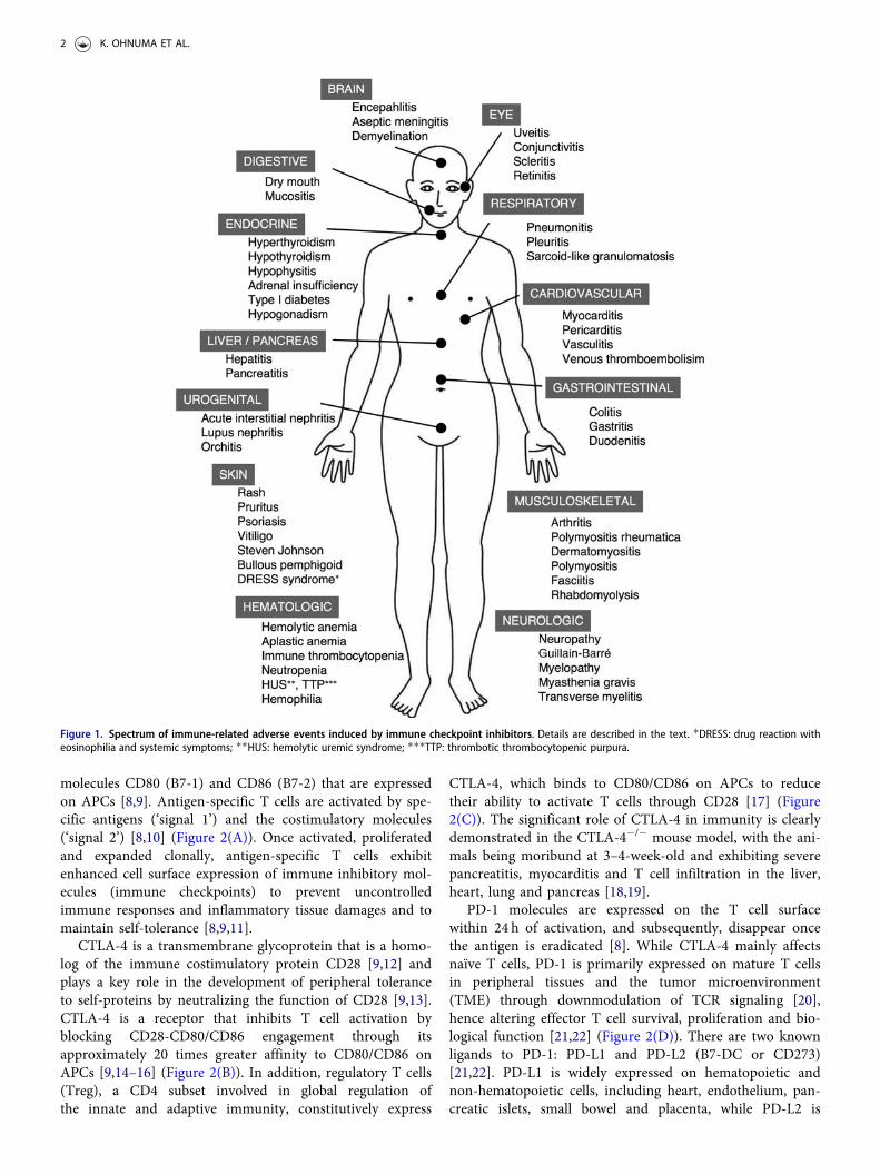
As a consequence of their mechanism of action, ICI ther-apy can induce nonspecific immune activation, which cantarget non-tumor tissues. These side effects are collectivelyreferred to as immune-related adverse events (irAEs) [3].
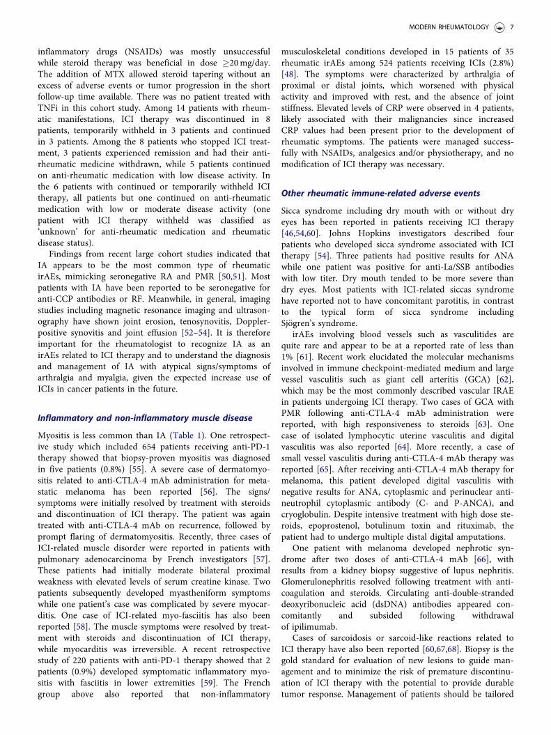
irAEs can resemble various rheumatic diseases, such asinflammatory arthritis (IA) [4], but also exhibit diversemanifestations throughout the body [5,6] (Figure 1). Asindications for ICIs use expand and as these novel agentsare combined with each other, it becomes increasinglyimportant for rheumatologists to recognize irAEs andappropriate management. In this paper, we summarize theunderlying immune mechanisms and the latest findingsregarding the rheumatic manifestations and the generalapproach to management of ICI-associated irAEs in cancerpatients treated with these novel agents. Reviewing manyrecently published work on rheumatic irAEs, this reviewwill provide rheumatologists an updated understanding ofthese emerging cancer therapies, with particularly a focus ontheir associated immunopathologic mechanisms and rheum-atic complications, and their management.
Normal immune response and immune homeostasis
The classical definition of immunity is protection frominfectious pathogens, and the mechanisms of host defensefall into two broad categories, innate immunity and adaptiveimmunity [7]. During the innate response process, activa-tion of antigen-presenting cells (APCs) leads to enhancedexpression of costimulatory molecules. The principal T cellcostimulatory molecule CD28 is recognized by the B7
CONTACT Kei Ohnuma [email protected] Department of Therapy Development and Innovation for Immune Disorders and Cancers, GraduateSchool of Medicine, Juntendo University, 2-1-1, Hongo, Bunkyo-ku, Tokyo 113-8421, Japan.� 2018 Japan College of Rheumatology
MODERN RHEUMATOLOGYhttps://doi.org/10.1080/14397595.2018.1532559
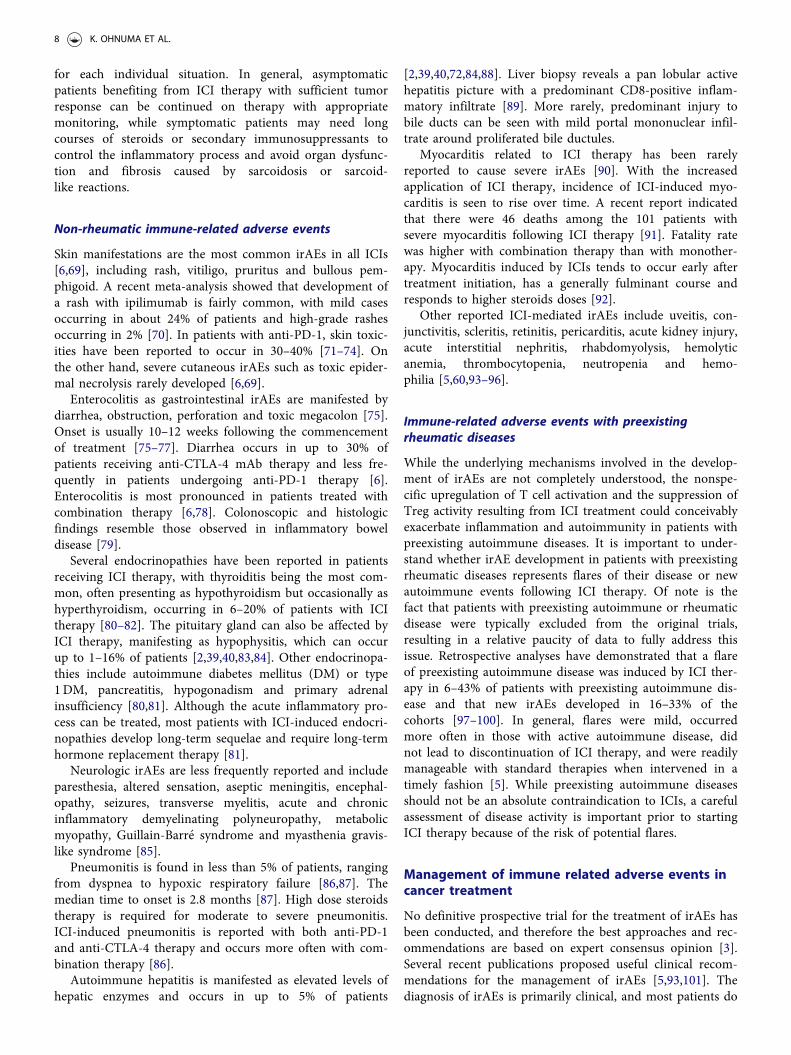
molecules CD80 (B7-1) and CD86 (B7-2) that are expressedon APCs [8,9]. Antigen-specific T cells are activated by spe-cific antigens (‘signal 1’) and the costimulatory molecules(‘signal 2’) [8,10] (Figure 2(A)). Once activated, proliferatedand expanded clonally, antigen-specific T cells exhibitenhanced cell surface expression of immune inhibitory mol-ecules (immune checkpoints) to prevent uncontrolledimmune responses and inflammatory tissue damages and tomaintain self-tolerance [8,9,11].
CTLA-4 is a transmembrane glycoprotein that is a homo-log of the immune costimulatory protein CD28 [9,12] andplays a key role in the development of peripheral toleranceto self-proteins by neutralizing the function of CD28 [9,13].CTLA-4 is a receptor that inhibits T cell activation byblocking CD28-CD80/CD86 engagement through itsapproximately 20 times greater affinity to CD80/CD86 onAPCs [9,14–16] (Figure 2(B)). In addition, regulatory T cells(Treg), a CD4 subset involved in global regulation ofthe innate and adaptive immunity, constitutively express
CTLA-4, which binds to CD80/CD86 on APCs to reducetheir ability to activate T cells through CD28 [17] (Figure2(C)). The significant role of CTLA-4 in immunity is clearlydemonstrated in the CTLA-4�/� mouse model, with the ani-mals being moribund at 3–4-week-old and exhibiting severepancreatitis, myocarditis and T cell infiltration in the liver,heart, lung and pancreas [18,19].
PD-1 molecules are expressed on the T cell surfacewithin 24 h of activation, and subsequently, disappear oncethe antigen is eradicated [8]. While CTLA-4 mainly affectsnaïve T cells, PD-1 is primarily expressed on mature T cellsin peripheral tissues and the tumor microenvironment(TME) through downmodulation of TCR signaling [20],hence altering effector T cell survival, proliferation and bio-logical function [21,22] (Figure 2(D)). There are two knownligands to PD-1: PD-L1 and PD-L2 (B7-DC or CD273)[21,22]. PD-L1 is widely expressed on hematopoietic andnon-hematopoietic cells, including heart, endothelium, pan-creatic islets, small bowel and placenta, while PD-L2 is
Figure 1. Spectrum of immune-related adverse events induced by immune checkpoint inhibitors. Details are described in the text. �DRESS: drug reaction witheosinophilia and systemic symptoms; ��HUS: hemolytic uremic syndrome; ���TTP: thrombotic thrombocytopenic purpura.
2 K. OHNUMA ET AL.
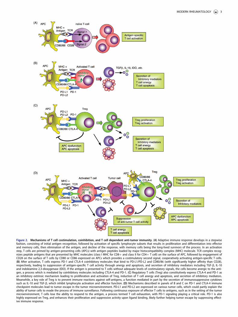
Figure 2. Mechanisms of T cell costimulation, coinhibition, and T cell dependent anti-tumor immunity. (A) Adaptive immune response develops in a stepwisefashion, consisting of initial antigen recognition, followed by activation of specific lymphocyte subsets that results in proliferation and differentiation into effectorand memory cells, then elimination of the antigen, and decline of the response, with memory cells being the long-lived survivors of the process. In an activationstep, T cells are primed by antigen-presenting cells (APCs) with antigen peptides loaded by major histocompatibility complex (MHC) molecule. TCR complex recog-nizes peptide antigens that are presented by MHC molecules (class I MHC for CD8þ and class II for CD4þ T cell) on the surface of APC, followed by engagement ofCD28 on the surface of T cells by CD80 or CD86 expressed on APCs which provides a costimulatory second signal, cooperatively activating antigen-specific T cells.(B) After activation, T cells express PD-1 and CTLA-4 coinhibitory molecules that bind to PD-L1/PD-L2 and CD80/86 (with significantly higher affinity than CD28),respectively, leading to suppression of antigen-specific T cell activity through anergy and apoptosis, and secretion of inhibitory mediators including TGF-b, IL-10and indoleamine 2,3-dioxygenase (IDO). If the antigen is presented to T cells without adequate levels of costimulatory signals, the cells become anergic to the anti-gen, a process which is mediated by coinhibitory molecules including CTLA-4 and PD-1. (C) Regulatory T cells (Treg) also constitutively express CTLA-4 and PD-1 asan inhibitory extrinsic mechanism leading to proliferation and activation of Treg, induction of T cell anergy and apoptosis, and secretion of inhibitory mediators.Meanwhile, a key role of Treg is to prevent immune reactions against self-antigens, a function mediated in part by the secretion of immunosuppressive cytokinessuch as IL-10 and TGF-b, which inhibit lymphocyte activation and effector function. (D) Mechanisms described in panels of B and C on PD-1 and CTLA-4 immunecheckpoint molecules lead to tumor escape in the tumor microenvironment. PD-L1 and PD-L2 are expressed on various tumor cells, which could partly explain theability of tumor cells to evade the process of immune surveillance. Following continuous exposure of effector T cells to antigens, such as in the setting of the tumormicroenvironment, T cells lose the ability to respond to the antigen, a process termed T cell exhaustion, with PD-1 signaling playing a critical role. PD-1 is alsohighly expressed on Treg, and enhances their proliferation and suppressive activity upon ligand binding, likely further helping tumor escape by suppressing effect-ive immune response.
MODERN RHEUMATOLOGY 3
expressed mainly on dendritic cells and macrophages [22].Induction of PD-L1 expression on tissue cells in the inflam-matory regions may be a protective mechanism to downre-gulate effector T cell activity and reduce immune-mediatedinjury [23] (Figure 2(B)). PD-1�/� mice demonstrate evi-dence of autoimmunity, specifically, mild lupus-like auto-immunity and dilated cardiomyopathy [23,24]. The PD-1knockout autoimmune effects appear to be less severe anddisplay a later onset than those observed in CTLA-4�/�
mice [22,25]. As is the case with CTLA-4, PD-1 is alsohighly expressed on Treg, and enhances their proliferationand suppressive activity upon ligand binding [26](Figure 2(C)).
An important group of diseases which reflects the failureof the normal control mechanisms described above is auto-immune diseases, which result from the lack of tolerance toself-antigens. The mechanisms of self-tolerance can bebroadly classified into two groups: central tolerance andperipheral tolerance [11]. In central tolerance, immatureself-reactive T and B lymphocyte clones that recognize self-antigens during their maturation in the central lymphoidorgans are eliminated or rendered harmless by negativeselection [11]. Autoreactive lymphocytes which manage toescape from the central tolerance mechanisms are subse-quently silenced in peripheral tolerance by anergy, Treg andapoptotic deletion [11] (Figure 2(B,C)).
Taken together, immune checkpoints such as CTLA-4and PD-1 systems are regulatory inhibitory pathways thatcontribute to immune homeostasis, being essential in pre-venting autoimmunity, maintaining self-tolerance and avoid-ing tissue damage that could result from persistentimmune activation.
Mechanism of action of immunecheckpoint inhibitors
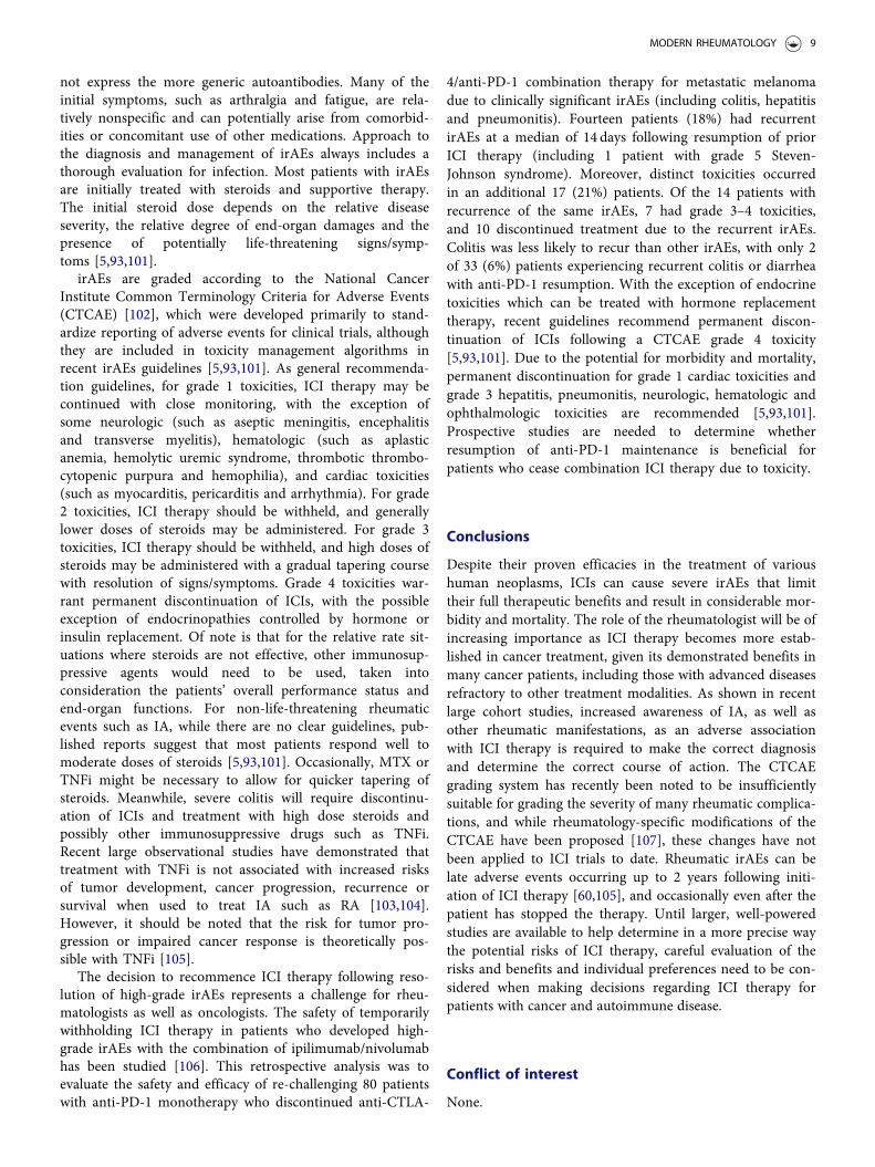
Multiple studies have demonstrated that many tumors usethe same pathways involved in immune regulation to evadeimmune attack [1]. This realization has led to the develop-ment of mAbs that block CTLA-4 and PD-1 for tumorimmunotherapy, by removing the brakes on the immuneresponse and promoting responses against tumors [1]. Thefirst approved ICI by FDA was ipilimumab, a fully humanIgG1 anti-CTLA-4 mAb, and subsequently, several agentsincluding anti-PD-1 mAb and anti-PD-L1 mAb have beendeveloped for clinical use as shown in Figure 3(A).
Anti-cytotoxic T-lymphocyte-associated antigen4 inhibitors
Following the discovery of the CTLA-4 receptor in 1986,work involving a murine preclinical model revealed theanti-tumor activity of anti-CTLA-4 Ab [13]. Clinical studiessubsequently demonstrated that ipilimumab extended sur-vival time by nearly four months in patients with advancedmelanoma [27,28]. Tremelimumab, a fully-human IgG2 thatalso targets CTLA-4, is currently under development asmonotherapy or combined therapy [29]. Treatment with
CTLA-4 mAb results in persistent T cell activation by block-ing the inhibitory pathway in the antigen priming phase(Figure 3(A,B)). Moreover, anti-CTLA-4 mAb-mediatedinhibition increases the ratio of effector T cells to Treg inthe TME, due to depletion of intratumoral Treg throughcomplement-dependent cytotoxicity (CDC) and antibody-dependent cell-mediated cytotoxicity (ADCC) [30] (Figure3(A)). Of note is that the therapeutic agent for rheumatoidarthritis (RA) abatacept, a fusion protein consisting of theextracellular domain of CTLA-4 and the Fc region of IgG1,acts in an opposite manner as ICIs, by facilitating coinhibi-tory signaling of T cells through its binding affinity forCD80/CD86 [31,32].
Anti-programmed cell-death protein-1 inhibitors
Generation of tumor-reactive CD8þ T cells requires thesuccessful processing and presentation of tumor-derivedpeptide antigens with class I major histocombatibility com-plex (MHC) molecules by APCs [10,33]. Once developed,tumor-specific CD8þ T cells subsequently differentiate intoeffector T cells, undergo clonal expansion, migrate to theTME, and ultimately eliminate tumor cells expressingtumor-specific antigens bound to class I MHC moleculesthrough the release of cytotoxic granules [10]. The presenceof enhanced PD-1 expression on CD8þ tumor infiltratinglymphocytes (TILs) may either reflect an anergic orexhausted state, consistent with the findings that cytokineproduction by PD-1þ TILs is decreased [34]. Initial studiesshowed that PD-1/PD-L1 blockade reversed the exhaustedstate of effector T cells in the TME, leading to the clinicaldevelopment of anti-PD-1 inhibitors for cancer immuno-therapy [20]. In addition, a large proportion of intratumoralCD4þ T cells are Treg with increased level of PD-1 expres-sion. These findings thus provide an important scientificrationale for a therapeutic approach involving anti-tumorimmunity through PD-1/PD-L1 blockade [35]. Currently,pembrolizumab, a humanized IgG4 mAb, and nivolumab, afully human IgG4 mAb, are approved as anti-PD-1 mAbsfor clinical use. Treatment with anti-PD-1 mAbs leads topersistent T cell activation by blocking the inhibitory path-way both in the antigen priming phase as well as theeffector phase (Figure 3(A,B)).
Anti-programmed cell-death protein-ligand 1 inhibitors
Atezolizumab is a humanized IgG1 anti-PD-L1 mAb, engi-neered to delete binding to the Fc receptor [36]. It upregu-lates T cell activation by blocking the interaction betweenPD-1 and PD-L1 or CD80 and PD-L1, with a safety profilesimilar to that of anti-PD-1 mAbs [37]. Other novel anti-PD-L1 mAbs being evaluated currently in various clinicaltrials are the fully human IgG1 mAbs durvalumaband avelumab.
4 K. OHNUMA ET AL.
Combined therapy
The combination of ipilimumab and nivolumab has beenapproved for the treatment of metastatic melanoma by theUS-FDA [38]. Other combined ICI therapy such as tremeli-mumab and durvalumab is under clinical trials for variouscancers. Although these combinations may improve efficacy,they can result in significantly increased toxicity [3,6,39–43].
Immune-related adverse events
As discussed earlier, human immune system normally existsin a state of equilibrium in which lymphocyte activation for
protection against pathogens is delicately balanced by themechanisms of tolerance to prevent deleterious reactionsagainst self-antigens, and the failure of tolerance allows forresponses against self-antigens, leading to autoimmune dis-eases [8,11,44]. Consequently, ICI-mediated blocking of theinhibitory checkpoints can enhance immune activation toresult in unwanted off-target effects, including immune-related and inflammatory events [3–6]. Involving any organsystem (Figure 1), irAEs from ICIs are increasingly recognizedas unique entities mimicking classical rheumatic diseases [4].The accurate diagnosis and management of these side effectsare of the utmost importance, given the fact that the use ofICIs in cancer patients with preexisting autoimmune disease
Figure 3. Points of action of anti-PD-1, anti-PD-L1 and anti-CTLA-4 inhibitors. (A) Anti-CTLA-4 inhibitor prevents CTLA-4 from binding to CD80/86, reinvigorat-ing the inhibited T cell. Anti-PD-1/PD-L1 inhibitors restore down-modulated TCR signaling and reinvigorate the exhausted T cell. Anti-CTLA-4 and anti-PD-1/PD-L1inhibitors also deplete regulatory T cells (Treg). (B) Cycle of tumor antigen loading to antigen-presenting cells (APCs), migration to lymph node of APC, tumor-spe-cific T cell activation by antigen-loaded APC, accumulation of activated tumor-specific T cells in the tumor microenvironment and targeting of tumor cells.Activation of Treg concomitantly leads to tumor escape. Anti-CTLA-4 inhibitor results in persistent T cell activation by blockade of inhibitory pathway in antigen pri-ming phase. Anti-PD-1/PD-L1 inhibitors result in persistent T cell activation by blockade of inhibitory pathway both in antigen priming phase and in effector phase.They also exert anti-tumor activity through depletion and suppression of Treg.
MODERN RHEUMATOLOGY 5
is expected to increase in the future as ICI therapy becomesmore prevalent in a variety of human neoplasms [3].
Arthritis
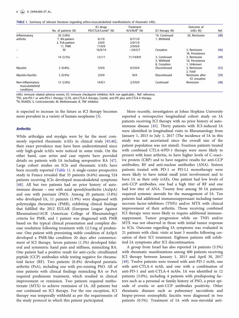
While arthralgia and myalgia were by far the most com-monly reported rheumatic irAEs in clinical trials [45,46],their exact prevalence may have been underestimated sinceonly high-grade irAEs were noticed in some trials. On theother hand, case series and case reports have provideddetails on patients with IA including seropositive RA [47].Large cohort studies on ICIs and rheumatic irAEs havebeen recently reported (Table 1). A single-center prospectivestudy in France revealed that 35 patients (6.6%) among 524patients receiving ICIs developed musculoskeletal symptoms[48]. All but two patients had no prior history of auto-immune disease – one with axial spondyloarthritis (AxSpA)and one with psoriasis (PSO). Among 20 patients (3.8%)who developed IA, 11 patients (1.9%) were diagnosed withpolymyalgia rheumatica (PMR), exhibiting clinical findingsthat fulfilled the 2012 EULAR (European League AgainstRheumatism)/ACR (American College of Rheumatology)criteria for PMR, and 1 patient was diagnosed with PMRbased on the typical clinical presentation and complete dis-ease resolution following treatment with 12.5mg of prednis-one. One patient with preexisting stable condition of AxSpAdeveloped a PMR-like condition 20 days after commence-ment of ICI therapy. Seven patients (1.3%) developed bilat-eral and symmetric hand pain and stiffness, mimicking RA.One patient had a positive result for anti-cyclic citrullinatedpeptide (CCP) antibodies while testing negative for rheuma-toid factor (RF). Two patients (0.4%) developed psoriaticarthritis (PsA), including one with pre-existing PSO. All ofnine patients with clinical findings mimicking RA or PsArequired prednisone treatment, which resulted in clinicalimprovement or remission. Two patients required metho-trexate (MTX) to achieve remission of IA. All patients butone continued on ICI therapy. For the one exception, ICItherapy was temporally withheld as per the requirements ofthe study protocol in which this patient participated.
More recently, investigators at Johns Hopkins Universityreported a retrospective longitudinal cohort study on IApatients receiving ICI therapy with no prior history of auto-immune disease [45]. Thirty patients with ICI-induced IAwere identified in longitudinal visits to Rheumatology fromJanuary 1, 2013 to July 1, 2017 (The incidence of IA in thisstudy was not ascertained since the overall size of thepatient population was not stated). Fourteen patients treatedwith combined CTLA-4/PD-1 therapy were more likely topresent with knee arthritis, to have higher levels of C-react-ive protein (CRP) and to have negative results for anti-CCPantibodies, RF and anti-nuclear antibodies (ANA). Sixteenpatients treated with PD-1 or PD-L1 monotherapy weremore likely to have initial small joint involvement and tohave IA as their only irAEs. One patient had low levels ofanti-CCP antibodies, one had a high titer of RF and onehad low titer of ANA. Twenty four among 30 IA patientsrequired systemic steroids for the management of IA. Tenpatients had additional immunosuppressant including tumornecrosis factor-inhibitors (TNFi) and/or MTX with clinicalimprovement of their arthritis. Those receiving combinedICI therapy were more likely to require additional immuno-suppressant. Tumor progression while on TNFi and/orMTX was not observed in those with initial tumor responseto ICIs. Outcome regarding IA symptoms was evaluated in21 patients with clinic visits at least 3 months following ces-sation of their ICI treatment. Eighteen patients still exhib-ited IA symptoms after ICI discontinuation.
A group from Israel has also reported 14 patients (3.5%)with rheumatic manifestations among 400 patients receivingICI therapy between January 1, 2013 and April 30, 2017[49]. Twelve patients were treated with anti-PD-1 mAb, onewith anti-CTLA-4 mAb, and one with a combination ofanti-PD-1 and anti-CTLA-4 mAbs. IA was identified in 12patients (3.0%), including 4 patients with predisposing fac-tors such as a personal or family history of PSO, a prior epi-sode of uveitis or anti-CCP antibodies positivity. Otherrheumatic diseases such as pulmonary sarcoidosis andbiopsy-proven eosinophilic fasciitis were diagnosed in twopatients (0.5%). Treatment of IA with non-steroidal anti-
TABLE 1. Summary of relevant literature regarding arthro-musculoskeletal manifestations of rheumatic irAEs.
No. of patients (N)ICI drugs
PD/CTLA/Combia (N)Treatment
N/S/M/Bb (N) ICI therapy (N)Outcome ofirAEs (N) Ref.
Inflammatoryarthritis
20 (3.8%) 19, Continued 20, Remission [48]7, RA-pattern 6/1/0 0/7/1/0 1, Withheld2, PsA-pattern 2/0/0 2/0/1/0
11, PMR 11/0/0 2/9/0/0– 30 16/0/14 –/24/3/7 Cessation 3, Remission [46]
18, Persistence– 14 (3.5%) 12/1/1 11/14/8/0 3, Continued 3, Remission [49]
3, Withheld 10, Persistence8, Cessation 1, Unknown
Myositis 5 (0.8%) 5/0/0 0/5/0/0 Discontinued 3, Remission [55]2, Fatal
Myositis-fasciitis 2 (0.9%) 2/0/0 N/A Discontinued Remission afterICI cessation
[59]
Non-inflammatorymusculoskeletalconditions
15 (2.8%) 14/0/1 2/3/0/0 Continued Remission [48]
irAEs: immune related adverse events; ICI: immune checkpoint inhibitor; N/A: not applicable;Ref: reference.aPD, anti-PD-1 or anti-PD-L1 therapy; CLTA, anti-CTLA-4 therapy, Combi, anti-PD plus anti-CTLA-4 therapy.bN, NSAIDS; S, Corticosteroids; M, Methotrexate; B, TNF inhibitor.
6 K. OHNUMA ET AL.
inflammatory drugs (NSAIDs) was mostly unsuccessfulwhile steroid therapy was beneficial in dose �20mg/day.The addition of MTX allowed steroid tapering without anexcess of adverse events or tumor progression in the shortfollow-up time available. There was no patient treated withTNFi in this cohort study. Among 14 patients with rheum-atic manifestations, ICI therapy was discontinued in 8patients, temporarily withheld in 3 patients and continuedin 3 patients. Among the 8 patients who stopped ICI treat-ment, 3 patients experienced remission and had their anti-rheumatic medicine withdrawn, while 5 patients continuedon anti-rheumatic medication with low disease activity. Inthe 6 patients with continued or temporarily withheld ICItherapy, all patients but one continued on anti-rheumaticmedication with low or moderate disease activity (onepatient with ICI therapy withheld was classified as‘unknown’ for anti-rheumatic medication and rheumaticdisease status).
Findings from recent large cohort studies indicated thatIA appears to be the most common type of rheumaticirAEs, mimicking seronegative RA and PMR [50,51]. Mostpatients with IA have been reported to be seronegative foranti-CCP antibodies or RF. Meanwhile, in general, imagingstudies including magnetic resonance imaging and ultrason-ography have shown joint erosion, tenosynovitis, Doppler-positive synovitis and joint effusion [52–54]. It is thereforeimportant for the rheumatologist to recognize IA as anirAEs related to ICI therapy and to understand the diagnosisand management of IA with atypical signs/symptoms ofarthralgia and myalgia, given the expected increase use ofICIs in cancer patients in the future.
Inflammatory and non-inflammatory muscle disease
Myositis is less common than IA (Table 1). One retrospect-ive study which included 654 patients receiving anti-PD-1therapy showed that biopsy-proven myositis was diagnosedin five patients (0.8%) [55]. A severe case of dermatomyo-sitis related to anti-CTLA-4 mAb administration for meta-static melanoma has been reported [56]. The signs/symptoms were initially resolved by treatment with steroidsand discontinuation of ICI therapy. The patient was againtreated with anti-CTLA-4 mAb on recurrence, followed byprompt flaring of dermatomyositis. Recently, three cases ofICI-related muscle disorder were reported in patients withpulmonary adenocarcinoma by French investigators [57].These patients had initially moderate bilateral proximalweakness with elevated levels of serum creatine kinase. Twopatients subsequently developed myastheniform symptomswhile one patient’s case was complicated by severe myocar-ditis. One case of ICI-related myo-fasciitis has also beenreported [58]. The muscle symptoms were resolved by treat-ment with steroids and discontinuation of ICI therapy,while myocarditis was irreversible. A recent retrospectivestudy of 220 patients with anti-PD-1 therapy showed that 2patients (0.9%) developed symptomatic inflammatory myo-sitis with fasciitis in lower extremities [59]. The Frenchgroup above also reported that non-inflammatory
musculoskeletal conditions developed in 15 patients of 35rheumatic irAEs among 524 patients receiving ICIs (2.8%)[48]. The symptoms were characterized by arthralgia ofproximal or distal joints, which worsened with physicalactivity and improved with rest, and the absence of jointstiffness. Elevated levels of CRP were observed in 4 patients,likely associated with their malignancies since increasedCRP values had been present prior to the development ofrheumatic symptoms. The patients were managed success-fully with NSAIDs, analgesics and/or physiotherapy, and nomodification of ICI therapy was necessary.
Other rheumatic immune-related adverse events
Sicca syndrome including dry mouth with or without dryeyes has been reported in patients receiving ICI therapy[46,54,60]. Johns Hopkins investigators described fourpatients who developed sicca syndrome associated with ICItherapy [54]. Three patients had positive results for ANAwhile one patient was positive for anti-La/SSB antibodieswith low titer. Dry mouth tended to be more severe thandry eyes. Most patients with ICI-related siccas syndromehave reported not to have concomitant parotitis, in contrastto the typical form of sicca syndrome includingSj€ogren’s syndrome.
irAEs involving blood vessels such as vasculitides arequite rare and appear to be at a reported rate of less than1% [61]. Recent work elucidated the molecular mechanismsinvolved in immune checkpoint-mediated medium and largevessel vasculitis such as giant cell arteritis (GCA) [62],which may be the most commonly described vascular IRAEin patients undergoing ICI therapy. Two cases of GCA withPMR following anti-CTLA-4 mAb administration werereported, with high responsiveness to steroids [63]. Onecase of isolated lymphocytic uterine vasculitis and digitalvasculitis was also reported [64]. More recently, a case ofsmall vessel vasculitis during anti-CTLA-4 mAb therapy wasreported [65]. After receiving anti-CTLA-4 mAb therapy formelanoma, this patient developed digital vasculitis withnegative results for ANA, cytoplasmic and perinuclear anti-neutrophil cytoplasmic antibody (C- and P-ANCA), andcryoglobulin. Despite intensive treatment with high dose ste-roids, epoprostenol, botulinum toxin and rituximab, thepatient had to undergo multiple distal digital amputations.
One patient with melanoma developed nephrotic syn-drome after two doses of anti-CTLA-4 mAb [66], withresults from a kidney biopsy suggestive of lupus nephritis.Glomerulonephritis resolved following treatment with anti-coagulation and steroids. Circulating anti-double-strandeddeoxyribonucleic acid (dsDNA) antibodies appeared con-comitantly and subsided following withdrawalof ipilimumab.
Cases of sarcoidosis or sarcoid-like reactions related toICI therapy have also been reported [60,67,68]. Biopsy is thegold standard for evaluation of new lesions to guide man-agement and to minimize the risk of premature discontinu-ation of ICI therapy with the potential to provide durabletumor response. Management of patients should be tailored
MODERN RHEUMATOLOGY 7
for each individual situation. In general, asymptomaticpatients benefiting from ICI therapy with sufficient tumorresponse can be continued on therapy with appropriatemonitoring, while symptomatic patients may need longcourses of steroids or secondary immunosuppressants tocontrol the inflammatory process and avoid organ dysfunc-tion and fibrosis caused by sarcoidosis or sarcoid-like reactions.
Non-rheumatic immune-related adverse events
Skin manifestations are the most common irAEs in all ICIs[6,69], including rash, vitiligo, pruritus and bullous pem-phigoid. A recent meta-analysis showed that development ofa rash with ipilimumab is fairly common, with mild casesoccurring in about 24% of patients and high-grade rashesoccurring in 2% [70]. In patients with anti-PD-1, skin toxic-ities have been reported to occur in 30–40% [71–74]. Onthe other hand, severe cutaneous irAEs such as toxic epider-mal necrolysis rarely developed [6,69].
Enterocolitis as gastrointestinal irAEs are manifested bydiarrhea, obstruction, perforation and toxic megacolon [75].Onset is usually 10–12 weeks following the commencementof treatment [75–77]. Diarrhea occurs in up to 30% ofpatients receiving anti-CTLA-4 mAb therapy and less fre-quently in patients undergoing anti-PD-1 therapy [6].Enterocolitis is most pronounced in patients treated withcombination therapy [6,78]. Colonoscopic and histologicfindings resemble those observed in inflammatory boweldisease [79].
Several endocrinopathies have been reported in patientsreceiving ICI therapy, with thyroiditis being the most com-mon, often presenting as hypothyroidism but occasionally ashyperthyroidism, occurring in 6–20% of patients with ICItherapy [80–82]. The pituitary gland can also be affected byICI therapy, manifesting as hypophysitis, which can occurup to 1–16% of patients [2,39,40,83,84]. Other endocrinopa-thies include autoimmune diabetes mellitus (DM) or type1DM, pancreatitis, hypogonadism and primary adrenalinsufficiency [80,81]. Although the acute inflammatory pro-cess can be treated, most patients with ICI-induced endocri-nopathies develop long-term sequelae and require long-termhormone replacement therapy [81].
Neurologic irAEs are less frequently reported and includeparesthesia, altered sensation, aseptic meningitis, encephal-opathy, seizures, transverse myelitis, acute and chronicinflammatory demyelinating polyneuropathy, metabolicmyopathy, Guillain-Barr�e syndrome and myasthenia gravis-like syndrome [85].
Pneumonitis is found in less than 5% of patients, rangingfrom dyspnea to hypoxic respiratory failure [86,87]. Themedian time to onset is 2.8 months [87]. High dose steroidstherapy is required for moderate to severe pneumonitis.ICI-induced pneumonitis is reported with both anti-PD-1and anti-CTLA-4 therapy and occurs more often with com-bination therapy [86].
Autoimmune hepatitis is manifested as elevated levels ofhepatic enzymes and occurs in up to 5% of patients
[2,39,40,72,84,88]. Liver biopsy reveals a pan lobular activehepatitis picture with a predominant CD8-positive inflam-matory infiltrate [89]. More rarely, predominant injury tobile ducts can be seen with mild portal mononuclear infil-trate around proliferated bile ductules.
Myocarditis related to ICI therapy has been rarelyreported to cause severe irAEs [90]. With the increasedapplication of ICI therapy, incidence of ICI-induced myo-carditis is seen to rise over time. A recent report indicatedthat there were 46 deaths among the 101 patients withsevere myocarditis following ICI therapy [91]. Fatality ratewas higher with combination therapy than with monother-apy. Myocarditis induced by ICIs tends to occur early aftertreatment initiation, has a generally fulminant course andresponds to higher steroids doses [92].
Other reported ICI-mediated irAEs include uveitis, con-junctivitis, scleritis, retinitis, pericarditis, acute kidney injury,acute interstitial nephritis, rhabdomyolysis, hemolyticanemia, thrombocytopenia, neutropenia and hemo-philia [5,60,93–96].
Immune-related adverse events with preexistingrheumatic diseases
While the underlying mechanisms involved in the develop-ment of irAEs are not completely understood, the nonspe-cific upregulation of T cell activation and the suppression ofTreg activity resulting from ICI treatment could conceivablyexacerbate inflammation and autoimmunity in patients withpreexisting autoimmune diseases. It is important to under-stand whether irAE development in patients with preexistingrheumatic diseases represents flares of their disease or newautoimmune events following ICI therapy. Of note is thefact that patients with preexisting autoimmune or rheumaticdisease were typically excluded from the original trials,resulting in a relative paucity of data to fully address thisissue. Retrospective analyses have demonstrated that a flareof preexisting autoimmune disease was induced by ICI ther-apy in 6–43% of patients with preexisting autoimmune dis-ease and that new irAEs developed in 16–33% of thecohorts [97–100]. In general, flares were mild, occurredmore often in those with active autoimmune disease, didnot lead to discontinuation of ICI therapy, and were readilymanageable with standard therapies when intervened in atimely fashion [5]. While preexisting autoimmune diseasesshould not be an absolute contraindication to ICIs, a carefulassessment of disease activity is important prior to startingICI therapy because of the risk of potential flares.
Management of immune related adverse events incancer treatment
No definitive prospective trial for the treatment of irAEs hasbeen conducted, and therefore the best approaches and rec-ommendations are based on expert consensus opinion [3].Several recent publications proposed useful clinical recom-mendations for the management of irAEs [5,93,101]. Thediagnosis of irAEs is primarily clinical, and most patients do
8 K. OHNUMA ET AL.
not express the more generic autoantibodies. Many of theinitial symptoms, such as arthralgia and fatigue, are rela-tively nonspecific and can potentially arise from comorbid-ities or concomitant use of other medications. Approach tothe diagnosis and management of irAEs always includes athorough evaluation for infection. Most patients with irAEsare initially treated with steroids and supportive therapy.The initial steroid dose depends on the relative diseaseseverity, the relative degree of end-organ damages and thepresence of potentially life-threatening signs/symp-toms [5,93,101].
irAEs are graded according to the National CancerInstitute Common Terminology Criteria for Adverse Events(CTCAE) [102], which were developed primarily to stand-ardize reporting of adverse events for clinical trials, althoughthey are included in toxicity management algorithms inrecent irAEs guidelines [5,93,101]. As general recommenda-tion guidelines, for grade 1 toxicities, ICI therapy may becontinued with close monitoring, with the exception ofsome neurologic (such as aseptic meningitis, encephalitisand transverse myelitis), hematologic (such as aplasticanemia, hemolytic uremic syndrome, thrombotic thrombo-cytopenic purpura and hemophilia), and cardiac toxicities(such as myocarditis, pericarditis and arrhythmia). For grade2 toxicities, ICI therapy should be withheld, and generallylower doses of steroids may be administered. For grade 3toxicities, ICI therapy should be withheld, and high doses ofsteroids may be administered with a gradual tapering coursewith resolution of signs/symptoms. Grade 4 toxicities war-rant permanent discontinuation of ICIs, with the possibleexception of endocrinopathies controlled by hormone orinsulin replacement. Of note is that for the relative rate sit-uations where steroids are not effective, other immunosup-pressive agents would need to be used, taken intoconsideration the patients’ overall performance status andend-organ functions. For non-life-threatening rheumaticevents such as IA, while there are no clear guidelines, pub-lished reports suggest that most patients respond well tomoderate doses of steroids [5,93,101]. Occasionally, MTX orTNFi might be necessary to allow for quicker tapering ofsteroids. Meanwhile, severe colitis will require discontinu-ation of ICIs and treatment with high dose steroids andpossibly other immunosuppressive drugs such as TNFi.Recent large observational studies have demonstrated thattreatment with TNFi is not associated with increased risksof tumor development, cancer progression, recurrence orsurvival when used to treat IA such as RA [103,104].However, it should be noted that the risk for tumor pro-gression or impaired cancer response is theoretically pos-sible with TNFi [105].
The decision to recommence ICI therapy following reso-lution of high-grade irAEs represents a challenge for rheu-matologists as well as oncologists. The safety of temporarilywithholding ICI therapy in patients who developed high-grade irAEs with the combination of ipilimumab/nivolumabhas been studied [106]. This retrospective analysis was toevaluate the safety and efficacy of re-challenging 80 patientswith anti-PD-1 monotherapy who discontinued anti-CTLA-
4/anti-PD-1 combination therapy for metastatic melanomadue to clinically significant irAEs (including colitis, hepatitisand pneumonitis). Fourteen patients (18%) had recurrentirAEs at a median of 14 days following resumption of priorICI therapy (including 1 patient with grade 5 Steven-Johnson syndrome). Moreover, distinct toxicities occurredin an additional 17 (21%) patients. Of the 14 patients withrecurrence of the same irAEs, 7 had grade 3–4 toxicities,and 10 discontinued treatment due to the recurrent irAEs.Colitis was less likely to recur than other irAEs, with only 2of 33 (6%) patients experiencing recurrent colitis or diarrheawith anti-PD-1 resumption. With the exception of endocrinetoxicities which can be treated with hormone replacementtherapy, recent guidelines recommend permanent discon-tinuation of ICIs following a CTCAE grade 4 toxicity[5,93,101]. Due to the potential for morbidity and mortality,permanent discontinuation for grade 1 cardiac toxicities andgrade 3 hepatitis, pneumonitis, neurologic, hematologic andophthalmologic toxicities are recommended [5,93,101].Prospective studies are needed to determine whetherresumption of anti-PD-1 maintenance is beneficial forpatients who cease combination ICI therapy due to toxicity.
Conclusions
Despite their proven efficacies in the treatment of varioushuman neoplasms, ICIs can cause severe irAEs that limittheir full therapeutic benefits and result in considerable mor-bidity and mortality. The role of the rheumatologist will be ofincreasing importance as ICI therapy becomes more estab-lished in cancer treatment, given its demonstrated benefits inmany cancer patients, including those with advanced diseasesrefractory to other treatment modalities. As shown in recentlarge cohort studies, increased awareness of IA, as well asother rheumatic manifestations, as an adverse associationwith ICI therapy is required to make the correct diagnosisand determine the correct course of action. The CTCAEgrading system has recently been noted to be insufficientlysuitable for grading the severity of many rheumatic complica-tions, and while rheumatology-specific modifications of theCTCAE have been proposed [107], these changes have notbeen applied to ICI trials to date. Rheumatic irAEs can belate adverse events occurring up to 2 years following initi-ation of ICI therapy [60,105], and occasionally even after thepatient has stopped the therapy. Until larger, well-poweredstudies are available to help determine in a more precise waythe potential risks of ICI therapy, careful evaluation of therisks and benefits and individual preferences need to be con-sidered when making decisions regarding ICI therapy forpatients with cancer and autoimmune disease.
Conflict of interest
None.
MODERN RHEUMATOLOGY 9
Funding
This study was supported in part by a grant of the Ministry of Health,Labour, and Welfare, Japan [Grant Number 150401-01 (C.M.) and180101-01 (C.M.)], JSPS KAKENHI Grant Numbers JP16H05345(C.M.), JP18H02782 (K.O.), and JP17K10008 (R.H.).
References
1. Ribas A, Wolchok JD. Cancer immunotherapy using check-point blockade. Science. 2018;359(6382):1350–5.
2. Hodi FS, O’Day SJ, McDermott DF, Weber RW, Sosman JA,Haanen JB, et al. Improved survival with ipilimumab inpatients with metastatic melanoma. N Engl J Med. 2010;363(8):711–23.
3. Postow MA, Sidlow R, Hellmann MD. Immune-related adverseevents associated with immune checkpoint blockade. N Engl JMed. 2018;378(2):158–68.
4. Suarez-Almazor ME, Kim ST, Abdel-Wahab N, Diab A.Immune-related adverse events with use of checkpoint inhibi-tors for immunotherapy of cancer. Arthritis Rheumatol. 2017;69(4):687–99.
5. Brahmer JR, Lacchetti C, Schneider BJ, Atkins MB, Brassil KJ,Caterino JM, et al. Management of immune-related adverseevents in patients treated with immune checkpoint inhibitortherapy: American Society of Clinical Oncology ClinicalPractice Guideline. J Clin Oncol. 2018;36(17):1714–68.
6. Michot JM, Bigenwald C, Champiat S, Collins M, Carbonnel F,Postel-Vinay S, et al. Immune-related adverse events withimmune checkpoint blockade: a comprehensive review. Eur JCancer. 2016; 54:139–48.
7. Iwasaki A, Medzhitov R. Control of adaptive immunity by theinnate immune system. Nat Immunol. 2015;16(4):343–53.
8. Chen L, Flies DB. Molecular mechanisms of T cell co-stimula-tion and co-inhibition. Nat Rev Immunol. 2013;13(4):227–42.
9. Rudd CE, Taylor A, Schneider H. CD28 and CTLA-4 corecep-tor expression and signal transduction. Immunol Rev. 2009;229(1):12–26.
10. Martinez-Lostao L, Anel A, Pardo J. How do cytotoxic lympho-cytes kill cancer cells? Clin Cancer Res. 2015;21(22):5047–56.
11. Luo X, Miller SD, Shea LD. Immune tolerance for autoimmunedisease and cell transplantation. Annu Rev Biomed Eng. 2016;18:181–205.
12. Walunas TL, Lenschow DJ, Bakker CY, Linsley PS, FreemanGJ, Green JM, et al. CTLA-4 can function as a negative regula-tor of T-cell activation. Immunity 1994;1(5):405–13.
13. Leach DR, Krummel MF, Allison JP. Enhancement of antitu-mor immunity by CTLA-4 blockade. Science. 1996;271(5256):1734–6.
14. Linsley PS, Greene JL, Brady W, Bajorath J, Ledbetter JA,Peach R. Human B7-1 (CD80) and B7-2 (CD86) bind withsimilar avidities but distinct kinetics to CD28 and CTLA-4receptors. Immunity. 1994;1(9):793–801.
15. Zou W, Chen L. Inhibitory B7-family molecules in the tumourmicroenvironment. Nat Rev Immunol. 2008;8(6):467–77.
16. Qureshi OS, Zheng Y, Nakamura K, Attridge K, Manzotti C,Schmidt EM, et al. Trans-endocytosis of CD80 and CD86: amolecular basis for the cell-extrinsic function of CTLA-4.Science. 2011;332(6029):600–3.
17. Wing K, Onishi Y, Prieto-Martin P, Yamaguchi T, Miyara M,Fehervari Z, et al. CTLA-4 control over Foxp3þ regulatory Tcell function. Science. 2008;322(5899):271–5.
18. Tivol EA, Borriello F, Schweitzer AN, Lynch WP, Bluestone JA,Sharpe AH. Loss of CTLA-4 leads to massive lymphoprolifera-tion and fatal multiorgan tissue destruction, revealing a criticalnegative regulatory role of CTLA-4. Immunity. 1995;3(5):541–7.
19. Waterhouse P, Penninger JM, Timms E, Wakeham A,Shahinian A, Lee KP, et al. Lymphoproliferative disorders with
early lethality in mice deficient in Ctla-4. Science. 1995;270(5238):985–8.
20. Dong H, Strome SE, Salomao DR, Tamura H, Hirano F, FliesDB, et al. Tumor-associated B7-H1 promotes T-cell apoptosis:a potential mechanism of immune evasion. Nat Med. 2002;8(8):793–800.
21. Boussiotis VA. Molecular and biochemical aspects of the PD-1checkpoint pathway. N Engl J Med. 2016;375(18):1767–78.
22. Keir ME, Butte MJ, Freeman GJ, Sharpe AH. PD-1 and itsligands in tolerance and immunity. Annu Rev Immunol. 2008;26:677–704.
23. Nishimura H, Nose M, Hiai H, Minato N, Honjo T.Development of lupus-like autoimmune diseases by disruptionof the PD-1 gene encoding an ITIM motif-carrying immunore-ceptor. Immunity. 1999;11(2):141–51.
24. Nishimura H, Okazaki T, Tanaka Y, Nakatani K, Hara M,Matsumori A, et al. Autoimmune dilated cardiomyopathy inPD-1 receptor-deficient mice. Science. 2001;291(5502):319–22.
25. Baumeister SH, Freeman GJ, Dranoff G, Sharpe AH.Coinhibitory pathways in immunotherapy for cancer. AnnuRev Immunol. 2016;34:539–73.
26. Ribas A. Adaptive immune resistance: how cancer protectsfrom immune attack. Cancer Discov. 2015;5(9):915.
27. Hodi FS, Mihm MC, Soiffer RJ, Haluska FG, Butler M, SeidenMV, et al. Biologic activity of cytotoxic T lymphocyte-associ-ated antigen 4 antibody blockade in previously vaccinatedmetastatic melanoma and ovarian carcinoma patients. ProcNatl Acad Sci U S A. 2003;100(8):4712–7.
28. Phan GQ, Yang JC, Sherry RM, Hwu P, Topalian SL,Schwartzentruber DJ, et al. Cancer regression and autoimmun-ity induced by cytotoxic T lymphocyte-associated antigen 4blockade in patients with metastatic melanoma. Proc Natl AcadSci USA. 2003;100(14):8372–7.
29. Ribas A. Clinical development of the anti-CTLA-4 antibodytremelimumab. Semin Oncol. 2010;37(5):450–4.
30. Peggs KS, Quezada SA, Chambers CA, Korman AJ, Allison JP.Blockade of CTLA-4 on both effector and regulatory T cellcompartments contributes to the antitumor activity of anti-CTLA-4 antibodies. J Exp Med. 2009;206(8):1717–25.
31. Kremer JM, Westhovens R, Leon M, Di Giorgio E, Alten R,Steinfeld S, et al. Treatment of rheumatoid arthritis by selectiveinhibition of T-cell activation with fusion protein CTLA4Ig. NEngl J Med. 2003;349(20):1907–15.
32. Harigai M, Ishiguro N, Inokuma S, Mimori T, Ryu J, Takei S,et al. Postmarketing surveillance of the safety and effectivenessof abatacept in Japanese patients with rheumatoid arthritis.Mod Rheumatol. 2016;26(4):491–8.
33. Golstein P, Griffiths GM. An early history of T cell-mediatedcytotoxicity. Nat Rev Immunol. 2018;18(8):527–35.
34. Ahmadzadeh M, Johnson LA, Heemskerk B, Wunderlich JR,Dudley ME, White DE, et al. Tumor antigen-specific CD8 Tcells infiltrating the tumor express high levels of PD-1 and arefunctionally impaired. Blood. 2009;114(8):1537–44.
35. Zou W. Regulatory T cells, tumour immunity and immuno-therapy. Nat Rev Immunol. 2006;6(4):295–307.
36. Shah NJ, Kelly WJ, Liu SV, Choquette K, Spira A. Productreview on the Anti-PD-L1 antibody atezolizumab. Hum VaccinImmunother. 2018;14(2):269–76.
37. Sun C, Mezzadra R, Schumacher TN. Regulation and functionof the PD-L1 checkpoint. Immunity. 2018;48(3):434–52.
38. Postow MA, Callahan MK, Wolchok JD. Immune checkpointblockade in cancer therapy. J Clin Oncol. 2015;33(17):1974–82.
39. Wolchok JD, Kluger H, Callahan MK, Postow MA, Rizvi NA,Lesokhin AM, et al. Nivolumab plus ipilimumab in advancedmelanoma. N Engl J Med. 2013;369(2):122–33.
40. Larkin J, Chiarion-Sileni V, Gonzalez R, Grob JJ, Cowey CL,Lao CD, et al. Combined nivolumab and ipilimumab or mono-therapy in untreated melanoma. N Engl J Med. 2015;373(1):23–34.
10 K. OHNUMA ET AL.
41. Friedman CF, Proverbs-Singh TA, Postow MA. Treatment ofthe immune-related adverse effects of immune checkpointinhibitors: a review. JAMA Oncol. 2016;2(10):1346–53.
42. Wu Y, Shi H, Jiang M, Qiu M, Jia K, Cao T, et al. The clinicalvalue of combination of immune checkpoint inhibitors in can-cer patients: a meta-analysis of efficacy and safety. Int J Cancer.2017;141(12):2562–70.
43. Zhang B, Wu Q, Zhou YL, Guo X, Ge J, Fu J. Immune-relatedadverse events from combination immunotherapy in cancerpatients: a comprehensive meta-analysis of randomized con-trolled trials. Int Immunopharmacol. 2018; 63:292–8.
44. Ohnuma K, Dang NH, Morimoto C. Revisiting an oldacquaintance: CD26 and its molecular mechanisms in T cellfunction. Trends Immunol. 2008;29(6):295–301.
45. Cappelli LC, Brahmer JR, Forde PM, Le DT, Lipson EJ, NaidooJ, et al. Clinical presentation of immune checkpoint inhibitor-induced inflammatory arthritis differs by immunotherapy regi-men. Semin Arthritis Rheum. 2018. [Epub ahead of print] DOI:10.1016/j.semarthrit.2018.02.011.
46. Cappelli LC, Gutierrez AK, Bingham CO, 3rd, Shah AA.Rheumatic and musculoskeletal immune-related adverse eventsdue to immune checkpoint inhibitors: a systematic review ofthe literature. Arthritis Care Res. 2017;69(11):1751–63.
47. Naidoo J, Cappelli LC, Forde PM, Marrone KA, Lipson EJ,Hammers HJ, et al. Inflammatory arthritis: a newly recognizedadverse event of immune checkpoint blockade. Oncologist.2017;22(6):627–30.
48. Kostine M, Rouxel L, Barnetche T, Veillon R, Martin F,Dutriaux C, et al. Rheumatic disorders associated with immunecheckpoint inhibitors in patients with cancer-clinical aspectsand relationship with tumour response: a single-centre pro-spective cohort study. Ann Rheum Dis. 2018;77(3):393–8.
49. Lidar M, Giat E, Garelick D, Horowitz Y, Amital H, Steinberg-Silman Y, et al. Rheumatic manifestations among cancerpatients treated with immune checkpoint inhibitors.Autoimmun Rev. 2018;17(3):284–9.
50. Cappelli LC, Naidoo J, Bingham CO, 3rd, Shah AA.Inflammatory arthritis due to immune checkpoint inhibitors:challenges in diagnosis and treatment. Immunotherapy. 2017;9(1):5–8.
51. Abdel-Rahman O, ElHalawani H, Fouad M. Risk of endocrinecomplications in cancer patients treated with immune checkpoint inhibitors: a meta-analysis. Future Oncol. 2016;12(3):413–25.
52. Chan MM, Kefford RF, Carlino M, Clements A, Manolios N.Arthritis and tenosynovitis associated with the anti-PD1 anti-body pembrolizumab in metastatic melanoma. J Immunother.2015;38(1):37–9.
53. Albayda J, Bingham CO, 3rd, Shah AA, Kelly RJ, Cappelli L.Metastatic joint involvement or inflammatory arthritis? A con-undrum with immune checkpoint inhibitor-related adverseevents. Rheumatology (Oxford). 2018;57(4):760–2.
54. Cappelli LC, Gutierrez AK, Baer AN, Albayda J, Manno RL,Haque U, et al. Inflammatory arthritis and sicca syndromeinduced by nivolumab and ipilimumab. Ann Rheum Dis. 2017;76(1):43–50.
55. Liewluck T, Kao JC, Mauermann ML. PD-1 inhibitor-associatedmyopathies: emerging immune-mediated myopathies. JImmunother. 2018;41(4):208–11.
56. Sheik Ali S, Goddard AL, Luke JJ, Donahue H, Todd DJ,Werchniak A, et al. Drug-associated dermatomyositis followingipilimumab therapy: a novel immune-mediated adverse eventassociated with cytotoxic T-lymphocyte antigen 4 blockade.JAMA Dermatol. 2015;151(2):195.
57. Gallay L, Bourgeois-Vionnet J, Joubert B, Streichenberger N,Hot A. Muscular disorder related to immune checkpoint inhib-itors: forewarned is forearmed. Neuro Oncol. 2018;20(6):861–2.
58. Daoussis D, Kraniotis P, Liossis SN, Solomou A. Immunecheckpoint inhibitor-induced myo-fasciitis. Rheumatology(Oxford). 2017;56(12):2161.
59. Narv�aez J, Juarez-Lopez P, Lluch J, Narv�aez JA, Palmero R,Garcia Del Muro X, et al. Rheumatic immune-related adverseevents in patients on anti-PD-1 inhibitors: fasciitis with myo-sitis syndrome as a new complication of immunotherapy.Autoimmun Rev. 2018;17(10):1040–5.
60. Le Burel S, Champiat S, Mateus C, Marabelle A, Michot JM,Robert C, et al. Prevalence of immune-related systemic adverseevents in patients treated with anti-Programmed cell Death 1/anti-Programmed cell Death-Ligand 1 agents: A single-centrepharmacovigilance database analysis. Eur J Cancer. 2017; 82:34–44.
61. Ipilimumab FDA, label. 2015. https://www.accessdata.fda.gov/drugsatfda_docs/label/2015/125377s073lbl.pdf.
62. Watanabe R, Zhang H, Berry G, Goronzy JJ, Weyand CM.Immune checkpoint dysfunction in large and medium vesselvasculitis. Am J Physiol Heart Circ Physiol. 2017;312(5):H1052–H9.
63. Goldstein BL, Gedmintas L, Todd DJ. Drug-associated poly-myalgia rheumatica/giant cell arteritis occurring in two patientsafter treatment with ipilimumab, an antagonist of CTLA-4.Arthritis Rheumatol. 2014;66(3):768–9.
64. Minor DR, Bunker SR, Doyle J. Lymphocytic vasculitis of theuterus in a patient with melanoma receiving ipilimumab. J ClinOncol. 2013;31(20):e356
65. Padda A, Schiopu E, Sovich J, Ma V, Alva A, Fecher L.Ipilimumab induced digital vasculitis. J Immunother Cancer.2018;6(1):12
66. Fadel F, El Karoui K, Knebelmann B. Anti-CTLA4 antibody-induced lupus nephritis. N Engl J Med. 2009;361(2):211
67. Gaughan EM. Sarcoidosis, malignancy and immune checkpointblockade. Immunotherapy. 2017;9(13):1051–3.
68. Cornejo CM, Haun P, English J, 3rd, Rosenbach M. Immunecheckpoint inhibitors and the development of granulomatousreactions. J Am Acad Dermatol. 2018. [Epub ahead of print]DOI: 10.1016/j.jaad.2018.07.051.
69. Sibaud V. Dermatologic reactions to immune checkpoint inhib-itors: skin toxicities and immunotherapy. Am J Clin Dermatol.2018;19(3):345–61.
70. Minkis K, Garden BC, Wu S, Pulitzer MP, Lacouture ME. Therisk of rash associated with ipilimumab in patients with cancer:a systematic review of the literature and meta-analysis. J AmAcad Dermatol. 2013;69(3):e121–8.
71. Robert C, Long GV, Brady B, Dutriaux C, Maio M, Mortier L,et al. Nivolumab in previously untreated melanoma withoutBRAF mutation. N Engl J Med. 2015;372(4):320–30.
72. Robert C, Schachter J, Long GV, Arance A, Grob JJ, Mortier L,et al. Pembrolizumab versus ipilimumab in advanced melan-oma. N Engl J Med. 2015;372(26):2521–32.
73. Naidoo J, Page DB, Li BT, Connell LC, Schindler K, LacoutureME, et al. Toxicities of the anti-PD-1 and anti-PD-L1 immunecheckpoint antibodies. Ann Oncol. 2016;27(7):1362.
74. Belum VR, Benhuri B, Postow MA, Hellmann MD, LesokhinAM, Segal NH, et al. Characterisation and management of der-matologic adverse events to agents targeting the PD-1 receptor.Eur J Cancer. 2016; 60:12–25.
75. Wang DY, Ye F, Zhao S, Johnson DB. Incidence of immunecheckpoint inhibitor-related colitis in solid tumor patients: Asystematic review and meta-analysis. Oncoimmunology. 2017;6(10):e1344805.
76. Palmieri DJ, Carlino MS. Immune checkpoint inhibitor toxicity.Curr Oncol Rep. 2018;20(9):72
77. Bertrand A, Kostine M, Barnetche T, Truchetet ME,Schaeverbeke T. Immune related adverse events associated withanti-CTLA-4 antibodies: systematic review and meta-analysis.BMC Med. 2015;13:211.
78. Tandon P, Bourassa-Blanchette S, Bishay K, Parlow S, LaurieSA, McCurdy JD. The risk of diarrhea and colitis in patientswith advanced melanoma undergoing immune checkpointinhibitor therapy: a systematic review and meta-analysis. JImmunother. 2018;41(3):101–8.
MODERN RHEUMATOLOGY 11
79. Adler BL, Pezhouh MK, Kim A, Luan L, Zhu Q, Gani F, et al.Histopathological and immunophenotypic features of ipilimu-mab-associated colitis compared to ulcerative colitis. J InternMed. 2018;283(6):568–77.
80. Konda B, Nabhan F, Shah MH. Endocrine dysfunction follow-ing immune checkpoint inhibitor therapy. Curr OpinEndocrinol Diabetes Obes. 2017;24(5):337–47.
81. Barroso-Sousa R, Ott PA, Hodi FS, Kaiser UB, Tolaney SM,Min L. Endocrine dysfunction induced by immune checkpointinhibitors: practical recommendations for diagnosis and clinicalmanagement. Cancer. 2018;124(6):1111–21.
82. Morganstein DL, Lai Z, Spain L, Diem S, Levine D, Mace C,et al. Thyroid abnormalities following the use of cytotoxic T-lymphocyte antigen-4 and programmed death receptor protein-1 inhibitors in the treatment of melanoma. Clin Endocrinol(Oxf). 2017;86(4):614–20.
83. Joshi MN, Whitelaw BC, Palomar MT, Wu Y, Carroll PV.Immune checkpoint inhibitor-related hypophysitis and endo-crine dysfunction: clinical review. Clin Endocrinol (Oxf). 2016;85(3):331–9.
84. Eggermont AM, Chiarion-Sileni V, Grob JJ, Dummer R,Wolchok JD, Schmidt H, et al. Prolonged survival in stage IIImelanoma with ipilimumab adjuvant therapy. N Engl J Med.2016;375(19):1845–55.
85. Fellner A, Makranz C, Lotem M, Bokstein F, Taliansky A,Rosenberg S, et al. Neurologic complications of immune check-point inhibitors. J Neurooncol. 2018;137(3):601–9.
86. Tabchi S, Messier C, Blais N. Immune-mediated respiratoryadverse events of checkpoint inhibitors. Curr Opin Oncol.2016;28(4):269–77.
87. Naidoo J, Wang X, Woo KM, Iyriboz T, Halpenny D,Cunningham J, et al. Pneumonitis in patients treated with anti-programmed death-1/programmed death ligand 1 therapy. JClin Oncol. 2017;35(7):709–17.
88. Sanjeevaiah A, Kerr T, Beg MS. Approach and management ofcheckpoint inhibitor-related immune hepatitis. J GastrointestOncol. 2018;9(1):220–4.
89. Karamchandani DM, Chetty R. Immune checkpoint inhibitor-induced gastrointestinal and hepatic injury: pathologists’ per-spective. J Clin Pathol. 2018;71(8):665–71.
90. Ganatra S, Neilan TG. Immune checkpoint inhibitor-associatedmyocarditis. The Oncologist. 2018;23(8):879–86.
91. Moslehi JJ, Salem JE, Sosman JA, Lebrun-Vignes B, JohnsonDB. Increased reporting of fatal immune checkpoint inhibitor-associated myocarditis. Lancet. 2018;391(10124):933.
92. Johnson DB, Balko JM, Compton ML, Chalkias S, Gorham J,Xu Y, et al. Fulminant myocarditis with combination immunecheckpoint blockade. N Engl J Med. 2016;375(18):1749–55.
93. Puzanov I, Diab A, Abdallah K, Bingham CO, 3rd, Brogdon C,Dadu R, et al. Managing toxicities associated with immunecheckpoint inhibitors: consensus recommendations from theSociety for Immunotherapy of Cancer (SITC) ToxicityManagement Working Group. J Immunother Cancer. 2017;5:95.
94. Wanchoo R, Karam S, Uppal NN, Barta VS, Deray G, DevoeC, et al. Adverse renal effects of immune checkpoint inhibitors:a narrative review. Am J Nephrol. 2017;45(2):160–9.
95. Antoun J, Titah C, Cochereau I. Ocular and orbital side-effectsof checkpoint inhibitors: a review article. Curr Opin Oncol.2016;28(4):288–94.
96. Cortazar FB, Marrone KA, Troxell ML, Ralto KM, Hoenig MP,Brahmer JR, et al. Clinicopathological features of acute kidneyinjury associated with immune checkpoint inhibitors. KidneyInt. 2016;90(3):638–47.
97. Johnson DB, Sullivan RJ, Ott PA, Carlino MS, Khushalani NI,Ye F, et al. Ipilimumab therapy in patients with advanced mel-anoma and preexisting autoimmune disorders. JAMA Oncol.2016;2(2):234–40.
98. Gutzmer R, Koop A, Meier F, Hassel JC, Terheyden P, ZimmerL, et al. Programmed cell death protein-1 (PD-1) inhibitor ther-apy in patients with advanced melanoma and preexisting auto-immunity or ipilimumab-triggered autoimmunity. Eur JCancer. 2017;75:24–32.
99. Menzies AM, Johnson DB, Ramanujam S, Atkinson VG, WongANM, Park JJ, et al. Anti-PD-1 therapy in patients withadvanced melanoma and preexisting autoimmune disorders ormajor toxicity with ipilimumab. Ann Oncol. 2017;28(2):368–76.
100. Richter MD, Pinkston O, Kottschade LA, Finnes HD, MarkovicSN, Thanarajasingam U. Brief report: cancer immunotherapy inpatients with preexisting rheumatic disease: the Mayo Clinicexperience. Arthritis Rheumatol. 2018;70(3):356–60.
101. Haanen J, Carbonnel F, Robert C, Kerr KM, Peters S, Larkin J,et al. Management of toxicities from immunotherapy: ESMOClinical Practice Guidelines for diagnosis, treatment and fol-low-up. Ann Oncol. 2017;28(suppl_4):iv119–iv42.
102. U.S. Departments of Health and Human Services. NationalInstitutes of Health. National Cancer Institutes. CommonTerminology Criteria for Adverse Events (CTCAE), 4.03.Bethesda, MD: National Institutes of Health; 2010. [Availablefrom: https://evs.nci.nih.gov/ftp1/CTCAE/CTCAE_4.03].
103. Mercer LK, Askling J, Raaschou P, Dixon WG, Dreyer L,Hetland ML, et al. Risk of invasive melanoma in patients withrheumatoid arthritis treated with biologics: results from a col-laborative project of 11 European biologic registers. AnnRheum Dis. 2017;76(2):386–91.
104. Mercer LK, Galloway JB, Lunt M, Davies R, Low AL, DixonWG, et al. Risk of lymphoma in patients exposed to antitumournecrosis factor therapy: results from the British Society forRheumatology Biologics Register for Rheumatoid Arthritis.Ann Rheum Dis. 2017;76(3):497–503.
105. Cappelli LC, Shah AA, Bingham CO, 3rd. Immune-relatedadverse effects of cancer immunotherapy- implications forrheumatology. Rheum. Dis Clin North Am. 2017;43(1):65–78.
106. Pollack MH, Betof A, Dearden H, Rapazzo K, Valentine I,Brohl AS, et al. Safety of resuming anti-PD-1 in patients withimmune-related adverse events (irAEs) during combined anti-CTLA-4 and anti-PD1 in metastatic melanoma. Ann Oncol.2018;29(1):250–5.
107. Woodworth T, Furst DE, Alten R, Bingham CO, 3rd, YocumD, Sloan V, et al. Standardizing assessment and reporting ofadverse effects in rheumatology clinical trials II: theRheumatology Common Toxicity Criteria v.2.0. J Rheumatol.2007;34:1401–14.
12 K. OHNUMA ET AL.
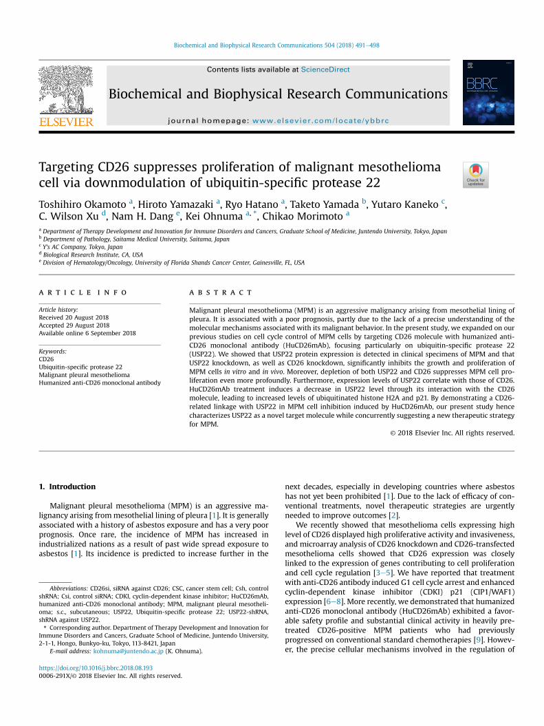
Targeting CD26 suppresses proliferation of malignant mesotheliomacell via downmodulation of ubiquitin-specific protease 22
Toshihiro Okamoto a, Hiroto Yamazaki a, Ryo Hatano a, Taketo Yamada b, Yutaro Kaneko c,C. Wilson Xu d, Nam H. Dang e, Kei Ohnuma a, *, Chikao Morimoto a
a Department of Therapy Development and Innovation for Immune Disorders and Cancers, Graduate School of Medicine, Juntendo University, Tokyo, Japanb Department of Pathology, Saitama Medical University, Saitama, Japanc Y's AC Company, Tokyo, Japand Biological Research Institute, CA, USAe Division of Hematology/Oncology, University of Florida Shands Cancer Center, Gainesville, FL, USA
a r t i c l e i n f o
Article history:Received 20 August 2018Accepted 29 August 2018Available online 6 September 2018
Keywords:CD26Ubiquitin-specific protease 22Malignant pleural mesotheliomaHumanized anti-CD26 monoclonal antibody
a b s t r a c t
Malignant pleural mesothelioma (MPM) is an aggressive malignancy arising from mesothelial lining ofpleura. It is associated with a poor prognosis, partly due to the lack of a precise understanding of themolecular mechanisms associated with its malignant behavior. In the present study, we expanded on ourprevious studies on cell cycle control of MPM cells by targeting CD26 molecule with humanized anti-CD26 monoclonal antibody (HuCD26mAb), focusing particularly on ubiquitin-specific protease 22(USP22). We showed that USP22 protein expression is detected in clinical specimens of MPM and thatUSP22 knockdown, as well as CD26 knockdown, significantly inhibits the growth and proliferation ofMPM cells in vitro and in vivo. Moreover, depletion of both USP22 and CD26 suppresses MPM cell pro-liferation even more profoundly. Furthermore, expression levels of USP22 correlate with those of CD26.HuCD26mAb treatment induces a decrease in USP22 level through its interaction with the CD26molecule, leading to increased levels of ubiquitinated histone H2A and p21. By demonstrating a CD26-related linkage with USP22 in MPM cell inhibition induced by HuCD26mAb, our present study hencecharacterizes USP22 as a novel target molecule while concurrently suggesting a new therapeutic strategyfor MPM.
© 2018 Elsevier Inc. All rights reserved.
1. Introduction
Malignant pleural mesothelioma (MPM) is an aggressive ma-lignancy arising frommesothelial lining of pleura [1]. It is generallyassociated with a history of asbestos exposure and has a very poorprognosis. Once rare, the incidence of MPM has increased inindustrialized nations as a result of past wide spread exposure toasbestos [1]. Its incidence is predicted to increase further in the
next decades, especially in developing countries where asbestoshas not yet been prohibited [1]. Due to the lack of efficacy of con-ventional treatments, novel therapeutic strategies are urgentlyneeded to improve outcomes [2].
We recently showed that mesothelioma cells expressing highlevel of CD26 displayed high proliferative activity and invasiveness,and microarray analysis of CD26 knockdown and CD26-transfectedmesothelioma cells showed that CD26 expression was closelylinked to the expression of genes contributing to cell proliferationand cell cycle regulation [3e5]. We have reported that treatmentwith anti-CD26 antibody induced G1 cell cycle arrest and enhancedcyclin-dependent kinase inhibitor (CDKI) p21 (CIP1/WAF1)expression [6e8]. More recently, we demonstrated that humanizedanti-CD26 monoclonal antibody (HuCD26mAb) exhibited a favor-able safety profile and substantial clinical activity in heavily pre-treated CD26-positive MPM patients who had previouslyprogressed on conventional standard chemotherapies [9]. Howev-er, the precise cellular mechanisms involved in the regulation of
Abbreviations: CD26si, siRNA against CD26; CSC, cancer stem cell; Csh, controlshRNA; Csi, control siRNA; CDKI, cyclin-dependent kinase inhibitor; HuCD26mAb,humanized anti-CD26 monoclonal antibody; MPM, malignant pleural mesotheli-oma; s.c., subcutaneous; USP22, Ubiquitin-specific protease 22; USP22-shRNA,shRNA against USP22.* Corresponding author. Department of Therapy Development and Innovation for
Immune Disorders and Cancers, Graduate School of Medicine, Juntendo University,2-1-1, Hongo, Bunkyo-ku, Tokyo, 113-8421, Japan
E-mail address: [email protected] (K. Ohnuma).
Contents lists available at ScienceDirect
Biochemical and Biophysical Research Communications
journal homepage: www.elsevier .com/locate/ybbrc
https://doi.org/10.1016/j.bbrc.2018.08.1930006-291X/© 2018 Elsevier Inc. All rights reserved.
Biochemical and Biophysical Research Communications 504 (2018) 491e498
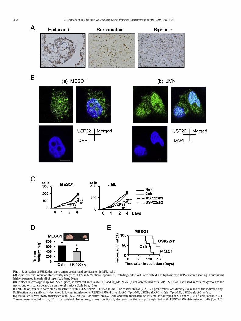
Fig. 1. Suppression of USP22 decreases tumor growth and proliferation in MPM cells.(A) Representative immunohistochemistry images of USP22 in MPM clinical specimens, including epithelioid, sarcomatoid, and biphasic type. USP22 (brown staining in nuceli) washighly expressed in each MPM type. Scale bars, 50 mm(B) Confocal microscopy images of USP22 (green) in MPM cell lines, (a) MESO1 and (b) JMN. Nuclei (blue) were stained with DAPI. USP22 was expressed in both the cytosol and thenuclei, and was barely detectable on the cell surface. Scale bars, 10 mm(C) MESO1 or JMN cells were stably transfected with USP22-shRNA-1, USP22-shRNA-2 or control shRNA (Csh). Cell proliferation was directly examined at the indicated days.Proliferation was significantly decreased following transfection of USP22-shRNA-1 or -shRNA-2. **p < 0.01, USP22-shRNA-1 vs Csh; ##p < 0.01, USP22-shRNA-2 vs Csh.(D) MESO1 cells were stably transfected with USP22-shRNA-1 or control shRNA (Csh), and were inoculated s.c. into the dorsal region of SCID mice (3� 105 cells/mouse, n ¼ 8).Tumors were resected at day 10 to be weighed. Tumor weight was significantly decreased in the group transplanted with USP22-shRNA-1-transfected cells (*p< 0.01).
T. Okamoto et al. / Biochemical and Biophysical Research Communications 504 (2018) 491e498492

Representative macroscopic plot is indicated in the upper panel. Scale bar, 1 cm(E) USP22-shRNA-1 or control shRNA (Csh) stably transfected MESO1 cells were injected into SCID mice intravenously (3� 105 cells/mouse, n¼ 8). Survival was evaluated byKaplan-Meier analysis. Survival of mice transplanted with USP22sh MESO1 cells was prolonged significantly. P value was calculated by log-rank test. (For interpretation of thereferences to color in this figure legend, the reader is referred to the Web version of this article.)
Fig. 2. Silencing of both USP22 and CD26 contributes to a more profound suppression of MPM cell growth than either alone.(A) Representative 2-D dot plots of CD26 (PE) and USP22 (FITC) in MESO1 and JMN cell lines. MESO1 cells contained CD26 þ USP22þ and CD26� USP22þ populations. JMN cellscontained mainly CD26þ USP22þ with faint amount of CD26þ USP22� cells.(B) CD26 þ or CD26� MESO1 cells were sorted by a flow cytometric cell sorter. As shown in (A), both CD26þ and CD26� MESO1 cells expressed USP22 (CD26þ USP22þ andCD26� USP22þ populations, respectively). In vitro proliferation of CD26þ USP22þ or CD26� USP22þ cells was evaluated by MTT assay at the indicated time points (left panel).CD26þ USP22þ cells exhibited significantly higher proliferative activity than CD26� USP22þ cells (*p < 0.05). For in vivo proliferation assay, each CD26þ USP22þ andCD26� USP22þ cell population was inoculated s.c. into the dorsal region of SCID mice (3 � 105 cells/mouse, each n ¼ 6). Tumors were resected at day 10 to be weighed. Tumorweight was significantly increased in the group transplanted with CD26þ USP22þ cells (*p< 0.01). Representative macroscopic plot is indicated in the upper panel. Scale bar, 1 cm(C) Cell cycle analysis of the MESO1 cells transfected with CD26-siRNA (CD26si) or control siRNA (Csi) (left two panels) and USP22-shRNA-1 (USP22sh) or control shRNA (Csh) (righttwo panels). Representative histograms are shown. Accumulation in G1 phase (green area) with decreased S (greenish brown area) and G2/M (light blue area) phase was observed inCD26si and USP22sh cells compared with each control cell population (similar results were obtained in five independent experiments) (p< 0.01).(D) In vitro proliferation of MESO1 cells stably transfected with USP22-shRNA-1 (USP22sh), CD26-siNRA (CD26si), both USP22sh and CD26si, or control shRNA and siRNA (Csh) wasevaluated by MTT assay at the indicated time points (left panel). The combined knockdown of USP22 and CD26 resulted in a more profound inhibition of MPM cell proliferation,compared with knockdown of USP22 or CD26 alone or Csh (*p< 0.05, USP22sh vs Csh; #p< 0.05, CD26si vs Csh; **p< 0.01, USP22sh plus CD26si vs Csh. For in vivo tumor growthassay, (right panel) MESO1 cells stably transfected with USP22sh, CD26si, both USP22sh and CD26si, or control shRNA and siRNA (Csh) were inoculated s.c. into the dorsal region ofSCID mice (3� 105 cells/mouse, n ¼ 8). Tumors were resected at day 10 to be weighed. Tumor weight was significantly decreased in the group transplanted with the combinedknockdown of USP22 and CD26 (*p < 0.05, **p < 0.01 vs Csh). Representative macroscopic plot is indicated in the upper panel. Scale bar, 1 cm. (For interpretation of the references tocolor in this figure legend, the reader is referred to the Web version of this article.)
T. Okamoto et al. / Biochemical and Biophysical Research Communications 504 (2018) 491e498 493
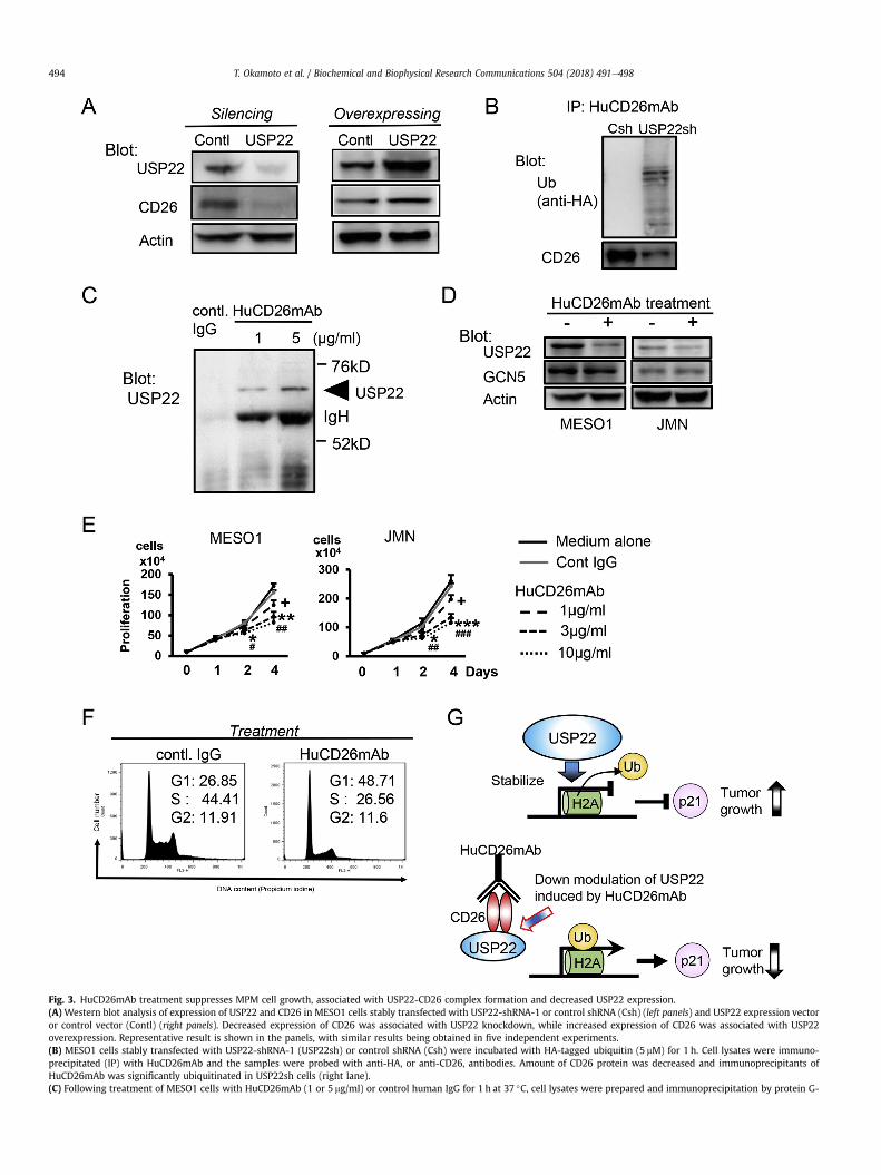
Fig. 3. HuCD26mAb treatment suppresses MPM cell growth, associated with USP22-CD26 complex formation and decreased USP22 expression.(A) Western blot analysis of expression of USP22 and CD26 in MESO1 cells stably transfected with USP22-shRNA-1 or control shRNA (Csh) (left panels) and USP22 expression vectoror control vector (Contl) (right panels). Decreased expression of CD26 was associated with USP22 knockdown, while increased expression of CD26 was associated with USP22overexpression. Representative result is shown in the panels, with similar results being obtained in five independent experiments.(B) MESO1 cells stably transfected with USP22-shRNA-1 (USP22sh) or control shRNA (Csh) were incubated with HA-tagged ubiquitin (5 mM) for 1 h. Cell lysates were immuno-precipitated (IP) with HuCD26mAb and the samples were probed with anti-HA, or anti-CD26, antibodies. Amount of CD26 protein was decreased and immunoprecipitants ofHuCD26mAb was significantly ubiquitinated in USP22sh cells (right lane).(C) Following treatment of MESO1 cells with HuCD26mAb (1 or 5 mg/ml) or control human IgG for 1 h at 37 �C, cell lysates were prepared and immunoprecipitation by protein G-
T. Okamoto et al. / Biochemical and Biophysical Research Communications 504 (2018) 491e498494
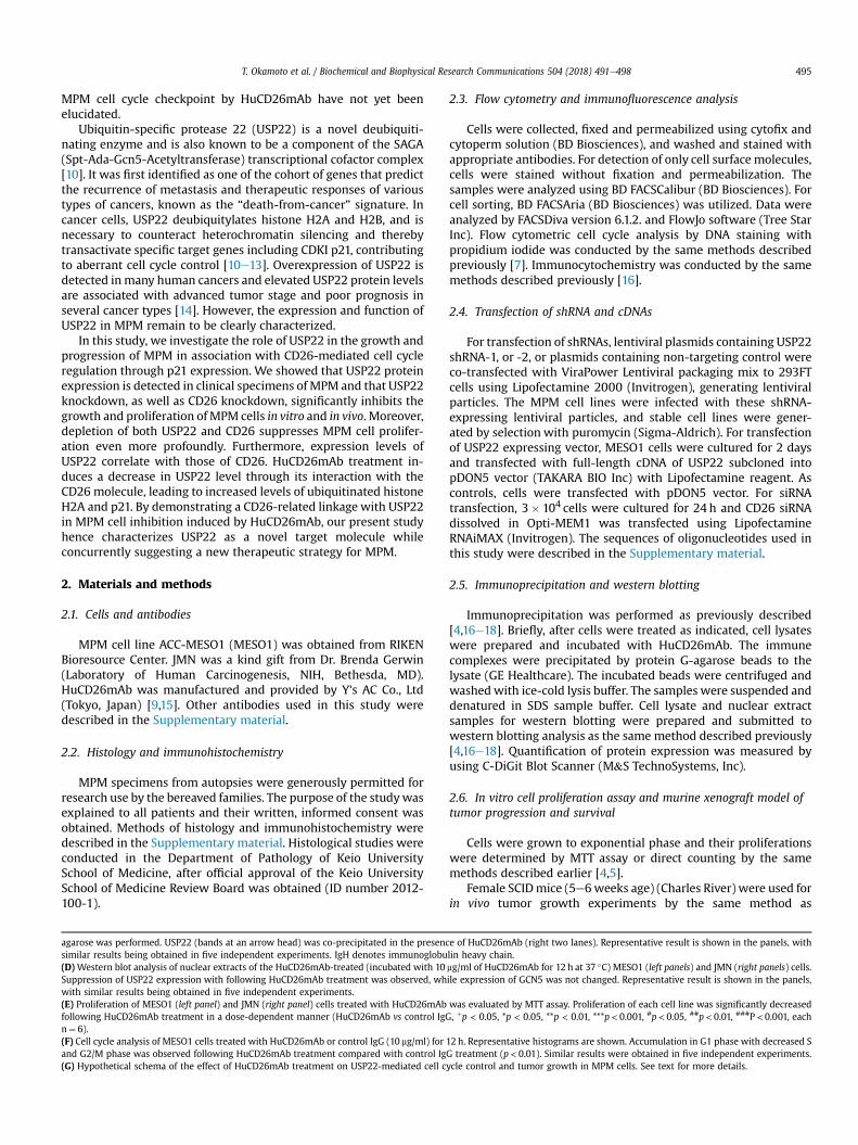
MPM cell cycle checkpoint by HuCD26mAb have not yet beenelucidated.
Ubiquitin-specific protease 22 (USP22) is a novel deubiquiti-nating enzyme and is also known to be a component of the SAGA(Spt-Ada-Gcn5-Acetyltransferase) transcriptional cofactor complex[10]. It was first identified as one of the cohort of genes that predictthe recurrence of metastasis and therapeutic responses of varioustypes of cancers, known as the “death-from-cancer” signature. Incancer cells, USP22 deubiquitylates histone H2A and H2B, and isnecessary to counteract heterochromatin silencing and therebytransactivate specific target genes including CDKI p21, contributingto aberrant cell cycle control [10e13]. Overexpression of USP22 isdetected inmany human cancers and elevated USP22 protein levelsare associated with advanced tumor stage and poor prognosis inseveral cancer types [14]. However, the expression and function ofUSP22 in MPM remain to be clearly characterized.
In this study, we investigate the role of USP22 in the growth andprogression of MPM in association with CD26-mediated cell cycleregulation through p21 expression. We showed that USP22 proteinexpression is detected in clinical specimens of MPM and that USP22knockdown, as well as CD26 knockdown, significantly inhibits thegrowth and proliferation ofMPM cells in vitro and in vivo. Moreover,depletion of both USP22 and CD26 suppresses MPM cell prolifer-ation even more profoundly. Furthermore, expression levels ofUSP22 correlate with those of CD26. HuCD26mAb treatment in-duces a decrease in USP22 level through its interaction with theCD26 molecule, leading to increased levels of ubiquitinated histoneH2A and p21. By demonstrating a CD26-related linkage with USP22in MPM cell inhibition induced by HuCD26mAb, our present studyhence characterizes USP22 as a novel target molecule whileconcurrently suggesting a new therapeutic strategy for MPM.
2. Materials and methods
2.1. Cells and antibodies
MPM cell line ACC-MESO1 (MESO1) was obtained from RIKENBioresource Center. JMN was a kind gift from Dr. Brenda Gerwin(Laboratory of Human Carcinogenesis, NIH, Bethesda, MD).HuCD26mAb was manufactured and provided by Y's AC Co., Ltd(Tokyo, Japan) [9,15]. Other antibodies used in this study weredescribed in the Supplementary material.
2.2. Histology and immunohistochemistry
MPM specimens from autopsies were generously permitted forresearch use by the bereaved families. The purpose of the studywasexplained to all patients and their written, informed consent wasobtained. Methods of histology and immunohistochemistry weredescribed in the Supplementary material. Histological studies wereconducted in the Department of Pathology of Keio UniversitySchool of Medicine, after official approval of the Keio UniversitySchool of Medicine Review Board was obtained (ID number 2012-100-1).
2.3. Flow cytometry and immunofluorescence analysis
Cells were collected, fixed and permeabilized using cytofix andcytoperm solution (BD Biosciences), and washed and stained withappropriate antibodies. For detection of only cell surface molecules,cells were stained without fixation and permeabilization. Thesamples were analyzed using BD FACSCalibur (BD Biosciences). Forcell sorting, BD FACSAria (BD Biosciences) was utilized. Data wereanalyzed by FACSDiva version 6.1.2. and FlowJo software (Tree StarInc). Flow cytometric cell cycle analysis by DNA staining withpropidium iodide was conducted by the same methods describedpreviously [7]. Immunocytochemistry was conducted by the samemethods described previously [16].
2.4. Transfection of shRNA and cDNAs
For transfection of shRNAs, lentiviral plasmids containing USP22shRNA-1, or -2, or plasmids containing non-targeting control wereco-transfected with ViraPower Lentiviral packaging mix to 293FTcells using Lipofectamine 2000 (Invitrogen), generating lentiviralparticles. The MPM cell lines were infected with these shRNA-expressing lentiviral particles, and stable cell lines were gener-ated by selection with puromycin (Sigma-Aldrich). For transfectionof USP22 expressing vector, MESO1 cells were cultured for 2 daysand transfected with full-length cDNA of USP22 subcloned intopDON5 vector (TAKARA BIO Inc) with Lipofectamine reagent. Ascontrols, cells were transfected with pDON5 vector. For siRNAtransfection, 3� 104 cells were cultured for 24 h and CD26 siRNAdissolved in Opti-MEM1 was transfected using LipofectamineRNAiMAX (Invitrogen). The sequences of oligonucleotides used inthis study were described in the Supplementary material.
2.5. Immunoprecipitation and western blotting
Immunoprecipitation was performed as previously described[4,16e18]. Briefly, after cells were treated as indicated, cell lysateswere prepared and incubated with HuCD26mAb. The immunecomplexes were precipitated by protein G-agarose beads to thelysate (GE Healthcare). The incubated beads were centrifuged andwashed with ice-cold lysis buffer. The samples were suspended anddenatured in SDS sample buffer. Cell lysate and nuclear extractsamples for western blotting were prepared and submitted towestern blotting analysis as the same method described previously[4,16e18]. Quantification of protein expression was measured byusing C-DiGit Blot Scanner (M&S TechnoSystems, Inc).
2.6. In vitro cell proliferation assay and murine xenograft model oftumor progression and survival
Cells were grown to exponential phase and their proliferationswere determined by MTT assay or direct counting by the samemethods described earlier [4,5].
Female SCIDmice (5e6weeks age) (Charles River) were used forin vivo tumor growth experiments by the same method as
agarose was performed. USP22 (bands at an arrow head) was co-precipitated in the presence of HuCD26mAb (right two lanes). Representative result is shown in the panels, withsimilar results being obtained in five independent experiments. IgH denotes immunoglobulin heavy chain.(D) Western blot analysis of nuclear extracts of the HuCD26mAb-treated (incubated with 10 mg/ml of HuCD26mAb for 12 h at 37 �C) MESO1 (left panels) and JMN (right panels) cells.Suppression of USP22 expression with following HuCD26mAb treatment was observed, while expression of GCN5 was not changed. Representative result is shown in the panels,with similar results being obtained in five independent experiments.(E) Proliferation of MESO1 (left panel) and JMN (right panel) cells treated with HuCD26mAb was evaluated by MTT assay. Proliferation of each cell line was significantly decreasedfollowing HuCD26mAb treatment in a dose-dependent manner (HuCD26mAb vs control IgG, þp < 0.05, *p < 0.05, **p < 0.01, ***p< 0.001, #p< 0.05, ##p< 0.01, ###P< 0.001, eachn¼ 6).(F) Cell cycle analysis of MESO1 cells treated with HuCD26mAb or control IgG (10 mg/ml) for 12 h. Representative histograms are shown. Accumulation in G1 phase with decreased Sand G2/M phase was observed following HuCD26mAb treatment compared with control IgG treatment (p< 0.01). Similar results were obtained in five independent experiments.(G) Hypothetical schema of the effect of HuCD26mAb treatment on USP22-mediated cell cycle control and tumor growth in MPM cells. See text for more details.
T. Okamoto et al. / Biochemical and Biophysical Research Communications 504 (2018) 491e498 495
described previously [4,5]. Briefly, for in vivo tumor progression,mice were anesthetized with isoflurane and subjected to subcu-taneous (s.c.) inoculation of MPM cells into the dorsal region. Formurine xenograft survival study, mice were intravenouslyimplanted with MESO1 cells transfected with USP22 shRNA-1 orcontrol vector shRNA.
2.7. Statistics
Data are represented as mean±standard deviations (SD) formurine xenograft study and mean±standard errors (SE) for otherassays. Data were analyzed by two-tailed Student's t-test for twogroup comparison or by ANOVA test for multiple comparisontesting followed by the Tukey-Kramer post-hoc test. P values� 0.05were considered statistically significant. In murine xenograft sur-vival study, prolonged survival was evaluated by Kaplan-Meieranalysis.
3. Results and discussion
3.1. Expression of USP22 in MPM clinical specimens and inhibitoryeffect of USP22 depletion on MPM cell proliferation
USP22 overexpression is detected in many human tumors,including non-small cell lung cancer, salivary duct carcinoma,bladder cancer, colorectal cancer, oral squamous cell carcinoma,and esophageal squamous cell carcinoma [14,19]. However, a rolefor USP22 in MPM has not yet been clearly elucidated. To addressthis issue, we first evaluated USP22 expression in clinical speci-mens of all three histopathologic subtypes (epithelioid, sarcoma-toid, and biphasic). Among 26 patients with epithelioid type, 21patients (81%) had USP22þ MPM histopathology. Moreover, 3 (60%)among 5 patients with sarcomatoid type and 7 (58%) among 12with biphasic type had USP22þ MPM histopathology. Fig. 1A showsa representative immunohistochemistry study demonstrating thatUSP22 protein expression was clearly detected in all three histo-pathologic subtypes. We next examined USP22 expression in theMPM cell lines used in our experimental studies. As shown inFig. 1B, USP22was found to be localized both in the nucleus and thecytosol of the MPM cell lines MESO1 and JMN (Fig. 1B). We there-fore used these cell lines for our present study. Since depletion ofUSP22 expression has been reported to suppress tumor growth invarious cancers other than MPM [14,19], we next examined thepotential regulatory effect of USP22 on MPM cell proliferation. Forthis purpose, we conducted knockdown experiments in MPM cellsutilizing shRNA transfection. As shown in Fig. 1C, knockdown ofUSP22 by shRNA (USP22-shRNA-1 or -2) significantly inhibitedin vitro proliferation of MESO1 (left panel) and JMN (right panel).Moreover, knockdown of USP22 in MESO1 cells suppressed in vivoproliferation in transplantation assay (Fig. 1D), which was associ-ated with prolonged survival of mice receiving USP22-abrogatedcells (Fig. 1E). Similar results were obtained in transplantationassay utilizing JMN cells with knockdown of USP22 (data notshown). Collectively, these results suggest that USP22 depletionattenuates tumor growth and proliferation of MPM.
3.2. Silencing of both USP22 and CD26 contributes to moreprofound suppression of MPM cell growth than either alone
We previously demonstrated that abrogation of CD26 expres-sion in MPM suppressed cell growth, invasion and proliferationin vitro and in vivo [4,5]. Moreover, we have found that cell surfaceexpression of CD26 is one of the cancer stem cell (CSC) markers thatcorrelated with CSC properties in MPM cells [20,21]. On the otherhand, we previously found that USP22 played a role in the CSC
property in human B-acute lymphocytic leukemia [22], as well as invarious other cancers [23]. We therefore explored the potentialcooperative effect of USP22 and CD26 on cell proliferation in MPM.Flow cytometric analysis revealed that the MESO1 cell line con-tained both CD26þ USP22þ and CD26- USP22þ cell populations andthat the JMN cell line contained CD26þ USP22þ cells almostexclusively (Fig. 2A). We then characterized the biological functionsof USP22þ MPM cells that differed in the expression of CD26. Forthis purpose, CD26þ (and USP22þ) and CD26� (and USP22þ) cellswere isolated from the MESO1 cell line through cell sorting anal-ysis, and then subjected to various biological assays. As shown inFig. 2B, USP22þ cells exhibited greater increase in in vitro prolifer-ation and in vivo growth in the CD26 þ population than theCD26�population (left and right panels, respectively). These datasuggest that CD26 and USP22 have a cooperative effect on tumorgrowth in MPM. We previously demonstrated that decreasedexpression of CD26 played a role in cell cycle control of tumor cellsvia enhanced expression of CDKI p21 [6e8]. Moreover, USP22expression counteracted heterochromatin silencing and therebytransactivated specific target genes including CDKI p21, contrib-uting to aberrant cell cycle control [10e13]. We therefore per-formed cell cycle analysis of CD26 or USP22-depleted MPM cells. Asshown in Fig. 2C, G1/S arrest was provoked by the abrogation ofCD26 or USP22 expression, suggesting that the inhibitory effect ofCD26 and USP22 depletion onMPM cell growthwas exerted via cellcycle arrest at the G1/S checkpoint. Further analysis showed thatthe combined knockdown of USP22 and CD26 resulted in a greaterlevel of inhibition of MPM cell proliferation in vitro (left panel ofFig. 2D) as well as in vivo (right panel of Fig. 2D), compared toknockdown of USP22 or CD26 alone. In the above clinical speci-mens of USP22þ MPM histology, co-expression of CD26 wasrevealed in 15 patients (71%) with epithelioid type, 1 (33%) withsarcomatoid type, and 5 (71%) with biphasic type. The clinicaloutcome was relatively worsened in these 21 patients withUSP22þ CD26þMPM histology than other groups, although smallnumber of each groups made it hard to have statistical significance.Taken together, our data indicate that USP22 and CD26 coopera-tively contribute to a more profound regulation of MPM cellgrowth.
3.3. HuCD26mAb induces suppression of MPM cell growth via adecrease in CD26-associated USP22
The results described above suggested a molecular associationbetween USP22 and CD26 in MPM cells. To further investigate themechanisms involved in this interaction, we analyzed the effect ofchanges in the expression level of USP22 on CD26 expression. Asshown in Fig. 3A, silencing or overexpression of USP22 led todecreased (left panel) or increased (right panel) CD26 expression,respectively. These data hence suggest that expression level of USP22regulates CD26 expression in MPM. Since USP22 contains a deubi-quitinating enzyme activity [10], we next examined a ubiquitinationstate of CD26 molecules in association with USP22 expression. Asshown in Fig. 3B, decreased USP22 expression led to increasedubiquitination of CD26 clearly. These data suggest that USP22expression regulates CD26 expression through its physical interac-tion of a deubiquitinating enzyme activity in USP22þ CD26þMPMcells.
We have previously shown that CD26 is expressedmainly on thecell surface of MPM cells [3,15,24], while USP22 is a nuclear protein,a component of the SAGA transcriptional cofactor complex [12], andis mainly localized in the nucleus (as shown Fig. 1A and 1B). In viewof their cellular localization, the mechanisms involved in CD26-USP22 interaction in MPM cells would need to be elucidated. Werecently demonstrated that treatment of MPM cells with
T. Okamoto et al. / Biochemical and Biophysical Research Communications 504 (2018) 491e498496
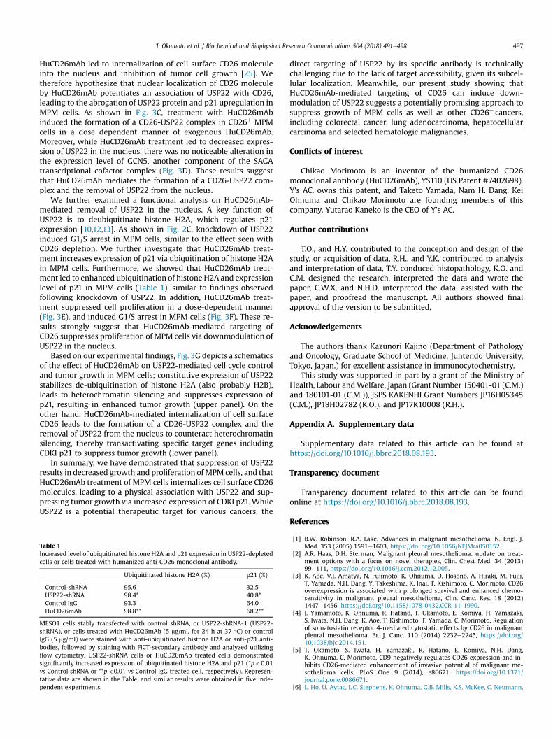
HuCD26mAb led to internalization of cell surface CD26 moleculeinto the nucleus and inhibition of tumor cell growth [25]. Wetherefore hypothesize that nuclear localization of CD26 moleculeby HuCD26mAb potentiates an association of USP22 with CD26,leading to the abrogation of USP22 protein and p21 upregulation inMPM cells. As shown in Fig. 3C, treatment with HuCD26mAbinduced the formation of a CD26-USP22 complex in CD26þ MPMcells in a dose dependent manner of exogenous HuCD26mAb.Moreover, while HuCD26mAb treatment led to decreased expres-sion of USP22 in the nucleus, there was no noticeable alteration inthe expression level of GCN5, another component of the SAGAtranscriptional cofactor complex (Fig. 3D). These results suggestthat HuCD26mAb mediates the formation of a CD26-USP22 com-plex and the removal of USP22 from the nucleus.
We further examined a functional analysis on HuCD26mAb-mediated removal of USP22 in the nucleus. A key function ofUSP22 is to deubiquitinate histone H2A, which regulates p21expression [10,12,13]. As shown in Fig. 2C, knockdown of USP22induced G1/S arrest in MPM cells, similar to the effect seen withCD26 depletion. We further investigate that HuCD26mAb treat-ment increases expression of p21 via ubiquitination of histone H2Ain MPM cells. Furthermore, we showed that HuCD26mAb treat-ment led to enhanced ubiquitination of histone H2A and expressionlevel of p21 in MPM cells (Table 1), similar to findings observedfollowing knockdown of USP22. In addition, HuCD26mAb treat-ment suppressed cell proliferation in a dose-dependent manner(Fig. 3E), and induced G1/S arrest in MPM cells (Fig. 3F). These re-sults strongly suggest that HuCD26mAb-mediated targeting ofCD26 suppresses proliferation of MPM cells via downmodulation ofUSP22 in the nucleus.
Based on our experimental findings, Fig. 3G depicts a schematicsof the effect of HuCD26mAb on USP22-mediated cell cycle controland tumor growth in MPM cells; constitutive expression of USP22stabilizes de-ubiquitination of histone H2A (also probably H2B),leads to heterochromatin silencing and suppresses expression ofp21, resulting in enhanced tumor growth (upper panel). On theother hand, HuCD26mAb-mediated internalization of cell surfaceCD26 leads to the formation of a CD26-USP22 complex and theremoval of USP22 from the nucleus to counteract heterochromatinsilencing, thereby transactivating specific target genes includingCDKI p21 to suppress tumor growth (lower panel).
In summary, we have demonstrated that suppression of USP22results in decreased growth and proliferation ofMPM cells, and thatHuCD26mAb treatment of MPM cells internalizes cell surface CD26molecules, leading to a physical association with USP22 and sup-pressing tumor growth via increased expression of CDKI p21. WhileUSP22 is a potential therapeutic target for various cancers, the
direct targeting of USP22 by its specific antibody is technicallychallenging due to the lack of target accessibility, given its subcel-lular localization. Meanwhile, our present study showing thatHuCD26mAb-mediated targeting of CD26 can induce down-modulation of USP22 suggests a potentially promising approach tosuppress growth of MPM cells as well as other CD26þ cancers,including colorectal cancer, lung adenocarcinoma, hepatocellularcarcinoma and selected hematologic malignancies.
Conflicts of interest
Chikao Morimoto is an inventor of the humanized CD26monoclonal antibody (HuCD26mAb), YS110 (US Patent #7402698).Y's AC. owns this patent, and Taketo Yamada, Nam H. Dang, KeiOhnuma and Chikao Morimoto are founding members of thiscompany. Yutarao Kaneko is the CEO of Y's AC.
Author contributions
T.O., and H.Y. contributed to the conception and design of thestudy, or acquisition of data, R.H., and Y.K. contributed to analysisand interpretation of data, T.Y. conduced histopathology, K.O. andC.M. designed the research, interpreted the data and wrote thepaper, C.W.X. and N.H.D. interpreted the data, assisted with thepaper, and proofread the manuscript. All authors showed finalapproval of the version to be submitted.
Acknowledgements
The authors thank Kazunori Kajino (Department of Pathologyand Oncology, Graduate School of Medicine, Juntendo University,Tokyo, Japan.) for excellent assistance in immunocytochemistry.
This study was supported in part by a grant of the Ministry ofHealth, Labour andWelfare, Japan (Grant Number 150401-01 (C.M.)and 180101-01 (C.M.)), JSPS KAKENHI Grant Numbers JP16H05345(C.M.), JP18H02782 (K.O.), and JP17K10008 (R.H.).
Appendix A. Supplementary data
Supplementary data related to this article can be found athttps://doi.org/10.1016/j.bbrc.2018.08.193.
Transparency document
Transparency document related to this article can be foundonline at https://doi.org/10.1016/j.bbrc.2018.08.193.
References
[1] B.W. Robinson, R.A. Lake, Advances in malignant mesothelioma, N. Engl. J.Med. 353 (2005) 1591e1603, https://doi.org/10.1056/NEJMra050152.
[2] A.R. Haas, D.H. Sterman, Malignant pleural mesothelioma: update on treat-ment options with a focus on novel therapies, Clin. Chest Med. 34 (2013)99e111, https://doi.org/10.1016/j.ccm.2012.12.005.
[3] K. Aoe, V.J. Amatya, N. Fujimoto, K. Ohnuma, O. Hosono, A. Hiraki, M. Fujii,T. Yamada, N.H. Dang, Y. Takeshima, K. Inai, T. Kishimoto, C. Morimoto, CD26overexpression is associated with prolonged survival and enhanced chemo-sensitivity in malignant pleural mesothelioma, Clin. Canc. Res. 18 (2012)1447e1456, https://doi.org/10.1158/1078-0432.CCR-11-1990.
[4] J. Yamamoto, K. Ohnuma, R. Hatano, T. Okamoto, E. Komiya, H. Yamazaki,S. Iwata, N.H. Dang, K. Aoe, T. Kishimoto, T. Yamada, C. Morimoto, Regulationof somatostatin receptor 4-mediated cytostatic effects by CD26 in malignantpleural mesothelioma, Br. J. Canc. 110 (2014) 2232e2245, https://doi.org/10.1038/bjc.2014.151.
[5] T. Okamoto, S. Iwata, H. Yamazaki, R. Hatano, E. Komiya, N.H. Dang,K. Ohnuma, C. Morimoto, CD9 negatively regulates CD26 expression and in-hibits CD26-mediated enhancement of invasive potential of malignant me-sothelioma cells, PLoS One 9 (2014), e86671, https://doi.org/10.1371/journal.pone.0086671.
[6] L. Ho, U. Aytac, L.C. Stephens, K. Ohnuma, G.B. Mills, K.S. McKee, C. Neumann,
Table 1Increased level of ubiquitinated histone H2A and p21 expression in USP22-depletedcells or cells treated with humanized anti-CD26 monoclonal antibody.
Ubiquitinated histone H2A (%) p21 (%)
Control-shRNA 95.6 32.5USP22-shRNA 98.4* 40.8*Control IgG 93.3 64.0HuCD26mAb 98.8** 68.2**
MESO1 cells stably transfected with control shRNA, or USP22-shRNA-1 (USP22-shRNA), or cells treated with HuCD26mAb (5 mg/ml, for 24 h at 37 �C) or controlIgG (5 mg/ml) were stained with anti-ubiquitinated histone H2A or anti-p21 anti-bodies, followed by staining with FICT-secondary antibody and analyzed utilizingflow cytometry. USP22-shRNA cells or HuCD26mAb treated cells demonstratedsignificantly increased expression of ubiquitinated histone H2A and p21 (*p< 0.01vs Control shRNA or **p< 0.01 vs Control IgG treated cell, respectively). Represen-tative data are shown in the Table, and similar results were obtained in five inde-pendent experiments.
T. Okamoto et al. / Biochemical and Biophysical Research Communications 504 (2018) 491e498 497
R. LaPushin, F. Cabanillas, J.L. Abbruzzese, C. Morimoto, N.H. Dang, In vitro andin vivo antitumor effect of the anti-CD26 monoclonal antibody 1F7 on humanCD30þ anaplastic large cell T-cell lymphoma Karpas 299, Clin. Canc. Res. 7(2001) 2031e2040.
[7] K. Ohnuma, T. Ishii, S. Iwata, O. Hosono, H. Kawasaki, M. Uchiyama, H. Tanaka,T. Yamochi, N.H. Dang, C. Morimoto, G1/S cell cycle arrest provoked in humanT cells by antibody to CD26, Immunology 107 (2002) 325e333.
[8] M. Hayashi, H. Madokoro, K. Yamada, H. Nishida, C. Morimoto, M. Sakamoto,T. Yamada, A humanized anti-CD26 monoclonal antibody inhibits cell growthof malignant mesothelioma via retarded G2/M cell cycle transition, Canc. CellInt. 16 (2016) 35, https://doi.org/10.1186/s12935-016-0310-9.
[9] E. Angevin, N. Isambert, V. Trillet-Lenoir, B. You, J. Alexandre, G. Zalcman,P. Vielh, F. Farace, F. Valleix, T. Podoll, Y. Kuramochi, I. Miyashita, O. Hosono,N.H. Dang, K. Ohnuma, T. Yamada, Y. Kaneko, C. Morimoto, First-in-humanphase 1 of YS110, a monoclonal antibody directed against CD26 in advancedCD26-expressing cancers, Br. J. Canc. 116 (2017) 1126e1134, https://doi.org/10.3892/or.2011.1449.
[10] X.Y. Zhang, M. Varthi, S.M. Sykes, C. Phillips, C. Warzecha, W. Zhu, A. Wyce,A.W. Thorne, S.L. Berger, S.B. McMahon, The putative cancer stem cell markerUSP22 is a subunit of the human SAGA complex required for activated tran-scription and cell-cycle progression, Mol. Cell 29 (2008) 102e111, https://doi.org/10.1016/j.molcel.2007.12.015.
[11] W.W. Pijnappel, H.T. Timmers, Dubbing SAGA unveils new epigenetic cross-talk, Mol. Cell 29 (2008) 152e154, https://doi.org/10.1016/j.molcel.2008.01.007.
[12] X.Y. Zhang, H.K. Pfeiffer, A.W. Thorne, S.B. McMahon, USP22, an hSAGA sub-unit and potential cancer stem cell marker, reverses the polycomb-catalyzedubiquitylation of histone H2A, Cell Cycle 7 (2008) 1522e1524, https://doi.org/10.4161/cc.7.11.5962.
[13] S.B. Ling, D.G. Sun, B. Tang, C. Guo, Y. Zhang, R. Liang, L.M. Wang, Knock-downof USP22 by small interfering RNA interference inhibits HepG2 cell prolifer-ation and induces cell cycle arrest, Cell. Mol. Biol. 58 (Suppl) (2012)OL1803eOL1808.
[14] R.S. Schrecengost, J.L. Dean, J.F. Goodwin, M.J. Schiewer, M.W. Urban,T.J. Stanek, R.T. Sussman, J.L. Hicks, R.C. Birbe, R.A. Draganova-Tacheva,T. Visakorpi, A.M. DeMarzo, S.B. McMahon, K.E. Knudsen, USP22 regulatesoncogenic signaling pathways to drive lethal cancer progression, Canc. Res. 74(2014) 272e286, https://doi.org/10.1158/0008-5472.CAN-13-1954.
[15] T. Inamoto, T. Yamada, K. Ohnuma, S. Kina, N. Takahashi, T. Yamochi,S. Inamoto, Y. Katsuoka, O. Hosono, H. Tanaka, N.H. Dang, C. Morimoto, Hu-manized anti-CD26 monoclonal antibody as a treatment for malignant me-sothelioma tumors, Clin. Canc. Res. 13 (2007) 4191e4200, https://doi.org/10.1158/1078-0432.CCR-07-0110.
[16] K. Ohnuma-Ishikawa, T. Morio, T. Yamada, Y. Sugawara, M. Ono, M. Nagasawa,
A. Yasuda, C. Morimoto, K. Ohnuma, N.H. Dang, H. Hosoi, E. Verdin,S. Mizutani, Knockdown of XAB2 enhances all-trans retinoic acid-inducedcellular differentiation in all-trans retinoic acid-sensitive and -resistant can-cer cells, Canc. Res. 67 (2007) 1019e1029, https://doi.org/10.1158/0008-5472.CAN-06-1638.
[17] K. Ohnuma, Y. Munakata, T. Ishii, S. Iwata, S. Kobayashi, O. Hosono,H. Kawasaki, N.H. Dang, C. Morimoto, Soluble CD26/dipeptidyl peptidase IVinduces T cell proliferation through CD86 up-regulation on APCs, J. Immunol.167 (2001) 6745e6755.
[18] K. Ohnuma, T. Yamochi, M. Uchiyama, K. Nishibashi, N. Yoshikawa, N. Shimizu,S. Iwata, H. Tanaka, N.H. Dang, C. Morimoto, CD26 up-regulates expression ofCD86 on antigen-presenting cells by means of caveolin-1, Proc. Natl. Acad. Sci.U. S. A. 101 (2004) 14186e14191, https://doi.org/10.1073/pnas.0405266101.
[19] H.D. Zhao, H.L. Tang, N.N. Liu, Y.L. Zhao, Q.Q. Liu, X.S. Zhu, L.T. Jia, C.F. Gao,A.G. Yang, J.T. Li, Targeting ubiquitin-specific protease 22 suppresses growthand metastasis of anaplastic thyroid carcinoma, Oncotarget 7 (2016)31191e31203, https://doi.org/10.18632/oncotarget.9098.
[20] H. Yamazaki, M. Naito, F.I. Ghani, N.H. Dang, S. Iwata, C. Morimoto, Charac-terization of cancer stem cell properties of CD24 and CD26-positive humanmalignant mesothelioma cells, Biochem. Biophys. Res. Commun. 419 (2012)529e536, https://doi.org/10.1016/j.bbrc.2012.02.054.
[21] F.I. Ghani, H. Yamazaki, S. Iwata, T. Okamoto, K. Aoe, K. Okabe, Y. Mimura,N. Fujimoto, T. Kishimoto, T. Yamada, C.W. Xu, C. Morimoto, Identification ofcancer stem cell markers in human malignant mesothelioma cells, Biochem.Biophys. Res. Commun. 404 (2011) 735e742, https://doi.org/10.1016/j.bbrc.2010.12.054.
[22] H. Yamazaki, C.W. Xu, M. Naito, H. Nishida, T. Okamoto, F.I. Ghani, S. Iwata,T. Inukai, K. Sugita, C. Morimoto, Regulation of cancer stem cell properties byCD9 in human B-acute lymphoblastic leukemia, Biochem. Biophys. Res.Commun. 409 (2011) 14e21, https://doi.org/10.1016/j.bbrc. 2011.04.098.
[23] G.V. Glinsky, O. Berezovska, A.B. Glinskii, Microarray analysis identifies adeath-from-cancer signature predicting therapy failure in patients withmultiple types of cancer, J. Clin. Invest. 115 (2005) 1503e1521, https://doi.org/10.1172/JCI23412.
[24] V.J. Amatya, Y. Takeshima, K. Kushitani, T. Yamada, C. Morimoto, K. Inai,Overexpression of CD26/DPPIV in mesothelioma tissue and mesothelioma celllines, Oncol. Rep. 26 (2011) 1369e1375, https://doi.org/10.3892/or.2011.1449.
[25] K. Yamada, M. Hayashi, H. Madokoro, H. Nishida, W. Du, K. Ohnuma,M. Sakamoto, C. Morimoto, T. Yamada, Nuclear localization of CD26 inducedby a humanized monoclonal antibody inhibits tumor cell growth by modu-lating of POLR2A transcription, PLoS One 8 (2013), e62304, https://doi.org/10.1371/journal.pone.0062304.
T. Okamoto et al. / Biochemical and Biophysical Research Communications 504 (2018) 491e498498
Data Article
Gene expression microarray data from mouseCBS treated with rTMS for 30 days, mousecerebrum and CBS treated with rTMS for 40 days
Tetsurou Ikeda a,b,⁎, Satoru Kobayashi c, Chikao Morimoto b
a Laboratory for Structural Neuropathology, RIKEN Brain Science Institute, 2-1 Hirosawa, Wako City, Saitama351-0198, Japanb Clinical Immunology, Institute of Medical Science, The University of Tokyo, 4-6-1 Shirokanedai, Minato-ku,Tokyo 108-8639, Japanc Technical Support, Thermo Fisher Scientific K.K., 3-9 Moriya, Kanagawa-ku, Yokohama 221-0022, Japan
a r t i c l e i n f o
Article history:Received 14 September 2017Received in revised form17 January 2018Accepted 29 January 2018Available online 8 February 2018
Keywords:rTMSMicroarray data
a b s t r a c t
This data article contains complementary tables related to theresearch article study entitled, ‘Effects of repetitive transcranialmagnetic stimulation on ER stress−related genes and glutamate,γ−aminobutyric acid, and glycine transporter genes in mousebrain’ (Ikeda et al. (2017) [1]), which showed that rTMS modulatesglutamate, GABA and glycine transporters and regulates ERstress−related genes. Here, we provide accompanying data col-lected using Affymetrix GeneChip microarrays to identify changesin gene expression in mouse CBS treated with rTMS for 30 days(Tables 1–21) and mouse cerebrum (Tables 22–57) and CBS(Tables 58–94) treated with rTMS for 40 days.
& 2018 The Authors. Published by Elsevier Inc. This is an openaccess article under the CC BY license
(http://creativecommons.org/licenses/by/4.0/).
Specifications Table
Subject area NeuroscienceMore specific subject area Gene expression
Contents lists available at ScienceDirect
journal homepage: www.elsevier.com/locate/dib
Data in Brief
http://dx.doi.org/10.1016/j.dib.2018.01.0792352-3409/& 2018 The Authors. Published by Elsevier Inc. This is an open access article under the CC BY license(http://creativecommons.org/licenses/by/4.0/).
Abbreviations: CBS, cerebellum with brain stem⁎ Correspondence to: Kochi Medical School, Kochi University, Kohasu, Oko, Nankoku-city, Kochi, Japan.E-mail address: [email protected] (T. Ikeda).
Data in Brief 17 (2018) 1078–1081
Type of data TablesHow data was acquired Affymetrix GeneChip RNA microarrayData format Filtered, analysedExperimental factors Mouse brain treated with rTMS for 30 and 40 daysExperimental features RNA isolation, global gene expression analysesData source location Wako, Saitama, JapanData accessibility Data are contained within this article
Value of the data
� Global gene expression analysis of mouse cerebellum with brain stem (CBS) treated with repetitivetranscranial magnetic stimulation (rTMS) for 30 and 40 days
� These data may be useful for comparison with microarray data obtained from rTMS of differentdurations.
� Genes identified as differentially expressed in this data set could be useful in further studiesinvestigating the effects of rTMS on mouse brain.
� In contrast to the advantage of using microelectrodes, inflammation never occurs while using TMSbecause it is non−invasive. Immune system may recognise the microelectrode and causeinflammation.
1. Data
Affymetrix GeneChip microarray analyses of mRNA isolated from mouse CBS after 30days rTMSshowed altered expression of several genes (Tables 1–21), including glutamatergic, GABAergic andglycinergic (e.g. glycine transporter) neurotransmission systems and ER stress−related genes. Affy-metrix GeneChip microarray analyses of mRNA isolated frommouse cerebrum after 40 days rTMS alsoshowed altered expression of several genes (Tables 22–57), including glutamatergic (e.g. glutamatetransporters), GABAergic and glycinergic neurotransmission systems. Furthermore, Affymetrix Gen-eChip microarray analyses of mRNA isolated from mouse CBS after 40days rTMS showed alteredexpressions of several genes (Tables 58–94), including glutamatergic (e.g. glutamate transporters),GABAergic and glycinergic neurotransmission systems. Mice CBS and cerebrum stimulated by rTMSfor 30 or 40days were denoted as M1 and M2, and sham control were denoted as C1 and C2 (n¼2). Allthe expression ratios were converted into the log2 (expression ratio) values. L1: Signal Log Ratio (M1/C1), L2: Signal Log Ratio (M2/C1), L3: Signal Log Ratio (M1/C2), L4: Signal Log Ratio (M1/C2). C1 andC2 were used as control, and M1 and M2 were normalized with regard to C1 and C2 to obtainexpression ratios. Expression Console (EC) Ver.1.4 and Transcriptome Analysis Console (TAC) Ver.3were used for comparison analysis as a manufacturer procedure. C means data analysis output for acomparison analysis showing change p−values with the associated Increase (I) or Decrease (D) call.Increase calls have change p−values closer to zero and Decrease calls have Change p−values closer toone. Finally, the change algorithm assesses probe pair saturation, calculates a change p−values, andassigns an (I), marginal increase (MI), no change (NC), marginal decrease (MD), or (D) call for C. Geneswith more than two significant difference calls were chosen. Abbreviations; TC ID: Transcript ClusterID, GS: gene symbol, *: Total number of increase, #: Total number of decrease. TC ID is available forpathway analysis.
2. Experimental design, materials and methods
We performed a comprehensive analysis of altered gene expression in CBS after chronic rTMSby using a high−density oligonucleotide array (GeneChip; Affymetrix, Santa Clara, CA, USA.MG_U74Av2 probe array), as described elsewhere [2]. Using the Affymetrix algorithm [3] andmultiple analysis comparison software for assessing gene expression differences, mRNAs that
T. Ikeda et al. / Data in Brief 17 (2018) 1078–1081 1079
increased or decreased in the mouse brain after chronic rTMS relative to levels in the controlmouse brain were identified. Pathway analysis was used to identify the significant pathway of thedifferential genes based on KEGG. Furthermore, gene ontology (GO) analysis was performed toanalyse the main function of the differentially expressed genes based on GO, which is the keyfunctional classification of NCBI that can organise genes into hierarchical categories and uncoverthe gene regulatory network based on biological process and molecular function [4]. Stimulationwas performed using a round−coil (7.5 cm outer diameter) and a Nihon Kohden Rapid Rate Sti-mulator (Nihon Kohden, Japan). Stimulation conditions were as follows: 20 Hz, 2 s; 20 times/day;inter−stimulus 1−min interval (30% machine output, representing approximately 0.75 T). The coilwas placed over the head without touching the skull. Sham control mice were ‘stimulated’410 cm from the head. rTMS did not produce either notable seizures or changes in behaviour,such as excessive struggling. The animals were killed 24 h after the last stimulation, and theirbrains were processed for further analysis [6,7]. Whole mouse brain was divided at the midbraininto the cerebrum and CBS. This method of stimulation is applied to the whole brain but not tospecific regions of brain. Hence, the feedback effect of the afferent pathway should be considered.Total RNA was isolated from the cerebrum and CBS by acid–phenol extraction [5]. Poly(A)þ RNAwas isolated from the samples using an mRNA purification kit (TaKaRa Bio, Japan).
Acknowledgements
The authors would like to thank Enago for the English language review. The authors would like tothank Mr. Masaru Kurosawa and Dr. Nobuyuki Nukina for the support for the experiment.
Funding sources
This work was partly supported by a grant from the Japanese Society for the Promotion of Science(No. 17109011). Furthermore, this work was supported by the Global COE Program ‘Center of Edu-cation and Research for the Advanced Genome−Based Medicine−For personalized medicine and thecontrol of worldwide infectious disease’ and MEXT Japan.
Transparency document. Supporting information
Supplementary data associated with this article can be found in the online version at http://dx.doi.org/10.1016/j.dib.2018.01.079.
Appendix A. Supporting information
Supplementary data associated with this article can be found in the online version at http://dx.doi.org/10.1016/j.dib.2018.01.079.
References
[1] T. Ikeda, W. Kobayashi and C. Morimoto, Effects of repetitive transcranial magnetic stimulation on ER stress−related genesand glutamate, γ−aminobutyric acid, and glycine transporter genes in mouse brain. Submitted for publication.
[2] S. Kotliarova, N.R. Jana, N. Sakamoto, et al., Decreased expression of hypothalamic neuropeptides in Huntington diseasetransgenic mice with expanded polyglutamine−EGFP fluorescent aggregates, J. Neurochem. 93 (2005) 641–653.
[3] R.J. Lipshutz, S.P. Fodor, T.R. Gingeras, et al., High density synthetic oligonucleotide arrays, Nat. Genet. 21 (1999) 20–24.[4] C. Gene Ontology, The Gene Ontology (GO) project in 2006, Nucleic. Acids. Res. 34 (2006) D322–D326.
T. Ikeda et al. / Data in Brief 17 (2018) 1078–10811080

[5] P. Chomczynski, N. Sacchi, Single−step method of RNA isolation by acid guanidinium thiocyanate−phenol−chloroformextraction, Anal. Biochem. 162 (1987) 156–159.
[6] T. Ikeda, M. Kurosawa, C. Uchikawa, et al., Modulation of monoamine transporter expression and function by repetitivetranscranial magnetic stimulation, Biochem. Biophys. Res. Commun. 327 (2005) 218–224.
[7] T. Ikeda, M. Kurosawa, C. Morimoto, et al., Multiple effects of repetitive transcranial magnetic stimulation on neu-ropsychiatric disorders, Biochem. Biophys. Res. Commun. 436 (2013) 121–127.
T. Ikeda et al. / Data in Brief 17 (2018) 1078–1081 1081

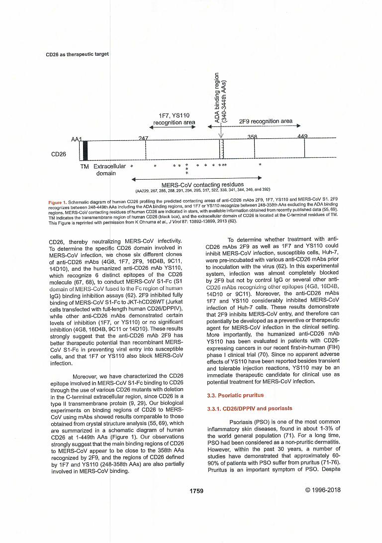
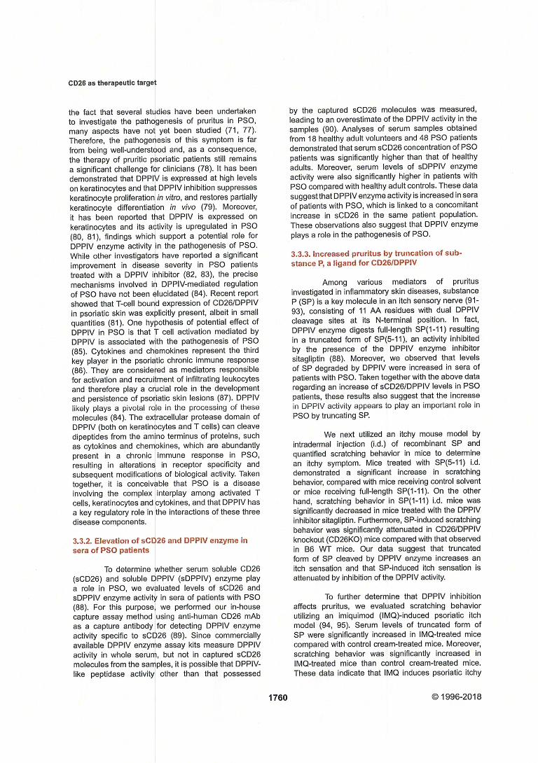
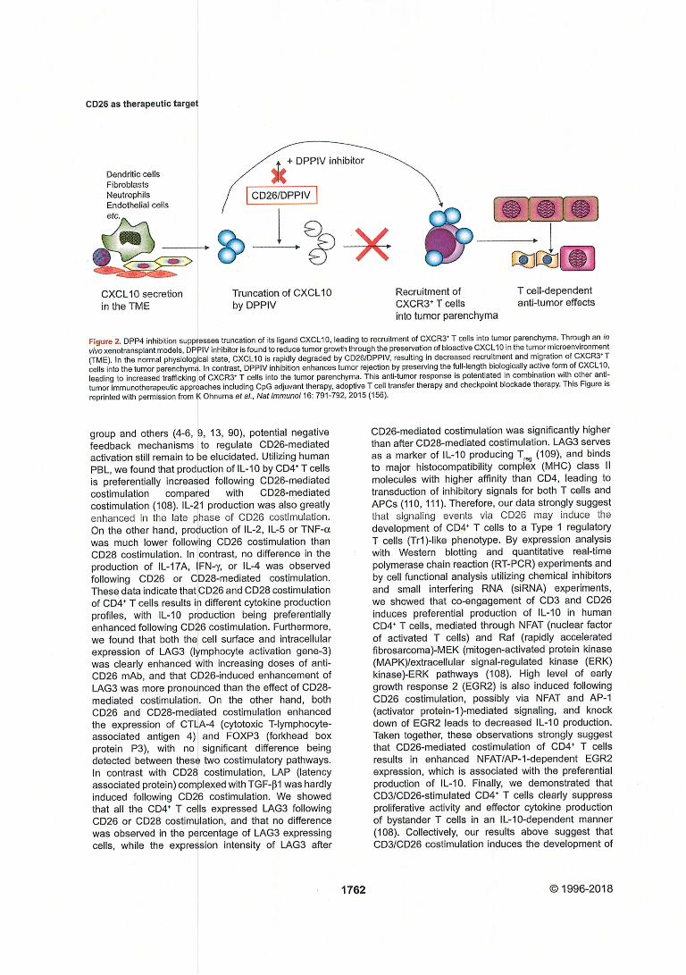
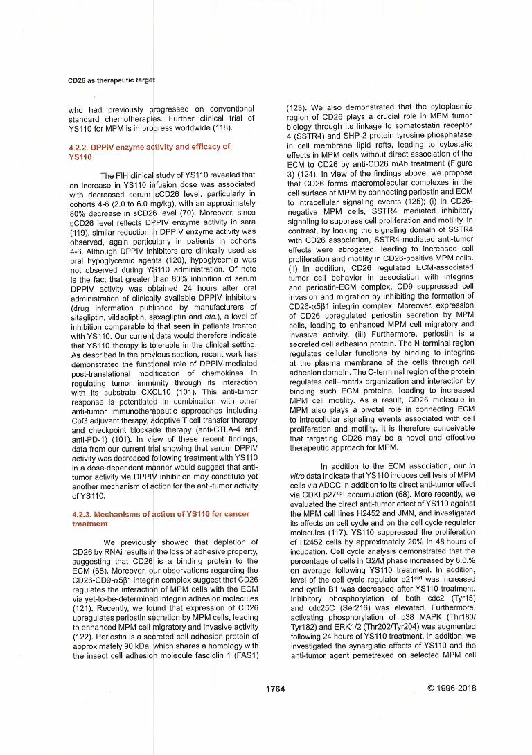
A novel derivative (GTN024) from a natural product,komaroviquinone, induced the apoptosis of high-risk myeloma cellsvia reactive oxygen production and ER stress
Mikio Okayama a, b, Shotaro Kitabatake a, Mariko Sato a, Kota Fujimori a, Daiju Ichikawa a,Maiko Matsushita a, Yutaka Suto c, Genji Iwasaki c, Taketo Yamada d, Fumiyuki Kiuchi e,Maki Hirao f, Hisako Kunieda f, Makoto Osada f, Shinichiro Okamoto b, Yutaka Hattori a, f, *
a Clinical Physiology and Therapeutics, Keio University Faculty of Pharmacy, Tokyo, Japanb Division of Hematology, Department of Internal Medicine, Keio University School of Medicine, Tokyo, Japanc Faculty of Pharmacy, Takasaki University of Health and Welfare, Gunma, Japand Department of Pathology, Saitama Medical University, Saitama, Japane Division of Natural Medicines, Keio University Faculty of Pharmacy, Tokyo, Japanf Department of Hematology, Tokyo Saiseikai Central Hospital, Tokyo, Japan
a r t i c l e i n f o
Article history:Received 19 September 2018Accepted 28 September 2018Available online 5 October 2018
Keywords:Multiple myelomaNatural productApoptosisReactive oxygen speciesER stress
a b s t r a c t
New drugs have significantly improved the survival of patients with multiple myeloma (MM), but theprognosis of MM patients with high-risk cytogenetic changes such as t(4; 14), t(14; 16) or del17p remainsvery poor. A natural product, komaroviquinone (KQN), was originally isolated from the perennial semi-shrub Dracocephalum komarovi and has anti-protozoal activity against Trypanosoma cruzi, the organismcausing Chagas’ disease. Here we demonstrate that a novel KQN-derivative, GTN024, has an anti-MMeffect both in vitro and in vivo. GTN024 induced the apoptosis of MM cell lines including those withhigh-risk cytogenetic changes. GTN024 produced reactive oxygen species (ROS) and increased phos-phorylated eIF2a. The ROS production and subsequent endoplasmic reticulum (ER) stress are thought toplay a key role in GTN024-induced apoptosis, as the apoptosis was completely abrogated by anti-oxidanttreatment. In a mouse xenograft model, an intraperitoneal injection of 20mg/kg of GTN024 significantlydelayed tumor growth. Hematological toxicity and systemic toxicity as indicated by weight loss were notobserved. These results suggest that the novel KQN-derivative GTN024 could become a candidate drugfor treating high-risk MM.
© 2018 Elsevier Inc. All rights reserved.
1. Introduction
Multiple myeloma (MM) is a B-cell neoplasm that causes clonalplasma cell proliferation in bone marrow and bone lesions. The 5-year prevalence rate of MM incidence in Japan is reported to be9.7 per 100,000 persons [1]. New agents such as proteasome in-hibitors and immunomodulatory drugs (IMiDs) have significantlyimproved the overall survival of MM patients [2e5], drugs fromdifferent categories such as a histone-deacetylase inhibitor [6], ananti-SLAMF7 antibody [7], and an anti-CD38 antibody [8] have also
been reported to be effective for refractory MM in combinationtherapy with IMiDs and proteasome inhibitors.
Despite these advances, the survival of certain groups of MMpatients remains unsatisfactory [9e11]. Those patients are knownas having ‘high-risk MM,’ and their MM cells frequently possesschromosomal abnormalities such as t(4; 14), t(14; 16), del17p, and1q21 amplification. The revised International Staging System in-dicates that the overall survival of the patients with high-riskcytogenic abnormalities is significantly short [12]. Another limita-tion of newly developed drugs is their toxicities [13,14], whichimpede the optimal drug efficacy and result in unsatisfactorytreatment outcomes, especially among elderly patients. New ther-apeutic modalities that are effective for high-risk MMwith less sideeffects are thus currently an unmet clinical need in MM treatment.
A series of anti-neoplastic drugs were developed from natural
* Corresponding author. Clinical Physiology and Therapeutics, Keio UniversityFaculty of Pharmacy, Tokyo, Japan.
E-mail address: [email protected] (Y. Hattori).
Contents lists available at ScienceDirect
Biochemical and Biophysical Research Communications
journal homepage: www.elsevier .com/locate/ybbrc
https://doi.org/10.1016/j.bbrc.2018.09.1770006-291X/© 2018 Elsevier Inc. All rights reserved.
Biochemical and Biophysical Research Communications 505 (2018) 787e793
products, and there are some reports describing anti-neoplasticactivities of anti-protozoal agents. For example, nifurtimox, adrug for Chagas’ disease, showed anti-tumor effects against neuraltumor cells. Nifurtimox induced the apoptosis of neuroblastomasby inhibiting extracellular signal-regulated kinase (ERK) phos-phorylation [15]. Artesunate, an anti-malaria drug, also showedanti-MM effects by inhibiting nuclear factor-kappa B (NFkB) func-tion [16]. With these drugs, the anti-tumor activities were discov-ered as off-target effects in drug repositioning studies.
Komaroviquinone (KQN) is one of the natural products isolatedfrom the perennial semi-shrub Dracocephalum komarovi (familyLaminaceae), which shows anti-protozoal activities. Suto et al. re-ported the asymmetric synthesis of KQN [17]. In a study of thestructure activity relationship study of KQN [17], a series of low-molecular-weight compounds were discovered to exhibit prom-ising anti-protozoal activities against Trypanosoma cruzi, which isthe causative pathogen of Chagas’ disease [18]. It is also reportedthat KQN was reduced by T. cruzi old yellow enzyme (TcOYE) toform its semiquinone and produced reactive oxygen species (ROS),which showed trypanocidal activities [19]. A biomedical assay ofboth KQN and its derivatives demonstrated that the new KQN-derivative GTN024 had high anti-proliferation activities againstMM cells [20]. In addition, in the above-cited structure-activityrelationship study, GTN024 was shown to be readily accessible anda valuable compound for the further pharmacodynamic study ofMM cells [20].
With this background, we carried out the present study todetermine the anti-tumor effects of GTN024 on MM cell linesincluding those with high-risk cytogenetic changes, and we clari-fied this promising new drug's mode of action and safety.
2. Materials and methods
2.1. Cells
The human myeloma cell lines KMM1, KMS11, KMS21, KMS26,KMS27, KMS28 and KMS34 were kindly provided by Dr. T. Otsuki(Kawasaki Medical School, Kurashiki, Japan) [21]. The cell lineMUM24 was established in our laboratory from a patient withthalidomide-resistant MM [22]. These cell lines were maintained inRPMI1640 medium (Sigma-Aldrich, St. Louis, MO, USA) containing10% fetal bovine serum (FBS, Gibco, Life Technologies, Carlsbad, CA)and 1% penicillin-streptomycin (Pen Strep, Gibco). Chronosomalabnormalities were detected by the fluorescence in situ hybridiza-tion (FISH) analysis. (LSI medience, Tokyo).
2.2. Reagents
GTN024 (Fig. 1A) was prepared as described by Suto et al. [18]. Inthe present in vitro study, GTN024 was diluted in phosphate-buffered saline (PBS, Sigma-Aldrich) containing 1% Tween®80(Otsuka Pharmaceutical, Tokyo) and 10% DMSO.
2.3. Patient's samples
Bone marrow samples were collected fromMM patients treatedat Tokyo Saiseikai Central Hospital. The collection of clinical sam-ples was approved by ethical committee of Saiseikai Central Hos-pital (No. 28e66) and the Faculty of Pharmacy, Keio University (No.170616e5, 180615e5). Written informed consent for their samplesto be used was obtained from all patients. Cells were isolated bycentrifugation with Lymphoprep™ (Axis-Shield, Oslo, Norway).Cells were labeled with CD138 MicroBeads (Miltenyi Biotec, Ber-gisch Gladbach, Germany). The magnetically labeled CD138-positive cells were purified by MACS Columns (Miltenyi Biotec).
2.4. Trypan blue exclusion assay
Cells (2� 105 cells/mL) were seeded on six-well plates andcultured in various concentrations of GTN024 (0e20 mM) with orwithout 6mMofN-acetyl cysteine (NAC, Sigma-Aldrich) or 3mMofglutathione (GSH, Sigma-Aldrich) at 37 �C in 5% CO2. The cells werestained with Trypan Blue Stain 0.4% (Gibco) and viable cells werecounted by an automatic cell counter TC20™ (Bio-Rad, Hercules,CA). Viable cells were counted three times, and the average wascalculated. The IC50 of GTN024 was calculated by approximation.
2.5. MTT assay
Collected clinical samples (6� 104 cells/mL) were seeded on 96-well plates and cultured with various concentrations of GTN024(0e30 mM) at 37 �C in 5% CO2 for 48 h. The viability of the cells wascalculated byMTT dye absorbance (Roche Diagnostics, Indianapolis,IN) according to the manufacturer's instructions.
2.6. Apoptosis detection assay
Cells (2� 105 cells/mL) were seeded on six-well plates andcultured at 37 �C in 10 mM GTN024 for 72 h. Apoptotic cells weredetected by an annexin V-FITC Apoptosis Detection Kit (BioVision,San Francisco, CA) following the manufacturer's protocol. Briefly,cells were collected and resuspended in 500 mL of 1� BindingBuffer and stained with annexin V eFITC and propidium iodide (PI)for 5min. The cells were analyzed using a BD™ LSRⅡ flow cytom-eter (Becton Dickinson, Lincoln Park, NJ).
2.7. Detection of reactive oxygen species
Cells (2� 105 cells/mL) were seeded on six-well plates andcultured at 37 �C with or without 6mM NAC or 3mM GSH for 2 h.Then, 20 mM GTN024 was added and incubated in 5% CO2. After72 h, 1 mM chloromethyl-dichlorodihydrofluorescein diacetate,acetyl ester (CM-H2DCFDA, Invitrogen, Carlsbad, CA) was addedand incubated for 30min. The stained cells were analyzed using theBD LSRⅡ flow cytometer.
2.8. Western blotting
Cells were cultured with GTN024 and lysed in 1% NP-40 buffercontaining 1mM PMSF, 1mM Na2PO4, 20mM NaF, 2mM Na2PO7,and protease inhibitors (Complete Protease Inhibitor Mixture,Roche Diagnostics, Mannheim, Germany). After incubation for10min on ice, the lysates were centrifuged at 15,000 rpm for10min at 4 �C, and the supernatants were collected. The amount ofprotein was evaluated by BCA Protein Assay Kit (Thermo FisherScientific, Waltham, MA).
The lysates were mixed with Laemmli's buffer (1.33% SDS, 10%glycerol, 0.083M Tris-HCl, 0.04% bromphenol blue, 2% 2-ME) andboiled for 5min. The lysates were subjected onto 10% sodiumdodecyl sulfate-polyacrylamide gel electrophoresis (SDS-PAGE),and transferred to a PVDF membrane. The membranes wereblocked with 5% skim milk and then immunoblotted. Antibodiesagainst eIF2a (Cell Signaling Technology, Danvers, MA), p-eIF2a(Cell Signaling Technology), b-actin (Santa Cruz Biotechnology,Santa Cruz, CA), and cleaved caspase-3 (Cell Signaling Technology)(diluted at 1:1000) was used. The second antigen-antibodies was ahorseradish peroxidase (HRP)-coupled anti-rabbit, anti-mouse Igantibody (diluted at 1:500). Antigen-antibody complexes weredetected using enhanced chemiluminescence (Amersham, Arling-ton Heights, IL).
M. Okayama et al. / Biochemical and Biophysical Research Communications 505 (2018) 787e793788
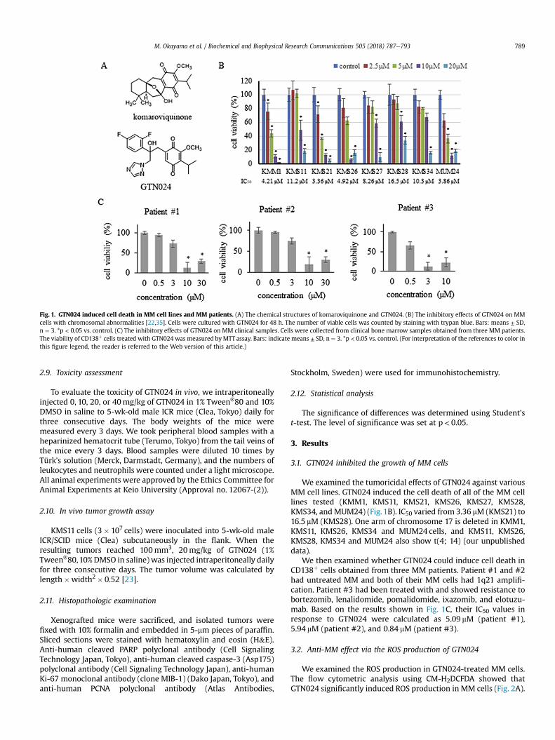
2.9. Toxicity assessment
To evaluate the toxicity of GTN024 in vivo, we intraperitoneallyinjected 0, 10, 20, or 40mg/kg of GTN024 in 1% Tween®80 and 10%DMSO in saline to 5-wk-old male ICR mice (Clea, Tokyo) daily forthree consecutive days. The body weights of the mice weremeasured every 3 days. We took peripheral blood samples with aheparinized hematocrit tube (Terumo, Tokyo) from the tail veins ofthe mice every 3 days. Blood samples were diluted 10 times byTürk's solution (Merck, Darmstadt, Germany), and the numbers ofleukocytes and neutrophils were counted under a light microscope.All animal experiments were approved by the Ethics Committee forAnimal Experiments at Keio University (Approval no. 12067-(2)).
2.10. In vivo tumor growth assay
KMS11 cells (3� 107 cells) were inoculated into 5-wk-old maleICR/SCID mice (Clea) subcutaneously in the flank. When theresulting tumors reached 100mm3, 20mg/kg of GTN024 (1%Tween®80,10% DMSO in saline) was injected intraperitoneally dailyfor three consecutive days. The tumor volume was calculated bylength�width2� 0.52 [23].
2.11. Histopathologic examination
Xenografted mice were sacrificed, and isolated tumors werefixed with 10% formalin and embedded in 5-mm pieces of paraffin.Sliced sections were stained with hematoxylin and eosin (H&E).Anti-human cleaved PARP polyclonal antibody (Cell SignalingTechnology Japan, Tokyo), anti-human cleaved caspase-3 (Asp175)polyclonal antibody (Cell Signaling Technology Japan), anti-humanKi-67 monoclonal antibody (clone MIB-1) (Dako Japan, Tokyo), andanti-human PCNA polyclonal antibody (Atlas Antibodies,
Stockholm, Sweden) were used for immunohistochemistry.
2.12. Statistical analysis
The significance of differences was determined using Student'st-test. The level of significance was set at p< 0.05.
3. Results
3.1. GTN024 inhibited the growth of MM cells
We examined the tumoricidal effects of GTN024 against variousMM cell lines. GTN024 induced the cell death of all of the MM celllines tested (KMM1, KMS11, KMS21, KMS26, KMS27, KMS28,KMS34, andMUM24) (Fig. 1B). IC50 varied from 3.36 mM (KMS21) to16.5 mM (KMS28). One arm of chromosome 17 is deleted in KMM1,KMS11, KMS26, KMS34 and MUM24 cells, and KMS11, KMS26,KMS28, KMS34 and MUM24 also show t(4; 14) (our unpublisheddata).
We then examined whether GTN024 could induce cell death inCD138þ cells obtained from three MM patients. Patient #1 and #2had untreated MM and both of their MM cells had 1q21 amplifi-cation. Patient #3 had been treated with and showed resistance tobortezomib, lenalidomide, pomalidomide, ixazomib, and elotuzu-mab. Based on the results shown in Fig. 1C, their IC50 values inresponse to GTN024 were calculated as 5.09 mM (patient #1),5.94 mM (patient #2), and 0.84 mM (patient #3).
3.2. Anti-MM effect via the ROS production of GTN024
We examined the ROS production in GTN024-treated MM cells.The flow cytometric analysis using CM-H2DCFDA showed thatGTN024 significantly induced ROS production in MM cells (Fig. 2A).
Fig. 1. GTN024 induced cell death in MM cell lines and MM patients. (A) The chemical structures of komaroviquinone and GTN024. (B) The inhibitory effects of GTN024 on MMcells with chromosomal abnormalities [22,35]. Cells were cultured with GTN024 for 48 h. The number of viable cells was counted by staining with trypan blue. Bars: means ± SD,n ¼ 3. *p < 0.05 vs. control. (C) The inhibitory effects of GTN024 on MM clinical samples. Cells were collected from clinical bone marrow samples obtained from three MM patients.The viability of CD138þ cells treated with GTN024 was measured by MTT assay. Bars: indicate means ± SD, n ¼ 3. *p < 0.05 vs. control. (For interpretation of the references to color inthis figure legend, the reader is referred to the Web version of this article.)
M. Okayama et al. / Biochemical and Biophysical Research Communications 505 (2018) 787e793 789
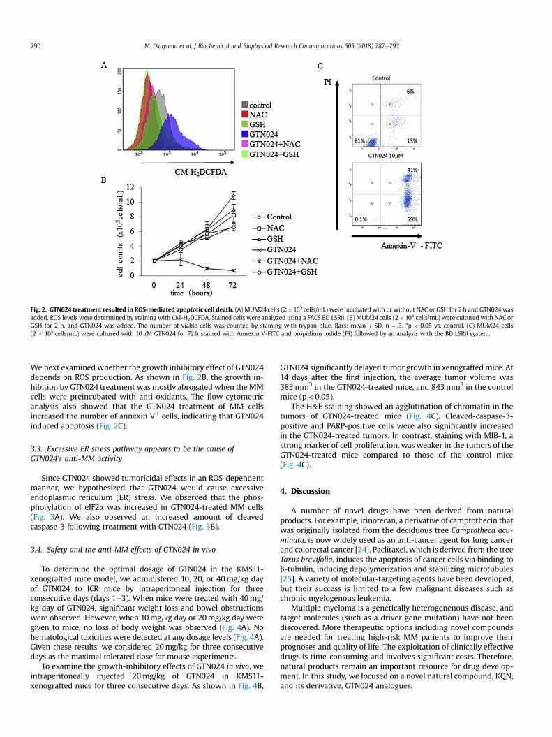
We next examined whether the growth inhibitory effect of GTN024depends on ROS production. As shown in Fig. 2B, the growth in-hibition by GTN024 treatment was mostly abrogated when the MMcells were preincubated with anti-oxidants. The flow cytometricanalysis also showed that the GTN024 treatment of MM cellsincreased the number of annexin Vþ cells, indicating that GTN024induced apoptosis (Fig. 2C).
3.3. Excessive ER stress pathway appears to be the cause ofGTN024's anti-MM activity
Since GTN024 showed tumoricidal effects in an ROS-dependentmanner, we hypothesized that GTN024 would cause excessiveendoplasmic reticulum (ER) stress. We observed that the phos-phorylation of eIF2a was increased in GTN024-treated MM cells(Fig. 3A). We also observed an increased amount of cleavedcaspase-3 following treatment with GTN024 (Fig. 3B).
3.4. Safety and the anti-MM effects of GTN024 in vivo
To determine the optimal dosage of GTN024 in the KMS11-xenografted mice model, we administered 10, 20, or 40mg/kg dayof GTN024 to ICR mice by intraperitoneal injection for threeconsecutive days (days 1e3). When mice were treated with 40mg/kg day of GTN024, significant weight loss and bowel obstructionswere observed. However, when 10mg/kg day or 20mg/kg day weregiven to mice, no loss of body weight was observed (Fig. 4A). Nohematological toxicities were detected at any dosage levels (Fig. 4A).Given these results, we considered 20mg/kg for three consecutivedays as the maximal tolerated dose for mouse experiments.
To examine the growth-inhibitory effects of GTN024 in vivo, weintraperitoneally injected 20mg/kg of GTN024 in KMS11-xenografted mice for three consecutive days. As shown in Fig. 4B,
GTN024 significantly delayed tumor growth in xenograftedmice. At14 days after the first injection, the average tumor volume was383mm3 in the GTN024-treated mice, and 843mm3 in the controlmice (p< 0.05).
The H&E staining showed an agglutination of chromatin in thetumors of GTN024-treated mice (Fig. 4C). Cleaved-caspase-3-positive and PARP-positive cells were also significantly increasedin the GTN024-treated tumors. In contrast, staining with MIB-1, astrong marker of cell proliferation, was weaker in the tumors of theGTN024-treated mice compared to those of the control mice(Fig. 4C).
4. Discussion
A number of novel drugs have been derived from naturalproducts. For example, irinotecan, a derivative of camptothecin thatwas originally isolated from the deciduous tree Camptotheca acu-minata, is now widely used as an anti-cancer agent for lung cancerand colorectal cancer [24]. Paclitaxel, which is derived from the treeTaxus brevifolia, induces the apoptosis of cancer cells via binding tob-tubulin, inducing depolymerization and stabilizing microtubules[25]. A variety of molecular-targeting agents have been developed,but their success is limited to a few malignant diseases such aschronic myelogenous leukemia.
Multiple myeloma is a genetically heterogenenous disease, andtarget molecules (such as a driver gene mutation) have not beendiscovered. More therapeutic options including novel compoundsare needed for treating high-risk MM patients to improve theirprognoses and quality of life. The exploitation of clinically effectivedrugs is time-consuming and involves significant costs. Therefore,natural products remain an important resource for drug develop-ment. In this study, we focused on a novel natural compound, KQN,and its derivative, GTN024 analogues.
Fig. 2. GTN024 treatment resulted in ROS-mediated apoptotic cell death. (A) MUM24 cells (2� 105 cells/mL) were incubated with or without NAC or GSH for 2 h and GTN024 wasadded. ROS levels were determined by staining with CM-H2DCFDA. Stained cells were analyzed using a FACS BD LSRⅡ. (B) MUM24 cells (2� 105 cells/mL) were cultured with NAC orGSH for 2 h, and GTN024 was added. The number of viable cells was counted by staining with trypan blue. Bars: mean ± SD, n ¼ 3. *p < 0.05 vs. control. (C) MUM24 cells(2 � 105 cells/mL) were cultured with 10 mM GTN024 for 72 h stained with Annexin V-FITC and propidium iodide (PI) followed by an analysis with the BD LSRⅡ system.
M. Okayama et al. / Biochemical and Biophysical Research Communications 505 (2018) 787e793790
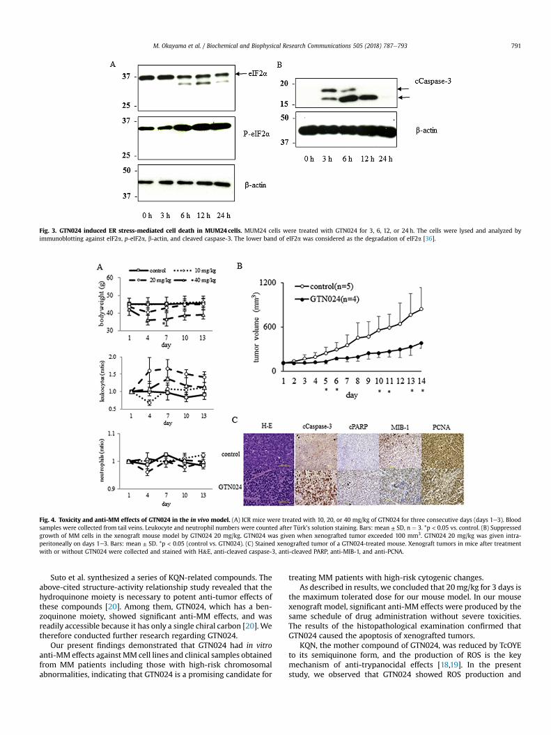
Suto et al. synthesized a series of KQN-related compounds. Theabove-cited structure-activity relationship study revealed that thehydroquinone moiety is necessary to potent anti-tumor effects ofthese compounds [20]. Among them, GTN024, which has a ben-zoquinone moiety, showed significant anti-MM effects, and wasreadily accessible because it has only a single chiral carbon [20]. Wetherefore conducted further research regarding GTN024.
Our present findings demonstrated that GTN024 had in vitroanti-MM effects against MM cell lines and clinical samples obtainedfrom MM patients including those with high-risk chromosomalabnormalities, indicating that GTN024 is a promising candidate for
treating MM patients with high-risk cytogenic changes.As described in results, we concluded that 20mg/kg for 3 days is
the maximum tolerated dose for our mouse model. In our mousexenograft model, significant anti-MM effects were produced by thesame schedule of drug administration without severe toxicities.The results of the histopathological examination confirmed thatGTN024 caused the apoptosis of xenografted tumors.
KQN, the mother compound of GTN024, was reduced by TcOYEto its semiquinone form, and the production of ROS is the keymechanism of anti-trypanocidal effects [18,19]. In the presentstudy, we observed that GTN024 showed ROS production and
Fig. 3. GTN024 induced ER stress-mediated cell death in MUM24 cells. MUM24 cells were treated with GTN024 for 3, 6, 12, or 24 h. The cells were lysed and analyzed byimmunoblotting against eIF2a, p-eIF2a, b-actin, and cleaved caspase-3. The lower band of eIF2a was considered as the degradation of eIF2a [36].
Fig. 4. Toxicity and anti-MM effects of GTN024 in the in vivo model. (A) ICR mice were treated with 10, 20, or 40 mg/kg of GTN024 for three consecutive days (days 1e3). Bloodsamples were collected from tail veins. Leukocyte and neutrophil numbers were counted after Türk's solution staining. Bars: mean ± SD, n ¼ 3. *p < 0.05 vs. control. (B) Suppressedgrowth of MM cells in the xenograft mouse model by GTN024 20 mg/kg. GTN024 was given when xenografted tumor exceeded 100 mm3. GTN024 20 mg/kg was given intra-peritoneally on days 1e3. Bars: mean ± SD. *p < 0.05 (control vs. GTN024). (C) Stained xenografted tumor of a GTN024-treated mouse. Xenograft tumors in mice after treatmentwith or without GTN024 were collected and stained with H&E, anti-cleaved caspase-3, anti-cleaved PARP, anti-MIB-1, and anti-PCNA.
M. Okayama et al. / Biochemical and Biophysical Research Communications 505 (2018) 787e793 791
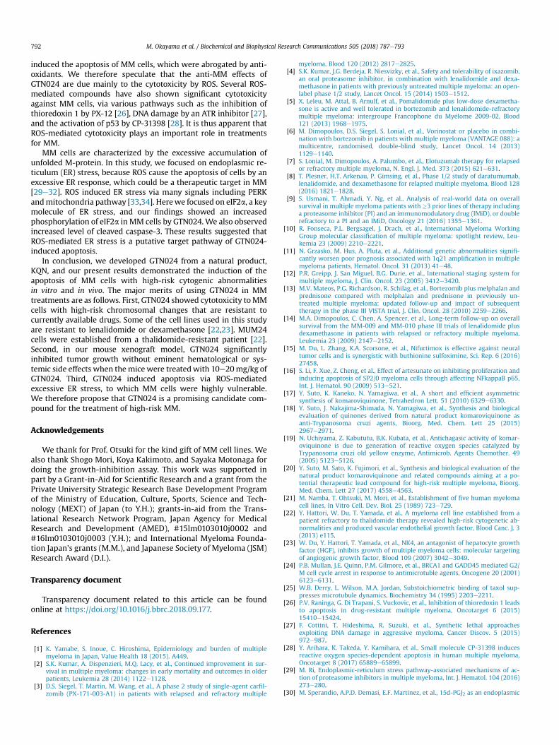
induced the apoptosis of MM cells, which were abrogated by anti-oxidants. We therefore speculate that the anti-MM effects ofGTN024 are due mainly to the cytotoxicity by ROS. Several ROS-mediated compounds have also shown significant cytotoxicityagainst MM cells, via various pathways such as the inhibition ofthioredoxin 1 by PX-12 [26], DNA damage by an ATR inhibitor [27],and the activation of p53 by CP-31398 [28]. It is thus apparent thatROS-mediated cytotoxicity plays an important role in treatmentsfor MM.
MM cells are characterized by the excessive accumulation ofunfolded M-protein. In this study, we focused on endoplasmic re-ticulum (ER) stress, because ROS cause the apoptosis of cells by anexcessive ER response, which could be a therapeutic target in MM[29e32]. ROS induced ER stress via many signals including PERKandmitochondria pathway [33,34]. Herewe focused on eIF2a, a keymolecule of ER stress, and our findings showed an increasedphosphorylation of eIF2a in MM cells by GTN024.We also observedincreased level of cleaved caspase-3. These results suggested thatROS-mediated ER stress is a putative target pathway of GTN024-induced apoptosis.
In conclusion, we developed GTN024 from a natural product,KQN, and our present results demonstrated the induction of theapoptosis of MM cells with high-risk cytogenic abnormalitiesin vitro and in vivo. The major merits of using GTN024 in MMtreatments are as follows. First, GTN024 showed cytotoxicity toMMcells with high-risk chromosomal changes that are resistant tocurrently available drugs. Some of the cell lines used in this studyare resistant to lenalidomide or dexamethasone [22,23]. MUM24cells were established from a thalidomide-resistant patient [22].Second, in our mouse xenograft model, GTN024 significantlyinhibited tumor growth without eminent hematological or sys-temic side effects when themice were treatedwith 10e20mg/kg ofGTN024. Third, GTN024 induced apoptosis via ROS-mediatedexcessive ER stress, to which MM cells were highly vulnerable.We therefore propose that GTN024 is a promising candidate com-pound for the treatment of high-risk MM.
Acknowledgements
We thank for Prof. Otsuki for the kind gift of MM cell lines. Wealso thank Shogo Mori, Koya Kakimoto, and Sayaka Motonaga fordoing the growth-inhibition assay. This work was supported inpart by a Grant-in-Aid for Scientific Research and a grant from thePrivate University Strategic Research Base Development Programof the Ministry of Education, Culture, Sports, Science and Tech-nology (MEXT) of Japan (to Y.H.); grants-in-aid from the Trans-lational Research Network Program, Japan Agency for MedicalResearch and Development (AMED), #15lm0103010j0002 and#16lm0103010j0003 (Y.H.); and International Myeloma Founda-tion Japan's grants (M.M.), and Japanese Society of Myeloma (JSM)Research Award (D.I.).
Transparency document
Transparency document related to this article can be foundonline at https://doi.org/10.1016/j.bbrc.2018.09.177.
References
[1] K. Yamabe, S. Inoue, C. Hiroshima, Epidemiology and burden of multiplemyeloma in Japan, Value Health 18 (2015). A449.
[2] S.K. Kumar, A. Dispenzieri, M.Q. Lacy, et al., Continued improvement in sur-vival in multiple myeloma: changes in early mortality and outcomes in olderpatients, Leukemia 28 (2014) 1122e1128.
[3] D.S. Siegel, T. Martin, M. Wang, et al., A phase 2 study of single-agent carfil-zomib (PX-171-003-A1) in patients with relapsed and refractory multiple
myeloma, Blood 120 (2012) 2817e2825.[4] S.K. Kumar, J.G. Berdeja, R. Niesvizky, et al., Safety and tolerability of ixazomib,
an oral proteasome inhibitor, in combination with lenalidomide and dexa-methasone in patients with previously untreated multiple myeloma: an open-label phase 1/2 study, Lancet Oncol. 15 (2014) 1503e1512.
[5] X. Leleu, M. Attal, B. Arnulf, et al., Pomalidomide plus low-dose dexametha-sone is active and well tolerated in bortezomib and lenalidomide-refractorymultiple myeloma: intergroupe Francophone du My�elome 2009-02, Blood121 (2013) 1968e1975.
[6] M. Dimopoulos, D.S. Siegel, S. Lonial, et al., Vorinostat or placebo in combi-nation with bortezomib in patients with multiple myeloma (VANTAGE 088): amulticentre, randomised, double-blind study, Lancet Oncol. 14 (2013)1129e1140.
[7] S. Lonial, M. Dimopoulos, A. Palumbo, et al., Elotuzumab therapy for relapsedor refractory multiple myeloma, N. Engl. J. Med. 373 (2015) 621e631.
[8] T. Plesner, H.T. Arkenau, P. Gimsing, et al., Phase 1/2 study of daratumumab,lenalidomide, and dexamethasone for relapsed multiple myeloma, Blood 128(2016) 1821e1828.
[9] S. Usmani, T. Ahmadi, Y. Ng, et al., Analysis of real-world data on overallsurvival in multiple myeloma patients with �3 prior lines of therapy includinga proteasome inhibitor (PI) and an immunomodulatory drug (IMiD), or doublerefractory to a PI and an IMiD, Oncology 21 (2016) 1355e1361.
[10] R. Fonseca, P.L. Bergsagel, J. Drach, et al., International Myeloma WorkingGroup molecular classification of multiple myeloma: spotlight review, Leu-kemia 23 (2009) 2210e2221.
[11] N. Grzasko, M. Hus, A. Pluta, et al., Additional genetic abnormalities signifi-cantly worsen poor prognosis associated with 1q21 amplification in multiplemyeloma patients, Hematol. Oncol. 31 (2013) 41e48.
[12] P.R. Greipp, J. San Miguel, B.G. Durie, et al., International staging system formultiple myeloma, J. Clin. Oncol. 23 (2005) 3412e3420.
[13] M.V. Mateos, P.G. Richardson, R. Schilag, et al., Bortezomib plus melphalan andprednisone compared with melphalan and prednisone in previously un-treated multiple myeloma: updated follow-up and impact of subsequenttherapy in the phase III VISTA trial, J. Clin. Oncol. 28 (2010) 2259e2266.
[14] M.A. Dimopoulos, C. Chen, A. Spencer, et al., Long-term follow-up on overallsurvival from the MM-009 and MM-010 phase III trials of lenalidomide plusdexamethasone in patients with relapsed or refractory multiple myeloma,Leukemia 23 (2009) 2147e2152.
[15] M. Du, L. Zhang, K.A. Scorsone, et al., Nifurtimox is effective against neuraltumor cells and is synergistic with buthionine sulfoximine, Sci. Rep. 6 (2016)27458.
[16] S. Li, F. Xue, Z. Cheng, et al., Effect of artesunate on inhibiting proliferation andinducing apoptosis of SP2/0 myeloma cells through affecting NFkappaB p65,Int. J. Hematol. 90 (2009) 513e521.
[17] Y. Suto, K. Kaneko, N. Yamagiwa, et al., A short and efficient asymmetricsynthesis of komaroviquinone, Tetrahedron Lett. 51 (2010) 6329e6330.
[18] Y. Suto, J. Nakajima-Shimada, N. Yamagiwa, et al., Synthesis and biologicalevaluation of quinones derived from natural product komaroviquinone asanti-Trypanosoma cruzi agents, Bioorg. Med. Chem. Lett 25 (2015)2967e2971.
[19] N. Uchiyama, Z. Kabututu, B.K. Kubata, et al., Antichagasic activity of komar-oviquinone is due to generation of reactive oxygen species catalyzed byTrypanosoma cruzi old yellow enzyme, Antimicrob. Agents Chemother. 49(2005) 5123e5126.
[20] Y. Suto, M. Sato, K. Fujimori, et al., Synthesis and biological evaluation of thenatural product komaroviquinone and related compounds aiming at a po-tential therapeutic lead compound for high-risk multiple myeloma, Bioorg.Med. Chem. Lett 27 (2017) 4558e4563.
[21] M. Namba, T. Ohtsuki, M. Mori, et al., Establishment of five human myelomacell lines, In Vitro Cell. Dev. Biol. 25 (1989) 723e729.
[22] Y. Hattori, W. Du, T. Yamada, et al., A myeloma cell line established from apatient refractory to thalidomide therapy revealed high-risk cytogenetic ab-normalities and produced vascular endothelial growth factor, Blood Canc. J. 3(2013) e115.
[23] W. Du, Y. Hattori, T. Yamada, et al., NK4, an antagonist of hepatocyte growthfactor (HGF), inhibits growth of multiple myeloma cells: molecular targetingof angiogenic growth factor, Blood 109 (2007) 3042e3049.
[24] P.B. Mullan, J.E. Quinn, P.M. Gilmore, et al., BRCA1 and GADD45 mediated G2/M cell cycle arrest in response to antimicrotuble agents, Oncogene 20 (2001)6123e6131.
[25] W.B. Derry, L. Wilson, M.A. Jordan, Substoichiometric binding of taxol sup-presses microtubule dynamics, Biochemistry 34 (1995) 2203e2211.
[26] P.V. Raninga, G. Di Trapani, S. Vuckovic, et al., Inhibition of thioredoxin 1 leadsto apoptosis in drug-resistant multiple myeloma, Oncotarget 6 (2015)15410e15424.
[27] F. Cottini, T. Hideshima, R. Suzuki, et al., Synthetic lethal approachesexploiting DNA damage in aggressive myeloma, Cancer Discov. 5 (2015)972e987.
[28] Y. Arihara, K. Takeda, Y. Kamihara, et al., Small molecule CP-31398 inducesreactive oxygen species-dependent apoptosis in human multiple myeloma,Oncotarget 8 (2017) 65889e65899.
[29] M. Ri, Endoplasmic-reticulum stress pathway-associated mechanisms of ac-tion of proteasome inhibitors in multiple myeloma, Int. J. Hematol. 104 (2016)273e280.
[30] M. Sperandio, A.P.D. Demasi, E.F. Martinez, et al., 15d-PGJ2 as an endoplasmic
M. Okayama et al. / Biochemical and Biophysical Research Communications 505 (2018) 787e793792

reticulum stress manipulator in multiple myeloma in vitro and in vivo, Exp.Mol. Pathol. 102 (2017) 434e445.
[31] S. Bustany, J. Cahu, P. Guardiola, et al., CyclinD1 sensitizes myeloma cells toendoplasmic reticulum stress-mediated apoptosis by activating the unfoldedprotein response pathway, BMC Canc. 15 (2015) 262.
[32] S. Manni, A. Brancalion, L.Q. Tubi, et al., Protein kinase CK2 protects multiplemyeloma cells from ER stress-induced apoptosis and from the cytotoxic effectof HSP90 inhibition through regulation of the unfolded protein response, Clin.Canc. Res. 18 (2012) 1888e1900.
[33] S. Moriya, S. Komatsu, K. Yamasaki, et al., Targeting the integrated networks ofaggresome formation, proteasome, and autophagy potentiates ER stress-
mediated cell death in multiple myeloma cells, Int. J. Oncol. 46 (2015)474e486.
[34] W. Rozpedek, D. Pytel, B. Mucha, et al., The Role of the PERK/eIF2a/ATF4/CHOPsignaling pathway in tumor progression during endoplasmic reticulum stress,Curr. Mol. Med. 16 (2016) 533e544.
[35] H. Shiheido, F. Terada, N. Tabata, et al., A phtalimide derivative that inhibitscentrosomal clustering is effective on multiple myeloma, PloS One 7 (2012),e38878.
[36] W.E. Marissen, Y. Guo, A.A. Thomas, et al., Identification of caspase 3-mediatedcleavage and functional alteration of eukaryotic initiation factor 2alpha inapoptosis, J. Biol. Chem. 275 (2000) 9314e9323.
M. Okayama et al. / Biochemical and Biophysical Research Communications 505 (2018) 787e793 793
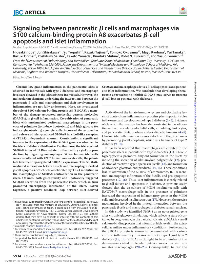
Signaling between pancreatic � cells and macrophages viaS100 calcium-binding protein A8 exacerbates �-cellapoptosis and islet inflammationReceived for publication, July 29, 2017, and in revised form, February 27, 2018 Published, Papers in Press, March 1, 2018, DOI 10.1074/jbc.M117.809228
Hideaki Inoue‡, Jun Shirakawa‡1, Yu Togashi‡2, Kazuki Tajima‡2, Tomoko Okuyama‡2, Mayu Kyohara‡, Yui Tanaka‡,Kazuki Orime‡§, Yoshifumi Saisho¶, Taketo Yamada�, Kimitaka Shibue§, Rohit N. Kulkarni§3, and Yasuo Terauchi‡4
From the ‡Department of Endocrinology and Metabolism, Graduate School of Medicine, Yokohama-City University, 3-9 Fuku-ura,Kanazawa-ku, Yokohama 236-0004, Japan, the Departments of ¶Internal Medicine and �Pathology, School of Medicine, KeioUniversity, Tokyo 108-8345, Japan, and the §Section of Islet Cell and Regenerative Biology, Joslin Diabetes Center, Department ofMedicine, Brigham and Women’s Hospital, Harvard Stem Cell Institute, Harvard Medical School, Boston, Massachusetts 02138
Edited by Jeffrey E. Pessin
Chronic low-grade inflammation in the pancreatic islets isobserved in individuals with type 2 diabetes, and macrophagelevels are elevated in the islets of these individuals. However, themolecular mechanisms underlying the interactions between thepancreatic � cells and macrophages and their involvement ininflammation are not fully understood. Here, we investigatedthe role of S100 calcium-binding protein A8 (S100A8), a mem-ber of the damage-associated molecular pattern molecules(DAMPs), in �-cell inflammation. Co-cultivation of pancreaticislets with unstimulated peritoneal macrophages in the pres-ence of palmitate (to induce lipotoxicity) and high glucose (toinduce glucotoxicity) synergistically increased the expressionand release of islet-produced S100A8 in a Toll-like receptor4 (TLR4)-independent manner. Consistently, a significantincrease in the expression of the S100a8 gene was observed inthe islets of diabetic db/db mice. Furthermore, the islet-derivedS100A8 induced TLR4-mediated inflammatory cytokine pro-duction by migrating macrophages. When human islet cellswere co-cultured with U937 human monocyte cells, the palmi-tate treatment up-regulated S100A8 expression. This S100A8-mediated interaction between islets and macrophages evoked�-cell apoptosis, which was ameliorated by TLR4 inhibition inthe macrophages or S100A8 neutralization in the pancreaticislets. Of note, both glucotoxicity and lipotoxicity triggeredS100A8 secretion from the pancreatic islets, which in turnpromoted macrophage infiltration of the islets. Takentogether, a positive feedback loop between islet-derived
S100A8 and macrophages drives �-cell apoptosis and pancre-atic islet inflammation. We conclude that developing thera-peutic approaches to inhibit S100A8 may serve to prevent�-cell loss in patients with diabetes.
Activation of the innate immune system and circulating lev-els of acute-phase inflammatory proteins play important rolesin the onset and development of type 2 diabetes (1–3). Evidenceof chronic inflammation has been demonstrated in the adiposetissue, liver, vascular endothelial cells, circulating leukocytes,and pancreatic islets in obese and/or diabetic humans (4 –8).Chronic islet inflammation evokes a decline in the �-cell massby promoting �-cell apoptosis, which is a hallmark of type 2diabetes (9, 10).
It has been reported that macrophages are elevated in thepancreatic islets in patients with type 2 diabetes (11). Chronichyperglycemia promotes amyloid formation in the islets byinducing the secretion of islet amyloid polypeptide (12), pro-duction of reactive oxygen species in � cells (13), and formationof advanced glycation end products (14, 15). These conditionslead to activation of the NLRP3 inflammasomes, IL-1� secre-tion, macrophage infiltration of the � cells, and pro-apoptoticprocesses (12, 16). Thus, islet inflammation is closely relatedto �-cell failure and apoptosis in diabetes. A previous studyshowed that the co-culture of MIN6 insulinoma cells withRAW264.7 macrophage cells in the presence of palmitateincreased the expression of inflammatory genes in the MIN6cells and decreased insulin secretion (17). However, the precisemechanisms involved in the mutual interaction between thepancreatic � cells and macrophages in diabetes remain unclear.
In this study, we identified S100a8 as an up-regulated geneafter chronic glucose stimulation, which reflects a state of sus-tained hyperglycemia, in the pancreatic islets. S100A8 is a smallcalcium-binding protein that is found at high levels in the extra-cellular milieu under inflammatory conditions. Furthermore,the S100A8 protein is known to be associated with variouschronic inflammatory diseases and both type 1 and type 2diabetes (18, 19). S100A8 is thought to be a member of thedamage-associated molecular pattern molecules and sti-mulates macrophages (20 –23). Consequently, to test the
This work was supported by Grant-in-Aid for Scientific Research (B) 16H05329(to Y. Terauchi) from the Ministry of Education, Culture, Sports, Science,and Technology (MEXT) of Japan, a grant-in-aid from the Japan Founda-tion for Applied Enzymology (to J. S.), and a Junior Scientist DevelopmentGrant supported by Novo Nordisk Pharma Ltd. (to J. S.). The authorsdeclare that they have no conflicts of interest with the contents of thisarticle. The content is solely the responsibility of the authors and does notnecessarily represent the official views of the National Institutes of Health.
This article contains Tables S1–S3.1 To whom correspondence may be addressed. Tel.: 81-45-787-2639; Fax:
81-45-781-5379. E-mail: [email protected] These authors contributed equally to this work.3 Supported by National Institutes of Health Grants RO1 DK67536 and
DK103215.4 To whom correspondence may be addressed. Tel.: 81-45-787-2639; Fax:
81-45-781-5379. E-mail: [email protected].
croARTICLE
5934 J. Biol. Chem. (2018) 293(16) 5934 –5946
© 2018 by The American Society for Biochemistry and Molecular Biology, Inc. Published in the U.S.A.
at JUN
TE
ND
O U
NIV
ER
SITY
on February 12, 2019http://w
ww
.jbc.org/D
ownloaded from

hypothesis that S100A8 contributes to islet inflammation,we established a co-culture system with freshly isolated pri-mary pancreatic islets and resident peritoneal macrophagesto investigate the role(s) of S100A8 in the sustenance of isletinflammation.
Results
S100A8/A9 expression in the islets was up-regulated bychronic glucose stimulation
Chronic hyperglycemia induces �-cell apoptosis, in part,through continuous glucokinase activation (24). We previouslyidentified the target genes of glucokinase by examining the geneexpression profiles of glucokinase activator (GKA)5-treatedisolated islets (NCBI GEO database GSE41248) (25). Amongthem, S100a8 and S100a9 (S100a8/a9) expression showed thegreatest increase following chronic glucokinase activation ofthe islets (90- and 254-fold increase, respectively). We validatedthe gene expression changes in isolated islets stimulated with aglucokinase activator (Fig. 1A) and confirmed that glucosestimulation increased the expression of S100a8/a9 in the isletsin a concentration-dependent manner (Fig. 1B). Thus, S100A8expression was induced by high glucose in the islets withoutmacrophages.
S100A8/A9 expression in the islets was enhanced by co-culturewith macrophages in the presence of palmitate
We co-cultured islets with macrophages using co-cultureinserts (Fig. 2A) and observed that the islet expression of
S100a8, S100a9, Il-1b, Tnf-a, Il-6, and Ccl2 was increased in thepresence of macrophages (Fig. 2B). The absence of elevatedexpression of the macrophage markers, Cd11b and F4/80, sug-gested that it was unlikely that there was contamination of theco-cultured islet samples with macrophages (Fig. 2C). Theexpression of S100a8 and S100a9, but not of Il-1b, Tnf-a, Il-6,or Ccl2, was enhanced in the islets co-cultured with macro-phages in the presence of the saturated fatty acid palmitate(16:0) (Fig. 2B). We confirmed the secretion of S100A8, but notof S100A9, by ELISAs in the supernatant of islets co-culturedwith macrophages in the presence of palmitate (Fig. 2D). Nota-bly, secretion of S100A8 from the macrophages was notaffected by the concentration of glucose or palmitate (Fig. 2E).S100A8 proteins were predominantly expressed in the mousepancreatic islets, but not in acinar cells (Fig. 2F).
To test the possibility that the adipocyte-derived fatty acidscontributed to the macrophage-mediated islet inflammation invivo, we added isolated white adipocytes from the epididymalfat to the co-culture of islets with macrophages and examinedislet gene expression. As expected, the adipocytes and macro-phages synergistically increased the expression of S100a8 andS100a9 in the islets, and this was not associated with elevationof the expression of macrophage or adipocyte markers (Fig.3A). Palmitate has been reported to induce islet inflammationthrough the TLR4/MyD88 pathway (17). Islets obtained fromTLR4-knockout mice and co-cultured with WT macrophagesin the presence of palmitate showed a significant increase in theexpression of S100a8/a9 (Fig. 3B). These results suggest thatTLR4-mediated signaling was not required for the S100A8 pro-duction induced by macrophage-derived factors and palmitatein the co-cultured islets.
Glucotoxicity further enhanced the induction of S100A8/A9 inco-cultured islets
Chronic high ambient glucose concentration has beenshown to accelerate inflammation in various tissues in diabetes(26, 27). We undertook experiments under normal glucose (5.6mmol/liter) conditions and under high glucose (11.1 mmol/liter) conditions to mimic the environment in diabetes. Theprotein expression of S100A8/A9 in the co-cultured islets wasenhanced following culture in the presence of 11.1 mmol/literglucose (high concentration) (Fig. 4A). S100A8 secretion fromislets co-cultured with macrophages in the presence of palmi-tate was also enhanced by glucose stimulation (Fig. 4B). Highglucose enhanced palmitate-induced S100a8 and S100a9 geneexpression, whereas expression of other inflammatory ormacrophage markers in the co-cultured islets was not influ-enced by the glucose concentration (Fig. 4C). In addition,inflammatory gene expression in the macrophages was up-reg-ulated by the ambient glucose level (Fig. 4D).
We next examined the expression of S100a8/a9 in the isletsof the db/db mouse, an established model of diabetes. Six- and12-week-old db/db mice exhibited morbid obesity, severehyperglycemia, and irregular �/�-cell distribution within theislets; however, the ratio of � to � cells and the proportion ofapoptotic � cells were not altered in the db/db mice comparedwith control db/� mice (Table S1 and Fig. 5 (A and B)). Isolatedpancreatic islets from db/db mice, at both 6 and 12 weeks of age,
5 The abbreviations used are: GKA, glucokinase activator; TLR, Toll-like recep-tor; TUNEL, TdT-mediated dUTP nick-end labeling; ER, endoplasmicreticulum.
Figure 1. Glucose stimulation up-regulated S100A8/A9 expression in theislets. A, mRNA expression levels in the islets stimulated with GKA Cpd A(glucokinase activator; 30 �mol/liter) for 2, 6, or 24 h. Horizontal bars, meanvalues. *, p � 0.05 versus other groups (n � 4/group). B, mRNA expressionlevels in islets cultured in the presence of 2.8, 5.6, 11.1, or 22.2 mmol/literglucose for 24 h. Horizontal bars, mean values. *, p � 0.05; **, p � 0.01 (n �4/group).
S100A8 in islet inflammation
J. Biol. Chem. (2018) 293(16) 5934 –5946 5935
at JUN
TE
ND
O U
NIV
ER
SITY
on February 12, 2019http://w
ww
.jbc.org/D
ownloaded from
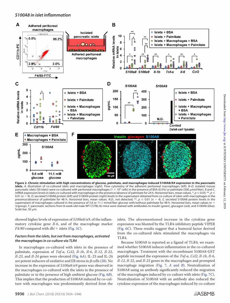
showed higher levels of expression of S100a8/a9, of the inflam-matory cytokine gene Il-6, and of the macrophage markerF4/80 compared with db/� islets (Fig. 5C).
Factors from the islets, but not from macrophages, activatedthe macrophages in co-culture via TLR4
In macrophages co-cultured with islets in the presence ofpalmitate, expression of Tnf-a, Ccl2, Il-1b, Il-6, Il-12, Il-22,Il-23, and Il-24 genes were elevated (Fig. 6A). IL-23 and IL-24are potent inducers of oxidative and ER stress in � cells (28). Noincrease in the expression of S100a8/a9 genes was observed inthe macrophages co-cultured with the islets in the presence ofpalmitate or in the presence of high ambient glucose (Fig. 6B).This implies that the production of S100A8 induced by co-cul-ture with macrophages was predominantly derived from the
islets. The aforementioned increase in the cytokine geneexpression was blunted by the TLR4-inhibitory peptide VIPER(Fig. 6C). These results suggest that a humoral factor derivedfrom the co-cultured islets stimulated the macrophages viaTLR4.
Because S100A8 is reported as a ligand of TLR4, we exam-ined whether S100A8 induces inflammation in the co-culturedmacrophages. Treatment with the recombinant S100A8-GSTpeptide increased the expression of the Tnf-a, Ccl2, Il-1b, Il-6,Il-12, Il-22, and Il-23 genes in the macrophages and promptedmacrophage migration (Fig. 7, A and B). Neutralization ofS100A8 using an antibody significantly reduced the migrationof the macrophages induced by co-culture with islets (Fig. 7C).Neutralization of S100A8 with an antibody also reduced thecytokine expression of the macrophages induced by co-culture
Figure 2. Chronic stimulation with high concentrations of glucose, palmitate, and macrophages induced S100A8/A9 expression in the pancreaticislets. A, illustration of co-cultured islets and macrophages (right). Flow cytometry of the adherent peritoneal macrophages (left). B–D, isolated mousepancreatic islets (50 islets) were co-cultured with peritoneal macrophages (1 � 105 cells) in the presence of BSA (0.5%) or palmitate (500 �mol/liter). B and C,mRNA expression levels in islets co-cultured with macrophages in the presence/absence of palmitate for 24 h. Horizontal bars, mean values. *, p � 0.05; **, p �0.01 (n � 9). D, secreted S100A8 protein (left) and S100A9 protein (right) levels in the supernatant obtained from co-culture of islets with macrophages in thepresence/absence of palmitate for 48 h. Horizontal bars, mean values. N.D., not detected; **, p � 0.01 (n � 4). E, secreted S100A8 protein levels in thesupernatant of macrophages cultured in the presence of 5.6 or 11.1 mmol/liter glucose with/without palmitate for 48 h. Horizontal bars, mean values (n �3/group). F, pancreatic sections from 8-week-old male WT C57BL/6J mice were stained with antibodies to insulin (green), glucagon (red), and S100A8 (blue).Scale bar, 50 �m.
S100A8 in islet inflammation
5936 J. Biol. Chem. (2018) 293(16) 5934 –5946
at JUN
TE
ND
O U
NIV
ER
SITY
on February 12, 2019http://w
ww
.jbc.org/D
ownloaded from
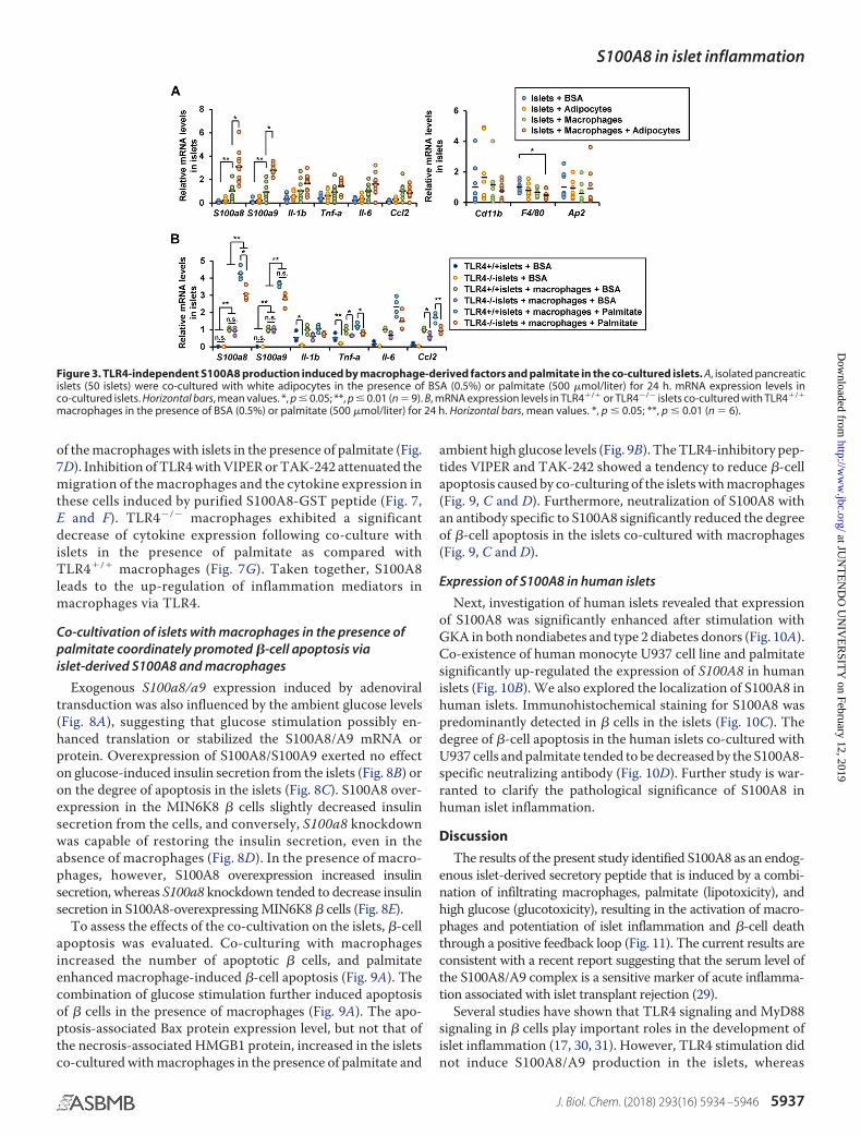
of the macrophages with islets in the presence of palmitate (Fig.7D). Inhibition of TLR4 with VIPER or TAK-242 attenuated themigration of the macrophages and the cytokine expression inthese cells induced by purified S100A8-GST peptide (Fig. 7,E and F). TLR4�/� macrophages exhibited a significantdecrease of cytokine expression following co-culture withislets in the presence of palmitate as compared withTLR4�/� macrophages (Fig. 7G). Taken together, S100A8leads to the up-regulation of inflammation mediators inmacrophages via TLR4.
Co-cultivation of islets with macrophages in the presence ofpalmitate coordinately promoted �-cell apoptosis viaislet-derived S100A8 and macrophages
Exogenous S100a8/a9 expression induced by adenoviraltransduction was also influenced by the ambient glucose levels(Fig. 8A), suggesting that glucose stimulation possibly en-hanced translation or stabilized the S100A8/A9 mRNA orprotein. Overexpression of S100A8/S100A9 exerted no effecton glucose-induced insulin secretion from the islets (Fig. 8B) oron the degree of apoptosis in the islets (Fig. 8C). S100A8 over-expression in the MIN6K8 � cells slightly decreased insulinsecretion from the cells, and conversely, S100a8 knockdownwas capable of restoring the insulin secretion, even in theabsence of macrophages (Fig. 8D). In the presence of macro-phages, however, S100A8 overexpression increased insulinsecretion, whereas S100a8 knockdown tended to decrease insulinsecretion in S100A8-overexpressing MIN6K8 � cells (Fig. 8E).
To assess the effects of the co-cultivation on the islets, �-cellapoptosis was evaluated. Co-culturing with macrophagesincreased the number of apoptotic � cells, and palmitateenhanced macrophage-induced �-cell apoptosis (Fig. 9A). Thecombination of glucose stimulation further induced apoptosisof � cells in the presence of macrophages (Fig. 9A). The apo-ptosis-associated Bax protein expression level, but not that ofthe necrosis-associated HMGB1 protein, increased in the isletsco-cultured with macrophages in the presence of palmitate and
ambient high glucose levels (Fig. 9B). The TLR4-inhibitory pep-tides VIPER and TAK-242 showed a tendency to reduce �-cellapoptosis caused by co-culturing of the islets with macrophages(Fig. 9, C and D). Furthermore, neutralization of S100A8 withan antibody specific to S100A8 significantly reduced the degreeof �-cell apoptosis in the islets co-cultured with macrophages(Fig. 9, C and D).
Expression of S100A8 in human islets
Next, investigation of human islets revealed that expressionof S100A8 was significantly enhanced after stimulation withGKA in both nondiabetes and type 2 diabetes donors (Fig. 10A).Co-existence of human monocyte U937 cell line and palmitatesignificantly up-regulated the expression of S100A8 in humanislets (Fig. 10B). We also explored the localization of S100A8 inhuman islets. Immunohistochemical staining for S100A8 waspredominantly detected in � cells in the islets (Fig. 10C). Thedegree of �-cell apoptosis in the human islets co-cultured withU937 cells and palmitate tended to be decreased by the S100A8-specific neutralizing antibody (Fig. 10D). Further study is war-ranted to clarify the pathological significance of S100A8 inhuman islet inflammation.
Discussion
The results of the present study identified S100A8 as an endog-enous islet-derived secretory peptide that is induced by a combi-nation of infiltrating macrophages, palmitate (lipotoxicity), andhigh glucose (glucotoxicity), resulting in the activation of macro-phages and potentiation of islet inflammation and �-cell deaththrough a positive feedback loop (Fig. 11). The current results areconsistent with a recent report suggesting that the serum level ofthe S100A8/A9 complex is a sensitive marker of acute inflamma-tion associated with islet transplant rejection (29).
Several studies have shown that TLR4 signaling and MyD88signaling in � cells play important roles in the development ofislet inflammation (17, 30, 31). However, TLR4 stimulation didnot induce S100A8/A9 production in the islets, whereas
Figure 3. TLR4-independent S100A8 production induced by macrophage-derived factors and palmitate in the co-cultured islets. A, isolated pancreaticislets (50 islets) were co-cultured with white adipocytes in the presence of BSA (0.5%) or palmitate (500 �mol/liter) for 24 h. mRNA expression levels inco-cultured islets. Horizontal bars, mean values. *, p � 0.05; **, p � 0.01 (n � 9). B, mRNA expression levels in TLR4�/� or TLR4�/� islets co-cultured with TLR4�/�
macrophages in the presence of BSA (0.5%) or palmitate (500 �mol/liter) for 24 h. Horizontal bars, mean values. *, p � 0.05; **, p � 0.01 (n � 6).
S100A8 in islet inflammation
J. Biol. Chem. (2018) 293(16) 5934 –5946 5937
at JUN
TE
ND
O U
NIV
ER
SITY
on February 12, 2019http://w
ww
.jbc.org/D
ownloaded from
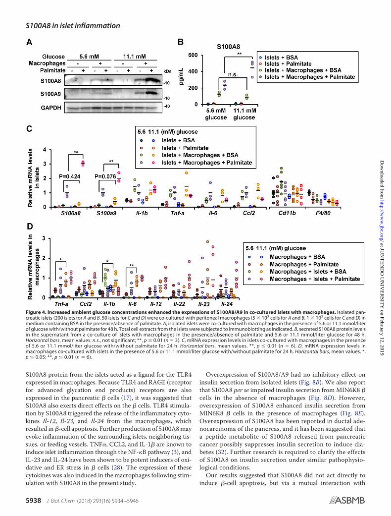
S100A8 protein from the islets acted as a ligand for the TLR4expressed in macrophages. Because TLR4 and RAGE (receptorfor advanced glycation end products) receptors are alsoexpressed in the pancreatic � cells (17), it was suggested thatS100A8 also exerts direct effects on the � cells. TLR4 stimula-tion by S100A8 triggered the release of the inflammatory cyto-kines Il-12, Il-23, and Il-24 from the macrophages, whichresulted in �-cell apoptosis. Further production of S100A8 mayevoke inflammation of the surrounding islets, neighboring tis-sues, or feeding vessels. TNF�, CCL2, and IL-1� are known toinduce islet inflammation through the NF-�B pathway (3), andIL-23 and IL-24 have been shown to be potent inducers of oxi-dative and ER stress in � cells (28). The expression of thesecytokines was also induced in the macrophages following stim-ulation with S100A8 in the present study.
Overexpression of S100A8/A9 had no inhibitory effect oninsulin secretion from isolated islets (Fig. 8B). We also reportthat S100A8 per se impaired insulin secretion from MIN6K8 �cells in the absence of macrophages (Fig. 8D). However,overexpression of S100A8 enhanced insulin secretion fromMIN6K8 � cells in the presence of macrophages (Fig. 8E).Overexpression of S100A8 has been reported in ductal ade-nocarcinoma of the pancreas, and it has been suggested thata peptide metabolite of S100A8 released from pancreaticcancer possibly suppresses insulin secretion to induce dia-betes (32). Further research is required to clarify the effectsof S100A8 on insulin secretion under similar pathophysio-logical conditions.
Our results suggested that S100A8 did not act directly toinduce �-cell apoptosis, but via a mutual interaction with
Figure 4. Increased ambient glucose concentrations enhanced the expressions of S100A8/A9 in co-cultured islets with macrophages. Isolated pan-creatic islets (200 islets for A and B, 50 islets for C and D) were co-cultured with peritoneal macrophages (5 � 105 cells for A and B, 1 � 105 cells for C and D) inmedium containing BSA in the presence/absence of palmitate. A, isolated islets were co-cultured with macrophages in the presence of 5.6 or 11.1 mmol/literof glucose with/without palmitate for 48 h. Total cell extracts from the islets were subjected to immunoblotting as indicated. B, secreted S100A8 protein levelsin the supernatant from a co-culture of islets with macrophages in the presence/absence of palmitate and 5.6 or 11.1 mmol/liter glucose for 48 h.Horizontal bars, mean values. n.s., not significant; **, p � 0.01 (n � 3). C, mRNA expression levels in islets co-cultured with macrophages in the presenceof 5.6 or 11.1 mmol/liter glucose with/without palmitate for 24 h. Horizontal bars, mean values. **, p � 0.01 (n � 6). D, mRNA expression levels inmacrophages co-cultured with islets in the presence of 5.6 or 11.1 mmol/liter glucose with/without palmitate for 24 h. Horizontal bars, mean values. *,p � 0.05; **, p � 0.01 (n � 6).
S100A8 in islet inflammation
5938 J. Biol. Chem. (2018) 293(16) 5934 –5946
at JUN
TE
ND
O U
NIV
ER
SITY
on February 12, 2019http://w
ww
.jbc.org/D
ownloaded from
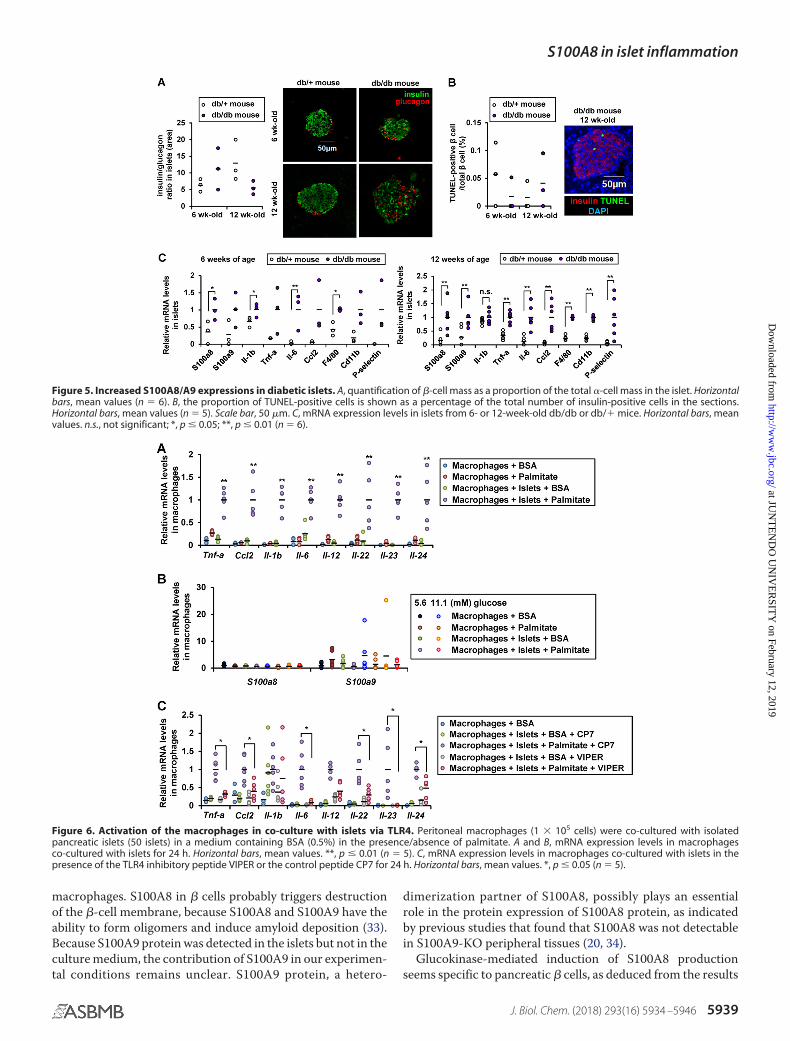
macrophages. S100A8 in � cells probably triggers destructionof the �-cell membrane, because S100A8 and S100A9 have theability to form oligomers and induce amyloid deposition (33).Because S100A9 protein was detected in the islets but not in theculture medium, the contribution of S100A9 in our experimen-tal conditions remains unclear. S100A9 protein, a hetero-
dimerization partner of S100A8, possibly plays an essentialrole in the protein expression of S100A8 protein, as indicatedby previous studies that found that S100A8 was not detectablein S100A9-KO peripheral tissues (20, 34).
Glucokinase-mediated induction of S100A8 productionseems specific to pancreatic � cells, as deduced from the results
Figure 5. Increased S100A8/A9 expressions in diabetic islets. A, quantification of �-cell mass as a proportion of the total �-cell mass in the islet. Horizontalbars, mean values (n � 6). B, the proportion of TUNEL-positive cells is shown as a percentage of the total number of insulin-positive cells in the sections.Horizontal bars, mean values (n � 5). Scale bar, 50 �m. C, mRNA expression levels in islets from 6- or 12-week-old db/db or db/� mice. Horizontal bars, meanvalues. n.s., not significant; *, p � 0.05; **, p � 0.01 (n � 6).
Figure 6. Activation of the macrophages in co-culture with islets via TLR4. Peritoneal macrophages (1 � 105 cells) were co-cultured with isolatedpancreatic islets (50 islets) in a medium containing BSA (0.5%) in the presence/absence of palmitate. A and B, mRNA expression levels in macrophagesco-cultured with islets for 24 h. Horizontal bars, mean values. **, p � 0.01 (n � 5). C, mRNA expression levels in macrophages co-cultured with islets in thepresence of the TLR4 inhibitory peptide VIPER or the control peptide CP7 for 24 h. Horizontal bars, mean values. *, p � 0.05 (n � 5).
S100A8 in islet inflammation
J. Biol. Chem. (2018) 293(16) 5934 –5946 5939
at JUN
TE
ND
O U
NIV
ER
SITY
on February 12, 2019http://w
ww
.jbc.org/D
ownloaded from
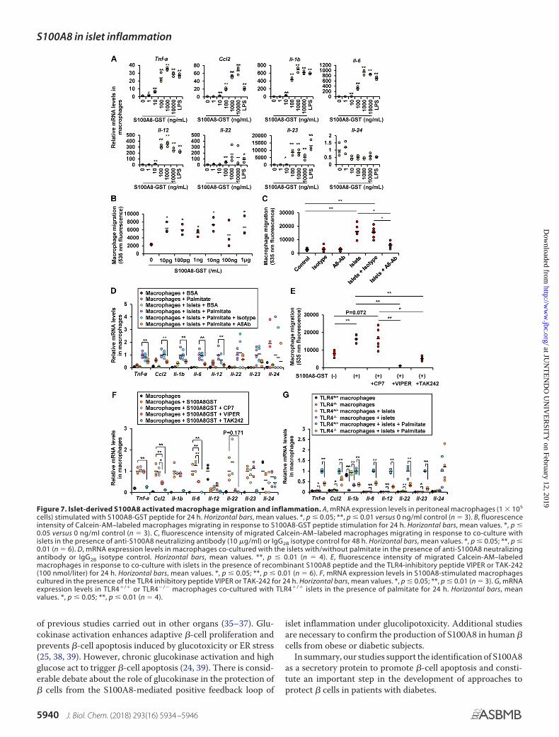
of previous studies carried out in other organs (35–37). Glu-cokinase activation enhances adaptive �-cell proliferation andprevents �-cell apoptosis induced by glucotoxicity or ER stress(25, 38, 39). However, chronic glucokinase activation and highglucose act to trigger �-cell apoptosis (24, 39). There is consid-erable debate about the role of glucokinase in the protection of� cells from the S100A8-mediated positive feedback loop of
islet inflammation under glucolipotoxicity. Additional studiesare necessary to confirm the production of S100A8 in human �cells from obese or diabetic subjects.
In summary, our studies support the identification of S100A8as a secretory protein to promote �-cell apoptosis and consti-tute an important step in the development of approaches toprotect � cells in patients with diabetes.
Figure 7. Islet-derived S100A8 activated macrophage migration and inflammation. A, mRNA expression levels in peritoneal macrophages (1 � 105
cells) stimulated with S100A8-GST peptide for 24 h. Horizontal bars, mean values. *, p � 0.05; **, p � 0.01 versus 0 ng/ml control (n � 3). B, fluorescenceintensity of Calcein-AM–labeled macrophages migrating in response to S100A8-GST peptide stimulation for 24 h. Horizontal bars, mean values. *, p �0.05 versus 0 ng/ml control (n � 3). C, fluorescence intensity of migrated Calcein-AM–labeled macrophages migrating in response to co-culture withislets in the presence of anti-S100A8 neutralizing antibody (10 �g/ml) or IgG2B isotype control for 48 h. Horizontal bars, mean values. *, p � 0.05; **, p �0.01 (n � 6). D, mRNA expression levels in macrophages co-cultured with the islets with/without palmitate in the presence of anti-S100A8 neutralizingantibody or IgG2B isotype control. Horizontal bars, mean values. **, p � 0.01 (n � 4). E, fluorescence intensity of migrated Calcein-AM–labeledmacrophages in response to co-culture with islets in the presence of recombinant S100A8 peptide and the TLR4-inhibitory peptide VIPER or TAK-242(100 nmol/liter) for 24 h. Horizontal bars, mean values. *, p � 0.05; **, p � 0.01 (n � 6). F, mRNA expression levels in S100A8-stimulated macrophagescultured in the presence of the TLR4 inhibitory peptide VIPER or TAK-242 for 24 h. Horizontal bars, mean values. *, p � 0.05; **, p � 0.01 (n � 3). G, mRNAexpression levels in TLR4�/� or TLR4�/� macrophages co-cultured with TLR4�/� islets in the presence of palmitate for 24 h. Horizontal bars, meanvalues. *, p � 0.05; **, p � 0.01 (n � 4).
S100A8 in islet inflammation
5940 J. Biol. Chem. (2018) 293(16) 5934 –5946
at JUN
TE
ND
O U
NIV
ER
SITY
on February 12, 2019http://w
ww
.jbc.org/D
ownloaded from
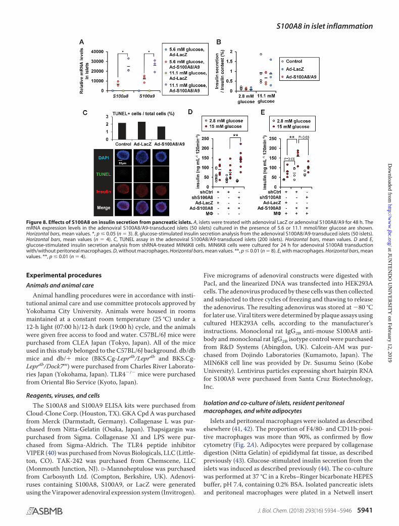
Experimental procedures
Animals and animal care
Animal handling procedures were in accordance with insti-tutional animal care and use committee protocols approved byYokohama City University. Animals were housed in roomsmaintained at a constant room temperature (25 °C) under a12-h light (07:00 h)/12-h dark (19:00 h) cycle, and the animalswere given free access to food and water. C57BL/6J mice werepurchased from CLEA Japan (Tokyo, Japan). All of the miceused in this study belonged to the C57BL/6J background. db/dbmice and db/� mice (BKS.Cg-Leprdb/Leprdb and BKS.Cg-Leprdb/Dock7m) were purchased from Charles River Laborato-ries Japan (Yokohama, Japan). TLR4�/� mice were purchasedfrom Oriental Bio Service (Kyoto, Japan).
Reagents, viruses, and cells
The S100A8 and S100A9 ELISA kits were purchased fromCloud-Clone Corp. (Houston, TX). GKA Cpd A was purchasedfrom Merck (Darmstadt, Germany). Collagenase L was pur-chased from Nitta-Gelatin (Osaka, Japan). Thapsigargin waspurchased from Sigma. Collagenase XI and LPS were pur-chased from Sigma-Aldrich. The TLR4 peptide inhibitorVIPER (40) was purchased from Novus Biologicals, LLC (Little-ton, CO). TAK-242 was purchased from Chemscene, LLC(Monmouth Junction, NJ). D-Mannoheptulose was purchasedfrom Carbosynth Ltd. (Compton, Berkshire, UK). Adenovi-ruses containing S100A8, S100A9, or LacZ were generatedusing the Virapower adenoviral expression system (Invitrogen).
Five micrograms of adenoviral constructs were digested withPacI, and the linearized DNA was transfected into HEK293Acells. The adenovirus produced by these cells was then collectedand subjected to three cycles of freezing and thawing to releasethe adenovirus. The resulting adenovirus was stored at �80 °Cfor later use. Viral titers were determined by plaque assays usingcultured HEK293A cells, according to the manufacturer’sinstructions. Monoclonal rat IgG2B anti-mouse S100A8 anti-body and monoclonal rat IgG2B isotype control were purchasedfrom R&D Systems (Abingdon, UK). Calcein-AM was pur-chased from Dojindo Laboratories (Kumamoto, Japan). TheMIN6K8 cell line was provided by Dr. Susumu Seino (KobeUniversity). Lentivirus particles expressing short hairpin RNAfor S100A8 were purchased from Santa Cruz Biotechnology,Inc.
Isolation and co-culture of islets, resident peritonealmacrophages, and white adipocytes
Islets and peritoneal macrophages were isolated as describedelsewhere (41, 42). The proportion of F4/80- and CD11b-posi-tive macrophages was more than 90%, as confirmed by flowcytometry (Fig. 2A). Adipocytes were prepared by collagenasedigestion (Nitta Gelatin) of epididymal fat tissue, as describedpreviously (43). Glucose-stimulated insulin secretion from theislets was induced as described previously (44). The co-culturewas performed at 37 °C in a Krebs–Ringer bicarbonate HEPESbuffer, pH 7.4, containing 0.2% BSA. Isolated pancreatic isletsand peritoneal macrophages were plated in a Netwell insert
Figure 8. Effects of S100A8 on insulin secretion from pancreatic islets. A, islets were treated with adenoviral LacZ or adenoviral S100A8/A9 for 48 h. ThemRNA expression levels in the adenoviral S100A8/A9-transduced islets (50 islets) cultured in the presence of 5.6 or 11.1 mmol/liter glucose are shown.Horizontal bars, mean values. *, p � 0.05 (n � 3). B, glucose-stimulated insulin secretion analysis from the adenoviral S100A8/A9-transduced islets (50 islets).Horizontal bars, mean values (n � 4). C, TUNEL assay in the adenoviral S100A8/A9-transduced islets (200 islets). Horizontal bars, mean values. D and E,glucose-stimulated insulin secretion analysis from shRNA-treated MIN6K8 cells. MIN6K8 cells were cultured for 24 h for adenoviral S100A8 transductionwith/without peritoneal macrophages. D, without macrophages. Horizontal bars, mean values. **, p � 0.01 (n � 8). E, with macrophages. Horizontal bars, meanvalues. **, p � 0.01 (n � 4).
S100A8 in islet inflammation
J. Biol. Chem. (2018) 293(16) 5934 –5946 5941
at JUN
TE
ND
O U
NIV
ER
SITY
on February 12, 2019http://w
ww
.jbc.org/D
ownloaded from
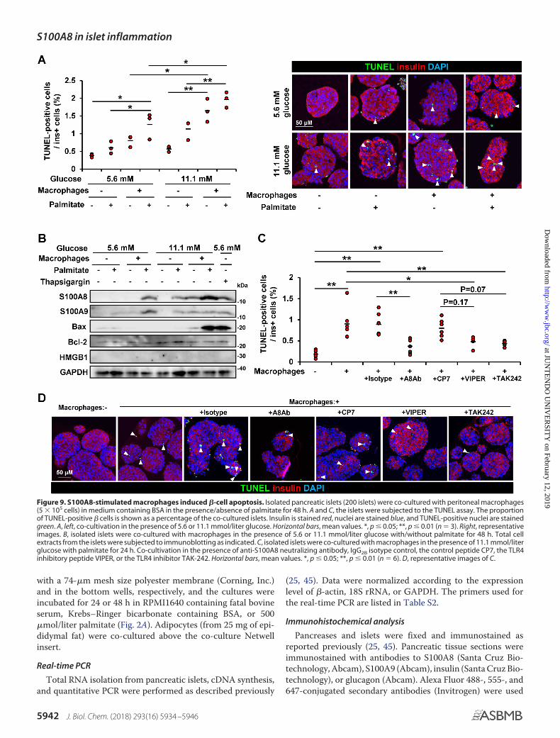
with a 74-�m mesh size polyester membrane (Corning, Inc.)and in the bottom wells, respectively, and the cultures wereincubated for 24 or 48 h in RPMI1640 containing fatal bovineserum, Krebs–Ringer bicarbonate containing BSA, or 500�mol/liter palmitate (Fig. 2A). Adipocytes (from 25 mg of epi-didymal fat) were co-cultured above the co-culture Netwellinsert.
Real-time PCR
Total RNA isolation from pancreatic islets, cDNA synthesis,and quantitative PCR were performed as described previously
(25, 45). Data were normalized according to the expressionlevel of �-actin, 18S rRNA, or GAPDH. The primers used forthe real-time PCR are listed in Table S2.
Immunohistochemical analysis
Pancreases and islets were fixed and immunostained asreported previously (25, 45). Pancreatic tissue sections wereimmunostained with antibodies to S100A8 (Santa Cruz Bio-technology, Abcam), S100A9 (Abcam), insulin (Santa Cruz Bio-technology), or glucagon (Abcam). Alexa Fluor 488-, 555-, and647-conjugated secondary antibodies (Invitrogen) were used
Figure 9. S100A8-stimulated macrophages induced �-cell apoptosis. Isolated pancreatic islets (200 islets) were co-cultured with peritoneal macrophages(5 � 105 cells) in medium containing BSA in the presence/absence of palmitate for 48 h. A and C, the islets were subjected to the TUNEL assay. The proportionof TUNEL-positive � cells is shown as a percentage of the co-cultured islets. Insulin is stained red, nuclei are stained blue, and TUNEL-positive nuclei are stainedgreen. A, left, co-cultivation in the presence of 5.6 or 11.1 mmol/liter glucose. Horizontal bars, mean values. *, p � 0.05; **, p � 0.01 (n � 3). Right, representativeimages. B, isolated islets were co-cultured with macrophages in the presence of 5.6 or 11.1 mmol/liter glucose with/without palmitate for 48 h. Total cellextracts from the islets were subjected to immunoblotting as indicated. C, isolated islets were co-cultured with macrophages in the presence of 11.1 mmol/literglucose with palmitate for 24 h. Co-cultivation in the presence of anti-S100A8 neutralizing antibody, IgG2B isotype control, the control peptide CP7, the TLR4inhibitory peptide VIPER, or the TLR4 inhibitor TAK-242. Horizontal bars, mean values. *, p � 0.05; **, p � 0.01 (n � 6). D, representative images of C.
S100A8 in islet inflammation
5942 J. Biol. Chem. (2018) 293(16) 5934 –5946
at JUN
TE
ND
O U
NIV
ER
SITY
on February 12, 2019http://w
ww
.jbc.org/D
ownloaded from
for the fluorescence microscopic analysis. �-cell apoptosis wasevaluated using a TdT-mediated dUTP nick-end labeling(TUNEL) assay of the co-cultured � cells. Co-cultured islets(200 islets) were attached to poly-L-lysine– coated coverslips(Falcon) and subjected to a TUNEL assay using the ApopTag insitu detection kit (EMD Millipore, MA). All of the images wereacquired using a FluoView FV1000-D confocal laser-scanningmicroscope (Olympus, Tokyo, Japan).
Immunoblotting
For immunoblotting, isolated islets (200 islets) were lysed inradioimmune precipitation buffer (Cell Signaling Technology,Danvers, MA) with complete protease inhibitor mixture
(Roche Diagnostics). After centrifugation, the extracts weresubjected to immunoblotting with antibodies. The primaryantibodies used were Calgranulin A (S100A8), Calgranulin B(S100A9), Bcl-2–associated X protein (BAX), Bcl-2 (Santa CruzBiotechnology), high-mobility group box 1 (HMGB1), and glyc-eraldehyde-3 phosphate dehydrogenase (GAPDH) (Abcam).
GST-fused proteins
GST-fused constructs comprising the mouse S100A8 andS100A9 proteins were generated in pGEX4T-1 and werereceived as kind gifts from Dr. Sachie Hiratsuka and Dr.Yoshihiro Maru (Department of Pharmacology, TokyoWomen’s Medical University) (46). The proteins were
Figure 10. The expression of S100A8 was induced by glucose stimulation, monocytes, and palmitate in human islets. A, mRNA expression levels inhuman islets from nondiabetes (non-DM) donors after stimulation with GKA Cpd A (glucokinase activator; 30 �mol/liter) for 24 h. Horizontal bars, mean values.*, p � 0.05; **, p � 0.01 (n � 5). B, mRNA expression levels in human islets co-cultured with U937 human monocyte cell line (1 � 105 cells) in the presence of BSA(0.5%) or palmitate (500 �mol/liter) for 24 h. Horizontal bars, mean values. *, p � 0.05; **, p � 0.01 (n � 5). C, embedded human islets from nondiabetes donorswere stained with antibodies to insulin (green), glucagon (red), and S100A8 (blue). Scale bar, 50 �m. D, human islets from nondiabetes donors were co-culturedwith U937 cells in the presence of 11.1 mmol/liter glucose with palmitate for 24 h. They were co-cultivated in the presence of anti-S100A8 neutralizing antibodyor IgG2B isotype control. The proportion of TUNEL-positive � cells is shown as a percentage of the co-cultured islets. Horizontal bars, mean values. *, p � 0.05(n � 5).
S100A8 in islet inflammation
J. Biol. Chem. (2018) 293(16) 5934 –5946 5943
at JUN
TE
ND
O U
NIV
ER
SITY
on February 12, 2019http://w
ww
.jbc.org/D
ownloaded from
expressed in Escherichia coli BL21 and purified on a GSH-Sepharose column.
Macrophage migration assay
The migrated macrophages were labeled with Calcein-AM(WAKO, Osaka, Japan) and measured using an ARVOTM MXplate reader (PerkinElmer Life Sciences) at an excitation wave-length of 485 nm and emission filter of 535 nm. The migrationof macrophages was evaluated using 8-�m pore Falcon BDFluoroBlokTM inserts and plates (BD Biosciences) with isolatedpancreatic islets (50 islets) or S100A8-GST in the presence orabsence of anti-S100A8 antibody, isotype control IgG2b,VIPER/CP7, or TAK-242. Macrophages were seeded onto theinsert mesh and incubated for 24 or 48 h at 37 °C under 5% CO2.
Human islets
Human islets were obtained from the Integrated Islet Distri-bution Program (National Institutes of Health). All studies andprotocols used were approved by the Joslin Diabetes Center’sCommittee on Human Studies (approval CHS#5-05). Detailsof human islets are described in Table S3. Upon receipt,islets were cultured overnight in Miami Medium 1A (Cell-gro). Co-culture was performed at 37 °C in final wash/cul-ture medium (Cellgro) or RPMI1640 medium. Cadaverichuman islets and U937 human monocyte cell line (ATCC)were plated in a Netwell insert with a 74-�m mesh size pol-yester membrane (Corning) and in the bottom wells,respectively.
Statistical analyses
All experiments were independently repeated at least threetimes. Horizontal bars indicate mean values. Statistical analyseswere conducted using IBM SPSS Statistics version 19. Equalityof variances was determined by using an F-test or Levene’s test.Statistical comparisons between groups were analyzed for sig-nificance by an unpaired two-tailed Student’s t test and one-way analysis of variance with post hoc Tukey tests for a para-metric test or Welch’s t test or Games–Howell test for anonparametric test. Differences were considered significant atp � 0.05.
Author contributions—J. S. designed the research; H. I., J. S., Y.Togashi, K. T., T. O., M. K., Y. Tanaka, K. O., Y. S., and T. Y., per-formed the experiments; K. O., K. S., and R. N. K contributed tohuman islet studies. H. I., J. S., Y. Togashi, K. T., T. O., and Y. Terau-chi analyzed the data; H. I., J. S., R. N. K., and Y. Terauchi wrote andedited the manuscript.
Acknowledgments—We thank Dr. Yoshihiro Maru and Dr. SachieHiratsuka (Tokyo Women’s Medical University) for kindly gifting usthe pGEX4T-1-S100A8 and -S100A9 plasmids and Dr. Susumu Seino(Kobe University) for providing the MIN6K8 cells. We also thank Mit-suyo Kaji, Eri Sakamoto (Yokohama City University), and HirokoMadokoro (Keio University) for excellent technical assistance andMisa Katayama for excellent secretarial assistance.
References1. Donath, M. Y., Schumann, D. M., Faulenbach, M., Ellingsgaard, H., Per-
ren, A., and Ehses, J. A. (2008) Islet inflammation in type 2 diabetes: frommetabolic stress to therapy. Diabetes Care 31, S161–S164 CrossRefMedline
2. Pickup, J. C., Mattock, M. B., Chusney, G. D., and Burt, D. (1997) NIDDMas a disease of the innate immune system: association of acute-phase re-actants and interleukin-6 with metabolic syndrome X. Diabetologia 40,1286 –1292 CrossRef Medline
3. Donath, M. Y., and Shoelson, S. E. (2011) Type 2 diabetes as an inflamma-tory disease. Nat. Rev. Immunol. 11, 98 –107 CrossRef Medline
4. Xu, H., Barnes, G. T., Yang, Q., Tan, G., Yang, D., Chou, C. J., Sole, J.,Nichols, A., Ross, J. S., Tartaglia, L. A., and Chen, H. (2003) Chronicinflammation in fat plays a crucial role in the development of obesity-related insulin resistance. J. Clin. Invest. 112, 1821–1830 CrossRefMedline
5. Cai, D., Yuan, M., Frantz, D. F., Melendez, P. A., Hansen, L., Lee, J., andShoelson, S. E. (2005) Local and systemic insulin resistance resulting fromhepatic activation of IKK-� and NF-�B. Nat. Med. 11, 183–190 CrossRefMedline
6. Brownlee, M. (2001) Biochemistry and molecular cell biology of diabeticcomplications. Nature 414, 813– 820 CrossRef Medline
7. Morigi, M., Angioletti, S., Imberti, B., Donadelli, R., Micheletti, G.,Figliuzzi, M., Remuzzi, A., Zoja, C., and Remuzzi, G. (1998) Leukocyte-endothelial interaction is augmented by high glucose concentrationsand hyperglycemia in a NF-�B-dependent fashion. J. Clin. Invest. 101,1905–1915 CrossRef Medline
8. Eguchi, K., and Manabe, I. (2013) Macrophages and islet inflammation intype 2 diabetes. Diabetes Obes. Metab. 15, 152–158 CrossRef Medline
9. Donath, M. Y., and Halban, P. A. (2004) Decreased beta-cell mass in dia-betes: significance, mechanisms and therapeutic implications. Diabetolo-gia 47, 581–589 CrossRef Medline
10. Donath, M. Y., Størling, J., Maedler, K., and Mandrup-Poulsen, T. (2003)Inflammatory mediators and islet beta-cell failure: a link between type 1and type 2 diabetes. J. Mol. Med. 81, 455– 470 CrossRef Medline
11. Ehses, J. A., Perren, A., Eppler, E., Ribaux, P., Pospisilik, J. A., Maor-Cahn,R., Gueripel, X., Ellingsgaard, H., Schneider, M. K., Biollaz, G., Fontana, A.,Reinecke, M., Homo-Delarche, F., and Donath, M. Y. (2007) Increasednumber of islet-associated macrophages in type 2 diabetes. Diabetes 56,2356 –2370 CrossRef Medline
12. Masters, S. L., Dunne, A., Subramanian, S. L., Hull, R. L., Tannahill, G. M.,Sharp, F. A., Becker, C., Franchi, L., Yoshihara, E., Chen, Z., Mullooly, N.,Mielke, L. A., Harris, J., Coll, R. C., Mills, K. H., et al. (2010) Activation ofthe NLRP3 inflammasome by islet amyloid polypeptide provides a mech-anism for enhanced IL-1� in type 2 diabetes. Nat. Immunol. 11, 897–904CrossRef Medline
13. Zhou, R., Tardivel, A., Thorens, B., Choi, I., and Tschopp, J. (2010) Thi-oredoxin-interacting protein links oxidative stress to inflammasome acti-vation. Nat. Immunol. 11, 136 –140 CrossRef Medline
Figure11.Anillustrativemodelofisletinflammation–induced�-cellapo-ptosis via S100A8 in diabetes. A combination of infiltrating macrophages,saturated fatty acids (palmitate), and hyperglycemia augmented the produc-tion of S100A8. S100A8 secreted from the islets induced further macrophagemigration and inflammation through TLR4. This positive feedback looppotentiates islet inflammation and �-cell death.
S100A8 in islet inflammation
5944 J. Biol. Chem. (2018) 293(16) 5934 –5946
at JUN
TE
ND
O U
NIV
ER
SITY
on February 12, 2019http://w
ww
.jbc.org/D
ownloaded from
14. Vlassara, H., and Palace, M. R. (2002) Diabetes and advanced glycationendproducts. J. Int. Med. 251, 87–101 CrossRef Medline
15. Zhu, Y., Shu, T., Lin, Y., Wang, H., Yang, J., Shi, Y., and Han, X. (2011)Inhibition of the receptor for advanced glycation endproducts (RAGE)protects pancreatic beta-cells. Biochem. Biophys. Res. Commun. 404,159 –165 CrossRef Medline
16. Maedler, K., Sergeev, P., Ris, F., Oberholzer, J., Joller-Jemelka, H. I., Spinas,G. A., Kaiser, N., Halban, P. A., and Donath, M. Y. (2002) Glucose-inducedbeta cell production of IL-1� contributes to glucotoxicity in human pan-creatic islets. J. Clin. Invest. 110, 851– 860 CrossRef Medline
17. Eguchi, K., Manabe, I., Oishi-Tanaka, Y., Ohsugi, M., Kono, N., Ogata, F.,Yagi, N., Ohto, U., Kimoto, M., Miyake, K., Tobe, K., Arai, H., Kadowaki,T., and Nagai, R. (2012) Saturated fatty acid and TLR signaling link betacell dysfunction and islet inflammation. Cell Metab. 15, 518 –533CrossRef Medline
18. Bouma, G., Lam-Tse, W. K., Wierenga-Wolf, A. F., Drexhage, H. A., andVersnel, M. A. (2004) Increased serum levels of MRP-8/14 in type 1 dia-betes induce an increased expression of CD11b and an enhanced adhesionof circulating monocytes to fibronectin. Diabetes 53, 1979 –1986 CrossRefMedline
19. Burkhardt, K., Schwarz, S., Pan, C., Stelter, F., Kotliar, K., Von Eynatten,M., Sollinger, D., Lanzl, I., Heemann, U., and Baumann, M. (2009) My-eloid-related protein 8/14 complex describes microcirculatory alterationsin patients with type 2 diabetes and nephropathy. Cardiovasc. Diabetol. 8,10 CrossRef Medline
20. Vogl, T., Tenbrock, K., Ludwig, S., Leukert, N., Ehrhardt, C., van Zoelen,M. A., Nacken, W., Foell, D., van der Poll, T., Sorg, C., and Roth, J. (2007)Mrp8 and Mrp14 are endogenous activators of Toll-like receptor 4, pro-moting lethal, endotoxin-induced shock. Nat. Med. 13, 1042–1049CrossRef Medline
21. Foell, D., and Roth, J. (2004) Proinflammatory S100 proteins in arthritisand autoimmune disease. Arthritis Rheum. 50, 3762–3771 CrossRefMedline
22. Riva, M., Kallberg, E., Bjork, P., Hancz, D., Vogl, T., Roth, J., Ivars, F., andLeanderson, T. (2012) Induction of nuclear factor-�B responses by theS100A9 protein is Toll-like receptor-4-dependent. Immunology 137,172–182 CrossRef Medline
23. Sunahori, K., Yamamura, M., Yamana, J., Takasugi, K., Kawashima, M.,Yamamoto, H., Chazin, W. J., Nakatani, Y., Yui, S., and Makino, H. (2006)The S100A8/A9 heterodimer amplifies proinflammatory cytokine pro-duction by macrophages via activation of nuclear factor �B and p38 mi-togen-activated protein kinase in rheumatoid arthritis. Arthritis Res. Ther.8, R69 CrossRef Medline
24. Tornovsky-Babeay, S., Dadon, D., Ziv, O., Tzipilevich, E., Kadosh, T.,Schyr-Ben Haroush, R., Hija, A., Stolovich-Rain, M., Furth-Lavi, J., Gra-not, Z., Porat, S., Philipson, L. H., Herold, K. C., Bhatti, T. R., Stanley, C., etal. (2014) Type 2 diabetes and congenital hyperinsulinism cause DNAdouble-strand breaks and p53 activity in beta cells. Cell Metab. 19,109 –121 CrossRef Medline
25. Shirakawa, J., Togashi, Y., Sakamoto, E., Kaji, M., Tajima, K., Orime, K.,Inoue, H., Kubota, N., Kadowaki, T., and Terauchi, Y. (2013) Glucokinaseactivation ameliorates ER stress-induced apoptosis in pancreatic beta-cells. Diabetes 62, 3448 –3458 CrossRef Medline
26. Dasu, M. R., and Jialal, I. (2011) Free fatty acids in the presence of highglucose amplify monocyte inflammation via Toll-like receptors. Am. J.Physiol. Endocrinol. Metab. 300, E145–E154 CrossRef Medline
27. Dasu, M. R., Devaraj, S., Zhao, L., Hwang, D. H., and Jialal, I. (2008) Highglucose induces toll-like receptor expression in human monocytes: mech-anism of activation. Diabetes 57, 3090 –3098 CrossRef Medline
28. Hasnain, S. Z., Borg, D. J., Harcourt, B. E., Tong, H., Sheng, Y. H., Ng, C. P.,Das, I., Wang, R., Chen, A. C., Loudovaris, T., Kay, T. W., Thomas, H. E.,Whitehead, J. P., Forbes, J. M., Prins, J. B., and McGuckin, M. A. (2014)Glycemic control in diabetes is restored by therapeutic manipulation ofcytokines that regulate beta cell stress. Nat. Med. 20, 1417–1426 CrossRefMedline
29. Ikemoto, M., Matsumoto, S., Egawa, H., Okitsu, T., Iwanaga, Y.,Umemoto, S., Itoh, H., Murayama, H., and Fujita, M. (2007) A case withtransient increases in serum S100A8/A9 levels implying acute inflamma-
tory responses after pancreatic islet transplantation. Ann. Clin. Biochem.44, 570 –572 CrossRef Medline
30. Boni-Schnetzler, M., Boller, S., Debray, S., Bouzakri, K., Meier, D. T.,Prazak, R., Kerr-Conte, J., Pattou, F., Ehses, J. A., Schuit, F. C., and Donath,M. Y. (2009) Free fatty acids induce a proinflammatory response in isletsvia the abundantly expressed interleukin-1 receptor I. Endocrinology 150,5218 –5229 CrossRef Medline
31. Gao, Q., Ma, L. L., Gao, X., Yan, W., Williams, P., and Yin, D. P. (2010)TLR4 mediates early graft failure after intraportal islet transplantation.Am. J. Transplant. 10, 1588 –1596 CrossRef Medline
32. Basso, D., Greco, E., Padoan, A., Fogar, P., Scorzeto, M., Fadi, E., Bozzato,D., Moz, S., Navaglia, F., Zambon, C. F., Seraglia, R., De Carlo, E., Valerio,A., Reggiani, C., Pedrazzoli, S., and Plebani, M. (2011) Altered intracellularcalcium fluxes in pancreatic cancer induced diabetes mellitus: Relevanceof the S100A8 N-terminal peptide (NT-S100A8). J. Cell. Physiol. 226,456 – 468 CrossRef Medline
33. Vogl, T., Gharibyan, A. L., and Morozova-Roche, L. A. (2012) Pro-inflam-matory S100A8 and S100A9 proteins: self-assembly into multifunctionalnative and amyloid complexes. Int. J. Mol. Sci. 13, 2893–2917 CrossRefMedline
34. Manitz, M. P., Horst, B., Seeliger, S., Strey, A., Skryabin, B. V., Gunzer, M.,Frings, W., Schonlau, F., Roth, J., Sorg, C., and Nacken, W. (2003) Loss ofS100A9 (MRP14) results in reduced interleukin-8-induced CD11b surfaceexpression, a polarized microfilament system, and diminished responsive-ness to chemoattractants in vitro. Mol. Cell. Biol. 23, 1034 –1043 CrossRefMedline
35. Sekimoto, R., Fukuda, S., Maeda, N., Tsushima, Y., Matsuda, K., Mori, T.,Nakatsuji, H., Nishizawa, H., Kishida, K., Kikuta, J., Maijima, Y., Fu-nahashi, T., Ishii, M., and Shimomura, I. (2015) Visualized macrophagedynamics and significance of S100A8 in obese fat. Proc. Natl. Acad. Sci.U.S.A. 112, E2058 –E2066 CrossRef Medline
36. Averill, M. M., Kerkhoff, C., and Bornfeldt, K. E. (2012) S100A8 andS100A9 in cardiovascular biology and disease. Arterioscler. Thromb. Vasc.Biol. 32, 223–229 CrossRef Medline
37. Perera, C., McNeil, H. P., and Geczy, C. L. (2010) S100 Calgranulinsin inflammatory arthritis. Immunol. Cell Biol. 88, 41– 49 CrossRefMedline
38. Oh, Y. S., Lee, Y. J., Park, K., Choi, H. H., Yoo, S., and Jun, H. S. (2014)Treatment with glucokinase activator, YH-GKA, increases cell prolifera-tion and decreases glucotoxic apoptosis in INS-1 cells. Eur. J. Pharm. Sci.51, 137–145 CrossRef Medline
39. Roma, L. P., Duprez, J., and Jonas, J. C. (2015) Glucokinase activation isbeneficial or toxic to cultured rat pancreatic islets depending on the pre-vailing glucose concentration. Am. J. Physiol. Endocrinol. Metab. 309,E632–E639 CrossRef Medline
40. Lysakova-Devine, T., Keogh, B., Harrington, B., Nagpal, K., Halle, A.,Golenbock, D. T., Monie, T., and Bowie, A. G. (2010) Viral inhibitorypeptide of TLR4, a peptide derived from vaccinia protein A46, specif-ically inhibits TLR4 by directly targeting MyD88 adaptor-like andTRIF-related adaptor molecule. J. Immunol. 185, 4261– 4271 CrossRefMedline
41. Shirakawa, J., Amo, K., Ohminami, H., Orime, K., Togashi, Y., Ito, Y.,Tajima, K., Koganei, M., Sasaki, H., Takeda, E., and Terauchi, Y. (2011)Protective effects of dipeptidyl peptidase-4 (DPP-4) inhibitor againstincreased beta cell apoptosis induced by dietary sucrose and linoleicacid in mice with diabetes. J. Biol. Chem. 286, 25467–25476 CrossRefMedline
42. Zhang, X., Goncalves, R., and Mosser, D. M. (2008) The isolation andcharacterization of murine macrophages. Curr. Protoc. Immunol. Chapter14, Unit 14.11 CrossRef Medline
43. Shirakawa, J., Fujii, H., Ohnuma, K., Sato, K., Ito, Y., Kaji, M., Sakamoto, E.,Koganei, M., Sasaki, H., Nagashima, Y., Amo, K., Aoki, K., Morimoto, C.,Takeda, E., and Terauchi, Y. (2011) Diet-induced adipose tissue inflam-mation and liver steatosis are prevented by DPP-4 inhibition in diabeticmice. Diabetes 60, 1246 –1257 CrossRef Medline
44. Shirakawa, J., Okuyama, T., Yoshida, E., Shimizu, M., Horigome, Y., Tuno,T., Hayasaka, M., Abe, S., Fuse, M., Togashi, Y., and Terauchi, Y. (2014)Effects of the antitumor drug OSI-906, a dual inhibitor of IGF-1 receptor
S100A8 in islet inflammation
J. Biol. Chem. (2018) 293(16) 5934 –5946 5945
at JUN
TE
ND
O U
NIV
ER
SITY
on February 12, 2019http://w
ww
.jbc.org/D
ownloaded from
and insulin receptor, on the glycemic control, beta-cell functions, andbeta-cell proliferation in male mice. Endocrinology 155, 2102–2111CrossRef Medline
45. Shirakawa, J., Fernandez, M., Takatani, T., El Ouaamari, A., Jungtrakoon,P., Okawa, E. R., Zhang, W., Yi, P., Doria, A., and Kulkarni, R. N. (2017)Insulin signaling regulates the FoxM1/PLK1/CENP-A pathway to pro-
mote adaptive pancreatic beta cell proliferation. Cell Metab. 25,868 – 882.e5 CrossRef Medline
46. Hiratsuka, S., Watanabe, A., Aburatani, H., and Maru, Y. (2006) Tumour-mediated upregulation of chemoattractants and recruitment of myeloidcells predetermines lung metastasis. Nat. Cell Biol. 8, 1369 –1375CrossRef Medline
S100A8 in islet inflammation
5946 J. Biol. Chem. (2018) 293(16) 5934 –5946
at JUN
TE
ND
O U
NIV
ER
SITY
on February 12, 2019http://w
ww
.jbc.org/D
ownloaded from
Shibue, Rohit N. Kulkarni and Yasuo TerauchiKyohara, Yui Tanaka, Kazuki Orime, Yoshifumi Saisho, Taketo Yamada, Kimitaka
Hideaki Inoue, Jun Shirakawa, Yu Togashi, Kazuki Tajima, Tomoko Okuyama, Mayu-cell apoptosis and islet inflammationβprotein A8 exacerbates
cells and macrophages via S100 calcium-bindingβSignaling between pancreatic
doi: 10.1074/jbc.M117.809228 originally published online March 1, 20182018, 293:5934-5946.J. Biol. Chem.
10.1074/jbc.M117.809228Access the most updated version of this article at doi:
Alerts:
When a correction for this article is posted•
When this article is cited•
to choose from all of JBC's e-mail alertsClick here
http://www.jbc.org/content/293/16/5934.full.html#ref-list-1
This article cites 46 references, 11 of which can be accessed free at
at JUN
TE
ND
O U
NIV
ER
SITY
on February 12, 2019http://w
ww
.jbc.org/D
ownloaded from
Case Report
Necrotizing enterocolitis associated with Clostridiumbutyricum in a Japanese man
Yukio Sato,1,2 Dai Kujirai,1,2 Katsura Emoto,3,4 Toshiaki Yagami,5 Taketo Yamada,3,4,6
Manabu Izumi,7 Masaki Ano,8 Kenichi Kase,1 and Kenji Kobayashi1,9
1Department of Emergency Medicine, Saiseikai Utsunomiya Hospital, Tochigi, Japan, 2Department of Emergencyand Critical Care Medicine, Keio University School of Medicine, Tokyo, Japan, 3Department of Pathology, SaiseikaiUtsunomiya Hospital, Tochigi, Japan, 4Department of Pathology, Keio University School of Medicine, Tokyo,Japan, 5Department of Radiology, Saiseikai Utsunomiya Hospital, Tochigi, Japan, 6Department of Pathology,Saitama Medical University, Saitama, Japan, 7Department of General Internal Medicine, Saiseikai UtsunomiyaHospital, Tochigi, Japan, 8Department of Critical Care Medicine, Saiseikai Utsunomiya Hospital, Tochigi, Japan,and 9Department of Surgery, Saiseikai Utsunomiya Hospital, Tochigi, Japan
Case: Necrotizing enterocolitis (NEC) caused by Clostridium butyricum is common in neonates; however, a case of NEC in adults hasnot been previously reported. An 84-year-old Japanese man developed C. butyricum-related NEC during hospitalization for treatmentof stab wounds to the left side of the neck and lower abdomen, without organ damage, and concomitant pneumonia.
Outcome: The patient developed acute onset of emesis accompanied by shock during his admission; partial resection of the smallintestine was carried out due to necrosis. Pathologic findings showed mucosal necrosis and extensive vacuolation with gram-positiverods in the necrotic small intestine. Blood culture tests revealed C. butyricum infection. The patient’s condition improved after thesurgery. He was moved to a rehabilitation hospital on day 66.
Conclusion: This study suggests that hospitalized adult patients who receive antibiotic treatment are at risk for NEC.
Key words: Adult, Asia, Clostridium butyricum, enterocolitis, necrotizing
INTRODUCTION
SEVERAL STRAINS OF Clostridium butyricum havebeen cultured from the stool of healthy children and
adults.1 One of those strains, MIYAIRI 588, is usedwidely as a probiotic in Asia, including Japan.2 It hasbeen reported that it inhibited the cytotoxicity ofClostridium difficile in an in vitro study and reducedC. difficile toxin A in an in vivo study.3,4 However, someof these strains produce endotoxins and cause necrotizingenterocolitis (NEC) in neonates.5 The only toxin C. bu-tyricum has been reported to produce is analogous to thetype E botulinum neurotoxin secreted by Clostridium
botulinum.6 After the first report of NEC due to C. bu-tyricum type E in an infant,7 many similar cases havebeen reported, including two cases of intestinal botulisminvolving adolescents,8 and one case of sepsis in anadult.9 Additionally, cases of food-borne botulism causedby C. butyricum have been reported.10 However, a caseof NEC due to C. butyricum in an adult has not beenreported to date.
CASE
AN 84-YEAR-OLD MAN visited our hospital owing toa neck (left) and abdominal penetrating injury by a
short sword in a suicide attempt. The patient had a medicalhistory of cerebral infarction and paroxysmal atrial fibrilla-tion on apixaban. He lived in his home with his family andhad no recent history of hospitalization or admission to anursing home.
On examination, his vital signs were normal except fordisturbed consciousness: Glasgow coma scale score, 6;blood pressure, 158/92 mmHg; respiratory rate, 18 breaths/
Corresponding: Yukio Sato, MD, PhD, Department of Emergency
& Critical Care Medicine, Keio University School of Medicine, 35
Shinanomachi, Shinjuku-ku, Tokyo 1608582, Japan. Email:
Received 24 Oct, 2017; accepted 25 Dec, 2017; online
publication 23 Jan, 2018
Funding Information
No funding information provided.
© 2018 The Authors. Acute Medicine & Surgery published by John Wiley & Sons Australia, Ltd on behalf ofJapanese Association for Acute Medicine.
194
This is an open access article under the terms of the Creative Commons Attribution License,which permits use, distribution and reproduction in any medium, provided the original work is properly cited.
Acute Medicine & Surgery 2018; 5: 194–198 doi: 10.1002/ams2.329
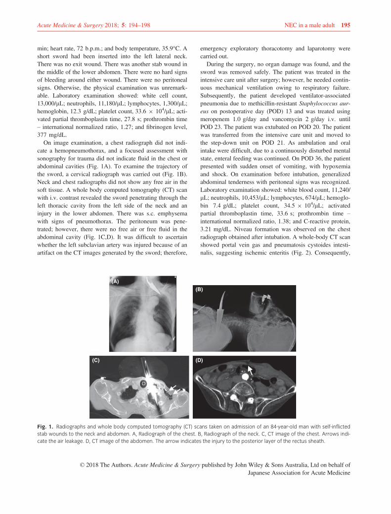
min; heart rate, 72 b.p.m.; and body temperature, 35.9°C. Ashort sword had been inserted into the left lateral neck.There was no exit wound. There was another stab wound inthe middle of the lower abdomen. There were no hard signsof bleeding around either wound. There were no peritonealsigns. Otherwise, the physical examination was unremark-able. Laboratory examination showed: white cell count,13,000/lL; neutrophils, 11,180/lL; lymphocytes, 1,300/lL;hemoglobin, 12.3 g/dL; platelet count, 33.6 9 104/lL; acti-vated partial thromboplastin time, 27.8 s; prothrombin time– international normalized ratio, 1.27; and fibrinogen level,377 mg/dL.
On image examination, a chest radiograph did not indi-cate a hemopneumothorax, and a focused assessment withsonography for trauma did not indicate fluid in the chest orabdominal cavities (Fig. 1A). To examine the trajectory ofthe sword, a cervical radiograph was carried out (Fig. 1B).Neck and chest radiographs did not show any free air in thesoft tissue. A whole body computed tomography (CT) scanwith i.v. contrast revealed the sword penetrating through theleft thoracic cavity from the left side of the neck and aninjury in the lower abdomen. There was s.c. emphysemawith signs of pneumothorax. The peritoneum was pene-trated; however, there were no free air or free fluid in theabdominal cavity (Fig. 1C,D). It was difficult to ascertainwhether the left subclavian artery was injured because of anartifact on the CT images generated by the sword; therefore,
emergency exploratory thoracotomy and laparotomy werecarried out.
During the surgery, no organ damage was found, and thesword was removed safely. The patient was treated in theintensive care unit after surgery; however, he needed contin-uous mechanical ventilation owing to respiratory failure.Subsequently, the patient developed ventilator-associatedpneumonia due to methicillin-resistant Staphylococcus aur-eus on postoperative day (POD) 13 and was treated usingmeropenem 1.0 g/day and vancomycin 2 g/day i.v. untilPOD 23. The patient was extubated on POD 20. The patientwas transferred from the intensive care unit and moved tothe step-down unit on POD 21. As ambulation and oralintake were difficult, due to a continuously disturbed mentalstate, enteral feeding was continued. On POD 36, the patientpresented with sudden onset of vomiting, with hypoxemiaand shock. On examination before intubation, generalizedabdominal tenderness with peritoneal signs was recognized.Laboratory examination showed: white blood count, 11,240/lL; neutrophils, 10,453/lL; lymphocytes, 674/lL; hemoglo-bin 7.4 g/dL; platelet count, 34.5 9 104/lL; activatedpartial thromboplastin time, 33.6 s; prothrombin time –international normalized ratio, 1.38; and C-reactive protein,3.21 mg/dL. Niveau formation was observed on the chestradiograph obtained after intubation. A whole-body CT scanshowed portal vein gas and pneumatosis cystoides intesti-nalis, suggesting ischemic enteritis (Fig. 2). Consequently,
(A)
(B)
(C) (D)
Fig. 1. Radiographs and whole body computed tomography (CT) scans taken on admission of an 84-year-old man with self-inflicted
stab wounds to the neck and abdomen. A, Radiograph of the chest. B, Radiograph of the neck. C, CT image of the chest. Arrows indi-
cate the air leakage. D, CT image of the abdomen. The arrow indicates the injury to the posterior layer of the rectus sheath.
© 2018 The Authors. Acute Medicine & Surgery published by John Wiley & Sons Australia, Ltd on behalf ofJapanese Association for Acute Medicine
Acute Medicine & Surgery 2018; 5: 194–198 NEC in a male adult 195
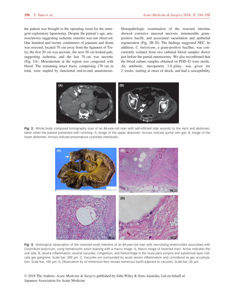
the patient was brought to the operating room for the emer-gent exploratory laparotomy. Despite the patient’s age, arte-riosclerosis suggesting ischemic enteritis was not observed.One hundred and twenty centimeters of jejunum and ileumwas resected, located 70 cm away from the ligament of Tre-itz; the first 20 cm was necrotic, the next 30 cm looked pale,suggesting ischemia, and the last 70 cm was necrotic(Fig. 3A). Mesenterium at the region was congested withblood. The remaining intact tracts, comprising 170 cm intotal, were stapled by functional end-to-end anastomosis.
Histopathologic examination of the resected intestineshowed extensive mucosal necrosis, innumerable gram-positive bacilli, and associated vacuolation and epithelialregeneration (Fig. 3B–D). The findings suggested NEC. Inaddition, C. butyricum, a gram-positive bacillus, was con-currently isolated from two cultured blood samples drawnjust before the partial enterectomy. We also reconfirmed thatthe blood culture samples obtained on POD 42 were sterile.An antibiotic, meropenem 1.0 g/day, was given for2 weeks, starting at onset of shock, and had a susceptibility
(A) (B)
Fig. 2. Whole body computed tomography scan of an 84-year-old man with self-inflicted stab wounds to the neck and abdomen,
taken when the patient presented with vomiting. A, Image of the upper abdomen. Arrows indicate portal vein gas. B, Image of the
lower abdomen. Arrows indicate pneumatosis cystoides intestinalis.
(A) (B)
(C) (D)
Fig. 3. Histological observation of the resected small intestine of an 84-year-old man with necrotizing enterocolitis associated with
Clostridium butyricum, using hematoxylin–eosin staining with a macro image. A, Macro image of resected tract. Arrow indicates the
oral side. B, Severe inflammation, several vacuoles, congestion, and hemorrhage in the muscularis propria and subserosal layer indi-
cate gas gangrene. Scale bar, 500 lm. C, Vacuoles are surrounded by acute severe inflammation and considered as gas accumula-
tion. Scale bar, 100 lm. D, Observation by oil immersion lens reveals numerous bacilli adjacent to vacuoles. Scale bar, 20 lm.
© 2018 The Authors. Acute Medicine & Surgery published by John Wiley & Sons Australia, Ltd on behalf ofJapanese Association for Acute Medicine
196 Y. Sato et al. Acute Medicine & Surgery 2018; 5: 194–198
to C. butyricum. The patient recovered and was moved to arehabilitation hospital on POD 66 following his firstsurgery.
DISCUSSION
NECROTIZING ENTEROCOLITIS CAUSED byC. butyricum is common in preterm neonates, and can
be life-threatening.5 However, to the best of our knowledge,no such cases have been reported in adults.
In the present case, we initially suspected ischemic enteri-tis; however, surgical and pathological findings suggestedNEC. In addition to the lack of obvious obstruction of ves-sels on surgery, histopathology results indicated that C. bu-tyricum isolation from the patient’s blood samples resultedfrom mucosal breakdown and transmigration of these bacte-ria into the bloodstream. These findings indicated that patho-genic C. butyricum infection in the gut resulted in septicshock, leading to ischemia of the small intestine. Withregard to the route of infection, in this case, the initialexploratory laparotomy for the stab wound did not involveany bowel injuries; therefore it was not related to the devel-opment of NEC. We hypothesized that the patient was aC. butyricum carrier, and treatment with several antibioticsfor methicillin-resistant Staphylococcus aureus pneumoniamight have resulted in the microbial substitution. Neverthe-less, the invasion route is still unclear. In addition, a previ-ous report suggested that lactose fermentation is involved inthe pathogenesis of NEC.11 However, the enteral nutrientadministered to this patient did not include lactose. More-over, the patient had been admitted for 30 days before thedevelopment of NEC. During that period, no outbreak ofthis kind of bacterial infection was observed in our hospital,thus, excluding the possibility of nosocomial infection.7
Therefore, the course of C. butyricum infection remainsunclear.
Regarding the differential diagnosis, we discussed thepossibility of the following three diseases. Neutropenicenterocolitis, also known as typhlitis, is confused withNEC; however, our patient’s background was completelydifferent to reported cases.12 Although its cause is stillunclear, it basically occurs in patients with neutropenia,such as leukemic patients receiving chemotherapy. Thepatient did not have any medical history suggesting neu-tropenia. In addition, a test for antibodies against HIVyielded negative results. The possibility of non-occlusivemesenteric ischemia (NOMI) was definitely difficult torule out, because of similar traits between both diseases:ischemia and infection.13 Moreover, segmental and discon-tinuous necrosis in a region perfused by the superior
mesenteric artery can be typically observed in NOMI,although it was difficult to confirm that necrotic andischemic regions were completely continuous in our case.Nonetheless, if our case was NOMI, the innumerable bac-teria propagated in such a short period would have beenincongruous, as the patient had no risk factors at theonset, such as cardiac failure, low flow states, multi-organdysfunction, or vasopressors.14 The probability of ischemicenteritis was low because there was no arteriosclerosis,and it typically occurs in a region perfused by the inferiormesenteric artery. Thus, based on the above reasons, wediagnosed NEC.
Regarding the prevention of adult NEC, a clonal strainwas found to be circulating in neonatal intensive care unit,during an outbreak of NEC in preterm neonates.5,7,15 There-fore, it is necessary for medical and nursing staff to controlinfection effectively, even in adult care units. In addition,although prediction is difficult, a risk for developing NEC,owing to microbial substitution, could be considered duringtreatment with antibiotics in the same manner as considera-tion for C. difficile colitis.
Limitations of this study include the inability to assess theneurogenic symptoms (vision impairment and dysphagia) ofthe patient, because of his continuous disturbed mental state.Furthermore, gene sequencing to confirm the type of strainisolated could not be carried out.
CONCLUSION
WE PRESENT A rare case of successful managementof a hospitalized elderly patient with NEC associated
with C. butyricum. The present case suggests that hospital-ized adults who receive antibiotic therapy carry a risk ofcritical illness associated with pathogenic C. butyricum.
ACKNOWLEDGEMENTS
WE WOULD LIKE to thank Editage for English lan-guage editing.
DISCLOSURE
Approval of the research protocol: N/A.Informed consent: Informed consent was obtained from thesubject.Registry and the registration no. of the study/trial: N/A.Animal studies: N/A.Conflict of interest: T.Y. was supported by grants from Kis-sei Pharmaceutical Co., Ltd. and Nichirei Biosciences Inc.The other authors have no conflict of interest.
© 2018 The Authors. Acute Medicine & Surgery published by John Wiley & Sons Australia, Ltd on behalf ofJapanese Association for Acute Medicine
Acute Medicine & Surgery 2018; 5: 194–198 NEC in a male adult 197
REFERENCES
1 Benno Y, Sawada K, Mitsuoka T. The intestinal microflora ofinfants: composition of fecal flora in breast-fed and bottle-fedinfants. Microbiol. Immunol. 1984; 28: 975–86.
2 Isa K, Oka K, Beauchamp N et al. Safety assessment of theClostridium butyricum MIYAIRI 588(R) probiotic strainincluding evaluation of antimicrobial sensitivity and presenceof Clostridium toxin genes in vitro and teratogenicity in vivo.Hum. Exp. Toxicol. 2016; 35: 818–32.
3 Woo TD, Oka K, Takahashi M et al. Inhibition of the cyto-toxic effect of Clostridium difficile in vitro by Clostridiumbutyricum MIYAIRI 588 strain. J. Med. Microbiol. 2011; 60:1617–25.
4 Imase K, Takahashi M, Tanaka A et al. Efficacy of Clostrid-ium butyricum preparation concomitantly with Helicobacterpylori eradication therapy in relation to changes in the intesti-nal microbiota. Microbiol. Immunol. 2008; 52: 156–61.
5 Cassir N, Benamar S, Khalil JB et al. Clostridium butyricumstrains and Dysbiosis linked to necrotizing enterocolitis inpreterm neonates. Clin. Infect. Dis. 2015; 61: 1107–15.
6 Popoff MR, Dodin A. Survey of neuraminidase productionby Clostridium butyricum, Clostridium beijerinckii, andClostridium difficile strains from clinical and nonclinicalsources. J. Clin. Microbiol. 1985; 22: 873–6.
7 Howard FM, Flynn DM, Bradley JM, Noone P, Sza-watkowski M. Outbreak of necrotising enterocolitis causedby Clostridium butyricum. Lancet 1977; 2: 1099–102.
8 Fenicia L, Franciosa G, Pourshaban M, Aureli P. Intestinaltoxemia botulism in two young people, caused by Clostrid-ium butyricum type E. Clin. Infect. Dis. 1999; 29: 1381–7.
9 Gardner EM, Kestler M, Beieler A, Belknap RW. Clostridiumbutyricum sepsis in an injection drug user with an indwellingcentral venous catheter. J. Med. Microbiol. 2008; 57: 236–9.
10 Meng X, Karasawa T, Zou K et al. Characterization of a neu-rotoxigenic Clostridium butyricum strain isolated from thefood implicated in an outbreak of food-borne type E botulism.J. Clin. Microbiol. 1997; 35: 2160–2.
11 Bousseboua H, Le Coz Y, Dabard J et al. Experimental ceci-tis in gnotobiotic quails monoassociated with Clostridiumbutyricum strains isolated from patients with neonatal necro-tizing enterocolitis and from healthy newborns. Infect.Immun. 1989; 57: 932–6.
12 Rodrigues FG, Dasilva G, Wexner SD. Neutropenic entero-colitis. World J. Gastroenterol. 2017; 23: 42–7.
13 Zachariah SK. Adult necrotizing enterocolitis and non occlu-sive mesenteric ischemia. J. Emerg. Trauma Shock. 2011; 4:430–2.
14 Bala M, Kashuk J, Moore EE et al. Acute mesenteric ische-mia: guidelines of the World Society of Emergency Surgery.World J. Emerg. Surg. 2017; 12: 38.
15 Benamar S, Cassir N, Merhej V et al. Multi-spacer typing asan effective method to distinguish the clonal lineage ofClostridium butyricum strains isolated from stool samplesduring a series of necrotizing enterocolitis cases. J. Hosp.Infect. 2017; 95: 300–5.
© 2018 The Authors. Acute Medicine & Surgery published by John Wiley & Sons Australia, Ltd on behalf ofJapanese Association for Acute Medicine
198 Y. Sato et al. Acute Medicine & Surgery 2018; 5: 194–198
Contents lists available at ScienceDirect
European Journal of Radiology
journal homepage: www.elsevier.com/locate/ejrad
Research article
Low-dose chest computed tomography screening of subjects exposed toasbestos
Katsuya Katoa, Kenichi Gembab, Kazuto Ashizawac, Hiroaki Arakawad, Satoshi Hondae,Naomi Noguchif, Sumihisa Hondac, Nobukazu Fujimotog,⁎, Takumi Kishimotoh
a Department of Diagnostic Radiology 2, Kawasaki Medical School, General Medical Center, 2-1-80 Nakasange, Okayama, 7008505, JapanbDepartment of Respiratory Medicine, Chugoku-chuo Hospital, 148-13 Oazakamiiwanari, Miyukicho, Fukuyama, 7200001, Japanc Department of Clinical Oncology, Nagasaki University Graduate School of Medicine, 1-7-1 Sakamoto, Nagasaki, 8528102, Japand Department of Radiology, Dokkyo Medical University, 880, Kita-Kobayashi, Mibu, Tochigi, 3210293, Japane Department of Radiology, Okayama Rosai Hospital, 1-10-25 Chikkomidorimachi, Okayama, 7028055, JapanfDepartment of Radiology, Tamano Mitsui Hospital, 3-2-1 Tama, Tamano, 7060012, Japang Department of Medical Oncology, Okayama Rosai Hospital, 1-10-25 Chikkomidorimachi, Okayama, 7028055, JapanhDepartment of Internal Medicine, Okayama Rosai Hospital, 1-10-25 Chikkomidorimachi, Okayama, 7028055, Japan
A R T I C L E I N F O
Keywords:AsbestosLow-dose CTScreeningLung cancerMesothelioma
A B S T R A C T
Objectives: The primary aim was to reveal the prevalence of lung cancer (LC) and malignant pleural mesothe-lioma (MPM) in subjects with past asbestos exposure (AE). We also examined pulmonary or pleural changescorrelated with the development of LC.Materials and methods: This was a prospective, multicenter, cross-sectional study. There were 2132 subjectsenrolled between 2010 and 2012. They included 96.2% men and 3.8% women, with a mean age of 76.1 years;78.8% former or current smokers; and 21.2% never smokers. We screened subjects using low-dose computedtomography (CT). The CT images were taken with a CT dose Index of 2.7 mGy. The evaluated CT findingsincluded subpleural curvilinear shadow/subpleural dots, ground glass opacity or interlobular reticular opacity,traction bronchiectasia, honeycombing change, parenchymal band, emphysema changes, pleural effusion, dif-fuse pleural thickening, rounded atelectasis, pleural plaques (PQs), and tumor formation.Results: The PQs were detected in most of subjects (89.4%) and emphysema changes were seen in 46.0%.Fibrotic changes were detected in 565 cases (26.5%). A pathological diagnosis of LC was confirmed in 45 cases(2.1%) and MPM was confirmed in 7 cases (0.3%). The prevalence of LC was 2.5% in patients with a smokinghistory, which was significantly higher than that in never smokers (0.7%, p=0.027). The prevalence of LC was2.8% in subjects with emphysema changes, which was higher than that of subjects without those findings(1.6%); although, the difference was not statistically significant (p=0.056). The prevalence of LC in subjectswith both fibrotic plus emphysema changes was 4.0%, which was significantly higher than that of subjects withneither of those findings (1.8%, p=0.011). Logistic regression analysis revealed smoking history, fibrotic plusemphysema changes, and pleural effusion as significant explanatory variables.Conclusions: Smoking history, fibrotic plus emphysema changes, and pleural effusion were correlated with theprevalence of LC.
1. Introduction
Asbestos was commonly used during the 20th century and remainsprevalent in many developing countries [1]. Asbestos causes patholo-gical changes in the lung or the pleura including asbestosis, pleuralplaques (PQs), benign asbestos pleural effusion [2], diffuse pleural
thickening, and malignant neoplasms such as malignant pleural me-sothelioma (MPM) and lung cancer (LC) [3,4]. According to the WorldHealth Organization, > 107,000 people die each year from asbestos-related diseases due to occupational exposure [1]. These diseasesusually develop after long latency periods of 40–50 years [5]. Thus,there will be more LC or MPM developing in the next few decades,
https://doi.org/10.1016/j.ejrad.2018.02.017Received 8 August 2017; Received in revised form 11 December 2017; Accepted 12 February 2018
⁎ Corresponding author.E-mail address: [email protected] (N. Fujimoto).
Abbreviations: AE, asbestos exposure; CT, computed tomography; LC, lung cancer; LDCT, low-dose computed tomography; MPM, malignant pleural mesothelioma; PQs, pleural plaques;SCLS/DOTS, subpleural curvilinear shadow/subpleural dots
European Journal of Radiology 101 (2018) 124–128
0720-048X/ © 2018 Elsevier B.V. All rights reserved.
T
despite that asbestos use was banned in Japan in 2004. Subjects withhistories of asbestos exposure (AE) in Japan are examined by annualchest X-ray; however, it is established that chest X-ray is not an efficientmethod forLC screening [6,7]. Thus, there is a need to establish a moreuseful screening strategy for subjects.
Mass screening of high-risk groups to detect LC could potentially bebeneficial. Multidetector computed tomography (CT) has made high-resolution volumetric imaging possible during a single breath hold withacceptable levels of radiation exposure [8]. There were several reportsthat low-dose helical CT of the lung detected more nodules and LCs,including early-stage, than chest X-ray [9]. Recently, the National LungScreening Trial, which recruited subjects at high-risk for LC, demon-strated that low-dose CT (LDCT) screening could decrease the deathrate due to LC by about 20% compared with screening using chest X-ray[7]. In addition, there are some recent reports that LDCT screening isuseful to detect LC at the earlier stages [10–12].
In the current study, we performed LDCT screening for subjects withhistories of AE. The primary aim of the study was to reveal the pre-valence of LC and MPM in the subjects. In addition, we focused on otherpulmonary or pleural changes, such as fibrotic or emphysema changesand plaques, to determine what findings correlated with the prevalenceof LC.
2. Materials and methods
2.1. Study approval
This study was conducted in compliance with the principles of theDeclaration of Helsinki. This study was carried out according to TheEthical Guidelines for Epidemiological Research by the JapaneseMinistry of Education, Culture, Sports, Science, and Technology, andthe Ministry of Health, Labour, and Welfare. This study was approvedby the Japan Health, Labour, and Welfare Organization and the in-stitutional review boards of each institution. Patient confidentiality wasstrictly maintained and written informed consent was obtained fromthe subjects.
2.2. Subjects
This was a prospective, multicenter, cross-sectional study to revealthe prevalence of LC and MPM, and the prevalence of CT findings due toAE. The inclusion criteria of the subjects are 1) those who had engagedin asbestos-product manufacturing for more than 1 year, 2) those whohad engaged in other industries related to AE for more than 10 years, or3) those who had engaged in industries related to AE and demonstratedpleural plaques on chest X-ray or CT (regardless of the duration of AE).There were 2132 subjects enrolled in this study between 2010 and2012. They included 2050 (96.2%) men and 82 (3.8%) women, with amean (range) age of 76.1 (51–101) years. There were 502 subjects fromOkayama Rosai Hospital, 392 from Chiba Rosai Hospital, 370 fromTamano Mitsui Hospital, 313 from Kinki Chuo Chest Medical Center,214 from Kagawa Rosai Hospital, 196 from Toyama Rosai Hospital, 96from Yamaguchi-Ube Medical Center, and 49 from Hokkaido ChuoRosai Hospital. The occupational categories associated with AE areshown in Fig. 1. The main categories included 612 subjects (28.7%) inshipbuilding, 260 (12.2%) in chemical manufacturing, 259 (12.2%) inasbestos-product manufacturing, and 245 (11.5%) in construction. Thesmoking history was obtained from 2095 subjects and revealed 1651(78.8%) former or current smokers and 444 (21.2%) never smokers.
2.3. CT acquisition and analysis
The CT images were taken in each institution with a median (range)CT dose Index of 2.7 (2.4–2.8) mGy. 2mm thick images were obtainedand stored in Digital Imaging and Communications in the Medicineformat. The evaluated CT findings included pulmonary fibrotic
changes, such as subpleural curvilinear shadow/subpleural dots (SCLS/DOTS), ground glass opacity or interlobular reticular opacity, tractionbronchiectasia, honeycombing change, and parenchymal band (Fig. 2).Other evaluated findings were emphysema change, pleural effusion,diffuse pleural thickening, rounded atelectasis, PQs with or withoutcalcification, and tumor formation. The CT images were taken with thesubject in a prone position to differentiate slight pulmonary changes onthe dorsal portion of the lungs from gravitational effects. Images wereanalyzed independently on the monitor, based on a quality standard,agreed on by two reference radiologists who were blinded to the clin-ical and demographic information of the subject and the results of oneanother’s assessments. If there was a difference between the inter-pretations of the two radiologists, more rigorous interpretation wasadopted with regard to emphysema changes, pleural effusion, diffusepleural thickening, PQs, and tumor formation. For fibrotic changes, athird radiologist made the second-round interpretation and gave thefinal decision. When LC or MPM was suspected in subjects, furtherexaminations such as bronchoscopy, needle biopsy, thoracentesis, and/or surgery were performed in the clinical practice.
This was a cross sectional study with only one CT performed in eachsubject. No follow up was performed for patients with a negative CT.
2.4. Statistical analysis
Comparisons between independent groups were performed usingthe chi-square test and the Mann-Whitney U test was used for non-parametric analysis. The average values were compared using the t-test.Overall survival of LC patients was obtained by using Kaplan-Meiermethods. Logistic regression analysis was conducted as a multivariateanalysis. Statistical calculations were performed using SPSS statisticalpackage version 22.0 (IBM, Armonk, USA).
3. Results
3.1. CT findings
The CT findings of the 2132 subjects are summarized in Table 1. ThePQs were detected in the majority of subjects (89.4%) and emphysemachanges in about half of the subjects (46.0%). Fibrotic changes (at leastone of: SCLS/DOTS, ground glass opacity or interlobular reticularopacity, traction bronchiectasia, honeycombing change, and par-enchymal band) were detected in 565 cases (26.5%). There were 116cases (5.4%) with suspected LC, including 101 with possible LC and 15with definite LC.
The pathological diagnosis of LC was confirmed in 45 cases (2.1%),44 men and 1 woman. Median (range) age at the diagnosis was 73(60–87) years old. There were 31(68.9%) adenocarcinoma, 10(22.2%)squamous cell carcinoma, 3(6.7%) small cell carcinoma, and 1(2.2%)adenosquamous carcinoma. According to the International Associationfor the Study of Lung Cancer staging (7th Edition), there were 13 StageIA, 14 Stage IB, 4 Stage IIA, 3 Stage IIB, 4 Stage IIIA, 2 Stage IIIB, and 1Stage IV patients. Median overall survival (95% confidence interval) ofthese 13 patients was 26.8 (4.01–71.93) months.
Pleural effusion was detected in 45 subjects. Among them, LC wasdiagnosed in six cases including four adenocarcinomas and two squa-mous cell carcinomas. Pleural carcinomatosis was revealed in 2 of the 4cases of adenocarcinoma. Another two subjects with adenocarcinomaand one of the 2 subjects with squamous cell carcinoma underwentthoracic surgery, suggesting they had post-operative pleural effusion.There were 16 subjects (0.8%) with suspected MPM and the patholo-gical diagnosis was confirmed in seven cases (0.3%) including 4 cases ofepithelioid, 2 cases of biphasic, and 1 case of sarcomatous subtype.
3.2. CT characteristics of LC cases
We examined the specific characteristics of patients in whom LC
K. Kato et al. European Journal of Radiology 101 (2018) 124–128
125
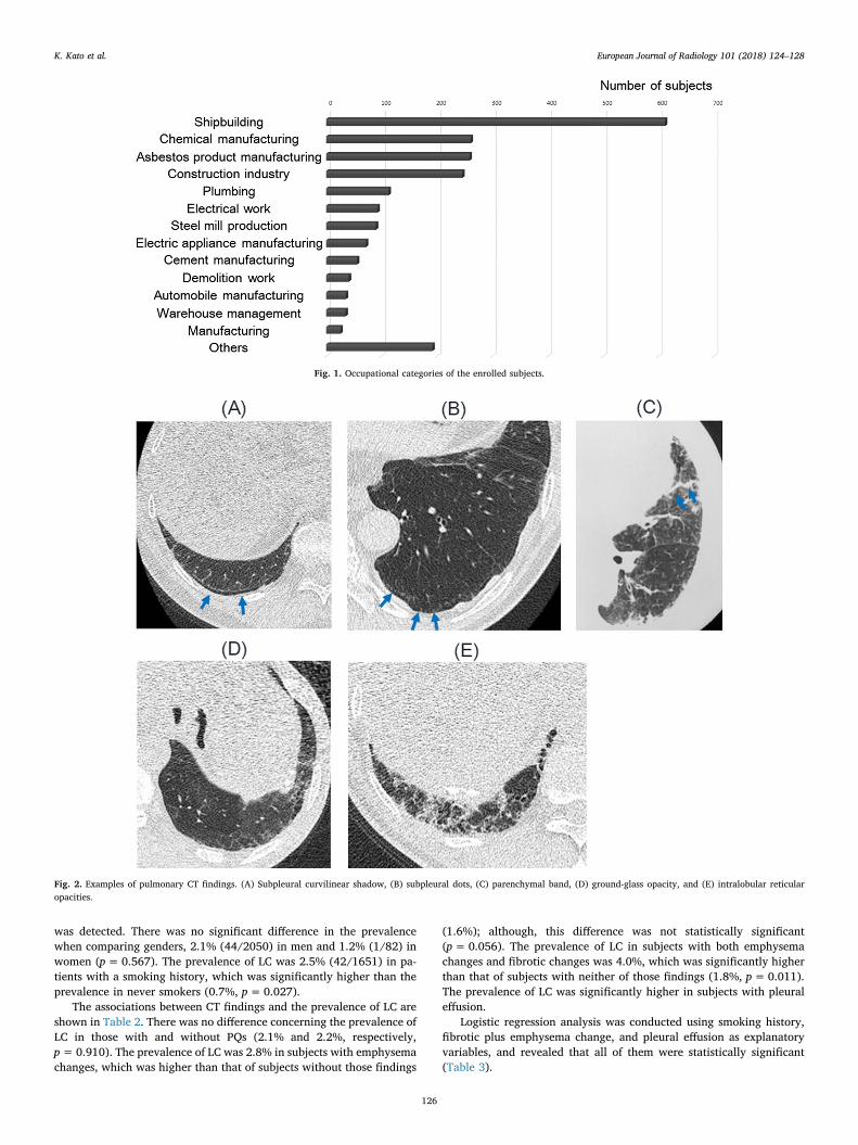
was detected. There was no significant difference in the prevalencewhen comparing genders, 2.1% (44/2050) in men and 1.2% (1/82) inwomen (p=0.567). The prevalence of LC was 2.5% (42/1651) in pa-tients with a smoking history, which was significantly higher than theprevalence in never smokers (0.7%, p=0.027).
The associations between CT findings and the prevalence of LC areshown in Table 2. There was no difference concerning the prevalence ofLC in those with and without PQs (2.1% and 2.2%, respectively,p=0.910). The prevalence of LC was 2.8% in subjects with emphysemachanges, which was higher than that of subjects without those findings
(1.6%); although, this difference was not statistically significant(p=0.056). The prevalence of LC in subjects with both emphysemachanges and fibrotic changes was 4.0%, which was significantly higherthan that of subjects with neither of those findings (1.8%, p=0.011).The prevalence of LC was significantly higher in subjects with pleuraleffusion.
Logistic regression analysis was conducted using smoking history,fibrotic plus emphysema change, and pleural effusion as explanatoryvariables, and revealed that all of them were statistically significant(Table 3).
Fig. 1. Occupational categories of the enrolled subjects.
Fig. 2. Examples of pulmonary CT findings. (A) Subpleural curvilinear shadow, (B) subpleural dots, (C) parenchymal band, (D) ground-glass opacity, and (E) intralobular reticularopacities.
K. Kato et al. European Journal of Radiology 101 (2018) 124–128
126
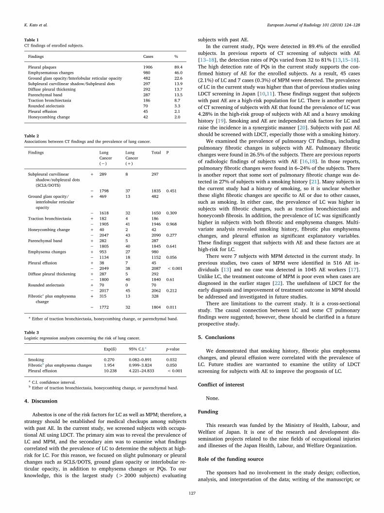
4. Discussion
Asbestos is one of the risk factors for LC as well as MPM; therefore, astrategy should be established for medical checkups among subjectswith past AE. In the current study, we screened subjects with occupa-tional AE using LDCT. The primary aim was to reveal the prevalence ofLC and MPM, and the secondary aim was to examine what findingscorrelated with the prevalence of LC to determine the subjects at high-risk for LC. For this reason, we focused on slight pulmonary or pleuralchanges such as SCLS/DOTS, ground glass opacity or interlobular re-ticular opacity, in addition to emphysema changes or PQs. To ourknowledge, this is the largest study (> 2000 subjects) evaluating
subjects with past AE.In the current study, PQs were detected in 89.4% of the enrolled
subjects. In previous reports of CT screening of subjects with AE[13–18], the detection rates of PQs varied from 32 to 81% [13,15–18].The high detection rate of PQs in the current study supports the con-firmed history of AE for the enrolled subjects. As a result, 45 cases(2.1%) of LC and 7 cases (0.3%) of MPM were detected. The prevalenceof LC in the current study was higher than that of previous studies usingLDCT screening in Japan [10,11]. These findings suggest that subjectswith past AE are a high-risk population for LC. There is another reportof CT screening of subjects with AE that found the prevalence of LC was4.28% in the high-risk group of subjects with AE and a heavy smokinghistory [19]. Smoking and AE are independent risk factors for LC andraise the incidence in a synergistic manner [20]. Subjects with past AEshould be screened with LDCT, especially those with a smoking history.
We examined the prevalence of pulmonary CT findings, includingpulmonary fibrotic changes in subjects with AE. Pulmonary fibroticchanges were found in 26.5% of the subjects. There are previous reportsof radiologic findings of subjects with AE [16,18]. In those reports,pulmonary fibrotic changes were found in 6–24% of the subjects. Thereis another report that some sort of pulmonary fibrotic change was de-tected in 27% of subjects with a smoking history [21]. Many subjects inthe current study had a history of smoking, so it is unclear whetherthese slight fibrotic changes are specific to AE or due to other causes,such as smoking. In either case, the prevalence of LC was higher insubjects with fibrotic changes, such as traction bronchiectasis andhoneycomb fibrosis. In addition, the prevalence of LC was significantlyhigher in subjects with both fibrotic and emphysema changes. Multi-variate analysis revealed smoking history, fibrotic plus emphysemachanges, and pleural effusion as significant explanatory variables.These findings suggest that subjects with AE and these factors are athigh-risk for LC.
There were 7 subjects with MPM detected in the current study. Inprevious studies, two cases of MPM were identified in 516 AE in-dividuals [13] and no case was detected in 1045 AE workers [17].Unlike LC, the treatment outcome of MPM is poor even when cases arediagnosed in the earlier stages [22]. The usefulness of LDCT for theearly diagnosis and improvement of treatment outcome in MPM shouldbe addressed and investigated in future studies.
There are limitations to the current study. It is a cross-sectionalstudy. The causal connection between LC and some CT pulmonaryfindings were suggested; however, these should be clarified in a futureprospective study.
5. Conclusions
We demonstrated that smoking history, fibrotic plus emphysemachanges, and pleural effusion were correlated with the prevalence ofLC. Future studies are warranted to examine the utility of LDCTscreening for subjects with AE to improve the prognosis of LC.
Conflict of interest
None.
Funding
This research was funded by the Ministry of Health, Labour, andWelfare of Japan. It is one of the research and development dis-semination projects related to the nine fields of occupational injuriesand illnesses of the Japan Health, Labour, and Welfare Organization.
Role of the funding source
The sponsors had no involvement in the study design; collection,analysis, and interpretation of the data; writing of the manuscript; or
Table 1CT findings of enrolled subjects.
Findings Cases %
Pleural plaques 1906 89.4Emphysematous changes 980 46.0Ground glass opacity/Interlobular reticular opacity 482 22.6Subpleural curvilinear shadow/Subpleural dots 297 13.9Diffuse pleural thickening 292 13.7Parenchymal band 287 13.5Traction bronchiectasia 186 8.7Rounded atelectasis 70 3.3Pleural effusion 45 2.1Honeycombing change 42 2.0
Table 2Associations between CT findings and the prevalence of lung cancer.
Findings LungCancer(−)
LungCancer(+)
Total P
Subpleural curvilinearshadow/subpleural dots(SCLS/DOTS)
+ 289 8 297
− 1798 37 1835 0.451Ground glass opacity/
interlobular reticularopacity
+ 469 13 482
− 1618 32 1650 0.309Traction bronchiectasia + 182 4 186
− 1905 41 1946 0.968Honeycombing change + 40 2 42
− 2047 43 2090 0.277Parenchymal band + 282 5 287
− 1805 40 1845 0.641Emphysema changes + 953 27 980
− 1134 18 1152 0.056Pleural effusion + 38 7 45
− 2049 38 2087 <0.001Diffuse pleural thickening + 287 5 292
− 1800 40 1840 0.61Rounded atelectasis + 70 0 70
− 2017 45 2062 0.212Fibrotica plus emphysema
change+ 315 13 328
− 1772 32 1804 0.011
a Either of traction bronchiectasia, honeycombing change, or parenchymal band.
Table 3Logistic regression analyses concerning the risk of lung cancer.
Exp(ß) 95% C.I.a p-value
Smoking 0.270 0.082–0.891 0.032Fibroticb plus emphysema changes 1.954 0.999–3.824 0.050Pleural effusion 10.238 4.221–24.833 < 0.001
a C.I. confidence interval.b Either of traction bronchiectasia, honeycombing change, or parenchymal band.
K. Kato et al. European Journal of Radiology 101 (2018) 124–128
127
the decision to submit the manuscript for publication.
Acknowledgements
The authors would like to thank all the participants in this study.
References
[1] L. Stayner, L.S. Welch, R. Lemen, The worldwide pandemic of asbestos-relateddiseases, Annu. Rev. Public Health 34 (2013) 205–216.
[2] N. Fujimoto, K. Gemba, K. Aoe, K. Kato, T. Yokoyama, I. Usami, K. Onishi,K. Mizuhashi, T. Yusa, T. Kishimoto, Clinical investigation of benign asbestospleural effusion, Pulm. Med. (2015) 416179, http://dx.doi.org/10.1155/2015/416179.
[3] Asbestos, asbestosis, and cancer: the Helsinki criteria for diagnosis and attribution,Scand. J. Work Environ. Health 23 (1997) 311–316.
[4] G. Hillerdal, Pleural plaques and risk for bronchial carcinoma and mesothelioma. Aprospective study, Chest 105 (1994) 144–150.
[5] K. Gemba, N. Fujimoto, K. Kato, K. Aoe, Y. Takeshima, K. Inai, T. Kishimoto,National survey of malignant mesothelioma and asbestos exposure in Japan, CancerSci. 103 (2012) 483–490.
[6] P.M. Marcus, E.J. Bergstralh, R.M. Fagerstrom, D.E. Williams, R. Fontana,W.F. Taylor, P.C. Prorok, Lung cancer mortality in the Mayo Lung Project: impact ofextended follow-up, J. Natl. Cancer Inst. 92 (2000) 1308–1316.
[7] D.R. Aberle, A.M. Adams, C.D. Berg, W.C. Black, J.D. Clapp, R.M. Fagerstrom,I.F. Gareen, C. Gatsonis, P.M. Marcus, J.D. Sicks, National lung screening trial re-search team, reduced lung-cancer mortality with low-dose computed tomographicscreening, N. Engl. J. Med. 365 (2011) 395–409.
[8] D.P. Naidich, C.H. Marshall, C. Gribbin, R.S. Arams, D.I. McCauley, Low-dose CT ofthe lungs: preliminary observations, Radiology 175 (1990) 729–731.
[9] V.P. Doria-Rose, E. Szabo, Screening and prevention of lung cancer, in:K.H. Kernstine, K. Reckamp (Eds.), Lung Cancer: A Multidisciplinary Approach toDiagnosis and Management, Demos Medical Publishing, New York, 2010, pp.53–72.
[10] S. Sone, S. Takashima, F. Li, Z. Yang, T. Honda, Y. Maruyama, M. Hasegawa,T. Yamanda, K. Kubo, K. Hanamura, K. Asakura, Mass screening for lung cancerwith mobile spiral computed tomography scanner, Lancet 351 (1998) 1242–1245.
[11] M. Kaneko, K. Eguchi, H. Ohmatsu, R. Kakinuma, T. Naruke, K. Suemasu,N. Moriyama, Peripheral lung cancer: screening and detection with low-dose spiralCT versus radiography, Radiology 201 (1996) 798–802.
[12] C.I. Henschke, D.I. McCauley, D.F. Yankelevitz, D.P. Naidich, G. McGuinness,O.S. Miettinen, D.M. Libby, M.W. Pasmantier, J. Koizumi, N.K. Altorki, J.P. Smith,Early Lung Cancer Action Project: overall design and findings from baselinescreening, Lancet 354 (1999) 99–105.
[13] H.C. Roberts, D.A. Patsios, N.S. Paul, M. DePerrot, W. Teel, H. Bayanati,F. Shepherd, M.R. Johnston, Screening for malignant pleural mesothelioma andlung cancer in individuals with a history of asbestos exposure, J. Thorac. Oncol. 4(2009) 620–628.
[14] T. Vierikko, R. Jarvenpaa, T. Autti, P. Oksa, M. Huuskonen, S. Kaleva, J. Laurikka,S. Kajander, K. Paakkola, S. Saarelainen, E.R. Salomaa, A. Tossavainen,P. Tukiainen, J. Uitti, T. Vehmas, Chest CT screening of asbestos-exposed workers:lung lesions and incidental findings, Eur. Respir. J. 29 (2007) 78–84.
[15] G. Mastrangelo, M.N. Ballarin, E. Bellini, R. Bizzotto, F. Zannol, F. Gioffre,M. Gobbi, G. Tessadri, L. Marchiori, G. Marangi, S. Bozzolan, J.H. Lange,F. Valentini, P. Spolaore, Feasibility of a screening programme for lung cancer informer asbestos workers, Occup. Med. (Lond.) 58 (2008) 175–180.
[16] C. Paris, A. Martin, M. Letourneux, P. Wild, Modelling prevalence and incidence offibrosis and pleural plaques in asbestos-exposed populations for screening andfollow-up: a cross-sectional study, Environ. Health 7 (2008) 30.
[17] G. Fasola, O. Belvedere, M. Aita, T. Zanin, A. Follador, P. Cassetti, S. Meduri,V. Pangher, G. Pignata, V. Rosolen, F. Barbone, F. Grossi, Low-dose computed to-mography screening for lung cancer and pleural mesothelioma in an asbestos-ex-posed population: baseline results of a prospective, nonrandomized feasibilitytrial—an Alpe-adria Thoracic Oncology Multidisciplinary Group Study (ATOM002), Oncologist 12 (2007) 1215–1224.
[18] M. Remy-Jardin, A. Sobaszek, A. Duhamel, I. Mastora, C. Zanetti, J. Remy,Asbestos-related pleuropulmonary diseases: evaluation with low-dose four-detectorrow spiral CT, Radiology 233 (2004) 182–190.
[19] M. Das, G. Muhlenbruch, A.H. Mahnken, K.G. Hering, H. Sirbu, W. Zschiesche,L. Knol, M.K. Felten, T. Kraus, R.W. Gunther, J.E. Wildberger, Asbestos SurveillanceProgram Aachen (ASPA): initial results from baseline screening for lung cancer inasbestos-exposed high-risk individuals using low-dose multidetector-row CT, Eur.Radiol. 17 (2007) 1193–1199.
[20] M. Akira, S. Yamamoto, Y. Inoue, M. Sakatani, High-resolution CT of asbestosis andidiopathic pulmonary fibrosis, AJR Am. J. Roentgenol. 181 (2003) 163–169.
[21] M. Remy-Jardin, J. Remy, B. Gosselin, V. Becette, J.L. Edme, Lung parenchymalchanges secondary to cigarette smoking: pathologic-CT correlations, Radiology 186(1993) 643–651.
[22] K. Gemba, N. Fujimoto, K. Aoe, K. Kato, Y. Takeshima, K. Inai, T. Kishimoto,Treatment and survival analyses of malignant mesothelioma in Japan, Acta Oncol.52 (2013) 803–808.
K. Kato et al. European Journal of Radiology 101 (2018) 124–128
128
RESEARCH ARTICLE Open Access
Quality of life of survivors of malignantpleural mesothelioma in Japan: a crosssectional studyYasuko Nagamatsu1, Isao Oze2, Keisuke Aoe3, Katsuyuki Hotta4, Katsuya Kato5, Junko Nakagawa6, Keiko Hara6,Takumi Kishimoto7 and Nobukazu Fujimoto8*
Abstract
Background: Previous studies have indicated that people with malignant pleural mesothelioma (MPM) have a poorquality of life (QOL); however, information about the QOL of people with MPM in Japan is anecdotal. The aims ofthis study were to investigate the QOL of survivors of MPM in Japan and to determine the factors that correlatewith their QOL.
Methods: This was a cross sectional study. The included patients were those diagnosed with MPM in Japan. Wecreated a self-administered questionnaire consisting of 64 questions. The questionnaires were sent to hospitals andpatient advocacy groups, distributed to the patients, completed, and sent back to the researchers by postal mail.QOL was assessed with the European Organization for Research and Treatment of Cancer 16 questionnaire (QLQ)and the short version of the core domains of the Comprehensive Quality of Life Outcome questionnaire (CoQoLo).
Results: In total, 133 questionnaires were collected. The QLQ assessments demonstrated that the survivors of MPMmost frequently complained of fatigue, pain, sleep disturbances, and dyspnea. The symptom scales wereacceptable, but the functional scales were significantly poorer for the patients with poor performance statuses (PSs).The short CoQoLo assessment was very unfavorable for ‘Being free from physical pain.’ Being a long-term survivorand a survivor with a poor PS were significantly correlated with poor global health status.
Conclusions: Survivors of MPM have impaired function, a variety of symptoms, and lower QOL. Survivors of MPM,even those in good physical condition, need broad support.
Keywords: Asbestos, CoQoLo, Mesothelioma, Palliative care, Quality of life, Questionnaire
BackgroundThe World Health Organization (WHO) reported that107,000 people die from occupational exposure to asbes-tos each year, and the WHO advocates for the elimin-ation of asbestos-related diseases [1, 2]. Mesothelioma isa rare malignancy caused by asbestos exposure that af-fects the pleura, peritoneum, and pericardium [3]. Ma-lignant pleural mesothelioma (MPM), which is the mostcommon mesothelioma, is almost always fatal [4]. Theoverall median survival time and 2-year survival rate ofpatients with resectable disease, who have undergone
trimodal treatment composed of induction chemother-apy followed by extrapleural pneumonectomy and post-operative radiation therapy, are 19.9 months and 42.9%,respectively [5], and the median overall survival of pa-tients with advanced surgically unresectable disease whoreceived cisplatin and pemetrexed is approximately12 months [6]. Additionally, MPM causes debilitatingphysical symptoms, such as pain, dyspnea, fatigue, andloss of appetite [7, 8]. The British Thoracic Society Stan-dards of Care Committee recommends that palliativecare and symptom control be central to any manage-ment plan for mesothelioma patients [9]. Recently,maintaining patients’ quality of life (QOL) has becomemore important in the treatment of MPM because of itspoor prognosis. The Australian guidelines were
* Correspondence: [email protected] of Medical Oncology, Okayama Rosai Hospital, 1-10-25Chikkomidorimachi, Okayama 7028055, JapanFull list of author information is available at the end of the article
© The Author(s). 2018 Open Access This article is distributed under the terms of the Creative Commons Attribution 4.0International License (http://creativecommons.org/licenses/by/4.0/), which permits unrestricted use, distribution, andreproduction in any medium, provided you give appropriate credit to the original author(s) and the source, provide a link tothe Creative Commons license, and indicate if changes were made. The Creative Commons Public Domain Dedication waiver(http://creativecommons.org/publicdomain/zero/1.0/) applies to the data made available in this article, unless otherwise stated.
Nagamatsu et al. BMC Cancer (2018) 18:350 https://doi.org/10.1186/s12885-018-4293-x
developed by employing questions about the QOL ofthe patient, interventions, comparisons, and outcomes[4]. The QOL has been assessed in studies of treat-ments for MPM, such as chemotherapy [10], pleurect-omy [11], and extrapleural pneumonectomy [12].There are previous reports that MPM impairs theQOL of patients and their care givers [13, 14]. Kao etal. reported that health-related QOL is associatedwith survival in MPM patients [15].Japan is one of the world’s largest importers and users
of asbestos [16, 17], and the number of deaths due toMPM reached 1500 in 2015 [18]. A total of 100,000deaths are expected in Japan in the next 40 years [19].Previous research has demonstrated that patients withMPM in Japan exhibit different care needs in the differ-ent stages of the disease. A previous study reported alack of information about their disease and treatmentoptions upon diagnosis, pain and deteriorated physicalcondition after extrapleural pneumonectomy, uncom-fortable symptoms from chemotherapy, shock of the re-currence of the disease, uncontrolled symptoms in theterminal stage, anxiety and anger about developing dis-ease due to asbestos, and burden of legal procedures inall stages [20]. Nurses who care for patients with MPMalso experienced difficulties, such as struggling with care,failure to introduce palliative care, limited support forpatients with decision making, difficulty in dealing withfamilies, unsuccessful communication, and emotionaldistress after being with patients with MPM [21]. Previ-ous studies indicate that people with MPM have a poorQOL. Moore et al. reported that support groups canprovide an important source of information and supportfor patients with MPM and their family members [22].However, information about the QOL of people withMPM in Japan is anecdotal.This study investigated the QOL of patients with
MPM in Japan and determined factors that correlatedwith their QOL.
MethodsStudy designThis was a cross-sectional study. The inclusion criteriawere 1) patients who were diagnosed with MPM and 2)those who could respond to self-administered question-naires written in Japanese. No exclusion criteria were ap-plied. A request for cooperation was sent to all hospitalsdesignated to promote quality oncologic care by the Jap-anese Ministry of Health and Welfare. Based on theiragreements, the questionnaires were sent to the hospi-tals and distributed to patients with MPM. Question-naires were also sent to 15 branches of a patientadvocacy group (Patients and Family Support Group inJapan) for distribution to survivors of MPM. Completed
questionnaires were returned to the researchers by pos-tal mail.
QOL assessmentA self-administered questionnaire was developed thatconsisted of 64 questions about QOL and collected in-formation about the patients’ age, gender, duration oftheir disease, and treatments received. The questionnairealso asked whether the patient had received worker’scompensation or support from the asbestos-relatedhealth damage relief system and whether the patient hadcontact with a patient advocacy group. QOL wasassessed with the European Organization for Researchand Treatment of Cancer questionnaire (EORTC-QLQC30; QLQ) [23] and the short version of the core do-mains of the Comprehensive Quality of Life Outcomequestionnaire (CoQoLo) [24]. These measures were in-cluded in the distributed questionnaire.The QLQ is a validated, patient-rated, core question-
naire for assessing the health-related QOL of cancer pa-tients. The questionnaire incorporates 5 functionalscales (physical, role, cognitive, emotional, and social),symptom scales (fatigue, pain, and nausea and vomiting),a global health and QOL scale, and single items forassessing additional symptoms commonly reported bycancer patients (i.e., dyspnea, loss of appetite, sleep dis-turbances, constipation, and diarrhea) as well as the per-ceived functional influence of the disease and itstreatment. All items are scored on a 4-point Likert scale(1 = not at all, 2 = a little, 3 = quite a bit, 4 = very much)except for the global health and QOL scale, which uses amodified 7-point linear analog scale (from 1 = very poorto 7 = excellent). The scores for each scale and single-item measures were averaged and linearly transformedinto a score ranging from 0 to 100. A high score for theGlobal Health Status/QOL represents health-relatedQOL, whereas high scores for the functional and symp-tom scales and single items represent worse functionalability or significant symptomatology.The CoQoLo consists of 10 subscales and 28 items
and has been validated for Japanese cancer patients. TheCoQoLo assesses the QOL of patients with advancedcancer in the terminal stage to support a ‘good death’based on the patient’s perspective [24]. In the currentstudy, we applied the short version of the CoQoLo(short CoQoLo) to minimize the burden on participants.The short CoQoLo includes the following 10 items thatassess physical and psychological comfort: staying in thepatient’s favorite place, maintaining hope and pleasure,good relationships with the medical staff, not being aburden to others, good relationships with family, inde-pendence, environmental comfort, being respected as anindividual, and having a fulfilling life. These items were
Nagamatsu et al. BMC Cancer (2018) 18:350 Page 2 of 7
answered on a 7-point Likert scale (from 1 = completelydisagree to 7 = completely agree).
Statistical analysisThe scores on the QLQ were calculated using a previ-ously described scoring procedure [25]. The Likert scalesfor each item on the short CoQoLo were used to scoreeach item. A multiple regression analysis was assessed toestimate the correlations between the QOL scores andthe clinical and social factors that potentially affectedthe factors for the QOL scores. Age was categorized asless than 60 years, 60–69 years, 70–79 years, and80 years or older. Sex, receiving surgery, receivingchemotherapy, receiving radiotherapy, receiving support-ive care, receiving compensation, and membership in anadvocacy group were treated as dichotomous variables.The years from diagnosis were divided into categories ofless than 2 years and two or more years. A p value lessthan 0.05 was considered statistically significant. Thestatistical analyses were performed using STATA version14.2 (STATA corporation, College Station, TX, USA).
ResultsCollection of questionnairesRequests for cooperation were sent to 422 cancer hospi-tals, and 64 (15.2%) agreed to participate. The main rea-son for nonparticipation was the absence of patientswith MPM. In February 2016, 438 questionnaires weredistributed throughout the hospitals to patients withMPM. By the end of April 2016, 88 patients hadreturned the questionnaires to the researchers by postalmail. Additionally, 94 questionnaires were mailed to sur-vivors of MPM through a patient advocacy group inMarch 2016. Among these, 45 (47.9%) were returned. Intotal, 133 questionnaires were collected.
Characteristics of the participantsThe characteristics of the participants are presented inTable 1. Overall, 83.5% were male, and the mean agewas 69.3 years. The mean (± standard deviation) dur-ation of MPM was 31.0 (± 43.6) months, 55.6% of thepatients had undergone surgery, 83.5% had receivedchemotherapy, 28.6% had received radiotherapy, and 45.9% had received palliative care. Either worker’s compen-sation or assistance from the asbestos-related healthdamage relief system was received by 74.4%, and 36.8%were members of a patient advocacy group.
QOL assessment in MPM survivorsThe QOL scores are presented in Table 2. The meanglobal QOL score was 47.9, and the mean scores for the5 functional scales, i.e., physical, role, cognitive, emo-tional, and social function, were 64.4, 54.1, 64.5, 70.1,and 67.0, respectively. Regarding the symptom scales,
the mean scores for fatigue, pain, nausea and vomiting,dyspnea, appetite loss, sleep disturbance, constipationand diarrhea were 50.8, 34.7, 12.9, 50.1, 38.3, 36.1, 38.1,and 14.8, respectively. The scores on the symptom scalesand functional scales were significantly worse amongthose with poor performance statuses (PSs).The results of the short CoQoLo assessment re-
vealed favorable scores for ‘Trusting physician’ (5.8),‘Being dependent in daily activities’ (5.4), ‘Being val-ued as a person’ (5.4), ‘Being able to stay at one's fa-vorite place’ (5.3), and ‘Spending enough time withone's family’ (5.0). The scores were not very favorable
Table 1 Sociodemographics of the participants
N (133) Percent
Sex
Male 111 83.5
Female 22 16.5
Age
≤ 59 17 12.8
60–69 56 42.1
70–79 47 35.3
≥ 80 13 9.8
Duration of disease (months)
0–11 49 36.8
12–23 35 26.3
24–35 17 12.8
36–47 6 4.5
48–60 6 4.5
≥ 61 20 15.0
Performance status
0 19 14.3
1 66 49.6
2 21 15.8
3 25 18.8
4 2 1.5
Received treatment 0 0
Surgery 57 55.6
Extra pleural pneumonectomy 31
Pleurectomy decortication 23
Unknown 3
Chemotherapy 111 83.5
Radiotherapy 38 28.6
Palliative Care 61 45.9
Compensated (there is some overlap) 99 74.4
Workmen’s accident compensation insurance 58
The asbestos-related health damage relief system 61
Patient and family support group membership 49 36.8
Nagamatsu et al. BMC Cancer (2018) 18:350 Page 3 of 7
for ‘Having some pleasure in daily life’ (4.4), ‘Feelingthat one's life was complete’ (4.4), ‘Feeling like thecause of trouble for others’ (4.0), and ‘Being free fromphysical pain’ (3.8). The mean total score across the10 items was 48.9.
Clinical factors correlated with QOLThe correlations between the QOL scores and the clin-ical factors are presented in Table 3. The score for theglobal health status on the QLQ among female survivorswas 10.89 points higher than that among males. Long-term survivors (≥ 2 years from diagnosis) and survivorswith poor PSs were significantly correlated with poorglobal health status. The total score on the core domainof the short CoQoLo was also significantly lower amongthe long-term survivors and survivors with poor PSs.
DiscussionIn this cross-sectional study, we intended to clarify theQOL of survivors with MPM at various stages of theirdisease, including diagnosis and during and after cancertreatment. To our knowledge, this is the first study inJapan to focus on the assessment of QOL of patientswith MPM and to include a considerable number oflong-term survivors.The QLQ assessment in the current study indi-
cated that emotional function and social functionwere relatively impaired in survivors of MPM, andthe survivors complained more frequently of fatigueand dyspnea. Arber et al. reported that patients withMPM receive insufficient psychososial support at thetime of the diagnosis [26]. Previous reports on theQOL of patients with MPM during systemic
chemotherapy revealed impairments on QOL scalesthat were similar to the results reported here [10].Another study that included patients with MPM whowere treated with either chemotherapy or best sup-portive care produced consistent impairments ofQOL [27]. Although the current study included sub-jects with poorer PSs, the results are quite similar tothose of the previous studies and support the notionthat patients with MPM experience diverse, overlap-ping symptoms that are often difficult to control[21, 28]. The QLQ scores reported in the currentstudy were similar to those reported in previousstudies of patients with MPM in other countries [10, 11].The short CoQoLo assessment revealed relatively fa-
vorable scores concerning items such as ‘Trusting phys-ician’, ‘Being dependent in daily activities’, ‘Being valuedas a person’, ‘Being able to stay at one's favorite place’,and ‘Spending enough time with one's family’. However,the score for ‘Being free from physical pain’ was not fa-vorable, which suggests that pain is an important elem-ent of QOL in patients with MPM.The results of the multiple regression analysis of
the QLQ indicated that a longer duration from diag-nosis and a poor PS were factors correlated with im-paired QOL. The results of multivariate regressionanalysis of the short CoQoLo scores also indicatedthat impaired QOL was correlated with poor PS anda longer duration from the diagnosis. A better QOLin patients with better PSs has been widely reportedin previous studies [10, 11, 29]. The current studyincludes a considerable number of people who hadsurvived for more than 2 years. We speculate thatMPM can be cured in only a few cases; therefore, a
Table 2 Quality of life scores of the survivors with MPM
EORTC QLQ C-30 Mean SD Short CoQoLo Mean SD
Global QOL 47.9 24.9 Total score 48.9 9.7
Physical functioning 64.4 25.8 Being free from physical pain 3.8 1.9
Role functioning 54.1 30.3 Being able to stay at one’s favorite place 5.3 1.4
Emotional functioning 70.1 24.8 Having some pleasure in daily life 4.4 1.7
Cognitive functioning 64.5 25.7 Trusting physician 5.8 1.5
Social functioning 67.0 28 Feeling like the cause of trouble for others 4.0 1.8
Fatigue 50.8 26.4 Spending enough time with one’s family 5.0 1.6
Nausea & Vomiting 12.9 21.7 Being dependent in daily activities 5.4 1.6
Pain 34.7 29.0 Living in calm circumstances 5.4 1.4
Dyspnea 50.1 29.0 Being valued as a person 5.4 1.3
Insomnia 36.1 30.9 Feeling that one’s life was complete 4.4 1.7
Appetite loss 38.3 34.7
Constipation 38.1 34.6
Diarrhea 14.8 23.0
Financial difficulties 33.1 31.9
Nagamatsu et al. BMC Cancer (2018) 18:350 Page 4 of 7
prolonged clinical course would result in more se-vere and continuous struggles with the disease.Several studies have been performed from a qualitative
perspective, focusing on the MPM patient’s perspective,suggesting that patients living with MPM undergo atraumatic shock and a “Damocles’ syndrome” character-ized by intense fears of death and anxiety along with the
awareness of the absence of effective treatments [30].There are other studies performed with semi-structuredinterviews, those seem to suggest that the reduced QOLof these cancer patients is strictly related with the sever-ity of symptoms, the poor prognosis, along with theawareness of the “unnatural” origin of MPM, the ethicalissues connected to the human responsibilities in the
Table 3 Multiple regression analysis of the QLQ-C30 and CoQoLo scores
QLQ-C30; Global health status CoQoLo; Core domain total
Coefficient 95% CI p-value Coefficient 95% CI p-value
Age at survey
−59 0 0
60–69 −3.08 −14.48, 8.33 0.594 2.44 −2.69, 7.56 0.348
70–79 −4.55 −16.52, 7.43 0.454 2.25 −3.13, 7.63 0.409
80- 1.10 −14.83, 17.03 0.891 4.97 − 2.19, 12.13 0.172
Sex
Male 0 0
Female 10.89 1.30, 20.48 0.026 4.03 −0.28, 8.34 0.067
Years from diagnosis
< 2 0 0
≥ 2 −10.36 −18.53, −2.19 0.011 −4.73 −8.40, − 1.06 0.012
Treatment
Surgery
(−) 0 0
(+) 4.99 −3.30, 13.27 0.235 1.10 −2.62, 4.82 0.558
Chemotherapy
(−) 0 0
(+) −5.75 −15.50, 4.00 0.245 0.65 −3.73, 5.04 0.768
Radiation
(−) 0 0
(+) 1.25 −2.56, 5.06 0.65 1.25 −2.56, 5.06 0.65
Palliative care
(−) 0 0
(+) −2.63 −5.93, 0.66 0.116 −2.63 −5.92, 0.66 0.116
Performance Status
0 0 0
1 −16.55 −27.26, −5.84 0.003 −3.54 −8.36, 1.26 0.147
2 −34.49 −47.69, −21.28 0.000 −7.64 −13.57, − 1.71 0.012
3 −40.97 −53.78, −28.15 0.000 −11.42 −17.18, − 5.67 0.000
4 −73.01 − 102.99, − 43.02 0.000 −24.09 − 37.56, −10.62 0.001
Compensation
Not approved 0 0
Approved 5.86 −2.98, 14.70 0.192 1.75 −2.22, 5.72 0.385
Patient advocacy group
Non-member 0 0
Member 0.97 −7.47, 9.41 0.821 0.15 −3.64, 3.95 0.936
Nagamatsu et al. BMC Cancer (2018) 18:350 Page 5 of 7
contamination, and intense worries for the beloved oneswho will survive [14, 26, 31, 32]. From a clinical point ofview of the subjective experience of patients living withthe disease, specific tailored psychological interventionsshould be developed in the understanding of the in-depth psychological functioning of these patients.This study has some limitations. First, the current
study included a small convenience sample. We re-cruited as many patients as possible from the hospitalsthat provide oncological care and through a patient ad-vocacy group in Japan. Our results may not be represen-tative of the general population of patients with MPM;however, our participants may at least be representativeof survivors to a certain extent. Second, our participantshad a relatively longer duration of disease, had receivedsurgery, chemotherapy, and/or palliative care, and hadbetter PSs. The data from the patients in the terminalstage and from those with poor general conditions mayhave been missed due mainly to the difficulty of acces-sing such people. The QOL of our participants might bebetter than those of the general population of patientswith MPM, which indicates that the QOL of patientswith MPM on site may be more impaired. Finally, thisstudy was a cross-sectional study of prevalent cases. Alongitudinal study of incident cases is warranted to iden-tify the factors that affect the QOL of incident cases ofMPM and to develop systems for the desired supportand care.
ConclusionsSurvivors of MPM have impaired function, experience avariety of symptoms, and have a lower QOL. The dur-ation of disease and a poor PS correlated with impairedQOL. Survivors of MPM, even those in good physicalcondition, need broader support.
AbbreviationsCoQoLo: Comprehensive quality of life outcome; EORTC: EuropeanOrganization for Research and Treatment of Cancer; MPM: Malignant pleuralmesothelioma; PS: Performance status; QOL: Quality of life; WHO: WorldHealth Organization
AcknowledgementsWe thank Ms. Riwa Koni for her support as a liaison nurse.
FundingThis study was supported by the Research and Development and theDissemination of Projects Related to the Nine Fields of Occupational Injuriesand Illnesses of the Japan Labour Health and Welfare Organization. This workis also supported by grants-in-aid from the Ministry of Health, Labor and Wel-fare, Japan.
Availability of data and materialsThe datasets used and analyzed in the current study are available from thecorresponding author on reasonable request.
Authors’ contributionsYN and IO made substantial contributions to the conception and design; YN,KA, JN, and KH made substantial contributions to the acquisition of the data;YN, IO, KA, KH, KK, and TK made substantial contributions to the analysis and
interpretation of the data; YN and NF were involved in drafting themanuscript; and NF provided the final approval of the version to bepublished. All authors read and approved the final manuscript.
Ethics approval and consent to participateThis study was approved by the institutional review board of Okayama RosaiHospital (approval no. 2017–22). This study was also approved by theinstitutional review board of each hospital or institution that distributed thequestionnaire to their patients according to their policy. The study wasconducted based on the ethical principles of avoiding harm, voluntaryparticipation, anonymity, and protection of privacy and personal information.The purpose, procedures, and confidentiality of the study were explained inwritten format. The participants were informed that nonparticipation wouldnot disadvantage them. Return of the answered questionnaire wasconsidered to constitute the patient’s consent.
Consent for publicationNot applicable.
Competing interestsThe authors declare that they have no competing interests.
Publisher’s NoteSpringer Nature remains neutral with regard to jurisdictional claims inpublished maps and institutional affiliations.
Author details1Graduate School of Nursing Science, St. Luke’s International University, 10-1Akashicho, Chuo-ku, Tokyo 1040044, Japan. 2Division of Molecular andClinical Epidemiology, Aichi Cancer Center Research Institute, 1-1 Kanokoden,Chigusa-ku, Nagoya 4648681, Japan. 3National Hospital OrganizationYamaguchi-Ube Medical Center, Department of Medical Oncology, 685Higashikiwa, Ube 7550241, Japan. 4Center for Innovative Clinical Medicine,Okayama University Hospital, 2-5-1 Shikatacho, Okayama 7008558, Japan.5Department of Radiology, Kawasaki General Medical Center, 2-6-1Nakasange, Okayama 7008505, Japan. 6Department of Nursing, OkayamaRosai Hospital, 1-10-25 Chikkomidorimachi, Okayama 7028055, Japan.7Department of Medicine, Okayama Rosai Hospital, 1-10-25Chikkomidorimachi, Okayama 7028055, Japan. 8Department of MedicalOncology, Okayama Rosai Hospital, 1-10-25 Chikkomidorimachi, Okayama7028055, Japan.
Received: 17 October 2017 Accepted: 22 March 2018
References1. World Health Organization. Chrysotile asbestos. 2014. www.who.int/ipcs/
assessment/.../chrysotile_asbestos_summary.pdf2. World Health Organization. Elimination of asbestos-related diseases. 2006.
whqlibdoc.who.int/hq/2006/WHO_SDE_OEH_06.03_eng.pdf.3. Kao SC, Reid G, Lee K, Vardy J, Clarke S, van Zandwijk N. Malignant
mesothelioma. Intern Med J. 2010;40:742–50.4. van Zandwijk N, Clarke C, Henderson D, Musk AW, Fong K, Nowak A, et al.
Guidelines for the diagnosis and treatment of malignant pleuralmesothelioma. J Thorac Dis. 2013;5:254–307.
5. Hasegawa S, Okada M, Tanaka F, Yamanaka T, Soejima T, Kamikonya N, et al.Trimodality strategy for treating malignant pleural mesothelioma: results ofa feasibility study of induction pemetrexed plus cisplatin followed byextrapleural pneumonectomy and postoperative hemithoracic radiation(Japan mesothelioma interest group 0601 trial). Int J Clin Oncol. 2016;21:523–30.
6. Vogelzang NJ, Rusthoven JJ, Symanowski J, Denham C, Kaukel E, Ruffie P, etal. Phase III study of pemetrexed in combination with cisplatin versuscisplatin alone in patients with malignant pleural mesothelioma. J ClinOncol. 2003;21:2636–44.
7. Bibby AC, Tsim S, Kanellakis N, Ball H, Talbot DC, Blyth KG, et al. Malignantpleural mesothelioma: an update on investigation, diagnosis and treatment.Eur Respir Rev. 2016;25:472–86.
8. Mercadante S, Degiovanni D, Casuccio A. Symptom burden inmesothelioma patients admitted to home palliative care. Curr Med ResOpin. 2016;32:1985–8.
Nagamatsu et al. BMC Cancer (2018) 18:350 Page 6 of 7
9. British Thoracic Society Standards of Care Committee. BTS statement onmalignant mesothelioma in the UK, 2007. Thorax. 2007;62(Suppl 2):ii1–ii19.
10. Nowak AK, Stockler MR, Byrne MJ. Assessing quality of life during chemotherapyfor pleural mesothelioma: feasibility, validity, and results of using the EuropeanOrganization for Research and Treatment of Cancer Core quality of lifequestionnaire and lung Cancer module. J Clin Oncol. 2004;22:3172–80.
11. Mollberg NM, Vigneswaran Y, Kindler HL, Warnes C, Salgia R, Husain AN, etal. Quality of life after radical pleurectomy decortication for malignantpleural mesothelioma. Ann Thorac Surg. 2012;94:1086–92.
12. Rena O, Casadio C. Extrapleural pneumonectomy for early stage malignantpleural mesothelioma: a harmful procedure. Lung Cancer. 2012;77:151–5.
13. Granieri A, Tamburello S, Tamburello A, Casale S, Cont C, Guglielmucci F, etal. Quality of life and personality traits in patients with malignant pleuralmesothelioma and their first-degree caregivers. Neuropsychiatr Dis Treat.2013;9:1193–202.
14. Guglielmucci F, Franzoi IG, Bonafede M, Borgogno FV, Grosso F, Granieri A.“the less I think about it, the better I feel”: a thematic analysis of thesubjective experience of malignant mesothelioma patients and theircaregivers. Front Psychol. 2018;9:205.
15. Kao SC, Vardy J, Harvie R, Chatfield M, van Zandwijk N, Clarke S, et al.Health-related quality of life and inflammatory markers in malignant pleuralmesothelioma. Support Care Cancer. 2013;21:697–705.
16. Furuya S, Takahashi K, Movahed M, Jiang Y. National asbestos profile ofJapan based on the national asbestos profile by the ILO and the WHO.2013. http://envepi.med.uoeh-u.ac.jp/NAPJ.pdf
17. Furuya S, Takahashi K. Experience of Japan in achieving a total ban onasbestos. Int J Environ Res Public Health. 2017;14:1261. https://doi.org/10.3390/ijerph14101261.
18. Ministry of Health, Labour and Welfare, Japan. Vital statistics in Japan. 2016.http://www.mhlw.go.jp/toukei/saikin/hw/jinkou/tokusyu/chuuhisyu15/dl/chuuhisyu.pdf (in Japanese).
19. Murayama T, Takahashi K, Natori Y, Kurumatani N. Estimation of futuremortality from pleural malignant mesothelioma in Japan based on an age-cohort model. Am J Ind Med. 2006;49:1–7.
20. Nagamatsu Y, Horinouchi S, Natori Y. The stages and difficulties of patientswith malignant pleural mesothelioma. J Hum Care Stud. 2012;12:69–81.
21. Nagamatsu Y, Horinouchi S, Natori Y. Difficulties faced by nurses in careing forpatients with malignant pleural mesothelioma. J Hum Care Stud. 2012;13:1–13.
22. Moore S, Teehan C, Cornwall A, Ball K, Thomas J. 'Hands of Time': theexperience of establishing a support group for people affected bymesothelioma. Eur J Cancer Care (Engl). 2008;17:585–92.
23. Aaronson NK, Ahmedzai S, Bergman B, Bullinger M, Cull A, Duez NJ, et al.The European Organization for Research and Treatment of Cancer QLQ-C30:a quality-of-life instrument for use in international clinical trials in oncology.J Natl Cancer Inst. 1993;85:365–76.
24. Miyashita M, Wada M, Morita T, Ishida M, Onishi H, Tsuneto S, et al.Development and validation of the comprehensive quality of life outcome(CoQoLo) inventory for patients with advanced cancer. BMJ Support PalliatCare. 2015; https://doi.org/10.1136/bmjspcare-2014-000725.
25. Fayers PM, Aaronson NK, Bjordal K, Groenvold M, Curran D, Bottomley A, onbehalf of the EORTC Quality of Life Group. EORTC QLQ-C30 Scoring Manual(3rd edition). 2001. https://wiki.nci.nih.gov/download/.../EORTC_QLQ_C30%20_scoring_Manual.pdf
26. Arber A, Spencer L. It’s all bad news’: the first 3 months following a diagnosisof malignant pleural mesothelioma. Psychooncology. 2013;22:1528–33.
27. Arnold DT, Hooper CE, Morley A, White P, Lyburn ID, Searle J, et al. Theeffect of chemotherapy on health-related quality of life in mesothelioma:results from the SWAMP trial. Br J Cancer. 2015;112:1183–9.
28. Wood H, Connors S, Dogan S, Peel T. Individual experiences and impacts ofa physiotherapist-led, non-pharmacological breathlessness programme forpatients with intrathoracic malignancy: a qualitative study. Palliat Med. 2013;27:499–507.
29. Moore A, Parker RJ, Wiggings J. Malignant mesothelioma. Orphanet J RareDis. 2008;3:34.
30. Clayson H, Seymour J, Noble B. Mesothelioma from the patient'sperspective. Hematol Oncol Clin North Am. 2005;19:1175–90. viii
31. Hughes N, Arber A. The lived experience of patients with pleuralmesothelioma. Int J Palliat Nurs. 2008;14:66–71.
32. Guglielmucci F, Franzoi IG, Barbasio CP, Borgogno FV, Granieri A. Helpingtraumatized people survive: a psychoanalytic intervention in acontaminated site. Front Psychol. 2014;5:1419.
• We accept pre-submission inquiries
• Our selector tool helps you to find the most relevant journal
• We provide round the clock customer support
• Convenient online submission
• Thorough peer review
• Inclusion in PubMed and all major indexing services
• Maximum visibility for your research
Submit your manuscript atwww.biomedcentral.com/submit
Submit your next manuscript to BioMed Central and we will help you at every step:
Nagamatsu et al. BMC Cancer (2018) 18:350 Page 7 of 7
Current Trial Report
A Phase II Trial of First-Line CombinationChemotherapy With Cisplatin, Pemetrexed, andNivolumab for Unresectable Malignant Pleural
Mesothelioma: A Study ProtocolNobukazu Fujimoto,1 Keisuke Aoe,2 Toshiyuki Kozuki,3 Isao Oze,4 Katsuya Kato,5
Takumi Kishimoto,6 Katsuyuki Hotta7
AbstractBackground: The purpose of this study is to assess the efficacy and safety of combination chemotherapy withcisplatin, pemetrexed, and nivolumab for unresectable malignant pleural mesothelioma (MPM). Patients andMethods: Patients with untreated, advanced, or metastatic MPM who meet the inclusion and exclusion criteria will beincluded. A total of 18 patients will be enrolled from 4 Japanese institutions within 1 year. Combination chemotherapywith cisplatin (75 mg/m2), pemetrexed (500 mg/m2), and nivolumab (360 mg/person) is administered every 3 weeks fora total of 4 to 6 cycles. Then, maintenance therapy with nivolumab will be administered until disease progression,unacceptable toxicities, or the patient’s condition meets the withdrawal criteria. The primary end point is the centrallyreviewed overall response rate. The secondary end points include the disease control rate, overall survival,progression-free survival, and adverse events. Conclusion: This phase II trial evaluating first-line combinationchemotherapy for unresectable MPM commenced in January 2018. This is the first prospective trial to evaluate theeffect of an anti-programmed death-1 antibody combined with cisplatin and pemetrexed for unresectable MPM.
Clinical Lung Cancer, Vol. -, No. -, --- ª 2018 The Author(s). Published by Elsevier Inc. This is an open access articleunder the CC BY-NC-ND license (http://creativecommons.org/licenses/by-nc-nd/4.0/).
Keywords: Asbestos, Immune checkpoint inhibitor, Maintenance, Programmed death-1, Prospective study
IntroductionMalignant pleural mesothelioma (MPM) is a highly aggressive
tumor that arises from mesothelial-lined surfaces and has a poorsurvival rate.1 MPM occurs more frequently in men (80%) than in
women, and the peak age of onset is between 60 and 80 years old.2
The industrial use of asbestos has been banned in Japan since 2006,but the incidence of MPM is expected to continue to increase for thenext few decades because of the past usage of asbestos.3 Treatment ofMPM is challenging. Most of the cases are diagnosed at an advancedstage and are treated with systemic chemotherapy. Combinationchemotherapy with cisplatin and pemetrexed is the standard treat-ment regimen; however, the median overall survival (OS) is onlyapproximately 12 .months.4 Recently, the additional use of bev-acizumab improved OS when used with cisplatin and pemetrexed inunresectable MPM.5 However, the prolongation of the OS was <3months. In addition, it can be administered only to bevacizumab-eligible patients. On the basis of these facts, cisplatin and peme-trexed is still considered the standard treatment regimen, thus, addi-tional treatment options are urgently needed.
Nivolumab is a human monoclonal antibody that targets theprogrammed death (PD)-1 cluster of differentiation 279 cell surfacemembrane receptor. Binding of PD-1 to its ligands, PD ligands 1and 2, results in the downregulation of lymphocyte activation.
This trial is registered in the UMIN Clinical Trials Registry: UMIN000030892.
1Department of Medical Oncology, Okayama Rosai Hospital, Okayama, Japan2Department of Medical Oncology, National Hospital Organization Yamaguchi-UbeMedical Center, Ube, Japan3Department of Thoracic Oncology, National Hospital Organization Shikoku CancerCenter, Matsuyama, Japan4Division of Molecular and Clinical Epidemiology, Aichi Cancer Center ResearchInstitute, Nagoya, Japan5Department of Diagnostic Radiology 2, Kawasaki Medical School, Okayama, Japan6Department of Medicine, Okayama Rosai Hospital, Okayama, Japan7Center of Innovative Clinical Medicine, Okayama University Hospital, Okayama,Japan
Submitted: Mar 25, 2018; Revised: Apr 12, 2018; Accepted: May 1, 2018
Address for correspondence: Nobukazu Fujimoto, MD, PhD, Department of MedicalOncology, Okayama Rosai Hospital, 1-10-25 Chikkomidorimachil, Okayama7028055, JapanFax: þ81-86-2623391; e-mail contact: [email protected]
1525-7304/ª 2018 The Author(s). Published by Elsevier Inc. This is an open access articleunder the CC BY-NC-ND license (http://creativecommons.org/licenses/by-nc-nd/4.0/).https://doi.org/10.1016/j.cllc.2018.05.001 Clinical Lung Cancer Month 2018 - 1
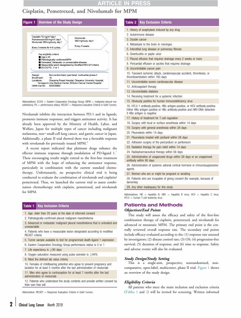
Nivolumab inhibits the interaction between PD-1 and its ligands,promotes immune responses, and triggers antitumor activity. It hasalready been approved by the Ministry of Health, Labor, andWelfare, Japan for multiple types of cancer including malignantmelanoma, nonesmall-cell lung cancer, and gastric cancer in Japan.Additionally, a phase II trial showed there was a favorable responsewith nivolumab for previously treated MPM.6
A recent report indicated that platinum drugs enhance theeffector immune response through modulation of PD-ligand 1.7
These encouraging results might extend to the first-line treatmentof MPM with the hope of enhancing the antitumor response,particularly in combination with the current standard chemo-therapy. Unfortunately, no prospective clinical trial is beingconducted to evaluate the combination of nivolumab and cisplatin/pemetrexed. Thus, we launched the current trial to assess combi-nation chemotherapy with cisplatin, pemetrexed, and nivolumabfor MPM.
Patients and MethodsObjectives/End Points
This study will assess the efficacy and safety of the first-linecombination therapy of cisplatin, pemetrexed, and nivolumab foradvanced or metastatic MPM. The primary end point is the cen-trally reviewed overall response rate. The secondary end pointsinclude efficacy evaluated according to the: (1) response rate assessedby investigators; (2) disease control rate; (3) OS; (4) progression-freesurvival; (5) duration of response; and (6) time to response. Safetyand adverse events will also be evaluated.
Study Design/Study SettingThis is a single-arm, prospective, nonrandomized, non-
comparative, open-label, multicenter, phase II trial. Figure 1 showsan overview of the study design.
Eligibility CriteriaAll patients who meet the main inclusion and exclusion criteria
(Tables 1 and 2) will be invited for screening. Written informed
Figure 1 Overview of the Study Design
Abbreviations: ECOG ¼ Eastern Cooperative Oncology Group; MPM ¼ malignant pleural me-sothelioma; PS ¼ performance status; RECIST ¼ Response Evaluation Criteria in Solid Tumors.
Table 1 Key Inclusion Criteria
1. Age: older than 20 years at the date of informed consent
2. Pathologically-confirmed pleural malignant mesothelioma
3. Advanced or metastatic malignant pleural mesothelioma that is untreated andunresectable
4. Patients who have a measurable lesion designated according to modifiedRECIST criteria
5. Tumor sample available to test for programmed death-ligand 1 expression
6. Eastern Cooperative Oncology Group performance status is 0 or 1
7. Life expectancy is �90 days
8. Oxygen saturation measured using pulse oximeter is �94%
9. Meet the defined lab value criteria
10. Females of childbearing potential who agree to prevent pregnancy andlactation for at least 5 months after the last administration of nivolumab
11. Men who agree to contraception for at least 7 months after the lastadministration of nivolumab
12. Patients who understand the study contents and provide written consent bytheir own free will
Abbreviation: RECIST ¼ Response Evaluation Criteria in Solid Tumors.
Table 2 Key Exclusion Criteria
1. History of anaphylaxis induced by any drug
2. Autoimmune disease
3. Double cancer
4. Metastasis to the brain or meninges
5. Interstitial lung disease or pulmonary fibrosis
6. Diverticulitis or peptic ulcer
7. Pleural effusion that requires drainage every 2 weeks or more
8. Pericardial effusion or ascites that requires drainage
9. Uncontrollable cancer pain
10. Transient ischemic attack, cerebrovascular accident, thrombosis, orthromboembolism within 180 days
11. Uncontrollable severe cardiovascular disease
12. Anticoagulant therapy
13. Uncontrollable diabetes
14. Receiving treatment for a systemic infection
15. Obviously positive for human immunodeficiency virus
16. HTLV-1 antibody-positive, HBs antigen-positive, or HCV antibody-positive.Either HBs antigen positive or HBc antibody-positive and HBV-DNA detectionif HBs antigen is negative
17. History of treatment for T-cell regulation
18. Surgery with local or surface anesthesia within 14 days
19. Surgery with general anesthesia within 28 days
20. Pleurodesis within 14 days
21. Pleurodesis treated with picibanil within 28 days
22. Adhesion surgery of the pericardium or peritoneum
23. Radiation therapy for pain relief within 14 days
24. Radiopharmaceutical therapy within 56 days
25. Administration of unapproved drugs within 28 days or an unapprovedantibody within 90 days
26. Administration of systemic adrenal cortical hormone or immunosuppressiveagents
27. Women who are or might be pregnant or lactating
28. Patients who are incapable of giving consent (for example, because ofdementia)
29. Any other inadequacy for this study
Abbreviations: HB ¼ hepatitis B; HBV ¼ hepatitis B virus; HCV ¼ hepatitis C virus;HTLV ¼ human T-cell leukemia virus.
2 - Clinical Lung Cancer Month 2018
Cisplatin, Pemetrexed, and Nivolumab for MPM
consent must be obtained by an investigator from the patient beforeany screening or inclusion procedure. This study will be conductedin compliance with the principles of the Declaration of Helsinki,and the protocol was approved by the institutional review board ofeach of the participating hospitals.
InterventionTreatment is composed of 2 sequential phases: the combination
phase and the maintenance phase. In the former, cisplatin (75 mg/m2),pemetrexed (500 mg/m2), and nivolumab (360 mg/person) will beadministered intravenously. Nivolumab was kindly provided by OnoPharmaceutical Co, Ltd. This treatment will be repeated every 3 weekswith a total of 4 to 6 cycles. If patients have not progressed during thecombination phase, maintenance therapy with nivolumab will beadministered until disease progression, unacceptable toxicities, or thepatient’s condition meets the withdrawal criteria.
Nivolumab was administered at a dose of 240 mg/personbiweekly in recent clinical trials6,8 including the one for MPM thatshowed encouraging clinical utility and acceptable toxicity profile.6
Both of cisplatin and pemetrexed are usually administered every 3weeks. Under the consideration of practical utility and dose in-tensity, we planned to administer nivolumab every 3 weeks with thedose of 360 mg/person. The combination of nivolumab (10 mg/kg)and pemetrexed/cisplatin every 3 weeks showed an acceptabletoxicity profile and encouraging antitumor activity in patients withadvanced nonesmall-cell lung cancer.9 On the basis of these find-ings, nivolumab will be administered at a dose of 360 mg/person,every 3 weeks, in the current study.
Outcome Measurement/Follow-upResponse is evaluated using the modified Response Evaluation
Criteria in Solid Tumors.10 The OS is defined as the duration fromstudy registration until the date of death or the last patient visit.
Statistical ConsiderationsThe target number of patients is 18 for the current phase II
study. If we assume that there would be 6 to 12 patients with aresponse, the response rate would be 33.3% to 66.7%. In this case,the estimated accuracy indicates the range between the point esti-mate of the response rate and the lower confidence limit (2-sided95% confidence coefficient on the basis of exact test) would be18% to 22%. An OS curve will be constructed using theKaplaneMeier product limit method.
DiscussionThere is a medical need for improved treatments for MPM. This
study is, to our knowledge, the first clinical trial to evaluate theeffect of combining an immune checkpoint inhibitor and platinum-based chemotherapy for MPM. In addition, to our knowledge, thisis the first investigator-initiated prospective clinical trial evaluatingsystemic chemotherapy for MPM that complies with Good ClinicalPractice in Japan.
ConclusionA phase II trial of first-line combination chemotherapy for
unresectable MPM commenced in January 2018. This study is, toour knowledge, the first prospective trial to evaluate the effect of anantiePD-1 antibody combined with cisplatin and pemetrexed forunresectable MPM.
AcknowledgmentsThis study is supported by a grant from Ono Pharmaceutical Co,
Ltd, and also by grants-in-aid from the Ministry of Health, Labor,and Welfare, Japan. Nivolumab was kindly provided by OnoPharmaceutical Co, Ltd.
DisclosureDr Fujimoto has received consultancy fees from Boehringer
Ingelheim, Bristol-Myers Squibb, Kyorin, and Kissei, and receivedhonoraria or research funding from Hisamitsu, Chugai, Ono,Taiho, Boehringer Ingelheim, Bristol-Myers Squibb, Novartis,GlaxoSmithKline, and MSD. Dr Aoe has received consultancy feesfrom Boehringer Ingelheim, Bristol-Myers Squibb, Ono, andreceived honoraria or research funding from Ono, Bristol-MyersSquibb, Novartis, MSD, AstraZeneca, and Eli Lilly. Dr Kozukihas received honoraria or research funding from Chugai,AstraZeneca, Eli Lilly, Pfizer, Ono, Boehringer Ingelheim, Bristol-Myers Squibb, Kyowa, Taiho, MSD, Merck, and Nippon Kayaku.Dr Hotta has received honoraria or research funding from Astra-Zeneca, Ono, Boehringer Ingelheim, Nippon Kayaku, Taiho,Chugai, Astellas, Novartis, Bristol-Myers Squibb, Eli Lilly, andMSD. The remaining authors have stated that they have noconflicts of interest.
References1. Gemba K, Fujimoto N, Aoe K, et al. Treatment and survival analyses of malignant
mesothelioma in Japan. Acta Oncol 2013; 52:803-8.2. Gemba K, Fujimoto N, Kato K, et al. National survey of malignant mesothelioma
and asbestos exposure in Japan. Cancer Sci 2012; 103:483-90.3. Robinson BW, Lake RA. Advances in malignant mesothelioma. N Engl J Med
2005; 353:1591-603.4. Vogelzang NJ, Rusthoven JJ, Symanowski J, et al. Phase III study of pemetrexed in
combination with cisplatin versus cisplatin alone in patients with malignant pleuralmesothelioma. J Clin Oncol 2003; 21:2636-44.
5. Zalcman G, Mazieres J, Margery J, et al. Bevacizumab for newly diagnosedpleural mesothelioma in the Mesothelioma Avastin Cisplatin Pemetrexed Study(MAPS): a randomised, controlled, open-label, phase 3 trial. Lancet 2016; 387:1405-14.
6. Goto Y, Okada M, Kijima T, et al. A phase II study of nivolumab: a multicenter,open-label, single arm study in malignant pleural mesothelioma (MERIT).J Thorac Oncol 2017; 12(11 suppl 2):S1883.
7. Hato SV, Khong A, de Vries IJ, Lesterhuis WJ. Molecular pathways: the immu-nogenic effects of platinum-based chemotherapeutics. Clin Cancer Res 2014; 20:2831-7.
8. Zhao X, Suryawanshi S, Hruska M, et al. Assessment of nivolumab benefit-riskprofile of a 240-mg flat dose relative to a 3-mg/kg dosing regimen in patientswith advanced tumors. Ann Oncol 2017; 28:2002-8.
9. Kanda S, Goto K, Shiraishi H, et al. Safety and efficacy of nivolumab andstandard chemotherapy drug combination in patients with advanced non-small-cell lung cancer: a four-arm phase Ib study. Ann Oncol 2016; 27:2242-50.
10. Byrne MJ, Nowak AK. Modified RECIST criteria for assessment of response inmalignant pleural mesothelioma. Ann Oncol 2004; 15:257-60.
Nobukazu Fujimoto et al
Clinical Lung Cancer Month 2018 - 3
ONCOLOGY LETTERS 15: 3540-3547, 20183540
Abstract. Histological distinction between epithelioid mesothelioma (EM) and reactive mesothelial hyperplasia (RMH) can be challenging. The aim of this study was to assess the diagnostic utility of Survivin, Ki-67, and loss of BRCA1-associated protein 1 (BAP1) expressions in distin-guishing EM from RMH using immunohistochemistry. Formalin‑fixed, paraffin‑embedded specimens from 78 cases of EM and 80 cases of RMH were immunohistochemically examined for Survivin, BAP1, and Ki-67. In addition, receiver operating characteristic curve analyses were performed to establish the cut-off values for Survivin and Ki-67 labelling indices. Survivin (cut-off value: 5%) had 67.7% sensitivity and 100% specificity, while Ki‑67 (cut‑off value: 10%) had 85.1% sensitivity and 87.5% specificity, and BAP1 had 66.2% sensitivity and 100% specificity for the differentiation of EM from RMH. Among the combinations of two markers, the combination of Survivin and BAP1 (Survivin-positive and/or BAP1‑loss finding) had the highest diagnostic accuracy (sensitivity: 89.8%; specificity: 100%; accuracy: 95.3%). We recommend using the combination of Survivin and BAP1 to distinguish EM from RMH.
Introduction
Malignant mesothelioma (MM) is a relatively rare but highly aggressive malignant neoplasm arising from mesothelial cells of the pleura, peritoneum, pericardium, and tunica vaginalis.
It is well-correlated with occupational and environmental asbestos exposure. (1,2) The incidence of MM has increased in many countries; (3) in Japan, mortality due to MM has increased since the 1990s, and is predicted to peak in the 2030s (4).
Epithelioid mesothelioma (EM) must be differentiated from reactive mesothelial hyperplasia (RMH), which is a non-neoplastic condition frequently caused by pleuritis, peri-tonitis, or serosal invasion of other cancers. Due to the close resemblance of EM to RMH, differentiation by routine histo-logical observation alone can be challenging.
Various established and novel immunohistochemical markers have been utilized to distinguish EM from other malignancies (5-8) and RMH (6,9-17) Multiple potential immunohistochemical markers, including Ki-67, desmin, epithelial membrane antigen (EMA), p53, glucose transporter 1, insulin-like growth factor 2 messenger RNA binding protein-3 and BRCA1-associated protein 1 (BAP1) have been evalu-ated. However, despite the use of these immunohistochemical markers, the distinction between EM and RMH remains chal-lenging in some cases.
Recently, detection of p16 (CDKN2A) homozygous deletion (p16 HD) using fluorescence in situ hybridization (FISH) has been used to differentiate MM from RMH, with 100% speci-ficity. However, the sensitivity of this marker for pleural EM varies between 45 and 86%, while its sensitivity for peritoneal EM ranges from 14 to 41% in different laboratories (10,18-20). In our unpublished experience, p16 HD (detected by FISH) was present in 63.2% (12/19) of EM cases, but absent in all RMH cases (0/20). Although the detection of p16 HD using FISH may be considered highly specific, its sensitivity in differentiating EM from RMH is not very high. In addition, FISH analysis cannot be applied in all cases or in all pathology laboratories, given its high cost and stringent experimental requirements.
We recently reported that phorbol 12‑myristate‑13‑ace-tate-induced protein-1 (PMAIP-1; Noxa) and baculoviral IAP repeat-containing 5 (BIRC5; Survivin) mRNA expression levels are significantly higher in EM than in non‑neoplastic pleural tissue, and discussed the utility of anti-Noxa antibody
Utility of Survivin, BAP1, and Ki‑67 immunohistochemistry in distinguishing epithelioid mesothelioma
from reactive mesothelial hyperplasiaKEI KUSHITANI1, VISHWA JEET AMATYA1, AMANY SAYED MAWAS2, RUI SUZUKI1, YOSHIHIRO MIYATA3,
MORIHITO OKADA3, KOUKI INAI1, TAKUMI KISHIMOTO4 and YUKIO TAKESHIMA1
1Department of Pathology, Institute of Biomedical and Health Sciences, Hiroshima University, Hiroshima 734-8551, Japan; 2Department of Pathology and Clinical Pathology, Faculty of Veterinary Medicine,
South Valley University, Qena 83523, Egypt; 3Department of Surgical Oncology, Research Center for Radiation Casualty Medicine, Research Institute for Radiation Biology and Medicine, Hiroshima University,
Hiroshima 734-8551; 4Department of Internal Medicine, Okayama Rosai Hospital, Okayama 702-8055, Japan
Received June 30, 2017; Accepted November 20, 2017
DOI: 10.3892/ol.2018.7765
Correspondence to: Professor Yukio Takeshima, Department of Pathology, Institute of Biomedical and Health Sciences, Hiroshima University, 1-2-3 Kasumi, Minami-ku, Hiroshima 734-8551, JapanE-mail: [email protected]
Key words: BAP1, immunohistochemistry, Ki-67, mesothelioma, reactive mesothelial hyperplasia, survivin
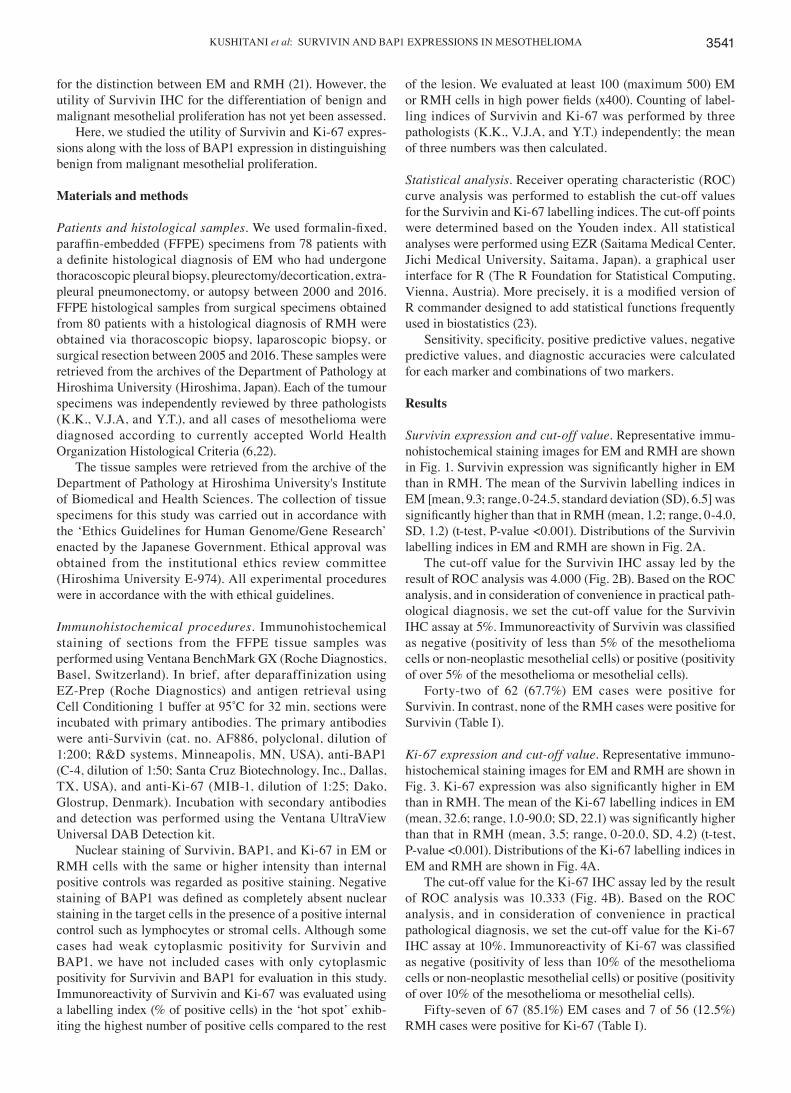
KUSHITANI et al: SURVIVIN AND BAP1 EXPRESSIONS IN MESOTHELIOMA 3541
for the distinction between EM and RMH (21). However, the utility of Survivin IHC for the differentiation of benign and malignant mesothelial proliferation has not yet been assessed.
Here, we studied the utility of Survivin and Ki-67 expres-sions along with the loss of BAP1 expression in distinguishing benign from malignant mesothelial proliferation.
Materials and methods
Patients and histological samples. We used formalin‑fixed, paraffin‑embedded (FFPE) specimens from 78 patients with a definite histological diagnosis of EM who had undergone thoracoscopic pleural biopsy, pleurectomy/decortication, extra-pleural pneumonectomy, or autopsy between 2000 and 2016. FFPE histological samples from surgical specimens obtained from 80 patients with a histological diagnosis of RMH were obtained via thoracoscopic biopsy, laparoscopic biopsy, or surgical resection between 2005 and 2016. These samples were retrieved from the archives of the Department of Pathology at Hiroshima University (Hiroshima, Japan). Each of the tumour specimens was independently reviewed by three pathologists (K.K., V.J.A, and Y.T.), and all cases of mesothelioma were diagnosed according to currently accepted World Health Organization Histological Criteria (6,22).
The tissue samples were retrieved from the archive of the Department of Pathology at Hiroshima University's Institute of Biomedical and Health Sciences. The collection of tissue specimens for this study was carried out in accordance with the ‘Ethics Guidelines for Human Genome/Gene Research’ enacted by the Japanese Government. Ethical approval was obtained from the institutional ethics review committee (Hiroshima University E-974). All experimental procedures were in accordance with the with ethical guidelines.
Immunohistochemical procedures. Immunohistochemical staining of sections from the FFPE tissue samples was performed using Ventana BenchMark GX (Roche Diagnostics, Basel, Switzerland). In brief, after deparaffinization using EZ-Prep (Roche Diagnostics) and antigen retrieval using Cell Conditioning 1 buffer at 95˚C for 32 min, sections were incubated with primary antibodies. The primary antibodies were anti-Survivin (cat. no. AF886, polyclonal, dilution of 1:200; R&D systems, Minneapolis, MN, USA), anti-BAP1 (C-4, dilution of 1:50; Santa Cruz Biotechnology, Inc., Dallas, TX, USA), and anti-Ki-67 (MIB-1, dilution of 1:25; Dako, Glostrup, Denmark). Incubation with secondary antibodies and detection was performed using the Ventana UltraView Universal DAB Detection kit.
Nuclear staining of Survivin, BAP1, and Ki-67 in EM or RMH cells with the same or higher intensity than internal positive controls was regarded as positive staining. Negative staining of BAP1 was defined as completely absent nuclear staining in the target cells in the presence of a positive internal control such as lymphocytes or stromal cells. Although some cases had weak cytoplasmic positivity for Survivin and BAP1, we have not included cases with only cytoplasmic positivity for Survivin and BAP1 for evaluation in this study. Immunoreactivity of Survivin and Ki-67 was evaluated using a labelling index (% of positive cells) in the ‘hot spot’ exhib-iting the highest number of positive cells compared to the rest
of the lesion. We evaluated at least 100 (maximum 500) EM or RMH cells in high power fields (x400). Counting of label-ling indices of Survivin and Ki-67 was performed by three pathologists (K.K., V.J.A, and Y.T.) independently; the mean of three numbers was then calculated.
Statistical analysis. Receiver operating characteristic (ROC) curve analysis was performed to establish the cut-off values for the Survivin and Ki-67 labelling indices. The cut-off points were determined based on the Youden index. All statistical analyses were performed using EZR (Saitama Medical Center, Jichi Medical University, Saitama, Japan), a graphical user interface for R (The R Foundation for Statistical Computing, Vienna, Austria). More precisely, it is a modified version of R commander designed to add statistical functions frequently used in biostatistics (23).
Sensitivity, specificity, positive predictive values, negative predictive values, and diagnostic accuracies were calculated for each marker and combinations of two markers.
Results
Survivin expression and cut‑off value. Representative immu-nohistochemical staining images for EM and RMH are shown in Fig. 1. Survivin expression was significantly higher in EM than in RMH. The mean of the Survivin labelling indices in EM [mean, 9.3; range, 0-24.5, standard deviation (SD), 6.5] was significantly higher than that in RMH (mean, 1.2; range, 0‑4.0, SD, 1.2) (t-test, P-value <0.001). Distributions of the Survivin labelling indices in EM and RMH are shown in Fig. 2A.
The cut-off value for the Survivin IHC assay led by the result of ROC analysis was 4.000 (Fig. 2B). Based on the ROC analysis, and in consideration of convenience in practical path-ological diagnosis, we set the cut-off value for the Survivin IHC assay at 5%. Immunoreactivity of Survivin was classified as negative (positivity of less than 5% of the mesothelioma cells or non-neoplastic mesothelial cells) or positive (positivity of over 5% of the mesothelioma or mesothelial cells).
Forty-two of 62 (67.7%) EM cases were positive for Survivin. In contrast, none of the RMH cases were positive for Survivin (Table Ⅰ).
Ki‑67 expression and cut‑off value. Representative immuno-histochemical staining images for EM and RMH are shown in Fig. 3. Ki‑67 expression was also significantly higher in EM than in RMH. The mean of the Ki-67 labelling indices in EM (mean, 32.6; range, 1.0‑90.0; SD, 22.1) was significantly higher than that in RMH (mean, 3.5; range, 0-20.0, SD, 4.2) (t-test, P-value <0.001). Distributions of the Ki-67 labelling indices in EM and RMH are shown in Fig. 4A.
The cut-off value for the Ki-67 IHC assay led by the result of ROC analysis was 10.333 (Fig. 4B). Based on the ROC analysis, and in consideration of convenience in practical pathological diagnosis, we set the cut-off value for the Ki-67 IHC assay at 10%. Immunoreactivity of Ki‑67 was classified as negative (positivity of less than 10% of the mesothelioma cells or non-neoplastic mesothelial cells) or positive (positivity of over 10% of the mesothelioma or mesothelial cells).
Fifty-seven of 67 (85.1%) EM cases and 7 of 56 (12.5%) RMH cases were positive for Ki‑67 (Table Ⅰ).
ONCOLOGY LETTERS 15: 3540-3547, 20183542
BAP1 expression. Loss of nuclear BAP1 expression was observed in 49 of 74 (66.2%) cases of EM (Table Ⅰ). Almost all cases without BAP1 expression had a homogenous expression loss pattern. No heterogeneous loss patterns were observed. In contrast, nuclear BAP1 expression was preserved in all 78 RMH cases (Table Ⅰ). Representative immunohistochemical staining images for EM and RMH are shown in Fig. 5.
Utilities of each marker and combinations of two markers. The sensitivity and specificity of each marker and combinations of
two markers for the distinction between EM and RMH are shown in Table Ⅱ. Among three single markers and six combination patterns of two markers, ‘Survivin-positive and/or BAP1-loss’ finding showed the highest diagnostic accuracy (95.3%).
Discussion
Accurate histopathological differentiation between MM and RMH is extremely important, not only for clinical manage-ment, but also for the appropriate operation of the public
Figure 1. Representative histological images of Survivin IHC. (A) EM with H&E staining. (B) Survivin IHC in EM; labelling index, 18.1. (C) RMH with H&E stain. (D) Survivin IHC in RMH; labelling index, 1.3. IHC, immunohistochemistry; EM, epithelioid mesothelioma; RMH, reactive mesothelial hyperplasia; H&E, haematoxylin and eosin.
Figure 2. (A) Distribution of Survivin labelling index in epithelioid mesothelioma and reactive mesothelial hyperplasia. The horizontal line in the dot chart shows the mean. (B) ROC analysis. ROC curve was estimated using Survivin labelling index. Cut-off value based on the Youden index is also shown. ROC, receiver operating characteristic.
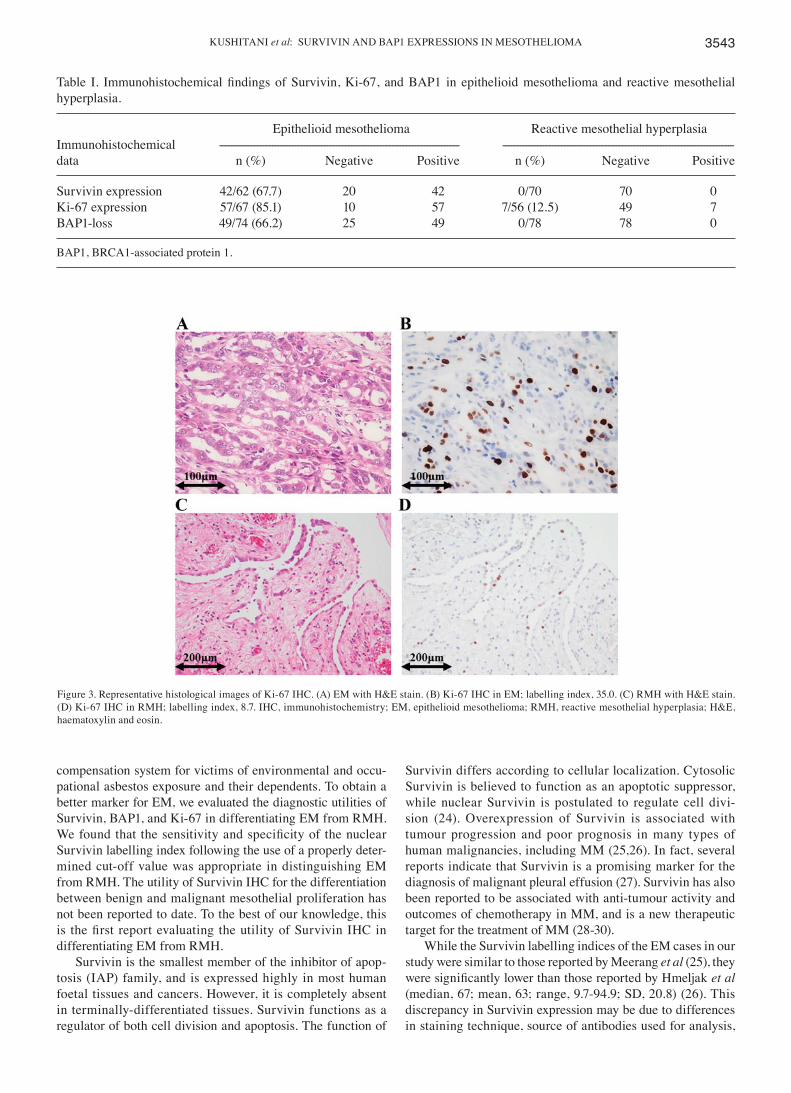
KUSHITANI et al: SURVIVIN AND BAP1 EXPRESSIONS IN MESOTHELIOMA 3543
compensation system for victims of environmental and occu-pational asbestos exposure and their dependents. To obtain a better marker for EM, we evaluated the diagnostic utilities of Survivin, BAP1, and Ki-67 in differentiating EM from RMH. We found that the sensitivity and specificity of the nuclear Survivin labelling index following the use of a properly deter-mined cut-off value was appropriate in distinguishing EM from RMH. The utility of Survivin IHC for the differentiation between benign and malignant mesothelial proliferation has not been reported to date. To the best of our knowledge, this is the first report evaluating the utility of Survivin IHC in differentiating EM from RMH.
Survivin is the smallest member of the inhibitor of apop-tosis (IAP) family, and is expressed highly in most human foetal tissues and cancers. However, it is completely absent in terminally-differentiated tissues. Survivin functions as a regulator of both cell division and apoptosis. The function of
Survivin differs according to cellular localization. Cytosolic Survivin is believed to function as an apoptotic suppressor, while nuclear Survivin is postulated to regulate cell divi-sion (24). Overexpression of Survivin is associated with tumour progression and poor prognosis in many types of human malignancies, including MM (25,26). In fact, several reports indicate that Survivin is a promising marker for the diagnosis of malignant pleural effusion (27). Survivin has also been reported to be associated with anti-tumour activity and outcomes of chemotherapy in MM, and is a new therapeutic target for the treatment of MM (28-30).
While the Survivin labelling indices of the EM cases in our study were similar to those reported by Meerang et al (25), they were significantly lower than those reported by Hmeljak et al (median, 67; mean, 63; range, 9.7-94.9; SD, 20.8) (26). This discrepancy in Survivin expression may be due to differences in staining technique, source of antibodies used for analysis,
Table Ⅰ. Immunohistochemical findings of Survivin, Ki‑67, and BAP1 in epithelioid mesothelioma and reactive mesothelial hyperplasia.
Epithelioid mesothelioma Reactive mesothelial hyperplasiaImmunohistochemical ---------------------------------------------------------------------------------- -------------------------------------------------------------------------------data n (%) Negative Positive n (%) Negative Positive
Survivin expression 42/62 (67.7) 20 42 0/70 70 0Ki-67 expression 57/67 (85.1) 10 57 7/56 (12.5) 49 7BAP1-loss 49/74 (66.2) 25 49 0/78 78 0
BAP1, BRCA1-associated protein 1.
Figure 3. Representative histological images of Ki-67 IHC. (A) EM with H&E stain. (B) Ki-67 IHC in EM; labelling index, 35.0. (C) RMH with H&E stain. (D) Ki-67 IHC in RMH; labelling index, 8.7. IHC, immunohistochemistry; EM, epithelioid mesothelioma; RMH, reactive mesothelial hyperplasia; H&E, haematoxylin and eosin.
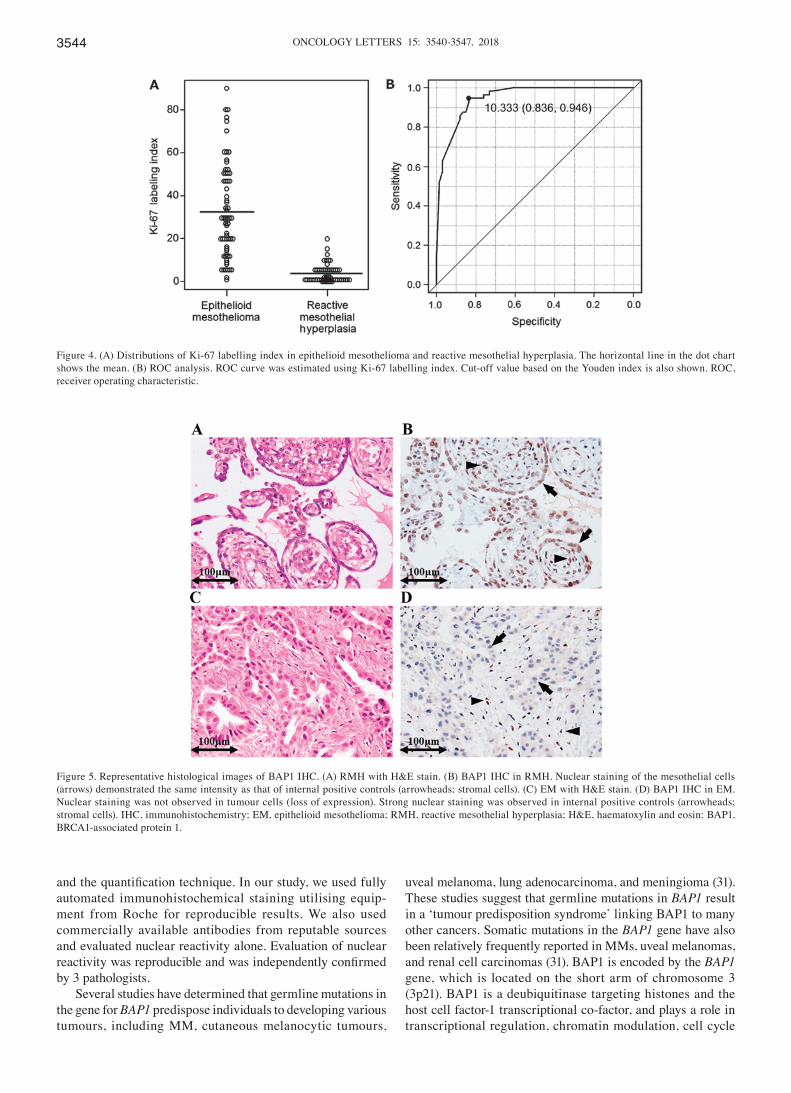
ONCOLOGY LETTERS 15: 3540-3547, 20183544
and the quantification technique. In our study, we used fully automated immunohistochemical staining utilising equip-ment from Roche for reproducible results. We also used commercially available antibodies from reputable sources and evaluated nuclear reactivity alone. Evaluation of nuclear reactivity was reproducible and was independently confirmed by 3 pathologists.
Several studies have determined that germline mutations in the gene for BAP1 predispose individuals to developing various tumours, including MM, cutaneous melanocytic tumours,
uveal melanoma, lung adenocarcinoma, and meningioma (31). These studies suggest that germline mutations in BAP1 result in a ‘tumour predisposition syndrome’ linking BAP1 to many other cancers. Somatic mutations in the BAP1 gene have also been relatively frequently reported in MMs, uveal melanomas, and renal cell carcinomas (31). BAP1 is encoded by the BAP1 gene, which is located on the short arm of chromosome 3 (3p21). BAP1 is a deubiquitinase targeting histones and the host cell factor-1 transcriptional co-factor, and plays a role in transcriptional regulation, chromatin modulation, cell cycle
Figure 4. (A) Distributions of Ki-67 labelling index in epithelioid mesothelioma and reactive mesothelial hyperplasia. The horizontal line in the dot chart shows the mean. (B) ROC analysis. ROC curve was estimated using Ki-67 labelling index. Cut-off value based on the Youden index is also shown. ROC, receiver operating characteristic.
Figure 5. Representative histological images of BAP1 IHC. (A) RMH with H&E stain. (B) BAP1 IHC in RMH. Nuclear staining of the mesothelial cells (arrows) demonstrated the same intensity as that of internal positive controls (arrowheads; stromal cells). (C) EM with H&E stain. (D) BAP1 IHC in EM. Nuclear staining was not observed in tumour cells (loss of expression). Strong nuclear staining was observed in internal positive controls (arrowheads; stromal cells). IHC, immunohistochemistry; EM, epithelioid mesothelioma; RMH, reactive mesothelial hyperplasia; H&E, haematoxylin and eosin; BAP1, BRCA1-associated protein 1.
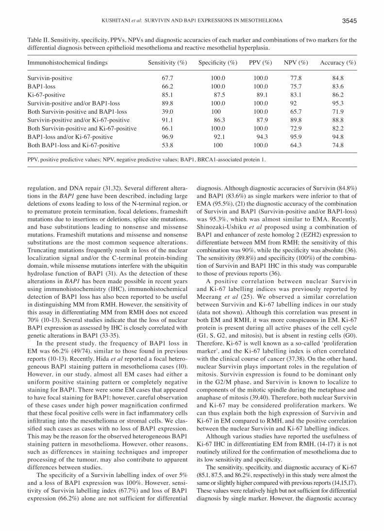
KUSHITANI et al: SURVIVIN AND BAP1 EXPRESSIONS IN MESOTHELIOMA 3545
regulation, and DNA repair (31,32). Several different altera-tions in the BAP1 gene have been described, including large deletions of exons leading to loss of the N-terminal region, or to premature protein termination, focal deletions, frameshift mutations due to insertions or deletions, splice site mutations, and base substitutions leading to nonsense and missense mutations. Frameshift mutations and missense and nonsense substitutions are the most common sequence alterations. Truncating mutations frequently result in loss of the nuclear localization signal and/or the C-terminal protein-binding domain, while missense mutations interfere with the ubiquitin hydrolase function of BAP1 (31). As the detection of these alterations in BAP1 has been made possible in recent years using immunohistochemistry (IHC), immunohistochemical detection of BAP1 loss has also been reported to be useful in distinguishing MM from RMH. However, the sensitivity of this assay in differentiating MM from RMH does not exceed 70% (10-13). Several studies indicate that the loss of nuclear BAP1 expression as assessed by IHC is closely correlated with genetic alterations in BAP1 (33-35).
In the present study, the frequency of BAP1 loss in EM was 66.2% (49/74), similar to those found in previous reports (10-13). Recently, Hida et al reported a focal hetero-geneous BAP1 staining pattern in mesothelioma cases (10). However, in our study, almost all EM cases had either a uniform positive staining pattern or completely negative staining for BAP1. There were some EM cases that appeared to have focal staining for BAP1; however, careful observation of these cases under high power magnification confirmed that these focal positive cells were in fact inflammatory cells infiltrating into the mesothelioma or stromal cells. We clas-sified such cases as cases with no loss of BAP1 expression. This may be the reason for the observed heterogeneous BAP1 staining pattern in mesothelioma. However, other reasons, such as differences in staining techniques and improper processing of the tumour, may also contribute to apparent differences between studies.
The specificity of a Survivin labelling index of over 5% and a loss of BAP1 expression was 100%. However, sensi-tivity of Survivin labelling index (67.7%) and loss of BAP1 expression (66.2%) alone are not sufficient for differential
diagnosis. Although diagnostic accuracies of Survivin (84.8%) and BAP1 (83.6%) as single markers were inferior to that of EMA (95.5%), (21) the diagnostic accuracy of the combination of Survivin and BAP1 (Survivin-positive and/or BAP1-loss) was 95.3%, which was almost similar to EMA. Recently, Shinozaki-Ushiku et al proposed using a combination of BAP1 and enhancer of zeste homolog 2 (EZH2) expression to differentiate between MM from RMH; the sensitivity of this combination was 90%, while the specificity was absolute (36). The sensitivity (89.8%) and specificity (100%) of the combina-tion of Survivin and BAP1 IHC in this study was comparable to those of previous reports (36).
A positive correlation between nuclear Survivin and Ki-67 labelling indices was previously reported by Meerang et al (25). We observed a similar correlation between Survivin and Ki-67 labelling indices in our study (data not shown). Although this correlation was present in both EM and RMH, it was more conspicuous in EM. Ki-67 protein is present during all active phases of the cell cycle (G1, S, G2, and mitosis), but is absent in resting cells (G0). Therefore, Ki-67 is well known as a so-called ‘proliferation marker’, and the Ki-67 labelling index is often correlated with the clinical course of cancer (37,38). On the other hand, nuclear Survivin plays important roles in the regulation of mitosis. Survivin expression is found to be dominant only in the G2/M phase, and Survivin is known to localize to components of the mitotic spindle during the metaphase and anaphase of mitosis (39,40). Therefore, both nuclear Survivin and Ki‑67 may be considered proliferation markers. We can thus explain both the high expression of Survivin and Ki-67 in EM compared to RMH, and the positive correlation between the nuclear Survivin and Ki-67 labelling indices.
Although various studies have reported the usefulness of Ki-67 IHC in differentiating EM from RMH, (14-17) it is not routinely utilized for the confirmation of mesothelioma due to its low sensitivity and specificity.
The sensitivity, specificity, and diagnostic accuracy of Ki‑67 (85.1, 87.5, and 86.2%, respectively) in this study were almost the same or slightly higher compared with previous reports (14,15,17). These values were relatively high but not sufficient for differential diagnosis by single marker. However, the diagnostic accuracy
Table II. Sensitivity, specificity, PPVs, NPVs and diagnostic accuracies of each marker and combinations of two markers for the differential diagnosis between epithelioid mesothelioma and reactive mesothelial hyperplasia.
Immunohistochemical findings Sensitivity (%) Specificity (%) PPV (%) NPV (%) Accuracy (%)
Survivin-positive 67.7 100.0 100.0 77.8 84.8BAP1-loss 66.2 100.0 100.0 75.7 83.6Ki-67-positive 85.1 87.5 89.1 83.1 86.2Survivin-positive and/or BAP1-loss 89.8 100.0 100.0 92 95.3Both Survivin-positive and BAP1-loss 39.0 100 100.0 65.7 71.9Survivin-positive and/or Ki-67-positive 91.1 86.3 87.9 89.8 88.8Both Survivin-positive and Ki-67-positive 66.1 100.0 100.0 72.9 82.2BAP1-loss and/or Ki-67-positive 96.9 92.1 94.3 95.9 94.8Both BAP1-loss and Ki-67-positive 53.8 100 100.0 64.3 74.8
PPV, positive predictive values; NPV, negative predictive values; BAP1, BRCA1-associated protein 1.
ONCOLOGY LETTERS 15: 3540-3547, 20183546
of the combination of Ki-67 and BAP1 was 94.8%, which was almost the same as that of the combination of Survivin and BAP1.
We evaluated the utility of Survivin, BAP1, and Ki‑67 IHC in distinguishing EM from RMH. Based on our results, ‘Survivin‑positive and/or BAP1‑loss’ finding strongly suggest EM, therefore we recommend the use of a combination of Survivin and BAP1. In addition, further evaluation of the Ki-67 labelling index may be useful for accurate differential diagnosis.
Acknowledgements
The authors would like to thank the Technical Centre of Hiroshima University for technical assistance. This study was funded in part by the Japanese Ministry of Health, Labour, and Welfare.
References
1. Robinson BW, Musk AW and Lake RA: Malignant mesothe-lioma. Lancet 366: 397-408, 2005.
2. Roggli VL, Sharma A, Butnor KJ, Sporn T and Vollmer RT: Malignant mesothelioma and occupational exposure to asbestos: A clinicopathological correlation of 1445 cases. Ultrastruct Pathol 26: 55-65, 2002.
3. Delgermaa V, Takahashi K, Park EK, Le GV, Hara T and Sorahan T: Global mesothelioma deaths reported to the World Health Organization between 1994 and 2008. Bull World Health Organ 89: 716-724, 2011.
4. Murayama T, Takahashi K, Natori Y and Kurumatani N: Estimation of future mortality from pleural malignant mesothe-lioma in Japan based on an age-cohort model. Am J Ind Med 49: 1-7, 2006.
5. Churg A, Roggli V, Galateau-Salle F, Cagle PhT, Gibbs AR, Hasleton PhS, Henderson DW, Vignaud JM, Inai K, Praet M, et al: Tumours of the pleura. In: WHO Classification of Tumours of the Lung, Pleura, Thymus and Heart. Travis WD, Brambilla E, Burke AP, Marx A and Nicholson AG (eds). IARC Press, Lyon, pp153-181, 2015.
6. Husain AN, Colby T, Ordonez N, Krausz T, Attanoos R, Beasley MB, Borczuk AC, Butnor K, Cagle PT, Chirieac LR, et al: Guidelines for pathologic diagnosis of malignant mesothelioma: 2012 update of the consensus statement from the International Mesothelioma Interest Group. Arch Pathol Lab Med 137: 647-667, 2013.
7. Ordóñez NG: Application of immunohistochemistry in the diag-nosis of epithelioid mesothelioma: A review and update. Hum Pathol 44: 1-19, 2013.
8. Kushitani K, Amatya VJ, Okada Y, Katayama Y, Mawas AS, Miyata Y, Okada M, Inai K, Kishimoto T and Takeshima Y: Utility and pitfall of immunohistochemistry in the differential diagnosis between epithelioid mesothelioma and poorly differ-entiated lung squamous cell carcinoma. Histopathology 70: 375-384, 2017.
9. Minato H, Kurose N, Fukushima M, Nojima T, Usuda K, Sagawa M, Sakuma T, Ooi A, Matsumoto I, Oda M, et al: Comparative immunohistochemical analysis of IMP3, GLUT1, EMA, CD146, and desmin for distinguishing malignant meso-thelioma from reactive mesothelial cells. Am J Clin Pathol 141: 85-93, 2014.
10. Hida T, Hamasaki M, Matsumoto S, Sato A, Tsujimura T, Kawahara K, Iwasaki A, Okamoto T, Oda Y, Honda H and Nabeshima K: BAP1 immunohistochemistry and p16 FISH results in combination provide higher confidence in malignant pleural mesothelioma diagnosis: ROC analysis of the two tests. Pathol Int 66: 563-570, 2016.
11. Hwang HC, Sheffield BS, Rodriguez S, Thompson K, Tse CH, Gown AM and Churg A: Utility of BAP1 immunohistochemistry and p16 (CDKN2A) FISH in the diagnosis of malignant meso-thelioma in effusion cytology specimens. Am J Surg Pathol 40: 120-126, 2016.
12. McGregor SM, Dunning R, Hyjek E, Vigneswaran W, Husain AN and Krausz T: BAP1 facilitates diagnostic objectivity, classifica-tion, and prognostication in malignant pleural mesothelioma. Hum Pathol 46: 1670-1678, 2015.
13. Cigognetti M, Lonardi S, Fisogni S, Balzarini P, Pellegrini V, Tironi A, Bercich L, Bugatti M, Rossi G, Murer B, et al: BAP1 (BRCA1‑associated protein 1) is a highly specific marker for differentiating mesothelioma from reactive mesothelial prolif-erations. Mod Pathol 28: 1043-1057, 2015.
14. Kimura F, Okayasu I, Kakinuma H, Satoh Y, Kuwao S, Saegusa M and Watanabe J: Differential diagnosis of reactive mesothelial cells and malignant mesothelioma cells using the cell prolifera-tion markers minichromosome maintenance protein 7, geminin, topoisomerase II alpha and Ki-67. Acta Cytol 57: 384-390, 2013.
15. Kimura F, Kawamura J, Watanabe J, Kamoshida S, Kawai K, Okayasu I and Kuwao S: Significance of cell proliferation markers (Minichromosome maintenance protein 7, topoisomerase IIalpha and Ki‑67) in cavital fluid cytology: Can we differentiate reactive meso-thelial cells from malignant cells? Diagn Cytopathol 38: 161-167, 2010.
16. Hasteh F, Lin GY, Weidner N and Michael CW: The use of immunohistochemistry to distinguish reactive mesothelial cells from malignant mesothelioma in cytologic effusions. Cancer Cytopathol 118: 90-96, 2010.
17. Taheri ZM, Mehrafza M, Mohammadi F, Khoddami M, Bahadori M and Masjedi MR: The diagnostic value of Ki-67 and repp86 in distinguishing between benign and malignant meso-thelial proliferations. Arch Pathol Lab Med 132: 694-697, 2008.
18. Churg A, Sheffield BS and Galateau‑Salle F: New markers for separating benign from malignant mesothelial proliferations: Are we there yet? Arch Pathol Lab Med 140: 318-321, 2016.
19. Hiroshima K, Wu D, Hasegawa M, Koh E, Sekine Y, Ozaki D, Yusa T, Walts AE, Marchevsky AM, Nabeshima K, et al: Cytologic differential diagnosis of malignant mesothelioma and reactive mesothelial cells with fish analysis of p16. Diagn Cytopathol 44: 591-598, 2016.
20. Walts AE, Hiroshima K, McGregor SM, Wu D, Husain AN and Marchevsky AM: BAP1 immunostain and CDKN2A (p16) FISH analysis: Clinical applicability for the diagnosis of malignant mesothelioma in effusions. Diagn Cytopathol 44: 599-606, 2016.
21. Kushitani K, Amatya VJ, Mawas AS, Miyata Y, Okada M and Takeshima Y: Use of anti-noxa antibody for differential diag-nosis between epithelioid mesothelioma and reactive mesothelial hyperplasia. Pathobiology 83: 33-40, 2016.
22. Travis WD, Brambilla E, Nicholson AG, Yatabe Y, Austin JHM, Beasley MB, Chirieac LR, Dacic S, Duhig E, Flieder DB, et al: The 2015 World Health Organization Classification of Lung Tumors: Impact of genetic, clinical and radiologic advances since the 2004 classification. J Thorac Oncol 10: 1243-1260, 2015.
23. Kanda Y: Investigation of the freely available easy-to-use soft-ware ‘EZR’ for medical statistics. Bone Marrow Transplant 48: 452-458, 2013.
24. Garg H, Suri P, Gupta JC, Talwar GP and Dubey S: Survivin: A unique target for tumor therapy. Cancer Cell Int 16: 49, 2016.
25. Meerang M, Bérard K, Friess M, Bitanihirwe BK, Soltermann A, Vrugt B, Felley‑Bosco E, Bueno R, Richards WG, Seifert B, et al: Low merlin expression and high survivin labeling index are indicators for poor prognosis in patients with malignant pleural mesothelioma. Mol Oncol 10: 1255-1265, 2016.
26. Hmeljak J, Erčulj N, Dolžan V, Pižem J, Kern I, Kovač V, Cemažar M and Cör A: Is survivin expression prognostic or predictive in malignant pleural mesothelioma? Virchows Arch 462: 315-321, 2013.
27. Chen S, Wang Y, An L, Fei ZT and Li T: The diagnostic value of survivin in malignant pleural effusion: A meta-analysis. Clin Chim Acta 441: 142-147, 2015.
28. Bertino P, Panigada M, Soprana E, Bianchi V, Bertilaccio S, Sanvito F, Rose AH, Yang H, Gaudino G, Hoffmann PR, et al: Fowlpox-based survivin vaccination for malignant mesothelioma therapy. Int J Cancer 133: 612-623, 2013.
29. De Cesare M, Cominetti D, Doldi V, Lopergolo A, Deraco M, Gandellini P, Friedlander S, Landesman Y, Kauffman MG, Shacham S, et al: Anti-tumor activity of selective inhibitors of XPO1/CRM1-mediated nuclear export in diffuse malignant peritoneal mesothelioma: The role of survivin. Oncotarget 6: 13119-13132, 2015.
30. Goričar K, Kovač V, Franko A, Dodič‑Fikfak M and Dolžan V: Serum survivin levels and outcome of chemotherapy in patients with malignant mesothelioma. Dis Markers 2015: 316739, 2015.
31. Murali R, Wiesner T and Scolyer RA: Tumours associated with BAP1 mutations. Pathology 45: 116-126, 2013.
32. Scheuermann JC, de Ayala Alonso AG, Oktaba K, Ly-Hartig N, McGinty RK, Fraterman S, Wilm M, Muir TW and Müller J: Histone H2A deubiquitinase activity of the Polycomb repressive complex PR-DUB. Nature 465: 243-247, 2010.

KUSHITANI et al: SURVIVIN AND BAP1 EXPRESSIONS IN MESOTHELIOMA 3547
33. Bott M, Brevet M, Taylor BS, Shimizu S, Ito T, Wang L, Creaney J, Lake RA, Zakowski MF, Reva B, et al: The nuclear deubiquitinase BAP1 is commonly inactivated by somatic muta-tions and 3p21.1 losses in malignant pleural mesothelioma. Nat Genet 43: 668-672, 2011.
34. Testa JR, Cheung M, Pei J, Below JE, Tan Y, Sementino E, Cox NJ, Dogan AU, Pass HI, Trusa S, et al: Germline BAP1 mutations predispose to malignant mesothelioma. Nat Genet 43: 1022-1025, 2011.
35. Yoshikawa Y, Sato A, Tsujimura T, Emi M, Morinaga T, Fukuoka K, Yamada S, Murakami A, Kondo N, Matsumoto S, et al: Frequent inactivation of the BAP1 gene in epithelioid-type malignant mesothelioma. Cancer Sci 103: 868-874, 2012.
36. Shinozaki-Ushiku A, Ushiku T, Morita S, Anraku M, Nakajima J and Fukayama M: Diagnostic utility of BAP1 and EZH2 expres-sion in malignant mesothelioma. Histopathology 70: 722-733, 2017.
37. Scholzen T and Gerdes J: The Ki-67 protein: From the known and the unknown. J Cell Physiol 182: 311-322, 2000.
38. Brown DC and Gatter KC: Monoclonal antibody Ki-67: Its use in histopathology. Histopathology 17: 489-503, 1990.
39. Altieri DC: Validating survivin as a cancer therapeutic target. Nat Rev Cancer 3: 46-54, 2003.
40. Kim JY, Chung JY, Lee SG, Kim YJ, Park JE, Yoo KS, Yoo YH, Park YC, Kim BG and Kim JM: Nuclear inter-action of Smac/DIABLO with survivin at G2/M arrest prompts docetaxel-induced apoptosis in DU145 prostate cancer cells. Biochem Biophys Res Commun 350: 949-954, 2006.
This work is licensed under a Creative Commons Attribution-NonCommercial-NoDerivatives 4.0 International (CC BY-NC-ND 4.0) License.
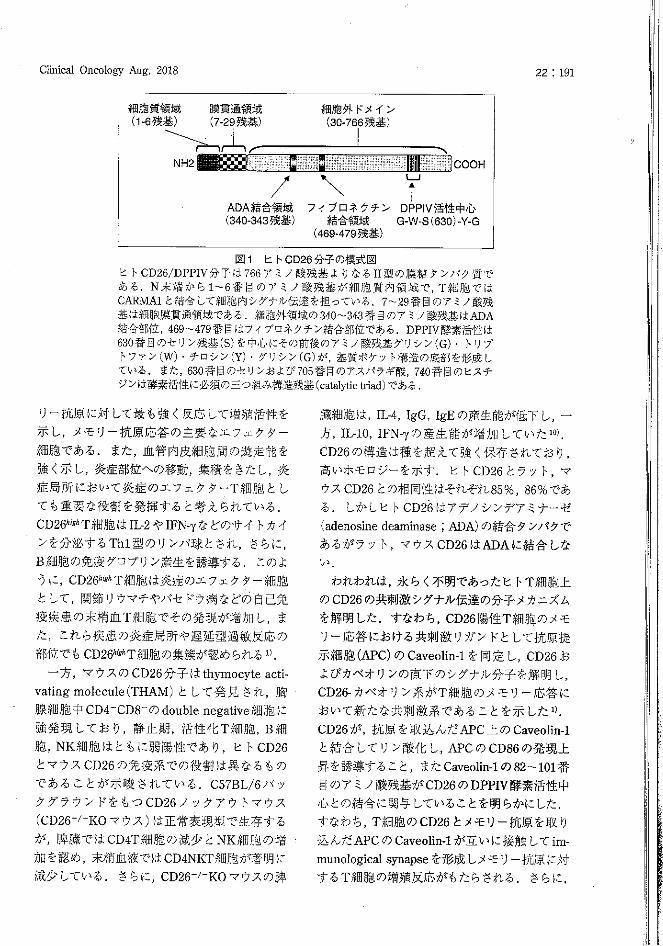
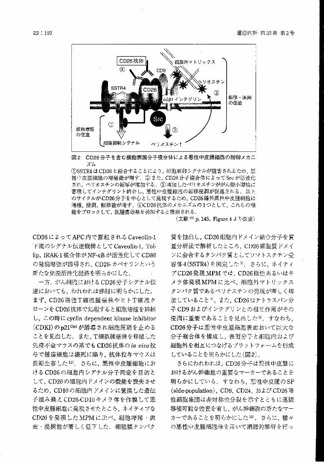
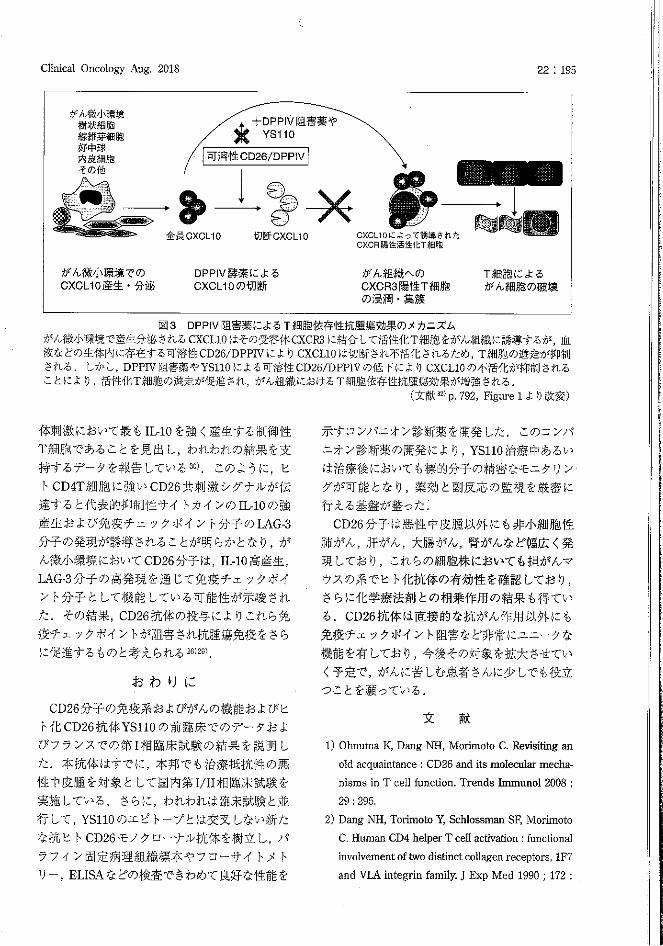
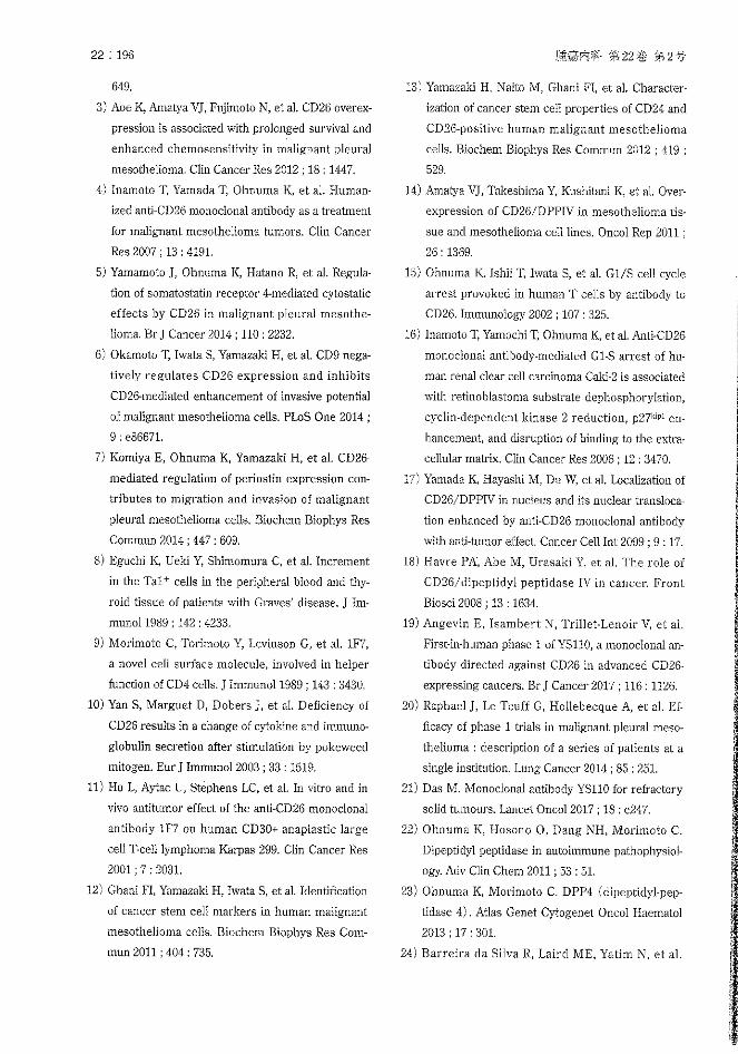
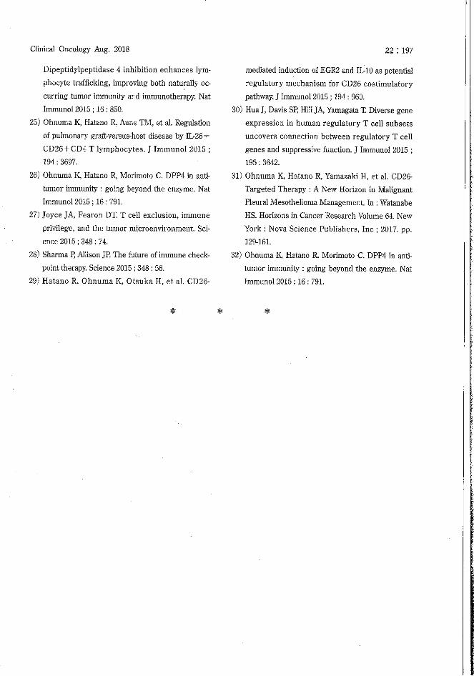
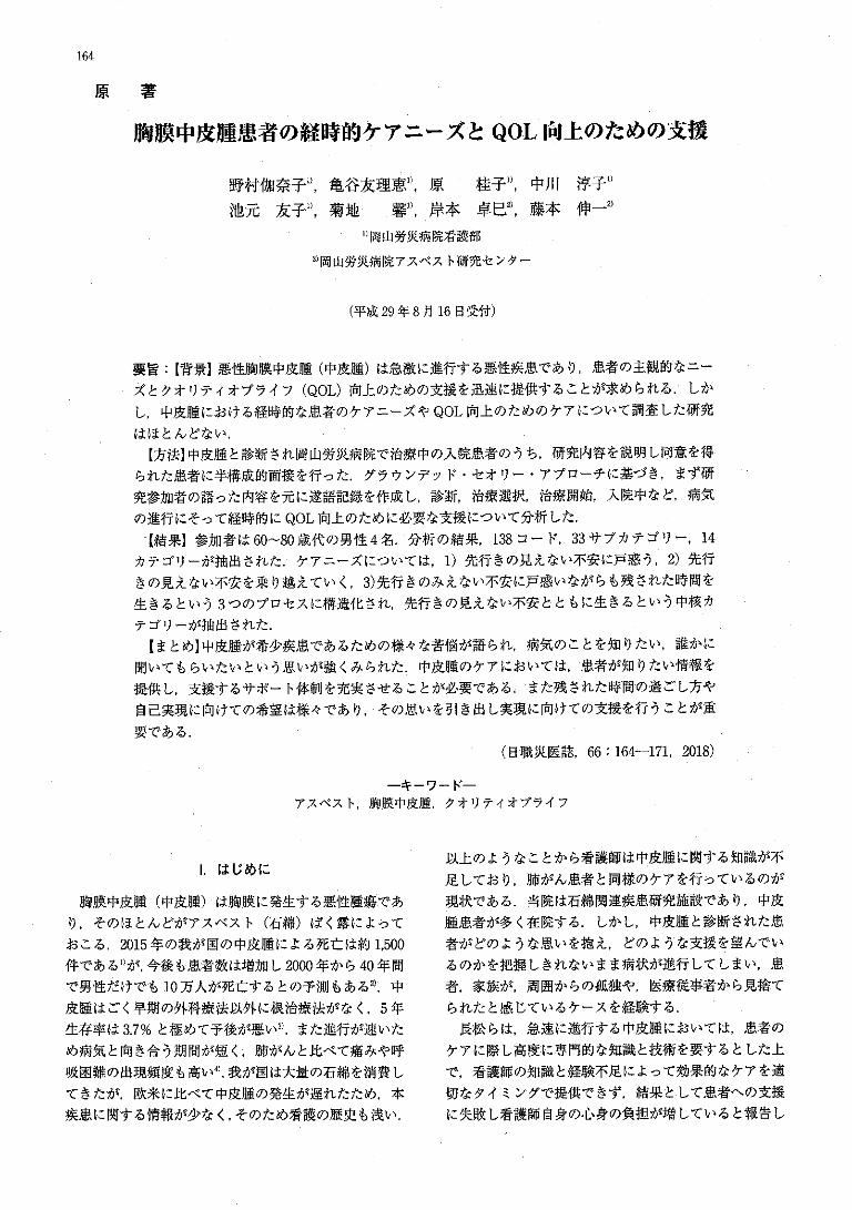
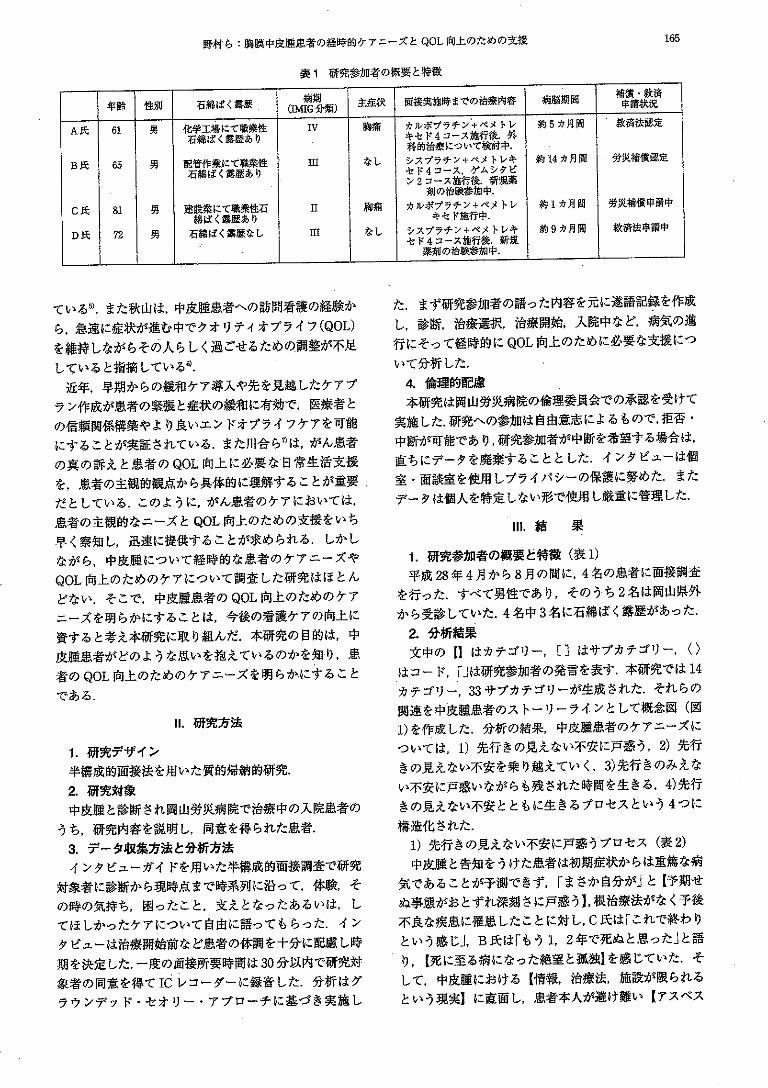
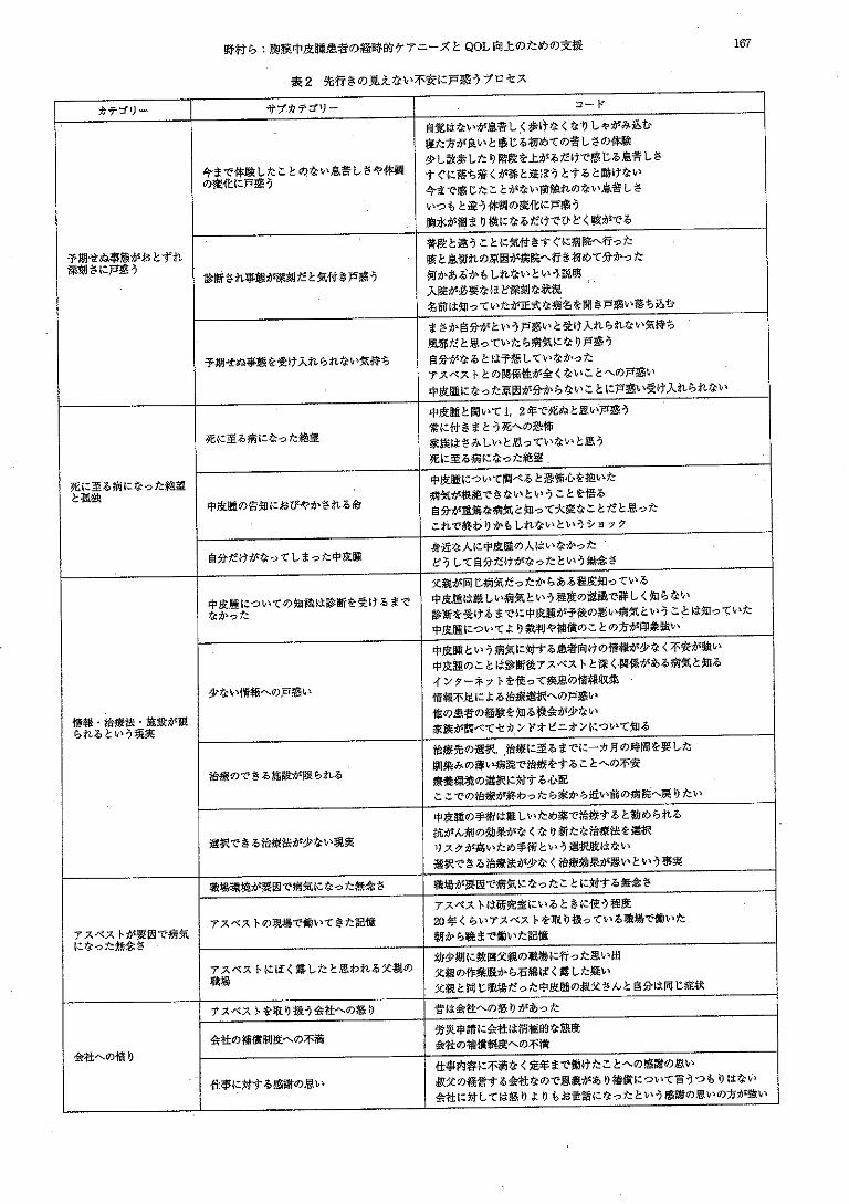
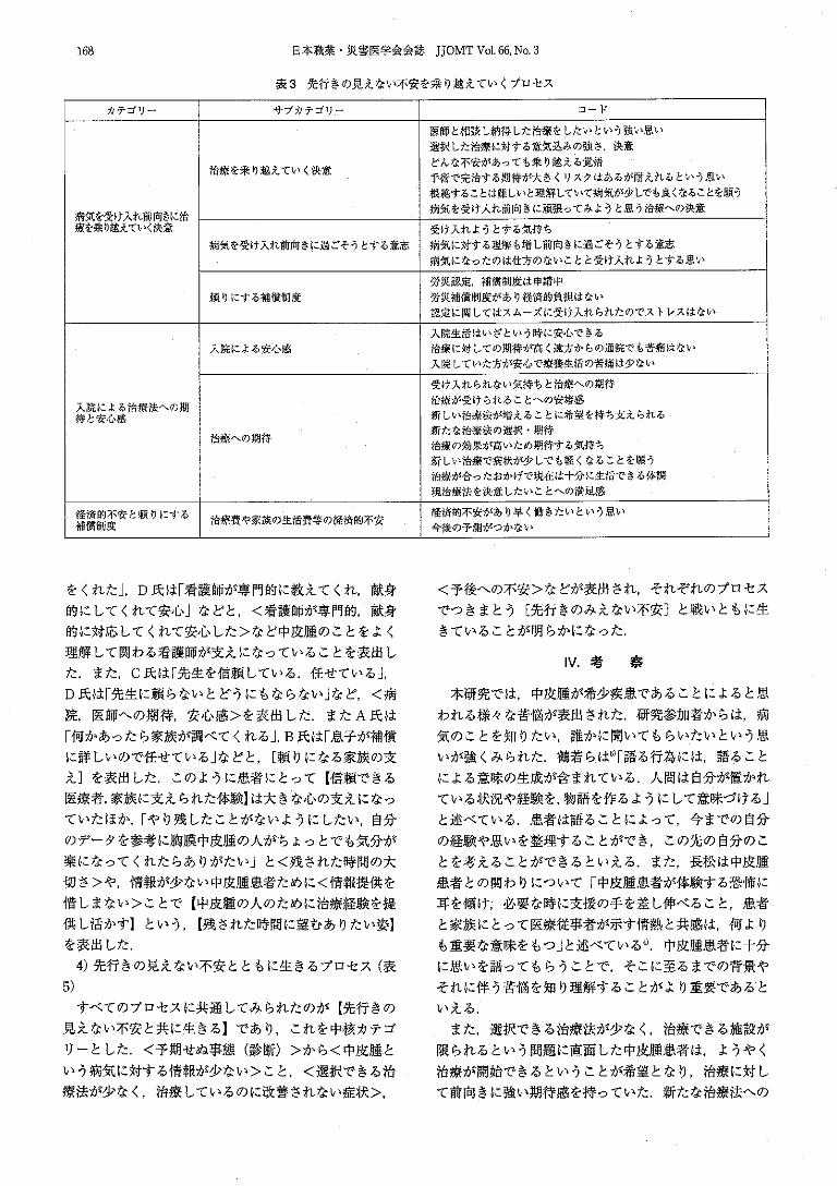
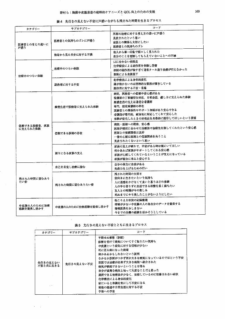
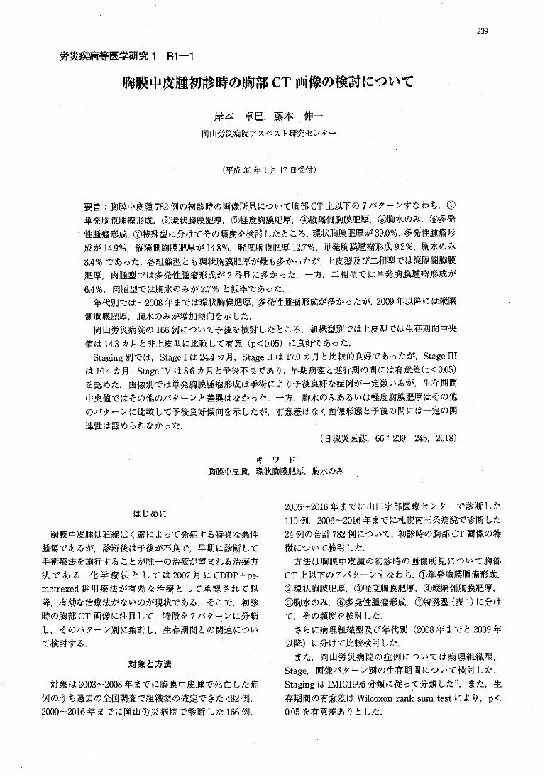
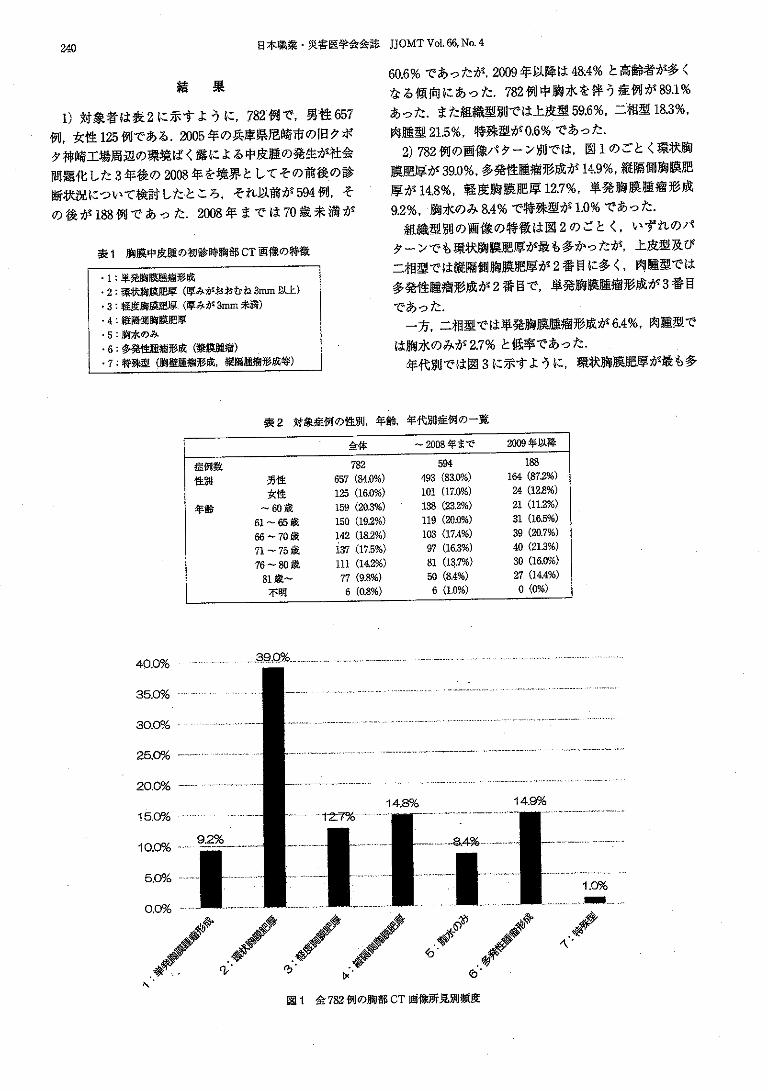
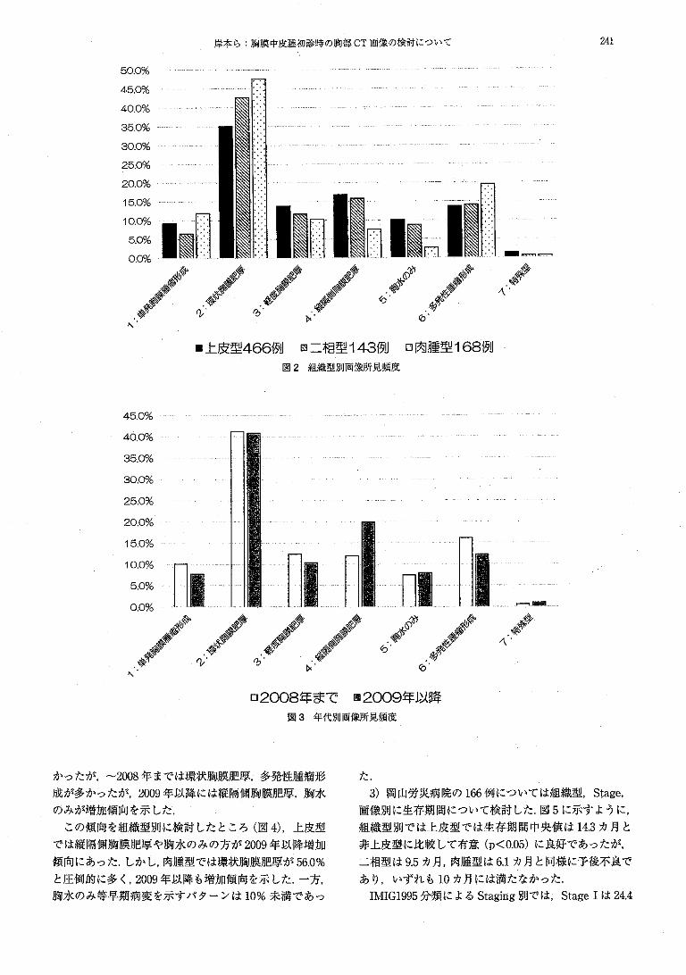
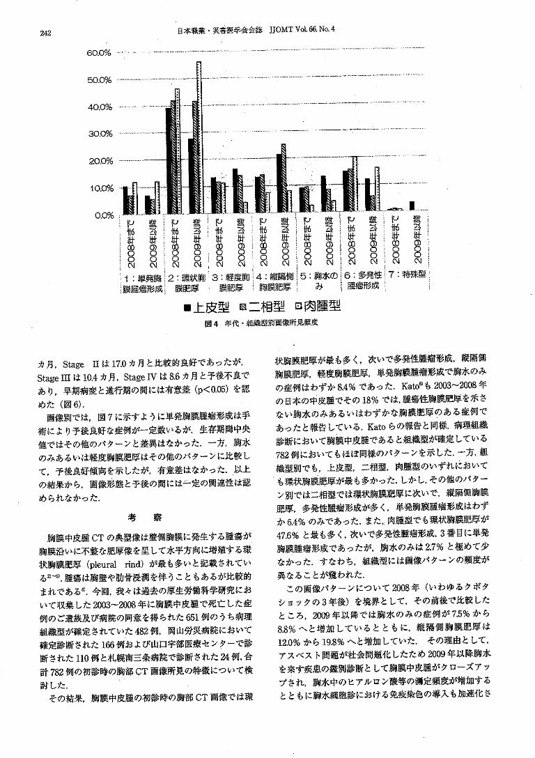
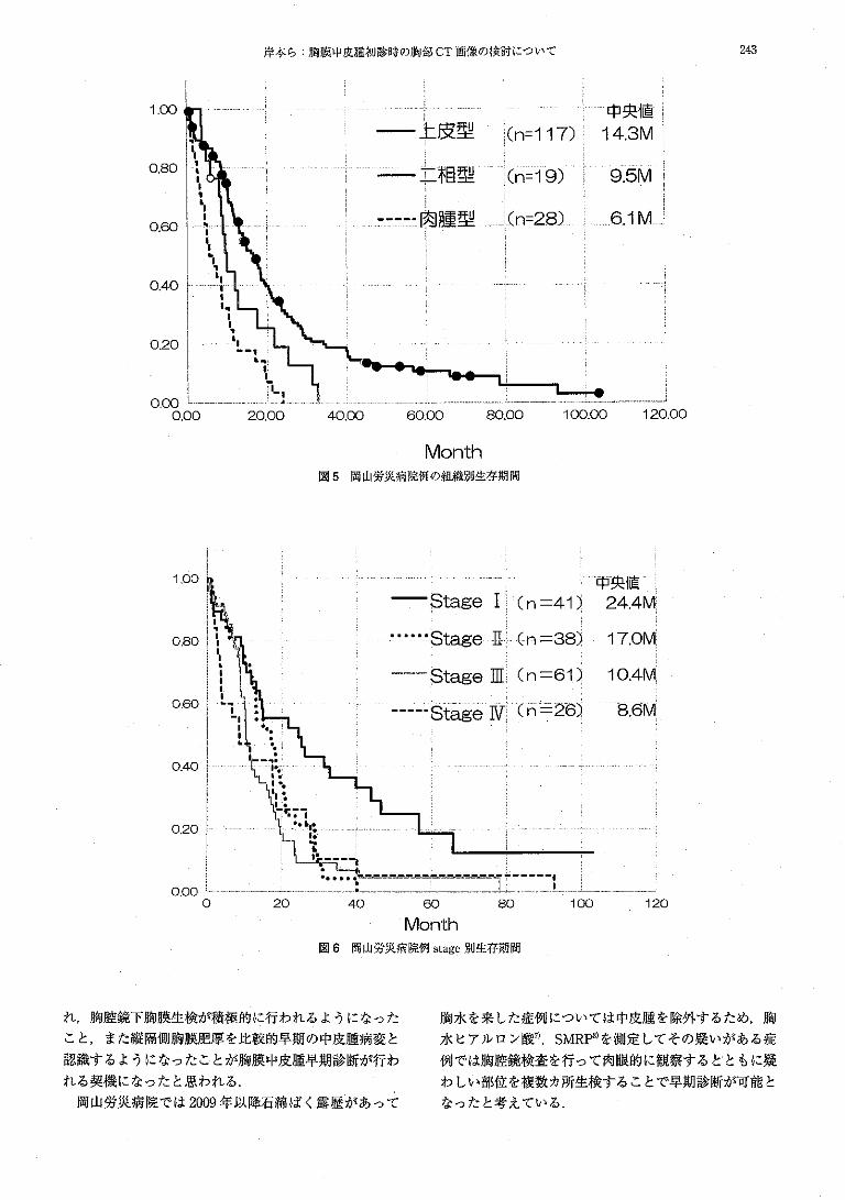
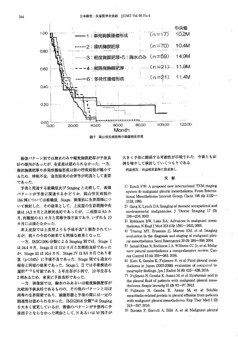
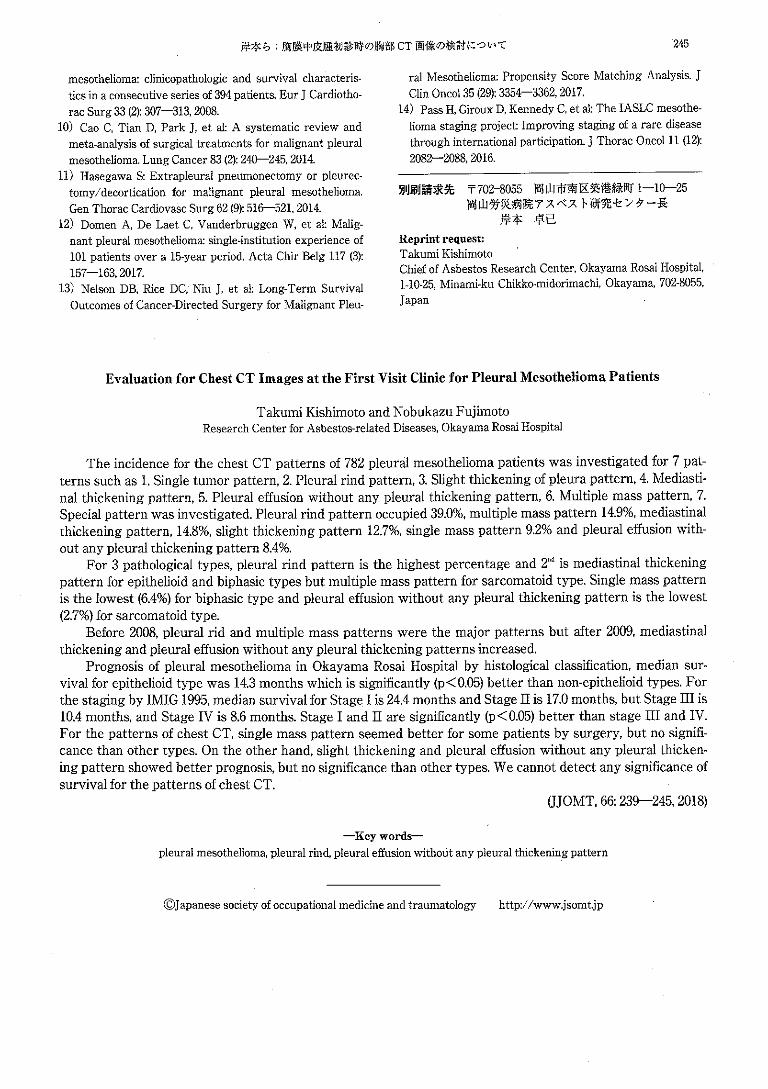
Related Documents