1/17/2019 1 Revitalization of the Anterior Segment: Corneal Transplantation and Secondary Lens Repair CATHERINE REPPA, MD CORNEA SPECIALIST, ASSISTANT PROFESSOR TTUHSC DEPARTMENT OF OPHTHALMOLOGY AND VISUAL SCIENCES CENTER I developed the course material and information independently. I have no relevant financial disclosures. I will be discussing off label use of some medications and devices.

Welcome message from author
This document is posted to help you gain knowledge. Please leave a comment to let me know what you think about it! Share it to your friends and learn new things together.
Transcript
1/17/2019
1
Revitalization of the Anterior Segment: Corneal Transplantation and Secondary Lens RepairCATHERINE REPPA, MDCORNEA SPECIALIST, ASSISTANT PROFESSORTTUHSC DEPARTMENT OF OPHTHALMOLOGY AND VISUAL SCIENCES CENTER
I developed the course material and information independently.
I have no relevant financial disclosures.
I will be discussing off label use of some medications and devices.
1/17/2019
2
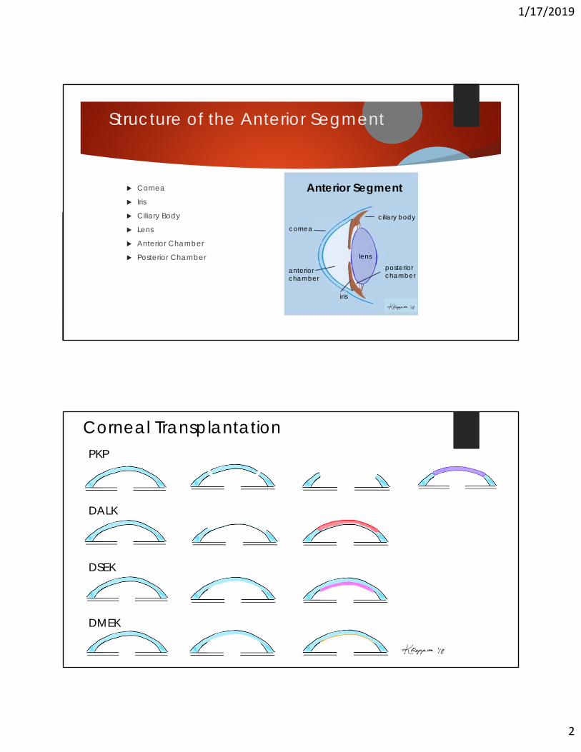
Structure of the Anterior Segment
Cornea
Iris
Ciliary Body
Lens
Anterior Chamber
Posterior Chamber
Anterior Segment
iris
ciliary body
cornea
lens
anteriorchamber
posterior chamber
PKP
DALK
DMEK
DSEK
Corneal Transplantation
1/17/2019
3
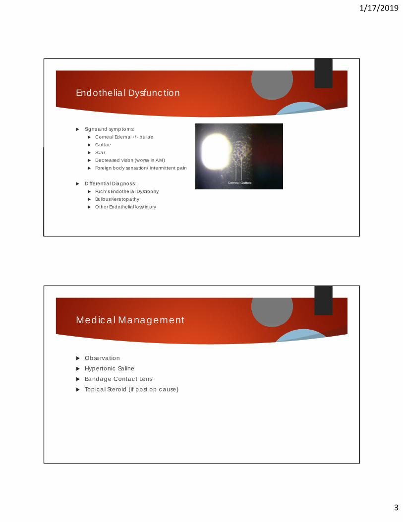
Endothelial Dysfunction
Signs and symptoms: Corneal Edema +/- bullae Guttae Scar Decreased vision (worse in AM) Foreign body sensation/ intermittent pain
Differential Diagnosis: Fuch’s Endothelial Dystrophy Bullous Keratopathy Other Endothelial loss/injury
Medical Management
Observation
Hypertonic Saline
Bandage Contact Lens
Topical Steroid (if post op cause)
1/17/2019
4
Indications for Surgery
Persistent corneal edema
Visual Obscuration (edema or guttae)
Painful Bullae/recurrent erosion
Concurrent Cataract Surgery
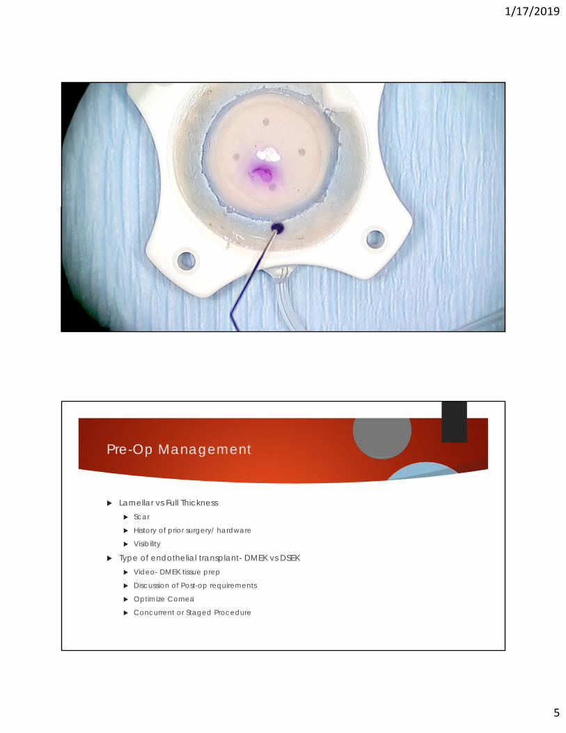
Pre-Op Management
Lamellar vs Full Thickness Scar
History of prior surgery/ hardware
Visibility
Type of endothelial transplant- DMEK vs DSEK Video- DMEK tissue prep
Discussion of Post-op requirements
Optimize Cornea
Concurrent or Staged Procedure
1/17/2019
5
Pre-Op Management
Lamellar vs Full Thickness Scar
History of prior surgery/ hardware
Visibility
Type of endothelial transplant- DMEK vs DSEK Video- DMEK tissue prep
Discussion of Post-op requirements
Optimize Cornea
Concurrent or Staged Procedure
Pre-Op Management
Lamellar vs Full Thickness Scar
History of prior surgery/ hardware
Visibility
Type of endothelial transplant- DMEK vs DSEK Video- DMEK tissue prep
Discussion of Post-op requirements
Optimize Cornea
Concurrent or Staged Procedure
1/17/2019
6
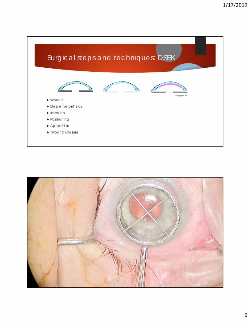
Surgical steps and techniques: DSEK
Wound
Descemetorrhexis
Insertion
Positioning
Apposition
Wound Closure
Surgical steps and techniques: DSEK
Wound
Descemetorrhexis
Insertion
Positioning
Apposition
Wound Closure
1/17/2019
7
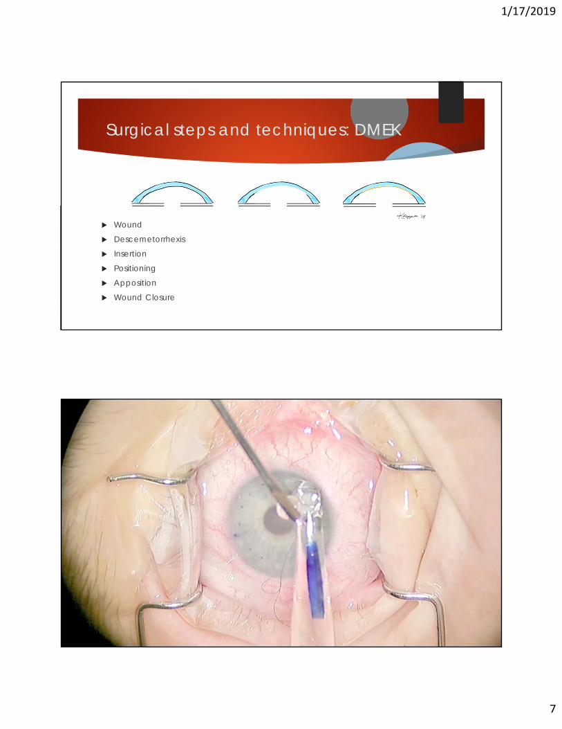
Surgical steps and techniques: DMEK
Wound
Descemetorrhexis
Insertion
Positioning
Apposition
Wound Closure
Surgical steps and techniques: DMEK
Wound
Descemetorrhexis
Insertion
Positioning
Apposition
Wound Closure
Video
1/17/2019
8
Post-op Management
Positioning
Medications
Re-bubble
Refraction
Possible Surgical Complications
Intra-Op
Post-Op Immediate
Long Term

1/17/2019
9
Endothelial Dysfunction: On the horizon
DWEK Stripping without graft
Currently small studies
Only for Fuch’s with centralized guttae
Longer healing time than DMEK
Not all patients clear
Rho-Kinase Inhibitors Activate endothelial cell migration
Decrease healing time
Not currently available in the US at proper concentration
Lens Replacement/Refixation
57 yo F presents for evaluation of corneal edema and dislocated IOL
OS: BCVA 20/400
H/o CEIOL OU in her 30s
H/o RD OS s/p repair 1 year prior with persistent macular edema
Multiple steroid injections and Ozurdex x1
Ozurdex migrated around dislocated IOL to AC
Now with corneal edema
3-piece PCIOL visibly subluxed inferonasally on exam
1/17/2019
10
Lens Replacement/Refixation
57 yo F presents for evaluation of corneal edema and dislocated IOL
OS: BCVA 20/400
H/o CEIOL OU in her 30s
H/o RD OS s/p repair 1 year prior with persistent macular edema
Multiple steroid injections and Ozurdex x1
Ozurdex migrated around dislocated IOL to AC
Now with corneal edema
3-piece PCIOL visibly subluxed inferonasally on exam
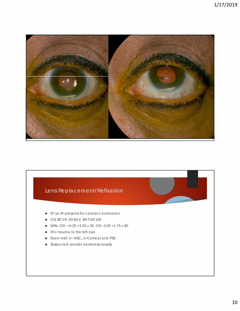
Lens Replacement/Refixation
67 yo M presents for cataract evaluation
OS: BCVA 20/60-2 BAT 20/100
MRx: OD: +0.25 +1.25 x 30 OS: -2.00 +1.75 x 90
H/o trauma to the left eye
Exam with 1+ NSC, 1+Cortical and PSC
Suspected zonular weakness nasally

1/17/2019
11
Lens Replacement/Refixation
67 yo M presents for cataract evaluation
Video
Lens Replacement/Refixation
Signs and symptoms Decreased vision
Visibly dislocated IOL
Chronic inflammation
Chronic low grade pain
Gonioscopy- IOL in angle
Indications for surgery Aphakia
Dislocated IOL
Uveitis-Glaucoma-Hyphema syndrome
1/17/2019
12
Medical Management
Observation
Contact lens
Topical NSAIDS or steroids
Surgical Management
Iris Fixation
Scleral Fixation
Concurrent or staged procedures
1/17/2019
13
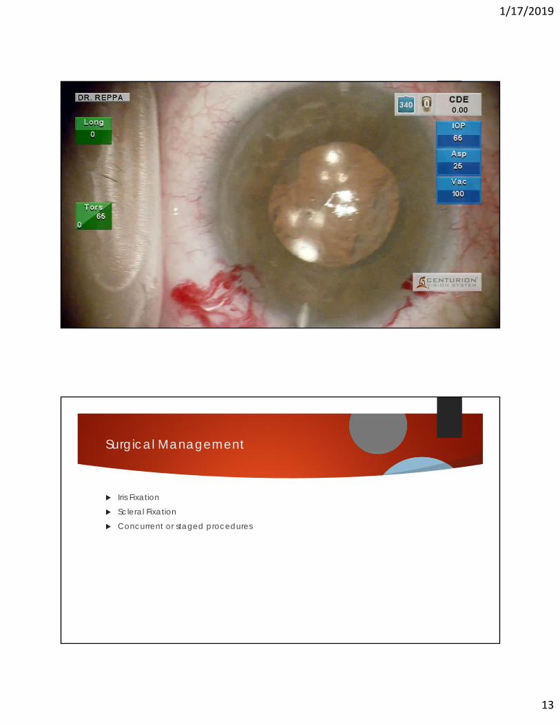
Surgical Management
Iris Fixation
Scleral Fixation
Concurrent or staged procedures
Videos
Surgical Management
Iris Fixation
Scleral Fixation
Concurrent or staged procedures
1/17/2019
14
Surgical Management
Iris Fixation
Scleral Fixation
Concurrent or staged procedures
Videos
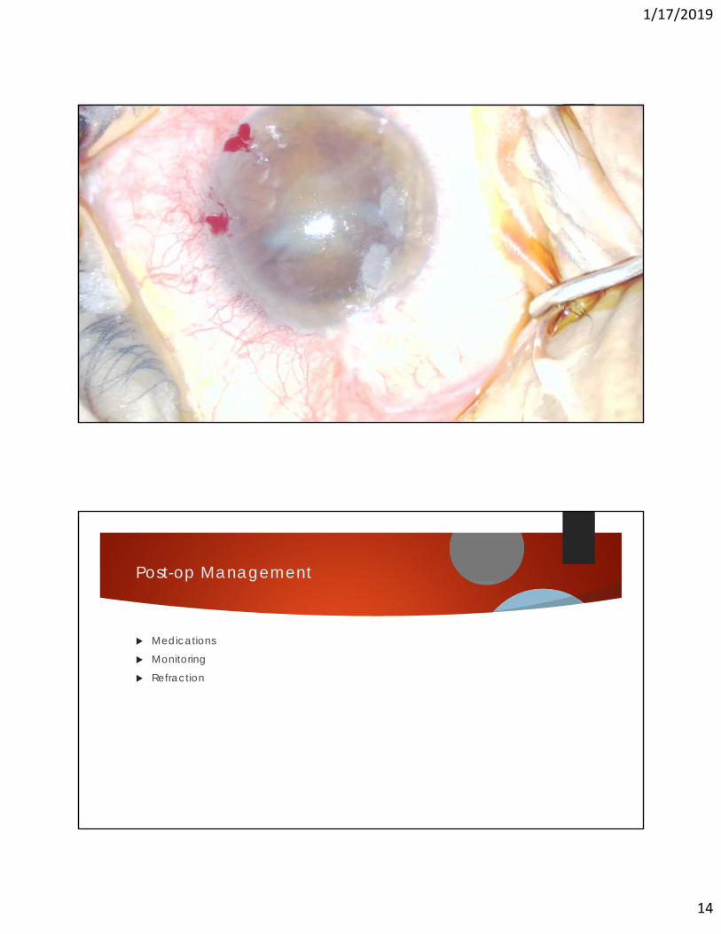
Post-op Management
Medications
Monitoring
Refraction

1/17/2019
15
Possible surgical complications
Immediate
Long term
Additional procedures Corneal transplant for endothelial dysfunction
Pupilloplasty
Corneal Transplantation
DMEKDSEKDALKPKP
1/17/2019
16
References
Borkar DS, Veldman P, Colby KA. Treatment of Fuchs Endothelial Dystrophy by Descemet Stripping Without Endothelial Keratoplasty Cornea. 2016 Oct; 35(10):1267-73.
Davies E, Jurkunas U, Pineda R 2nd. Predictive Factors for Corneal Clearance After Descemetorhexis Without Endothelial Keratoplasty Cornea. 2018 Feb; 37(2):137-140
Huang M, Kane S, Dhaliwal DK. Descemetorhexis Without Endothelial Keratoplasty Versus DMEK for Treatment of Fuchs Endothelial Corneal Dystrophy Cornea. 2018 Dec; 37(12):1479-1483
Iovieno A, Neri A, Soldani AM, Adani C et al. Descemetorhexis Without Graft Placement for the Treatment of Fuchs Endothelial Dystrophy: Preliminary Results and Review of the Literature Cornea. 2017 Jun; 36 (6):637-641
Questions?
Related Documents


![Double mutation (R124H, N544S) of TGFBI in two sisters ... · Groenouw type I corneal dystrophy, and Reis-Bücklers corneal dystrophy, respectively [2]. Many additional mutations](https://static.cupdf.com/doc/110x72/5fd80a4db453983ed540e753/double-mutation-r124h-n544s-of-tgfbi-in-two-sisters-groenouw-type-i-corneal.jpg)
