Citation: Sundararajan, V.; Burk, U.C.; Bajdak-Rusinek, K. Revisiting the miR-200 Family: A Clan of Five Siblings with Essential Roles in Development and Disease. Biomolecules 2022, 12, 781. https:// doi.org/10.3390/biom12060781 Academic Editors: Klaudia Skrzypek and Agnieszka Loboda Received: 29 March 2022 Accepted: 1 June 2022 Published: 3 June 2022 Publisher’s Note: MDPI stays neutral with regard to jurisdictional claims in published maps and institutional affil- iations. Copyright: © 2022 by the authors. Licensee MDPI, Basel, Switzerland. This article is an open access article distributed under the terms and conditions of the Creative Commons Attribution (CC BY) license (https:// creativecommons.org/licenses/by/ 4.0/). biomolecules Review Revisiting the miR-200 Family: A Clan of Five Siblings with Essential Roles in Development and Disease Vignesh Sundararajan 1 , Ulrike C. Burk 2 and Karolina Bajdak-Rusinek 3, * 1 Cancer Science Institute of Singapore, National University of Singapore, Center for Translational Medicine, Singapore 117599, Singapore; [email protected] 2 Institute of Molecular Medicine and Cell Research, Faculty of Medicine, University of Freiburg, 79104 Freiburg, Germany; [email protected] 3 Department of Medical Genetics, Faculty of Medical Sciences, Medical University of Silesia, 40-752 Katowice, Poland * Correspondence: [email protected]; Tel.: +48-32-208-8382 Abstract: Over two decades of studies on small noncoding RNA molecules illustrate the significance of microRNAs (miRNAs/miRs) in controlling multiple physiological and pathological functions through post-transcriptional and spatiotemporal gene expression. Among the plethora of miRs that are essential during animal embryonic development, in this review, we elaborate the indispens- able role of the miR-200 family (comprising miR-200a, -200b, 200c, -141, and -429) in governing the cellular functions associated with epithelial homeostasis, such as epithelial differentiation and neurogenesis. Additionally, in pathological contexts, miR-200 family members are primarily involved in tumor-suppressive roles, including the reversal of the cancer-associated epithelial–mesenchymal transition dedifferentiation process, and are dysregulated during organ fibrosis. Moreover, recent eminent studies have elucidated the crucial roles of miR-200s in the pathophysiology of multiple neurodegenerative diseases and tissue fibrosis. Lastly, we summarize the key studies that have recognized the potential use of miR-200 members as biomarkers for the diagnosis and prognosis of cancers, elaborating the application of these small biomolecules in aiding early cancer detection and intervention. Keywords: miRNA/microRNA; miR-200 family; cancer-associated miRNAs; epithelial-to-mesenchymal transition (EMT); development; neurodegenerative diseases 1. Introduction In classical molecular biology, the flow of genetic information from DNA to messenger RNA (mRNA) and thereafter to protein is regarded as the “central dogma” of life. How- ever, the fraction of protein-coding genes accounts for merely ~3% of the human genome, whereas almost 97% of the genome is non-protein coding, which remained puzzling for sev- eral years. Until the early 1970s, this bulk of genetic material was largely considered to be functionally inactive and even termed as “junk DNA” [1]. However, with the advent of sys- temic whole-genomic sequencing analyses and extensive genome-wide association studies, thousands of “silent” genetic elements such as cis/trans-gene-regulatory elements, introns, repetitive sequences, transposable elements, telomeres, and pseudogenes were identified and functionally characterized within the so-called “junk DNA”. Such non-protein-coding, gene regulatory elements were attributed as one of the paramount discoveries in genomics ever since the decoding of the DNA structure in 1953 [2–4]. Further exploration of these non-protein-coding segments of the genome showed that a significant portion of these genes are transcribed as non-coding RNA (ncRNA), which are capable of influencing multi- ple and highly specific biochemical and molecular processes such as chromatin accessibility, RNA splicing, and transcription and/or protein translation-rate determination. Besides the fundamental ncRNAs (transfer RNA (tRNA) and ribosomal RNA (rRNA)) that are integral Biomolecules 2022, 12, 781. https://doi.org/10.3390/biom12060781 https://www.mdpi.com/journal/biomolecules

Welcome message from author
This document is posted to help you gain knowledge. Please leave a comment to let me know what you think about it! Share it to your friends and learn new things together.
Transcript
Citation: Sundararajan, V.;
Burk, U.C.; Bajdak-Rusinek, K.
Revisiting the miR-200 Family: A
Clan of Five Siblings with Essential
Roles in Development and Disease.
Biomolecules 2022, 12, 781. https://
doi.org/10.3390/biom12060781
Academic Editors: Klaudia Skrzypek
and Agnieszka Łoboda
Received: 29 March 2022
Accepted: 1 June 2022
Published: 3 June 2022
Publisher’s Note: MDPI stays neutral
with regard to jurisdictional claims in
published maps and institutional affil-
iations.
Copyright: © 2022 by the authors.
Licensee MDPI, Basel, Switzerland.
This article is an open access article
distributed under the terms and
conditions of the Creative Commons
Attribution (CC BY) license (https://
creativecommons.org/licenses/by/
4.0/).
biomolecules
Review
Revisiting the miR-200 Family: A Clan of Five Siblings withEssential Roles in Development and DiseaseVignesh Sundararajan 1, Ulrike C. Burk 2 and Karolina Bajdak-Rusinek 3,*
1 Cancer Science Institute of Singapore, National University of Singapore, Center for Translational Medicine,Singapore 117599, Singapore; [email protected]
2 Institute of Molecular Medicine and Cell Research, Faculty of Medicine, University of Freiburg,79104 Freiburg, Germany; [email protected]
3 Department of Medical Genetics, Faculty of Medical Sciences, Medical University of Silesia,40-752 Katowice, Poland
* Correspondence: [email protected]; Tel.: +48-32-208-8382
Abstract: Over two decades of studies on small noncoding RNA molecules illustrate the significanceof microRNAs (miRNAs/miRs) in controlling multiple physiological and pathological functionsthrough post-transcriptional and spatiotemporal gene expression. Among the plethora of miRs thatare essential during animal embryonic development, in this review, we elaborate the indispens-able role of the miR-200 family (comprising miR-200a, -200b, 200c, -141, and -429) in governingthe cellular functions associated with epithelial homeostasis, such as epithelial differentiation andneurogenesis. Additionally, in pathological contexts, miR-200 family members are primarily involvedin tumor-suppressive roles, including the reversal of the cancer-associated epithelial–mesenchymaltransition dedifferentiation process, and are dysregulated during organ fibrosis. Moreover, recenteminent studies have elucidated the crucial roles of miR-200s in the pathophysiology of multipleneurodegenerative diseases and tissue fibrosis. Lastly, we summarize the key studies that haverecognized the potential use of miR-200 members as biomarkers for the diagnosis and prognosisof cancers, elaborating the application of these small biomolecules in aiding early cancer detectionand intervention.
Keywords: miRNA/microRNA; miR-200 family; cancer-associated miRNAs; epithelial-to-mesenchymaltransition (EMT); development; neurodegenerative diseases
1. Introduction
In classical molecular biology, the flow of genetic information from DNA to messengerRNA (mRNA) and thereafter to protein is regarded as the “central dogma” of life. How-ever, the fraction of protein-coding genes accounts for merely ~3% of the human genome,whereas almost 97% of the genome is non-protein coding, which remained puzzling for sev-eral years. Until the early 1970s, this bulk of genetic material was largely considered to befunctionally inactive and even termed as “junk DNA” [1]. However, with the advent of sys-temic whole-genomic sequencing analyses and extensive genome-wide association studies,thousands of “silent” genetic elements such as cis/trans-gene-regulatory elements, introns,repetitive sequences, transposable elements, telomeres, and pseudogenes were identifiedand functionally characterized within the so-called “junk DNA”. Such non-protein-coding,gene regulatory elements were attributed as one of the paramount discoveries in genomicsever since the decoding of the DNA structure in 1953 [2–4]. Further exploration of thesenon-protein-coding segments of the genome showed that a significant portion of thesegenes are transcribed as non-coding RNA (ncRNA), which are capable of influencing multi-ple and highly specific biochemical and molecular processes such as chromatin accessibility,RNA splicing, and transcription and/or protein translation-rate determination. Besides thefundamental ncRNAs (transfer RNA (tRNA) and ribosomal RNA (rRNA)) that are integral
Biomolecules 2022, 12, 781. https://doi.org/10.3390/biom12060781 https://www.mdpi.com/journal/biomolecules
Biomolecules 2022, 12, 781 2 of 27
to the central dogma, eukaryotic genomes encode for equally important classes of ncRNAssuch as microRNAs (miRNA/miR), small interfering RNAs (siRNA), small nuclear RNAs(snRNA), Piwi-interacting RNAs (piRNA), and long non-coding RNAs (lncRNA).
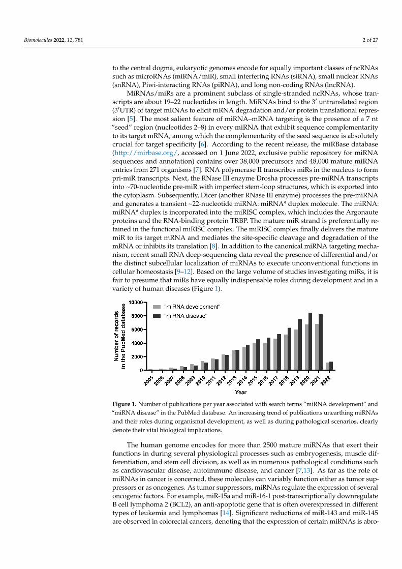
MiRNAs/miRs are a prominent subclass of single-stranded ncRNAs, whose tran-scripts are about 19–22 nucleotides in length. MiRNAs bind to the 3′ untranslated region(3′UTR) of target mRNAs to elicit mRNA degradation and/or protein translational repres-sion [5]. The most salient feature of miRNA–mRNA targeting is the presence of a 7 nt“seed” region (nucleotides 2–8) in every miRNA that exhibit sequence complementarityto its target mRNA, among which the complementarity of the seed sequence is absolutelycrucial for target specificity [6]. According to the recent release, the miRBase database(http://mirbase.org/, accessed on 1 June 2022, exclusive public repository for miRNAsequences and annotation) contains over 38,000 precursors and 48,000 mature miRNAentries from 271 organisms [7]. RNA polymerase II transcribes miRs in the nucleus to formpri-miR transcripts. Next, the RNase III enzyme Drosha processes pre-miRNA transcriptsinto ~70-nucleotide pre-miR with imperfect stem-loop structures, which is exported intothe cytoplasm. Subsequently, Dicer (another RNase III enzyme) processes the pre-miRNAand generates a transient ~22-nucleotide miRNA: miRNA* duplex molecule. The miRNA:miRNA* duplex is incorporated into the miRISC complex, which includes the Argonauteproteins and the RNA-binding protein TRBP. The mature miR strand is preferentially re-tained in the functional miRISC complex. The miRISC complex finally delivers the maturemiR to its target mRNA and mediates the site-specific cleavage and degradation of themRNA or inhibits its translation [8]. In addition to the canonical miRNA targeting mecha-nism, recent small RNA deep-sequencing data reveal the presence of differential and/orthe distinct subcellular localization of miRNAs to execute unconventional functions incellular homeostasis [9–12]. Based on the large volume of studies investigating miRs, it isfair to presume that miRs have equally indispensable roles during development and in avariety of human diseases (Figure 1).
Figure 1. Number of publications per year associated with search terms “miRNA development” and“miRNA disease” in the PubMed database. An increasing trend of publications unearthing miRNAsand their roles during organismal development, as well as during pathological scenarios, clearlydenote their vital biological implications.
The human genome encodes for more than 2500 mature miRNAs that exert theirfunctions in during several physiological processes such as embryogenesis, muscle dif-ferentiation, and stem cell division, as well as in numerous pathological conditions suchas cardiovascular disease, autoimmune disease, and cancer [7,13]. As far as the role ofmiRNAs in cancer is concerned, these molecules can variably function either as tumor sup-pressors or as oncogenes. As tumor suppressors, miRNAs regulate the expression of severaloncogenic factors. For example, miR-15a and miR-16-1 post-transcriptionally downregulateB cell lymphoma 2 (BCL2), an anti-apoptotic gene that is often overexpressed in differenttypes of leukemia and lymphomas [14]. Significant reductions of miR-143 and miR-145are observed in colorectal cancers, denoting that the expression of certain miRNAs is abro-
Biomolecules 2022, 12, 781 3 of 27
gated during carcinogenesis [15]. As oncogenes, the aberrant activation of certain miRNAscould directly or indirectly lead to cancer progression. The mitogen-activated proteinkinase (MAPK) signaling-mediated overexpression of miR-21 induces tumorigenesis- andmetastasis-associated phenotypes, including cell migration and invasion in lung cancer [16],EGF-induced pancreatic cancer [17], and Her2/neu-overexpressing breast cancer [18]. Mir-21 expression is associated with cancer stem cell properties and is involved in stemnessmaintenance in multiple cancers, including pancreatic ductal adenocarcinoma cells [19–22].In addition, miR-21 contributes to drug resistance in head and neck squamous cell carci-noma [23], breast cancer [24], acute myeloid leukemia [25], ovarian cancer [26], and colonadenocarcinoma [27]. The abundant expression of circulating miRNAs such as miR-629*,miR-660, and miR-141 were detected in the plasma of prostate cancer xenograft modelsystem, leading to the potential use of circulating miRNAs as blood-based biomarkers ofhuman cancer detection [28]. Furthermore, miRNA-based clinical therapy has made signifi-cant progress over the past decade. MiR-16-based nanoparticle-encapsulated microRNAmimics have been employed in phase I trials in recurrent malignant pleural mesotheliomapatients [29]. Another prospective phase II clinical trial encompassing the stratificationof chemo-refractory metastatic colorectal cancer patients with low miR-31-3p positivelycorrelated with clinical benefits from anti-EGFR monoclonal antibodies [30]. Hence, inthis special issue focusing on “MicroRNAs—Small Molecules with Great Potential in Tu-morigenesis”, we intend to focus on a specific family of miRNAs that is indispensable foranimal development and its deregulation among the pathogenesis of various diseases: themiR-200 family.
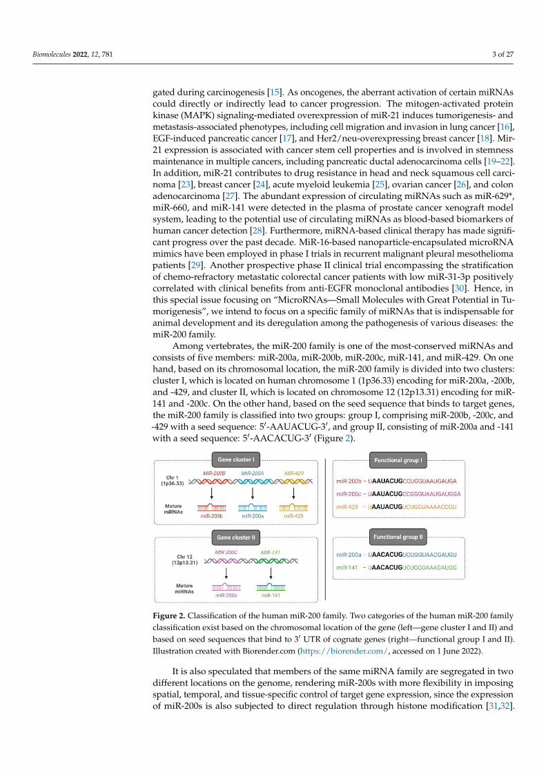
Among vertebrates, the miR-200 family is one of the most-conserved miRNAs andconsists of five members: miR-200a, miR-200b, miR-200c, miR-141, and miR-429. On onehand, based on its chromosomal location, the miR-200 family is divided into two clusters:cluster I, which is located on human chromosome 1 (1p36.33) encoding for miR-200a, -200b,and -429, and cluster II, which is located on chromosome 12 (12p13.31) encoding for miR-141 and -200c. On the other hand, based on the seed sequence that binds to target genes,the miR-200 family is classified into two groups: group I, comprising miR-200b, -200c, and-429 with a seed sequence: 5′-AAUACUG-3′, and group II, consisting of miR-200a and -141with a seed sequence: 5′-AACACUG-3′ (Figure 2).
Figure 2. Classification of the human miR-200 family. Two categories of the human miR-200 familyclassification exist based on the chromosomal location of the gene (left—gene cluster I and II) andbased on seed sequences that bind to 3′ UTR of cognate genes (right—functional group I and II).Illustration created with Biorender.com (https://biorender.com/, accessed on 1 June 2022).
It is also speculated that members of the same miRNA family are segregated in twodifferent locations on the genome, rendering miR-200s with more flexibility in imposingspatial, temporal, and tissue-specific control of target gene expression, since the expressionof miR-200s is also subjected to direct regulation through histone modification [31,32].
Biomolecules 2022, 12, 781 4 of 27
Nevertheless, several exogenous stimuli and signaling cascades activate miR-200 membersto mediate key cellular functions, which we will elaborate upon in the coming sections.Similarly, the deregulation of miR-200s is frequently observed in multiple pathologicalconditions such as tissue fibrosis, neurodegenerative diseases, and cancer, which arediscussed in the later part of the article.
2. miR-200 Family in Development
The formation and establishment of two distinct layers of tissue, epithelium andmesenchyme, demarcate the basis of organ development and coordinated multicellular-ity in metazoans. The epithelium is characterized by a layer of cells adjacently linkedthrough interconnecting cellular structures such as adherens junctions, tight junctions,desmosomes, and gap junctions. The obligate roles of each of these structures in epithelialcellular organization, adhesion, and selective permeability are extensively discussed inrecent reviews [33–36]. In addition, epithelial cells display characteristic apical–basolateralcellular polarity (the apical pole facing the outer lumen and the basal pole facing thebasement extracellular matrix), which is crucial for the asymmetric localization of proteinsat distinct membrane domains and the orientation of microtubule networks during intra-cellular trafficking [37,38]. During early embryonic events such as embryonic implantation,gastrulation, and neural crest formation, a subset of epithelial cells from the epitheliallayer of the embryo systematically loses its epithelial features through a process calledepithelial-to-mesenchymal transition (EMT), thereby acquiring a mesenchymal phenotype.Unlike epithelial cells, mesenchymal cells show weakened cell–cell adhesion properties andlack apical–basal cellular polarity, resulting in cells with enhanced migratory behavior aswell as the potential to degrade the underlying extracellular matrix. The salient bifurcationof these two contrasting cellular types during early embryogenesis is vital for successivevertebral column development and organogenesis [39]. Recently, studies focusing on therole of miRNAs during early embryogenesis have identified that the miR-200 family playsessential roles in the establishment and functioning of the epithelial phenotype duringearly embryogenesis and organogenesis that are detailed below, and a summary of specificmiR-200 family members, their target genes, and the regulated function is listed in Table 1.
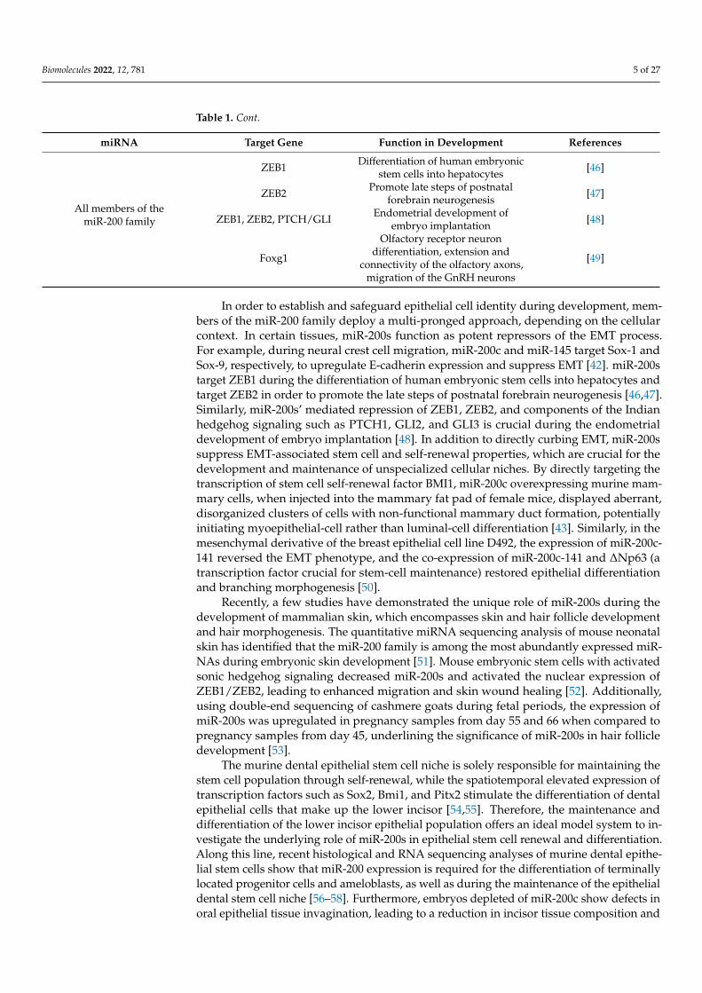
Table 1. miR-200s, target genes, and their roles in development.
miRNA Target Gene Function in Development References
miR-200a/-200b TET3
Olfactory-mediated behaviors andglobose basal cell proliferation
and differentiation in the mousemain olfactory epithelium (MOE)
[40]
miR-200a/-200b/-429 Sox2 Taste bud formation [41]
miR-200c
Sox-1 Neural crest cell migration [42]
BMI Regulating self-renewal anddifferentiation of stem cells [43]
VldlrFGFR-mediated epithelial end bud
proliferation during branchingmorphogenesis
[44]
miR-141/-200c noggin Epithelial cell differentiation andtooth development [45]
Biomolecules 2022, 12, 781 5 of 27
Table 1. Cont.
miRNA Target Gene Function in Development References
All members of themiR-200 family
ZEB1 Differentiation of human embryonicstem cells into hepatocytes [46]
ZEB2 Promote late steps of postnatalforebrain neurogenesis [47]
ZEB1, ZEB2, PTCH/GLI Endometrial development ofembryo implantation [48]
Foxg1
Olfactory receptor neurondifferentiation, extension and
connectivity of the olfactory axons,migration of the GnRH neurons
[49]
In order to establish and safeguard epithelial cell identity during development, mem-bers of the miR-200 family deploy a multi-pronged approach, depending on the cellularcontext. In certain tissues, miR-200s function as potent repressors of the EMT process.For example, during neural crest cell migration, miR-200c and miR-145 target Sox-1 andSox-9, respectively, to upregulate E-cadherin expression and suppress EMT [42]. miR-200starget ZEB1 during the differentiation of human embryonic stem cells into hepatocytes andtarget ZEB2 in order to promote the late steps of postnatal forebrain neurogenesis [46,47].Similarly, miR-200s’ mediated repression of ZEB1, ZEB2, and components of the Indianhedgehog signaling such as PTCH1, GLI2, and GLI3 is crucial during the endometrialdevelopment of embryo implantation [48]. In addition to directly curbing EMT, miR-200ssuppress EMT-associated stem cell and self-renewal properties, which are crucial for thedevelopment and maintenance of unspecialized cellular niches. By directly targeting thetranscription of stem cell self-renewal factor BMI1, miR-200c overexpressing murine mam-mary cells, when injected into the mammary fat pad of female mice, displayed aberrant,disorganized clusters of cells with non-functional mammary duct formation, potentiallyinitiating myoepithelial-cell rather than luminal-cell differentiation [43]. Similarly, in themesenchymal derivative of the breast epithelial cell line D492, the expression of miR-200c-141 reversed the EMT phenotype, and the co-expression of miR-200c-141 and ∆Np63 (atranscription factor crucial for stem-cell maintenance) restored epithelial differentiationand branching morphogenesis [50].
Recently, a few studies have demonstrated the unique role of miR-200s during thedevelopment of mammalian skin, which encompasses skin and hair follicle developmentand hair morphogenesis. The quantitative miRNA sequencing analysis of mouse neonatalskin has identified that the miR-200 family is among the most abundantly expressed miR-NAs during embryonic skin development [51]. Mouse embryonic stem cells with activatedsonic hedgehog signaling decreased miR-200s and activated the nuclear expression ofZEB1/ZEB2, leading to enhanced migration and skin wound healing [52]. Additionally,using double-end sequencing of cashmere goats during fetal periods, the expression ofmiR-200s was upregulated in pregnancy samples from day 55 and 66 when compared topregnancy samples from day 45, underlining the significance of miR-200s in hair follicledevelopment [53].
The murine dental epithelial stem cell niche is solely responsible for maintaining thestem cell population through self-renewal, while the spatiotemporal elevated expression oftranscription factors such as Sox2, Bmi1, and Pitx2 stimulate the differentiation of dentalepithelial cells that make up the lower incisor [54,55]. Therefore, the maintenance anddifferentiation of the lower incisor epithelial population offers an ideal model system to in-vestigate the underlying role of miR-200s in epithelial stem cell renewal and differentiation.Along this line, recent histological and RNA sequencing analyses of murine dental epithe-lial stem cells show that miR-200 expression is required for the differentiation of terminallylocated progenitor cells and ameloblasts, as well as during the maintenance of the epithelialdental stem cell niche [56–58]. Furthermore, embryos depleted of miR-200c show defects inoral epithelial tissue invagination, leading to a reduction in incisor tissue composition and
Biomolecules 2022, 12, 781 6 of 27
length. Using a conditional knockout system of dental stem cells and ameloblasts (cellsthat generate enamel), Cao et al. have shown that Pitx2 directly activates the expression ofthe miR-200c/141 cluster and miR-203, which subsequently inhibits noggin expression andleads to increased BMP signaling activity, leading to epithelial cell differentiation [45]. Inaddition, another independent investigation has shown that elevated BMP signaling duringthe early stages of mouse somatic cell reprogramming induces the expression of miR-205and miR-200 family members, to reinforce an MET phenotype. This reveals the presenceof miR-200-mediated epithelial differentiation as being vital during the initiation phaseof reprogramming [59]. Therefore, these findings illustrate that BMP signaling-mediatedmiR-200 expression is crucial during the maintenance of dental epithelial stem cells, as wellas for the differentiation of progenitor cells.
While the significance of the miR-200 family during EMT suppression was initiallyrecognized in cancers, investigations along the same period have shown that the miR-200family indeed plays distinct roles during embryogenesis and development, which is notconfined just to the establishment and maintenance of the epithelial phenotype. In this sec-tion, we elaborate upon the role of the miR-200 family in the neurosensory epithelium suchas olfactory neurogenesis and taste sensory organs. In mammals, the vomeronasal organ(VNO) and the main olfactory epithelium (MOE) are made of pseudostratified epithelialcells and bipolar sensory neurons, which are essential for the detection of pheromones andvolatile odorants, respectively [60]. In situ hybridization experiments in mouse embryonicMOE showed strong expressions of miR-200a, -b, and -429, detected as early as E9.5 (firstdistinguishable stage of olfactory development) and maintained stable expression untilE13.5 [61]. Moreover, the expression of all miR-200 members was observed in immatureand mature neuronal cell layers of MOE and VNO, highlighting the persistent role ofmiR-200s in adult MOE and VNO neurogenesis. Intriguingly, the intranasal delivery ofCRISPR-Cas9-mediated miR-200b/a knockdown in the MOE caused a significant loss ofdifferentiated olfactory sensory neurons, accompanied by a dramatic reduction in olfactory-mediated male–male aggressive behavior and male–female mating behavior, which ismechanistically mediated through miR-200/TET3/REST signaling [40]. Mutant mice nullfor Dlx5, a homeogene that controls olfactory receptor neuron differentiation, showed areduced expression of miR-9 and miR-200s, and the subsequent knockdown of miR-9 andmiR-200s in zebrafish embryos led to defective olfactory placode organization as well asthe altered differentiation and migration of olfactory receptor neurons [49]. In addition toregulating olfactory epithelial differentiation, miR-200s are implicated in the developmentof other essential neurosensory organs. Zebrafish embryos carrying morpholinos (anti-sense oligonucleotide analogs that are used for generating knockdown embryos) targetedagainst miR-200 expression resulted in a reduction in taste bud cells, and the upstreamactivation of FGF and Notch signaling is essential for miR-200 activity [41]. Similarly, thecharacterization of miRNAs in the developing submandibular gland revealed that miR-200c specifically regulates FGFR-dependent epithelial end bud proliferation and branchingmorphogenesis [44]. Based on the abovementioned investigations, the function of miR-200sis evidently essential for the development and functioning of selected sensory organs.
Furthermore, miR-8, the insect homolog of miR-200, is regarded as a pleiotropicregulator of Drosophila development, including neuroepithelial expansion [62], steroidsignaling-mediated body size regulation [63,64], and pigmentation patterning [65]. miR-8activity is also essential during mosquito oogenesis, such as the proper secretion of yolkprotein precursors like vitellogenin and lipophorin in developing oocytes, through theWingless signaling pathway [66].
3. miR-200 Family in Pathophysiology
The aberrant expression of the miR-200 family is involved in a group of pathophysio-logic conditions such as cancer, diabetes, asthma, autoimmune diseases, kidney diseases,and neurodegenerative diseases. In this section, we would like to elaborate upon the roleof miR-200s on the pathophysiology of neurogenerative diseases and fibrosis.
Biomolecules 2022, 12, 781 7 of 27
3.1. miR-200 Family in Neurodegenerative Diseases
In general, disorders characterized by the progressive degeneration of the structure orfunction of the central nervous system (CNS) are collectively referred as neurodegenerativediseases. Representative neurodegenerative diseases include Alzheimer’s disease (AD),Parkinson’s disease (PD), amyotrophic lateral sclerosis (ALS), Huntington’s disease (HD),multiple sclerosis, and prion diseases. Consequently, they cause significant defects inmotor and cognitive ability [67]. An eminent study of the miR-8 mutants has shown thatthese mutants displayed high levels of apoptosis in the brain, behavioral defects, and animpaired neuromuscular coordination phenotype in Drosophila legs, which prompted thepotential role of miR-200s in neurodegenerative disorders in mammals [68]. Recently, anumber of studies have revealed that members of the miR-200 family are differentiallyexpressed in the human brain and, more importantly, modulate genes associated withspecific neurodegenerative disorders (Table 2).
Table 2. miR-200s, target genes and their roles in neurodegenerative diseases.
Disease miRNA Target Gene Function in Neurodegenerative Diseases References
Alzheimer’sdisease
miR-141SIRT1 Promote Aβ-induced neuronal apoptosis [69]
miR-200a [70]
miR-200b/-429 APPHigh expression of APP correlating with
accelerated accumulation of the Aβ in brainand take part in the progression of AD
[71,72]
miR-200b/c S6K1Reduction in Aβ secretion and/or Aβ-induced
spatial memory impairment by promotingactivation of the insulin signaling pathway
[73]
Parkinson’sdisease
miR-200aSIRT
Involved in DA neurons cell death via P53 andFOXO signaling pathways as a possible reason
for PD pathogenesis[74]
miR-141 Induce neuronal apoptosis and oxidative stress [75]
Amyotrophic lateralsclerosis
miR-141 FUS, EWS,TAF15 Involved in the differentiation of neuronal cells [76]
miR-200c FUS Promote miR-200c-mediated gene silencing [77]
3.1.1. miR-200 Family in Alzheimer’s Disease
Alzheimer’s disease (AD) is a progressive neurological disorder, characterized by anincreasing level of memory loss and deterioration of cognitive functions that eventuallylead to dementia [78]. In particular, the pathological landscapes of AD brains are wellrecognized by the deposition of beta amyloid peptide (Aβ) and the formation of intracellularneurofibrillary tangles (NFT) [79], and ER stress is believed to be the initial driver forneuronal cell loss in AD [80]. Several studies have demonstrated that members of the miR-200 family are found to be linked with AD progression. The overexpression of miR-200ain peripheral blood mononuclear cells from AD patients targets genes related to cell cycleand DNA repair [81]. Another report showed that the upregulation of miR-200a-3p targetsSIRT1, an anti-apoptotic protein, in order to stimulate Aβ-induced neuronal apoptosis [70].Similarly, miR-200c is overexpressed in the plasma of patients with moderate to severeforms of AD, indicating its association with the progression of the disease [78].
In turn, the decreased expression of miR-200b and -429 by Aβ42 leads to the expressionof the amyloid precursor protein (APP). Increased APP levels, in turn, lead to an increasein Aβ42, which causes a further reduction of miR-200b. This generates a kind of viciouscycle that contributes to the progression of AD [71]. Furthermore, folic acid deficiencycan also decrease the level of miR-200b. Thus, a folic acid-deficient diet stimulates APPoverexpression and promotes Aβ generation [72,82]. Higaki et al. report that miR-200b/creduces insulin resistance by targeting the ribosomal protein kinase S6 B1 [73]. Thiscontributes to a reduction in the production of Aβ peptide and thus alleviates the Aβ-induced toxicity. On the contrary, the upregulation of miR-200c levels was observed in theserum of AD patients [78], denoting that the precise role of miR-200s in Aβ-induced ERstress remains to be clearly investigated. These findings highlight that the miR-200 family
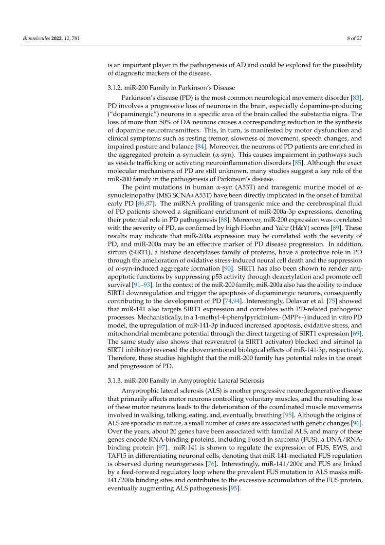
Biomolecules 2022, 12, 781 8 of 27
is an important player in the pathogenesis of AD and could be explored for the possibilityof diagnostic markers of the disease.
3.1.2. miR-200 Family in Parkinson’s Disease
Parkinson’s disease (PD) is the most common neurological movement disorder [83].PD involves a progressive loss of neurons in the brain, especially dopamine-producing(“dopaminergic”) neurons in a specific area of the brain called the substantia nigra. Theloss of more than 50% of DA neurons causes a corresponding reduction in the synthesisof dopamine neurotransmitters. This, in turn, is manifested by motor dysfunction andclinical symptoms such as resting tremor, slowness of movement, speech changes, andimpaired posture and balance [84]. Moreover, the neurons of PD patients are enriched inthe aggregated protein α-synuclein (α-syn). This causes impairment in pathways suchas vesicle trafficking or activating neuroinflammation disorders [85]. Although the exactmolecular mechanisms of PD are still unknown, many studies suggest a key role of themiR-200 family in the pathogenesis of Parkinson’s disease.
The point mutations in human α-syn (A53T) and transgenic murine model of α-synucleinopathy (M83 SCNA∗A53T) have been directly implicated in the onset of familialearly PD [86,87]. The miRNA profiling of transgenic mice and the cerebrospinal fluidof PD patients showed a significant enrichment of miR-200a-3p expressions, denotingtheir potential role in PD pathogenesis [88]. Moreover, miR-200 expression was correlatedwith the severity of PD, as confirmed by high Hoehn and Yahr (H&Y) scores [89]. Theseresults may indicate that miR-200a expression may be correlated with the severity ofPD, and miR-200a may be an effective marker of PD disease progression. In addition,sirtuin (SIRT1), a histone deacetylases family of proteins, have a protective role in PDthrough the amelioration of oxidative stress-induced neural cell death and the suppressionof α-syn-induced aggregate formation [90]. SIRT1 has also been shown to render anti-apoptotic functions by suppressing p53 activity through deacetylation and promote cellsurvival [91–93]. In the context of the miR-200 family, miR-200a also has the ability to induceSIRT1 downregulation and trigger the apoptosis of dopaminergic neurons, consequentlycontributing to the development of PD [74,94]. Interestingly, Delavar et al. [75] showedthat miR-141 also targets SIRT1 expression and correlates with PD-related pathogenicprocesses. Mechanistically, in a 1-methyl-4-phenylpyridinium- (MPP+-) induced in vitro PDmodel, the upregulation of miR-141-3p induced increased apoptosis, oxidative stress, andmitochondrial membrane potential through the direct targeting of SIRT1 expression [69].The same study also shows that resveratrol (a SIRT1 activator) blocked and sirtinol (aSIRT1 inhibitor) reversed the abovementioned biological effects of miR-141-3p, respectively.Therefore, these studies highlight that the miR-200 family has potential roles in the onsetand progression of PD.
3.1.3. miR-200 Family in Amyotrophic Lateral Sclerosis
Amyotrophic lateral sclerosis (ALS) is another progressive neurodegenerative diseasethat primarily affects motor neurons controlling voluntary muscles, and the resulting lossof these motor neurons leads to the deterioration of the coordinated muscle movementsinvolved in walking, talking, eating, and, eventually, breathing [95]. Although the origins ofALS are sporadic in nature, a small number of cases are associated with genetic changes [96].Over the years, about 20 genes have been associated with familial ALS, and many of thesegenes encode RNA-binding proteins, including Fused in sarcoma (FUS), a DNA/RNA-binding protein [97]. miR-141 is shown to regulate the expression of FUS, EWS, andTAF15 in differentiating neuronal cells, denoting that miR-141-mediated FUS regulationis observed during neurogenesis [76]. Interestingly, miR-141/200a and FUS are linkedby a feed-forward regulatory loop where the prevalent FUS mutation in ALS masks miR-141/200a binding sites and contributes to the excessive accumulation of the FUS protein,eventually augmenting ALS pathogenesis [95].
Biomolecules 2022, 12, 781 9 of 27
In addition, miR-200c is directly regulated by FUS, which contributes to gene silencing.In turn, mutations in FUS reduce the silencing of genes targeted by miR-200c, which may beone of mechanisms involved in the development of ALS [77]. Additionally, Zhou et al. [98]found that the expression level of miR-200a was increased in the early stage and decreasedin the later stage in ALS transgenic mice, indicating this microRNA as a potential markerfor detecting the progression of ALS.
3.1.4. miR-200 Family in Multiple Sclerosis and Prion Disease
Multiple sclerosis (MS) is the most common demyelinating disease of the CNS. It isconsidered an autoimmune disease in which the body’s immune system attacks its owntissues [99]. The epidemiology of multiple sclerosis in developing countries shows thatthere has been a sharp increase in the incidence of patients, with an overall incidence of85.8 per 100,000 [100]. The cause of MS is unknown, but it appears that a combinationof environmental factors, epigenetics, and genetics lead to ongoing immune attacks onthe CNS [101]. Two preliminary assessments on MS patient samples showed that miR-141 and -200a levels were increased in the relapsing phase of MS patients compared tothe remission and control groups [102,103]. In addition, elevated miR levels induce thedifferentiation of Th17 cells that are involved in the development of MS [102]. Prion diseaseis a rare, fatal, neurodegenerative disease caused by abnormally folded prion proteins inbrain (PrP) [104]. Most cases of prion disease in humans arise spontaneously and theirsigns and symptoms typically begin in adulthood and worsen with time [105]. A pilotstudy has potentially revealed a correlation between prion disease and miR-200 familyexpression, where decreased levels of all members of the miR-200 family correlated withmorphological changes of dendritic spines and synaptic dysfunction [106]. Although thedifferential expressions of the miR-200 family members are beginning to be documented insuch neurodegenerative conditions, specific roles of these miRNAs in their etiology remainlargely unknown.
3.2. miR-200 Family in Fibrosis
Fibrosis is one of the major pathological processes that affect vital organs such asthe kidneys, liver, lungs, and intestines. It is characterized by impaired epithelial archi-tecture and excessive deposition of extracellular matrix and fibrous connective tissue,which generates multiple inflamed scar tissues within the organ, eventually leading toorgan dysfunction and failure. Furthermore, patients diagnosed with cystic fibrosis poseincreased risk towards cancer progression, denoting that fibrosis might serve as a gate-way to life-threatening illnesses [107]. At the molecular level, several studies have vali-dated the activation of EMT during the early stages of fibrosis and is referred to as type 2EMT [108,109]. Accordingly, with EMT-inhibiting roles, miR-200s have been also implicatedduring tissue fibrosis.
Using a unilateral ureter obstruction model, Oba et al. have shown that the injection of0.5 nM of pre-miR-200b (precursor) efficiently inhibited the rise of collagen and fibronectinlevels in obstructed kidneys, and could ameliorate renal tubulointerstitial fibrosis [110].Similarly, the collection duct-specific inhibition of miR-200 activity in a transgenic mousemodel evoked the expression of profibrotic target genes and inflammatory cytokines suchas Mcp1, Il6, and Cxcl2 [111]. Furthermore, the downregulation of miR-200 members inrenal fibrosis is primarily mediated through TGF-β1, and accordingly, the overexpressionof miR-141 or -200b hindered Smad-dependent TGF-β signaling and downstream EMTphenotypes [112–114].
Levels of miR-200a and miR-200c were also significantly downregulated in murinelungs in a bleomycin-mediated fibrosis model as well as in patients with idiopathic pul-monary fibrosis (IPF), and the restoration of miR-200 expression in senescent IPF cellsresumed normal regenerative functions [115–117]. In the case of liver fibrosis, miR-200sseem to play a pro-fibrotic role. In a CCL4-induced liver fibrosis model, miR-199 andmiR-200 levels were significantly upregulated in comparison to the control groups [118].
Biomolecules 2022, 12, 781 10 of 27
Accordingly, the expression of miR-200s were elevated in serums of patients diagnosedwith non-alcoholic fatty liver disease (NAFLD), and NAFLD mice treated with miR-200inhibitor ameliorated liver fibrosis. Mechanistically, GRHL2 was overexpressed in theserum of NAFLD patients, which in turn negatively targeted SIRT1 and also activated miR-200 and the MAPK signaling pathway, aggravating liver fibrosis and intestinal mucosalbarrier dysfunction [119]. GRHL2 is regarded as a gatekeeper of epithelial phenotype anddifferentiation, also involved in the direct activation of the miR-200 family [120].
3.3. miR-200 Family in Cancer
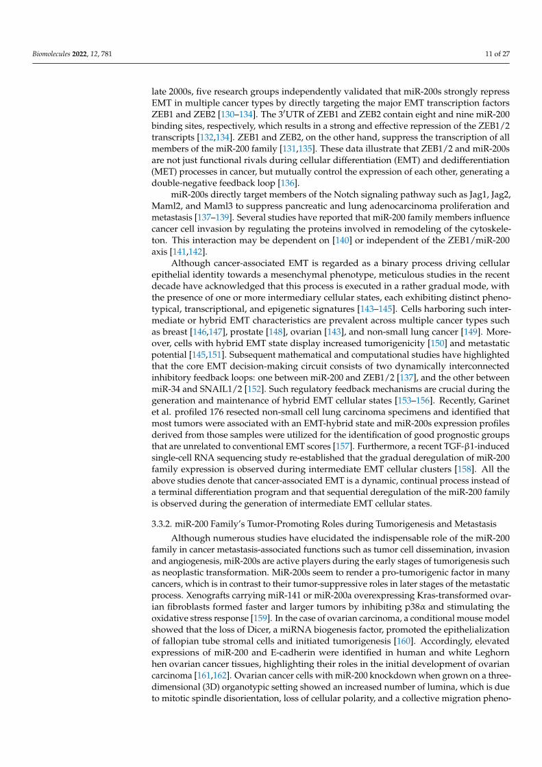
Over the past two decades, the role of miR-200s in cancer has been extensively studied,which has led us to distinguish the established (well-explored) and emerging functions thatare exclusively observed during cancer progression (Figure 3).
Figure 3. Established and novel functions of the miR-200 family in human cancers. Although certainfunctions of miR-200s such as EMT suppression are quite well understood, members of the miR-200family have recently been studied in detail for their novel roles such as RNA editing and reversalof chemoresistance. Illustration created with Biorender.com (https://biorender.com/, accessed on1 June 2022).
The dysregulation of miR-200 expression in various entities of human cancer (reviewedin [121]) assigns it with tumor-promoting as well as tumor-suppressive effects. Throughthe direct targeting of genes that are involved in tumor-promoting mechanisms, membersof the miR-200 family, individually or collectively, have been reported to be involvedin the regulation of EMT/MET and metastasis [122,123], in the regulation of the cellcycle and apoptosis [74,124], as well as its deregulation during chemoresistance [125],cancer stemness [43,126], and, more recently, also in modulating intra-tumoral immune cellfunction [127–129]
3.3.1. miR-200 Family in Cancer-Associated EMT and MET
In the metastatic cascade, miR-200 exerts a context-dependent role. In general, theexpression of miR-200 family members in the primary tumor strengthens an epithelialphenotype, thereby preventing EMT. Members of the miR-200 family primarily block EMTthrough direct repression of the EMT inducers ZEB1 and ZEB2, and impede tumor celldissociation, migration, and invasion, which potentially combats the initial stages of themetastatic cascade, thus implicating the miRNAs’ tumor-suppressive effect. During the
Biomolecules 2022, 12, 781 11 of 27
late 2000s, five research groups independently validated that miR-200s strongly repressEMT in multiple cancer types by directly targeting the major EMT transcription factorsZEB1 and ZEB2 [130–134]. The 3′UTR of ZEB1 and ZEB2 contain eight and nine miR-200binding sites, respectively, which results in a strong and effective repression of the ZEB1/2transcripts [132,134]. ZEB1 and ZEB2, on the other hand, suppress the transcription of allmembers of the miR-200 family [131,135]. These data illustrate that ZEB1/2 and miR-200sare not just functional rivals during cellular differentiation (EMT) and dedifferentiation(MET) processes in cancer, but mutually control the expression of each other, generating adouble-negative feedback loop [136].
miR-200s directly target members of the Notch signaling pathway such as Jag1, Jag2,Maml2, and Maml3 to suppress pancreatic and lung adenocarcinoma proliferation andmetastasis [137–139]. Several studies have reported that miR-200 family members influencecancer cell invasion by regulating the proteins involved in remodeling of the cytoskele-ton. This interaction may be dependent on [140] or independent of the ZEB1/miR-200axis [141,142].
Although cancer-associated EMT is regarded as a binary process driving cellularepithelial identity towards a mesenchymal phenotype, meticulous studies in the recentdecade have acknowledged that this process is executed in a rather gradual mode, withthe presence of one or more intermediary cellular states, each exhibiting distinct pheno-typical, transcriptional, and epigenetic signatures [143–145]. Cells harboring such inter-mediate or hybrid EMT characteristics are prevalent across multiple cancer types suchas breast [146,147], prostate [148], ovarian [143], and non-small lung cancer [149]. More-over, cells with hybrid EMT state display increased tumorigenicity [150] and metastaticpotential [145,151]. Subsequent mathematical and computational studies have highlightedthat the core EMT decision-making circuit consists of two dynamically interconnectedinhibitory feedback loops: one between miR-200 and ZEB1/2 [137], and the other betweenmiR-34 and SNAIL1/2 [152]. Such regulatory feedback mechanisms are crucial during thegeneration and maintenance of hybrid EMT cellular states [153–156]. Recently, Garinetet al. profiled 176 resected non-small cell lung carcinoma specimens and identified thatmost tumors were associated with an EMT-hybrid state and miR-200s expression profilesderived from those samples were utilized for the identification of good prognostic groupsthat are unrelated to conventional EMT scores [157]. Furthermore, a recent TGF-β1-inducedsingle-cell RNA sequencing study re-established that the gradual deregulation of miR-200family expression is observed during intermediate EMT cellular clusters [158]. All theabove studies denote that cancer-associated EMT is a dynamic, continual process instead ofa terminal differentiation program and that sequential deregulation of the miR-200 familyis observed during the generation of intermediate EMT cellular states.
3.3.2. miR-200 Family’s Tumor-Promoting Roles during Tumorigenesis and Metastasis
Although numerous studies have elucidated the indispensable role of the miR-200family in cancer metastasis-associated functions such as tumor cell dissemination, invasionand angiogenesis, miR-200s are active players during the early stages of tumorigenesis suchas neoplastic transformation. MiR-200s seem to render a pro-tumorigenic factor in manycancers, which is in contrast to their tumor-suppressive roles in later stages of the metastaticprocess. Xenografts carrying miR-141 or miR-200a overexpressing Kras-transformed ovar-ian fibroblasts formed faster and larger tumors by inhibiting p38α and stimulating theoxidative stress response [159]. In the case of ovarian carcinoma, a conditional mouse modelshowed that the loss of Dicer, a miRNA biogenesis factor, promoted the epithelializationof fallopian tube stromal cells and initiated tumorigenesis [160]. Accordingly, elevatedexpressions of miR-200 and E-cadherin were identified in human and white Leghornhen ovarian cancer tissues, highlighting their roles in the initial development of ovariancarcinoma [161,162]. Ovarian cancer cells with miR-200 knockdown when grown on a three-dimensional (3D) organotypic setting showed an increased number of lumina, which is dueto mitotic spindle disorientation, loss of cellular polarity, and a collective migration pheno-
Biomolecules 2022, 12, 781 12 of 27
type, facilitating ovarian carcinogenesis [163]. At the molecular level, the loss of collectivemigration in these 3D structures is due to the disruption of ROCK-mediated myosin-IIphosphorylation and SRC signaling. The increased expression of miR-200 in early-stagelung adenocarcinomas in vitro and in vivo activated PI3K/AKT signaling through FOG2targeting and promoted tumor-initiating spheroid growth formation [164]. Early ductalcarcinomas in situ (DCIS) lesions of the breast tissue are non-obligatory precursors ofinvasive breast cancer, yet their severe microenvironment with hypoxia, nutrient depri-vation, and acidosis facilitate tumorigenesis [165,166]. Subtype-specific miRNA profilingof normal, DCIS, and invasive breast carcinoma cohorts showed significant enrichmentof miR-21-5p and all members of the miR-200 family in the DCIS samples, denoting theirpotential tumorigenic role in breast carcinogenesis [167].
In the context of cancer progression and metastasis, it is important to note that miR-200s support an MET of mesenchymal tumor cells at distant organ sites, thereby promotingmetastatic colonization. In breast cancer, miR-200s promote the metastatic colonizationof secondary organs by targeting the cancer cell secretome, subsequently influencing thetumor microenvironment [168,169]. In addition, miR-200c, miR-192, and miR-17 wereidentified as targeting genes involved in extracellular matrix remodeling. The expressionof the three miRNAs in tumor-associated fibroblasts may therefore suppress the invasionof colorectal cancer cells [170]. Further studies on miR-200 in the tumor microenvironmentrecently revealed that apoptotic MCF7 breast cancer cells release miR-200c, which is takenup by tumor-associated macrophages. This leads to the overexpression of miR-200c inthe tumor-associated macrophages, followed by the reduced expression of a set of pro-migratory miR-200c targets and a reduced capacity of the macrophages to infiltrate intotumor spheroids [129]. Taken together, these findings show that miR-200 is a crucial playerin the crosstalk between tumor cells and tumor-associated stromal and immune cells. Thiscrosstalk may modulate the cells’ invasive behavior with tumor-promoting or suppressingeffects, depending on the releasing and receiving cell. This points to the fact that the role ofmiR-200s in tumor progression extends beyond the regulation of transition between EMTand MET and the ZEB/miR-200 feedback loop.
3.3.3. miR200 Family in Radiotherapy
Radiotherapy still remains one of the main front-line treatment options in several can-cer types including breast, lung, prostate, cervical, and head and neck cancers. The responseof cancer cells towards radiotherapy is mostly affected by the tumor size. However, recentpreclinical studies have highlighted that miRNAs, especially miR-200s, contribute towardsradiotherapy response in multiple cancer types [171]. In breast cancer cells, it has beenshown that high miR-200c expression is important for radiosensitivity, while decreasedexpression is associated with radioresistance [172]. This has been demonstrated in studieswith cell lines showing different basal levels of miR-200c expression. The MCF-7 cell line,which has a high level of miR-200c expression, showed a higher sensitivity to radiother-apy compared to the MDA-MB-231 cell line. Additionally, higher expression of miR-200cincreases the sensitivity to radiation by inhibiting cell proliferation and by increasing apop-tosis and DNA double-strand breaks [173]. Further studies showed that the sensitizationof cancer cells to radiation therapy with high levels of miR-200c is mediated by targetingTBK1 and VEGF-VEGFR2, resulting in cell apoptosis [174]. Moreover, Sun et al. identifiedubiquilin 1 (UBQLN1) as a functional target of miR-200c, where miR-200c sensitizes breastcancer cells to radiation in a manner associated with the inhibition of radiation-inducedautophagy [174]. In turn, Wang et al. speculated that radiation resistance is promotedby the interaction of the long coding RNA, LINC02582, with USP7 to deubiquitinate andstabilize CHK1. Therefore, they proposed that the miR-200c/LINC02582/USP7/CHK1signaling axis could be a therapeutic target to improve the breast cancer response to radio-therapy [175].
In addition, miR-200c plays a role in the effectiveness of radiotherapy in lung cancer.The high expression of miR-200c increases radiation sensitivity by regulating the oxidative
Biomolecules 2022, 12, 781 13 of 27
stress response by targeting PRDX2, GAPB/Nrf2, and SESN1 and by inhibiting the repairof radiation-induced double-strand breaks [176]. Next, miR-200a affects the radiosensitivityof non-small cell lung cancer (NSCLC) cells. The analysis of clinical data in NSCLC patientsshowed that miR-200a negatively regulates HGF expression, resulting in reduced cancercell invasion and metastasis. Moreover, the miR-200a/HGF pathway also influences theradiosensitivity of NSCLC cells. This was confirmed in experiments on cell lines A549 andH1299, where the overexpression of miR-200a after irradiation resulted in the increasedapoptosis and DNA double-stranded breaks of these cancer cells [177].
Interestingly, miR-200a is detected in the saliva of patients with head and neck squa-mous cell carcinoma after radiation treatment. The analysis of the distribution of miR-200aexpression during and after radiotherapy showed that the miR level is significantly higher12 months after treatment compared with the baseline state [178]. It may constitute apotential tool in monitoring the response to radiotherapy in patients with HNSCC. Alongthis line, radiation-induced oral mucositis (RIOM) is one of the more prevalent side effectsof radiotherapy in HNSCC patients. A preliminary study identified that all members of themiR-200 family were significantly upregulated in RIOM formation, and the knockdown ofmiR-200c led to a reduction of proinflammatory cytokine synthesis and reactive oxygenspecies generation [179].
In addition to all of the abovementioned studies, two new reports this year implicatethe association of miR-200s with radiosensitivity in other cancer types. The suppressionof miR-200b/c in esophageal squamous cell carcinoma impaired with cell sensitivity toconcurrent chemoradiotherapy treatment with or without surgery [180]. Similarly, tumorxenografts carrying miR-200a/b/-429 overexpressing cervical carcinoma cell lines weresignificantly sensitized to radiotherapy, which is also independent of tumor hypoxia [181].Therefore, the prospective role of the miR-200 family as a predictor of response to cancerradiotherapy has been revealed in work over the last decade and certainly demands moreresearch.
3.3.4. miR-200 Family in Chemotherapy
Dealing with the emergence of therapy resistance poses a major challenge in thetreatment of cancer patients. Recent literature has reported that miR-200s are involved inmodulating the response of cancer cells to anti-cancer therapy by influencing the statusof certain adenocarcinomas towards chemo- and radiosensitivity. Importantly, therapyresistance occurs through numerous resistance mechanisms such as increased DNA repaircapacity, elevated drug efflux through ATP-binding cassette (ABC) transporters, varyingexpression of β-tubulin isotypes, changes in cell cycle, inhibition of apoptosis, and responseto oxidative stress [182]. The participation of miR-200 in these processes has been welldescribed, thereby miR-200s may enhance the therapeutic possibilities of EMT-associatedcancer metastasis and may even predict therapeutic response [140,183,184]. Crudele et al.reviewed the miR-200 family network’s connection with resistance to six different anti-cancer treatments [185]. In this regard, the restoration of miR-200c expression in ovarian andendometrial cancer cells leads to increased sensitivity to microtubule-binding chemother-apeutic agents through the reduction of class III β-tubulin (TUBB3) [125]. Similarly, inchemoresistant pancreatic cells, the induction of miR-200s and miR-203 expression throughthe class I HDAC inhibitor mocetinostat restored drug sensitivity to standard chemothera-peutics such as gemcitabine and docetaxel [186]. The oxaliplatin-resistant colorectal cancercell line showed increased expression of SUZ12, a Polycomb-repressive complex 2 subunit,and decreased expression of the miR-200 family [187]. In a similar fashion, the expressionof miR-200c alone enhanced the chemosensitivity and reduced the metastatic potentialof p53null claudin-low breast cancer mouse models [188]. The depletion of miR-200csignificantly reduced 5-flurouracil-induced apoptosis and caspase 3 activity in colorectalHCT-116 cells [189], and the inhibition of miR-141/200c in the ovarian cancer cell lineOVCAR-3 led to resistance to paclitaxel and carboplatin [190]. The overexpression ofmiR-200c in melanoma cells decreased resistance to cisplatin as well as a BRAF- and a
Biomolecules 2022, 12, 781 14 of 27
MEK-inhibitor through the downregulation of the ABC transporters ABCG2, ABCG5, andMDR1 directly or indirectly through the downregulation of BMI-1 [183,191]. Intriguingly, arecent pilot study has shown that the anticancer drug cisplatin interacts with pre-miR-200band decreases the production of mature miR-200b expression in ovarian cancer cells [184].Therefore, a direct correlation between miR-200s and a drug sensitivity phenotype hasbeen established primarily using adenocarcinomas; however, the underlying mechanisticinsights are still being delineated.
It is important to mention that there are also reports that observe the opposite ef-fects, and describe miR-200 as conferring chemoresistance. In a study by Yu et al., theexpression of miR-200a correlated with poor response to preoperative chemotherapy andpoor prognosis in breast cancer patients. Additionally, miR-200a was described to conferchemoresistance to gemcitabine by targeting TP53INP1 and YAP1 [192]. TP53INP1 is a p73target gene that inhibits cell growth and promotes cell death, and is induced by stress inresponse to DNA damage, i.e., by cisplatin [193]. In hepatocellular carcinoma, the overex-pression of miR-200a-3p increased resistance to 5-FU by targeting DUSP6. In this setting,the inhibition of miR-200a-3p decreased cell growth and viability after 5-FU treatment,thereby sensitizing Hep3B cells to different anti-cancer drugs, including 5-FU, cisplatin,and doxorubicin [194].
The administration of anticancer drugs or radiotherapy is proposed to lead to anaccumulation of reactive oxygen species (ROS). These ROS interfere with different cellularprocesses such as cell survival and motility, and the oxidative stress influences miRNAexpression patterns. The miR-200 family was described to be involved in the oxidativestress response by regulating KEAP1 expression in breast and ovarian cancer [149,150,179].The introduction of miR-141/-200c into a paclitaxel-resistant ovarian cell line confersresistance to carboplatin, while altering the expression of genes involved in balancingoxidative stress [190].
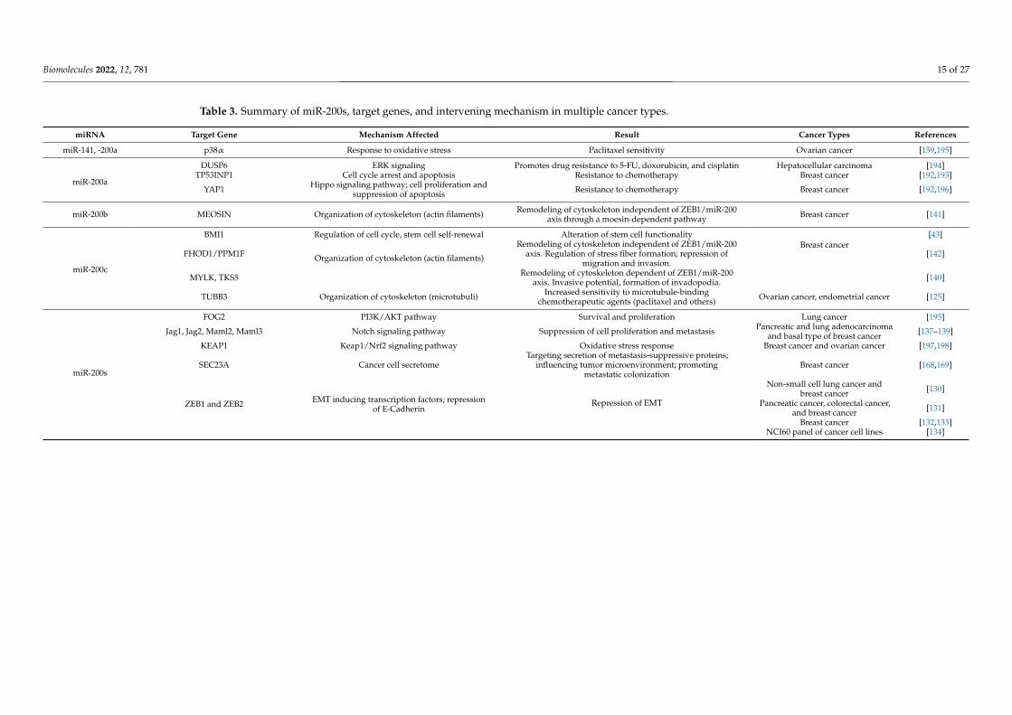
In summary, miR-200 family members target a plethora of genes involved in fatedetermining processes within cancer cells as well as in cells of the tumor microenvironment.A selection of these miR-200 target genes is compiled in Table 3. Seemingly contradictoryeffects may occur, which require a precise analysis of cellular context and spatiotemporalexpression pattern of the miR-200s in order to predict the outcome of a potential therapeuticinterference. The fact that cells may release miRNAs assigns miR-200s a role in inter cellularcommunication and makes them detectable in body fluids for a potential use as biomarkerswhich is discussed in the following section.
Biomolecules 2022, 12, 781 15 of 27
Table 3. Summary of miR-200s, target genes, and intervening mechanism in multiple cancer types.
miRNA Target Gene Mechanism Affected Result Cancer Types References
miR-141, -200a p38α Response to oxidative stress Paclitaxel sensitivity Ovarian cancer [159,195]
miR-200a
DUSP6 ERK signaling Promotes drug resistance to 5-FU, doxorubicin, and cisplatin Hepatocellular carcinoma [194]TP53INP1 Cell cycle arrest and apoptosis Resistance to chemotherapy Breast cancer [192,193]
YAP1 Hippo signaling pathway; cell proliferation andsuppression of apoptosis Resistance to chemotherapy Breast cancer [192,196]
miR-200b MEOSIN Organization of cytoskeleton (actin filaments) Remodeling of cytoskeleton independent of ZEB1/miR-200axis through a moesin-dependent pathway Breast cancer [141]
miR-200c
BMI1 Regulation of cell cycle, stem cell self-renewal Alteration of stem cell functionalityBreast cancer
[43]
FHOD1/PPM1F Organization of cytoskeleton (actin filaments)
Remodeling of cytoskeleton independent of ZEB1/miR-200axis. Regulation of stress fiber formation; repression of
migration and invasion.[142]
MYLK, TKS5 Remodeling of cytoskeleton dependent of ZEB1/miR-200axis. Invasive potential, formation of invadopodia. [140]
TUBB3 Organization of cytoskeleton (microtubuli) Increased sensitivity to microtubule-bindingchemotherapeutic agents (paclitaxel and others) Ovarian cancer, endometrial cancer [125]
miR-200s
FOG2 PI3K/AKT pathway Survival and proliferation Lung cancer [195]
Jag1, Jag2, Maml2, Maml3 Notch signaling pathway Suppression of cell proliferation and metastasis Pancreatic and lung adenocarcinomaand basal type of breast cancer [137–139]
KEAP1 Keap1/Nrf2 signaling pathway Oxidative stress response Breast cancer and ovarian cancer [197,198]
SEC23A Cancer cell secretomeTargeting secretion of metastasis-suppressive proteins;
influencing tumor microenvironment; promotingmetastatic colonization
Breast cancer [168,169]
ZEB1 and ZEB2 EMT inducing transcription factors; repressionof E-Cadherin
Repression of EMT
Non-small cell lung cancer andbreast cancer [130]
Pancreatic cancer, colorectal cancer,and breast cancer [131]
Breast cancer [132,133]NCI60 panel of cancer cell lines [134]
Biomolecules 2022, 12, 781 16 of 27
4. miR-200 Family as Predictive Markers
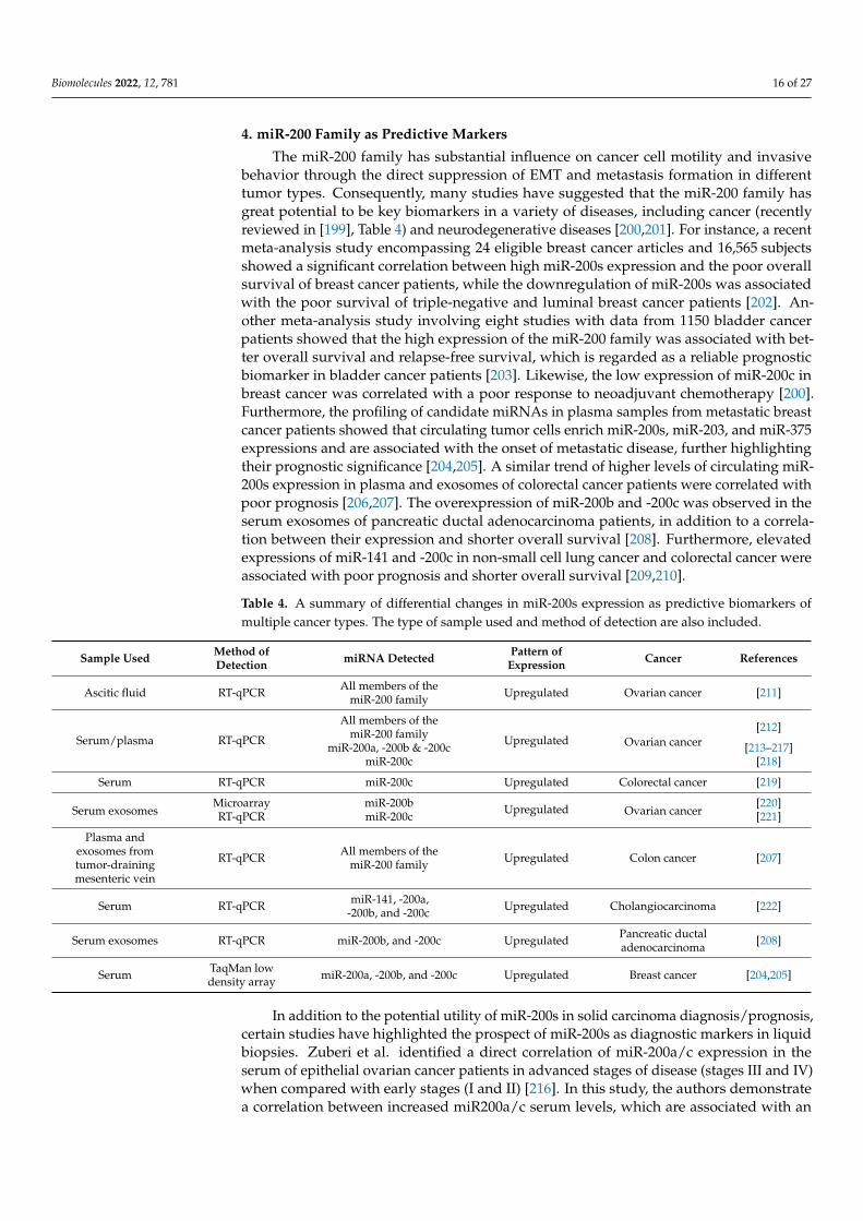
The miR-200 family has substantial influence on cancer cell motility and invasivebehavior through the direct suppression of EMT and metastasis formation in differenttumor types. Consequently, many studies have suggested that the miR-200 family hasgreat potential to be key biomarkers in a variety of diseases, including cancer (recentlyreviewed in [199], Table 4) and neurodegenerative diseases [200,201]. For instance, a recentmeta-analysis study encompassing 24 eligible breast cancer articles and 16,565 subjectsshowed a significant correlation between high miR-200s expression and the poor overallsurvival of breast cancer patients, while the downregulation of miR-200s was associatedwith the poor survival of triple-negative and luminal breast cancer patients [202]. An-other meta-analysis study involving eight studies with data from 1150 bladder cancerpatients showed that the high expression of the miR-200 family was associated with bet-ter overall survival and relapse-free survival, which is regarded as a reliable prognosticbiomarker in bladder cancer patients [203]. Likewise, the low expression of miR-200c inbreast cancer was correlated with a poor response to neoadjuvant chemotherapy [200].Furthermore, the profiling of candidate miRNAs in plasma samples from metastatic breastcancer patients showed that circulating tumor cells enrich miR-200s, miR-203, and miR-375expressions and are associated with the onset of metastatic disease, further highlightingtheir prognostic significance [204,205]. A similar trend of higher levels of circulating miR-200s expression in plasma and exosomes of colorectal cancer patients were correlated withpoor prognosis [206,207]. The overexpression of miR-200b and -200c was observed in theserum exosomes of pancreatic ductal adenocarcinoma patients, in addition to a correla-tion between their expression and shorter overall survival [208]. Furthermore, elevatedexpressions of miR-141 and -200c in non-small cell lung cancer and colorectal cancer wereassociated with poor prognosis and shorter overall survival [209,210].
Table 4. A summary of differential changes in miR-200s expression as predictive biomarkers ofmultiple cancer types. The type of sample used and method of detection are also included.
Sample Used Method ofDetection miRNA Detected Pattern of
Expression Cancer References
Ascitic fluid RT-qPCR All members of themiR-200 family Upregulated Ovarian cancer [211]
Serum/plasma RT-qPCR
All members of themiR-200 family Upregulated Ovarian cancer
[212]
miR-200a, -200b & -200c [213–217]miR-200c [218]
Serum RT-qPCR miR-200c Upregulated Colorectal cancer [219]
Serum exosomesMicroarray miR-200b Upregulated Ovarian cancer
[220]RT-qPCR miR-200c [221]
Plasma andexosomes fromtumor-drainingmesenteric vein
RT-qPCR All members of themiR-200 family Upregulated Colon cancer [207]
Serum RT-qPCR miR-141, -200a,-200b, and -200c Upregulated Cholangiocarcinoma [222]
Serum exosomes RT-qPCR miR-200b, and -200c Upregulated Pancreatic ductaladenocarcinoma [208]
Serum TaqMan lowdensity array miR-200a, -200b, and -200c Upregulated Breast cancer [204,205]
In addition to the potential utility of miR-200s in solid carcinoma diagnosis/prognosis,certain studies have highlighted the prospect of miR-200s as diagnostic markers in liquidbiopsies. Zuberi et al. identified a direct correlation of miR-200a/c expression in theserum of epithelial ovarian cancer patients in advanced stages of disease (stages III and IV)when compared with early stages (I and II) [216]. In this study, the authors demonstratea correlation between increased miR200a/c serum levels, which are associated with an
Biomolecules 2022, 12, 781 17 of 27
aggressive tumor progression, combined with poor prognosis in ovarian cancer. Similarly,miR-21, miR-200s, and miR-205 were among the abundant RNA biomarkers detectedin the biofluids of ovarian cancer patients when compared to controls [223]. The RNAprofiling of exosomes isolated from serum, plasma, or pleural effusions of patients withlung cancer [224], cholangiocarcinoma [222], colon cancer [207], and pancreatic ductaladenocarcinomas [208] showed the miR-200 family as being one of the top differentiallyexpressed microRNAs in distinguishing those diagnosed with benign disease. Takentogether, the differential expression of miR-200s during varying stages of cancer progressionand their distinctive expression in different cancer types render a prognostic and/orpredictive value in multiple tumor types.
5. Conclusions and Future Perspectives
In spite of being single-stranded, non-coding oligonucleotides and encoded by lessthan 1% of the human genome, miRNAs are known to regulate over 60% of human protein-coding genes [225]. Therefore, it is imperative to claim that miRNAs are one of the majorgene-regulatory elements, which exert control on almost every aspect of cellular events suchas growth, differentiation, homeostasis, and death. It is also apparent that the deregulationof miRNAs is directly associated with the onset and progression of multiple diseasesand pathological conditions. Furthermore, recent reviews have determined that miRNAshave roles in newer avenues, including drug addiction [226], neonatal sepsis [227], andthe alteration of synaptic plasticity in depression [228,229], illustrating that this class ofncRNAs displays versatile functions in several aspects of cellular development as well asin pathological conditions.
Among the most versatile miRNAs, one or multiple members of the miR-200 family aredirectly involved in regulating almost all of the abovementioned aspects of cellular events,which we have comprehensively elaborated in the earlier sections. The initial function ofmiR-200s begin as early as the migration of neural crest cells and are crucial players inepithelial differentiation and branching morphogenesis. In particular, miR-200-mediatedgene expression regulation is essential during mammalian skin, hair follicle, dental, andsensory organ development. Interestingly, recent studies are beginning to understand themiR-200s’ role in stem cell/progenitor cell populations [58,230], denoting that the role ofmiR-200s in embryonic and organ development is still underexplored.
In terms of the role of miR-200s in cancer, five pioneering yet independent publica-tions in 2008 [130–134] revealing the EMT- and metastasis-suppressing roles of miR-200s inmultiple cancer entities not only cemented an indispensable spot for the miR-200 family incomprehending cancer progression, but also prompted the scientific community to contem-plate and investigate miR-200s and other miRNAs from a pathological perspective. Sincethen, an incessant surge in the number of publications identifying the association betweenmiR-200s expressions and the pathogenesis of several human ailments has been observed.In this review, we have reviewed both the established functions of miR-200s, such as EMTsuppression and cancer stemness inhibition, as well as newly emerging functions, such asRNA editing, alternative splicing, and the reversal of chemoresistance. Such new emergingfunctions underline the fact that miR-200s are not mere deregulators of oncogenes, butpossess diverse potentials in combating and intervening in cancer metastasis. In contrast tothe suppressive roles of miR-200s listed here, researchers have also found that members ofthe miR-200 family impose pro-tumorigenic functions, such as during tumor initiation andmetastasis progression. This paradoxical phenomenon is not unique to the miR-200 family,serving as a reminder that these molecules execute functions based on the cellular contextand upstream signaling events. Furthermore, the expression and, thereby, the functionalityof miR-200s are often directly regulated or deregulated by transcription factors that areon the other side of the functional spectrum. Recent studies on three-dimensional (3D)chromatin interaction studies at the genomic level have speculated that local chromatinconformational changes at the 3D genome levels influence the expression of ncRNAs, in-cluding miRNAs [231,232]. Therefore, genome-wide chromatin conformation studies on the
Biomolecules 2022, 12, 781 18 of 27
miR-200 locus across multiple pathological situations (chromatin conformational changesof cancer cells on miR-200 loci before and after EMT, drug sensitivity, etc.) could exposenovel mechanisms through which miR-200-mediated disease progression or repressioncould be achieved.
Lastly, several studies and systemic reviews have already highlighted the prospectivefunctions of miR-200s as a prognostic biomarker. However, efforts to utilize miR-200s andother related ncRNAs as commercial biomarker test kits are still lacking. On the other hand,simple and inexpensive commercial test kits are now available for quantifying other geneticabnormalities such as TERT promoter mutations [233], and the number of CGG repeats inthe fragile X mental retardation 1 (FMR1) gene in Fragile X syndrome (FXS) [234]. Suchinitiatives in the coming years are essential for the successful utilization of miRNA-basedbiomarkers in the preclinical and clinical settings.
Author Contributions: Conceptualization, V.S.; writing—original draft preparation, review andediting, V.S., U.C.B. and K.B.-R.; funding acquisition, K.B.-R. All authors have read and agreed to thepublished version of the manuscript.
Funding: This research received no external funding.
Conflicts of Interest: The authors declare no conflict of interest.
References1. Ohno, S. So Much “Junk” DNA in Our Genome. Brookhaven Symp. Biol. 1972, 23, 366–370. [PubMed]2. Lander, E.S. Initial Impact of the Sequencing of the Human Genome. Nature 2011, 470, 187–197. [CrossRef] [PubMed]3. Lander, E.S.; Linton, L.M.; Birren, B.; Nusbaum, C.; Zody, M.C.; Baldwin, J.; Devon, K.; Dewar, K.; Doyle, M.; FitzHugh, W.; et al.
Initial Sequencing and Analysis of the Human Genome. Nature 2001, 409, 860–921. [CrossRef] [PubMed]4. Pennisi, E. Genomics. ENCODE Project Writes Eulogy for Junk DNA. Science 2012, 337, 1159–1161. [CrossRef]5. Bartel, D.P. MicroRNAs: Genomics, Biogenesis, Mechanism, and Function. Cell 2004, 116, 281–297. [CrossRef]6. Lewis, B.P.; Shih, I.; Jones-Rhoades, M.W.; Bartel, D.P.; Burge, C.B. Prediction of Mammalian MicroRNA Targets. Cell 2003, 115,
787–798. [CrossRef]7. Kozomara, A.; Birgaoanu, M.; Griffiths-Jones, S. MiRBase: From MicroRNA Sequences to Function. Nucleic Acids Res. 2019, 47,
D155–D162. [CrossRef]8. Esquela-Kerscher, A.; Slack, F.J. Oncomirs—MicroRNAs with a Role in Cancer. Nat. Rev. Cancer 2006, 6, 259–269. [CrossRef]9. Dragomir, M.P.; Knutsen, E.; Calin, G.A. SnapShot: Unconventional MiRNA Functions. Cell 2018, 174, 1038–1038.e1. [CrossRef]10. Jie, M.; Feng, T.; Huang, W.; Zhang, M.; Feng, Y.; Jiang, H.; Wen, Z. Subcellular Localization of MiRNAs and Implications in
Cellular Homeostasis. Genes 2021, 12, 856. [CrossRef]11. Dexheimer, P.J.; Cochella, L. MicroRNAs: From Mechanism to Organism. Front. Cell Dev. Biol. 2020, 8, 409. [CrossRef]12. Xu, M.; Chen, Y.; Xu, Z.; Zhang, L.; Jiang, H.; Pian, C. MiRLoc: Predicting MiRNA Subcellular Localization by Incorporating
MiRNA-MRNA Interactions and MRNA Subcellular Localization. Brief. Bioinform. 2022, 23, bbac044. [CrossRef]13. Chang, T.-C.; Mendell, J.T. MicroRNAs in Vertebrate Physiology and Human Disease. Annu. Rev. Genom. Hum. Genet. 2007, 8,
215–239. [CrossRef]14. Cimmino, A.; Calin, G.A.; Fabbri, M.; Iorio, M.V.; Ferracin, M.; Shimizu, M.; Wojcik, S.E.; Aqeilan, R.I.; Zupo, S.; Dono, M.; et al.
MiR-15 and MiR-16 Induce Apoptosis by Targeting BCL2. Proc. Natl. Acad. Sci. USA 2005, 102, 13944–13949. [CrossRef]15. Michael, M.Z.; O’ Connor, S.M.; van Holst Pellekaan, N.G.; Young, G.P.; James, R.J. Reduced Accumulation of Specific MicroRNAs
in Colorectal Neoplasia. Mol. Cancer Res. 2003, 1, 882–891.16. Shi, L.; Middleton, J.; Jeon, Y.-J.; Magee, P.; Veneziano, D.; Laganà, A.; Leong, H.-S.; Sahoo, S.; Fassan, M.; Booton, R.; et al. KRAS
Induces Lung Tumorigenesis through MicroRNAs Modulation. Cell Death Dis. 2018, 9, 219. [CrossRef]17. Zhao, Q.; Chen, S.; Zhu, Z.; Yu, L.; Ren, Y.; Jiang, M.; Weng, J.; Li, B. MiR-21 Promotes EGF-Induced Pancreatic Cancer Cell
Proliferation by Targeting Spry2. Cell Death Dis. 2018, 9, 1157. [CrossRef]18. Huang, T.-H.; Wu, F.; Loeb, G.B.; Hsu, R.; Heidersbach, A.; Brincat, A.; Horiuchi, D.; Lebbink, R.J.; Mo, Y.-Y.; Goga, A.; et al.
Up-Regulation of MiR-21 by HER2/Neu Signaling Promotes Cell Invasion. J. Biol. Chem. 2009, 284, 18515–18524. [CrossRef]19. Xia, H.; Zhang, W.; Zhang, B.; Zhao, Y.; Zhao, Y.; Li, S.; Liu, Y. MiR-21 Modulates the Effect of EZH2 on the Biological Behavior of
Human Lung Cancer Stem Cells in Vitro. Oncotarget 2017, 8, 85442–85451. [CrossRef]20. Gao, Z.; Liu, H.; Shi, Y.; Yin, L.; Zhu, Y.; Liu, R. Identification of Cancer Stem Cell Molecular Markers and Effects of has-MiR-21-3p
on Stemness in Esophageal Squamous Cell Carcinoma. Cancers 2019, 11, 518. [CrossRef]21. Li, A.; Yang, P.-M. Overexpression of MiR-21-5p in Colorectal Cancer Cells Promotes Self-Assembly of E-Cadherin-Dependent
Multicellular Tumor Spheroids. Tissue Cell 2020, 65, 101365. [CrossRef]22. Mortoglou, M.; Miralles, F.; Arisan, E.D.; Dart, A.; Jurcevic, S.; Lange, S.; Uysal-Onganer, P. MicroRNA-21 Regulates Stemness in
Pancreatic Ductal Adenocarcinoma Cells. Int. J. Mol. Sci. 2022, 23, 1275. [CrossRef]
Biomolecules 2022, 12, 781 19 of 27
23. Sheng, S.; Su, W.; Mao, D.; Li, C.; Hu, X.; Deng, W.; Yao, Y.; Ji, Y. MicroRNA-21 Induces Cisplatin Resistance in Head and NeckSquamous Cell Carcinoma. PLoS ONE 2022, 17, e0267017. [CrossRef]
24. Najjary, S.; Mohammadzadeh, R.; Mokhtarzadeh, A.; Mohammadi, A.; Kojabad, A.B.; Baradaran, B. Role of MiR-21 as anAuthentic Oncogene in Mediating Drug Resistance in Breast Cancer. Gene 2020, 738, 144453. [CrossRef]
25. Vandewalle, V.; Essaghir, A.; Bollaert, E.; Lenglez, S.; Graux, C.; Schoemans, H.; Saussoy, P.; Michaux, L.; Valk, P.J.M.;Demoulin, J.-B.; et al. MiR-15a-5p and MiR-21-5p Contribute to Chemoresistance in Cytogenetically Normal Acute MyeloidLeukaemia by Targeting PDCD4, ARL2 and BTG2. J. Cell Mol. Med. 2021, 25, 575–585. [CrossRef]
26. Zhang, X.-Y.; Li, Y.-F.; Ma, H.; Gao, Y.-H. Regulation of MYB Mediated Cisplatin Resistance of Ovarian Cancer Cells InvolvesMiR-21-Wnt Signaling Axis. Sci. Rep. 2020, 10, 6893. [CrossRef]
27. Sun, L.-H.; Tian, D.; Yang, Z.-C.; Li, J.-L. Exosomal MiR-21 Promotes Proliferation, Invasion and Therapy Resistance of ColonAdenocarcinoma Cells through Its Target PDCD4. Sci. Rep. 2020, 10, 8271. [CrossRef]
28. Mitchell, P.S.; Parkin, R.K.; Kroh, E.M.; Fritz, B.R.; Wyman, S.K.; Pogosova-Agadjanyan, E.L.; Peterson, A.; Noteboom, J.;O’Briant, K.C.; Allen, A.; et al. Circulating MicroRNAs as Stable Blood-Based Markers for Cancer Detection. Proc. Natl. Acad. Sci.USA 2008, 105, 10513–10518. [CrossRef] [PubMed]
29. Van Zandwijk, N.; Pavlakis, N.; Kao, S.C.; Linton, A.; Boyer, M.J.; Clarke, S.; Huynh, Y.; Chrzanowska, A.; Fulham, M.J.;Bailey, D.L.; et al. Safety and Activity of MicroRNA-Loaded Minicells in Patients with Recurrent Malignant Pleural Mesothelioma:A First-in-Man, Phase 1, Open-Label, Dose-Escalation Study. Lancet Oncol. 2017, 18, 1386–1396. [CrossRef]
30. Anandappa, G.; Lampis, A.; Cunningham, D.; Khan, K.H.; Kouvelakis, K.; Vlachogiannis, G.; Hedayat, S.; Tunariu, N.; Rao, S.;Watkins, D.; et al. MiR-31-3p Expression and Benefit from Anti-EGFR Inhibitors in Metastatic Colorectal Cancer Patients Enrolledin the Prospective Phase II PROSPECT-C Trial. Clin. Cancer Res. 2019, 25, 3830–3838. [CrossRef] [PubMed]
31. Lim, Y.-Y.; Wright, J.A.; Attema, J.L.; Gregory, P.A.; Bert, A.G.; Smith, E.; Thomas, D.; Lopez, A.F.; Drew, P.A.; Khew-Goodall, Y.; et al.Epigenetic Modulation of the MiR-200 Family Is Associated with Transition to a Breast Cancer Stem-Cell-like State. J. Cell Sci.2013, 126, 2256–2266. [CrossRef]
32. Humphries, B.; Wang, Z.; Oom, A.L.; Fisher, T.; Tan, D.; Cui, Y.; Jiang, Y.; Yang, C. MicroRNA-200b Targets Protein Kinase Cα andSuppresses Triple-Negative Breast Cancer Metastasis. Carcinogenesis 2014, 35, 2254–2263. [CrossRef]
33. Green, K.J.; Jaiganesh, A.; Broussard, J.A. Desmosomes: Essential Contributors to an Integrated Intercellular Junction Network.F1000Research 2019, 8, F1000. [CrossRef]
34. Landschaft, D. Gaps and Barriers: Gap Junctions as a Channel of Communication between the Soma and the Germline. Semin.Cell Dev. Biol. 2020, 97, 167–171. [CrossRef]
35. Otani, T.; Furuse, M. Tight Junction Structure and Function Revisited. Trends Cell Biol. 2020, 30, 805–817. [CrossRef]36. Veeraval, L.; O’Leary, C.J.; Cooper, H.M. Adherens Junctions: Guardians of Cortical Development. Front. Cell Dev. Biol. 2020, 8, 6.
[CrossRef]37. Akhtar, N.; Streuli, C.H. An Integrin-ILK-Microtubule Network Orients Cell Polarity and Lumen Formation in Glandular
Epithelium. Nat. Cell Biol. 2013, 15, 17–27. [CrossRef]38. Rodriguez-Boulan, E.; Macara, I.G. Organization and Execution of the Epithelial Polarity Programme. Nat. Rev. Mol. Cell Biol.
2014, 15, 225–242. [CrossRef]39. Hay, E.D. The Mesenchymal Cell, Its Role in the Embryo, and the Remarkable Signaling Mechanisms That Create It. Dev. Dyn.
2005, 233, 706–720. [CrossRef]40. Yang, D.; Wu, X.; Zhou, Y.; Wang, W.; Wang, Z. The MicroRNA/TET3/REST Axis Is Required for Olfactory Globose Basal Cell
Proliferation and Male Behavior. EMBO Rep. 2020, 21, e49431. [CrossRef]41. Kapsimali, M.; Kaushik, A.-L.; Gibon, G.; Dirian, L.; Ernest, S.; Rosa, F.M. Fgf Signaling Controls Pharyngeal Taste Bud Formation
through MiR-200 and Delta-Notch Activity. Development 2011, 138, 3473–3484. [CrossRef]42. Banerjee, P.; Dutta, S.; Pal, R. Dysregulation of Wnt-Signaling and a Candidate Set of MiRNAs Underlie the Effect of Metformin
on Neural Crest Cell Development. Stem Cells 2016, 34, 334–345. [CrossRef]43. Shimono, Y.; Zabala, M.; Cho, R.W.; Lobo, N.; Dalerba, P.; Qian, D.; Diehn, M.; Liu, H.; Panula, S.P.; Chiao, E.; et al. Downregulation
of MiRNA-200c Links Breast Cancer Stem Cells with Normal Stem Cells. Cell 2009, 138, 592–603. [CrossRef]44. Rebustini, I.T.; Hayashi, T.; Reynolds, A.D.; Dillard, M.L.; Carpenter, E.M.; Hoffman, M.P. MiR-200c Regulates FGFR-Dependent
Epithelial Proliferation via Vldlr during Submandibular Gland Branching Morphogenesis. Development 2012, 139, 191–202.[CrossRef]
45. Cao, H.; Jheon, A.; Li, X.; Sun, Z.; Wang, J.; Florez, S.; Zhang, Z.; McManus, M.T.; Klein, O.D.; Amendt, B.A. The Pitx2:MiR-200c/141:Noggin Pathway Regulates Bmp Signaling and Ameloblast Differentiation. Development 2013, 140, 3348–3359. [CrossRef]
46. Beclin, C.; Follert, P.; Stappers, E.; Barral, S.; Coré, N.; de Chevigny, A.; Magnone, V.; Lebrigand, K.; Bissels, U.;Huylebroeck, D.; et al. MiR-200 Family Controls Late Steps of Postnatal Forebrain Neurogenesis via Zeb2 Inhibition. Sci. Rep.2016, 6, 35729. [CrossRef]
47. Kim, Y.; Kim, N.; Park, S.-W.; Kim, H.; Park, H.-J.; Han, Y.-M. Lineage-Specific Expression of MiR-200 Family in Human EmbryonicStem Cells during In Vitro Differentiation. Int. J. Stem Cells 2017, 10, 28–37. [CrossRef]
48. Jimenez, P.T.; Mainigi, M.A.; Word, R.A.; Kraus, W.L.; Mendelson, C.R. MiR-200 Regulates Endometrial Development DuringEarly Pregnancy. Mol. Endocrinol. 2016, 30, 977–987. [CrossRef] [PubMed]

Biomolecules 2022, 12, 781 20 of 27
49. Garaffo, G.; Conte, D.; Provero, P.; Tomaiuolo, D.; Luo, Z.; Pinciroli, P.; Peano, C.; D’Atri, I.; Gitton, Y.; Etzion, T.; et al. The Dlx5and Foxg1 Transcription Factors, Linked via MiRNA-9 and -200, Are Required for the Development of the Olfactory and GnRHSystem. Mol. Cell Neurosci. 2015, 68, 103–119. [CrossRef] [PubMed]
50. Hilmarsdóttir, B.; Briem, E.; Sigurdsson, V.; Franzdóttir, S.R.; Ringnér, M.; Arason, A.J.; Bergthorsson, J.T.; Magnusson, M.K.;Gudjonsson, T. MicroRNA-200c-141 and ∆Np63 Are Required for Breast Epithelial Differentiation and Branching Morphogenesis.Dev. Biol. 2015, 403, 150–161. [CrossRef] [PubMed]
51. Hoefert, J.E.; Bjerke, G.A.; Wang, D.; Yi, R. The MicroRNA-200 Family Coordinately Regulates Cell Adhesion and Proliferation inHair Morphogenesis. J. Cell Biol. 2018, 217, 2185–2204. [CrossRef]
52. Suh, H.N.; Han, H.J. Sonic Hedgehog Increases the Skin Wound-Healing Ability of Mouse Embryonic Stem Cells through theMicroRNA 200 Family. Br. J. Pharmacol. 2015, 172, 815–828. [CrossRef]
53. Liu, Y.; Wang, L.; Li, X.; Han, W.; Yang, K.; Wang, H.; Zhang, Y.; Su, R.; Liu, Z.; Wang, R.; et al. High-Throughput Sequencing ofHair Follicle Development-Related Micrornas in Cashmere Goat at Various Fetal Periods. Saudi J. Biol. Sci. 2018, 25, 1494–1508.[CrossRef]
54. Sun, Z.; Yu, W.; Sanz Navarro, M.; Sweat, M.; Eliason, S.; Sharp, T.; Liu, H.; Seidel, K.; Zhang, L.; Moreno, M.; et al. Sox2 and Lef-1Interact with Pitx2 to Regulate Incisor Development and Stem Cell Renewal. Development 2016, 143, 4115–4126. [CrossRef]
55. Sanz-Navarro, M.; Seidel, K.; Sun, Z.; Bertonnier-Brouty, L.; Amendt, B.A.; Klein, O.D.; Michon, F. Plasticity within the NicheEnsures the Maintenance of a Sox2+ Stem Cell Population in the Mouse Incisor. Development 2018, 145, dev155929. [CrossRef]
56. Michon, F.; Tummers, M.; Kyyrönen, M.; Frilander, M.J.; Thesleff, I. Tooth Morphogenesis and Ameloblast Differentiation AreRegulated by Micro-RNAs. Dev. Biol. 2010, 340, 355–368. [CrossRef]
57. Jheon, A.H.; Li, C.-Y.; Wen, T.; Michon, F.; Klein, O.D. Expression of MicroRNAs in the Stem Cell Niche of the Adult MouseIncisor. PLoS ONE 2011, 6, e24536. [CrossRef]
58. Sweat, M.; Sweat, Y.; Yu, W.; Su, D.; Leonard, R.J.; Eliason, S.L.; Amendt, B.A. The MiR-200 Family Is Required for EctodermalOrgan Development through the Regulation of the Epithelial Stem Cell Niche. Stem Cells 2021, 39, 761–775. [CrossRef]
59. Samavarchi-Tehrani, P.; Golipour, A.; David, L.; Sung, H.; Beyer, T.A.; Datti, A.; Woltjen, K.; Nagy, A.; Wrana, J.L. FunctionalGenomics Reveals a BMP-Driven Mesenchymal-to-Epithelial Transition in the Initiation of Somatic Cell Reprogramming. CellStem Cell 2010, 7, 64–77. [CrossRef]
60. Trinh, K.; Storm, D.R. Detection of Odorants through the Main Olfactory Epithelium and Vomeronasal Organ of Mice. Nutr. Rev.2004, 62, S189–S192. [CrossRef]
61. Choi, P.S.; Zakhary, L.; Choi, W.-Y.; Caron, S.; Alvarez-Saavedra, E.; Miska, E.A.; McManus, M.; Harfe, B.; Giraldez, A.J.;Horvitz, H.R.; et al. Members of the MiRNA-200 Family Regulate Olfactory Neurogenesis. Neuron 2008, 57, 41–55. [CrossRef]
62. Morante, J.; Vallejo, D.M.; Desplan, C.; Dominguez, M. Conserved MiR-8/MiR-200 Defines a Glial Niche That Controls Neuroep-ithelial Expansion and Neuroblast Transition. Dev. Cell 2013, 27, 174–187. [CrossRef]
63. Hyun, S.; Lee, J.H.; Jin, H.; Nam, J.; Namkoong, B.; Lee, G.; Chung, J.; Kim, V.N. Conserved MicroRNA MiR-8/MiR-200 and ItsTarget USH/FOG2 Control Growth by Regulating PI3K. Cell 2009, 139, 1096–1108. [CrossRef]
64. Jin, H.; Kim, V.N.; Hyun, S. Conserved MicroRNA MiR-8 Controls Body Size in Response to Steroid Signaling in Drosophila.Genes Dev. 2012, 26, 1427–1432. [CrossRef]
65. Kennell, J.A.; Cadigan, K.M.; Shakhmantsir, I.; Waldron, E.J. The MicroRNA MiR-8 Is a Positive Regulator of Pigmentation andEclosion in Drosophila. Dev. Dyn. 2012, 241, 161–168. [CrossRef]
66. Lucas, K.J.; Roy, S.; Ha, J.; Gervaise, A.L.; Kokoza, V.A.; Raikhel, A.S. MicroRNA-8 Targets the Wingless Signaling Pathway in theFemale Mosquito Fat Body to Regulate Reproductive Processes. Proc. Natl. Acad. Sci. USA 2015, 112, 1440–1445. [CrossRef]
67. Pentón-Rol, G.; Cervantes-Llanos, M. Report on the Symposium “Molecular Mechanisms Involved in Neurodegeneration”. Behav.Sci. 2018, 8, 16. [CrossRef] [PubMed]
68. Karres, J.S.; Hilgers, V.; Carrera, I.; Treisman, J.; Cohen, S.M. The Conserved MicroRNA MiR-8 Tunes Atrophin Levels to PreventNeurodegeneration in Drosophila. Cell 2007, 131, 136–145. [CrossRef] [PubMed]
69. Zheng, Y.; Dong, L.; Liu, N.; Luo, X.; He, Z. Mir-141-3p Regulates Apoptosis and Mitochondrial Membrane Potential viaTargeting Sirtuin1 in a 1-Methyl-4-Phenylpyridinium in Vitro Model of Parkinson’s Disease. BioMed Res. Int. 2020, 2020, e7239895.[CrossRef] [PubMed]
70. Zhang, Q.-S.; Liu, W.; Lu, G.-X. MiR-200a-3p Promotes b-Amyloid-Induced Neuronal Apoptosis through down-Regulation ofSIRT1 in Alzheimer’s Disease. J. Biosci. 2017, 42, 397–404. [CrossRef]
71. Liu, C.-G.; Wang, J.-L.; Li, L.; Xue, L.-X.; Zhang, Y.-Q.; Wang, P.-C. MicroRNA-135a and -200b, Potential Biomarkers forAlzheimer’s Disease, Regulate β Secretase and Amyloid Precursor Protein. Brain Res. 2014, 1583, 55–64. [CrossRef]
72. Liu, R.; Wang, M.; Su, L.; Li, X.; Zhao, S.; Yu, M. The Expression Pattern of MicroRNAs and the Associated Pathways Involved inthe Development of Porcine Placental Folds That Contribute to the Expansion of the Exchange Surface Area. Biol. Reprod. 2015,93, 62. [CrossRef]
73. Higaki, S.; Muramatsu, M.; Matsuda, A.; Matsumoto, K.; Satoh, J.-I.; Michikawa, M.; Niida, S. Defensive Effect of MicroRNA-200b/c against Amyloid-Beta Peptide-Induced Toxicity in Alzheimer’s Disease Models. PLoS ONE 2018, 13, e0196929. [CrossRef]
74. Salimian, N.; Peymani, M.; Ghaedi, K.; Nasr Esfahani, M.H. Modulation in MiR-200a/SIRT1axis Is Associated with Apoptosis inMPP+-Induced SH-SY5Y Cells. Gene 2018, 674, 25–30. [CrossRef]

Biomolecules 2022, 12, 781 21 of 27
75. Rostamian Delavar, M.; Baghi, M.; Safaeinejad, Z.; Kiani-Esfahani, A.; Ghaedi, K.; Nasr-Esfahani, M.H. Differential Expression ofMiR-34a, MiR-141, and MiR-9 in MPP+-Treated Differentiated PC12 Cells as a Model of Parkinson’s Disease. Gene 2018, 662,54–65. [CrossRef]
76. Svetoni, F.; De Paola, E.; La Rosa, P.; Mercatelli, N.; Caporossi, D.; Sette, C.; Paronetto, M.P. Post-Transcriptional Regulation ofFUS and EWS Protein Expression by MiR-141 during Neural Differentiation. Hum. Mol. Genet. 2017, 26, 2732–2746. [CrossRef]
77. Zhang, T.; Wu, Y.-C.; Mullane, P.; Ji, Y.J.; Liu, H.; He, L.; Arora, A.; Hwang, H.-Y.; Alessi, A.F.; Niaki, A.G.; et al. FUS RegulatesActivity of MicroRNA-Mediated Gene Silencing. Mol. Cell 2018, 69, 787–801.e8. [CrossRef]
78. Wu, Q.; Ye, X.; Xiong, Y.; Zhu, H.; Miao, J.; Zhang, W.; Wan, J. The Protective Role of MicroRNA-200c in Alzheimer’s DiseasePathologies Is Induced by Beta Amyloid-Triggered Endoplasmic Reticulum Stress. Front. Mol. Neurosci. 2016, 9, 140. [CrossRef]
79. Vinters, H.V. Emerging Concepts in Alzheimer’s Disease. Annu. Rev. Pathol. 2015, 10, 291–319. [CrossRef]80. Cornejo, V.H.; Hetz, C. The Unfolded Protein Response in Alzheimer’s Disease. Semin. Immunopathol. 2013, 35, 277–292.
[CrossRef]81. Schipper, H.M.; Maes, O.C.; Chertkow, H.M.; Wang, E. MicroRNA Expression in Alzheimer Blood Mononuclear Cells. Gene Regul.
Syst. Biol. 2007, 1, 263–274. [CrossRef]82. Shen, L.; Ji, H.-F. Associations between Homocysteine, Folic Acid, Vitamin B12 and Alzheimer’s Disease: Insights from Meta-
Analyses. J. Alzheimer’s Dis 2015, 46, 777–790. [CrossRef]83. Goh, S.Y.; Chao, Y.X.; Dheen, S.T.; Tan, E.-K.; Tay, S.S.-W. Role of MicroRNAs in Parkinson’s Disease. Int. J. Mol. Sci. 2019, 20, 5649.
[CrossRef]84. Jankovic, J. Parkinson’s Disease: Clinical Features and Diagnosis. J. Neurol. Neurosurg. Psychiatry 2008, 79, 368–376. [CrossRef]
[PubMed]85. Hallett, P.J.; Engelender, S.; Isacson, O. Lipid and Immune Abnormalities Causing Age-Dependent Neurodegeneration and
Parkinson’s Disease. J. Neuroinflamm. 2019, 16, 153. [CrossRef]86. Li, J.; Uversky, V.N.; Fink, A.L. Conformational Behavior of Human Alpha-Synuclein Is Modulated by Familial Parkinson’s
Disease Point Mutations A30P and A53T. Neurotoxicology 2002, 23, 553–567. [CrossRef]87. Puentes, L.N.; Lengyel-Zhand, Z.; Lee, J.Y.; Hsieh, C.-J.; Schneider, M.E.; Edwards, K.J.; Luk, K.C.; Lee, V.M.-Y.; Trojanowski, J.Q.;
Mach, R.H. Poly (ADP-Ribose) Interacts With Phosphorylated α-Synuclein in Post Mortem PD Samples. Front. Aging Neurosci.2021, 13, 704041. [CrossRef]
88. Mo, M.; Xiao, Y.; Huang, S.; Cen, L.; Chen, X.; Zhang, L.; Luo, Q.; Li, S.; Yang, X.; Lin, X.; et al. MicroRNA Expressing Profiles inA53T Mutant Alpha-Synuclein Transgenic Mice and Parkinsonian. Oncotarget 2017, 8, 15–28. [CrossRef]
89. Zhao, Y.J.; Wee, H.L.; Chan, Y.-H.; Seah, S.H.; Au, W.L.; Lau, P.N.; Pica, E.C.; Li, S.C.; Luo, N.; Tan, L.C.S. Progression ofParkinson’s Disease as Evaluated by Hoehn and Yahr Stage Transition Times. Mov. Disord. 2010, 25, 710–716. [CrossRef]
90. Singh, P.; Hanson, P.S.; Morris, C.M. SIRT1 Ameliorates Oxidative Stress Induced Neural Cell Death and Is Down-Regulated inParkinson’s Disease. BMC Neurosci. 2017, 18, 46. [CrossRef] [PubMed]
91. Cheng, H.-L.; Mostoslavsky, R.; Saito, S.; Manis, J.P.; Gu, Y.; Patel, P.; Bronson, R.; Appella, E.; Alt, F.W.; Chua, K.F. DevelopmentalDefects and P53 Hyperacetylation in Sir2 Homolog (SIRT1)-Deficient Mice. Proc. Natl. Acad. Sci. USA 2003, 100, 10794–10799.[CrossRef]
92. Vaziri, H.; Dessain, S.K.; Eaton, E.N.; Imai, S.-I.; Frye, R.A.; Pandita, T.K.; Guarente, L.; Weinberg, R.A. HSIR2SIRT1 Functions asan NAD-Dependent P53 Deacetylase. Cell 2001, 107, 149–159. [CrossRef]
93. Kim, E.-J.; Kho, J.-H.; Kang, M.-R.; Um, S.-J. Active Regulator of SIRT1 Cooperates with SIRT1 and Facilitates Suppression of P53Activity. Mol. Cell 2007, 28, 277–290. [CrossRef]
94. Talepoor Ardakani, M.; Rostamian Delavar, M.; Baghi, M.; Nasr-Esfahani, M.H.; Kiani-Esfahani, A.; Ghaedi, K. Upregulation ofMiR-200a and MiR-204 in MPP+ -Treated Differentiated PC12 Cells as a Model of Parkinson’s Disease. Mol. Genet. Genom. Med.2019, 7, e548. [CrossRef]
95. Dini Modigliani, S.; Morlando, M.; Errichelli, L.; Sabatelli, M.; Bozzoni, I. An ALS-Associated Mutation in the FUS 3’-UTRDisrupts a MicroRNA-FUS Regulatory Circuitry. Nat. Commun. 2014, 5, 4335. [CrossRef]
96. Figueroa-Romero, C.; Hur, J.; Lunn, J.S.; Paez-Colasante, X.; Bender, D.E.; Yung, R.; Sakowski, S.A.; Feldman, E.L. Expression ofMicroRNAs in Human Post-Mortem Amyotrophic Lateral Sclerosis Spinal Cords Provides Insight into Disease Mechanisms. Mol.Cell Neurosci. 2016, 71, 34–45. [CrossRef]
97. Taylor, J.P.; Brown, R.H.; Cleveland, D.W. Decoding ALS: From Genes to Mechanism. Nature 2016, 539, 197–206. [CrossRef]98. Zhou, F.; Zhang, C.; Guan, Y.; Chen, Y.; Lu, Q.; Jie, L.; Gao, H.; Du, H.; Zhang, H.; Liu, Y.; et al. Screening the Expression
Characteristics of Several MiRNAs in G93A-SOD1 Transgenic Mouse: Altered Expression of MiRNA-124 Is Associated withAstrocyte Differentiation by Targeting Sox2 and Sox9. J. Neurochem. 2018, 145, 51–67. [CrossRef]
99. Brownlee, W.J.; Hardy, T.A.; Fazekas, F.; Miller, D.H. Diagnosis of Multiple Sclerosis: Progress and Challenges. Lancet 2017, 389,1336–1346. [CrossRef]
100. Milo, R.; Kahana, E. Multiple Sclerosis: Geoepidemiology, Genetics and the Environment. Autoimmun. Rev. 2010, 9, A387–A394.[CrossRef]
101. Waubant, E.; Lucas, R.; Mowry, E.; Graves, J.; Olsson, T.; Alfredsson, L.; Langer-Gould, A. Environmental and Genetic RiskFactors for MS: An Integrated Review. Ann. Clin. Transl. Neurol. 2019, 6, 1905–1922. [CrossRef] [PubMed]

Biomolecules 2022, 12, 781 22 of 27
102. Naghavian, R.; Ghaedi, K.; Kiani-Esfahani, A.; Ganjalikhani-Hakemi, M.; Etemadifar, M.; Nasr-Esfahani, M.H. MiR-141 andMiR-200a, Revelation of New Possible Players in Modulation of Th17/Treg Differentiation and Pathogenesis of Multiple Sclerosis.PLoS ONE 2015, 10, e0124555. [CrossRef] [PubMed]
103. Izadi, M.; Tahmasebi, S.; Pustokhina, I.; Yumashev, A.V.; Lakzaei, T.; Alvanegh, A.G.; Roshangar, L.; Dadashpour, M.; Yousefi, M.;Ahmadi, M. Changes in Th17 Cells Frequency and Function after Ozone Therapy Used to Treat Multiple Sclerosis Patients. Mult.Scler. Relat. Disord. 2020, 46, 102466. [CrossRef] [PubMed]
104. Vallabh, S.M.; Minikel, E.V.; Schreiber, S.L.; Lander, E.S. Towards a Treatment for Genetic Prion Disease: Trials and Biomarkers.Lancet Neurol. 2020, 19, 361–368. [CrossRef]
105. Thompson, A.G.B.; Lowe, J.; Fox, Z.; Lukic, A.; Porter, M.-C.; Ford, L.; Gorham, M.; Gopalakrishnan, G.S.; Rudge, P.;Walker, A.S.; et al. The Medical Research Council Prion Disease Rating Scale: A New Outcome Measure for Prion DiseaseTherapeutic Trials Developed and Validated Using Systematic Observational Studies. Brain 2013, 136, 1116–1127. [CrossRef]
106. Boese, A.S.; Saba, R.; Campbell, K.; Majer, A.; Medina, S.; Burton, L.; Booth, T.F.; Chong, P.; Westmacott, G.; Dutta, S.M.; et al.MicroRNA Abundance Is Altered in Synaptoneurosomes during Prion Disease. Mol. Cell Neurosci. 2016, 71, 13–24. [CrossRef]
107. Maisonneuve, P.; Marshall, B.C.; Knapp, E.A.; Lowenfels, A.B. Cancer Risk in Cystic Fibrosis: A 20-Year Nationwide Study Fromthe United States. J. Natl. Cancer Inst. 2013, 105, 122–129. [CrossRef]
108. Kalluri, R.; Weinberg, R.A. The Basics of Epithelial-Mesenchymal Transition. J. Clin. Investig. 2009, 119, 1420–1428. [CrossRef]109. Li, M.; Luan, F.; Zhao, Y.; Hao, H.; Zhou, Y.; Han, W.; Fu, X. Epithelial-Mesenchymal Transition: An Emerging Target in Tissue
Fibrosis. Exp. Biol. Med. 2016, 241, 1–13. [CrossRef]110. Oba, S.; Kumano, S.; Suzuki, E.; Nishimatsu, H.; Takahashi, M.; Takamori, H.; Kasuya, M.; Ogawa, Y.; Sato, K.; Kimura, K.; et al.
MiR-200b Precursor Can Ameliorate Renal Tubulointerstitial Fibrosis. PLoS ONE 2010, 5, e13614. [CrossRef]111. Hajarnis, S.; Yheskel, M.; Williams, D.; Brefort, T.; Glaudemans, B.; Debaix, H.; Baum, M.; Devuyst, O.; Patel, V. Suppression of
MicroRNA Activity in Kidney Collecting Ducts Induces Partial Loss of Epithelial Phenotype and Renal Fibrosis. JASN 2018, 29,518–531. [CrossRef]
112. Kato, M.; Arce, L.; Wang, M.; Putta, S.; Lanting, L.; Natarajan, R. A MicroRNA Circuit Mediates Transforming Growth Factor-B1Autoregulation in Renal Glomerular Mesangial Cells. Kidney Int. 2011, 80, 358–368. [CrossRef]
113. Xiong, M.; Jiang, L.; Zhou, Y.; Qiu, W.; Fang, L.; Tan, R.; Wen, P.; Yang, J. The MiR-200 Family Regulates TGF-B1-Induced RenalTubular Epithelial to Mesenchymal Transition through Smad Pathway by Targeting ZEB1 and ZEB2 Expression. Am. J. Physiol.Renal. Physiol. 2012, 302, F369–F379. [CrossRef]
114. Huang, Y.; Tong, J.; He, F.; Yu, X.; Fan, L.; Hu, J.; Tan, J.; Chen, Z. MiR-141 Regulates TGF-B1-Induced Epithelial-MesenchymalTransition through Repression of HIPK2 Expression in Renal Tubular Epithelial Cells. Int. J. Mol. Med. 2015, 35, 311–318.[CrossRef]
115. Yang, S.; Banerjee, S.; de Freitas, A.; Sanders, Y.Y.; Ding, Q.; Matalon, S.; Thannickal, V.J.; Abraham, E.; Liu, G. Participation ofMiR-200 in Pulmonary Fibrosis. Am. J. Pathol. 2012, 180, 484–493. [CrossRef]
116. Chilosi, M.; Caliò, A.; Rossi, A.; Gilioli, E.; Pedica, F.; Montagna, L.; Pedron, S.; Confalonieri, M.; Doglioni, C.; Ziesche, R.; et al.Epithelial to Mesenchymal Transition-Related Proteins ZEB1, β-Catenin, and β-Tubulin-III in Idiopathic Pulmonary Fibrosis.Mod. Pathol. 2017, 30, 26–38. [CrossRef]
117. Moimas, S.; Salton, F.; Kosmider, B.; Ring, N.; Volpe, M.C.; Bahmed, K.; Braga, L.; Rehman, M.; Vodret, S.; Graziani, M.L.; et al.MiR-200 Family Members Reduce Senescence and Restore Idiopathic Pulmonary Fibrosis Type II Alveolar Epithelial CellTransdifferentiation. ERJ Open Res. 2019, 5, 00138–02019. [CrossRef]
118. Hassan, Z.K.; Al-Olayan, E.M. Curcumin Reorganizes MiRNA Expression in a Mouse Model of Liver Fibrosis. Asian Pac. J. CancerPrev. 2012, 13, 5405–5408. [CrossRef]
119. Wang, Y.; Zeng, Z.; Guan, L.; Ao, R. GRHL2 Induces Liver Fibrosis and Intestinal Mucosal Barrier Dysfunction in Non-AlcoholicFatty Liver Disease via MicroRNA-200 and the MAPK Pathway. J. Cell Mol. Med. 2020, 24, 6107–6119. [CrossRef]
120. Sundararajan, V.; Pang, Q.Y.; Choolani, M.; Huang, R.Y.-J. Spotlight on the Granules (Grainyhead-Like Proteins)—From anEvolutionary Conserved Controller of Epithelial Trait to Pioneering the Chromatin Landscape. Front. Mol. Biosci. 2020, 7, 213.[CrossRef]
121. Feng, X.; Wang, Z.; Fillmore, R.; Xi, Y. MiR-200, a New Star MiRNA in Human Cancer. Cancer Lett. 2014, 344, 166–173. [CrossRef]122. Lamouille, S.; Subramanyam, D.; Blelloch, R.; Derynck, R. Regulation of Epithelial-Mesenchymal and Mesenchymal-Epithelial
Transitions by Micrornas. Curr. Opin. Cell Biol. 2013, 25, 200–207. [CrossRef]123. Gollavilli, P.N.; Parma, B.; Siddiqui, A.; Yang, H.; Ramesh, V.; Napoli, F.; Schwab, A.; Natesan, R.; Mielenz, D.; Asangani, I.A.; et al.
The Role of MiR-200b/c in Balancing EMT and Proliferation Revealed by an Activity Reporter. Oncogene 2021, 40, 2309. [CrossRef]124. Uhlmann, S.; Zhang, J.D.; Schwäger, A.; Mannsperger, H.; Riazalhosseini, Y.; Burmester, S.; Ward, A.; Korf, U.; Wiemann, S.;
Sahin, Ö. MiR-200bc/429 Cluster Targets PLCγ1 and Differentially Regulates Proliferation and EGF-Driven Invasion thanMiR-200a/141 in Breast Cancer. Oncogene 2010, 29, 4297–4306. [CrossRef]
125. Cochrane, D.R.; Spoelstra, N.S.; Howe, E.N.; Nordeen, S.K.; Richer, J.K. MicroRNA-200c Mitigates Invasiveness and RestoresSensitivity to Microtubule-Targeting Chemotherapeutic Agents. Mol. Cancer Ther. 2009, 8, 1055–1066. [CrossRef]
126. Ceppi, P.; Peter, M.E. MicroRNAs Regulate Both Epithelial-to-Mesenchymal Transition and Cancer Stem Cells. Oncogene 2014, 33,269–278. [CrossRef]
Biomolecules 2022, 12, 781 23 of 27
127. Noman, M.Z.; Janji, B.; Abdou, A.; Hasmim, M.; Terry, S.; Tan, T.Z.; Mami-Chouaib, F.; Thiery, J.P.; Chouaib, S. The ImmuneCheckpoint Ligand PD-L1 Is Upregulated in EMT-Activated Human Breast Cancer Cells by a Mechanism Involving ZEB-1 andMiR-200. OncoImmunology 2017, 6, e1263412. [CrossRef]
128. Chen, L.; Gibbons, D.L.; Goswami, S.; Cortez, M.A.; Ahn, Y.-H.; Byers, L.A.; Zhang, X.; Yi, X.; Dwyer, D.; Lin, W.; et al. MetastasisIs Regulated via MicroRNA-200/ZEB1 Axis Control of Tumour Cell PD-L1 Expression and Intratumoral Immunosuppression.Nat. Commun. 2014, 5, 5241. [CrossRef]
129. Raue, R.; Frank, A.-C.; Fuhrmann, D.C.; de la Cruz-Ojeda, P.; Rösser, S.; Bauer, R.; Cardamone, G.; Weigert, A.; Syed, S.N.;Schmid, T.; et al. MicroRNA-200c Attenuates the Tumor-Infiltrating Capacity of Macrophages. Biology 2022, 11, 349. [CrossRef]
130. Hurteau, G.J.; Carlson, J.A.; Spivack, S.D.; Brock, G.J. Overexpression of the MicroRNA Hsa-MiR-200c Leads to ReducedExpression of Transcription Factor 8 and Increased Expression of E-Cadherin. Cancer Res. 2007, 67, 7972–7976. [CrossRef]
131. Burk, U.; Schubert, J.; Wellner, U.; Schmalhofer, O.; Vincan, E.; Spaderna, S.; Brabletz, T. A Reciprocal Repression between ZEB1and Members of the MiR-200 Family Promotes EMT and Invasion in Cancer Cells. EMBO Rep. 2008, 9, 582–589. [CrossRef][PubMed]
132. Gregory, P.A.; Bert, A.G.; Paterson, E.L.; Barry, S.C.; Tsykin, A.; Farshid, G.; Vadas, M.A.; Khew-Goodall, Y.; Goodall, G.J. TheMiR-200 Family and MiR-205 Regulate Epithelial to Mesenchymal Transition by Targeting ZEB1 and SIP1. Nat. Cell Biol. 2008, 10,593–601. [CrossRef] [PubMed]
133. Korpal, M.; Lee, E.S.; Hu, G.; Kang, Y. The MiR-200 Family Inhibits Epithelial-Mesenchymal Transition and Cancer Cell Migrationby Direct Targeting of E-Cadherin Transcriptional Repressors ZEB1 and ZEB2. J. Biol. Chem. 2008, 283, 14910–14914. [CrossRef][PubMed]
134. Park, S.-M.; Gaur, A.B.; Lengyel, E.; Peter, M.E. The MiR-200 Family Determines the Epithelial Phenotype of Cancer Cells byTargeting the E-Cadherin Repressors ZEB1 and ZEB2. Genes Dev. 2008, 22, 894–907. [CrossRef]
135. Bracken, C.P.; Gregory, P.A.; Kolesnikoff, N.; Bert, A.G.; Wang, J.; Shannon, M.F.; Goodall, G.J. A Double-Negative Feedback Loopbetween ZEB1-SIP1 and the MicroRNA-200 Family Regulates Epithelial-Mesenchymal Transition. Cancer Res. 2008, 68, 7846–7854.[CrossRef]
136. Brabletz, S.; Brabletz, T. The ZEB/MiR-200 Feedback Loop—A Motor of Cellular Plasticity in Development and Cancer? EMBORep. 2010, 11, 670–677. [CrossRef]
137. Brabletz, S.; Bajdak, K.; Meidhof, S.; Burk, U.; Niedermann, G.; Firat, E.; Wellner, U.; Dimmler, A.; Faller, G.; Schubert, J.; et al. TheZEB1/MiR-200 Feedback Loop Controls Notch Signalling in Cancer Cells: ZEB1 Activates Notch Signalling. EMBO J. 2011, 30,770–782. [CrossRef]
138. Yang, Y.; Ahn, Y.-H.; Gibbons, D.L.; Zang, Y.; Lin, W.; Thilaganathan, N.; Alvarez, C.A.; Moreira, D.C.; Creighton, C.J.;Gregory, P.A.; et al. The Notch Ligand Jagged2 Promotes Lung Adenocarcinoma Metastasis through a MiR-200-DependentPathway in Mice. J. Clin. Investig. 2011, 121, 1373–1385. [CrossRef]
139. Xue, B.; Chuang, C.H.; Prosser, H.M.; Fuziwara, C.S.; Chan, C.; Sahasrabudhe, N.; Kühn, M.; Wu, Y.; Chen, J.; Biton, A.; et al.MiR-200 Deficiency Promotes Lung Cancer Metastasis by Activating Notch Signaling in Cancer-Associated Fibroblasts. Genes Dev.2021, 35, 1109–1122. [CrossRef]
140. Sundararajan, V.; Gengenbacher, N.; Stemmler, M.P.; Kleemann, J.A.; Brabletz, T.; Brabletz, S. The ZEB1/MiR-200c Feedback LoopRegulates Invasion via Actin Interacting Proteins MYLK and TKS5. Oncotarget 2015, 6, 27083–27096. [CrossRef]
141. Li, X.; Roslan, S.; Johnstone, C.N.; Wright, J.A.; Bracken, C.P.; Anderson, M.; Bert, A.G.; Selth, L.A.; Anderson, R.L.;Goodall, G.J.; et al. MiR-200 Can Repress Breast Cancer Metastasis through ZEB1-Independent but Moesin-Dependent Pathways.Oncogene 2014, 33, 4077–4088. [CrossRef]
142. Jurmeister, S.; Baumann, M.; Balwierz, A.; Keklikoglou, I.; Ward, A.; Uhlmann, S.; Zhang, J.D.; Wiemann, S.; Sahin, Ö. MicroRNA-200c Represses Migration and Invasion of Breast Cancer Cells by Targeting Actin-Regulatory Proteins FHOD1 and PPM1F. Mol.Cell Biol. 2012, 32, 633–651. [CrossRef]
143. Huang, R.Y.-J.; Wong, M.K.; Tan, T.Z.; Kuay, K.T.; Ng, A.H.C.; Chung, V.Y.; Chu, Y.-S.; Matsumura, N.; Lai, H.-C.; Lee, Y.F.; et al.An EMT Spectrum Defines an Anoikis-Resistant and Spheroidogenic Intermediate Mesenchymal State That Is Sensitive toe-Cadherin Restoration by a Src-Kinase Inhibitor, Saracatinib (AZD0530). Cell Death Dis. 2013, 4, e915. [CrossRef]
144. Hong, T.; Watanabe, K.; Ta, C.H.; Villarreal-Ponce, A.; Nie, Q.; Dai, X. An Ovol2-Zeb1 Mutual Inhibitory Circuit GovernsBidirectional and Multi-Step Transition between Epithelial and Mesenchymal States. PLoS Comput. Biol. 2015, 11, e1004569.[CrossRef] [PubMed]
145. Pastushenko, I.; Brisebarre, A.; Sifrim, A.; Fioramonti, M.; Revenco, T.; Boumahdi, S.; Van Keymeulen, A.; Brown, D.; Moers, V.;Lemaire, S.; et al. Identification of the Tumour Transition States Occurring during EMT. Nature 2018, 556, 463–468. [CrossRef]
146. Grosse-Wilde, A.; Fouquier d’Hérouël, A.; McIntosh, E.; Ertaylan, G.; Skupin, A.; Kuestner, R.E.; del Sol, A.; Walters, K.-A.;Huang, S. Stemness of the Hybrid Epithelial/Mesenchymal State in Breast Cancer and Its Association with Poor Survival. PLoSONE 2015, 10, e0126522. [CrossRef]
147. Yamashita, N.; Tokunaga, E.; Iimori, M.; Inoue, Y.; Tanaka, K.; Kitao, H.; Saeki, H.; Oki, E.; Maehara, Y. Epithelial Paradox: ClinicalSignificance of Coexpression of E-Cadherin and Vimentin With Regard to Invasion and Metastasis of Breast Cancer. Clin. BreastCancer 2018, 18, e1003–e1009. [CrossRef]
Biomolecules 2022, 12, 781 24 of 27
148. Ruscetti, M.; Quach, B.; Dadashian, E.L.; Mulholland, D.J.; Wu, H. Tracking and Functional Characterization of Epithelial-Mesenchymal Transition and Mesenchymal Tumor Cells during Prostate Cancer Metastasis. Cancer Res. 2015, 75, 2749–2759.[CrossRef]
149. Fustaino, V.; Presutti, D.; Colombo, T.; Cardinali, B.; Papoff, G.; Brandi, R.; Bertolazzi, P.; Felici, G.; Ruberti, G. Characterization ofEpithelial-Mesenchymal Transition Intermediate/Hybrid Phenotypes Associated to Resistance to EGFR Inhibitors in Non-SmallCell Lung Cancer Cell Lines. Oncotarget 2017, 8, 103340–103363. [CrossRef]
150. Kröger, C.; Afeyan, A.; Mraz, J.; Eaton, E.N.; Reinhardt, F.; Khodor, Y.L.; Thiru, P.; Bierie, B.; Ye, X.; Burge, C.B.; et al. Acquisitionof a Hybrid E/M State Is Essential for Tumorigenicity of Basal Breast Cancer Cells. Proc. Natl. Acad. Sci. USA 2019, 116, 7353–7362.[CrossRef]
151. Jolly, M.K.; Somarelli, J.A.; Sheth, M.; Biddle, A.; Tripathi, S.C.; Armstrong, A.J.; Hanash, S.M.; Bapat, S.A.; Rangarajan, A.;Levine, H. Hybrid Epithelial/Mesenchymal Phenotypes Promote Metastasis and Therapy Resistance across Carcinomas. Pharma-col. Ther. 2019, 194, 161–184. [CrossRef] [PubMed]
152. Siemens, H.; Jackstadt, R.; Hünten, S.; Kaller, M.; Menssen, A.; Götz, U.; Hermeking, H. MiR-34 and SNAIL Form a Double-Negative Feedback Loop to Regulate Epithelial-Mesenchymal Transitions. Cell Cycle 2011, 10, 4256–4271. [CrossRef] [PubMed]
153. Lu, M.; Jolly, M.K.; Levine, H.; Onuchic, J.N.; Ben-Jacob, E. MicroRNA-Based Regulation of Epithelial-Hybrid-Mesenchymal FateDetermination. Proc. Natl. Acad. Sci. USA 2013, 110, 18144–18149. [CrossRef]
154. Jolly, M.K.; Tripathi, S.C.; Jia, D.; Mooney, S.M.; Celiktas, M.; Hanash, S.M.; Mani, S.A.; Pienta, K.J.; Ben-Jacob, E.; Levine, H.Stability of the Hybrid Epithelial/Mesenchymal Phenotype. Oncotarget 2016, 7, 27067–27084. [CrossRef]
155. Burger, G.A.; Danen, E.H.J.; Beltman, J.B. Deciphering Epithelial–Mesenchymal Transition Regulatory Networks in Cancerthrough Computational Approaches. Front. Oncol. 2017, 7, 162. [CrossRef]
156. Jia, W.; Tripathi, S.; Chakraborty, P.; Chedere, A.; Rangarajan, A.; Levine, H.; Jolly, M.K. Epigenetic Feedback and StochasticPartitioning during Cell Division Can Drive Resistance to EMT. Oncotarget 2020, 11, 2611–2624. [CrossRef]
157. Garinet, S.; Didelot, A.; Denize, T.; Perrier, A.; Beinse, G.; Leclere, J.-B.; Oudart, J.-B.; Gibault, L.; Badoual, C.; Le Pimpec-Barthes, F.; et al.Clinical Assessment of the MiR-34, MiR-200, ZEB1 and SNAIL EMT Regulation Hub Underlines the Differential Prognostic Valueof EMT MiRs to Drive Mesenchymal Transition and Prognosis in Resected NSCLC. Br. J. Cancer 2021, 125, 1544–1551. [CrossRef]
158. Deshmukh, A.P.; Vasaikar, S.V.; Tomczak, K.; Tripathi, S.; den Hollander, P.; Arslan, E.; Chakraborty, P.; Soundararajan, R.;Jolly, M.K.; Rai, K.; et al. Identification of EMT Signaling Cross-Talk and Gene Regulatory Networks by Single-Cell RNASequencing. Proc. Natl. Acad. Sci. USA 2021, 118, e2102050118. [CrossRef]
159. Mateescu, B.; Batista, L.; Cardon, M.; Gruosso, T.; de Feraudy, Y.; Mariani, O.; Nicolas, A.; Meyniel, J.-P.; Cottu, P.;Sastre-Garau, X.; et al. MiR-141 and MiR-200a Act on Ovarian Tumorigenesis by Controlling Oxidative Stress Response. Nat.Med. 2011, 17, 1627–1635. [CrossRef]
160. Hua, Y.; Choi, P.-W.; Trachtenberg, A.J.; Ng, A.C.; Kuo, W.P.; Ng, S.-K.; Dinulescu, D.M.; Matzuk, M.M.; Berkowitz, R.S.; Ng, S.-W.Epithelialization of Mouse Ovarian Tumor Cells Originating in the Fallopian Tube Stroma. Oncotarget 2016, 7, 66077–66086.[CrossRef]
161. Ansenberger, K.; Zhuge, Y.; Lagman, J.A.J.; Richards, C.; Barua, A.; Bahr, J.M.; Hales, D.B. E-Cadherin Expression in OvarianCancer in the Laying Hen, Gallus Domesticus, Compared to Human Ovarian Cancer. Gynecol. Oncol. 2009, 113, 362–369.[CrossRef] [PubMed]
162. Hales, K.H.; Speckman, S.C.; Kurrey, N.K.; Hales, D.B. Uncovering Molecular Events Associated with the ChemosuppressiveEffects of Flaxseed: A Microarray Analysis of the Laying Hen Model of Ovarian Cancer. BMC Genom. 2014, 15, 709. [CrossRef][PubMed]
163. Choi, P.-W.; So, W.W.; Yang, J.; Liu, S.; Tong, K.K.; Kwan, K.M.; Kwok, J.S.-L.; Tsui, S.K.W.; Ng, S.-K.; Hales, K.H.; et al. MicroRNA-200 Family Governs Ovarian Inclusion Cyst Formation and Mode of Ovarian Cancer Spread. Oncogene 2020, 39, 4045–4060.[CrossRef] [PubMed]
164. Guo, L.; Wang, J.; Yang, P.; Lu, Q.; Zhang, T.; Yang, Y. MicroRNA-200 Promotes Lung Cancer Cell Growth through FOG2-Independent AKT Activation. IUBMB Life 2015, 67, 720–725. [CrossRef]
165. Damaghi, M.; West, J.; Robertson-Tessi, M.; Xu, L.; Ferrall-Fairbanks, M.C.; Stewart, P.A.; Persi, E.; Fridley, B.L.; Altrock, P.M.;Gatenby, R.A.; et al. The Harsh Microenvironment in Early Breast Cancer Selects for a Warburg Phenotype. Proc. Natl. Acad. Sci.USA 2021, 118, e2011342118. [CrossRef]
166. Sinha, V.C.; Piwnica-Worms, H. Intratumoral Heterogeneity in Ductal Carcinoma In Situ: Chaos and Consequence. J. MammaryGland Biol. Neoplasia 2018, 23, 191–205. [CrossRef]
167. Haakensen, V.D.; Nygaard, V.; Greger, L.; Aure, M.R.; Fromm, B.; Bukholm, I.R.K.; Lüders, T.; Chin, S.-F.; Git, A.; Caldas, C.; et al.Subtype-Specific Micro-RNA Expression Signatures in Breast Cancer Progression. Int. J. Cancer 2016, 139, 1117–1128. [CrossRef]
168. Dykxhoorn, D.M.; Wu, Y.; Xie, H.; Yu, F.; Lal, A.; Petrocca, F.; Martinvalet, D.; Song, E.; Lim, B.; Lieberman, J. MiR-200 EnhancesMouse Breast Cancer Cell Colonization to Form Distant Metastases. PLoS ONE 2009, 4, e7181. [CrossRef]
169. Korpal, M.; Ell, B.J.; Buffa, F.M.; Ibrahim, T.; Blanco, M.A.; Celià-Terrassa, T.; Mercatali, L.; Khan, Z.; Goodarzi, H.; Hua, Y.; et al.Direct Targeting of Sec23a by MiR-200s Influences Cancer Cell Secretome and Promotes Metastatic Colonization. Nat. Med. 2011,17, 1101–1108. [CrossRef]
170. Ast, V.; Korda, T.; Oswald, M.; Kolte, A.; Eisel, D.; Osen, W.; Eichmüller, S.B.; Berndt, A.; König, R. MiR-192, MiR-200c and MiR-17Are Fibroblast-Mediated Inhibitors of Colorectal Cancer Invasion. Oncotarget 2018, 9, 35559–35580. [CrossRef]
Biomolecules 2022, 12, 781 25 of 27
171. Oghabi Bakhshaiesh, T.; Esmaeili, R. Effects of Noncoding RNAs in Radiotherapy Response in Breast Cancer: A SystematicReview. Cell Cycle 2022, 21, 883–893. [CrossRef]
172. Kozak, J.; Jonak, K.; Maciejewski, R. The Function of MiR-200 Family in Oxidative Stress Response Evoked in Cancer Chemother-apy and Radiotherapy. Biomed. Pharm. 2020, 125, 110037. [CrossRef]
173. Lin, J.; Liu, C.; Gao, F.; Mitchel, R.E.J.; Zhao, L.; Yang, Y.; Lei, J.; Cai, J. MiR-200c Enhances Radiosensitivity of Human BreastCancer Cells. J. Cell Biochem. 2013, 114, 606–615. [CrossRef]
174. Sun, Q.; Liu, T.; Yuan, Y.; Guo, Z.; Xie, G.; Du, S.; Lin, X.; Xu, Z.; Liu, M.; Wang, W.; et al. MiR-200c Inhibits Autophagy andEnhances Radiosensitivity in Breast Cancer Cells by Targeting UBQLN1. Int. J. Cancer 2015, 136, 1003–1012. [CrossRef]
175. Wang, B.; Zheng, J.; Li, R.; Tian, Y.; Lin, J.; Liang, Y.; Sun, Q.; Xu, A.; Zheng, R.; Liu, M.; et al. Long Noncoding RNA LINC02582Acts Downstream of MiR-200c to Promote Radioresistance through CHK1 in Breast Cancer Cells. Cell Death Dis. 2019, 10, 764.[CrossRef]
176. Cortez, M.A.; Valdecanas, D.; Zhang, X.; Zhan, Y.; Bhardwaj, V.; Calin, G.A.; Komaki, R.; Giri, D.K.; Quini, C.C.; Wolfe, T.; et al.Therapeutic Delivery of MiR-200c Enhances Radiosensitivity in Lung Cancer. Mol. Ther. 2014, 22, 1494–1503. [CrossRef]
177. Du, M.; Wang, J.; Chen, H.; Wang, S.; Chen, L.; Xu, Y.; Su, F.; Lu, X. MicroRNA-200a Suppresses Migration and Invasion andEnhances the Radiosensitivity of NSCLC Cells by Inhibiting the HGF/C-Met Signaling Pathway. Oncol. Rep. 2019, 41, 1497–1508.[CrossRef]
178. Greither, T.; Vorwerk, F.; Kappler, M.; Bache, M.; Taubert, H.; Kuhnt, T.; Hey, J.; Eckert, A.W. Salivary MiR-93 and MiR-200a asPost-Radiotherapy Biomarkers in Head and Neck Squamous Cell Carcinoma. Oncol. Rep. 2017, 38, 1268–1275. [CrossRef]
179. Tao, J.; Fan, M.; Zhou, D.; Hong, Y.; Zhang, J.; Liu, H.; Sharma, S.; Wang, G.; Dong, Q. MiR-200c Modulates the Pathogenesis ofRadiation-Induced Oral Mucositis. Oxid. Med. Cell Longev. 2019, 2019, 2352079. [CrossRef]
180. Lee, Y.-C.; Lin, C.-H.; Chang, W.-L.; Lin, W.-D.; Pan, J.-K.; Wang, W.-J.; Su, B.-C.; Chung, H.-H.; Tsai, C.-H.; Lin, F.-C.; et al.Concurrent Chemoradiotherapy-Driven Cell Plasticity by MiR-200 Family Implicates the Therapeutic Response of EsophagealSquamous Cell Carcinoma. Int. J. Mol. Sci. 2022, 23, 4367. [CrossRef]
181. Nilsen, A.; Hillestad, T.; Skingen, V.E.; Aarnes, E.-K.; Fjeldbo, C.S.; Hompland, T.; Evensen, T.S.; Stokke, T.; Kristensen, G.B.;Grallert, B.; et al. MiR-200a/b/-429 Downregulation Is a Candidate Biomarker of Tumor Radioresistance and Independent ofHypoxia in Locally Advanced Cervical Cancer. Mol. Oncol. 2022, 16, 1402–1419. [CrossRef]
182. Loret, N.; Denys, H.; Tummers, P.; Berx, G. The Role of Epithelial-to-Mesenchymal Plasticity in Ovarian Cancer Progression andTherapy Resistance. Cancers 2019, 11, 838. [CrossRef]
183. Liu, S.; Tetzlaff, M.T.; Cui, R.; Xu, X. MiR-200c Inhibits Melanoma Progression and Drug Resistance through Down-Regulation ofBmi-1. Am. J. Pathol. 2012, 181, 1823–1835. [CrossRef]
184. Mezencev, R.; Wartell, R.M. Cisplatin Binds to Pre-MiR-200b and Impairs Its Processing to Mature MicroRNA. Neoplasma 2018, 65,222–227. [CrossRef]
185. Crudele, F.; Bianchi, N.; Astolfi, A.; Grassilli, S.; Brugnoli, F.; Terrazzan, A.; Bertagnolo, V.; Negrini, M.; Frassoldati, A.; Volinia, S.The Molecular Networks of MicroRNAs and Their Targets in the Drug Resistance of Colon Carcinoma. Cancers 2021, 13, 4355.[CrossRef]
186. Meidhof, S.; Brabletz, S.; Lehmann, W.; Preca, B.-T.; Mock, K.; Ruh, M.; Schüler, J.; Berthold, M.; Weber, A.; Burk, U.; et al.ZEB1-associated Drug Resistance in Cancer Cells Is Reversed by the Class I HDAC Inhibitor Mocetinostat. EMBO Mol. Med. 2015,7, 831–847. [CrossRef]
187. San, K.; Horita, M.; Ganapathy, A.; Chinnadurai, G.; Ezekiel, U.R. Deregulated Expression of MicroRNA-200b/c and SUZ12, aPolycomb Repressive Complex 2 Subunit, in Chemoresistant Colorectal Cancer Cells. Genes Cancer 2017, 8, 673–681. [CrossRef]
188. Knezevic, J.; Pfefferle, A.D.; Petrovic, I.; Greene, S.B.; Perou, C.M.; Rosen, J.M. Expression of MiR-200c in Claudin-Low BreastCancer Alters Stem Cell Functionality, Enhances Chemosensitivity and Reduces Metastatic Potential. Oncogene 2015, 34, 5997–6006.[CrossRef]
189. Heydari, K.; Saidijam, M.; Reza Sharifi, M.; Dermani, F.K.; Soleimani Asl, S.; Shabab, N.; Najafi, R. The Effect of MiR-200cInhibition on Chemosensitivity (5- FluoroUracil) in Colorectal Cancer. Pathol. Oncol. Res. 2018, 24, 145–151. [CrossRef]
190. Brozovic, A.; Duran, G.E.; Wang, Y.C.; Francisco, E.B.; Sikic, B.I. The MiR-200 Family Differentially Regulates Sensitivity toPaclitaxel and Carboplatin in Human Ovarian Carcinoma OVCAR-3 and MES-OV Cells. Mol. Oncol. 2015, 9, 1678–1693.[CrossRef]
191. Haenisch, S.; Werk, A.N.; Cascorbi, I. MicroRNAs and Their Relevance to ABC Transporters. Br. J. Clin. Pharmacol. 2014, 77, 587.[CrossRef] [PubMed]
192. Yu, S.J.; Yang, L.; Hong, Q.; Kuang, X.Y.; Di, G.H.; Shao, Z.M. MicroRNA-200a Confers Chemoresistance by AntagonizingTP53INP1 and YAP1 in Human Breast Cancer. BMC Cancer 2018, 18, 74. [CrossRef] [PubMed]
193. Tomasini, R.; Seux, M.; Nowak, J.; Bontemps, C.; Carrier, A.; Dagorn, J.C.; Pébusque, M.J.; Iovanna, J.L.; Dusetti, N.J. TP53INP1 Isa Novel P73 Target Gene That Induces Cell Cycle Arrest and Cell Death by Modulating P73 Transcriptional Activity. Oncogene2005, 24, 8093–8104. [CrossRef] [PubMed]
194. Lee, H.; Kim, C.; Kang, H.; Tak, H.; Ahn, S.; Yoon, S.K.; Kuh, H.J.; Kim, W.; Lee, E.K. MicroRNA-200a-3p Increases 5-FluorouracilResistance by Regulating Dual Specificity Phosphatase 6 Expression. Exp. Mol. Med. 2017, 49, e327. [CrossRef] [PubMed]
195. Choi, P.-W.; Ng, S.-W. The Functions of MicroRNA-200 Family in Ovarian Cancer: Beyond Epithelial-Mesenchymal Transition.Int. J. Mol. Sci. 2017, 18, 1207. [CrossRef]

Biomolecules 2022, 12, 781 26 of 27
196. Yu, S.-J.; Hu, J.-Y.; Kuang, X.-Y.; Luo, J.-M.; Hou, Y.-F.; Di, G.-H.; Wu, J.; Shen, Z.-Z.; Song, H.-Y.; Shao, Z.-M. MicroRNA-200aPromotes Anoikis Resistance and Metastasis by Targeting YAP1 in Human Breast Cancer. Clin. Cancer Res. 2013, 19, 1389–1399.[CrossRef]
197. Van Jaarsveld, M.T.M.; Helleman, J.; Boersma, A.W.M.; Van Kuijk, P.F.; Van Ijcken, W.F.; Despierre, E.; Vergote, I.;Mathijssen, R.H.J.; Berns, E.M.J.J.; Verweij, J.; et al. MiR-141 Regulates KEAP1 and Modulates Cisplatin Sensitivity inOvarian Cancer Cells. Oncogene 2012, 32, 4284–4293. [CrossRef]
198. Eades, G.; Yang, M.; Yao, Y.; Zhang, Y.; Zhou, Q. MiR-200a Regulates Nrf2 Activation by Targeting Keap1 MRNA in Breast CancerCells. J. Biol. Chem. 2011, 286, 40725–40733. [CrossRef]
199. Cavallari, I.; Ciccarese, F.; Sharova, E.; Urso, L.; Raimondi, V.; Silic-Benussi, M.; D’Agostino, D.M.; Ciminale, V. The MiR-200Family of MicroRNAs: Fine Tuners of Epithelial-Mesenchymal Transition and Circulating Cancer Biomarkers. Cancers 2021,13, 5874. [CrossRef]
200. Chen, J.; Tian, W.; Cai, H.; He, H.; Deng, Y. Down-Regulation of MicroRNA-200c Is Associated with Drug Resistance in HumanBreast Cancer. Med. Oncol. 2012, 29, 2527–2534. [CrossRef]
201. Men, D.; Liang, Y.; Chen, L. Decreased Expression of MicroRNA-200b Is an Independent Unfavorable Prognostic Factor forGlioma Patients. Cancer Epidemiol. 2014, 38, 152–156. [CrossRef]
202. Thi Chung Duong, T.; Nguyen, T.H.N.; Thi Ngoc Nguyen, T.; Huynh, L.H.; Ngo, H.P.; Thi Nguyen, H. Diagnostic and PrognosticValue of MiR-200 Family in Breast Cancer: A Meta-Analysis and Systematic Review. Cancer Epidemiol. 2022, 77, 102097. [CrossRef]
203. Mei, Y.; Zheng, J.; Xiang, P.; Liu, C.; Fan, Y. Prognostic Value of the MiR-200 Family in Bladder Cancer: A Systematic Review andMeta-Analysis. Medicine 2020, 99, e22891. [CrossRef]
204. Madhavan, D.; Zucknick, M.; Wallwiener, M.; Cuk, K.; Modugno, C.; Scharpff, M.; Schott, S.; Heil, J.; Turchinovich, A.;Yang, R.; et al. Circulating MiRNAs as Surrogate Markers for Circulating Tumor Cells and Prognostic Markers in MetastaticBreast Cancer. Clin. Cancer Res. 2012, 18, 5972–5982. [CrossRef]
205. Madhavan, D.; Peng, C.; Wallwiener, M.; Zucknick, M.; Nees, J.; Schott, S.; Rudolph, A.; Riethdorf, S.; Trumpp, A.; Pantel, K.; et al.Circulating MiRNAs with Prognostic Value in Metastatic Breast Cancer and for Early Detection of Metastasis. Carcinogenesis 2016,37, 461–470. [CrossRef]
206. Maierthaler, M.; Benner, A.; Hoffmeister, M.; Surowy, H.; Jansen, L.; Knebel, P.; Chang-Claude, J.; Brenner, H.; Burwinkel, B.Plasma MiR-122 and MiR-200 Family Are Prognostic Markers in Colorectal Cancer. Int. J. Cancer Res. 2017, 140, 176–187.[CrossRef]
207. Santasusagna, S.; Moreno, I.; Navarro, A.; Martinez Rodenas, F.; Hernández, R.; Castellano, J.J.; Muñoz, C.; Monzo, M. PrognosticImpact of MiR-200 Family Members in Plasma and Exosomes from Tumor-Draining versus Peripheral Veins of Colon CancerPatients. Oncology 2018, 95, 309–318. [CrossRef]
208. Reese, M.; Flammang, I.; Yang, Z.; Dhayat, S.A. Potential of Exosomal MicroRNA-200b as Liquid Biopsy Marker in PancreaticDuctal Adenocarcinoma. Cancers 2020, 12, 197. [CrossRef]
209. Cheng, H.; Zhang, L.; Cogdell, D.E.; Zheng, H.; Schetter, A.J.; Nykter, M.; Harris, C.C.; Chen, K.; Hamilton, S.R.; Zhang, W.Circulating Plasma MiR-141 Is a Novel Biomarker for Metastatic Colon Cancer and Predicts Poor Prognosis. PLoS ONE 2011,6, e17745. [CrossRef]
210. Tejero, R.; Navarro, A.; Campayo, M.; Viñolas, N.; Marrades, R.M.; Cordeiro, A.; Ruíz-Martínez, M.; Santasusagna, S.; Molins, L.;Ramirez, J.; et al. MiR-141 and MiR-200c as Markers of Overall Survival in Early Stage Non-Small Cell Lung Cancer Adenocarci-noma. PLoS ONE 2014, 9, e101899. [CrossRef]
211. Záveský, L.; Jandáková, E.; Weinberger, V.; Minár, L.; Hanzíková, V.; Dušková, D.; Drábková, L.Z.; Svobodová, I.; Horínek, A.Ascites-Derived Extracellular MicroRNAs as Potential Biomarkers for Ovarian Cancer. Reprod. Sci. 2019, 26, 510–522. [CrossRef]
212. Márton, É.; Lukács, J.; Penyige, A.; Janka, E.; Hegedüs, L.; Soltész, B.; Méhes, G.; Póka, R.; Nagy, B.; Szilágyi, M. CirculatingEpithelial-Mesenchymal Transition-Associated MiRNAs Are Promising Biomarkers in Ovarian Cancer. J. Biotechnol. 2019, 297,58–65. [CrossRef]
213. Meng, X.; Müller, V.; Milde-Langosch, K.; Trillsch, F.; Pantel, K.; Schwarzenbach, H. Diagnostic and Prognostic Relevance ofCirculating Exosomal MiR-373, MiR-200a, MiR-200b and MiR-200c in Patients with Epithelial Ovarian Cancer. Oncotarget 2016, 7,16923–16935. [CrossRef]
214. Choi, P.-W.; Bahrampour, A.; Ng, S.-K.; Liu, S.K.; Qiu, W.; Xie, F.; Kuo, W.P.; Kwong, J.; Hales, K.H.; Hales, D.B.; et al.Characterization of MiR-200 Family Members as Blood Biomarkers for Human and Laying Hen Ovarian Cancer. Sci. Rep. 2020,10, 20071. [CrossRef]
215. Savolainen, K.; Scaravilli, M.; Ilvesmäki, A.; Staff, S.; Tolonen, T.; Mäenpää, J.U.; Visakorpi, T.; Auranen, A. Expression of theMiR-200 Family in Tumor Tissue, Plasma and Urine of Epithelial Ovarian Cancer Patients in Comparison to Benign Counterparts.BMC Res. Notes 2020, 13, 311. [CrossRef] [PubMed]
216. Zuberi, M.; Mir, R.; Das, J.; Ahmad, I.; Javid, J.; Yadav, P.; Masroor, M.; Ahmad, S.; Ray, P.C.; Saxena, A. Expression ofSerum MiR-200a, MiR-200b, and MiR-200c as Candidate Biomarkers in Epithelial Ovarian Cancer and Their Association withClinicopathological Features. Clin. Transl. Oncol. 2015, 17, 779–787. [CrossRef]
217. Kan, C.W.S.; Hahn, M.A.; Gard, G.B.; Maidens, J.; Huh, J.Y.; Marsh, D.J.; Howell, V.M. Elevated Levels of Circulating MicroRNA-200 Family Members Correlate with Serous Epithelial Ovarian Cancer. BMC Cancer 2012, 12, 627. [CrossRef]
Biomolecules 2022, 12, 781 27 of 27
218. Oliveira, D.N.P.; Carlsen, A.L.; Heegaard, N.H.H.; Prahm, K.P.; Christensen, I.J.; Høgdall, C.K.; Høgdall, E.V. Diagnostic PlasmaMiRNA-Profiles for Ovarian Cancer in Patients with Pelvic Mass. PLoS ONE 2019, 14, e0225249. [CrossRef] [PubMed]
219. Toiyama, Y.; Hur, K.; Tanaka, K.; Inoue, Y.; Kusunoki, M.; Boland, C.R.; Goel, A. Serum MiR-200c Is a Novel Prognostic andMetastasis-Predictive Biomarker in Patients with Colorectal Cancer. Ann. Surg. 2014, 259, 735–743. [CrossRef] [PubMed]
220. Pan, C.; Stevic, I.; Müller, V.; Ni, Q.; Oliveira-Ferrer, L.; Pantel, K.; Schwarzenbach, H. Exosomal MicroRNAs as Tumor Markers inEpithelial Ovarian Cancer. Mol. Oncol. 2018, 12, 1935–1948. [CrossRef] [PubMed]
221. Kim, S.; Choi, M.C.; Jeong, J.-Y.; Hwang, S.; Jung, S.G.; Joo, W.D.; Park, H.; Song, S.H.; Lee, C.; Kim, T.H.; et al. Serum ExosomalMiRNA-145 and MiRNA-200c as Promising Biomarkers for Preoperative Diagnosis of Ovarian Carcinomas. J. Cancer 2019, 10,1958–1967. [CrossRef]
222. Shen, L.; Chen, G.; Xia, Q.; Shao, S.; Fang, H. Exosomal MiR-200 Family as Serum Biomarkers for Early Detection and PrognosticPrediction of Cholangiocarcinoma. Int. J. Clin. Exp. Pathol. 2019, 12, 3870–3876.
223. Hulstaert, E.; Morlion, A.; Levanon, K.; Vandesompele, J.; Mestdagh, P. Candidate RNA Biomarkers in Biofluids for EarlyDiagnosis of Ovarian Cancer: A Systematic Review. Gynecol. Oncol. 2021, 160, 633–642. [CrossRef]
224. Hydbring, P.; De Petris, L.; Zhang, Y.; Brandén, E.; Koyi, H.; Novak, M.; Kanter, L.; Hååg, P.; Hurley, J.; Tadigotla, V.; et al. ExosomalRNA-Profiling of Pleural Effusions Identifies Adenocarcinoma Patients through Elevated MiR-200 and LCN2 Expression. LungCancer 2018, 124, 45–52. [CrossRef]
225. Gebert, L.F.R.; MacRae, I.J. Regulation of MicroRNA Function in Animals. Nat. Rev. Mol. Cell Biol. 2019, 20, 21–37. [CrossRef]226. Zhao, Y.; Qin, F.; Han, S.; Li, S.; Zhao, Y.; Wang, H.; Tian, J.; Cen, X. MicroRNAs in Drug Addiction: Current Status and Future
Perspectives. Pharmacol. Ther. 2022, 108215. [CrossRef]227. Jouza, M.; Bohosova, J.; Stanikova, A.; Pecl, J.; Slaby, O.; Jabandziev, P. MicroRNA as an Early Biomarker of Neonatal Sepsis.
Front. Pediatr. 2022, 10, 854324. [CrossRef]228. Gao, Y.-N.; Zhang, Y.-Q.; Wang, H.; Deng, Y.-L.; Li, N.-M. A New Player in Depression: MiRNAs as Modulators of Altered
Synaptic Plasticity. Int. J. Mol. Sci. 2022, 23, 4555. [CrossRef]229. Zurawek, D.; Turecki, G. The MiRNome of Depression. Int. J. Mol. Sci. 2021, 22, 11312. [CrossRef]230. Hu, C.; Liang, X.; Fang, S.; Xu, L.; Gong, M.; Wang, Y.; Bi, Y.; Hong, S.; He, Y. ATRA Induces the Differentiation of Hepatic
Progenitor Cells by Upregulating MicroRNA-200a. In Vitro Cell Dev. Biol. Anim. 2019, 55, 713–722. [CrossRef]231. Milevskiy, M.J.G.; Gujral, U.; Del Lama Marques, C.; Stone, A.; Northwood, K.; Burke, L.J.; Gee, J.M.W.; Nephew, K.; Clark, S.;
Brown, M.A. MicroRNA-196a Is Regulated by ER and Is a Prognostic Biomarker in ER+ Breast Cancer. Br. J. Cancer 2019, 120,621–632. [CrossRef]
232. Nuñez-Olvera, S.I.; Puente-Rivera, J.; Ramos-Payán, R.; Pérez-Plasencia, C.; Salinas-Vera, Y.M.; Aguilar-Arnal, L.;López-Camarillo, C. Three-Dimensional Genome Organization in Breast and Gynecological Cancers: How ChromatinFolding Influences Tumorigenic Transcriptional Programs. Cells 2021, 11, 75. [CrossRef]
233. Kang, S.Y.; Kim, D.G.; Kim, H.; Cho, Y.A.; Ha, S.Y.; Kwon, G.Y.; Jang, K.-T.; Kim, K.-M. Direct Comparison of the Next-GenerationSequencing and ITERT PCR Methods for the Diagnosis of TERT Hotspot Mutations in Advanced Solid Cancers. BMC Med.Genom. 2022, 15, 25. [CrossRef]
234. Lim, G.X.Y.; Yeo, M.; Koh, Y.Y.; Winarni, T.I.; Rajan-Babu, I.-S.; Chong, S.S.; Faradz, S.M.H.; Guan, M. Validation of a CommerciallyAvailable Test That Enables the Quantification of the Numbers of CGG Trinucleotide Repeat Expansion in FMR1 Gene. PLoS ONE2017, 12, e0173279. [CrossRef]
Related Documents