REVISION OF AULOSPONGUS AND OTHER RASPAILIIDAE WITH RHABDOSTYLES (PORIFERA: DEMOSPONGIAE: POECILOSCLERIDA) JOHN N.A. HOOPER, HELMUT LEHNERT AND SVEN ZEA Hooper, J.N.A., Lehnert, H. & Zea, S. 1999 06 30: Revison of Aulospongus and other Raspailiidae with rhabdostyles (Porifera: Demospongiae). Memoirs of the Queensland Museum 43(2): 649-707. Brisbane. ISSN 0079-8835. Aulospongus is revised to contain 10 species ( cerebella Dickinson, flabellum Pultizer-Finali, gardineri (Dendy), involutum (Kirkpatrick), monticularis (Ridley & Dendy), novaecaledoniensis sp. nov., samariensis sp. nov., spinosum (Topsent), tubulatus (Bowerbank) and villosa (Thiele)). Other species previously included in Aulospongus are re- ferred to Raspailia (Raspaxilla), most being new combinations. Raspailia (Raspaxilla) and Endectyon (Hemectyon) are also reviewed and some re-illustrated, containing 17 and 1 spe- cies, respectively. Aulospongus is contrasted with these genera, differing in having two homologous size categories of rhabdostyles, apparently of common derivation, coring and echinating fibres; plumose skeletal structure persisting throughout choanosomal and periph- eral skeletons composed of ascending compressed fibre-bundles with few or no reticulate elements; lacking any differentiation between axial and extra-axial skeletons. Phylogenetic analysis delineates 2 groups of Aulospongus species based primarily on skeletal structure: one group exclusively plumose, the other with rudimentary plumo-reticulate skeletons, with the non-rhabdose raspailiid outgroup predominantly plumo-reticulate or reticulate, with loss of ectosomal specialisation being highly homoplasic and unstable throughout the classifica- tion of Raspailiidae. Biogeographic comparisons among rhabdose raspailiids (Aulospongus versus Raspaxilla and Hemectyon) show essential differences in distributions (pan-equatorial versus Pacific rim, respectively). r Porifera, Demospongiae, Raspailiidae, Aulospongus, Raspaxilla, Hemectyon, new species, new records, new combinations, taxo- nomic revision, rhabdostyles. John N.A. Hooper, (email:[email protected]), Queensland Museum, PO Box 3300, South Brisbane 4101, Australia; Helmut Lehnert, Institut & Museum für Geologie und Paläontologie, Goldschmidtstr. 3, 37077 Göttingen, Germany; Sven Zea, Universidad Nacional de Colombia, INVEMAR, Apartado Aereo 10-16, Santa Marta (Magd.), Colombia; 30 November 1998. Rhabdostyles (structural stylote megascleres with a prominent bend or rhabd at the basal extremity), are found amongst several groups of demosponges. They have been recorded from the order Poecilosclerida, families Raspailiidae (Aulospongus Norman, Raspaxilla Topsent, Echinaxia Hallmann, Axinectya Hallmann, Hemectyon Topsent), Rhabderemiidae (Rhabderemia Topsent), and Desmacellidae ( Biemna Gray), and order Halichondrida, families Desmoxyidae (Halicnemia Bowerbank, Higginsia Higgin), and Axinellidae (Rhabdoploca Topsent, Hymerhabdia Topsent, Lithobubaris Vacelet, Monocrepidium Topsent, Perissinella Topsent), with the implication that they have been derived independently within each group (homoplasic developments). Amongst Raspailiidae there may be two forms of rhabdostyles. Smaller echinating (usually acanthose) rhabdostyles occur in the three rhabdose genera, and are probably homologous to typical echinating acanthostyles found throughout Raspailiidae. In Raspaxilla (including the synonyms Echinaxia Hallmann, Axinectya Hallmann) and Hemectyon Topsent, as in most raspailiids, fibres are cored by non-rhabdose, smooth styles of distinctly different geometry and origin from the rhabdose echinating spicules. In Aulospongus larger, smooth or partially spined choanosomal principal rhabdostyles bear a strikingly resemblance to the smaller rhabdostyles, from which they are probably derived. Nevertheless, despite these apparently straightforward generic differences there are several species currently assigned to Aulospongus that do not easily rest there, mostly because they possess characters intermediate to both groups. The present work revises Aulospongus, as a consequence of discovering several characters in the type species (A. tubulatus); redescribes and illustrates all known species; describes a new

Welcome message from author
This document is posted to help you gain knowledge. Please leave a comment to let me know what you think about it! Share it to your friends and learn new things together.
Transcript
REVISION OF AULOSPONGUS AND OTHER RASPAILIIDAE WITH RHABDOSTYLES (PORIFERA:DEMOSPONGIAE: POECILOSCLERIDA)
JOHN N.A. HOOPER, HELMUT LEHNERT AND SVEN ZEA
Hooper, J.N.A., Lehnert, H. & Zea, S. 1999 06 30: Revison of Aulospongus and otherRaspailiidae with rhabdostyles (Porifera: Demospongiae). Memoirs of the QueenslandMuseum 43(2): 649-707. Brisbane. ISSN 0079-8835.
Aulospongus is revised to contain 10 species (cerebella Dickinson, flabellumPultizer-Finali, gardineri (Dendy), involutum (Kirkpatrick), monticularis (Ridley &Dendy), novaecaledoniensis sp. nov., samariensis sp. nov., spinosum (Topsent), tubulatus(Bowerbank) and villosa (Thiele)). Other species previously included in Aulospongus are re-ferred to Raspailia (Raspaxilla), most being new combinations. Raspailia (Raspaxilla) andEndectyon (Hemectyon) are also reviewed and some re-illustrated, containing 17 and 1 spe-cies, respectively. Aulospongus is contrasted with these genera, differing in having twohomologous size categories of rhabdostyles, apparently of common derivation, coring andechinating fibres; plumose skeletal structure persisting throughout choanosomal and periph-eral skeletons composed of ascending compressed fibre-bundles with few or no reticulateelements; lacking any differentiation between axial and extra-axial skeletons. Phylogeneticanalysis delineates 2 groups of Aulospongus species based primarily on skeletal structure:one group exclusively plumose, the other with rudimentary plumo-reticulate skeletons, withthe non-rhabdose raspailiid outgroup predominantly plumo-reticulate or reticulate, with lossof ectosomal specialisation being highly homoplasic and unstable throughout the classifica-tion of Raspailiidae. Biogeographic comparisons among rhabdose raspailiids (Aulospongusversus Raspaxilla and Hemectyon) show essential differences in distributions(pan-equatorial versus Pacific rim, respectively). � Porifera, Demospongiae, Raspailiidae,Aulospongus, Raspaxilla, Hemectyon, new species, new records, new combinations, taxo-nomic revision, rhabdostyles.
John N.A. Hooper, (email:[email protected]), Queensland Museum, PO Box 3300,South Brisbane 4101, Australia; Helmut Lehnert, Institut & Museum für Geologie undPaläontologie, Goldschmidtstr. 3, 37077 Göttingen, Germany; Sven Zea, UniversidadNacional de Colombia, INVEMAR, Apartado Aereo 10-16, Santa Marta (Magd.),Colombia; 30 November 1998.
Rhabdostyles (structural stylote megasclereswith a prominent bend or rhabd at the basalextremity), are found amongst several groups ofdemosponges. They have been recorded from theorder Poecilosclerida, families Raspailiidae(Aulospongus Norman, Raspaxilla Topsent,Echinaxia Hallmann, Axinectya Hallmann,Hemectyon Topsent) , Rhabderemiidae(Rhabderemia Topsent), and Desmacellidae(Biemna Gray), and order Halichondrida,families Desmoxyidae (Halicnemia Bowerbank,Higginsia Higgin) , and Axinel l idae(Rhabdoploca Topsent, Hymerhabdia Topsent,Lithobubaris Vacelet, Monocrepidium Topsent,Perissinella Topsent), with the implication thatthey have been derived independently withineach group (homoplasic developments).
Amongst Raspailiidae there may be two formsof rhabdostyles. Smaller echinating (usuallyacanthose) rhabdostyles occur in the threerhabdose genera, and are probably homologous
to typical echinating acanthostyles foundthroughout Raspail i idae. In Raspaxilla(including the synonyms Echinaxia Hallmann,Axinectya Hallmann) and Hemectyon Topsent, asin most raspailiids, fibres are cored bynon-rhabdose, smooth styles of distinctlydifferent geometry and origin from the rhabdoseechinating spicules. In Aulospongus larger,smooth or partially spined choanosomalprincipal rhabdostyles bear a strikinglyresemblance to the smaller rhabdostyles, fromwhich they are probably derived. Nevertheless,despite these apparently straightforward genericdifferences there are several species currentlyassigned to Aulospongus that do not easily restthere, mostly because they possess charactersintermediate to both groups.
The present work revises Aulospongus, as aconsequence of discovering several characters inthe type species (A. tubulatus); redescribes andillustrates all known species; describes a new
species from the Caribbean fauna (a new localityrecord for the genus), and New Caledonia; andreviews and compares all known species ofraspailiids with rhabdostyles (Raspailia(Raspaxilla) and Endectyon (Hemectyon).
Aulospongus presently contains 15 species(Hooper, 1991; Pul i tzer-Final i , 1993;Desqueyroux-Faundez & van Soest, 1997),including the two new species described in thispresent work, whereas five of these species arereferred here to Raspailia (Raspaxilla) based onmajor differences between the two genera in theirskeletal structure and geometry of structuralmegascleres.
Aulospongus, as revised here, contains 10 speciesand has a disjunct geographic distribution, fromthe N Atlantic (São Vicente and Cape VerdeIslands), SW Indian Ocean (Natal), W and centralIndian Ocean (Zanzibar, Kenya, Gulf of Aden,Arabian Gulf, S Arabian coast, Amirante, India,Sri Lanka), NW Pacific (Japan) and SW PacificOcean (New Caledonia), E Pacific (Gulf ofCalifornia), and Caribbean (Colombia andJamaica). Raspailia (Raspaxilla) and Endectyon(Hemectyon) now contain 17 and 1 species,respectively, with wide but very differentpatterns of distribution than Aulospongus.
Australasian and New Caledonian raspailiidfaunas (Hooper, 1991; Hooper & Lévi, 1993) arewell known compared to most regional faunas,containing 56 and 7 species, respectively. To datethere has not been any synthesis of the publishedCaribbean raspailiid fauna (including the Gulf ofMexico and West Indies),with species recordsscattered throughout many isolated publications(e.g. see Wiedenmayer, 1977; Zea, 1987). It istherefore appropriate to list the published faunahere, containing 20 raspailiid species for theentire region. These include: Genus CeratopsionStrand (C. crustosum Alvarez & van Soest, 1993:629). Genus Cyamon Gray (C. vickersi (Bower-bank, 1864: 234) (Dendy, 1922: 109; Arndt,1927: 149; Pulitzer-Finali, 1986: 199; van Soest& Stentoft, 1988: 115; Hooper, 1991: 1304)).Genus Ectyoplasia Topsent (E. ferox(Duchaissaing & Michelotti , 1864: 81)(Wiedenmayer, 1977: 158; Pulitzer-Finali, 1986:105, 199; Zea, 1987: 202; van Soest et al., 1983:198, 204; Hooper, 1991: 1273)). GenusEndectyon Topsent (E. tenax (Schmidt, 1870: 62)(Topsent, 1920: 23; Wells et al., 1960: 218;Pulitzer-Finali, 1986: 199; Hooper, 1991: 1284);E. (Hemectyon) hamatum (Schmidt, 1870: 62)(Topsent, 1920: 26; Pulitzer-Finali, 1986: 199;
Hooper, 1991: 1285)). Genus EchinodictyumRidley (E. lugubre (Duchaissaing & Michelotti,1864: 89) (de Laubenfels, 1936: 63; Wieden-mayer, 1977: 254; Pulitzer-Finali, 1986: 106,199; Hooper, 1991: 1349); E. pennatum(Duchaissaing & Michelotti, 1864: 88) (deLaubenfels, 1936: 63; Wiedenmayer, 1977: 254;Pulitzer-Finali, 1986: 199; Hooper, 1991: 1349)).Genus Eurypon Gray (E. clavatella Little, 1963:49 (Pulitzer-Finali, 1986: 199); E. cf. clavatum(Bowerbank, 1866: 143) (sensu Topsent, 1889:29; Wells et al., 1960: 217; Desqueyroux-Faundez, 1981: 737; Pulitzer-Finali, 1986: 199;Hooper, 1991: 1314); E. coronula (Bowerbank,1874: 246) (Topsent, 1936: 66; Pulitzer-Finali,1986: 199); E. laughlini Diaz et al., 1987: 33; E.topsenti (Burton, 1954: 235); E. toureti (Topsent,1894: 30); E. viride (Topsent, 1889: 43) (deLaubenfels, 1950: 81; Wiedenmayer, 1977: 160;Pulitzer-Finali, 1986: 199; Hooper, 1991:1314)). Genus Plocamione Topsent (P.clopetaria (Schmidt, 1870: 63) (Burton, 1935:402; Pulitzer-Finali, 1986: 203; van Soest &Stentoft, 1988: 115; Hooper, 1991: 1319). GenusRaspailia Nardo (R. acanthifera (George &Wilson, 1919: 159); R. pearsi (Wells et al., 1960:218); R. cf. tenuis Ridley & Dendy, 1886 (vanSoest & Stentof t , 1988: 113) . GenusThrinacophora Ridley (T. spinosa Wilson, 1902:400 (Pulitzer-Finali, 1986: 199; Hooper, 1991:1340); T. funiformis Ridley & Dendy, 1886: 484(1887: 195; Zea, 1987: 198; Hooper, 1991:1339)).
MATERIALS AND METHODS
Terminology for Raspailiidae follows Hooper(1991). Preparation techniques for lightmicroscopy follows Hooper (1996). Spiculemeasurements are based on 25 random samplesof each spicule category for each species,indicated as range of lengths and widths, or range(and mean) for the new taxa. Spicule and sectionillustrations were produced using digital lightmicroscopy. Phylogenetic analyses wereperformed using Paup 3.1.1 (Swofford, 1993),and character changes further explored withMacClade (Maddison & Maddison, 1992).Statistical support for phylogenetic treebranching was undertaken using Bootstrap index(under Paup) and Autodecay (Version 3.0;Eriksson & Wikstrom, 1997). The latter indexmeasures Bremer (Branch) support for the nodes.Bremer (1994) defined branch support as theextra total tree length needed for the specified
650 MEMOIRS OF THE QUEENSLAND MUSEUM
branch to be lost in the strict consensus of near-most parsimonious tree. The Autodecay programexamines a consensus of all trees of a certainlength, increasing by 1 from the most parsimon-ious tree (MPT) length, and saves the consensustrees until all the nodes in the MPT havedisappeared. It then determines the BranchSupport by counting the increase in the lengthbefore that particular node disappeared. Decayvalues of <0 indicate that MPT has beenconstrained and that shorter, unconstrained treesmay exist, or that an error has been made with theMPT length. Decay value of 0 indicates there areother MPTs which do not have this branch; andvalues >1 indicate that all MPTs have this node,with potential level of statistical support fornodes increasing on a scale of 1-10.
Abbreviations: AHF, Alan Hancock Foundation(University of Southern California, LosAngeles); AM, Australian Museum, Sydney;BMNH, The Natural History Museum, London;ICN-MHN, Istituto de Ciencias Naturales –Museo de Historia Natural (Porifera collection) –Universidad Nacional de Colombia, AA 74-95,Santafé de Bogotá DC, Colombia; INV, Institutode Investigaciones Marinas y Costeras ‘JoséBenito Vives de Andreis’; INVEMAR, Poriferacollection, AA 10-16, Santa Marta, Colombia;MOM, Musée Oceanographie Monaco; MNHN,Muséum National d’Histoire Naturelle, Paris;Munsell: Munsell color charts (Munsell, 1977);MSNG, Museum of Natural History of Genoa;MZUS, Museé Zoologique de Strasbourg,France; NCG, Naturalist’s Color Guide (seeSmithe, 1975); MONZ, National Museum ofNew Zealand (Dominion Museum), Wellington;NTM, Northern Territory Museum of Arts andSciences, Darwin; ORSTOM, Institut Françaisde Recherche Scientifique pour le Develop-pement en Cooperation, Centre de Noumea; QM,Queensland Museum, Brisbane; USC, Univers-ity of Southern California, Los Angeles; USNM,National Museum of Natural History,Smithsonian Institution, Washington; ZMA,Zoological Museum, University of Amsterdam;ZMB, Zoologisches Museum für Naturkunde ander Humboldt-Universität zu Berlin.
ACKNOWLEDGEMENTS
We thank Rob van Soest and Belinda Alvarezde Glasby for their comments which greatlyimproved this manuscript. We also thank M.G.(Jojo) Bargibant (ORSTOM Centre de Noumea)for kindly providing the photograph of Raspailia
(Raspaxilla) clathrioides; Ms Kylie Dwine (QM)for digital spicule imaging; Prof. Jerry Bakus(USC) for searching for AHF type material; DrKlaus Ruetzler and Ms Kathleen Smith (USNM),Ms Clare Valentine (BMNH), Prof. Claude Lévi(MNHN), and Dr Deiter Kühlman (ZMB) for theloan of type material. Sven Zea’s work isContribution No. 614 of the Instituto deInvestigaciones Marinas y Costeras ‘José BenitoVives de Andreis’ - INVEMAR, and No. 148 ofthe Marine Biology Graduate Program of theUniversidad Nacional de Colombia, Faculty ofSciences.
SYSTEMATICS
Family Raspailiidae Hentschel, 1923
KEY TO GENERA WITH RHABDOSTYLES.Those genera with echinating acanthostyles withbasal rhabds.1. Both smaller echinating (acanthose) styles and larger
choanosomal (smooth or acanthose) principal styleshave basal rhabds with more-or-less similar geometry;both categories of rhabdostyles distributed throughoutthe skeleton, the latter predominantly confined withinfibres; axial skeleton slightly more compressed butotherwise virtually undifferentiated from the extra-axialskeleton, both regions dominated by ascending plumosefibre-bundles . . . . . . . . . . . . . . Aulospongus
Choanosomal principal styles geometrically differentf rom echinat ing rhabdostyles /acanthostyles ;choanosomal principal styles entirely smooth, withoutbasal rhabd, often with anisoxeote/strongylotemodifications; axial and extra-axial skeletons welldifferentiated . . . . . . . . . . . . . . . . . . . . . 2
2. Echinating rhabdostyles predominant in (although notstrictly localised to) peripheral skeleton; axial skeletoncompressed and more-or-less reticulate; extra-axialskeleton plumoreticulate cored by choanosomalprincipal styles and longer subectosomal extra-axialstyles, with transverse fibres/tracts interconnectingascending plumose tracts/fibres all the way to thesurface, or reduced to a radial skeleton of singlesubectosomal extra-axial spicules . . . . . . . . . . .. . . . . . . . . . . . . . . . . Raspailia (Raspaxilla)
Echinating acanthostyles with clavulate spines on apex,bases smooth and sometimes with slight basal rhabd;echinating spicules localised at junction of axial andextra-axial skeletons, outside the axis (in compressedforms with radial extra-axial skeleton) or echinatingplumose extra-axial fibres, and often producing spiculebrushes at the surface . . . . Endectyon (Hemectyon)
Aulospongus Norman, 1878
Aulospongus Norman, 1878: 267; Dendy, 1889: 89; Dendy,1922: 61; Burton, 1938: 38; Hooper, 1991: 1307;Hooper & Lévi, 1993: 1294 (not Aulospongus; deLaubenfels, 1936: 100). Type species Haliphysematubulatus Bowerbank, 1873: 29 (by original design-ation).
REVISION OF AULOSPONGUS 651
Heterectya Hallmann, 1917: 393. Type species: Raspailia(?) villosa Thiele, 1898: 60 (by original designation).
Rhaphidectyon Topsent, 1927: 15. Type species:Rhaphidectyon spinosum Topsent, 1927: 15 (by originaldesignation and monotypy; schizotypes MNHN LBIMDT 1139, BMNH 1930.7.1.39).
Aulospongiella Burton, 1956: 141. Type species Axinellamonticularis Ridley & Dendy, 1886: 481 (by originaldesignation and monotypy).
Hemectyonilla Burton, 1959: 254. Type species:Stylostichon involutum Kirkpatrick, 1903: 250 (by orig-inal designation and monotypy).
DEFINITION. Raspailiidae with at least two sizeclasses of rhabdostyles of similar geometry, thelarger (smooth or partially spined) core sponginfibres, and the smaller (partially spined) echinatefibres although neither are localised to any regionof the skeleton; choanosomal skeletal structure ispredominantly plumose, with spicules and fibresamalgamated into bulbous tracts (‘fibre-bundles’), more-or-less complicated in the axialskeleton, becoming increasingly plumose as theyascend to the periphery, eventually producing ashaggy, compartmentalised or conulose surface;axial and extra-axial skeletons undifferentiatedapart from greater amalgamation of fibre-bundles in the axis.
DIAGNOSIS (emended). Growth forms tubular,cup-shaped, lobate , lamella te or erectcylindrical-digitate; individual lobes or branchesare composed of large fibre-bundles amalgam-ated at the core or base of the sponge, divergingand becoming increasingly plumose towards theperiphery, eventually producing a compartment-alised surface of discrete lobes or shaggy surfaceprocesses. Ectosomal skeleton ranges from: welldeveloped, ‘specialised raspailiid’ (consisting oflong subectosomal extra-axial styles protrudingthrough the surface, surrounded by sparsebrushes of ectosomal auxiliary spicules);vestigial (with wispy raphidiform or sinuousectosomal auxiliary spicules scattered sparselyand indiscriminately over the surface); or absentcompletely (with only choanosomal principalspicules protruding through the surface, formingdiscrete surface bundles). Long subectosomalextra-axial spicules produce a hispid surface insome species. Choanosomal skeletal structurepredominantly plumose (with very few reticulateconnecting fibres, and these mainly towards theaxis), with virtually no differentiation betweenaxial and peripheral skeletons. Ascending fibresnearly fully cored by larger choanosomalprincipal rhabdostyles, forming dense plumosebundles particularly on fibre nodes, andechinated by smaller rhabdostyles, together
producing bulbous spiculo-spongin tracts(termed here ‘fibre-bundles’); smaller echinatingrhabdostyles more-or-less evenly dispersedthroughout the skeleton; interconnecting fibres,if present, are aspicular or paucispicular, andgenerally confined to the axial region.Megascleres consist of larger choanosomalprincipal rhabdostyles usually with a relativelyslight basal rhabd, entirely smooth or withrecurved spines only on apical part of spicules.Smaller echinating rhabdostyles in one or twocategories have more pronounced basal rhabd,often prominently subtylote, entirely smooth orwith spines only the apex of spicules, or coveringmost of the spicule except for the base, or rarelycompletely spined. Subectosomal extra-axialstyles or anisoxeas, if present are long andprotrude through the surface. Ectosomalauxiliary styles or anisoxeas, if present, arewispy, sinuous or raphidiform, often vestigial.Raphide microscleres are present in only onespecies.
Aulospongus tubulatus (Bowerbank, 1873)(Figs 1-2, Table 1)
Haliphysema tubulatus Bowerbank, 1873: 29, pl. 7.Aulospongus tubulatus; Norman, 1878: 267; Dendy, 1905:
176; Dendy, 1922: 61; Burton & Rao, 1932: 347; Bur-ton, 1938: 32, pl. 3, fig. 24; Burton, 1959: 253; Thomas,1985: 269, pl. 3, fig. 10; Hooper, 1991: 1307, fig.66g-k.
Axinella tubulata; Dendy, 1889: 89, pl. 5, fig. 11.
MATERIAL. HOLOTYPE. BMNH1873.7.21.9: Ceylon(Sri Lanka), coll. E.W.H. Holdsworth. OTHERMATERIAL. BMNH1931.11.28.18 (fragmentMNHNLBIMDCL51): Off Megapatam, Amirante, coll.‘Investigator’, 16.vi.1930, 18-22m.
DISTRIBUTION. Amirante, Gulf of Aden,South Arabian Coast, SE coast India and SriLanka, W Indian Ocean.
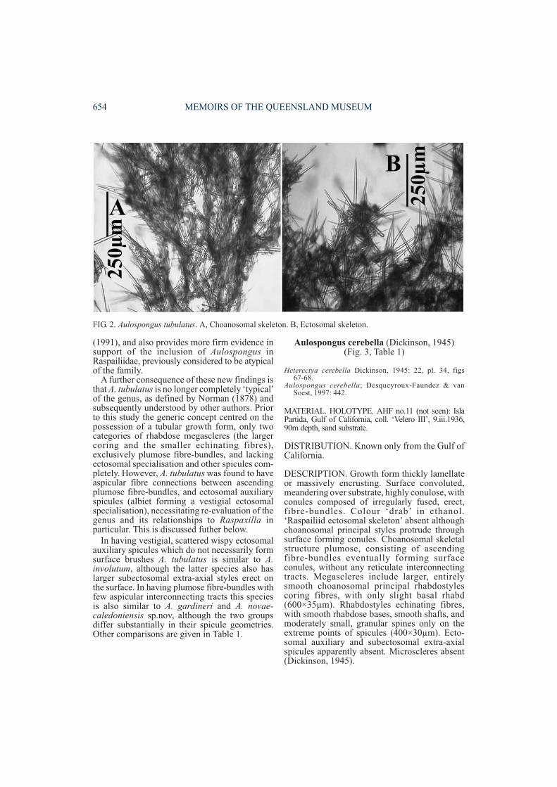
DESCRIPTION. Growth form subspherical,massive, tubular, composed of amalgamatedfibre-bundles that extend to the surface andproduce a compartmentalised surface of discreteconules. Colour red or pinkish-red alive.Ectosome with vestigial ‘raspailiid skeleton’composed of sinuous or raphidiform ectosomalauxil iary styles scattered sparsely andindiscriminantly over the surface, and withplumose bundles of both larger and smallerrhabdostyles protruding through the surfacemainly at the ends of conules. No subectosomalextra-axial spicules. Adjacent surface conulesinterconnected by aspicular (membranous)collagen. Choanosomal skeleton exclusively
652 MEMOIRS OF THE QUEENSLAND MUSEUM
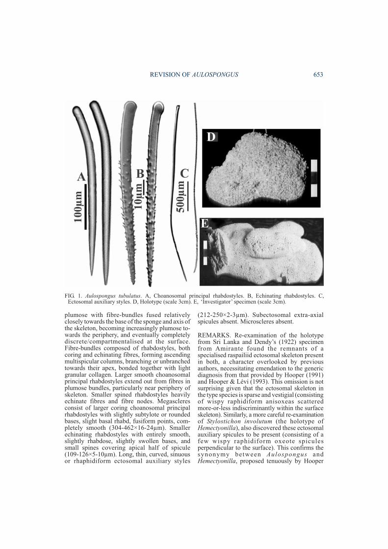
plumose with fibre-bundles fused relativelyclosely towards the base of the sponge and axis ofthe skeleton, becoming increasingly plumose to-wards the periphery, and eventually completelydiscrete/compartmentalised at the surface.Fibre-bundles composed of rhabdostyles, bothcoring and echinating fibres, forming ascendingmultispicular columns, branching or unbranchedtowards their apex, bonded together with lightgranular collagen. Larger smooth choanosomalprincipal rhabdostyles extend out from fibres inplumose bundles, particularly near periphery ofskeleton. Smaller spined rhabdostyles heavilyechinate fibres and fibre nodes. Megascleresconsist of larger coring choanosomal principalrhabdostyles with slightly subtylote or roundedbases, slight basal rhabd, fusiform points, com-pletely smooth (304-462×16-24µm). Smallerechinating rhabdostyles with entirely smooth,slightly rhabdose, slightly swollen bases, andsmall spines covering apical half of spicule(109-126×5-10µm). Long, thin, curved, sinuousor rhaphidiform ectosomal auxiliary styles
(212-250×2-3µm). Subectosomal extra-axialspicules absent. Microscleres absent.
REMARKS. Re-examination of the holotypefrom Sri Lanka and Dendy’s (1922) specimenfrom Amirante found the remnants of aspecialised raspailiid ectosomal skeleton presentin both, a character overlooked by previousauthors, necessitating emendation to the genericdiagnosis from that provided by Hooper (1991)and Hooper & Lévi (1993). This omission is notsurprising given that the ectosomal skeleton inthe type species is sparse and vestigial (consistingof wispy raphidiform anisoxeas scatteredmore-or-less indiscriminantly within the surfaceskeleton). Similarly, a more careful re-examinationof Stylostichon involutum (the holotype ofHemectyonilla), also discovered these ectosomalauxiliary spicules to be present (consisting of afew wispy raphidiform oxeote spiculesperpendicular to the surface). This confirms thesynonymy between Aulospongus andHemectyonilla, proposed tenuously by Hooper
REVISION OF AULOSPONGUS 653
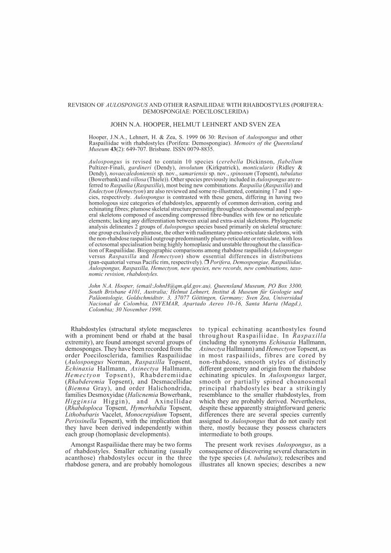
FIG. 1. Aulospongus tubulatus. A, Choanosomal principal rhabdostyles. B, Echinating rhabdostyles. C,Ectosomal auxiliary styles. D, Holotype (scale 3cm). E, ‘Investigator’ specimen (scale 3cm).
(1991), and also provides more firm evidence insupport of the inclusion of Aulospongus inRaspailiidae, previously considered to be atypicalof the family.
A further consequence of these new findings isthat A. tubulatus is no longer completely ‘typical’of the genus, as defined by Norman (1878) andsubsequently understood by other authors. Priorto this study the generic concept centred on thepossession of a tubular growth form, only twocategories of rhabdose megascleres (the largercoring and the smaller echinating fibres),exclusively plumose fibre-bundles, and lackingectosomal specialisation and other spicules com-pletely. However, A. tubulatus was found to haveaspicular fibre connections between ascendingplumose fibre-bundles, and ectosomal auxiliaryspicules (albiet forming a vestigial ectosomalspecialisation), necessitating re-evaluation of thegenus and its relationships to Raspaxilla inparticular. This is discussed futher below.
In having vestigial, scattered wispy ectosomalauxiliary spicules which do not necessarily formsurface brushes A. tubulatus is similar to A.involutum, although the latter species also haslarger subectosomal extra-axial styles erect onthe surface. In having plumose fibre-bundles withfew aspicular interconnecting tracts this speciesis also similar to A. gardineri and A. novae-caledoniensis sp.nov, although the two groupsdiffer substantially in their spicule geometries.Other comparisons are given in Table 1.
Aulospongus cerebella (Dickinson, 1945)(Fig. 3, Table 1)
Heterectya cerebella Dickinson, 1945: 22, pl. 34, figs67-68.
Aulospongus cerebella; Desqueyroux-Faundez & vanSoest, 1997: 442.
MATERIAL. HOLOTYPE. AHF no.11 (not seen): IslaPartida, Gulf of California, coll. ‘Velero III’, 9.iii.1936,90m depth, sand substrate.
DISTRIBUTION. Known only from the Gulf ofCalifornia.
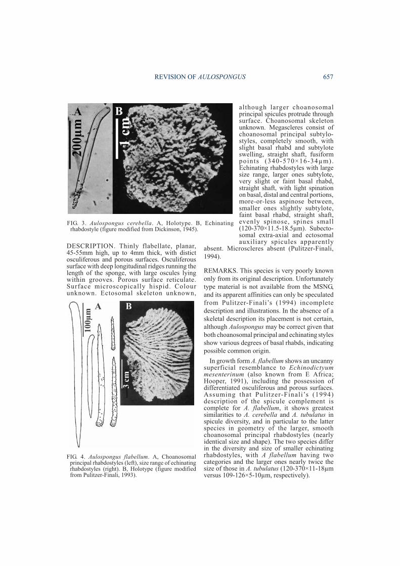
DESCRIPTION. Growth form thickly lamellateor massively encrusting. Surface convoluted,meandering over substrate, highly conulose, withconules composed of irregularly fused, erect,fibre-bundles. Colour ‘drab’ in ethanol.‘Raspailiid ectosomal skeleton’ absent althoughchoanosomal principal styles protrude throughsurface forming conules. Choanosomal skeletalstructure plumose, consisting of ascendingfibre-bundles eventually forming surfaceconules, without any reticulate interconnectingtracts. Megascleres include larger, entirelysmooth choanosomal principal rhabdostylescoring fibres, with only slight basal rhabd(600×35µm). Rhabdostyles echinating fibres,with smooth rhabdose bases, smooth shafts, andmoderately small, granular spines only on theextreme points of spicules (400×30µm). Ecto-somal auxiliary and subectosomal extra-axialspicules apparently absent. Microscleres absent(Dickinson, 1945).
654 MEMOIRS OF THE QUEENSLAND MUSEUM
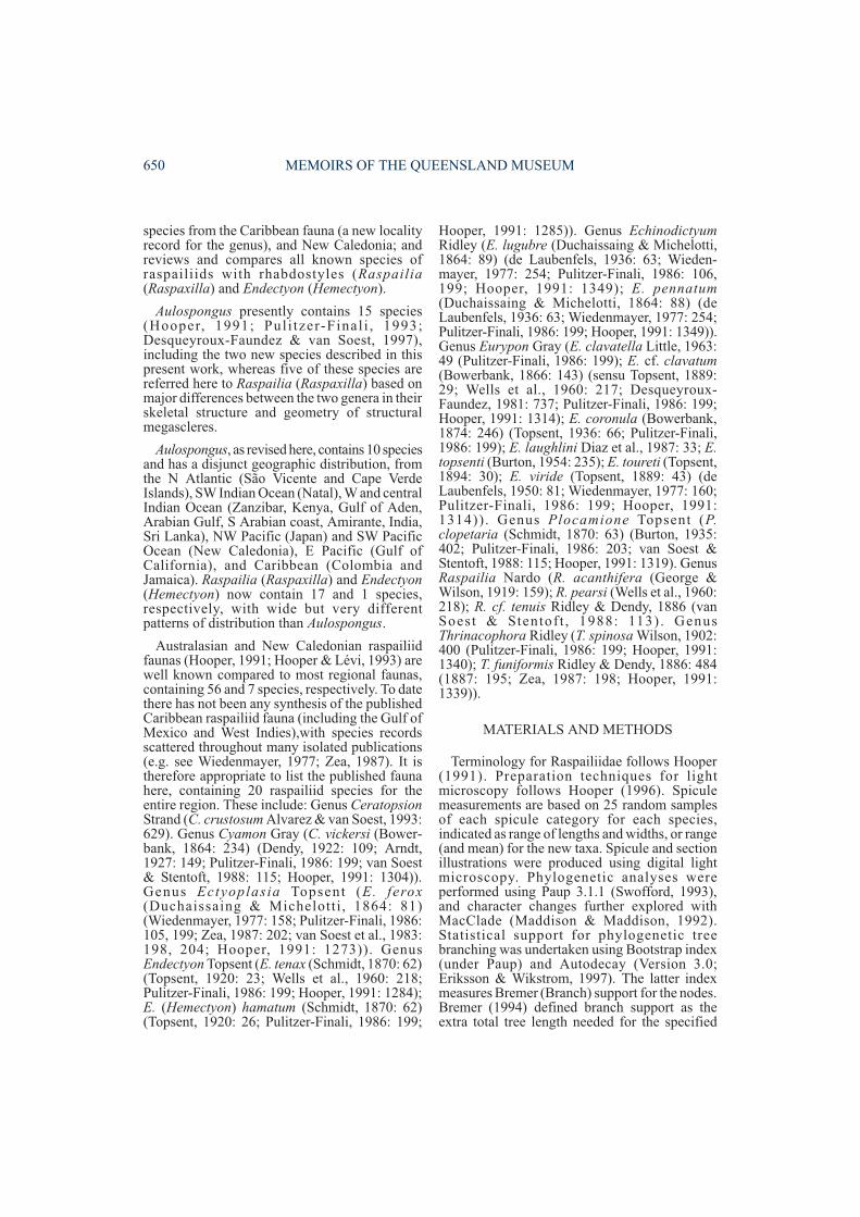
FIG. 2. Aulospongus tubulatus. A, Choanosomal skeleton. B, Ectosomal skeleton.
REVISION OF AULOSPONGUS 655
SpeciesSurface features
& skeletalfibre-bundles
Skeletal reticu-lation & axialvs. extra-axial
skeleton
Choanosomalprincipalspicules
(spicule size)
Echinating spicules(spicule size)
Specialisedectosomal
auxiliary skeleton(spicule size)
Subectosomalextra-axial
spicules(spicule size)
Raphides(spicule
size)
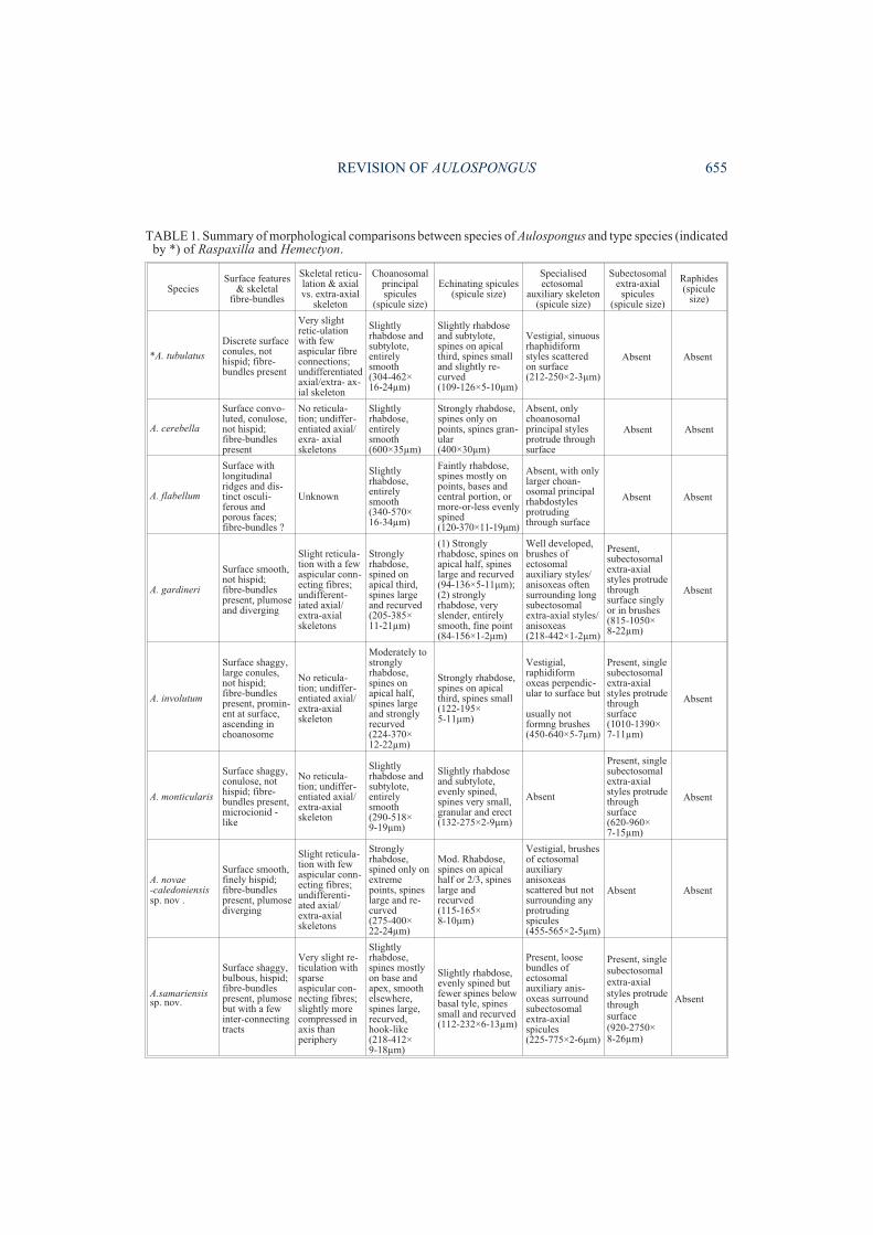
*A. tubulatus
Discrete surfaceconules, nothispid; fibre-bundles present
Very slightretic-ulationwith fewaspicular fibreconnections;undifferentiatedaxial/extra- ax-ial skeleton
Slightlyrhabdose andsubtylote,entirelysmooth(304-462×16-24µm)
Slightly rhabdoseand subtylote,spines on apicalthird, spines smalland slightly re-curved(109-126×5-10µm)
Vestigial, sinuousrhaphidiformstyles scatteredon surface(212-250×2-3µm)
Absent Absent
A. cerebella
Surface convo-luted, conulose,not hispid;fibre-bundlespresent
No reticula-tion; undiffer-entiated axial/exra- axialskeletons
Slightlyrhabdose,entirelysmooth(600×35µm)
Strongly rhabdose,spines only onpoints, spines gran-ular(400×30µm)
Absent, onlychoanosomalprincipal stylesprotrude throughsurface
Absent Absent
A. flabellum
Surface withlongitudinalridges and dis-tinct osculi-ferous andporous faces;fibre-bundles ?
Unknown
Slightlyrhabdose,entirelysmooth(340-570×16-34µm)
Faintly rhabdose,spines mostly onpoints, bases andcentral portion, ormore-or-less evenlyspined(120-370×11-19µm)
Absent, with onlylarger choan-osomal principalrhabdostylesprotrudingthrough surface
Absent Absent
A. gardineri
Surface smooth,not hispid;fibre-bundlespresent, plumoseand diverging
Slight reticula-tion with a fewaspicular conn-ecting fibres;undifferent-iated axial/extra-axialskeletons
Stronglyrhabdose,spined onapical third,spines largeand recurved(205-385×11-21µm)
(1) Stronglyrhabdose, spines onapical half, spineslarge and recurved(94-136×5-11µm);(2) stronglyrhabdose, veryslender, entirelysmooth, fine point(84-156×1-2µm)
Well developed,brushes ofectosomalauxiliary styles/anisoxeas oftensurrounding longsubectosomalextra-axial styles/anisoxeas(218-442×1-2µm)
Present,subectosomalextra-axialstyles protrudethroughsurface singlyor in brushes(815-1050×8-22µm)
Absent
A. involutum
Surface shaggy,large conules,not hispid;fibre-bundlespresent, promin-ent at surface,ascending inchoanosome
No reticula-tion; undiffer-entiated axial/extra-axialskeleton
Moderately tostronglyrhabdose,spines onapical half,spines largeand stronglyrecurved(224-370×12-22µm)
Strongly rhabdose,spines on apicalthird, spines small(122-195×5-11µm)
Vestigial,raphidiformoxeas perpendic-ular to surface but
usually notformng brushes(450-640×5-7µm)
Present, singlesubectosomalextra-axialstyles protrudethroughsurface(1010-1390×7-11µm)
Absent
A. monticularis
Surface shaggy,conulose, nothispid; fibre-bundles present,microcionid -like
No reticula-tion; undiffer-entiated axial/extra-axialskeleton
Slightlyrhabdose andsubtylote,entirelysmooth(290-518×9-19µm)
Slightly rhabdoseand subtylote,evenly spined,spines very small,granular and erect(132-275×2-9µm)
Absent
Present, singlesubectosomalextra-axialstyles protrudethroughsurface(620-960×7-15µm)
Absent
A. novae-caledoniensissp. nov .
Surface smooth,finely hispid;fibre-bundlespresent, plumosediverging
Slight reticula-tion with fewaspicular conn-ecting fibres;undifferenti-ated axial/extra-axialskeletons
Stronglyrhabdose,spined only onextremepoints, spineslarge and re-curved(275-400×22-24µm)
Mod. Rhabdose,spines on apicalhalf or 2/3, spineslarge andrecurved(115-165×8-10µm)
Vestigial, brushesof ectosomalauxiliaryanisoxeasscattered but notsurrounding anyprotrudingspicules(455-565×2-5µm)
Absent Absent
A.samariensissp. nov.
Surface shaggy,bulbous, hispid;fibre-bundlespresent, plumosebut with a fewinter-connectingtracts
Very slight re-ticulation withsparseaspicular con-necting fibres;slightly morecompressed inaxis thanperiphery
Slightlyrhabdose,spines mostlyon base andapex, smoothelsewhere,spines large,recurved,hook-like(218-412×9-18µm)
Slightly rhabdose,evenly spined butfewer spines belowbasal tyle, spinessmall and recurved(112-232×6-13µm)
Present, loosebundles ofectosomalauxiliary anis-oxeas surroundsubectosomalextra-axialspicules(225-775×2-6µm)
Present, singlesubectosomalextra-axialstyles protrudethroughsurface(920-2750×8-26µm)
Absent
TABLE 1. Summary of morphological comparisons between species of Aulospongus and type species (indicatedby *) of Raspaxilla and Hemectyon.
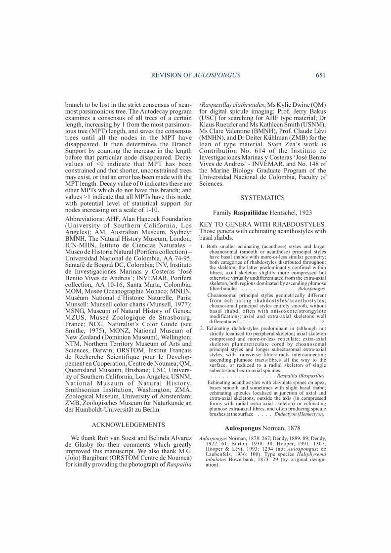
REMARKS. This species has not been recordedsince it was first described, and regrettably theholotype cannot be located in the AHFcollections (Prof. G. Bakus, pers.comm.).Dickinson (1945) stated that it was a sisterspecies of R. inaequalis Dendy, 1924 (which healso suggested belonged to Echinaxia inpossessing ectosomal auxiliary oxeas, and whichHooper (1991) subsequently referred to Raspailia(Raspaxilla)), but this comparison is very super-ficial: R. inaequalis has a distinct, compressedaxial and plumo-reticulate extra-axial region,possesses ectosomal specialisation, and hasnon-rhabdose choanosomal principal mega-scleres of distinctly different geometry thanrhabdose echinating megascleres. Althoughincompletely described A. cerebella appears to bemost similar to A. flabellum and A. villosa inhaving a reduced spiculation consisting only of
choanosomal principal styles and echinating rhab-dostyles, lacking any form of ectosomal auxiliaryspicules or long subectosomal extra-axialspicules. Unlike both these species the largerchoanosomal principal styles in A. cerebella donot appear to have a basal rhabd, which haspresumably been secondarily modified.
Aulospongus flabellum Pulitzer-Finali, 1994(Fig. 4, Table 1)
Aulospongus flabellum Pulitzer-Finali, 1994: 308, figs38-39.
MATERIAL. HOLOTYPE. MSNG 48305 (not seen):North Kenya Banks, off Manda I., Kenya, 02°23’S,41°04’E, 17.vi.1971, 110-170m depth.
DISTRIBUTION. Kenya, W Indian Ocean.
656 MEMOIRS OF THE QUEENSLAND MUSEUM
Species
Surfacefeatures &skeletal
fibre-bundles
Skeletalreticulation &axial vs. extra-axial skeleton
Choanosomalprincipalspicules
(spicule size)
Echinating spicules(spicule size)
Specialisedectosomal
auxiliary skeleton(spicule size)
Subectosomalextra-axial
spicules(spicule size)
Raphides(spicule size)
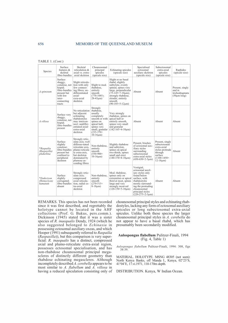
A.spinosum
Surfaceshaggy,conulose, nothispid;fibre-bundlespresent butwith fewinter-connectingtracts
Slight reticula-tion with onlyfew connect-ing fibres; un-differentiatedaxial/exra-axialskeleton
Slight to mod.rhabdose,entirelysmooth(770-1085×28-43µm)
Slight or no basalrhabd, slightlysubtylote, evenlyspined, spines verylarge, perpendicular(75-145×7-10µm);strongly rhabdose,slender, entirelysmooth(90-185×5-12µm)
Absent Absent
Present, singlyand intrichodragmata(50µm long)
A.villosa
Surface veryshaggy,conulose, nothispid;fibre-bundlespresent
No reticulationbut adjacentechinatingrhabdostylesmay intercon-nect; undiffer-entiated axial/extra-axialskeleton
Stronglyrhabdose,usuallycompletelysmooth or withspines onapical half,spines verysmall, granular(235-370×10-16µm)
Very stronglyrhabdose, spines onapical half orentirely smooth,spines very smalland granular(142-165×4-10µm)
Absent Absent Absent
*Raspailia(Raspaxilla)phakellina
Surfaceslightlyconulose;fibre-bundlespresent butconfined toperipheralskeleton
Strongly retic-ulate axis; welldifferen-tiatedreticulate axis,plumo-reticulate extra-axis,but skeletondominated byplumose as-cending fibres
Non-rhabdose,entirelysmooth(550-900×10-16µm)
Slightly rhabdoseand subtylote,spines on apicaltwo-thirds, spinessmall and erect(140-370×8-18µm)
Present, brushesof ectosomal aux-iliary stylessurroundingsubectosomalextra-axial styles(450-650×2-3µm)
Present, singlesubectosomalextra-axialstyles protrudethroughsurface(1100-1450×12-18µm)
Absent
*Endectyon(Hemectyon)hamatum
Surfaceslightlycorrugated;fibre-bundlesabsent
Strongly retic-ulate axis;compressedaxial reticula-tion, radial ex-tra-axialskeleton
Non-rhabdose,entirelysmooth(270-515×8-18µm)
Mod. rhabdose,spines only onextremities or apicalthird at most, spineslarge and verystrongly recurved(120-150×5-10µm)
Vestigial,ectosomal auxil-iary styles onlyfound belowsurface, withrhabdostylesmostly surround-ing the protrudingchoanosomalprincipal styles(220-275×2-3µm)
Absent Absent
TABLE 1. (Cont.)
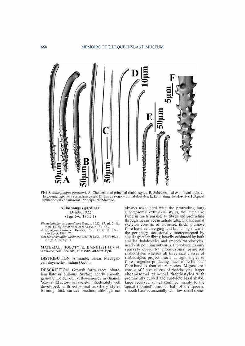
DESCRIPTION. Thinly flabellate, planar,45-55mm high, up to 4mm thick, with distictosculiferous and porous surfaces. Osculiferoussurface with deep longitudinal ridges running thelength of the sponge, with large oscules lyingwithin grooves. Porous surface reticulate.Surface microscopically hispid. Colourunknown. Ectosomal skeleton unknown,
although larger choanosomalprincipal spicules protrude throughsurface. Choanosomal skeletonunknown. Megascleres consist ofchoanosomal principal subtylo-styles, completely smooth, withslight basal rhabd and subtyloteswelling, straight shaft, fusiformpoints (340-570×16-34µm).Echinating rhabdostyles with largesize range, larger ones subtylote,very slight or faint basal rhabd,straight shaft, with light spinationon basal, distal and central portions,more-or-less aspinose between,smaller ones slightly subtylote,faint basal rhabd, straight shaft,evenly spinose, spines small(120-370×11.5-18.5µm). Subecto-somal extra-axial and ectosomalauxiliary spicules apparently
absent. Microscleres absent (Pulitzer-Finali,1994).
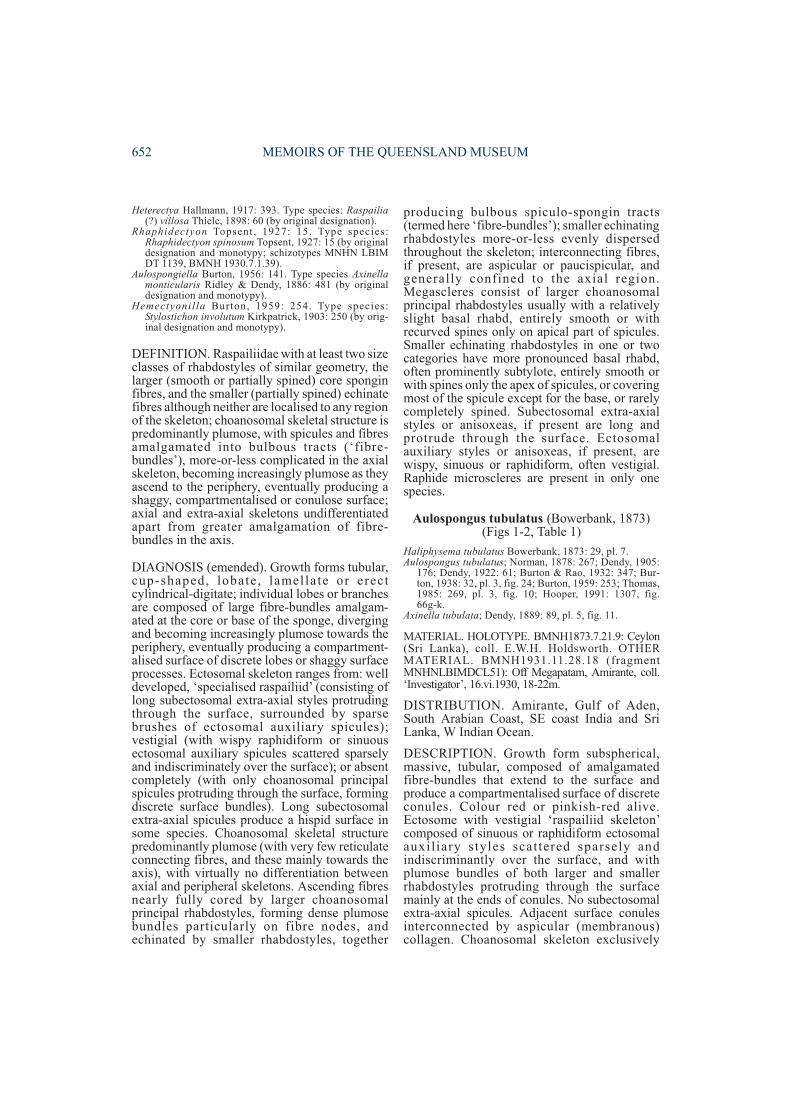
REMARKS. This species is very poorly knownonly from its original description. Unfortunatelytype material is not available from the MSNG,and its apparent affinities can only be speculatedfrom Pulitzer-Finali’s (1994) incompletedescription and illustrations. In the absence of askeletal description its placement is not certain,although Aulospongus may be correct given thatboth choanosomal principal and echinating stylesshow various degrees of basal rhabds, indicatingpossible common origin.
In growth form A. flabellum shows an uncannysuperficial resemblance to Echinodictyummesenterinum (also known from E Africa;Hooper, 1991), including the possession ofdifferentiated osculiferous and porous surfaces.Assuming that Pulitzer-Finali’s (1994)description of the spicule complement iscomplete for A. flabellum, it shows greatestsimilarities to A. cerebella and A. tubulatus inspicule diversity, and in particular to the latterspecies in geometry of the larger, smoothchoanosomal principal rhabdostyles (nearlyidentical size and shape). The two species differin the diversity and size of smaller echinatingrhabdostyles, with A flabellum having twocategories and the larger ones nearly twice thesize of those in A. tubulatus (120-370×11-18µmversus 109-126×5-10µm, respectively).
REVISION OF AULOSPONGUS 657
FIG. 3. Aulospongus cerebella. A, Holotype. B, Echinatingrhabdostyle (figure modified from Dickinson, 1945).
FIG. 4. Aulospongus flabellum. A, Choanosomalprincipal rhabdostyles (left), size range of echinatingrhabdostyles (right). B, Holotype (figure modifiedfrom Pulitzer-Finali, 1993).
Aulospongus gardineri(Dendy, 1922)
(Figs 5-6, Table 1)
Plumohalichondria gardineri Dendy, 1922: 87, pl. 2, fig.9, pl. 15, fig. 4a-d; Vacelet & Vasseur, 1971: 83.
Aulospongus gardineri; Hooper, 1991: 1309, fig. 67e-h;van Soest, 1994: 72.
Not Hemectyonilla gardineri; Lévi & Lévi, 1983: 950, pl.2, figs 2,3,5, fig. 14.
MATERIAL. HOLOTYPE. BMNH1921.11.7.74:Amirante, coll. ‘Sealark’, 18.x.1905, 40-88m depth.
DISTRIBUTION. Amirante, Tulear, Madagas-car, Seychelles, Indian Ocean.
DESCRIPTION. Growth form erect lobate,lamellate or bulbous. Surface nearly smooth,granular. Colour dull yellowish-grey in ethanol.‘Raspailiid ectosomal skeleton’ moderately welldeveloped, with ectosomal auxiliary stylesforming thick surface brushes, although not
always associated with the protruding longsubectosomal extra-axial styles, the latter alsolying in tracts parallel to fibres and protrudingthrough the surface in radiate tufts. Choanosomalskeleton consists of close-set, thick, plumosefibre-bundles diverging and branching towardsthe periphery, occasionally interconnected bysmall aspicular fibres, heavily echinated by bothsmaller rhabdostyles and smooth rhabdostyles,nearly all pointing outwards. Fibre-bundles onlysparsely cored by choanosomal principalrhabdostyles whereas all three size classes ofrhabdostyles project nearly at right angles tofibres, together producing much more bulbousfibre-bundles than other species. Megascleresconsist of 3 size classes of rhabdostyles: largerchoanosomal principal rhabdostyles withprominently curved and subtylote basal rhabd,large recurved spines confined mainly to theapical (pointed) third or half of the spicule,smooth base occasionally with few small spines
658 MEMOIRS OF THE QUEENSLAND MUSEUM
FIG. 5. Aulospongus gardineri. A, Choanosomal principal rhabdostyles. B, Subectosomal extra-axial style. C,Ectosomal auxiliary styles/anisoxeas. D, Third category of rhabdostyles. E, Echinating rhabdostyles. F, Apicalspination on choanosomal principal rhabdostyle.
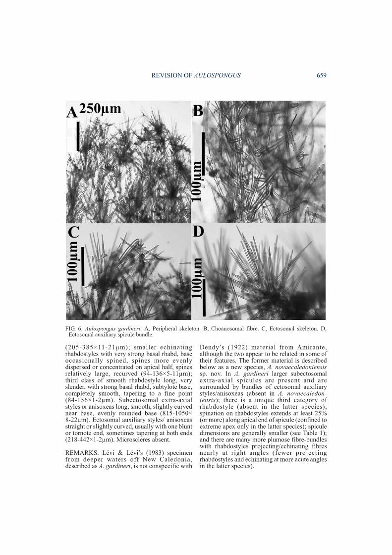
(205-385×11-21µm); smaller echinatingrhabdostyles with very strong basal rhabd, baseoccasionally spined, spines more evenlydispersed or concentrated on apical half, spinesrelatively large, recurved (94-136×5-11µm);third class of smooth rhabdostyle long, veryslender, with strong basal rhabd, subtylote base,completely smooth, tapering to a fine point(84-156×1-2µm). Subectosomal extra-axialstyles or anisoxeas long, smooth, slightly curvednear base, evenly rounded base (815-1050×8-22µm). Ectosomal auxiliary styles/ anisoxeasstraight or slightly curved, usually with one bluntor tornote end, sometimes tapering at both ends(218-442×1-2µm). Microscleres absent.
REMARKS. Lévi & Lévi’s (1983) specimenfrom deeper waters off New Caledonia,described as A. gardineri, is not conspecific with
Dendy’s (1922) material from Amirante,although the two appear to be related in some oftheir features. The former material is describedbelow as a new species, A. novaecaledoniensissp. nov. In A. gardineri larger subectosomalextra-axial spicules are present and aresurrounded by bundles of ectosomal auxiliarystyles/anisoxeas (absent in A. novaecaledon-iensis); there is a unique third category ofrhabdostyle (absent in the latter species);spination on rhabdostyles extends at least 25%(or more) along apical end of spicule (confined toextreme apex only in the latter species); spiculedimensions are generally smaller (see Table 1);and there are many more plumose fibre-bundleswith rhabdostyles projecting/echinating fibresnearly at right angles (fewer projectingrhabdostyles and echinating at more acute anglesin the latter species).
REVISION OF AULOSPONGUS 659
FIG. 6. Aulospongus gardineri. A, Peripheral skeleton. B, Choanosomal fibre. C, Ectosomal skeleton. D,Ectosomal auxiliary spicule bundle.
In rhabdostyle geometry this species is alsosimilar to A. involutum (which prompted Burton(1959) to synonymise the two), but they differ inmany other respects (fibre characteristics,choanosomal and ectosomal skeletal structure,spicule sizes and spicule diversity (see Table 1)),which Lévi & Lévi (1983) and Hooper (1991)indicated they were dis t inct species .Aulospongus gardineri is unusual in having amacroscopically smooth surface (althoughmicroscopically it is hispid from the protrudingsubectosomal extra-axial styles), and in possess-ing of a third category of rhabdostyle (similaronly to A. spinosum in this respect, although thetwo species differ in virtually all other characters;Table 1).
Aulospongus involutum (Kirkpatrick, 1903)(Figs 7-8, Table 1)
Stylostichon involutum Kirkpatrick, 1903: 250, pl. 5, fig.16, pl. 6, fig. 17a-e.
Hemectyonilla involutum; Burton, 1959: 254.Aulospongus involutum; Pultizer-Finali, 1993: 308-309;
Hooper, 1991: 1307, fig. 66a-f.
MATERIAL. HOLOTYPE. BMNH1902.11.16.33(fragment MNHN LBIM DCL61): Cone Point, Natal,South Africa, 68m depth. OTHER MATERIAL.BMNH1936.3.4.118: Gulf of Aden, 11°56-57’N,50°35-39’E, coll. ‘John Murray’, 12.x.1933, 37m depth.
DISTRIBUTION. Natal, Zanzibar, Kenya, Gulfof Aden, South Arabian coast and Arabian Gulf,Indian Ocean.
660 MEMOIRS OF THE QUEENSLAND MUSEUM
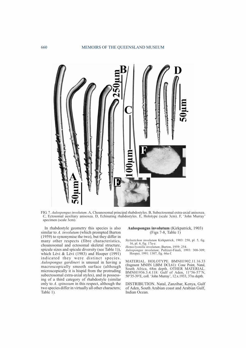
FIG. 7. Aulospongus involutum. A, Choanosomal principal rhabdostyles. B, Subectosomal extra-axial anisoxea.C, Ectosomal auxiliary anisoxea. D, Echinating rhabdostyles. E, Holotype (scale 3cm). F, ‘John Murray’specimen (scale 3cm).
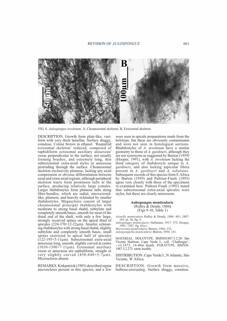
DESCRIPTION. Growth form plate-like, vasi-form with very thick lamellae. Surface shaggy,conulose. Colour brown in ethanol. ‘Raspailiidectosomal skeleton’ reduced, composed ofraphidiform ectosomal auxiliary anisoxeas/oxeas perpendicular to the surface, not usuallyforming brushes, and extremely long, thinsubectosomal extra-axial styles or anisoxeasprotruding through the surface. Choanosomalskeleton exclusively plumose, lacking any axialcompression or obvious differentiation betweenaxial and extra-axial regions, although peripheralskeleton tracts form prominent tufts at thesurface, producing relatively large conules.Larger rhabdostyles form plumose tufts alongfibre-bundles, which are radial, microcionid-like, plumose, and heavily echinated by smallerrhabdostyles. Megascleres consist of largerchoanosomal principal rhabdostyles withmoderate to strong basal rhabd, subtylote andcompletely smooth bases, smooth for most of thedistal end of the shaft, with only a few large,strongly recurved spines on the apical third ofspicules (224-370×12-22µm). Smaller echinat-ing rhabdostyles with strong basal rhabd, slightlysubtylote and completely smooth bases, smallspines restricted to apical half of spicules(122-195×5-11µm). Subectosomal extra-axialanisoxeas long, smooth, slightly curved at centre(1010-1390×7-11µm). Ectosomal auxiliaryoxeas or anisoxeas are raphidiform, straight orvery slightly curved (450-640×5-7µm).Microscleres absent.
REMARKS. Kirkpatrick (1903) described sigmamicroscleres present in this species, and a few
were seen in spicule preparations made from theholotype, but these are obviously contaminantsand were not seen in histological sections.Rhabdostyles of A. involutum have a similargeometry to those of A. gardineri, although theyare not synonyms as suggested by Burton (1959)(Hooper, 1991), with A. involutum lacking thethird category of rhabdostyle unique to A.gardineri, and also lacking aspicular fibrespresent in A. gardineri and A. tubulatus.Subsequent records of this species from E Africaby Burton (1959) and Pultizer-Finali (1993)agree very closely with those of the specimensre-examined here. Pulitzer-Finali (1993) statedthat subectosomal extra-axial spicules werestyles, but these are clearly anisoxeote.
Aulospongus monticularis(Ridley & Dendy, 1886)
(Figs 9-10, Table 1)
Axinella monticularis Ridley & Dendy, 1886: 481; 1887:185, pl. 38, fig. 5.
Aulospongus monticularis; Hallmann, 1917: 373; Hooper,1991: 1307, fig. 65a-c.
Microciona monticularis; Burton, 1956: 132.Aulospongiella monticularis; Burton, 1956: 141.
MATERIAL. HOLOTYPE. BMNH1887.5.2.20: SãoVicente Harbour, Cape Verde I., coll. ‘Challenger’,-.vii.1873, 14-40m depth. PARATYPE. BMNH-1887.5.2.273: same locality.
DISTRIBUTION. Cape Verde I., N Atlantic, SãoVicente, W Africa.
DESCRIPTION. Growth form massive,bulbous-encrusting. Surface shaggy, conulose.
REVISION OF AULOSPONGUS 661
FIG. 8. Aulospongus involutum. A, Choanosomal skeleton. B, Ectosomal skeleton.

662 MEMOIRS OF THE QUEENSLAND MUSEUM
FIG. 10. Aulospongus monticularis. A, Microcionid-like choanosomal skeleton arising from basal detritus. B,Peripheral skeleton.
FIG. 9. Aulospongus monticularis. A, Choanosomal principal rhabdostyles. B, Basal end of subectosomalextra-axial styles. C, Echinating rhabdostyles. D, Holotype (scale 3cm).
Colour yellowish-grey in ethanol. ‘Raspailiidectosomal skeleton’ absent, although largersubectosomal extra-axial styles occasionallyprotrude through surface. Choanosomal skeletonmicrocionid-like, composed of large, non-anastomosing, plumose fibre-bundles, withoutany trace of axial compression or differentiationbetween axial and extra-axial regions. Ascendingfibre-bundles arise from detritus-encrusted basalskeleton, and foreign particles also incorporatedinto choanosomal skeleton. Ascending fibre-bundles cored and echinated by both categoriesof rhabdostyles, although larger rhabdostylescomprise most of the coring spicules, as well asprotruding through fibres in plumose bundles.Megascleres consist of larger choanosomalprincipal rhabdostyles with only slight basalrhabd, completely smooth, rounded or slightlysubtylote bases (290-518×9-19µm). Smallerechinating rhabdostyles more-or-less evenlyspined, microcionid-like, slightly curved atcentre, with very small, granular, erect spines,only slight basal rhabd and slight to moderatesubtylote basal swelling (132-275×2-9µm).Subectosomal extra-axial styles slightly curvednear basal end, with slightly subtylote bases andvery long tapering points (620-960×7-15µm).Ectosomal auxiliary spicules absent. Micro-scleres absent.
REMARKS. This species is similar to A. involutumin having long subectosomal extra-axial spiculesprotruding through the surface, which are notsurrounded by ectosomal auxiliary spicules, astypical for most raspailiids. It differs from A.involutum in lacking rhaphidiform ectosomalauxiliary spicules completely, as well as in otherimportant characters such as the geometry, smallsize and vestigial spination of rhabdostyles(Table 1). It should also be compared to A. villosaand A. cerebella which also lack any ectosomalspecialisation, differing from A. villosa in havingcompletely smooth larger rhabdostyles (general-ly longer than those of A. villosa), microcionid-like, evenly spined smaller rhabdostyles(partially spined in A. villosa), and a bulbousgrowth form (bushy growth form in A. villosa)(see Table 2). Rhabdostyle morphology differssubstantially between A. monticularis and A.cerebella: larger rhabdostyles are of similar size,but those in the latter species have only a slightbasal rhabd; smaller rhabdostyles are evenlyspined in both species but about half the size in A.monticularis.
Aulospongus novaecaledoniensis sp. nov.(Figs 11-12, Table 1)
Hemectyonilla gardineri; Lévi & Lévi, 1983: 950, pl. 2,figs 2,3,5, fig. 14.
ETYMOLOGY. For the type locality.
MATERIAL. HOLOTYPE. MNHN LBIM DCL2941:Havannah, New Caledonia, 22°17’S, 167°14’E, coll.‘Vauban’ (stn.DP36), 24.v.1978, 425-430m depth.PARATYPE. MNHN LBIM DCL2940: same locality,22°19’S, 167°11’E, 300-315m depth.
DISTRIBUTION. New Caledonia.
DESCRIPTION. Growth form massive, tubular.Surface finely hispid, generally smooth apartfrom several large surface conules, eachsurmounted by a terminal oscule. Colour inethanol brownish ocre with slight pinkish tinge.‘Raspailiid ectosomal skeleton’ moderately welldeveloped, with ectosomal auxiliary stylesforming thick surface brushes, although notassociated with any subectosomal extra-axialspicules (the latter absent completely from thisspecies). Choanosomal skeleton essentiallyplumose, consisting of close-set, thick, ascendingfibre-bundles, diverging and branching towardsthe periphery. Ascending fibres moderatelyheavily cored by larger rhabdostyles, formingmainly axial bundles within fibres and onlyslightly plumose tracts of single rhabdostylesprotruding through fibres, with their pointsascending towards the surface. Smallerrhabdostyles concentrated mainly at the base ofmain ascending fibres. Ascending fibresinterconnected by few, aspicular, transversefibres sparsely echinated by smaller rhabdostyles.Megascleres consist of larger choanosomalprincipal rhabdostyles with variably developedsmooth basal rhabds, ranging from nearlystraight to prominently rhabdose, with basalrhabd occupying between 15-40% of spiculelength, shaft smooth except for a few (4-12) largerecurved spines located only on the extremeapical (pointed) end of the spicule (275-415×19-26µm). Smaller echinating rhabdostyles withonly moderate basal rhabd (sometimes slight),base usually subtylote, smooth or occasionallywith a few spines, shaft with spines concentratedon apical half (not merely confined to extremepoint of spicule as for larger rhabdostyles), spinesrelatively large, recurved (122-195×7-13µm).Subectosomal extra-axial megascleres absent.Ectosomal auxiliary anisoxeas (occasionallystyles) large, slightly curved at the centre, usuallywith one blunt or tornote end, sometimes tapering
REVISION OF AULOSPONGUS 663
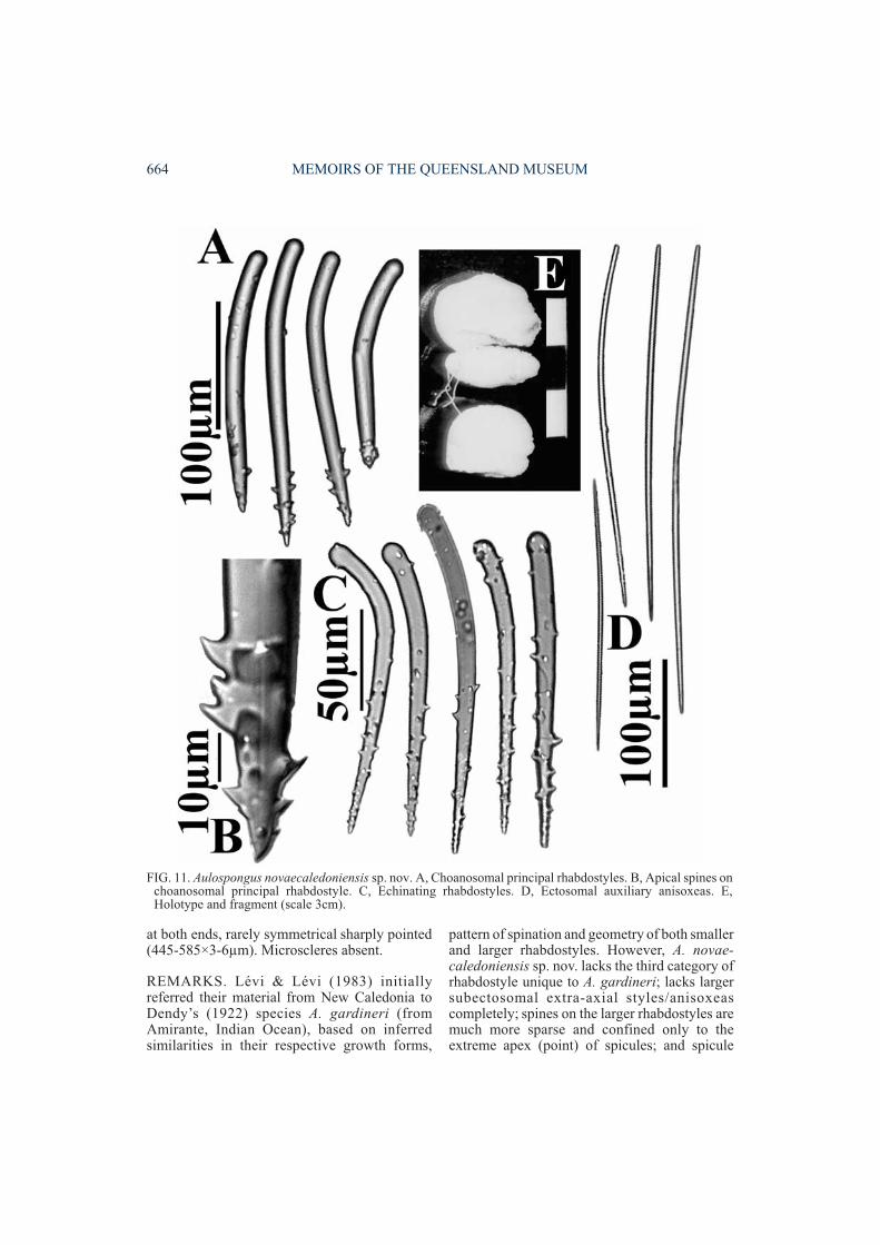
at both ends, rarely symmetrical sharply pointed(445-585×3-6µm). Microscleres absent.
REMARKS. Lévi & Lévi (1983) initiallyreferred their material from New Caledonia toDendy’s (1922) species A. gardineri (fromAmirante, Indian Ocean), based on inferredsimilarities in their respective growth forms,
pattern of spination and geometry of both smallerand larger rhabdostyles. However, A. novae-caledoniensis sp. nov. lacks the third category ofrhabdostyle unique to A. gardineri; lacks largersubectosomal extra-axial styles/anisoxeascompletely; spines on the larger rhabdostyles aremuch more sparse and confined only to theextreme apex (point) of spicules; and spicule
664 MEMOIRS OF THE QUEENSLAND MUSEUM
FIG. 11. Aulospongus novaecaledoniensis sp. nov. A, Choanosomal principal rhabdostyles. B, Apical spines onchoanosomal principal rhabdostyle. C, Echinating rhabdostyles. D, Ectosomal auxiliary anisoxeas. E,Holotype and fragment (scale 3cm).
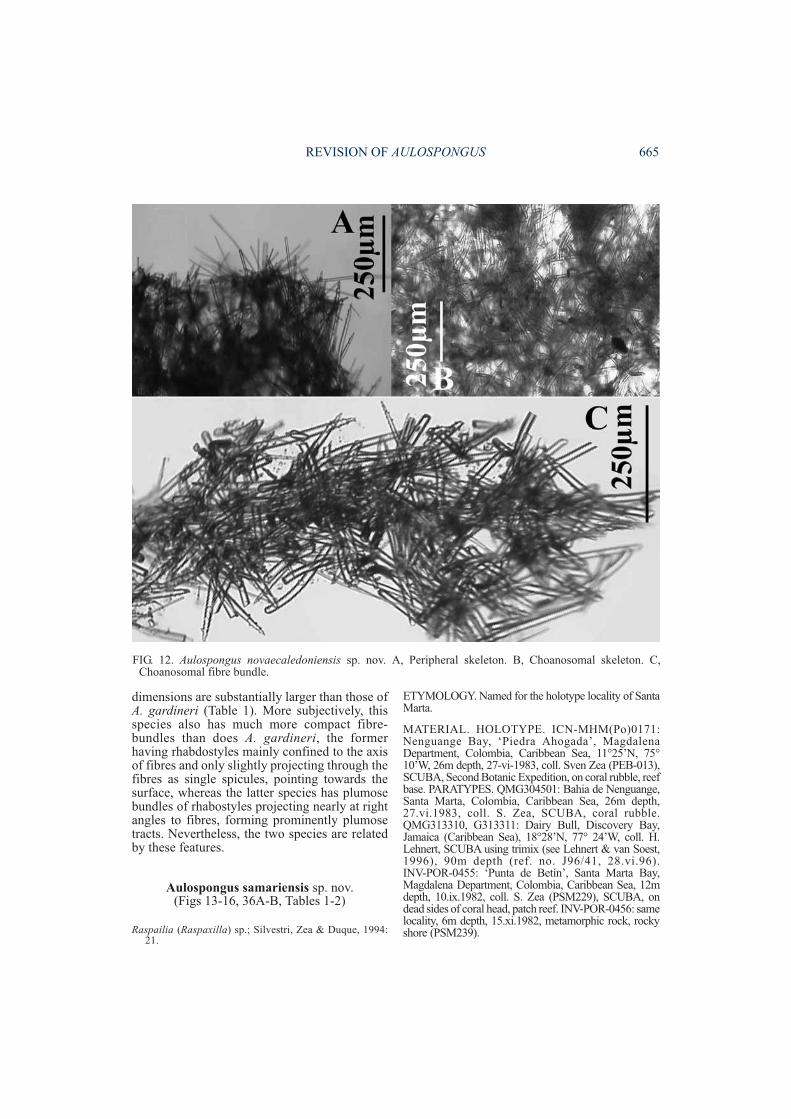
dimensions are substantially larger than those ofA. gardineri (Table 1). More subjectively, thisspecies also has much more compact fibre-bundles than does A. gardineri, the formerhaving rhabdostyles mainly confined to the axisof fibres and only slightly projecting through thefibres as single spicules, pointing towards thesurface, whereas the latter species has plumosebundles of rhabostyles projecting nearly at rightangles to fibres, forming prominently plumosetracts. Nevertheless, the two species are relatedby these features.
Aulospongus samariensis sp. nov.(Figs 13-16, 36A-B, Tables 1-2)
Raspailia (Raspaxilla) sp.; Silvestri, Zea & Duque, 1994:21.
ETYMOLOGY. Named for the holotype locality of SantaMarta.
MATERIAL. HOLOTYPE. ICN-MHM(Po)0171:Nenguange Bay, ‘Piedra Ahogada’, MagdalenaDepartment, Colombia, Caribbean Sea, 11�25’N, 75�10’W, 26m depth, 27-vi-1983, coll. Sven Zea (PEB-013),SCUBA, Second Botanic Expedition, on coral rubble, reefbase. PARATYPES. QMG304501: Bahia de Nenguange,Santa Marta, Colombia, Caribbean Sea, 26m depth,27.vi.1983, coll. S. Zea, SCUBA, coral rubble.QMG313310, G313311: Dairy Bull, Discovery Bay,Jamaica (Caribbean Sea), 18�28’N, 77� 24’W, coll. H.Lehnert, SCUBA using trimix (see Lehnert & van Soest,1996), 90m depth (ref. no. J96/41, 28.vi.96).INV-POR-0455: ‘Punta de Betín’, Santa Marta Bay,Magdalena Department, Colombia, Caribbean Sea, 12mdepth, 10.ix.1982, coll. S. Zea (PSM229), SCUBA, ondead sides of coral head, patch reef. INV-POR-0456: samelocality, 6m depth, 15.xi.1982, metamorphic rock, rockyshore (PSM239).
REVISION OF AULOSPONGUS 665
FIG. 12. Aulospongus novaecaledoniensis sp. nov. A, Peripheral skeleton. B, Choanosomal skeleton. C,Choanosomal fibre bundle.
DISTRIBUTION. Santa Marta region, W Carib-bean, Jamaica, E Caribbean
DESCRIPTION. Growth form. Erect, stalked,vaguely cylindrical, club-shaped, slightly bushy,5-12cm high, 0.5-2.5cm diameter, with severalsmall irregular bulbous lobate branches up to0.7-3cm diameter, partially fused and becomingmore swollen at their tips. Protruding fibre-bundles from underlying skeleton producing ashaggy appearance at the surface, superficiallyresembling Pandaros acanthi fol ium(Duchassaing & Michelotti, 1864) (Microcion-idae). Fibre-bundles at the centre of the spongeare dense, narrow, winding and branching, withthe longitudinal axis produced by fusion of fibres
clearly dominant. Numerous short thin brancheslocated towards periphery which subdividerepeatedly.
Surface. Shaggy, slightly bulbous, prominenthispid ridges running longitudinally, subparallelalong branches, with individual ridges composedof smaller lamellae or tuberculate conules;valleys between ridges thickly collagenous,smooth, with ectosome stretched between ridgesand towards apex of branches forming a shinysurface in life, or with deep valleys when ecto-some collapses out of water.
Colour. Brownish yellow (Munsell 5YR 6/6) todark brown alive (NCG 23 (raw amber), 36(amber) to 219 (sepia)). Apical branch tips with
666 MEMOIRS OF THE QUEENSLAND MUSEUM
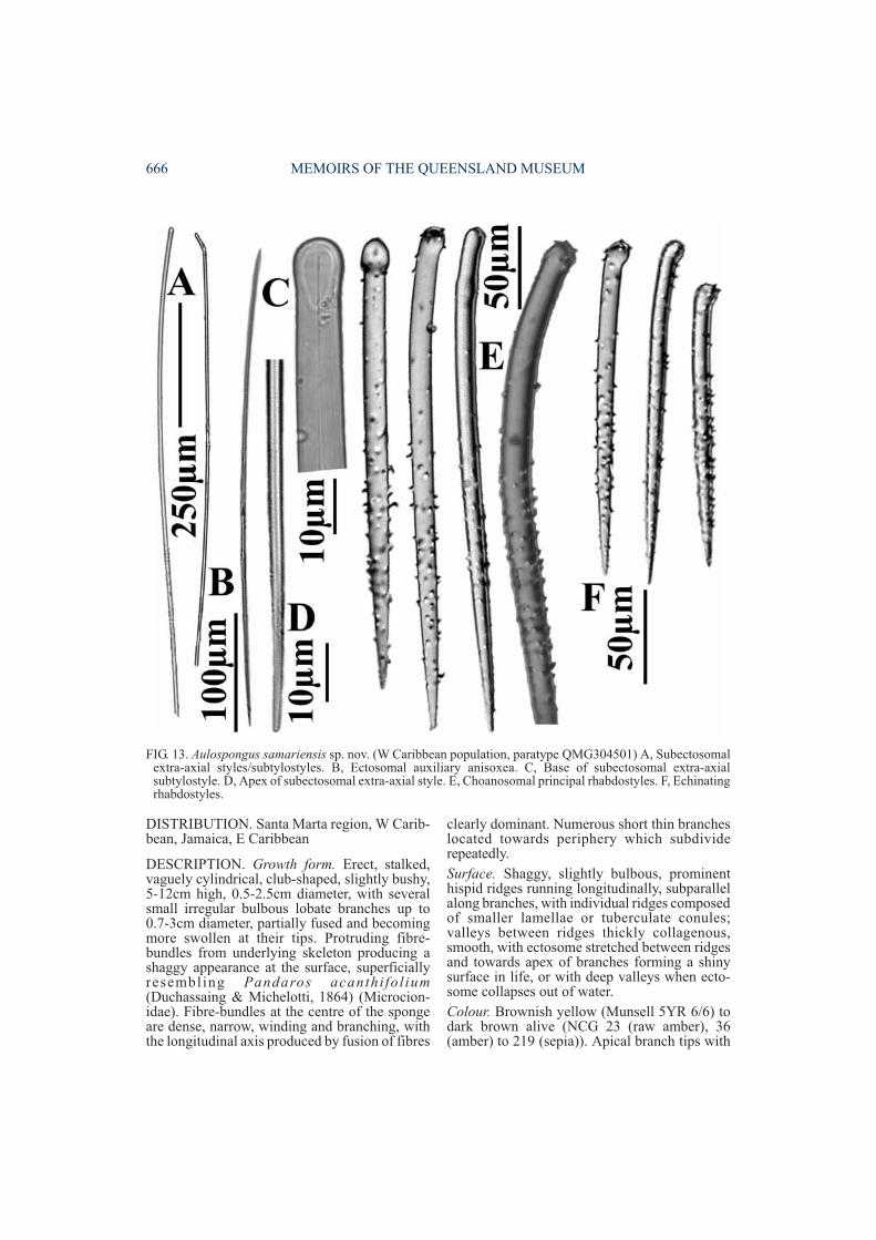
FIG. 13. Aulospongus samariensis sp. nov. (W Caribbean population, paratype QMG304501) A, Subectosomalextra-axial styles/subtylostyles. B, Ectosomal auxiliary anisoxea. C, Base of subectosomal extra-axialsubtylostyle. D, Apex of subectosomal extra-axial style. E, Choanosomal principal rhabdostyles. F, Echinatingrhabdostyles.
mustard yellow tinge (NCG 24 (buff)). Preservedspecimens evenly brown.
Oscules. Small,<0.5-2mm diameter, inter-dispersed in cavities on produced by folding ofsurface ridges, collapsing out of water.
Texture. Firm, compressible, with stiff, flexible,elastic branches.
Ectosomal skeleton. Ectosome with thick,organic, heavily collagenous matrix up to 200µmthick. ‘Raspailiid ectosomal skeleton’ presentconsisting of clusters of loose ectosomalauxiliary anisoxeas, forming bouquets on thesurface conules, surrounding the usually single,long subectosomal extra-axial styles at the pointthey protrude through the surface. Occasionalplumose bundles of larger choanosomal principalrhabdostyles also protrude through the surface(on conules), and individual rhabdostyles form
an evenly spaced palisade in between surfaceconules.
Choanosomal skeleton. Skeletal structure pre-dominantly plumose, only very faintly morecompressed, slightly reticulate, in axis than inperiphery. Axial skeletal reticulation composedof fibre-bundles more-or-less amalgamated intolarge tracts, sparsely interconnected by collagenand/or aspicular or paucispicular ‘secondaryfibres’. Fibres in peripheral skeleton with veryfew reticulate elements, disappearing closer tothe surface, with ascending fibre-bundlesdiverging and forming discrete conules at thesurface. Primary reticulate fibres and ascendingfibres fully cored by larger rhabdostyles,protruding through fibres at obtuse angles, andheavily echinated by both smaller and largerrhabdostyles forming heavy plumose tracts,producing clumps of spicules particularly at fibre
REVISION OF AULOSPONGUS 667
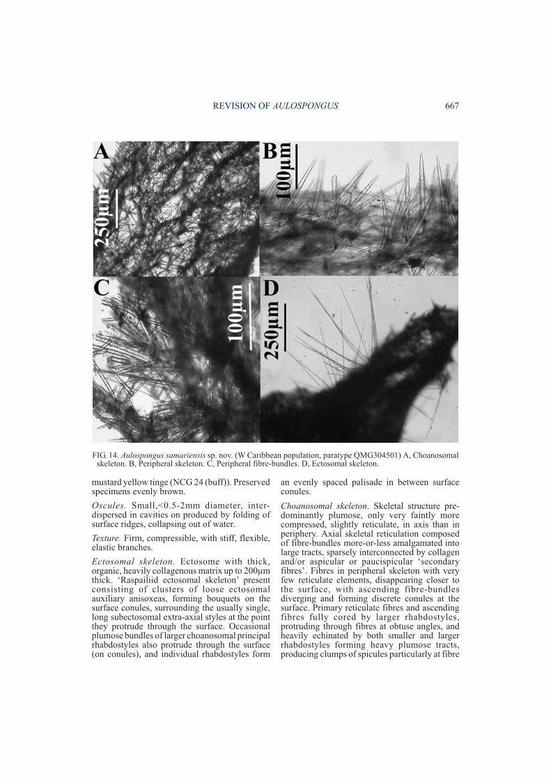
FIG. 14. Aulospongus samariensis sp. nov. (W Caribbean population, paratype QMG304501) A, Choanosomalskeleton. B, Peripheral skeleton. C, Peripheral fibre-bundles. D, Ectosomal skeleton.
nodes, with larger rhabdostyles dominating tractsand smaller ones interdispersed between them.Choanosomal principal rhabdostyles appearlarger in the periphery than in axial regions of theskeleton. Long subectosomal extra-axialsubtylostyles have their bases embedded inspongin fibres, forming sparse radial tracts
protruding a long way through the surfaceCollagen between the fibres is light, generallyaspicular, although multispicular tracts ofsubectosomal extra-axial styles run along thelongitudinal axis of branches towards thesurface, more-or-less parallel to (and external of)spongin fibres. Meshes between fibre are
668 MEMOIRS OF THE QUEENSLAND MUSEUM
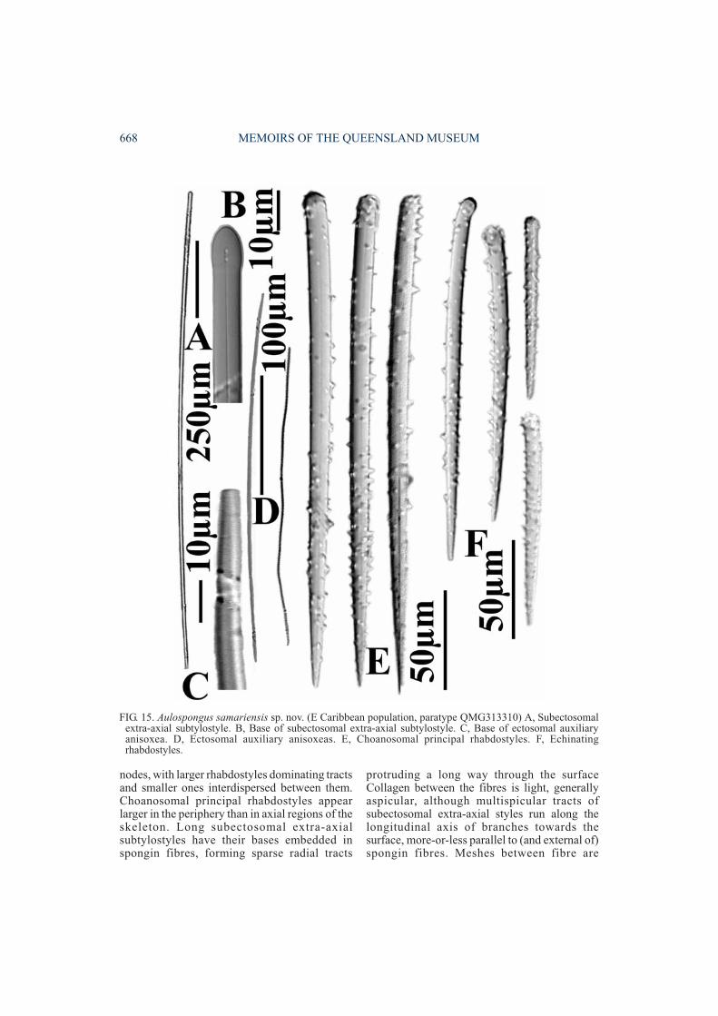
FIG. 15. Aulospongus samariensis sp. nov. (E Caribbean population, paratype QMG313310) A, Subectosomalextra-axial subtylostyle. B, Base of subectosomal extra-axial subtylostyle. C, Base of ectosomal auxiliaryanisoxea. D, Ectosomal auxiliary anisoxeas. E, Choanosomal principal rhabdostyles. F, Echinatingrhabdostyles.
relatively small, close-set, 150-250µm diameter,generally smaller in the axis than periphery of theskeleton.
Megascleres. Larger choanosomal principalrhabdostyles long, thick; shaft is nearly straight;bases slightly rhabdose; bases also lightlysubtylote to prominently tylote, with tylesterminal or subterminal; spination on spiculesvaries between E & W Caribbean populations(the former with more-or-less even spinationalthough generally heavier at distal and apicalends than at centre, spines relatively large, re-curved, hook-like; the latter with bases lightlyspined or occasionally completely smooth, withsmall recurved spines concentrated on apical endof spicule, aspinose below basal swelling); pointsare tapering, fusiform (218-(322.2)-412×9-(13.2)-18µm). Smaller echinat ing
rhabdostyles short, relatively thick; shaft straightor slightly curved at centre; basal rhabd slight oroccasionally completely straight; points fusiformtapering; bases range from slightly subtylote towell developed tylote; spines more evenlydispersed over whole spicule (as compared to thelarger size class), sometimes less spinose belowbasal swelling than elsewhere, sometimescompletely smooth below basal swelling; spinessmall, recurved (112-(181.2)-232×6-(9.3)-13µm). Several intermediate-sized rhabdostylespresent, linking larger and smaller classes.Subectosomal extra-axial styles very long, thin;shaft straight or slightly curved near basal end,occasionally flexuous; base smooth, rounded orslightly subtylote; points tapering to longfusiform (920-(1814.5)-2750×8-(18.3)-26µm).Ectosomal auxiliary anisoxeas moderately long,
REVISION OF AULOSPONGUS 669
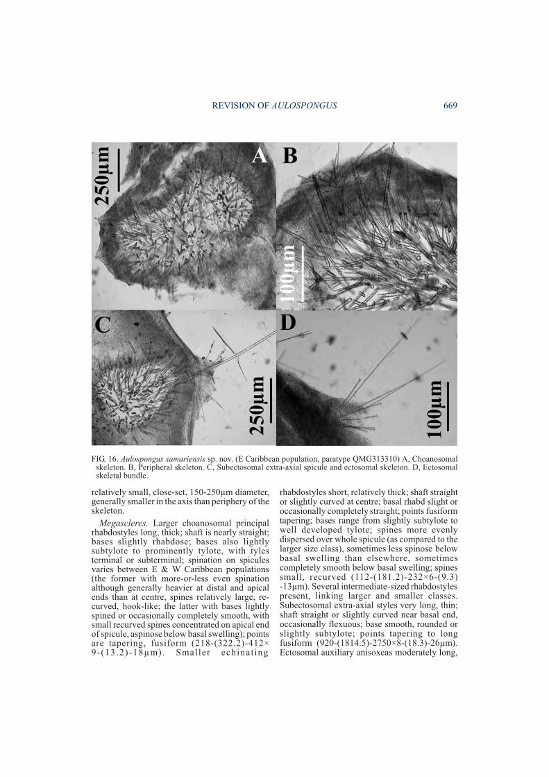
FIG. 16. Aulospongus samariensis sp. nov. (E Caribbean population, paratype QMG313310) A, Choanosomalskeleton. B, Peripheral skeleton. C, Subectosomal extra-axial spicule and ectosomal skeleton. D, Ectosomalskeletal bundle.
thin; shaft slightly curved at centre, occasionallyflexuous, sinuous or raphidiform; ends areasymmetrical with long tapering points andtapering rounded hastate base (225-(508.5)-775×2.5-(4.1)-5.5µm)Microscleres. Absent.
ECOLOGY. Aulospongus samariensis has notbeen recorded from any other locality in theColombian Caribbean. One of us (SZ) hasextensively investigated rocky-reef complexareas down to a depth of 40m from the border ofPanamá to Santa Marta in the continental coast ofColombia, and in the islands, atolls and banks ofthe San Andrés and Providencia Archipelago(San Andrés island, Old Providence island, Cour-town Cays, Albuquerque Cays, Serrana Bank,Roncador Bank, Quitasueño Bank), SWCaribbean. The fact that this species has beenfound elsewhere only in deep, insular drop-offs(i.e. Jamaica), suggests it may be a deep waterspecies, that comes up to shallower reefs (above18m depth) at Santa Marta, where there is aseasonal upwelling of the colder water mass(called ‘Subtropical Underwater’, 19-25°C,usually localized between 100-200m depth in theCaribbean; Bula-Meyer, 1985). There areunpublished examples of other sponges (andmany published records of algae), that follow thispattern, but this is the first published record ofthis phenomenon for sponges.
REMARKS. Initially the E (Jamaican) and W(Colombian) Caribbean populations werethought to be distinct species, showing someconsistent differences in growth form (moreelongate versus more bushy), ectosomal aux-iliary spicule geometry (anisoxeas with fusiformpoints versus those with hastate points), ecto-somal specialisation (ectosomal auxiliary spiculebrushes concentrated mainly around surfaceconules versus evenly hispid surface), andskeletal structure (axial skeleton morecompressed versus more reticulate, respective-ly). Spicule dimensions also varied slightlybetween the two populations (Table 2). However,upon further consideration these differences are
less obvious than their similarities, particularly inspicule geometry, and the two populations areconsidered to be conspecific.
This species belongs to Aulospongus in havingcharacteristic fused fibre-bundles forming adenser core in the axial part of the skeleton, and apredominantly plumose structure towards theperiphery; lacking any prominent differentiationbetween axial and extra-axial skeletons apartfrom the amalgamation of these fibres towardsthe centre of the skeleton, and having fibreswhich are cored and echinated by heavy bundlesof rhabdostyles, in two size classes.
Aulospongus samariensis differs from the‘typical tubular’ Aulospongus in its growth form(cylindr ical c lub-shaped) , rhabdostylemorphology (more-or-less even spination onboth size classes of spicules, slightly less spinedin the basal end, and with only a slight basalrhabd), possession of very long subectosomalextra-axial spicules protruding through the sur-face (in this regard similar only to A. involutum(Kirkpatrick)), and possession of a more-or-lesswell developed, specialised raspailiid ectosomalskeleton. Ectosomal skeletons are welldeveloped in only two species of Aulospongus(A. gardineri and the present species), consistingof plumose brushes of ectosomal auxiliary styles/anisoxeas surrounding longer subectosomalextra-axial styles/anisoxeas. By comparison,vestigial ectosomal skeletons are present in threespecies (A. involutum, A. novaecaledoniensis sp.nov., A. tubulatus), consisting of ectosomalauxiliary styles/anisoxeas scattered on or belowthe surface, but not forming brushes and notnecessarily associated with protruding subecto-somal extra-axial styles/anisoxeas. Ectosomalauxiliary spicules and a specialised ‘raspailiid’ectosomal structure are absent in four species (A.cerebella, A. monticularis, A. spinosum, and A.villosa), presumably a derived condition.
This species is similar to A. gardineri inectosomal skeletal structure, A. involutum inpossession of long subectosomal extra-axialspicules, and to both these species plus A.novaecaledoniensis in having both categories of
670 MEMOIRS OF THE QUEENSLAND MUSEUM
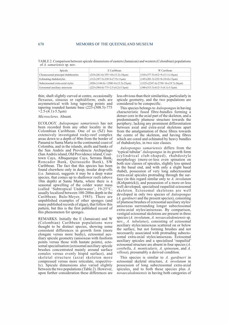
Spicule E Caribbean W Carribean
Choanosomal principal rhabdostyles (218-(261.6)-355×10-(13.2)-18µm) (310-(377.5)-412×9-(13.1)-16µm)
Echinating rhabdostyles (112-(187.3)-210×6-(7.5)-11µm) (145-(201.3)-232×8-(10.4)-13µm)
Subectosomal extra-axial styles (920-(1144.6)->2500×8-(15.5)-25µm) (1325-(2247.6)-2750×16-(19.7)-26µm)
Ectosomal auxiliary anisoxeas (225-(584.4)-775×2.5-(4.2)-5.5µm) (340-(515.3)-612×3-(4.1)-5.5µm)
TABLE 2. Comparison between spicule dimensions of eastern (Jamaican) and western (Colombian) populationsof A. samariensis sp. nov.
rhabdostyles partially spined. In other details,however, it differs substantially from all otherspecies, particularly in spicule geometries (Table1) and dimensions (Table 2).
To some extent this species also resemblesRaspailia acanthifera (George & Wilson, 1919:159) from North Carolina, particularly in itsgrowth form (lobate, with lamellate branches),some aspects of skeletal architecture (longitud-inal multispicular fibres at the core of theskeleton with only few interconnectingpaucispicular transverse fibres; peripheral fibresbecoming more radial with fewerinterconnecting tracts towards the surface;peripheral fibres fully cored by styles whicheventually project through surface in bundlesforming surface conules), and ectosomal char-acteristics (projecting long subectosomalextra-axial styles forming loose bundles at thesurface). Conversely, spicule morphology andspicule distribution within the skeleton differsubstantially between the two species. In R.acanthifera there are five categories ofmegascleres, each substantially different fromthose of A. samariensis sp. nov., and skeletalstructure of R. acanthifera is also markedlyaxially compressed, indicating the latter speciesshould be assigned to Raspailia (Raspaxilla) (seebelow), whereas this species is more approp-riately referred to Aulospongus.
Colombian populations of A. samariensis werefound to contain both slight antimicrobial againstStaphylococcus aureus (for ethanol andchloroform extracts) (Silvestri, Zea & Duque,1994), and strong antitumor activity (Zea,unpublished data), further supporting therelatively high incidence of ‘biological activity’reported amongst species of Raspailiidae(Hooper et al., 1992).
Aulospongus spinosum (Topsent, 1927)(Fig. 17, Table 1)
Rhaphidectyon spinosum Topsent, 1927: 15; 1928: 288, pl.2, fig. 5, pl. 9, fig. 28, pl. 10, figs 2-3; Lévi, 1960: 752,fig. 7.
Aulospongus spinosum; Hooper, 1991: 1307, fig. 65g-i;Maldonado, 1992: 1149-1150, fig. 9e-j.
MATERIAL. HOLOTYPE. MOM (schizotypes MNHNLBIM DT1139, BMNH1930.7.1.39): Cape Verde Is, nearSão Vicente I., 16 48’N, 25 06’W, coll. ‘Princesse-Alice’,29.vii.1901, 219m depth.
DISTRIBUTION. São Vicente I., North Atlantic,Alboran I., Mediterranean.
DESCRIPTION. Growth form bulbous, erect.Surface shaggy, conulose. Colour dark grey inethanol. ‘Raspailiid ectosomal skeleton’ absent,with only larger smooth rhabdostyles protrudingthrough the surface forming shaggy surfaceprocesses. Choanosomal skeleton distinctlyplumose in both axial and extra-axial regions,composed of very stout, widely separatedascending fibre-bundles with very fewinterconnecting tracts. Fibres cored by larger(smooth) styles and rhabdostyles (virtuallyinseparable in their morphology), forming radialtracts in the axis of the skeleton but becomingprogressively thicker towards the periphery,ending in discrete plumose bundles at the surface.Largest rhabdostyles/styles appear to be locatedin the peripheral skeleton. Megascleres includelong, thick, choanosomal principal styles andrhabdostyles, completely smooth, with veryslight to moderate basal rhabd, the largestoccasionally nearly straight at the base(770-1085×28-43µm). Two sizes of smallerrhabdostyles present: long, slender ones,completely smooth, with well curved basalrhabd, predominantly found in choanosomal andperipheral fibres, protruding from fibres atslightly acute angles (90-185×5-12µm), and trueechinating acanthostyles with only slight or nobasal rhabd, relatively evenly spined, Eurypon-like, with swollen subtylote bases bearing verylarge perpendicular spines (75-145×7-10µm).Subectosomal extra-axial and ectosomalauxiliary spicules absent. Microscleres areraphides occurring singly or in trichodragmata(40-50µm long).
REMARKS. The second category of (smooth)echinating rhabdostyles was overlooked byprevious authors, and appears to be quitedifferent from the acanthose echinatingacanthostyles. In this regard it is similar to A.gardineri, although differing in most othercharacters (e.g. spicule morphology andspination, spicule sizes, growth form, lack ofspecialised ectosomal skeleton and possession ofraphides in A. spinosum).
This species is also highly derived, reduced inmost of its morphological characters, and differsfrom other known Aulospongus in havingraphide microscleres dispersed throughout theskeleton. Although several other species ofAulospongus have been described at some time oranother with raphide microscleres ,re-examination of relevant type and othermaterial has confirmed that in all cases these were
REVISION OF AULOSPONGUS 671
vestigial (raphidiform) ectosomal auxiliaryspicules, whereas in A. spinosum these appear tobe genuine raphides/ tr ichodragmata.Nevertheless, Maldonado (1992) notes thatraphides appear to be associated with, and pos-sibly reinforce, the pinacoderm. If this correct, itis possible that these raphides may be extremelyvestigial ectosomal auxiliary megascleres ratherthan typical microscleres. Unfortunately nowell-fixed material is available to undertakemore comprehensive histological analysis toexplore this possibility, and for the moment thesespicules are assumed to be microscleres.
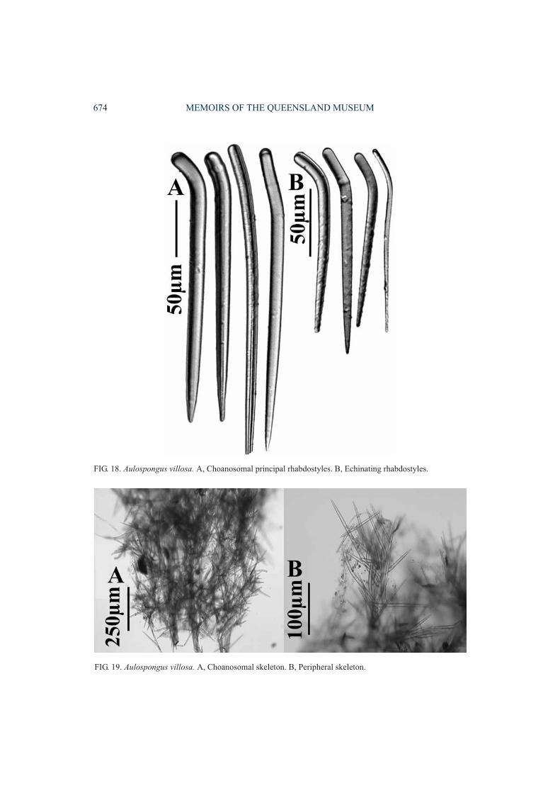
Aulospongus villosa (Thiele, 1898)(Figs 18-19, Table 1)
Raspailia (?) villosa Thiele, 1898: 60, pl. 4, fig. 10, pl. 8,fig. 48; Koltun, 1970: 270, fig. 32, pl. 8, fig. 4; Hoshino,1987: 19; Tanita & Hoshino, 1990: 102; Sim, 1990: 317.
Heterectya villosa; Hallmann, 1917: 393; [? doubtful re-cord of Burton, 1959: 45].
Aulospongus villosa; Hooper, 1991: 1307, fig. 65d-f.
MATERIAL. HOLOTYPE. ZMB2204: Hakodate, Japan,coll. Hilgendorf.
DISTRIBUTION. Japan (Hakodate, SagamiBay), Korea (Japan Sea & Jeju I.) and Russia(Iturup I., Kurile Is, Sea of Ochotsk). Burton’s(1959) record from Iceland is dubious. He did notprovide a description of the specimen, novoucher material was cited, and it is thereforeignored here.
DESCRIPTION. Growth form massive, sub-spherical, bushy. Surface prominently shaggy,conulose. Colour light brown in ethanol.‘Raspailiid ectosomal skeleton’absent, with onlytufts of choanosomal principal rhabdostylesprotruding. Choanosomal skeleton exclusively
672 MEMOIRS OF THE QUEENSLAND MUSEUM
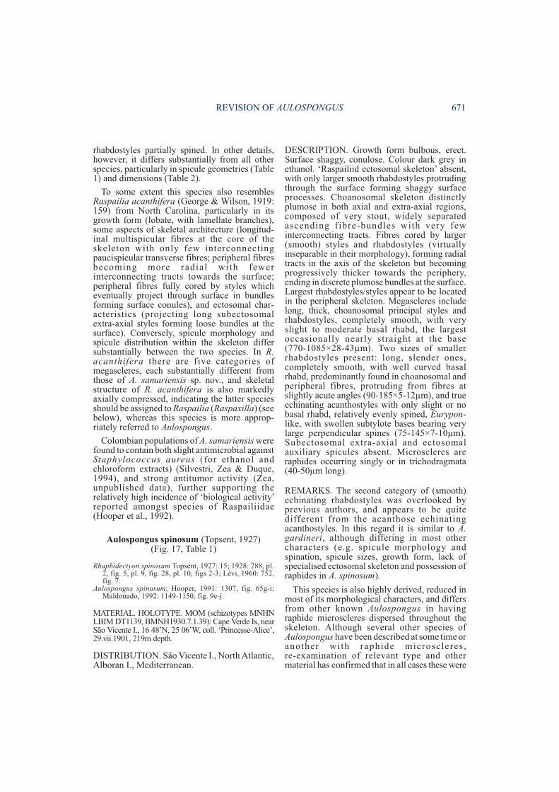
FIG. 17. Aulospongus spinosum. A, Third category of rhabdostyle. B, Echinating style/rhabdostyle. C,Choanosomal principal rhabdostyle. D, Raphides. E, Ectosomal auxiliary spicule bundle. F, Choanosomal fibrebundle. G, Choanosomal skeleton.
plumose, composed of relatively thick,compressed fibre-bundles in which the largerrhabdostyles are confined completely withinascending fibres. Smaller rhabdostyles ex-clusively echinate fibres, standing nearlyperpendicular to them, often touching those onadjacent, opposing fibres, together producing thesuperficial impression of a lattice-like, reticulateskeleton. No notable compression of the axialskeleton, and no specialised subectosomalextra-axial megascleres present. Megascleresconsist of larger choanosomal principalrhabdostyles with moderate to stronglydeveloped basal rhabd, usually completelysmooth or with small granular spines scatteredover the apical half (235-370×10-16µm).Smaller echinating rhabdostyles vary fromcompletely smooth to partially spined on theapical half, usually with a very strong basal rhabdbut occasionally straight, spines very small,granular (142-165×4-10µm). Subectosomalextra-axial and ectosomal auxiliary spiculesabsent. Microscleres absent.
REMARKS. This is a very reduced species ofAulospongus, similar to A. cerebella, A. flabel-lum and A. spinosum, lacking any ectosomalauxiliary or subectosomal extra-axial spicules,and having only two categories of rhabdostyles.Rhabdostyles in A. villosa resemble to someextent those of A. involutum, A. gardineri and A.novaecaledoniensis sp.nov, in geometry andapproximate size, but whereas those of A. villosaare often completely smooth or have smallgranular spines the other three species have verylarge, recurved spines covering only the apex ofrhabdostyles.
REVIEW OF OTHER RASPAILIIDAE WITHRHABDOSTYLES
Raspailia Nardo, 1833Subgenus Raspaxilla Topsent, 1913
Raspaxilla Topsent, 1913: 616; Bergquist, 1970: 28;Hooper, 1991: 1195, 1245. Type species: Raspaxillaphakellina Topsent, 1913: 617, by monotypy.
Echinaxia Hallmann, 1916a: 543; 1917: 391; deLaubenfels, 1936: 102; Bergquist, 1970: 30; Hooper,1991: 1195. Type species : Axinel la frondulaWhitelegge, 1907: 509, by original designation.
Axinectya Hallmann, 1917: 393; Hooper, 1991: 1195. Typespecies: Axinella mariana Ridley & Dendy, 1886: 480,by original designation.
DEFINITION. Raspailia with echinatingacanthose rhabdostyles. Larger choanosomalprincipal styles completely smooth, without any
basal rhabd, geometrically distinct from smalleracanthose echinating rhabdostyles. Axialskeleton well differentiated from extra-axialskeleton: axial skeletal compressed, composed ofreticulate tracts cored by choanosomal principalstyles; extra-axial skeleton plumo-reticulate,with plumose ascending tracts interconnected bytransverse tracts both cored by choanosomalprincipal styles (forming a reticulation), or reduc-ed to radial tracts of single long subectosomalextra-axial styles embedded in and perpendicularto axis, protruding through the surface.Echinating rhabdostyles generally moreabundant in peripheral skeleton than in axis.
REMARKS. Seventeen species are currentlyassigned to Raspailia (Raspaxilla), includingspecies transferred here from Endectyon,Hemectyon and Aulospongus. Raspaxilla has awide geographic distribution, ranging from theIndo-west Pacific (N and S New Zealand, NWAustralia, N Great Barrier Reef, central NSW,New Caledonia, Japan, Micronesia), E coast ofthe United States of America (North Carolina),central E Pacific and the antarctic-subantarcticregion (Fig. 35). Apart from the Southern (sub-antarctic) Ocean, Raspaxilla has not yet beenrecorded in either the Atlantic or the E or centralIndian Oceans, and is assumed (from presentdata) to be a Pacific rim species (Fig. 35). It ispossible that the specimen described byPulitzer-Finali (1994) as ‘Endectyon hamatum‘from Kenya belongs to Raspaxilla, but thisspecies is barely recognisable from his descript-ion and for the moment is incertae sedis.
Essentially Raspailia (Raspaxilla) differs fromAulospongus in having echinating rhabdostylesgeometrically very different from the usuallylonger choanosomal principal styles (the latterwithout any basal rhabd); the subectosomalextra-axial styles form a radial skeletonperpendicular to the axis; and axial andextra-axial skeletons are well differentiated (theformer compressed, the latter plumoreticulateand/or radial). Placement of all species, however,is not always straightforward given that sometaxa may lose certain characters (e.g. extra-axialskeleton becomes reduced to single longsubectosomal extra-axial spicules embedded inthe axis and forming a radial skeleton; or thesubectosomal extra-axial spicules are lostcompletely). There is also a correlation betweenthe localisation of echinating rhabdostyles in theperipheral skeleton and the degree of axialcompression. In species with very compressed
REVISION OF AULOSPONGUS 673
674 MEMOIRS OF THE QUEENSLAND MUSEUM
FIG. 19. Aulospongus villosa. A, Choanosomal skeleton. B, Peripheral skeleton.
FIG. 18. Aulospongus villosa. A, Choanosomal principal rhabdostyles. B, Echinating rhabdostyles.
skeletons the extra-axial skeleton is reduced tosingle long subectosomal extra-axial spicules(without reticulate connections) and theechinating rhabdostyles are literally ‘pushed’into the ectosomal skeleton where they formbrushes or produce a continuous palisade.
Raspailia (Raspaxilla) phakellina(Topsent, 1913)
(Fig. 20, Table 1)
Raspaxilla phakellina Topsent, 1913: 617, pl. 1, fig. 4, pl.6, fig. 15; Burton, 1932: 326; Boury-Esnault & vanBeveren, 1982: 51, pl. 7, fig. 26, fig. 12.
Raspailia (Raspaxilla) phakellina; Hooper, 1991: 1196,fig. 7k-l.
MATERIAL. HOLOTYPE. MOM (fragment MNHNLBIM DT1614): Burwood Bank, Antarctica, 54�25’S,57�32’E, 112m depth, 1.xii.1903. OTHER MATERIAL.BMNH1928.2.15.781a, 846a: Falkland Islands,Argentina, RRS ‘Discovery’, 75-82m depth.
DISTRIBUTION. Antarctic - subantarcticregion (Antarctica, Falkland Is, Kerguelen Is).
DESCRIPTION. Erect, digitate, arborescent,with enlarged basal holdfast attachment andbranching in one plane. Surface slightly conu-lose, hispid. Colour whitish cream to yellowish inethanol. ‘Raspailiid ectosomal skeleton’ presentbut not well developed, consisting of longsubectosomal extra-axial styles protrudingthrough the surface, surrounded at their bases bywispy bundles of ectosomal auxiliary anisoxeasforming stellate bundles nearly parallel to thesurface. Choanosomal skeleton with welldifferentiated axial and extra-axial regions. Axialskeleton compressed, strongly reticulate,composed of multispicular fibres cored bychoanosomal principal styles and echinatedsparsely by echinating rhabdostyles. Extra-axialskeleton plumo-reticulate, with ascendingmultispicular fibres cored by choanosomalprincipal styles and profusely echinated byrhabdostyles, diverging towards the periphery,interconnected by transverse pauci- ormultispicular fibres which persist all the way tothe surface. Echinating rhabdostyles predom-inant in extra-axial skeleton. Megascleresinclude long choanosomal principal styles,slightly curved centrally or straight, with evenlyrounded, smooth, non-rhabdose bases (550-900×10-16µm). Echinating rhabdostyles moderatelylong, with slightly rhabdose and subtylote bases,completely smooth or with small, erect spinesconfined to apical two-thirds of spicule(140-370×8-18µm). Subectosomal extra-axial
styles long, thick, straight or slightly curved,entirely smooth (1100-1450×12-18µm).Ectosomal auxiliary styles wispy, raphidiform,straight, centrally curved or sinuous (450-650×2-3µm). Micrscleres absent.
REMARKS. Comparisons between the holotype(Antarctica) and Kerguelen specimens, with the‘Discovery’ material described by Burton (1932)show some differences: notably the completelack of spines on rhabdostyles in the latterspecimen (whereas some rhabdostyles haveapical spines in the former specimens), and thepersistence of the plumo-reticulate extra-axialskeleton for most of the sponge diameter, withonly a relatively small, confused, slightlycompressed axial component (as compared to arelatively larger central axial skeleton in theholotype and Kerguelen specimen). Never-theless, there is no doubt that all three specimensbelong to the same species . Raspail ia(Raspaxilla) phakellina differs substantiallyfrom other Raspailia species only in having basalrhabds on echinating acanthostyles, and havingits extra-axial skeleton dominated by ascendingfibres reminiscent of Aulospongus, but in whichtransverse connecting fibres persist all the way tothe surface and produce a plumo-reticulateskeletal structure (rather than a purely plumoseskeleton characteristic of Aulospongus).
Raspailia (Raspaxilla) acanthiferaGeorge & Wilson, 1919
Raspailia acanthifera George & Wilson, 1919: 159, pl. 62,fig. 34, pl. 63, fig. 38-39, pl. 66, fig. 59.
MATERIAL. HOLOTYPE. USNM (not seen): FortMacon beach, Beaufort, North Carolina.
DISTRIBUTION. E coast of USA (North Car-olina).
DESCRIPTION.Growth form stalked, lobate,with lamellate branches. Surface with hispidridges but smooth between ridges. Colourgrey-brown with tinge of yellow in ethanol.‘Raspailiid ectosomal skeleton’ well developedwith projecting long subectosomal extra-axialstyles surrounded by ectosomal auxiliaryanisoxeas forming loose bundles at the surface.Skeletal architecture with well differentiatedaxial and extra-axial components. Axial skeletoncompressed, with mainly thicker longitudinalfibres fully cored with spicules, looselyinterconnected by few transverse paucispicularfibres, together producing a loose, openreticulation. Extra-axial skeleton radial, with
REVISION OF AULOSPONGUS 675
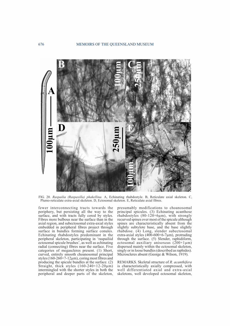
fewer interconnecting tracts towards theperiphery, but persisting all the way to thesurface, and with tracts fully cored by styles.Fibres more bulbous near the surface than in theaxial region, and subectosomal extra-axial stylesembedded in peripheral fibres project throughsurface in bundles forming surface conules.Echinating rhabdostyles predominant in theperipheral skeleton, participating in ‘raspailiidectosomal spicule brushes’, as well as echinatingradial (connecting) fibres near the surface. Fivecategories of megascleres present. (1) Short,curved, entirely smooth choanosomal principalstyles (160-260×7-12µm), coring most fibres andproducing the spicule bundles at the surface. (2)Straight, thick styles (160-240×12-20µm)intermingled with the shorter styles in both theperipheral and deeper parts of the skeleton,
presumably modifications to choanosomalprincipal spicules. (3) Echinating acanthoserhabdostyles (80-120×6µm), with stronglyrecurved spines over most of the spicule althoughspines are characteristically absent from theslightly subtylote base, and the base slightlyrhabdose. (4) Long, slender subectosomalextra-axial styles (400-600×6-7µm), protrudingthrough the surface. (5) Slender, raphidiform,ectosomal auxiliary anisoxeas (200×1µm)dispersed mainly within the ectosomal skeleton,singly or in loose bundles (described as raphides).Microscleres absent (George & Wilson, 1919).
REMARKS. Skeletal structure of R. acanthiferais characteristically axially compressed, withwell differentiated axial and extra-axialskeletons, well developed ectosomal skeleton,
676 MEMOIRS OF THE QUEENSLAND MUSEUM
FIG. 20. Raspailia (Raspaxilla) phakellina. A, Echinating rhabdostyle. B, Reticulate axial skeleton. C,Plumo-reticulate extra-axial skeleton. D, Ectosomal skeleton. E, Reticulate axial fibres.
and geometrical ly different echinatingrhabdostyles and choanosomal principal styles,indicating that it is most appropriately assignedto Raspailia (Raspaxilla) and not to Aulo-spongus.
Raspailia (Raspaxilla) clathrioides(Lévi, 1967)
(Figs 21, 36C)
Aulospongus clathrioides Lévi, 1967: 21, text-fig. 5, pl. 2,fig. c; Hooper, 1991: 1311, fig. 67a-d; Hooper & Lévi,1993: 1295, figs 40-41; Hooper & Battershill, 1998:136.
MATERIAL. HOLOTYPE. MNHN LBIM DCL823:Canala, Melasceu, New Caledonia, coll.‘Singer-Polignac’, 4.i.1962, 22°30’S, 166°45’E, 35mdepth. OTHER MATERIAL. New Caledonia (refer toHooper & Lévi, 1993).
DISTRIBUTION. Known only from the SWNew Caledonian lagoon.
DESCRIPTION. Growth form stalked, arbor-escent, bushy, cylindrical-branching. Surfaceshaggy, prominently conulose. Colour yellow-orange alive. ‘Raspailiid ectosomal skeleton’either completely absent or with very fewvestigial ectosomal auxiliary styles/anisoxeas,forming vestigial brushes surrounding pro-truding choanosomal principal styles or scatteredbelow the surface. Subectosomal extra-axialspicules absent. Surface dominated by swollenfibres and plumose brushes of smooth styles bothproducing surface conules. Choanosomalskeleton predominantly plumose orplumo-reticulate, with a slightly compressedreticulate axial core dominated by welldeveloped spongin fibres, with the spiculeskeleton proportionally reduced. Axial fibres arevestigially cored by choanosomal principalstyles. Extra-axial fibres are prominentlyplumose and heavily multispicular, with fewerpaucispicular interconnecting fibres persisting inthe peripheral skeleton and to the surface.Echinating rhabdostyles are slightly moreabundant in the peripheral skeleton than in theaxis. Megascleres include choanosomal principalstyles, smooth, slightly curved or occasionallysinuous, sometimes modified to anisoxeas, onlyoccasionally with a very faint basal curvature(145-454×3-7µm). Echinating rhabdostyles,microcionid-like, long, slender, straight orslightly curved towards the basal end, with arelatively well developed basal swelling, straightbase or only slight basal rhabd, evenly spinedexcept for smooth base, spines small, granular
(58-82×1.5-4µm). Subectosomal extra-axialspicules absent. Ectosomal auxiliary styles andanisoxeas present or absent, always very rare,raphidiform, straight or sinuous (97-134×0.8-1.5µm). Microscleres absent.
REMARKS. This species is a borderline casebetween Aulospongus and Raspaxilla, and isreferred here to Raspailia (Raspaxilla) given thatthe ascending fibre system has transverseconnecting fibres extending all the way from theaxis to the surface (producing a plumo-reticulaterather than a strictly plumose skeleton as istypical for Aulospongus); the axial andextra-axial skeletons are slightly differentiated(reticulate versus plumo-reticulate), with the axisshowing slight compression; the largerchoanosomal principal styles are entirelysmooth, and they have no, or at most only veryfaint, basal curvature (i.e. only a few might betermed vaguely rhabdose), and furthermore,anisoxeote modifications of choanosomalprincipal spicules are relatively common,indicating that choanosomal principal andechinating spicules are morphologically quitedistinct; and the smaller echinating rhabdostylesalso have only slight basal rhabds, more incommon with species of Raspaxilla than withAulospongus.
This is a sister species of R. (Raspaxilla)reticulata (N Great Barrier Reef) and R. (R.)topsenti (N New Zealand), having a similararborescent growth form, echinat ingrhabdostyles with swollen, smooth, straight- orrelatively poorly rhabdose-bases (at leastcompared to most species of Raspaxilla andAulospongus); the reticulate axial region isslightly compressed; and the extra-axial skeletonis plumo-reticulate in which the ascendingplumose fibres dominate but the reticulate fibreconnections persist all the way to the surface. Thethree species differ in their spicule compositions(presence of a specialised ectosomal skeleton inR. (R.) reticulata, including possession of longsubectosomal extra-axial styles; vestigialectosomal auxiliary styles/anisoxeas in R. (R.)topsenti), spicule sizes (compare with therespective descriptions below), morphology ofchoanosomal principal styles (particularly R. (R.)topsenti), and greater dominance of the reticulateversus plumose components of the skeleton inboth the other species.
Re-examination of the material described asAulospongus clathrioides by Hooper & Lévi(1993) also found that one specimen out of four
REVISION OF AULOSPONGUS 677
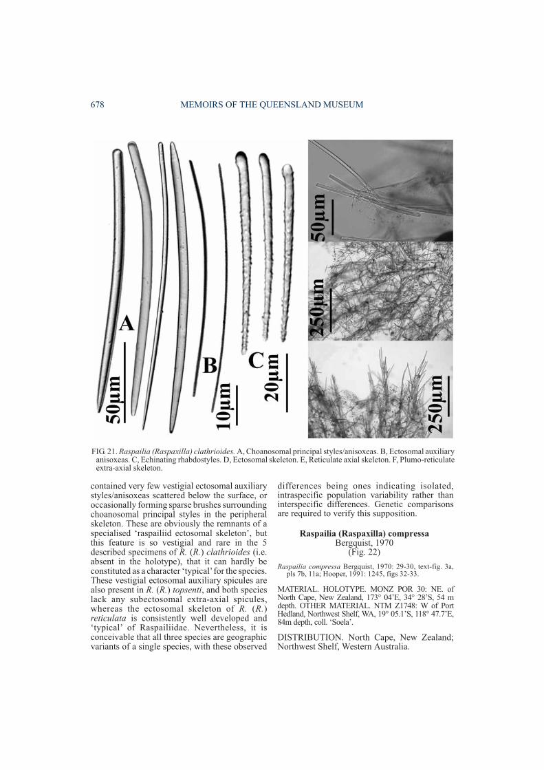
contained very few vestigial ectosomal auxiliarystyles/anisoxeas scattered below the surface, oroccasionally forming sparse brushes surroundingchoanosomal principal styles in the peripheralskeleton. These are obviously the remnants of aspecialised ‘raspailiid ectosomal skeleton’, butthis feature is so vestigial and rare in the 5described specimens of R. (R.) clathrioides (i.e.absent in the holotype), that it can hardly beconstituted as a character ‘typical’for the species.These vestigial ectosomal auxiliary spicules arealso present in R. (R.) topsenti, and both specieslack any subectosomal extra-axial spicules,whereas the ectosomal skeleton of R. (R.)reticulata is consistently well developed and‘typical’ of Raspailiidae. Nevertheless, it isconceivable that all three species are geographicvariants of a single species, with these observed
differences being ones indicating isolated,intraspecific population variability rather thaninterspecific differences. Genetic comparisonsare required to verify this supposition.
Raspailia (Raspaxilla) compressaBergquist, 1970
(Fig. 22)
Raspailia compressa Bergquist, 1970: 29-30, text-fig. 3a,pls 7b, 11a; Hooper, 1991: 1245, figs 32-33.
MATERIAL. HOLOTYPE. MONZ POR 30: NE. ofNorth Cape, New Zealand, 173° 04’E, 34° 28’S, 54 mdepth. OTHER MATERIAL. NTM Z1748: W of PortHedland, Northwest Shelf, WA, 19° 05.1’S, 118° 47.7’E,84m depth, coll. ‘Soela’.
DISTRIBUTION. North Cape, New Zealand;Northwest Shelf, Western Australia.
678 MEMOIRS OF THE QUEENSLAND MUSEUM
FIG. 21. Raspailia (Raspaxilla) clathrioides. A, Choanosomal principal styles/anisoxeas. B, Ectosomal auxiliaryanisoxeas. C, Echinating rhabdostyles. D, Ectosomal skeleton. E, Reticulate axial skeleton. F, Plumo-reticulateextra-axial skeleton.
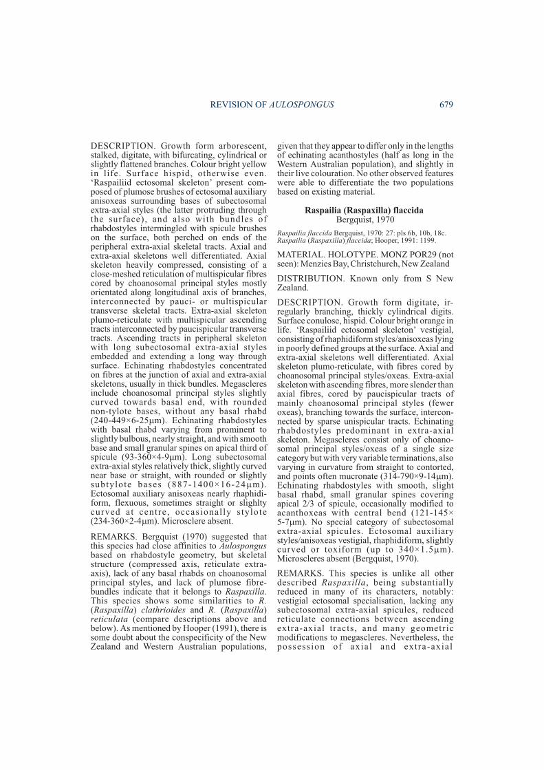
DESCRIPTION. Growth form arborescent,stalked, digitate, with bifurcating, cylindrical orslightly flattened branches. Colour bright yellowin life. Surface hispid, otherwise even.‘Raspailiid ectosomal skeleton’ present com-posed of plumose brushes of ectosomal auxiliaryanisoxeas surrounding bases of subectosomalextra-axial styles (the latter protruding throughthe surface), and also with bundles ofrhabdostyles intermingled with spicule brusheson the surface, both perched on ends of theperipheral extra-axial skeletal tracts. Axial andextra-axial skeletons well differentiated. Axialskeleton heavily compressed, consisting of aclose-meshed reticulation of multispicular fibrescored by choanosomal principal styles mostlyorientated along longitudinal axis of branches,interconnected by pauci- or multispiculartransverse skeletal tracts. Extra-axial skeletonplumo-reticulate with multispicular ascendingtracts interconnected by paucispicular transversetracts. Ascending tracts in peripheral skeletonwith long subectosomal extra-axial stylesembedded and extending a long way throughsurface. Echinating rhabdostyles concentratedon fibres at the junction of axial and extra-axialskeletons, usually in thick bundles. Megascleresinclude choanosomal principal styles slightlycurved towards basal end, with roundednon-tylote bases, without any basal rhabd(240-449×6-25�m). Echinating rhabdostyleswith basal rhabd varying from prominent toslightly bulbous, nearly straight, and with smoothbase and small granular spines on apical third ofspicule (93-360×4-9�m). Long subectosomalextra-axial styles relatively thick, slightly curvednear base or straight, with rounded or slightlysubtylote bases (887-1400×16-24�m).Ectosomal auxiliary anisoxeas nearly rhaphidi-form, flexuous, sometimes straight or slighltycurved at centre , occasional ly stylote(234-360×2-4�m). Microsclere absent.
REMARKS. Bergquist (1970) suggested thatthis species had close affinities to Aulospongusbased on rhabdostyle geometry, but skeletalstructure (compressed axis, reticulate extra-axis), lack of any basal rhabds on choanosomalprincipal styles, and lack of plumose fibre-bundles indicate that it belongs to Raspaxilla.This species shows some similarities to R.(Raspaxilla) clathrioides and R. (Raspaxilla)reticulata (compare descriptions above andbelow). As mentioned by Hooper (1991), there issome doubt about the conspecificity of the NewZealand and Western Australian populations,
given that they appear to differ only in the lengthsof echinating acanthostyles (half as long in theWestern Australian population), and slightly intheir live colouration. No other observed featureswere able to differentiate the two populationsbased on existing material.
Raspailia (Raspaxilla) flaccidaBergquist, 1970
Raspailia flaccida Bergquist, 1970: 27: pls 6b, 10b, 18c.Raspailia (Raspaxilla) flaccida; Hooper, 1991: 1199.
MATERIAL. HOLOTYPE. MONZ POR29 (notseen): Menzies Bay, Christchurch, New Zealand
DISTRIBUTION. Known only from S NewZealand.
DESCRIPTION. Growth form digitate, ir-regularly branching, thickly cylindrical digits.Surface conulose, hispid. Colour bright orange inlife. ‘Raspailiid ectosomal skeleton’ vestigial,consisting of rhaphidiform styles/anisoxeas lyingin poorly defined groups at the surface. Axial andextra-axial skeletons well differentiated. Axialskeleton plumo-reticulate, with fibres cored bychoanosomal principal styles/oxeas. Extra-axialskeleton with ascending fibres, more slender thanaxial fibres, cored by paucispicular tracts ofmainly choanosomal principal styles (feweroxeas), branching towards the surface, intercon-nected by sparse unispicular tracts. Echinatingrhabdostyles predominant in extra-axialskeleton. Megascleres consist only of choano-somal principal styles/oxeas of a single sizecategory but with very variable terminations, alsovarying in curvature from straight to contorted,and points often mucronate (314-790×9-14�m).Echinating rhabdostyles with smooth, slightbasal rhabd, small granular spines coveringapical 2/3 of spicule, occasionally modified toacanthoxeas with central bend (121-145×5-7�m). No special category of subectosomalextra-axial spicules. Ectosomal auxiliarystyles/anisoxeas vestigial, rhaphidiform, slightlycurved or toxiform (up to 340×1.5�m).Microscleres absent (Bergquist, 1970).
REMARKS. This species is unlike all otherdescribed Raspaxilla, being substantiallyreduced in many of its characters, notably:vestigial ectosomal specialisation, lacking anysubectosomal extra-axial spicules, reducedreticulate connections between ascendingextra-axial tracts, and many geometricmodifications to megascleres. Nevertheless, thepossession of axial and extra-axial
REVISION OF AULOSPONGUS 679
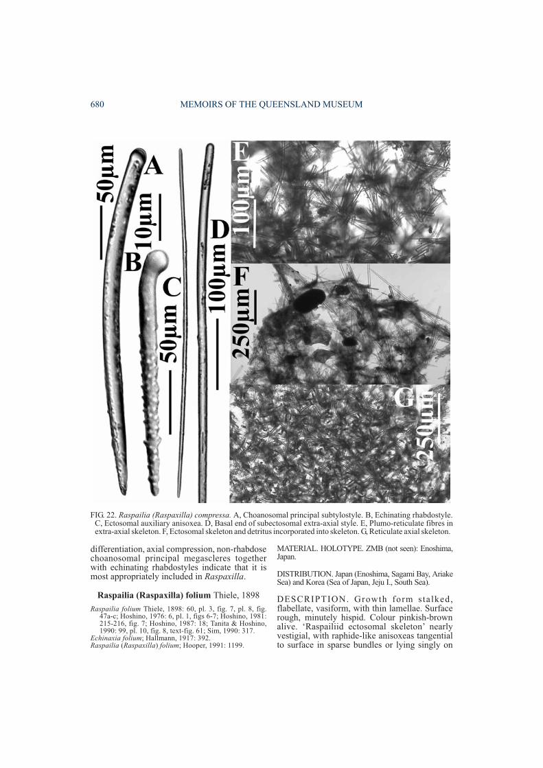
differentiation, axial compression, non-rhabdosechoanosomal principal megascleres togetherwith echinating rhabdostyles indicate that it ismost appropriately included in Raspaxilla.
Raspailia (Raspaxilla) folium Thiele, 1898
Raspailia folium Thiele, 1898: 60, pl. 3, fig. 7, pl. 8, fig.47a-c; Hoshino, 1976: 6, pl. 1, figs 6-7; Hoshino, 1981:215-216, fig. 7; Hoshino, 1987: 18; Tanita & Hoshino,1990: 99, pl. 10, fig. 8, text-fig. 61; Sim, 1990: 317.
Echinaxia folium; Hallmann, 1917: 392.Raspailia (Raspaxilla) folium; Hooper, 1991: 1199.
MATERIAL. HOLOTYPE. ZMB (not seen): Enoshima,Japan.
DISTRIBUTION. Japan (Enoshima, Sagami Bay, AriakeSea) and Korea (Sea of Japan, Jeju I., South Sea).
DESCRIPTION. Growth form stalked,flabellate, vasiform, with thin lamellae. Surfacerough, minutely hispid. Colour pinkish-brownalive. ‘Raspailiid ectosomal skeleton’ nearlyvestigial, with raphide-like anisoxeas tangentialto surface in sparse bundles or lying singly on
680 MEMOIRS OF THE QUEENSLAND MUSEUM
FIG. 22. Raspailia (Raspaxilla) compressa. A, Choanosomal principal subtylostyle. B, Echinating rhabdostyle.C, Ectosomal auxiliary anisoxea. D, Basal end of subectosomal extra-axial style. E, Plumo-reticulate fibres inextra-axial skeleton. F, Ectosomal skeleton and detritus incorporated into skeleton. G, Reticulate axial skeleton.
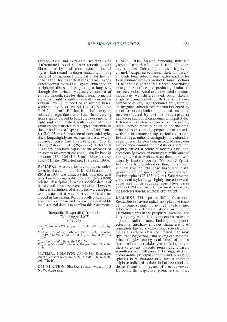
surface. Axial and extra-axial skeletons welldifferentiated. Axial skeleton reticulate, withfibres cored by small choanosomal principalstyles. Extra-axial skeleton radial, with longtracts of choanosomal principal styles heavilyechinated by rhabdostyles , and largersubectosomal extra-axial styles embedded inperipheral fibres and projecting a long waythrough the surface. Megascleres consist ofentirely smooth, slender choanosomal principalstyles, straight, slightly centrally curved orsinuous, evenly rounded or anisoxeote bases,without any basal rhabd (240-(292)-325×5-(6.7)-11�m). Echinating rhabdostylesrelatively large, thick, with basal rhabd varyingfrom slightly curved to basal curvature nearly atright angles to the shaft, with smooth base andsmall spines restricted to the apical extremity orthe apical 1/3 of spicule (141-(264)-500×8-(12.5)-25�m). Subectosomal extra-axial stylesthick, long, slightly curved near basal end, evenlyrounded base and hastate point (up to1120-(1236)-2000×16-(25)-36�m). Ectosomalauxiliary spicules raphidiform oxeotes oranisoxeas (asymmetrical ends), usually bent orsinuous (270-320×1.5-3�m). Microscleresabsent (Thiele, 1898; Hoshino, 1981; Sim, 1990).
REMARKS. A search for the holotype, under-taken by the author and Dr D. Kühlmann at theZMB in 1988, was unsuccessful. This species isonly barely recognisable from Thiele’s (1898)original description, in which specific details ofits skeletal structure were missing. However,Thiele’s illustrations of its spicules were adequateto indicate that it was most appropriately in-cluded in Raspaxilla. Recent recollections of thespecies from Japan and Korea provided addit-ional skeletal details to confirm this placement.
Raspailia (Raspaxilla) frondula(Whitelegge, 1907)
(Fig. 23)
Axinella frondula Whitelegge, 1907: 509-510, pl. 46, fig.32.
Echinaxia frondula; Hallmann 1916a: 543; Hallmann1917: 394-398, text-fig. 1, pl. 21, figs 3-4, pl. 22, figs1-2.
Raspailia frondula; Bergquist 1970: 30.Raspailia (Raspaxilla) frondula; Hooper, 1991: 1248, fig.
34
MATERIAL. HOLOTYPE. AM G4349: ShoalhavenBight, S coast of NSW, 34° 51’S, 150° 45’E, 60 m depth,coll. ‘Thetis’.
DISTRIBUTION. Shallow coastal waters of SNSW, Australia.
DESCRIPTION. Stalked branching flabellategrowth form. Surface with fine, close-setmicroconules. Colour light brownish-grey inethanol. ‘Raspailiid ectosomal skeleton’ absent,although long subectosomal extra-axial stylesform plumose brushes around terminal portionsof ascending peripheral fibres, protrudingthrough the surface and producing distinctivesurface conules. Axial and extra-axial skeletonsmoderately well-differentiated. Axial skeletalslightly compressed, with the axial corecomposed of very light spongin fibres, formingan irregular subrenieroid reticulation cored bypauci- or multispicular longitudinal tracts andinterconnected by uni- or paucispiculartransverse tracts of choanosomal principal styles.Extra-axial skeleton composed of prominentlyradial, non-plumose bundles of choanosomalprincipal styles arising perpendicular to axis,without interconnecting reticulate tracts.Echinating acanthostyles slightly more abundantin peripheral skeleton than in axis. Megascleresinclude choanosomal principal styles, short, thin,slightly curved at centre or towards basal end,occasionally oxeote or strongylote, with roundednon-tylote bases, without basal rhabd, and withslightly hastate points (87-165×3-8µm).Echinating rhabdostyles short, thin, with smooth,slightly swollen, rhabdose bases and distal(pointed) 2/3 of spicule evenly covered withvestigial spines (72-133×4-9µm). Subectosomalextra-axial styles long, slightly curved towardsbasal end, with rounded non-tylote bases(278-710×4-16µm). Ectosomal auxiliarymegascleres absent. Microscleres absent.
REMARKS. This species differs from mostRaspaxilla in having radial, non-plumose tractsof choanosomal pr incipal s tyles andsubectosomal extra-axial styles forming theascending fibres in the peripheral skeleton, andlacking any reticulate connections betweenadjacent radial tracts; lacking the specialectosomal auxiliary spicules characteristic ofraspailiids; having a wide-meshed reticulation inthe axial skeleton (less compressed than mostspecies of Raspaxilla); and having choanosomalprincipal styles (coring axial fibres) of similarsize to echinating rhabdostyles, differing only intheir thickness, hastate points and entirelysmooth surface. Hallmann (1917) suggested thatchoanosomal principal (coring) and echinatingspicules in R. frondula may have a commonorigin, as indicated by their similar size, similar tothose found in species of Aulospongus.However, the respective geometries of these
REVISION OF AULOSPONGUS 681
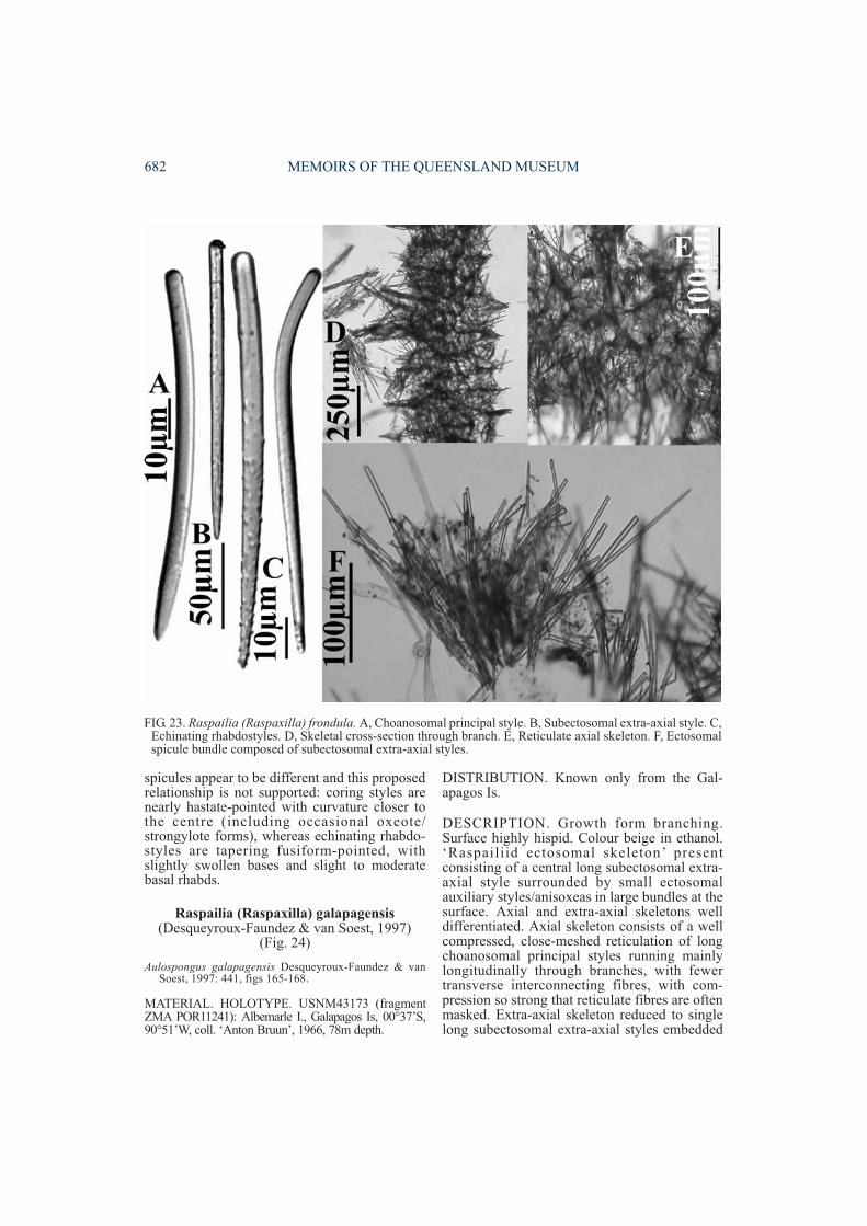
spicules appear to be different and this proposedrelationship is not supported: coring styles arenearly hastate-pointed with curvature closer tothe centre (including occasional oxeote/strongylote forms), whereas echinating rhabdo-styles are tapering fusiform-pointed, withslightly swollen bases and slight to moderatebasal rhabds.
Raspailia (Raspaxilla) galapagensis(Desqueyroux-Faundez & van Soest, 1997)
(Fig. 24)
Aulospongus galapagensis Desqueyroux-Faundez & vanSoest, 1997: 441, figs 165-168.
MATERIAL. HOLOTYPE. USNM43173 (fragmentZMA POR11241): Albemarle I., Galapagos Is, 00°37’S,90°51’W, coll. ‘Anton Bruun’, 1966, 78m depth.
DISTRIBUTION. Known only from the Gal-apagos Is.
DESCRIPTION. Growth form branching.Surface highly hispid. Colour beige in ethanol.‘Raspailiid ectosomal skeleton’ presentconsisting of a central long subectosomal extra-axial style surrounded by small ectosomalauxiliary styles/anisoxeas in large bundles at thesurface. Axial and extra-axial skeletons welldifferentiated. Axial skeleton consists of a wellcompressed, close-meshed reticulation of longchoanosomal principal styles running mainlylongitudinally through branches, with fewertransverse interconnecting fibres, with com-pression so strong that reticulate fibres are oftenmasked. Extra-axial skeleton reduced to singlelong subectosomal extra-axial styles embedded
682 MEMOIRS OF THE QUEENSLAND MUSEUM
FIG. 23. Raspailia (Raspaxilla) frondula. A, Choanosomal principal style. B, Subectosomal extra-axial style. C,Echinating rhabdostyles. D, Skeletal cross-section through branch. E, Reticulate axial skeleton. F, Ectosomalspicule bundle composed of subectosomal extra-axial styles.
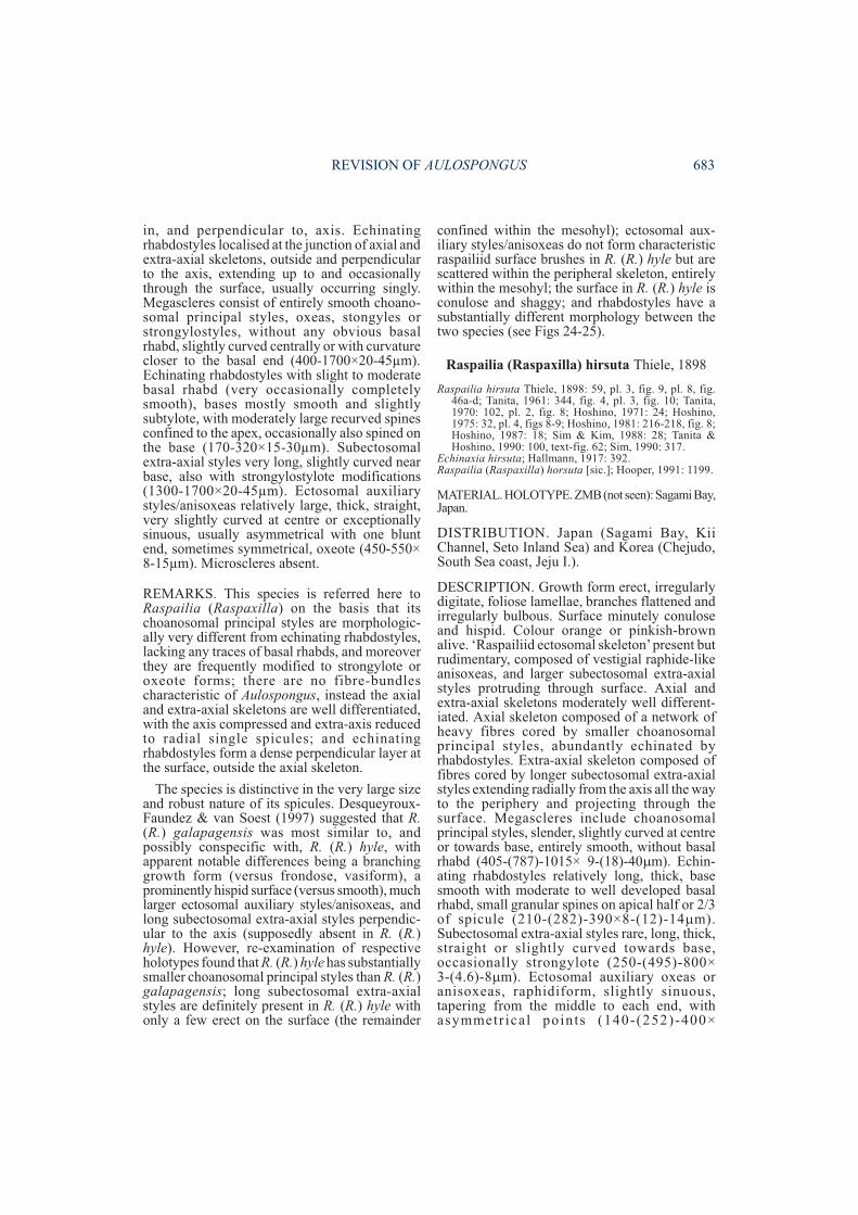
in, and perpendicular to, axis. Echinatingrhabdostyles localised at the junction of axial andextra-axial skeletons, outside and perpendicularto the axis, extending up to and occasionallythrough the surface, usually occurring singly.Megascleres consist of entirely smooth choano-somal principal styles, oxeas, stongyles orstrongylostyles, without any obvious basalrhabd, slightly curved centrally or with curvaturecloser to the basal end (400-1700×20-45µm).Echinating rhabdostyles with slight to moderatebasal rhabd (very occasionally completelysmooth), bases mostly smooth and slightlysubtylote, with moderately large recurved spinesconfined to the apex, occasionally also spined onthe base (170-320×15-30µm). Subectosomalextra-axial styles very long, slightly curved nearbase, also with strongylostylote modifications(1300-1700×20-45µm). Ectosomal auxiliarystyles/anisoxeas relatively large, thick, straight,very slightly curved at centre or exceptionallysinuous, usually asymmetrical with one bluntend, sometimes symmetrical, oxeote (450-550×8-15µm). Microscleres absent.
REMARKS. This species is referred here toRaspailia (Raspaxilla) on the basis that itschoanosomal principal styles are morphologic-ally very different from echinating rhabdostyles,lacking any traces of basal rhabds, and moreoverthey are frequently modified to strongylote oroxeote forms; there are no fibre-bundlescharacteristic of Aulospongus, instead the axialand extra-axial skeletons are well differentiated,with the axis compressed and extra-axis reducedto radial single spicules; and echinatingrhabdostyles form a dense perpendicular layer atthe surface, outside the axial skeleton.
The species is distinctive in the very large sizeand robust nature of its spicules. Desqueyroux-Faundez & van Soest (1997) suggested that R.(R.) galapagensis was most similar to, andpossibly conspecific with, R. (R.) hyle, withapparent notable differences being a branchinggrowth form (versus frondose, vasiform), aprominently hispid surface (versus smooth), muchlarger ectosomal auxiliary styles/anisoxeas, andlong subectosomal extra-axial styles perpendic-ular to the axis (supposedly absent in R. (R.)hyle). However, re-examination of respectiveholotypes found that R. (R.) hyle has substantiallysmaller choanosomal principal styles than R. (R.)galapagensis; long subectosomal extra-axialstyles are definitely present in R. (R.) hyle withonly a few erect on the surface (the remainder
confined within the mesohyl); ectosomal aux-iliary styles/anisoxeas do not form characteristicraspailiid surface brushes in R. (R.) hyle but arescattered within the peripheral skeleton, entirelywithin the mesohyl; the surface in R. (R.) hyle isconulose and shaggy; and rhabdostyles have asubstantially different morphology between thetwo species (see Figs 24-25).
Raspailia (Raspaxilla) hirsuta Thiele, 1898
Raspailia hirsuta Thiele, 1898: 59, pl. 3, fig. 9, pl. 8, fig.46a-d; Tanita, 1961: 344, fig. 4, pl. 3, fig. 10; Tanita,1970: 102, pl. 2, fig. 8; Hoshino, 1971: 24; Hoshino,1975: 32, pl. 4, figs 8-9; Hoshino, 1981: 216-218, fig. 8;Hoshino, 1987: 18; Sim & Kim, 1988: 28; Tanita &Hoshino, 1990: 100, text-fig. 62; Sim, 1990: 317.
Echinaxia hirsuta; Hallmann, 1917: 392.Raspailia (Raspaxilla) horsuta [sic.]; Hooper, 1991: 1199.
MATERIAL. HOLOTYPE. ZMB(not seen): Sagami Bay,Japan.
DISTRIBUTION. Japan (Sagami Bay, KiiChannel, Seto Inland Sea) and Korea (Chejudo,South Sea coast, Jeju I.).
DESCRIPTION. Growth form erect, irregularlydigitate, foliose lamellae, branches flattened andirregularly bulbous. Surface minutely conuloseand hispid. Colour orange or pinkish-brownalive. ‘Raspailiid ectosomal skeleton’present butrudimentary, composed of vestigial raphide-likeanisoxeas, and larger subectosomal extra-axialstyles protruding through surface. Axial andextra-axial skeletons moderately well different-iated. Axial skeleton composed of a network ofheavy fibres cored by smaller choanosomalprincipal styles, abundantly echinated byrhabdostyles. Extra-axial skeleton composed offibres cored by longer subectosomal extra-axialstyles extending radially from the axis all the wayto the periphery and projecting through thesurface. Megascleres include choanosomalprincipal styles, slender, slightly curved at centreor towards base, entirely smooth, without basalrhabd (405-(787)-1015× 9-(18)-40�m). Echin-ating rhabdostyles relatively long, thick, basesmooth with moderate to well developed basalrhabd, small granular spines on apical half or 2/3of spicule (210-(282)-390×8-(12)-14�m).Subectosomal extra-axial styles rare, long, thick,straight or slightly curved towards base,occasionally strongylote (250-(495)-800×3-(4.6)-8�m). Ectosomal auxiliary oxeas oranisoxeas, raphidiform, slightly sinuous,tapering from the middle to each end, withasymmetr ical points (140-(252)-400×
REVISION OF AULOSPONGUS 683
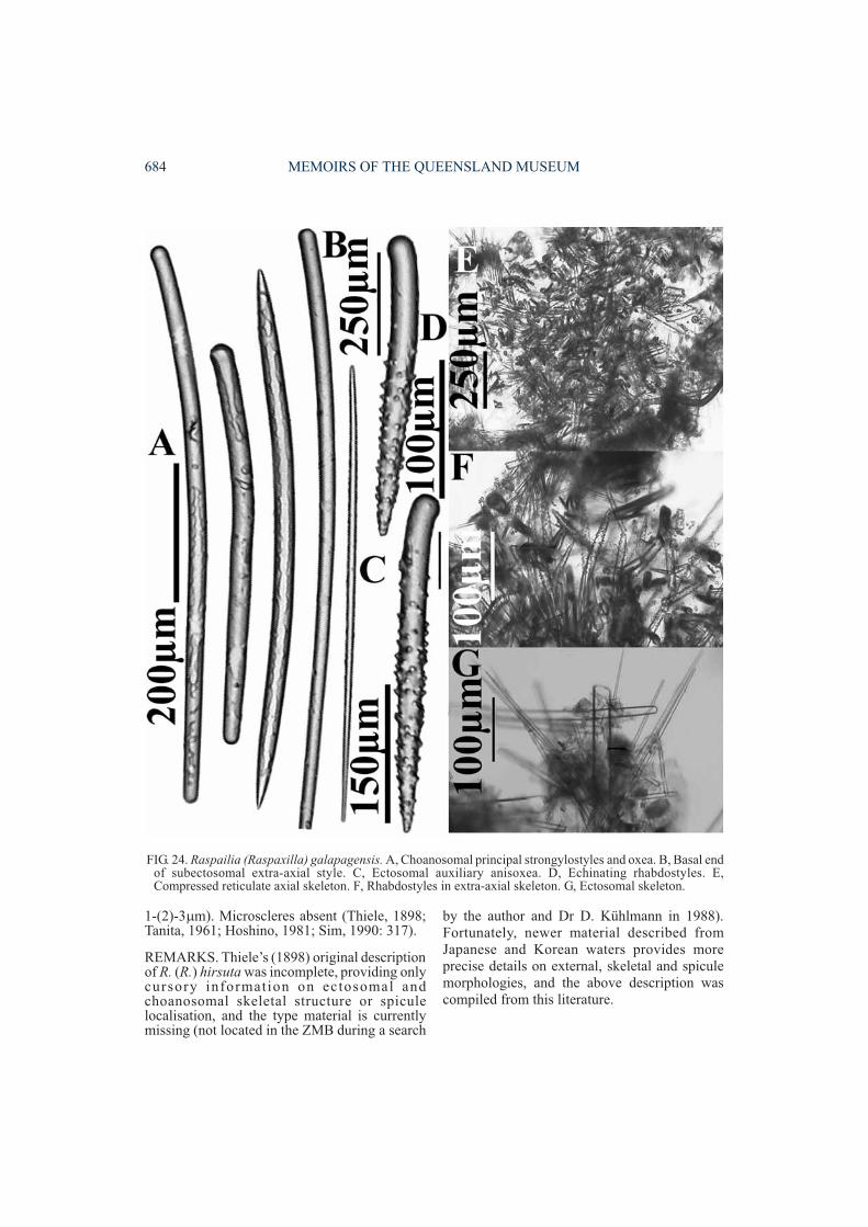
1-(2)-3�m). Microscleres absent (Thiele, 1898;Tanita, 1961; Hoshino, 1981; Sim, 1990: 317).
REMARKS. Thiele’s (1898) original descriptionof R. (R.) hirsuta was incomplete, providing onlycursory information on ectosomal andchoanosomal skeletal structure or spiculelocalisation, and the type material is currentlymissing (not located in the ZMB during a search
by the author and Dr D. Kühlmann in 1988).Fortunately, newer material described fromJapanese and Korean waters provides moreprecise details on external, skeletal and spiculemorphologies, and the above description wascompiled from this literature.
684 MEMOIRS OF THE QUEENSLAND MUSEUM
FIG. 24. Raspailia (Raspaxilla) galapagensis. A, Choanosomal principal strongylostyles and oxea. B, Basal endof subectosomal extra-axial style. C, Ectosomal auxiliary anisoxea. D, Echinating rhabdostyles. E,Compressed reticulate axial skeleton. F, Rhabdostyles in extra-axial skeleton. G, Ectosomal skeleton.

Raspailia (Raspaxilla) hyle(de Laubenfels, 1930)
(Fig. 25)
Hemectyon hyle de Laubenfels, 1930: 28; 1932: 107, fig.64; Dickinson, 1945: 21, pl. 31, figs 61-62, pl. 32, figs63-64; Bakus & Green, 1987: 79; Green & Bakus,1994: 37-39, fig. 20.
Aulospongus hyle; Desqueyroux-Faundez & van Soest,1997: 442.
MATERIAL. HOLOTYPE. USNM21418 (fragmentBMNH1929.9.30.4): Point Fermin, San Pedro, California,coll. USC, 16.ii.1924, 30-150m depth.
DISTRIBUTION. Queen Charlotte Is, BritishColumbia, Canada to Tanner Bank, California coast,Gulf of California, Mexico.
DESCRIPTION. Growth form frondose, vasiform.Surface smooth. Colour pale drab in ethanol.‘Raspailiid ectosomal skeleton’ vestigial, withectosomal auxiliary styles/anisoxeas scattered on orbelow the surface, usually in bundles parallel to thesurface, not forming brushes and not associated withprotruding erect subectosomal extra-axial styles.Axial and extra-axial skeletons well differentiated.Axial skeleton more-or-less a loose-meshedreticulation of multispicular fibres cored by smoothchoanosomal principal styles enclosed within lightspongin fibres forming a nearly halichondroidcriss-cross reticulation. Extra-axial skeleton consistsof erect subectosomal extra-axial styles per-pendicular to axis, protruding through the surface,and also lying parallel to the surface within themesohyl, with a dense layer of erect rhabdostylesembedded and perpendicular to the axial skeleton,not protruding through the surface. Megascleresconsist of slender, entirely smooth choanosomalprincipal styles, sometimes modified to strongyles,with curvature varying from slight to greatly curved,curvature central or slightly basal, without any basalrhabd (322-585×12-190µm). Echinating rhabdo-styles with slight to moderate basal rhabd,sometimes completely straight, base slightlysubtylote, entirely smooth on basal half with large,erect, slightly recurved spines sparsely scattered onapical (pointed) half of spicule (155-364×9-22µm).Subectosomal extra-axial styles long, slender,straight or slightly curved near basal end,occasionally strongylote (715-1650×9-16µm).Ectosomal auxiliary styles, or less commonlyanisoxeas, raphidiform, curved in basal third orsinuous (165-385×0.8-1.5µm).Microscleresabsent.
REMARKS. The species has been discussedabove in comparison with R. (R.) galapagensis.De Laubenfels (1930, 1932) and Dickinson(1945) recorded raphides in their material but
these are vestigial ectosomal auxiliary styles/anisoxeas. Several other inconsistencies betweenthe type material and original descriptions havebeen corrected in the redescription above.
De Laubenfels (1932) originally included thisspecies in Hemectyon on the basis of its allegedlystrong similarities with the type species,Hemectyon hamatum (Schmidt, 1870): viz. pos-session of rhabdostyles with strongly recurvedspines more-or-less restricted to the apex ofspicules; localisation of echinating rhabdostylesto the peripheral skeleton; and ‘unusual’ axialcore of styles enclosed in spongin (although thisis now widely known to occur throughout theRaspailiidae). However, the resemblancebetween these two species is minimal: they areneither closely related nor does this speciesbelong in Endectyon (Hemectyon). Desqueyroux-Faundez & van Soest (1997) subsequentlyreferred H. hyle to Aulospongus based on itssimilarities with R. (R.) galapagensis (althoughthese similarities are also only very slight; seeremarks for R. (R.) galapagensis), whereas it issuggested here that both these species are moreappropriately included in Raspailia (Raspaxilla).
Raspailia (Raspaxilla) hymani(Dickinson, 1945)
(Fig. 26)
Hemectyon hymani Dickinson, 1945: 21, pl. 33, figs 65-66;Green & Bakus, 1994: 39-40, fig. 21.
Aulospongus hymani; Desqueyroux-Faundez & van Soest,1997: 442.
MATERIAL. HOLOTYPE. AHF no.10 (not seen): SanJaime Banks, Cabo San Lucas, Mexico, 22°50’N,110°15’W, coll. ‘Velero III’, 3.iii.1937, 150m depth.
DISTRIBUTION. California to Cape San Lucas,Gulf of California, Mexico.
DESCRIPTION. Growth form flabellate,reticulate branching. Surface slightly hispid.Colour light drab, almost white, in ethanol.‘Raspailiid ectosomal skeleton’ vestigial, withraphidiform ectosomal auxiliary styles/anisoxeasforming sparse brushes or scattered below thesurface, but not necessarily surrounding theprotruding spicules. Axial and extra-axialskeletons well differentiated. Axial skeletoncomposed of a highly compressed solid core ofchoanosomal principal styles aligned longi-tudinally through branches. Extra-axial skeletonplumo-reticulate composed of tracts ofchoanosomal principal styles embedded in axisand protruding at right angles through thesurface, echinated on their outer edge by
REVISION OF AULOSPONGUS 685
rhabdostyles. Echinating rhabdostyles predom-inant in peripheral skeleton. Megascleres consistof long, smooth choanosomal principal styles,straight or slightly curved, entirely smooth,without any basal rhabd (650->1700×12-36µm).Echinating rhabdostyles with basal rhabdvarying from slight to well curved, bases entirelysmooth, with large recurved spines only on theapical half of spicule, sometimes modified toacanthotylostyles (130-300×6-30µm). Subecto-somal extra-axial spicules absent. Ectosomalauxi l iary styles/anisoxeas raphidiform(dimensions unknown). Microscleres absent(Dickinson, 1945; Green & Bakus, 1994).
REMARKS. This species has been recorded onlytwice, and regrettably the holotype cannot belocated in the AHF collections (Prof. G. Bakus,pers.comm.). From its published descriptions the
species has a distinctive reticulate-branching,flabellate growth form resembling Echino-dictyum cancellatum (Lamarck) (Raspailiidae)and Clathria coppingeri (Ridley) (Micro-cionidae). It is assigned here to Raspailia(Raspaxilla) in having coring and echinatingspicules of different morphology; echinatingrhabdostyles localised outside the axis, perchedon the junction of the axial and extra-axialskeletons; having well differentiated axial andextra-axial skeletons; and lacking the char-acteristic fibre-bundles found in Aulospongus.The E Pacific species R. (R.) hymani, R. (R.) hyleand R. (R.) galapagensis are similar in many oftheir characteristics and probably representallopatric sibling species, analogous to therelationship between the sister species R. (R.)clathrioides, R. (R.) reticulata and R. (R.)topsenti from the SW. Pacific.
686 MEMOIRS OF THE QUEENSLAND MUSEUM
FIG. 25. Raspailia (Raspaxilla) hyle. A, Basal portion of subectosomal extra-axial style. B, Basal portion ofectosomal auxiliary style. C, Choanosomal principal styles. D, Echinating rhabdostyles. E, Axial andextra-axial skeletons. F, Ectosomal skeleton. G, Specimens AHF (modified from Dickinson, 1945).
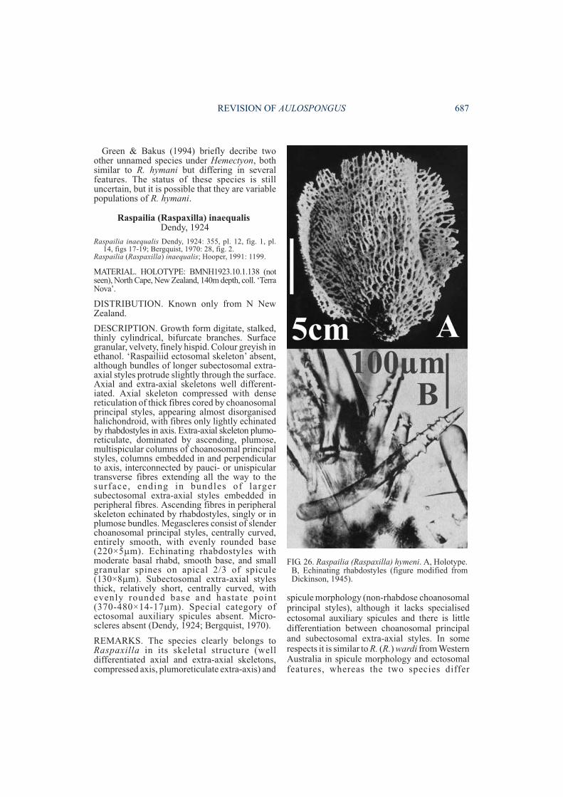
Green & Bakus (1994) briefly decribe twoother unnamed species under Hemectyon, bothsimilar to R. hymani but differing in severalfeatures. The status of these species is stilluncertain, but it is possible that they are variablepopulations of R. hymani.
Raspailia (Raspaxilla) inaequalisDendy, 1924
Raspailia inaequalis Dendy, 1924: 355, pl. 12, fig. 1, pl.14, figs 17-19; Bergquist, 1970: 28, fig. 2.
Raspailia (Raspaxilla) inaequalis; Hooper, 1991: 1199.
MATERIAL. HOLOTYPE: BMNH1923.10.1.138 (notseen), North Cape, New Zealand, 140m depth, coll. ‘TerraNova’.
DISTRIBUTION. Known only from N NewZealand.
DESCRIPTION. Growth form digitate, stalked,thinly cylindrical, bifurcate branches. Surfacegranular, velvety, finely hispid. Colour greyish inethanol. ‘Raspailiid ectosomal skeleton’ absent,although bundles of longer subectosomal extra-axial styles protrude slightly through the surface.Axial and extra-axial skeletons well different-iated. Axial skeleton compressed with densereticulation of thick fibres cored by choanosomalprincipal styles, appearing almost disorganisedhalichondroid, with fibres only lightly echinatedby rhabdostyles in axis. Extra-axial skeleton plumo-reticulate, dominated by ascending, plumose,multispicular columns of choanosomal principalstyles, columns embedded in and perpendicularto axis, interconnected by pauci- or unispiculartransverse fibres extending all the way to thesurface, ending in bundles of largersubectosomal extra-axial styles embedded inperipheral fibres. Ascending fibres in peripheralskeleton echinated by rhabdostyles, singly or inplumose bundles. Megascleres consist of slenderchoanosomal principal styles, centrally curved,entirely smooth, with evenly rounded base(220×5�m). Echinating rhabdostyles withmoderate basal rhabd, smooth base, and smallgranular spines on apical 2/3 of spicule(130×8�m). Subectosomal extra-axial stylesthick, relatively short, centrally curved, withevenly rounded base and hastate point(370-480×14-17�m). Special category ofectosomal auxiliary spicules absent. Micro-scleres absent (Dendy, 1924; Bergquist, 1970).
REMARKS. The species clearly belongs toRaspaxilla in its skeletal structure (welldifferentiated axial and extra-axial skeletons,compressed axis, plumoreticulate extra-axis) and
spicule morphology (non-rhabdose choanosomalprincipal styles), although it lacks specialisedectosomal auxiliary spicules and there is littledifferentiation between choanosomal principaland subectosomal extra-axial styles. In somerespects it is similar to R. (R.) wardi from WesternAustralia in spicule morphology and ectosomalfeatures, whereas the two species differ
REVISION OF AULOSPONGUS 687
FIG. 26. Raspailia (Raspaxilla) hymeni. A, Holotype.B, Echinating rhabdostyles (figure modified fromDickinson, 1945).
substantially in their growth form, surfacefeatures, density and location of echinatingspicules, and spicule sizes.
Raspailia (Raspaxilla) mariana(Ridley & Dendy, 1886)
(Fig. 27)
Axinella mariana Ridley & Dendy, 1886: 480; 1887: 180,pl. 34, fig. 1, pl. 40, fig. 2; Koltun, 1964: 83, pl. 13, figs7-10.
Axinectya mariana; Hallmann, 1917: 393.Raspailia (Raspaxilla) mariana; Hooper, 1991: 1196, fig.
7d-f.
MATERIAL. HOLOTYPE. BMNH1887.5.2.28: MarionI., Prince Edward Islands, subantarctic, 100-150m depth,coll. ‘Challenger’.
DISTRIBUTION. Known only from the sub-antarctic islands.
DESCRIPTION. Arborescent, bifurcate branch-ing, flattened branches. Surface prominentlyhispid. Colour greyish-yellow in ethanol.‘Raspailiid ectosomal skeleton’ absent, althoughextremely long subectosomal extra-axial stylesprotrude through the surface, singly or inbundles, surrounded by plumose brushes ofrhabdostyles at their point of insertion throughthe surface. Axial and extra-axial skeletons welldifferent iated. Axial skeleton great lycompressed, heavy fibres, without any coringspicules but with the bases of long subectosomalextra-axial styles embedded. Extra-axial skeletonradial, with long subectosomal extra-axial stylesradiating from the axis to the surface, surroundedalong most of their length (within the spongebody) by echinating rhabdostyles in plumosebundles. Megascleres consist only of two forms.Principal spicules absent. Echinating rhabdo-styles moderately thick, with smooth non-tylotebase, basal rhabd varying from slight to welldeveloped (strongly curved), and either com-pletely smooth point or with small granularspines covering apical 2/3 of spicule (185-370×8-18�m). Subectosomal extra-axial styles long,thick or slender, slightly curved near the basalend, slightly subtylote, completely smooth(1550-2400×12-29�m). Ectosomal auxiliarymegascleres absent. Microscleres absent.
REMARKS. This species is unusual amongstRaspaxilla in lacking principal spiculescompletely, having only long subectosomalextra-axial styles radially oriented and embeddedwithin axial fibres (i.e. no reticulate connectingtracts), and plumose bundles of echinatingrhabdostyles throughout the axial and extra-axial
skeletons. The absence of principal spiculesmakes it difficult to determine whether it belongsto Aulospongus (principal spicules with basalrhabds) or Raspaxilla (without basal rhabds),whereas the possession of a compressed axis andabsence of fibre-bundles suggests it belongs tothe latter genus. The species is a highly reducedRaspaxilla (also lacking specialised ectosomalauxiliary spicules).
Raspailia (Raspaxilla) pearsei(Wells, Wells & Gray, 1960)
Hemectyon pearsei Wells, Wells & Gray, 1960: 218, figs14,26.
MATERIAL. HOLOTYPE. USNM 23651 (notseen): off Beaufort, North Carolina, 5m depth.
DISTRIBUTION. Known only from E coastUSA.
DESCRIPTION. Small ellipsoidal masscomposed of many compressed verticallyelongated lamellae, 30mm long, 20mm high,encrusting rock. Texture stiff. Surface with manyupright ridges 1-1.5mm apart, hispid particularlyon ridges, with glabrous ectosomal membranestretched between adjacent ridges. Colourunknown. Oscules up to 0.6mm diameterscattered over ectosomal membrane. Ectosomalspecialisation unknown. Choanosomal skeletoncomposed of a axial reticulation of vertical andradial spongin fibres cored by choanosomalprincipal subtylostyles and echinated byrhabdostyles, with radial fibres and choanosomalprincipal spicules forming plumose bundles atthe surface. Megascleres include choanosomalprincipal subtylostyles, entirely smooth, slightlycurved at centre, with prominent tylote base andlacking any basal rhabd (190-340×10-14µm).Echinating rhabdostyles with prominent basalrhabd, entirely spined or with smooth base(85-115×3-7µm). Subectosomal extra-axialspicules not recorded. Ectosomal auxiliaryspicules not recorded. Microscleres absent(Wells, Wells & Gray, 1960).
REMARKS. The type material of this species hasnot been re-examined, and it is only known so farfrom its original description. The true nature ofits ectosomal and choanosomal skeletalstructures is still uncertain, and it is possible thatectosomal auxiliary spicules and/or subecto-somal extra-axial spicules were overlooked byWells et al. (1960), or that it is indeed a veryreduced species in it spicule complement. Fromits description it appears to conform best to
688 MEMOIRS OF THE QUEENSLAND MUSEUM
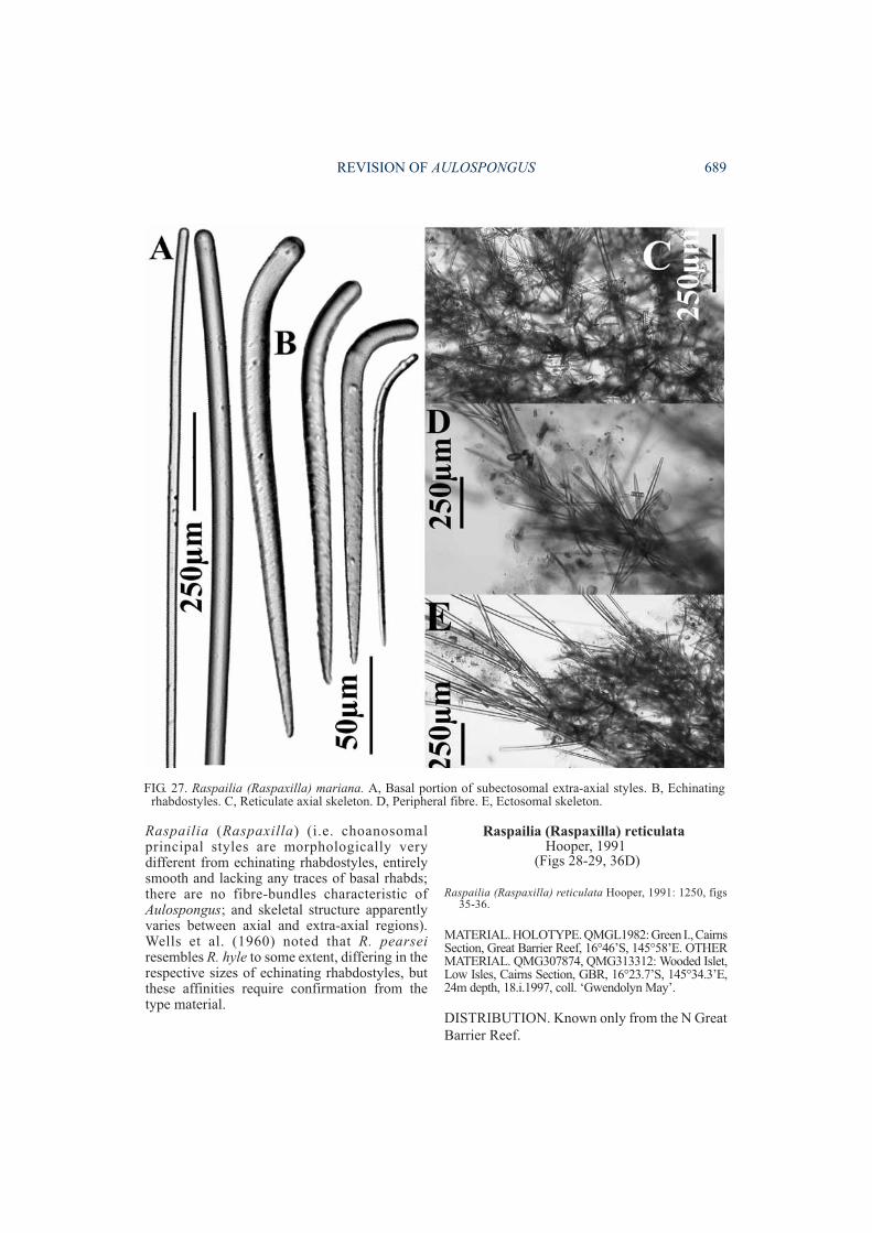
Raspailia (Raspaxilla) (i.e. choanosomalprincipal styles are morphologically verydifferent from echinating rhabdostyles, entirelysmooth and lacking any traces of basal rhabds;there are no fibre-bundles characteristic ofAulospongus; and skeletal structure apparentlyvaries between axial and extra-axial regions).Wells et al. (1960) noted that R. pearseiresembles R. hyle to some extent, differing in therespective sizes of echinating rhabdostyles, butthese affinities require confirmation from thetype material.
Raspailia (Raspaxilla) reticulataHooper, 1991
(Figs 28-29, 36D)
Raspailia (Raspaxilla) reticulata Hooper, 1991: 1250, figs35-36.
MATERIAL. HOLOTYPE. QMGL1982: Green I., CairnsSection, Great Barrier Reef, 16°46’S, 145°58’E. OTHERMATERIAL. QMG307874, QMG313312: Wooded Islet,Low Isles, Cairns Section, GBR, 16°23.7’S, 145°34.3’E,24m depth, 18.i.1997, coll. ‘Gwendolyn May’.
DISTRIBUTION. Known only from the N GreatBarrier Reef.
REVISION OF AULOSPONGUS 689
FIG. 27. Raspailia (Raspaxilla) mariana. A, Basal portion of subectosomal extra-axial styles. B, Echinatingrhabdostyles. C, Reticulate axial skeleton. D, Peripheral fibre. E, Ectosomal skeleton.
DESCRIPTION. Erect, arborescent, stalked,cylindrical branches, bulbous nodes. Surfacefinely conulose, fleshy, hispid, slightly shaggy.Colour yellow-orange (Munsell 2.5Y 8/10) alive.Oscules very small (<1mm diameter), slightlyraised above the surface with membranous lipwhen alive, with small surface drainage canalsradiating towards oscules. Texture stiff (basalstalk), flexible (branches), fleshy when alive.Ectosome hispid, moderately collagenous, withdarkly pigmented collagen layer up to 200µmthick. Specialised ‘raspailiid ectosomal skeleton’present, consisting of long thin subectosomalextra-axial styles and stout choanosomalprincipal styles protruding through surface insmall bundles of 2-5 spicules, or singly,surrounded at their bases by ectosomal auxiliaryanisoxeas, which form large bundles in plumose,radial or clumped-sinuous tracts. Projectingectosomal and subectosomal spicules extend upto 500µm from the surface, producing irregularlyspaced, long, slender conules. Collagenouscomponent of conules extends 100-350µm fromthe surface. Axial and extra-axial skeletons onlyslightly differentiated. Axial skeleton predom-inantly reticulate, only slightly compressed, withrelatively tight meshes formed by well developedspongin fibres (35-70µm diameter), bulbous atfibre nodes (up to 90µm diameter), formingelongate-oval meshes (150-320µm long,90-140µm wide), with abundant granularcollagen within mesohyl; fibre size relativelyhomogeneous throughout axial skeleton,although ascending fibres are cored by pauci-spicular tracts of 2-3 choanosomal principalstyles and transverse fibres are aspicular or uni-spicular. Extra-axial skeleton plumo-reticulate,with skeletal tracts diverging as they ascend,becoming more plumose, with progressivelyfewer interconnecting tracts towards theperipheral skeleton, although interconnectionsbetween adjacent fibres persist all the way to thesurface. Peripheral skeleton with pauci- ormultispicular ascending fibres, cored by 2-5choanosomal principal styles per tract; transverseconnecting fibres are aspicular or unispicular.Coring styles in peripheral skeleton are generallylarger than those in axial fibres. Echinatingrhabdostyles more-or-less evenly scatteredthroughout the skeleton, perhaps slightly moredense at fibre nodes, protruding perpendicularlyfrom fibres. Megascleres consist of largerchoanosomal principal styles, entirely smooth,slightly curved at centre, with straight base oroccasionally with a slight basal rhabd, bases
evenly rounded, occasionally modified to stron-gylote forms (165-(344.1)-402×8-(10.6)-13µm).Smaller echinating rhabdostyles club-shaped,with prominent smooth subtylote base, with orwithout well-developed basal rhabd, spinesgranular, evenly dispersed but restricted to apicaltwo-thirds of spicule (58-(80.3)-97×3-(6.6)-9µm).Subectosomal extra-axial styles very long, slen-der, entirely smooth, with variable shape rangingfrom slightly curved near basal end, straight, withseveral asymmetrical curves, sinuous or raphidiform(455-(726.7)-1025× 2-(6.4)-10µm). Ectosomalauxiliary anisoxeas raphidiform, slightly curvednear basal end, asymmetrical ends with pointstapering, fusiform and bases tapering, hastate,rounded, completely smooth (255-(325.3)-445×1.5-(2.2)-3µm). Microscleres absent.
REMARKS. This species was known previouslyonly from a single dead, trawled sample, withliving populations recently discovered on theLow Isles providing additional characters for thespecies (hence redescribed in detail above;compare spicule measurements with those ofHooper, 1991: 1252). This species is asister-species to (and potentially a synonym of),R. (R.) clathrioides (New Caledonia) and R. (R.)topsenti (N New Zealand), with observeddifferences significant certainly at the populationlevel, but equivocally at the species level (refer toremarks under R. (R.) clathrioides).
Raspailia (Raspaxilla) topsenti Dendy, 1924(Fig. 30)
Raspailia topsenti Dendy, 1924: 354, pl. 12, fig. 4, pl. 14,figs 14-16; de Laubenfels, 1936: 102; Bergquist, 1970:28, fig. 3b, pls 6c-d, 7a, 10d, 18d, 19d.
Raspailia (Raspaxilla) topsenti; Hooper, 1991: 1199.
MATERIAL. HOLOTYPE. BMNH1923.10.1.135: NorthCape, New Zealand, 140m depth, coll. ‘Terra Nova’.
DISTRIBUTION. Known only from N NewZealand.
DESCRIPTION. Arborescent , s ta lked,dichotomously branching, cylindrical branches.Surface granular, minutely hispid. Colour deepdull-orange alive. ‘Raspailiid ectosomalskeleton’ vestigial, with sparse, wispy ectosomalauxiliary styles/anisoxeas scattered on or belowsurface, singly or in sparse brushes. Axial andextra-axial skeletons moderately welldifferentiated. Axial skeleton compressed,close-meshed reticulation of heavy fibres coredby choanosomal principal styles, with fewechinating spicules. Extra-axial skeleton
690 MEMOIRS OF THE QUEENSLAND MUSEUM
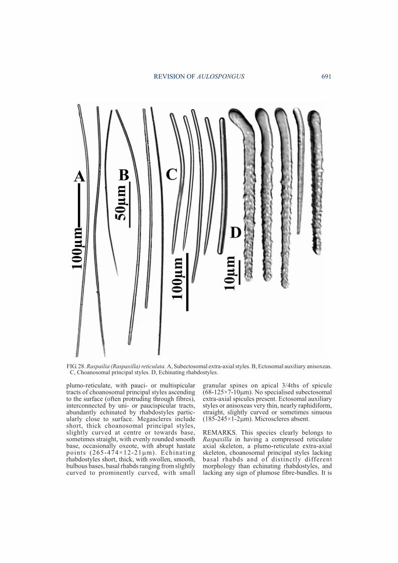
plumo-reticulate, with pauci- or multispiculartracts of choanosomal principal styles ascendingto the surface (often protruding through fibres),interconnected by uni- or paucispicular tracts,abundantly echinated by rhabdostyles partic-ularly close to surface. Megascleres includeshort, thick choanosomal principal styles,slightly curved at centre or towards base,sometimes straight, with evenly rounded smoothbase, occasionally oxeote, with abrupt hastatepoints (265-474×12-21�m). Echinatingrhabdostyles short, thick, with swollen, smooth,bulbous bases, basal rhabds ranging from slightlycurved to prominently curved, with small
granular spines on apical 3/4ths of spicule(68-125×7-10�m). No specialised subectosomalextra-axial spicules present. Ectosomal auxiliarystyles or anisoxeas very thin, nearly raphidiform,straight, slightly curved or sometimes sinuous(185-245×1-2�m). Microscleres absent.
REMARKS. This species clearly belongs toRaspaxilla in having a compressed reticulateaxial skeleton, a plumo-reticulate extra-axialskeleton, choanosomal principal styles lackingbasal rhabds and of distinctly differentmorphology than echinating rhabdostyles, andlacking any sign of plumose fibre-bundles. It is
REVISION OF AULOSPONGUS 691
FIG. 28. Raspailia (Raspaxilla) reticulata. A, Subectosomal extra-axial styles. B, Ectosomal auxiliary anisoxeas.C, Choanosomal principal styles. D, Echinating rhabdostyles.
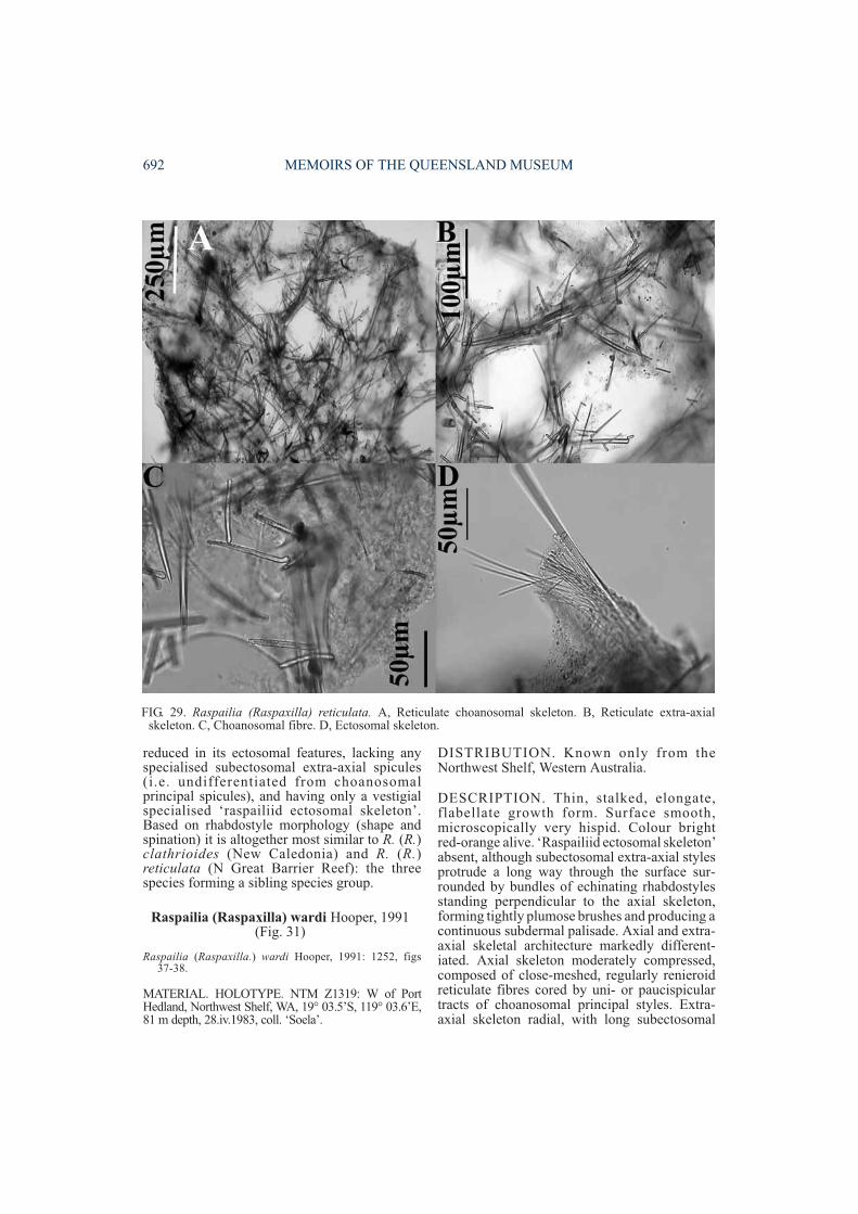
reduced in its ectosomal features, lacking anyspecialised subectosomal extra-axial spicules(i.e. undifferentiated from choanosomalprincipal spicules), and having only a vestigialspecialised ‘raspailiid ectosomal skeleton’.Based on rhabdostyle morphology (shape andspination) it is altogether most similar to R. (R.)clathrioides (New Caledonia) and R. (R.)reticulata (N Great Barrier Reef): the threespecies forming a sibling species group.
Raspailia (Raspaxilla) wardi Hooper, 1991(Fig. 31)
Raspailia (Raspaxilla.) wardi Hooper, 1991: 1252, figs37-38.
MATERIAL. HOLOTYPE. NTM Z1319: W of PortHedland, Northwest Shelf, WA, 19° 03.5’S, 119° 03.6’E,81 m depth, 28.iv.1983, coll. ‘Soela’.
DISTRIBUTION. Known only from theNorthwest Shelf, Western Australia.
DESCRIPTION. Thin, stalked, elongate,flabellate growth form. Surface smooth,microscopically very hispid. Colour brightred-orange alive. ‘Raspailiid ectosomal skeleton’absent, although subectosomal extra-axial stylesprotrude a long way through the surface sur-rounded by bundles of echinating rhabdostylesstanding perpendicular to the axial skeleton,forming tightly plumose brushes and producing acontinuous subdermal palisade. Axial and extra-axial skeletal architecture markedly different-iated. Axial skeleton moderately compressed,composed of close-meshed, regularly renieroidreticulate fibres cored by uni- or paucispiculartracts of choanosomal principal styles. Extra-axial skeleton radial, with long subectosomal
692 MEMOIRS OF THE QUEENSLAND MUSEUM
FIG. 29. Raspailia (Raspaxilla) reticulata. A, Reticulate choanosomal skeleton. B, Reticulate extra-axialskeleton. C, Choanosomal fibre. D, Ectosomal skeleton.
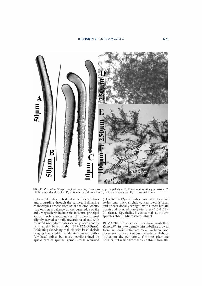
extra-axial styles embedded in peripheral fibresand protruding through the surface. Echinatingrhabdostyles absent from axial skeleton, occur-ring only as a palisade on the outer edge of theaxis. Megascleres include choanosomal principalstyles, rarely anisoxeas, entirely smooth, mostslightly curved centrally towards basal end, withrounded non-tylote bases or very occasionallywith slight basal rhabd (147-222×5-9µm).Echinating rhabdostyles thick, with basal rhabdsranging from slight to moderately curved, with afew basal spines but more heavily spined onapical part of spicule, spines small, recurved
(112-165×8-12µm). Subectosomal extra-axialstyles long, thick, slightly curved towards basalend or occasionally straight, with almost hastatepoints and rounded non-tylote bases (515-1122×7-16µm). Specialised ectosomal auxiliaryspicules absent. Microscleres absent.
REMARKS. This species differs from most otherRaspaxilla in its extremely thin flabellate growthform, renieroid reticulate axial skeleton, andpossession of a continuous palisade of rhabdo-styles on the ectosome, forming plumosebrushes, but which are otherwise absent from the
REVISION OF AULOSPONGUS 693
FIG. 30. Raspailia (Raspaxilla) topsenti. A, Choanosomal principal style. B, Ectosomal auxiliary anisoxea. C,Echinating rhabdostyles. D, Reticulate axial skeleton. E, Ectosomal skeleton. F, Extra-axial fibres.
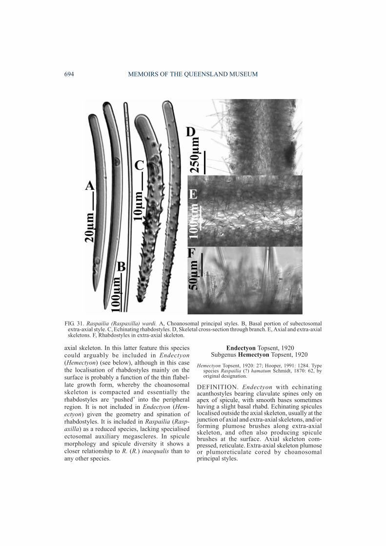
axial skeleton. In this latter feature this speciescould arguably be included in Endectyon(Hemectyon) (see below), although in this casethe localisation of rhabdostyles mainly on thesurface is probably a function of the thin flabel-late growth form, whereby the choanosomalskeleton is compacted and essentially therhabdostyles are ‘pushed’ into the peripheralregion. It is not included in Endectyon (Hem-ectyon) given the geometry and spination ofrhabdostyles. It is included in Raspailia (Rasp-axilla) as a reduced species, lacking specialisedectosomal auxiliary megascleres. In spiculemorphology and spicule diversity it shows acloser relationship to R. (R.) inaequalis than toany other species.
Endectyon Topsent, 1920Subgenus Hemectyon Topsent, 1920
Hemectyon Topsent, 1920: 27; Hooper, 1991: 1284. Typespecies Raspailia (?) hamatum Schmidt, 1870: 62, byoriginal designation.
DEFINITION. Endectyon with echinatingacanthostyles bearing clavulate spines only onapex of spicule, with smooth bases sometimeshaving a slight basal rhabd. Echinating spiculeslocalised outside the axial skeleton, usually at thejunction of axial and extra-axial skeletons, and/orforming plumose brushes along extra-axialskeleton, and often also producing spiculebrushes at the surface. Axial skeleton com-pressed, reticulate. Extra-axial skeleton plumoseor plumoreticulate cored by choanosomalprincipal styles.
694 MEMOIRS OF THE QUEENSLAND MUSEUM
FIG. 31. Raspailia (Raspaxilla) wardi. A, Choanosomal principal styles. B, Basal portion of subectosomalextra-axial style. C, Echinating rhabdostyles. D, Skeletal cross-section through branch. E, Axial and extra-axialskeletons. F, Rhabdostyles in extra-axial skeleton.
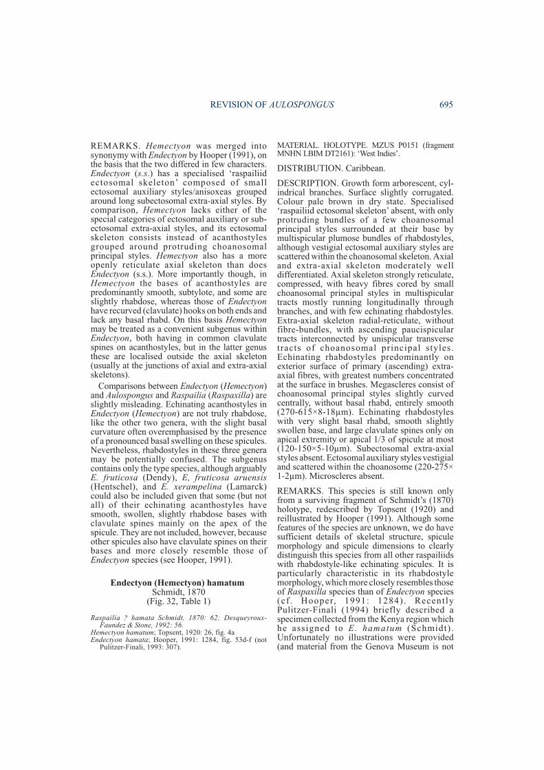
REMARKS. Hemectyon was merged intosynonymy with Endectyon by Hooper (1991), onthe basis that the two differed in few characters.Endectyon (s.s.) has a specialised ‘raspailiidectosomal skeleton’ composed of smallectosomal auxiliary styles/anisoxeas groupedaround long subectosomal extra-axial styles. Bycomparison, Hemectyon lacks either of thespecial categories of ectosomal auxiliary or sub-ectosomal extra-axial styles, and its ectosomalskeleton consists instead of acanthostylesgrouped around protruding choanosomalprincipal styles. Hemectyon also has a moreopenly reticulate axial skeleton than doesEndectyon (s.s.). More importantly though, inHemectyon the bases of acanthostyles arepredominantly smooth, subtylote, and some areslightly rhabdose, whereas those of Endectyonhave recurved (clavulate) hooks on both ends andlack any basal rhabd. On this basis Hemectyonmay be treated as a convenient subgenus withinEndectyon, both having in common clavulatespines on acanthostyles, but in the latter genusthese are localised outside the axial skeleton(usually at the junctions of axial and extra-axialskeletons).
Comparisons between Endectyon (Hemectyon)and Aulospongus and Raspailia (Raspaxilla) areslightly misleading. Echinating acanthostyles inEndectyon (Hemectyon) are not truly rhabdose,like the other two genera, with the slight basalcurvature often overemphasised by the presenceof a pronounced basal swelling on these spicules.Nevertheless, rhabdostyles in these three generamay be potentially confused. The subgenuscontains only the type species, although arguablyE. fruticosa (Dendy), E, fruticosa aruensis(Hentschel), and E. xerampelina (Lamarck)could also be included given that some (but notall) of their echinating acanthostyles havesmooth, swollen, slightly rhabdose bases withclavulate spines mainly on the apex of thespicule. They are not included, however, becauseother spicules also have clavulate spines on theirbases and more closely resemble those ofEndectyon species (see Hooper, 1991).
Endectyon (Hemectyon) hamatumSchmidt, 1870
(Fig. 32, Table 1)
Raspailia ? hamata Schmidt, 1870: 62; Desqueyroux-Faundez & Stone, 1992: 56.
Hemectyon hamatum; Topsent, 1920: 26, fig. 4aEndectyon hamata; Hooper, 1991: 1284, fig. 53d-f (not
Pulitzer-Finali, 1993: 307).
MATERIAL. HOLOTYPE. MZUS P0151 (fragmentMNHN LBIM DT2161): ‘West Indies’.
DISTRIBUTION. Caribbean.
DESCRIPTION. Growth form arborescent, cyl-indrical branches. Surface slightly corrugated.Colour pale brown in dry state. Specialised‘raspailiid ectosomal skeleton’ absent, with onlyprotruding bundles of a few choanosomalprincipal styles surrounded at their base bymultispicular plumose bundles of rhabdostyles,although vestigial ectosomal auxiliary styles arescattered within the choanosomal skeleton. Axialand extra-axial skeleton moderately welldifferentiated. Axial skeleton strongly reticulate,compressed, with heavy fibres cored by smallchoanosomal principal styles in multispiculartracts mostly running longitudinally throughbranches, and with few echinating rhabdostyles.Extra-axial skeleton radial-reticulate, withoutfibre-bundles, with ascending paucispiculartracts interconnected by unispicular transversetracts of choanosomal principal styles.Echinating rhabdostyles predominantly onexterior surface of primary (ascending) extra-axial fibres, with greatest numbers concentratedat the surface in brushes. Megascleres consist ofchoanosomal principal styles slightly curvedcentrally, without basal rhabd, entirely smooth(270-615×8-18µm). Echinating rhabdostyleswith very slight basal rhabd, smooth slightlyswollen base, and large clavulate spines only onapical extremity or apical 1/3 of spicule at most(120-150×5-10µm). Subectosomal extra-axialstyles absent. Ectosomal auxiliary styles vestigialand scattered within the choanosome (220-275×1-2µm). Microscleres absent.
REMARKS. This species is still known onlyfrom a surviving fragment of Schmidt’s (1870)holotype, redescribed by Topsent (1920) andreillustrated by Hooper (1991). Although somefeatures of the species are unknown, we do havesufficient details of skeletal structure, spiculemorphology and spicule dimensions to clearlydistinguish this species from all other raspailiidswith rhabdostyle-like echinating spicules. It isparticularly characteristic in its rhabdostylemorphology, which more closely resembles thoseof Raspaxilla species than of Endectyon species(cf . Hooper, 1991: 1284) . Recent lyPulitzer-Finali (1994) briefly described aspecimen collected from the Kenya region whichhe assigned to E. hamatum (Schmidt) .Unfortunately no illustrations were provided(and material from the Genova Museum is not
REVISION OF AULOSPONGUS 695
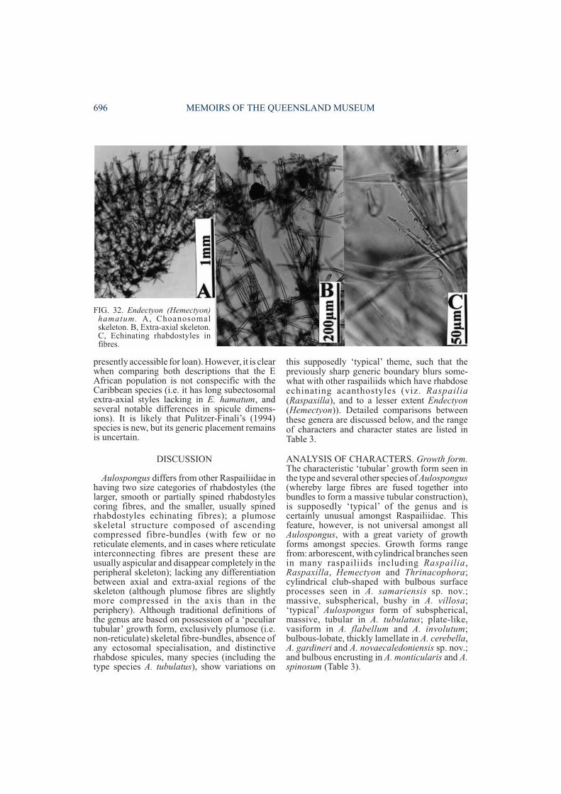
presently accessible for loan). However, it is clearwhen comparing both descriptions that the EAfrican population is not conspecific with theCaribbean species (i.e. it has long subectosomalextra-axial styles lacking in E. hamatum, andseveral notable differences in spicule dimens-ions). It is likely that Pulitzer-Finali’s (1994)species is new, but its generic placement remainsis uncertain.
DISCUSSION
Aulospongus differs from other Raspailiidae inhaving two size categories of rhabdostyles (thelarger, smooth or partially spined rhabdostylescoring fibres, and the smaller, usually spinedrhabdostyles echinating fibres); a plumoseskeletal structure composed of ascendingcompressed fibre-bundles (with few or noreticulate elements, and in cases where reticulateinterconnecting fibres are present these areusually aspicular and disappear completely in theperipheral skeleton); lacking any differentiationbetween axial and extra-axial regions of theskeleton (although plumose fibres are slightlymore compressed in the axis than in theperiphery). Although traditional definitions ofthe genus are based on possession of a ‘peculiartubular’ growth form, exclusively plumose (i.e.non-reticulate) skeletal fibre-bundles, absence ofany ectosomal specialisation, and distinctiverhabdose spicules, many species (including thetype species A. tubulatus), show variations on
this supposedly ‘typical’ theme, such that thepreviously sharp generic boundary blurs some-what with other raspailiids which have rhabdoseechinating acanthostyles (viz. Raspailia(Raspaxilla), and to a lesser extent Endectyon(Hemectyon)). Detailed comparisons betweenthese genera are discussed below, and the rangeof characters and character states are listed inTable 3.
ANALYSIS OF CHARACTERS. Growth form.The characteristic ‘tubular’ growth form seen inthe type and several other species of Aulospongus(whereby large fibres are fused together intobundles to form a massive tubular construction),is supposedly ‘typical’ of the genus and iscertainly unusual amongst Raspailiidae. Thisfeature, however, is not universal amongst allAulospongus, with a great variety of growthforms amongst species. Growth forms rangefrom: arborescent, with cylindrical branches seenin many raspailiids including Raspailia,Raspaxilla, Hemectyon and Thrinacophora;cylindrical club-shaped with bulbous surfaceprocesses seen in A. samariensis sp. nov.;massive, subspherical, bushy in A. villosa;‘typical’ Aulospongus form of subspherical,massive, tubular in A. tubulatus; plate-like,vasiform in A. flabellum and A. involutum;bulbous-lobate, thickly lamellate in A. cerebella,A. gardineri and A. novaecaledoniensis sp. nov.;and bulbous encrusting in A. monticularis and A.spinosum (Table 3).
696 MEMOIRS OF THE QUEENSLAND MUSEUM
FIG. 32. Endectyon (Hemectyon)hamatum. A, Choanosomalskeleton. B, Extra-axial skeleton.C, Echinating rhabdostyles infibres.
Determination of whether particular growthforms are derived or ancestral is relativelysubjective and equivocal in any characteranalysis (e.g. Hooper, 1991), but there is alreadysome precedence for such interpretationsamongst several groups of demosponges:Axinel l idae (Alvarez & Crisp, 1994) ,Rhabderemiidae (van Soest & Hooper, 1993),Acarnus (van Soest et al., 1991), Microcionidae(Hooper, 1996), Mycalidae (Hajdu, in press).Based on outgroup analyses these studieshypothesise that the more massive, erect growthforms are less derived than thinly encrustingones, the latter frequently indicative ofsuccessful adaptation and survival in ephemeralhabits, such as the intertidal zone and interstitialhabitats. Amongst Raspailiidae ‘typical’ growthforms are erect, digitate, branching sponges, withfar fewer encrusting or bulbous species, and thisinterpretation is consistent for Aulospongus,which is interpreted here as being a highlyderived raspailiid. However, this interpretationmust be taken with caution because it is likelythat species independently colonise theseephemeral habitats, and more unlikely that theyhave evolved there.
Plumose fibre-bundles. Species of Aulospongusare characterised in having their fibres and spic-ule tracts amalgamated into bundles, termed here‘fibre-bundles’, composed of bulbous sponginfibres cored and echinated by rhabdostyles inplumose tracts, forming individual plumoseascending (usually non-reticulate) branches, andterminating as surface lobes and/or processes.This feature is apparently unrelated to overallgrowth form and also appears to be unique withinRaspailidae. These fibre-bundles are super-ficially reminiscent of the spiculo-spongin tractsseen in Pseudaxinella (Axinellidae) (see Hooper& Lévi, 1993) and Clathria (Microciona)(Microcionidae) (see Hooper, 1996), althoughfibre structure and localisation of spiculesassociated with these fibres are substantiallydifferent in both these genera, and there is noinferred relationship between them andAulospongus. Similarly, in R. (Raspaxilla)phakellina, and some other species assigned toRaspaxilla, ascending fibres are plumose anddominate the extra-axial skeleton, with (e.g. R.(R.) reticulata) or without (e.g. R. (R.)compressa), interconnecting fibres extending allthe way to the peripheral skeleton. However, inthese species fibres are generally not bulbous,nor do spicules form dense plumose tracts aroundthe bulbs, amalgamated at the core but diverging
towards the periphery, as in Aulospongus.Instead, fibres in Raspaxilla arise from a welldeveloped, compressed reticulate axis, con-necting fibres are paucispicular, and skeletalstructure can best be described as plumo-reticulate. ‘Fibre-bundles’ are also absent fromEndectyon.Choanosomal skeletal structure. In Aulospongusthere is no appreciable difference between theaxial or basal region and the peripheral (extra-axial) regions of the skeleton, although there isoften a greater degree of amalgamation (orfusion) of the system of plumose fibre-bundles atthe core of the skeleton (or base of the sponge),than in the peripheral skeleton (where fibressimply diverge further apart). Reticulate fibresare present in the axial skeletons of severalspecies (A. tubulatus, A. gardineri, A. spinosum,A. novaecaledoniensis sp. nov., A. samariensissp. nov.), in which there are few aspicular orrarely paucispicular (transverse) fibres inter-connecting adjacent (ascending) plumosefibre-bundles, but these do not persist into theperipheral region and cannot be classed as‘reticulate’ skeletons.
By comparison, Raspailia (Raspaxilla) andEndectyon (Hemectyon) have well differentiatedaxial and extra-axial skeletons; well developed,compressed, reticulate axial skeletons; andplumoreticulate or radial extra-axial skeletons(similar to most other raspailiids). In these generaextra-axial skeletal structure consists of plumoseascending multispicular fibres interconnected byuni- or paucispicular transverse fibres that persistall the way to the surface. In some of these specieswhere axial compression is extreme, or where thediameter of the skeleton is narrow (e.g. thinbranches), extra-axial tracts are reduced to singleradial subectosomal extra-axial styles embeddedin and perpendicular to the axis, protrudingthrough the surface, with echinating rhabdostylesappearing to be ‘pushed’ to the outer edge of theskeleton.
In choanosomal skeletal structure species ofAulospongus fall into two groups, with two othercharacter states seen in the Raspailiidaeoutgroups (Raspailia (Raspailia), Raspailia(Raspaxi l la ) , Endectyon (Hemectyon ) ,Thrinacophora). Skeletal structures include:species with compressed axial and radialextra-axial skeletons (the latter composed ofsubectosomal extra-axial styles embedded in andperpendicular to the axial skeleton), seen in manyraspailiids including Endectyon (Hemectyon)and Thrinacophora (this is probably a reduced,
REVISION OF AULOSPONGUS 697
derived character related to the degree of axialcompression and loss of a more extensiveextra-axial skeleton); species with a compressedreticulate axis, and a reticulate or plumo-reticulate extra-axis in which interconnectingfibres persist throughout the entire peripheralskeleton, also seen in many raspailiids includingRaspailia (Raspaxilla) and R. (Raspailia);species with few aspicular or occasionally pauci-spicular fibres interconnecting the ascendingplumose fibre-bundles, with reticulate connect-ing fibres diminishing and disappearing towardsthe periphery, seen in A. gardineri, A. spinosum,
A. tubulatus, A. novaecaledoniensis sp. nov. andA. samariensis sp. nov.; species with exclusivelyplumose fibre-bundles arising from a slightlycompressed base, with fibres diverging towardsthe periphery, without any reticulate connections,seen in A. cerebella , A. involutum , A.monticularis and A. villosa. These skeletalstructures are subdivided into several characters,based on the subdivision of the skeleton intoaxial, extra-axial and peripheral skeletons, andthe presence, absence and nature of reticulateskeletal tracts (Table 3).
698 MEMOIRS OF THE QUEENSLAND MUSEUM
1. Growth form: 1(1), arborescent, cylindrical branches; 1(2), cylindrical club-shaped withbulbous surface processes; 1(3), massive, subspherical, bushy; 1(4), subspherical, massive, tubular,erect digitate cup-shaped; 1(5), plate-like, vasiform; 1(6), bulbous-lobate; 1(7), bulbous encrusting.2. Fibre-bundles: 2(1), absent; 2(2), present. 3. Axial skeleton: 3(1), compressed, solid; 3(2),compressed, more open-reticulate; 3(3), slightly compressed, predominantly plumose, fewreticulate connections; 3(4), exclusively plumose, slightly compressed, no fibre reticulation. 4.Extra-axial skeleton: 4(1), radial, with longer extra-axial auxiliary styles/ anisoxeas embedded inand perpendicular to axis, with or without unispicular reticulate connections; 4(2), reticulate orslightly plumoreticulate, reticulate connections persistent throughout; 4(3), plumose, few aspicularor unispicular reticulate connections disappearing in periphery; 4(4), exlusively plumose, withoutreticulate connections. 5. Location of protruding extra-axial styles/ anisoxeas: 5(1), absent; 5(2),embedded in ascending peripheral fibres and protruding through surface, singly or in bundles. 6.Interconnecting reticulate fibres: 6(1), well developed, persistent throughout skeleton; 6(2), welldeveloped in axis, poorly developed in extra-axial skeleton; 6(3), vestigial, with aspicular orunispicular interconnecting fibres diminishing along ascending fibres and absent from periphery;6(4), absent completely. 7. Location of (smaller) echinating styles or rhabdostyles: 7(1), evenlydispersed throughout skeleton; 7(2), restricted to axis, absent from periphery; 7(3), reduced in axis,concentrated mainly in peripheral skeleton embedded in primary ascending fibres, protrudingthrough surface; 7(4), localised to outer surface of peripheral fibres, echinating surface and/orforming brushes around protruding extra-axial auxiliary spicules; 7(5), absent (presumedsecondary loss). 8. Specialised ‘raspailiid ectosomal skeleton’: 8(1), well developed, composed ofbrushes of ectosomal auxiliary styles/ anisoxeas surrounding the longer protruding subectosomalextra-axial styles; 8(2), vestigial, composed of sparse ectosomal auxiliary styles/ anisoxeasscattered on or below the surface, not forming bundles, with or without the longer protrudingsubectosomal extra-axial styles; 8(3), vestigial, with only protruding long subectosomal extra-axialstyles but no ectosomal auxiliary styles; 8(4), absent, lacking any ectosomal or extra-axial auxiliaryspicules. 9. Basal rhabds on styles: 9(1), absent (never present); 9(2), present only on echinatingstyles/ acanthostyles; 9(3), present on both echinating and principal styles/ acanthostyles, 9(4),absent (secondarily modified). 10. Spination on rhabdostyles: 10(1), rhabdose spicules absentcompletely; 10(2), smaller echinating acanthostyles spined or partially spined, and at least partiallyrhabdose, whereas larger choanosomal principal styles entirely smooth and non-rhabdose; 10(3),larger choanosomal principal rhabdostyles entirely smooth, echinating rhabdostyles spined orpartially spined, basal rhabd moderate or slightly developed on both sorts of spicule; 10(4) bothechinating and choanosomal principal rhabdostyles at least partially spined, basal rhabd stronglydeveloped on both. 11. Number of categories of (smaller) echinating rhabdostyles: 11(1), none;11(2), one; 11(3), two. 12. Raphide microscleres: 12(1), present; 12(2), absent.
TABLE 3. List of characters and character states used to judge apomorphy in the construction of the cladogram ofrelationships between species of Aulospongus based on outgroup comparisons with members of the familyRaspailiidae.
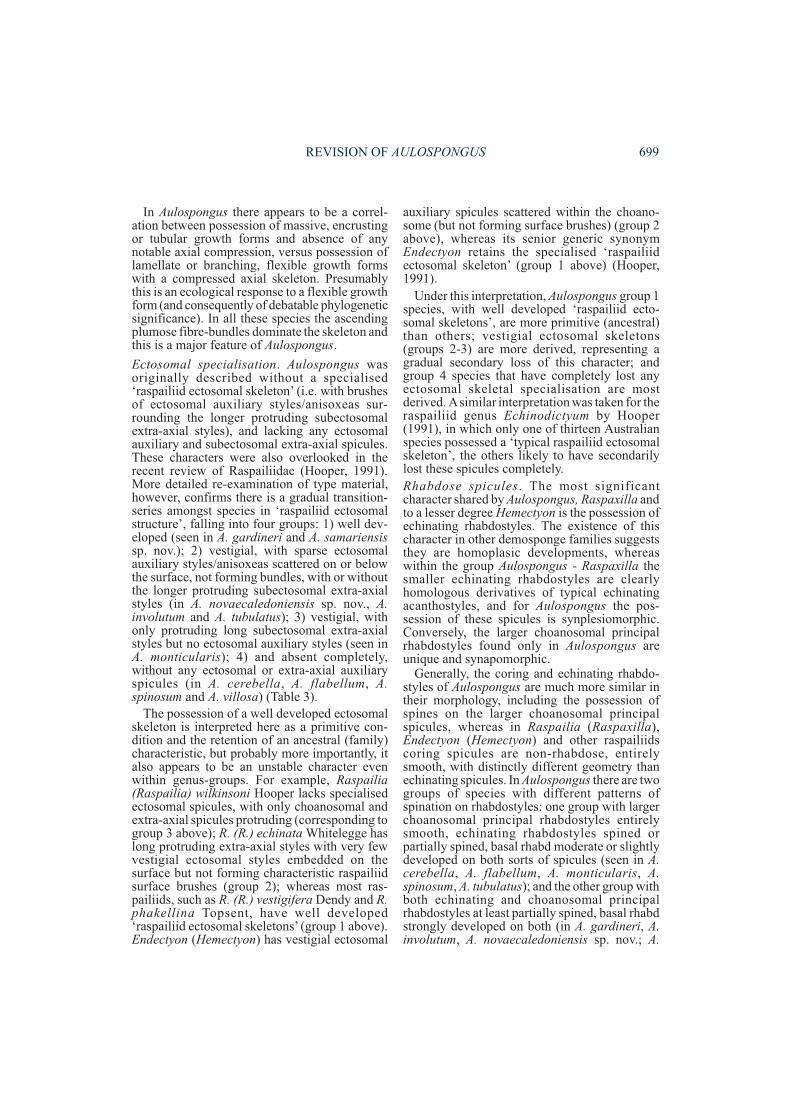
In Aulospongus there appears to be a correl-ation between possession of massive, encrustingor tubular growth forms and absence of anynotable axial compression, versus possession oflamellate or branching, flexible growth formswith a compressed axial skeleton. Presumablythis is an ecological response to a flexible growthform (and consequently of debatable phylogeneticsignificance). In all these species the ascendingplumose fibre-bundles dominate the skeleton andthis is a major feature of Aulospongus.
Ectosomal specialisation. Aulospongus wasoriginally described without a specialised‘raspailiid ectosomal skeleton’ (i.e. with brushesof ectosomal auxiliary styles/anisoxeas sur-rounding the longer protruding subectosomalextra-axial styles), and lacking any ectosomalauxiliary and subectosomal extra-axial spicules.These characters were also overlooked in therecent review of Raspailiidae (Hooper, 1991).More detailed re-examination of type material,however, confirms there is a gradual transition-series amongst species in ‘raspailiid ectosomalstructure’, falling into four groups: 1) well dev-eloped (seen in A. gardineri and A. samariensissp. nov.); 2) vestigial, with sparse ectosomalauxiliary styles/anisoxeas scattered on or belowthe surface, not forming bundles, with or withoutthe longer protruding subectosomal extra-axialstyles (in A. novaecaledoniensis sp. nov., A.involutum and A. tubulatus); 3) vestigial, withonly protruding long subectosomal extra-axialstyles but no ectosomal auxiliary styles (seen inA. monticularis); 4) and absent completely,without any ectosomal or extra-axial auxiliaryspicules (in A. cerebella, A. flabellum, A.spinosum and A. villosa) (Table 3).
The possession of a well developed ectosomalskeleton is interpreted here as a primitive con-dition and the retention of an ancestral (family)characteristic, but probably more importantly, italso appears to be an unstable character evenwithin genus-groups. For example, Raspailia(Raspailia) wilkinsoni Hooper lacks specialisedectosomal spicules, with only choanosomal andextra-axial spicules protruding (corresponding togroup 3 above); R. (R.) echinata Whitelegge haslong protruding extra-axial styles with very fewvestigial ectosomal styles embedded on thesurface but not forming characteristic raspailiidsurface brushes (group 2); whereas most ras-pailiids, such as R. (R.) vestigifera Dendy and R.phakellina Topsent, have well developed‘raspailiid ectosomal skeletons’ (group 1 above).Endectyon (Hemectyon) has vestigial ectosomal
auxiliary spicules scattered within the choano-some (but not forming surface brushes) (group 2above), whereas its senior generic synonymEndectyon retains the specialised ‘raspailiidectosomal skeleton’ (group 1 above) (Hooper,1991).
Under this interpretation, Aulospongus group 1species, with well developed ‘raspailiid ecto-somal skeletons’, are more primitive (ancestral)than others; vestigial ectosomal skeletons(groups 2-3) are more derived, representing agradual secondary loss of this character; andgroup 4 species that have completely lost anyectosomal skeletal specialisation are mostderived. Asimilar interpretation was taken for theraspailiid genus Echinodictyum by Hooper(1991), in which only one of thirteen Australianspecies possessed a ‘typical raspailiid ectosomalskeleton’, the others likely to have secondarilylost these spicules completely.Rhabdose spicules. The most significantcharacter shared by Aulospongus, Raspaxilla andto a lesser degree Hemectyon is the possession ofechinating rhabdostyles. The existence of thischaracter in other demosponge families suggeststhey are homoplasic developments, whereaswithin the group Aulospongus - Raspaxilla thesmaller echinating rhabdostyles are clearlyhomologous derivatives of typical echinatingacanthostyles, and for Aulospongus the pos-session of these spicules is synplesiomorphic.Conversely, the larger choanosomal principalrhabdostyles found only in Aulospongus areunique and synapomorphic.
Generally, the coring and echinating rhabdo-styles of Aulospongus are much more similar intheir morphology, including the possession ofspines on the larger choanosomal principalspicules, whereas in Raspailia (Raspaxilla),Endectyon (Hemectyon) and other raspailiidscoring spicules are non-rhabdose, entirelysmooth, with distinctly different geometry thanechinating spicules. In Aulospongus there are twogroups of species with different patterns ofspination on rhabdostyles: one group with largerchoanosomal principal rhabdostyles entirelysmooth, echinating rhabdostyles spined orpartially spined, basal rhabd moderate or slightlydeveloped on both sorts of spicules (seen in A.cerebella, A. flabellum, A. monticularis, A.spinosum, A. tubulatus); and the other group withboth echinating and choanosomal principalrhabdostyles at least partially spined, basal rhabdstrongly developed on both (in A. gardineri, A.involutum, A. novaecaledoniensis sp. nov.; A.
REVISION OF AULOSPONGUS 699
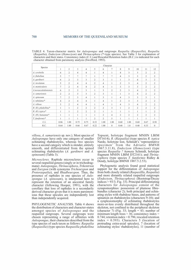
villosa, A. samariensis sp. nov.). Most species ofAulospongus have only one category of smallerechinating rhabdostyles, whereas two specieshave a second category which is slender, entirelysmooth, and differentiated from the spinedechinating rhabdostyles (A. gardineri and A.spinosum) (Table 3).
Microscleres. Raphide microscleres occur inseveral raspailiid genera (singly or in trichodrag-mata): Aulospongus, Thrinacophora, Trikentrionand Eurypon (with synonyms Tricheurypon andProtoraspailia), and Rhadbeurypon. Thus, thepresence of raphides in one species of Aulo-spongus (A. spinosum), is interpreted here torepresent the retention of an ancestral familycharacter (following Hooper, 1991), with thecorollary that loss of raphides is a secondarilyderived character given that it is more parsimon-ious that these spicules are independently lostthan independently acquired.
PHYLOGENETIC ANALYSIS. Table 4 showsthe distribution of characters and character-statesamongst species of Aulospongus and theraspailiid outgroups. Several outgroups werechosen representing a range of affinities withAulospongus, their characters described from thetype species of each taxon as follows: Raspailia(Raspaxilla) (type species Raspaxilla phakellina
Topsent, holotype fragment MNHN LBIMDT1614); R. (Raspailia) (type species R. typicaNardo, holotype lost, Schmidt’s ‘representativespecimen’ from the Adriat ic BMNH1867.3.11.8), Endectyon (Hemectyon) (typespecies Raspailia ? hamata Schmidt, holotypefragment MNHN LBIM DT2161); and Thrina-cophora (type species T. funiformis Ridley &Dendy, holotype BMNH 1887.5.2.53).
Phylogenetic analysis found good statisticalsupport for the differentiation of Aulospongusfrom both closely related (Raspaxillla, Raspailia)and more distantly related raspailiid outgroups(Endectyon, Thrinacophora) (Bootstrap/Decayindices = 93/3; Fig. 33). Principal differentiatingcharacters for Aulospongus consist of thesynapomorphies: possession of plumose fibre-bundles (character 2); both principal and echin-ating styles with rhabdose bases, and patterns ofspination on rhabdostyles (characters 9-10); anda synplesiomorphy of echinating rhabdostylesmore-or-less evenly distributed throughout theskeleton, not confined to the peripheral skeleton(character 7) (Fig. 33; length = 43; number ofminimum length trees = 36; consistency index =0.744; retention index = 0.796; rescaled retentionindex = 0.593). Characters 5 (location ofprotruding extra-axial spicules), 7 (location ofechinating styles/ rhabdostyles), 11 (number of
700 MEMOIRS OF THE QUEENSLAND MUSEUM
SpeciesCharacter
1 2 3 4 5 6 7 8 9 10 11 12
A. cerebella 6 2 4 4 1 4 1 4 3 3 2 2
A. flabellum 5 ? ? ? 1 ? ? 4 3 3 2 2
A. gardineri 6 2 3 3 2 3 1 1 3 4 3 2
A. involutum 5 2 4 4 2 4 1 2 3 4 2 2
A. monticularis 7 2 4 4 2 4 1 3 3 3 2 2
A.novaecaledoniensis 6 2 3 3 1 3 2 2 3 4 2 2
A. samariensis 2 2 3 3 2 3 1 1 3 4 2 2
A. spinosum 7 2 3 4 1 3 1 4 3 3 3 1
A. tubulatus* 4 2 4 4 1 4 1 2 3 3 2 2
A. villosa 3 2 4 4 1 4 1 4 3 4 2 2
R. (R.) phakellina* 1 1 2 2 2 2 3 1 2 2 2 2
R. (R.) typica* 1 1 2 2 2 1 1 1 1 1 1 2
E. (H.) hamatum* 1 1 1 1 1 2 4 2 2 2 2 2
T. funiformis* 1 1 1 1 2 2 5 1 1 1 1 1
C.I. 0.86 1.00 0.75 0.75 0.33 1.00 1.00 0.60 1.00 0.60 0.67 0.50
R.C. 0.64 1.00 0.66 0.67 0.22 1.00 0 0.40 1.0 0.40 0.33 0
TABLE 4. Taxon-character matrix for Aulospongus and outgroups Raspailia (Raspaxilla), Raspailia(Raspailia), Endectyon (Hemectyon) and Thrinacophora (*=type species). See Table 3 for explanation ofcharacters and their states. Consistency index (C.I.) and Rescaled Retention Index (R.C.) is indicated for eachcharacter obtained from parsimony analysis (Swofford, 1993).
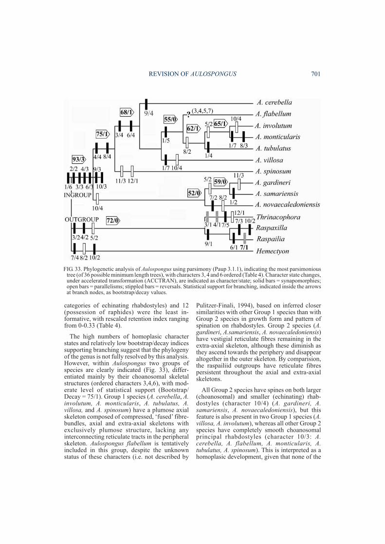
categories of echinating rhabdostyles) and 12(possession of raphides) were the least in-formative, with rescaled retention index rangingfrom 0-0.33 (Table 4).
The high numbers of homoplasic characterstates and relatively low bootstrap/decay indicessupporting branching suggest that the phylogenyof the genus is not fully resolved by this analysis.However, within Aulospongus two groups ofspecies are clearly indicated (Fig. 33), differ-entiated mainly by their choanosomal skeletalstructures (ordered characters 3,4,6), with mod-erate level of statistical support (Bootstrap/Decay = 75/1). Group 1 species (A. cerebella, A.involutum, A. monticularis, A. tubulatus, A.villosa, and A. spinosum) have a plumose axialskeleton composed of compressed, ‘fused’ fibre-bundles, axial and extra-axial skeletons withexclusively plumose structure, lacking anyinterconnecting reticulate tracts in the peripheralskeleton. Aulospongus flabellum is tentativelyincluded in this group, despite the unknownstatus of these characters (i.e. not described by
Pulitzer-Finali, 1994), based on inferred closersimilarities with other Group 1 species than withGroup 2 species in growth form and pattern ofspination on rhabdostyles. Group 2 species (A.gardineri, A.samariensis, A. novaecaledoniensis)have vestigial reticulate fibres remaining in theextra-axial skeleton, although these diminish asthey ascend towards the periphery and disappearaltogether in the outer skeleton. By comparision,the raspailiid outgroups have reticulate fibrespersistent throughout the axial and extra-axialskeletons.
All Group 2 species have spines on both larger(choanosomal) and smaller (echinating) rhab-dostyles (character 10/4) (A. gardineri, A.samariensis, A. novaecaledoniensis), but thisfeature is also present in two Group 1 species (A.villosa, A. involutum), whereas all other Group 2species have completely smooth choanosomalprincipal rhabdostyles (character 10/3: A.cerebella, A. flabellum, A. monticularis, A.tubulatus, A. spinosum). This is interpreted as ahomoplasic development, given that none of the
REVISION OF AULOSPONGUS 701
FIG. 33. Phylogenetic analysis of Aulospongus using parsimony (Paup 3.1.1), indicating the most parsimonioustree (of 36 possible minimum length trees), with characters 3, 4 and 6 ordered (Table 4). Character state changes,under accelerated transformation (ACCTRAN), are indicated as character/state; solid bars = synapomorphies;open bars = parallelisms; stippled bars = reversals. Statistical support for branching, indicated inside the arrowsat branch nodes, as bootstrap/decay values.
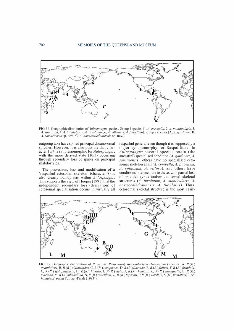
outgroup taxa have spined principal choanosomalspicules. However, it is also possible that char-acter 10/4 is synplesiomorphic for Aulospongus,with the more derived state (10/3) occurringthrough secondary loss of spines on principalrhabdostyles.
The possession, loss and modification of a‘raspailiid ectosomal skeleton’ (character 8) isalso clearly homoplasic within Aulospongus.This supports the view of Hooper (1991) that theindependent secondary loss (derivation) ofectosomal specialisation occurs in virtually all
raspailiid genera, even though it is supposedly amajor synapomorphy for Raspaillidae. InAulospongus several species retain (theancestral) specialised condition (A. gardineri, A.samariensis), others have no specialised ecto-somal skeleton at all (A. cerebella, A. flabellum,A. spinosum, A. villosa), and others haveconditions intermediate to these, with partial lossof spicules types and/or ectosomal skeletalstructures (A. involutum, A. monticularis, A.novaecaledoniensis, A. tubulatus). Thus,ectosomal skeletal structure is the most easily
702 MEMOIRS OF THE QUEENSLAND MUSEUM
FIG. 35. Geographic distribution of Raspailia (Raspaxilla) and Endectyon (Hemectyon) species. A, R.(R.)acanthifera, B, R.(R.) clathrioides, C, R.(R.) compressa, D, R.(R.) flaccida, E, R.(R.) folium, F, R.(R.) frondula,G, R.(R.) galapagensis, H, R.(R.) hirsuta, I, R.(R.) hyle, J, R.(R.) hymani, K, R.(R.) inaequalis, L, R.(R.)mariana, M, R.(R.) phakellina, N, R.(R.) reticulata, O, R.(R.) topsenti, P, R.(R.) wardi, 1, E.(H.) hamatum, 2, ‘E.hamatum’ sensu Pulitzer-Finali (1993)).
FIG. 34. Geographic distribution of Aulospongus species. Group 1 species (1, A. cerebella, 2, A. monticularis, 3,A. spinosum, 4, A. tubulatus, 5, A. involutum, 6, A. villosa, 7, A. flabellum); group 2 species (A, A. gardineri, B,A. samariensis sp. nov., C, A. novaecaledoniensis sp. nov.).
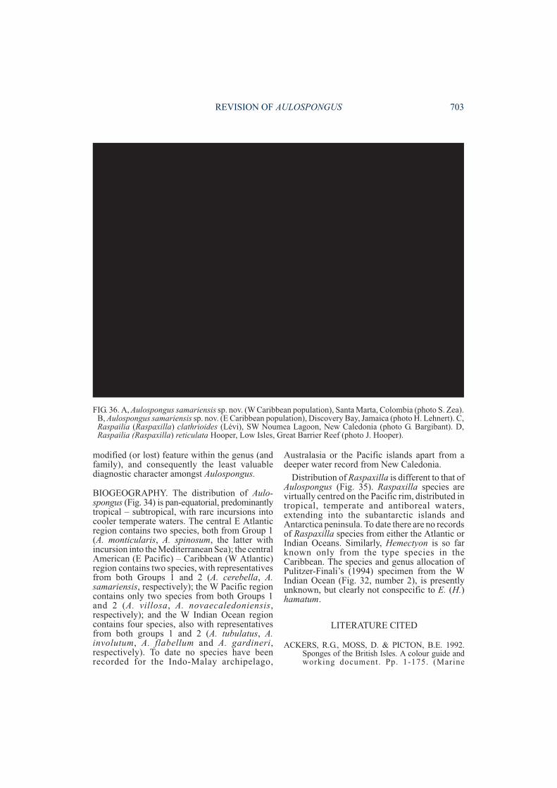
modified (or lost) feature within the genus (andfamily), and consequently the least valuablediagnostic character amongst Aulospongus.
BIOGEOGRAPHY. The distribution of Aulo-spongus (Fig. 34) is pan-equatorial, predominantlytropical – subtropical, with rare incursions intocooler temperate waters. The central E Atlanticregion contains two species, both from Group 1(A. monticularis, A. spinosum, the latter withincursion into the Mediterranean Sea); the centralAmerican (E Pacific) – Caribbean (W Atlantic)region contains two species, with representativesfrom both Groups 1 and 2 (A. cerebella, A.samariensis, respectively); the W Pacific regioncontains only two species from both Groups 1and 2 (A. villosa, A. novaecaledoniensis,respectively); and the W Indian Ocean regioncontains four species, also with representativesfrom both groups 1 and 2 (A. tubulatus, A.involutum, A. flabellum and A. gardineri,respectively). To date no species have beenrecorded for the Indo-Malay archipelago,
Australasia or the Pacific islands apart from adeeper water record from New Caledonia.
Distribution of Raspaxilla is different to that ofAulospongus (Fig. 35). Raspaxilla species arevirtually centred on the Pacific rim, distributed intropical, temperate and antiboreal waters,extending into the subantarctic islands andAntarctica peninsula. To date there are no recordsof Raspaxilla species from either the Atlantic orIndian Oceans. Similarly, Hemectyon is so farknown only from the type species in theCaribbean. The species and genus allocation ofPulitzer-Finali’s (1994) specimen from the WIndian Ocean (Fig. 32, number 2), is presentlyunknown, but clearly not conspecific to E. (H.)hamatum.
LITERATURE CITED
ACKERS, R.G., MOSS, D. & PICTON, B.E. 1992.Sponges of the British Isles. A colour guide andworking document. Pp. 1-175. (Marine
REVISION OF AULOSPONGUS 703
FIG. 36. A, Aulospongus samariensis sp. nov. (W Caribbean population), Santa Marta, Colombia (photo S. Zea).B, Aulospongus samariensis sp. nov. (E Caribbean population), Discovery Bay, Jamaica (photo H. Lehnert). C,Raspailia (Raspaxilla) clathrioides (Lévi), SW Noumea Lagoon, New Caledonia (photo G. Bargibant). D,Raspailia (Raspaxilla) reticulata Hooper, Low Isles, Great Barrier Reef (photo J. Hooper).
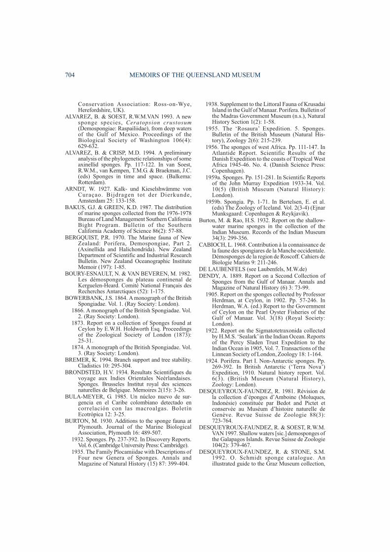
Conservation Association: Ross-on-Wye,Herefordshire, UK).
ALVAREZ, B. & SOEST, R.W.M.VAN 1993. A newsponge species, Ceratopsion crustosum(Demospongiae: Raspailiidae), from deep watersof the Gulf of Mexico. Proceedings of theBiological Society of Washington 106(4):629-632.
ALVAREZ, B. & CRISP, M.D. 1994. A preliminaryanalysis of the phylogenetic relationships of someaxinellid sponges. Pp. 117-122. In van Soest,R.W.M., van Kempen, T.M.G. & Braekman, J.C.(eds) Sponges in time and space. (Balkema:Rotterdam).
ARNDT, W. 1927. Kalk- und Kieselshwämme vonCuraçao. Bijdragen tot der Dierkunde,Amsterdam 25: 133-158.
BAKUS, G.J. & GREEN, K.D. 1987. The distributionof marine sponges collected from the 1976-1978Bureau of Land Management Southern CaliforniaBight Program. Bulletin of the SouthernCalifornia Academy of Science 86(2): 57-88.
BERGQUIST, P.R. 1970. The Marine fauna of NewZealand: Porifera, Demospongiae, Part 2.(Axinellida and Halichondrida). New ZealandDepartment of Scientific and Industrial ResearchBulletin. New Zealand Oceanographic InstituteMemoir (197): 1-85.
BOURY-ESNAULT, N. & VAN BEVEREN, M. 1982.Les démosponges du plateau continenal deKerguelen-Heard. Comité National Français desRecherches Antarctiques (52): 1-175.
BOWERBANK, J.S. 1864. A monograph of the BritishSpongiadae. Vol. 1. (Ray Society: London).
1866. A monograph of the British Spongiadae. Vol.2. (Ray Society: London).
1873. Report on a collection of Sponges found atCeylon by E.W.H. Holdworth Esq. Proceedingsof the Zoological Society of London (1873):25-31.
1874. A monograph of the British Spongiadae. Vol.3. (Ray Society: London).
BREMER, K. 1994. Branch support and tree stability.Cladistics 10: 295-304.
BRONDSTED, H.V. 1934. Resultats Scientifiques duvoyage aux Indies Orientales Neérlandaises.Sponges. Brusseles Institut royal des sciencesnaturelles de Belgique. Memoires 2(15): 3-26.
BULA-MEYER, G. 1985. Un núcleo nuevo de sur-gencia en el Caribe colombiano detectado encorrelación con las macroalgas. BoletínEcotrópica 12: 3-25.
BURTON, M. 1930. Additions to the sponge fauna atPlymouth. Journal of the Marine BiologicalAssociation, Plymouth 16: 489-507.
1932. Sponges. Pp. 237-392. In Discovery Reports.Vol. 6. (Cambridge University Press: Cambridge).
1935. The Family Plocamiidae with Descriptions ofFour new Genera of Sponges. Annals andMagazine of Natural History (15) 87: 399-404.
1938. Supplement to the Littoral Fauna of KrusadaiIsland in the Gulf of Manaar. Porifera. Bulletin ofthe Madras Government Museum (n.s.), NaturalHistory Section 1(2): 1-58.
1955. The ‘Rosaura’ Expedition. 5. Sponges.Bulletin of the British Museum (Natural His-tory), Zoology 2(6): 215-239.
1956. The sponges of west Africa. Pp. 111-147. InAtlantide Report. Scientific Results of theDanish Expedition to the coasts of Tropical WestAfrica 1945-46. No. 4. (Danish Science Press:Copenhagen).
1959a. Sponges. Pp. 151-281. In Scientific Reportsof the John Murray Expedition 1933-34. Vol.10(5) (British Museum (Natural History):London).
1959b. Spongia. Pp. 1-71. In Bertelsen, E. et al.(eds) The Zoology of Iceland. Vol. 2(3-4) (EjnarMunksgaard: Copenhagen & Reykjavik).
Burton, M. & Rao, H.S. 1932. Report on the shallow-water marine sponges in the collection of theIndian Museum. Records of the Indian Museum34(3): 299-356.
CABIOCH, L. 1968. Contribution à la connaissance dela faune des spongiares de la Manche occidentale.Démosponges de la region de Roscoff. Cahiers deBiologie Marins 9: 211-246.
DE LAUBENFELS (see Laubenfels, M.W.de)DENDY, A. 1889. Report on a Second Collection of
Sponges from the Gulf of Manaar. Annals andMagazine of Natural History (6) 3: 73-99.
1905. Report on the sponges collected by ProfessorHerdman, at Ceylon, in 1902. Pp. 57-246. InHerdman, W.A. (ed.) Report to the Governmentof Ceylon on the Pearl Oyster Fisheries of theGulf of Manaar. Vol. 3(18) (Royal Society:London).
1922. Report on the Sigmatotetraxonida collectedby H.M.S. ‘Sealark’in the Indian Ocean. Reportsof the Percy Sladen Trust Expedition to theIndian Ocean in 1905, Vol. 7. Transactions of theLinnean Society of London, Zoology 18: 1-164.
1924. Porifera. Part I. Non-Antarctic sponges. Pp.269-392. In British Antarctic (‘Terra Nova’)Expedition, 1910. Natural history report. Vol.6(3). (British Museum (Natural History),Zoology: London).
DESQUEYROUX-FAUNDEZ, R. 1981. Révision dela collection d’éponges d’Amboine (Moluques,Indonésie) constituée par Bedot and Pictet etconservée au Muséum d’histoire naturelle deGenève. Revue Suisse de Zoologie 88(3):723-764.
DESQUEYROUX-FAUNDEZ, R. & SOEST, R.W.M.VAN 1997. Shallow waters [sic.] demosponges ofthe Galapagos Islands. Revue Suisse de Zoologie104(2): 379-467.
DESQUEYROUX-FAUNDEZ, R. & STONE, S.M.1992. O. Schmidt sponge catalogue. Anillustrated guide to the Graz Museum collection,
704 MEMOIRS OF THE QUEENSLAND MUSEUM
with notes on additional material. (Muséumd’Histoire naturelle: Geneva).
DIAZ, M.C., ALVAREZ, B. & VAN SOEST, R.W.M.1987. New species of Demospongiae (Porifera)from the National Park ‘Archipiélago de LosRoques’, Venezuela. Bijdragen tot de Dierkunde57(1): 31-41.
DICKINSON, M.G. 1945. Sponges of the Gulf ofCalifornia. Pp. 1-57. In Reports on the collectionsobtained by Allan Hancock Pacific Expeditionsof Velero III off the coast of Mexico, CentralAmerica, South America, and Galapagos Islandsin 1932-40. Vol. 11(1). (Allan Hancock Found-ation: California).
DUCHASSAING, DE FONBRESSIN, P. & MICH-ELOTTI, G. 1864. Spongiaires de la mer Caraïbe.Natuurkkundige Verhandelingen HollandscheMaatschappij Wetenschappen Haarlem (2) 21(3):1-124.
ERIKSSON, T. & WIKSTROM, N. 1997. AutodecayVersion 3.0 (freeware). ([email protected]).
GEORGE, W.C. & WILSON, H.V. 1919. Sponges ofBeaufort (N.C.) Harbor and Vicinity. FisheryBulletin, United States National Fisheries Service(36): 130-179.
GREEN, K.D. & BAKUS, G.J. 1994. The Porifera. Pp.1-82. In Blake, J.A., Lissner, A.L. & Scott, P.H.(eds) Taxonomic atlas of the benthic fauna of theSanta Maria Basin and Western Santa BarbaraChannel. Vol. 2 (Santa Barbara Museum ofNatural History: Santa Barbara, California).
HALLMANN, E.F. 1916a. A Revision of the Generawith Microscleres included, or provisionallyincluded, in the Family Axinellidae, withDescriptions of some Australian Species. Part ii.(Porifera). Proceedings of the Linnean Society ofNew South Wales 41(3): 495-552.
1917. On the Genera Echinaxia and Rhabdosigma[Porifera]. Proceedings of the Linnean Society ofNew South Wales 42(2): 391-405.
HAJDU, E. in press. Taxonomy and phylogeny of themarine sponge genus Mycale. SubgenusGrapelia. Invertebrate Taxonomy.
HENTSCHEL, E. 1914. Monaxone Kieselschwämmeund Hornschwämme der DeutschenSüdpolar-Expedition 1901-1903. DeutscheSudpolar-Expedition 1901-1903 15(Zoologie 7):37-141.
HOOPER, J.N.A. 1991. Revision of the FamilyRaspailiidae (Porifera: Demospongiae), withdescription of Australian species. InvertebrateTaxonomy 5(6): 1179-1415.
1996. Revision of Microcionidae (Porifera:Poeci loscler ida: Demospongiae) , withdescription of Australian species. Memoirs ofthe Queensland Museum 40: 1-626.
HOOPER, J.N.A. & BATTERSHILL, C.N. 1998.Order Poecilosclerida. Pp. 114-136. In Lévi, C.,Laboute, P., Bargibant, G. & Menou, J.-L. (eds)Sponges of the New Caledonia Lagoon.
Collection Faune et flore tropicales. No. 33.(Éditions de l’Orstom: Paris).
HOOPER, J.N.A., CAPON, R.J., KEENAN, C.P.,PARRY, D.L. & SMIT, N. 1992. Chemotaxonomyof marine sponges: families Microcionidae,Raspailiidae and Axinellidae, and theirrelationships with other families in the ordersPoecilosclerida and Axinellida (Porifera: Demo-spongiae). Invertebrate Taxonomy 6: 261-301.
HOOPER, J.N.A. & LÉVI, C. 1993. Poeciloscleridafrom the New Caledonia lagoon (Porifera:Demospongiae). Invertebrate Taxonomy 7(5):1221-1302.
HOSHINO, T. 1971. Sponge fauna of Seto Inland Sea(Demospongiae, Calcarea). Bulletin of theBiological Society of Hiroshima University 38:21-30.
1975. The sponges of the Anan coast. ZoologicalMagazine 84(1): 30-38.
1976. Demosponges obtained from the vicinity ofthe Aitsu Marine Biological Station. Calanus 5:3-11.
1981. Shallow-water demosponges of WesternJapan I-II. Journal of Science of the HiroshimaUniversity (B,1,Zoology) 29(1,2): 47-276.
1987. Apreliminary catalogue of the marine speciesof the class Demospongiae (Porifera) fromJapanese waters. Mukaishima Marine BiologicalStation Faculty of Science, HiroshimaUniversity, Contribution (279): 1-48.
KIRKPATRICK, R. 1903. Descriptions of SouthAfrican Sponges. Part III. Cape of Good Hope,Department of Agriculture Bulletin. MarineInvestigations in South Africa 2(16): 233-264.
KOLTUN, V.M. 1964. Sponges of the Antarctic. Part 1.Tetraxonida and Cornucuspongida. Pp. 6-116,428-433. In Pavlovskii, E.P., Andriyashev, A.P. &Ushakov, P.V. (eds) Biological Reports of theSoviet Antarctic Expedition (1955-1958). Vol. 2.Academy of Sciences of the USSR, ZoologicalInstitute: Explorations of the fauna of the seas II(10). (Izdatel’stovo ‘Nauka’: Moscow).
1970. Sponge fauna of the northwestern Pacificfrom the shallows to the hadal depths. TrudyAkademiya Nauk SSSR Institut Okeanologii 86:177-233.
LAUBENFELS, M.W.DE 1930. The Sponges ofCalifornia. Stanford University Bulletin 5(98):24-29.
1932. The Marine and Freshwater Sponges ofCalifornia. Proceedings of the United StatesNational Museum Washington 81(4): 1-140.
1936. A discussion of the sponge fauna of the DryTortugas in particular, and the West Indies ingeneral, with material for a revision of thefamilies and orders of the Porifera. CarnegieInstitute of Washington Publication. Papers ofthe Tortugas Laboratory 30(467): 1-225.
1950. The Porifera of the Bermuda Archipelago.Transactions of the Zoological Society ofLondon 27: 1-154.
REVISION OF AULOSPONGUS 705
LEHNERT, H. & SOEST, R.W.M. VAN 1996. NorthJamaican deep fore-reef sponges. Beaufortia46(4): 53-81.
LÉVI, C. 1960. Spongiaires des côtes occidentalesAfricaines. Bulletin de l’Institut Françaisd’Afrique Noire (A) 22(3): 743-769.
1967. Démosponges récoltées en Nouvelle-Calédonie par la Mission Singer-Polignac.Expédition Française sur les récifs coralliens dela Nouvel le-Calédonie. Édit ions de laFoundation Singer-Polignac 2: 13-26.
LÉVI, C. & LÉVI, P. 1983. Démosponges bathyalesrécoltées par le N/O “Vauban” au sud de la Nouvelle-Calédonie. Bulletin du Muséum Nationald’Histoire Naturelle (4) 5(A, 4): 931-997.
LITTLE, E.J. 1963. The Sponge fauna of the St.George’s Sound, Apalachee Bay, and PanamaCity regions of the Florida Gulf Coast. TulaneStudies in Zoology 11: 31-71.
MADDISON, W.P & MADDISON, D.R. 1992.MacClade. Version 3.0 (Sinauer Associates, Inc.Publishers: Sunderland, Massachusetts).
MALDONADO, M. 1992. Demosponges of the redcoral bottoms from the Alboran Sea. Journal ofNatural History 26: 1131-1161.
MUNSELL, 1977. Munsell Color Charts for PlantTissues. 2nd Edition (Munsell Color: Universityof Wisconsin).
NARDO, G.D. 1833. Auszug aus einem neuen Systemder Spongiarien, wornach bereits die Aufstellungin der Universitäts-Sammlung zu Padua gemachtist. Isis Oken (1833): 519-523.
NORMAN, A.M. 1878. On the Genus Haliphysema,with descriptions of several forms apparentlyallied to it. Annals and Magazine of NaturalHistory (5) 1: 264-284.
PULITZER-FINALI, G. 1986. A collection of WestIndian Demospongiae (Porifera). In Appendix, alist of the Demospongiae hitherto recorded fromthe West Indies. Annali del Museo Civico diStoria Naturale di Genova 86: 65-216.
1993. A collection of marine sponges from EastAfrica. Annali del Museo Civico di StoriaNaturale di Genova 89: 247-350.
RIDLEY, S.O. & DENDY, A. 1886. Preliminary Reporton the Monaxonida collected by the H.M.S.‘Challenger’. Annals and Magazine of NaturalHistory (5) 18: 325-351, 470-493.
1887. Report on the Monaxonida collected byH.M.S. ‘Challenger’ during the Years 1873-76.Pp. 1-275. In Report on the Scientific Results ofthe Voyage of H.M.S. ‘Challenger’ during theYears 1873-76. Vol. 20 (Her Majesty’s StationeryOffice: London, Edinburgh, Dublin).
SCHMIDT, E.O. 1870. Grundzüge einer Spongien-Fauna des Atlantischen Gebietes Pp. 1-88.(Wilhelm Engelmann: Leipzig).
SMITHE, F.B. 1975. Naturalist’s Color Guide. Part I,Color Guide, 86+96 colors. Part II (1974), ColorGuide Supplement. (The American Museum ofNatural History: New York).
SILVESTRI, J.DE, ZEA, S. & DUQUE, C. 1994.Actividad antibacteriana de algunas esponjas delCaribe Colombiano. Revista Colombiana deCiencias Químico Farmacéuticas (22): 21-26.
SIM, C.J. 1990. Distribution of the Tetractinomorpha inSouth Korea. Pp. 316-319. In Rützler, K. (ed.)New perspectives in sponge biology(Smithsonian Institution Press: Washington).
SIM, C.J. & KIM, M.H. 1988. Asystematic study on themarine sponges in Korea. 7. Demospongiae andHexactinellida. The Korean Journal of SystematicZoology 4(1): 21-42.
SOEST, R.W.M. VAN 1994. Sponges of the Seychelles.Pp. 65-74. In Van der Land, J. (ed.) Oceanic Reefsof the Seychelles. Vol. 2. (Netherlands IndianOcean Programme Cruise Reports: Leiden).
SOEST, R.W.M. VAN & HOOPER, J.N.A. 1993.Taxonomy, phylogeny and biogeography of themarine sponge genus Rhabderemia Topsent, 1890(Demospongiae: Poecilosclerida). In Uriz, M.J. &Ruetzler, K. (eds) Recent advances in ecology andsystematics of sponges. Scientia Marina 57(4):319-351.
SOEST, R.W.M. VAN, HOOPER, J.N.A. &HIEMSTRA, F. 1991. Taxonomy, phylogeny andbiogeography of the marine sponge genusAcarnus (Porifera: Poecilosclerida). Beaufortia42(3): 49-88.
SOEST, R.W.M. VAN & STENTOFT, N. 1988.Barbados deep-water sponges. Studies on theFauna of Curaçao and Other Caribbean Islands70(215): 1-175.
SOEST, R.W.M. VAN, STONE, S.M., BOURY-ESNAULT, N. & RÜTZLER, K. 1983. Catalogueof the Duchassaing and Michelotti (1864)collection of West Indian sponges (Porifera).Bulletin Zoologisch Museum. Universiteit vanAmsterdam 9(21): 189-205.
SWOFFORD, D.L. 1993. PAUP (PhylogeneticAnalysis Using Parsimony). Version 3.1.1 forApple Macintosh. (Smithsonian Insitution:Washington).
TANITA, S. 1961. Report on the sponges collectedfrom the Kurushima Strait, Seto Inland Sea.Memoirs of the Ehime University (II,B) 4(2):335-354.
1970. The sponges in the Tokushima Museum.Bulletin of the Tohoku Regional FisheriesResearch Laboratory 30: 99-105.
TANITA, S. & HOSHINO, T. 1989. TheDemospongiae of Sagami Bay, collected by HisMajesty Emperor Showa. (Biological Laboratory,Imperial Household: Japan).
THIELE, J. 1898. Studien über pazifische Spongien. I.Heft. Zoologica 24: 1-72.
THOMAS, P.A. 1985. Demospongiae of the Gulf ofMannar and Palk Bay. Pp. 205-365. In James,P.S.B.R. (ed.). Recent Advances in MarineBiology. (Today Tomorrow’s Printers andPublishers: New Delhi).
706 MEMOIRS OF THE QUEENSLAND MUSEUM
TOPSENT, E. 1889. Quelques Spongiaires du Banc deCampêche et de la Pointe-a-Pître. Mémoires de laSociété Zoologique de France 2: 30-52.
1894. Application de la taxonomie actuelle à unecollection de Spongiaires du Banc de Campêcheet de Guadeloupe décrite précédement. Mém-oires de la Société Zoologique de France 7: 27-36.
1913. Spongiaires de l’Expédition AntarctiqueNationale Ecossaise. Transactions of the RoyalSociety of Edinburgh 49(3,9): 579-643.
1920. Spongiaires du Musée Zoologique deStrasbourg. Monaxonides. Bulletin de l’InstitutOcéanographique Monaco (381): 1-36.
1927. Diagnoses d’Éponges nouvelles recueilliespar le Prince Albert Ier de Monaco. Bulletin del’Institut Océanographique Monaco (502): 1-19.
1928. Spongiaires de l’Atlantique et de laMéditerranée provenant des croisières du PrinceAlbert Ier de Monaco. Résultats des CampagnesScientifiques Accomplies sur son Yacht parAlbert Ier Prince Souverain de Monaco 74:1-376.
1936. Éponges observées dans les parages deMonaco. (Deuxième Partie). Bulletin del’Institut Océanographique Monaco (686): 1-70.
VACELET, J. & VASSEUR, P. 1971. Éponges desrécifs coralliens de Tulear (Madagascar). Tethys,Supplement 1: 51-126.
VAN SOEST (see Soest, R.W.M. van)WELLS, H.W., WELLS, M.J. & GRAY, I.E. 1960.
Marine sponges of North Carolina. Journal of theElisha Mitchell Scientific Society, University ofNorth Carolina at Chapel Hill 76: 200-245.
WIEDENMAYER, F. 1977. Shallow-Water Sponges ofthe Western Bahamas. ExperimentiaSupplementa (28): 1-287. (Birkhäuser: Basel).
WILSON, H.V. 1902. The Sponges collected in PortoRico in 1899 by the U.S. Fish CommissionSteamer ‘Fish Hawk’. Bulletin of the UnitedStates Fish Commission for 1900 2: 375-411.
WHITELEGGE, T. 1907. Sponges. Part I. Addenda.Part 2. Monaxonida continued. In ScientificResults of the Trawling Expedition of H.M.C.S.‘Thetis’ Off the Coast of New South Wales inFebruary and March, 1898. Memoirs of theAustralian Museum 4(10): 487-515.
ZEA, S. 1987. Esponjas del Carïbe Colombiano.(Editorial Catalogo Cientìfico: Bogota D.E.Colombia).
REVISION OF AULOSPONGUS 707
Related Documents