1 Prediction without Pigment Cliff Rosendahl, Alan Cameron, Phil Tschandl, Agata Bulinska, Harald Kittler This algorithmic method is intended as a diagnostic guide. No algorithm can claim 100% sensitivity for diagnosing malignancy. Any method can miss malignancies including melanoma. In particular any elevated, firm, growing (EFG) lesion should be excised.[1] Dermatoscopy has been shown to improve diagnostic accuracy with respect to pigmented skin lesions, both melanocytic and non-melanocytic [2] and there are an increasing number of evaluations with respect to non-pigmented lesions [3-10]. A recent study on the dermatoscopic criteria of squamous cell carcinoma (SCC) and keratoacanthoma (KA) has revealed that there are clinical and dermatoscopic features of these conditions apart from blood vessel patterns that can assist with diagnosis of non-pigmented skin lesions [11] Revised Pattern Analysis This presentation will use the terminology of Revised Pattern Analysis (RPA). This method describes structures with clearly defined geometric terms rather than with metaphorical language which carries pre-conceived diagnostic implications [12]. For example, using a pigmented lesion as an example, figure 1 is the dermatoscopic image of a pigmented skin lesion which has the structure which would be named ‘lines radial segmental’ according to RPA. There are metaphorical “equivalents” such as “radial streaming” if it was believed to be a melanoma or “leaf-like areas” if it was believed to be a pigmented BCC but there is no metaphorical term for this structure if it occurs in a case of pigmented intraepidermal carcinoma (pIEC), which this is [13] (Pigmented IEC is also known as pigmented SCC in-situ and pigmented Bowen’s disease). Figure 1: A pigmented IEC (SCC in-situ) with structures defined as lines radial segmental (upper left) in the language of revised pattern analysis (RPA). This language uses clearly defined terminology which is not burdened with preconceived diagnostic implications.

Welcome message from author
This document is posted to help you gain knowledge. Please leave a comment to let me know what you think about it! Share it to your friends and learn new things together.
Transcript

1
Prediction without Pigment
Cliff Rosendahl, Alan Cameron, Phil Tschandl, Agata Bulinska, Harald Kittler
This algorithmic method is intended as a diagnostic guide. No algorithm can claim 100%
sensitivity for diagnosing malignancy. Any method can miss malignancies including
melanoma. In particular any elevated, firm, growing (EFG) lesion should be excised.[1]
Dermatoscopy has been shown to improve diagnostic accuracy with respect to pigmented skin
lesions, both melanocytic and non-melanocytic [2] and there are an increasing number of
evaluations with respect to non-pigmented lesions [3-10]. A recent study on the dermatoscopic
criteria of squamous cell carcinoma (SCC) and keratoacanthoma (KA) has revealed that there are
clinical and dermatoscopic features of these conditions apart from blood vessel patterns that can
assist with diagnosis of non-pigmented skin lesions [11]
Revised Pattern Analysis
This presentation will use the terminology of Revised Pattern Analysis (RPA). This method describes
structures with clearly defined geometric terms rather than with metaphorical language which
carries pre-conceived diagnostic implications [12]. For example, using a pigmented lesion as an
example, figure 1 is the dermatoscopic image of a pigmented skin lesion which has the structure
which would be named ‘lines radial segmental’ according to RPA. There are metaphorical
“equivalents” such as “radial streaming” if it was believed to be a melanoma or “leaf-like areas” if it
was believed to be a pigmented BCC but there is no metaphorical term for this structure if it occurs
in a case of pigmented intraepidermal carcinoma (pIEC), which this is [13] (Pigmented IEC is also
known as pigmented SCC in-situ and pigmented Bowen’s disease).
Figure 1: A pigmented IEC (SCC in-situ) with structures defined as lines radial segmental (upper left)
in the language of revised pattern analysis (RPA). This language uses clearly defined terminology
which is not burdened with preconceived diagnostic implications.

2
Revised pattern Analysis with respect to vessel patterns was first published in 2008 [14]. There are
eight types of vessel structure (figure 2) and eight different vessel arrangements (figure 3). As with
all terms use in revised pattern analysis the vessel types and arrangements are clearly defined.
Figure 2: The eight types of vessel structure defined in revised pattern analysis.
Vessel structures are defined as follows:-
Dot: any vessel too small to have a discernable shape.
Clod: any well circumscribed, non-linear vessel larger than a dot. Clods may take any shape.
Line: a two-dimensional continuous object with length greatly exceeding width
Linear straight: linear vessels without a bend.
Linear looped: linear vessels with a sharp bend (180 degrees)resulting in a reversal of direction (U-
turn)
Linear curved: Linear vessels with one gentle curve.

3
Linear serpentine: linear vessels with more than one bend with the bends arranged in a snakelike
fashion.
Linear helical: serpentine vessels with the bends arranged about a central axis.
Linear coiled: linear vessels with more than one bend with the bends convoluted compactly.
Figure 3: The eight types of vessel arrangement defined in revised pattern analysis.
Vessel arrangements are defined as follows:-
Random: The vessels are not arranged in any of the other seven specific patterns defined in this
section.
Clustered: the vessels are arranged in identifiable similar groups.
Serpiginous: coiled or dot vessels arranged in serpentine lines.
Linear: coiled or dot vessels arranged in straight lines

4
Centred: curved or coiled vessels arranged in the centre of skin-coloured clods (Note: this is
regarded as a benign pattern but the clods must be skin-coloured and the lesion must otherwise be
biologically stable – not actively growing- for this to be assumed)
Radial: linear vessels radiating from a central point but not crossing the centre
Reticular: linear vessels crossing at right angles to form a fish-net like pattern.
Branched: linear vessels comprising a thicker trunk with successively narrower vessels branching
from that trunk
An Algorithm for Diagnosing Non-Pigmented Skin Lesions
With non-pigmented lesions it is often possible to make a confident specific benign
diagnosis clinically – for example in cases of verruca vulgaris, seborrhoeic keratosis and
dermal (Unna or Meischer) naevus. An algorithmic flowchart is useful when this is not
possible and the ‘Prediction without Pigment’ flowchart is presented in figure 4.
Pigmented structures, when present, should always be used to diagnose skin lesions and
non-pigmented structures in these circumstances, at most, should be regarded as clues to a
more specific diagnosis. If there are no structures pigmented by melanin (no black, brown,
grey or blue) then other clues, both clinical and dermatoscopic, should be used firstly to
reach a decision about the need for biopsy and secondly to attempt to make a specific
diagnosis.

5
Figure 4 – Flowchart for Prediction without Pigment, an algorithm for diagnosing non
pigmented skin malignancy. Abbreviations used include:-BCC: basal cell carcinoma; SCC:
squamous cell carcinoma; DF: dermatofibroma; LPLK: lichen-planus-like-keratosis; KA:
keratoacanthoma; IEC: intraepidermal carcinoma (SCC in-situ); SK: seborrhoeic keratosis;
CCA: clear cell acanthoma; PG: pyogenic granuloma.
ULCERATION
The first assessment in the ‘Prediction without Pigment’ algorithm is for the presence of
ulceration and this takes priority over all other clues with respect to the decision to
perform a biopsy.
Ulceration is common in basal cell carcinoma (BCC) and in a non-pigmented lesion the
presence of ulceration either clinically or dermatoscopically, without a clear history of
trauma, should lead to a biopsy. Ulceration is most often appreciated on the clinical view
and with non-pigmented lesions, with fewer dermatoscopic clues; the clinical view takes on
greater significance (figure 5A).
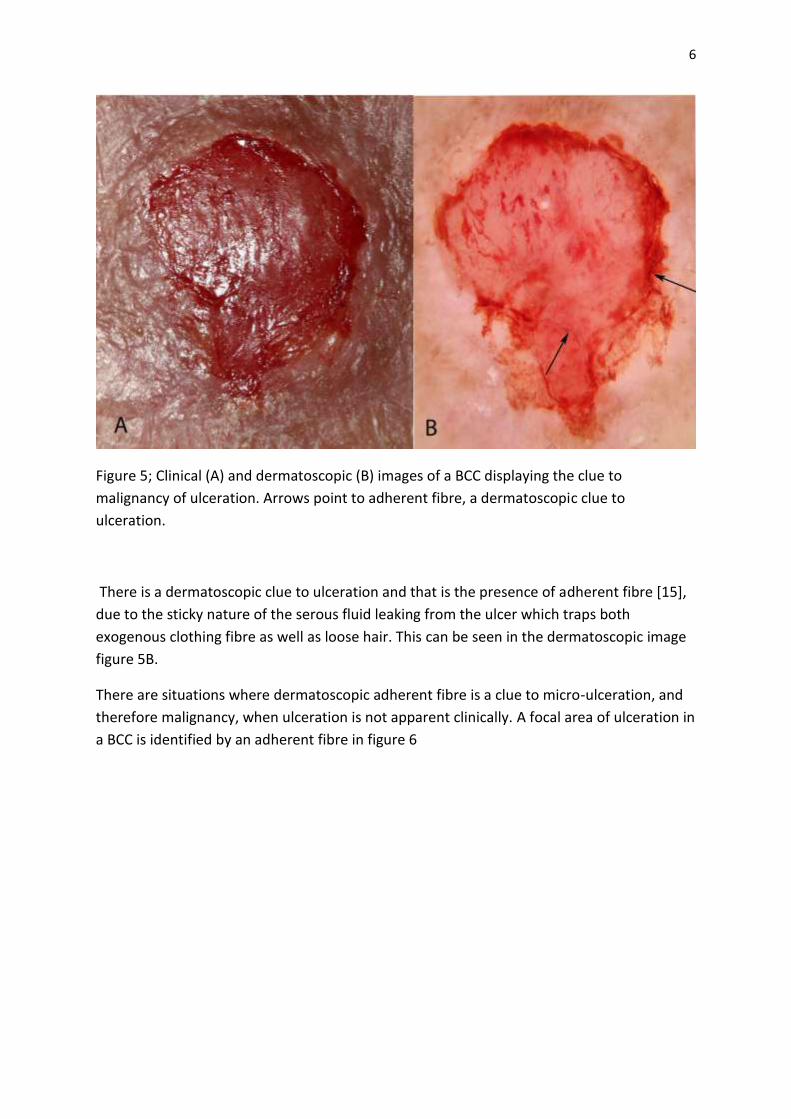
6
Figure 5; Clinical (A) and dermatoscopic (B) images of a BCC displaying the clue to
malignancy of ulceration. Arrows point to adherent fibre, a dermatoscopic clue to
ulceration.
There is a dermatoscopic clue to ulceration and that is the presence of adherent fibre [15],
due to the sticky nature of the serous fluid leaking from the ulcer which traps both
exogenous clothing fibre as well as loose hair. This can be seen in the dermatoscopic image
figure 5B.
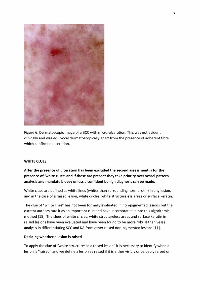
There are situations where dermatoscopic adherent fibre is a clue to micro-ulceration, and
therefore malignancy, when ulceration is not apparent clinically. A focal area of ulceration in
a BCC is identified by an adherent fibre in figure 6
7
Figure 6; Dermatoscopic image of a BCC with micro-ulceration. This was not evident
clinically and was equivocal dermatoscopically apart from the presence of adherent fibre
which confirmed ulceration.
WHITE CLUES
After the presence of ulceration has been excluded the second assessment is for the
presence of ‘white clues’ and if these are present they take priority over vessel pattern
analysis and mandate biopsy unless a confident benign diagnosis can be made.
White clues are defined as white lines (whiter than surrounding normal skin) in any lesion,
and in the case of a raised lesion, white circles, white structureless areas or surface keratin.
The clue of “white lines” has not been formally evaluated in non-pigmented lesions but the
current authors rate it as an important clue and have incorporated it into this algorithmic
method [15]. The clues of white circles, white structureless areas and surface keratin in
raised lesions have been evaluated and have been found to be more robust than vessel
analysis in differentiating SCC and KA from other raised non-pigmented lesions [11].
Deciding whether a lesion is raised
To apply the clue of “white structures in a raised lesion” it is necessary to identify when a
lesion is “raised” and we define a lesion as raised if it is either visibly or palpably raised or if
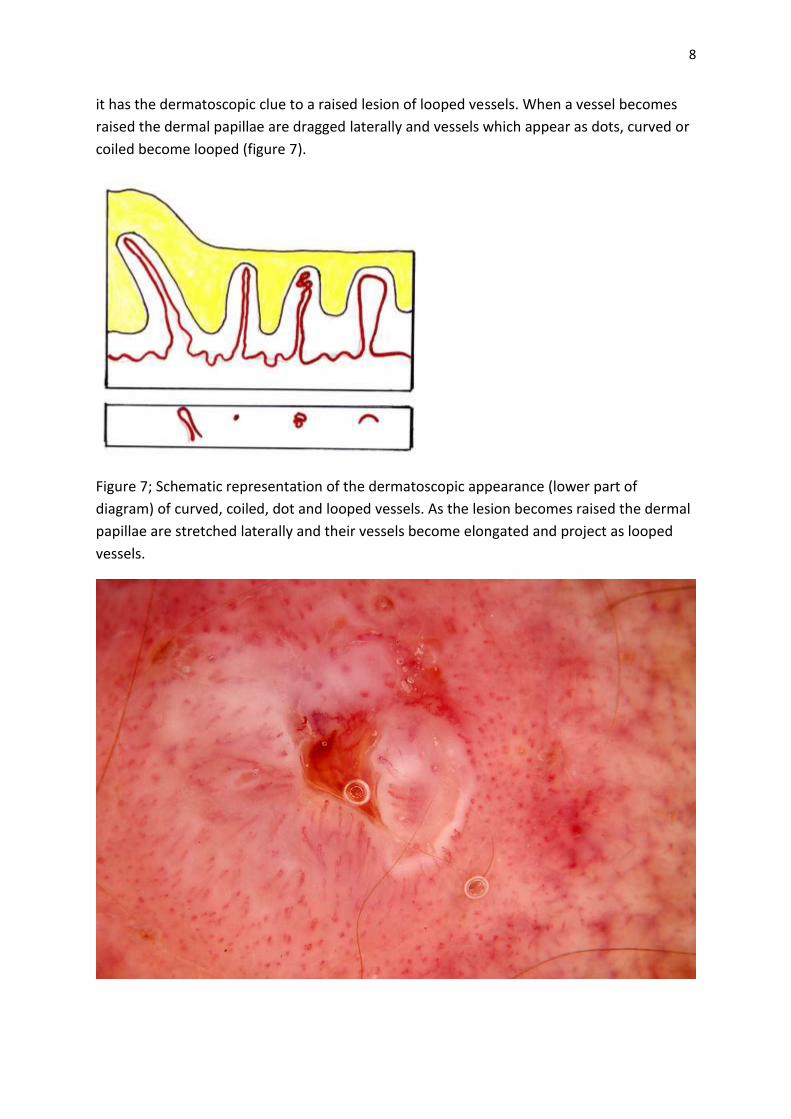
8
it has the dermatoscopic clue to a raised lesion of looped vessels. When a vessel becomes
raised the dermal papillae are dragged laterally and vessels which appear as dots, curved or
coiled become looped (figure 7).
Figure 7; Schematic representation of the dermatoscopic appearance (lower part of
diagram) of curved, coiled, dot and looped vessels. As the lesion becomes raised the dermal
papillae are stretched laterally and their vessels become elongated and project as looped
vessels.

9
Figure 8: Dermatoscopic image of an IEC (SCC in-situ) with coiled vessels peripherally.
Centrally the lesion has evolved into an invasive SCC and become raised. This was not
evident visually or by palpation but looped vessels identified the raised portion and this
lesion was evaluated as a raised lesion. Surface keratin centrally was therefore interpreted
as pointing to SCC and the lesion was excised.
WHITE LINES
There are two types of white lines which can be present in both pigmented and non-
pigmented lesions. Firstly there are white lines with may be seen with both polarising and
non-polarising dermatoscopy (see fig.9) and then there are ‘perpendicular white lines –
polarising artefact’ which are only seen with polarising dermatoscopes. The polarising
artefact types of white lines are oriented at right angle to each other but do not cross. The
authors of Prediction without Pigment regard both types of white lines as a clue to
malignancy which mandate biopsy but the specificity for malignancy is not 100%. For
example, perpendicular white lines polarising artefact (originally named ‘chrysallis’ by
Cowel) are a published clue to Spitz naevus, and DF as well as the malignancies melanoma
and BCC but they occur in lichen planus as Wickham’s striae and the authors have also seen
them frequently in LPLK as well as in (the malignancy) IEC (SCC in-situ).

10
Figure 9: This non-pigmented lesion does not have the clue of ulceration but there are white
lines. According to the Prediction without Pigment algorithm the presence of white lines
takes priority over vessel pattern analysis and mandates biopsy. This is a BCC.
Figure 10: A nonpigmented lesion on the back of a 70 year-old man cannot be confidently
diagnosed clinically.

11
Figure 11: Polarised and nonpolarised dermatoscopy of the lesion shown in figure 10 reveals
the dermatoscopic clue of perpendicular white lines – polarising artefact. This is a
fibroepithelioma of Pinkus – a variant of BCC.
IN A RAISED LESION: SURFACE KERATIN, WHITE STRUCTURELESS AREAS AND WHITE
CIRCLES
If a non-pigmented lesion is not ulcerated and there are no white lines as a clue to
malignancy then there is one step to assess prior to having to fall back on vessel pattern
analysis. If the lesion is raised then the ‘white clues’ of surface keratin, white structureless
areas or white circles are valuable clues to malignancy, specifically SCC and KA and they
have been found to be more robust in this regard than vessel patterns. Surface keratin is
best appreciated clinically, as one of the purposes of dermatoscopy is to render keratin
invisible to better appreciate structures beneath the stratum corneum.

12
Figure 12: A keratin horn is appreciated clinically but dermatoscopy of such a lesion would
be challenging! (image courtesy Alan Cameron)
Figure 13: Surface keratin and a white structureless area are seen clearly on this close-up
clinical image of a non-pigmented lesion on the ear of a 60 year old man.

13
Figure 14: Dermatoscopy of the lesion shown in figure 13. This shows the dermatoscopic
appearance of the surface keratin and white structureless area. It is an SCC.

14
Figure 15: Dense heaped-up parakeratotic keratin is the dermatopathological correlate of
the surface keratin on the lesion shown in figure 13. This densely packed keratin tends to
remain white as does the similar keratin over an actinic keratosis (unlike the orthokeratotic
keratin on the surface of a seborrhoeic keratosis which absorbs impurities to become
discoloured orange or brown (orange clods/keratin crypts)).
Figure 16: An aggregated mass of highly keratinised malignant squamous cells is the
dermatopathological correlate of white structureless areas in this SCC shown in figure 13.
“White clues” are not expected in poorly differentiated SCCs because these have lost the
ability to produce keratin.

15
Figure 17: A lesion on the hand of a 72 year old man cannot be confidently diagnosed as
benign clinically.
16
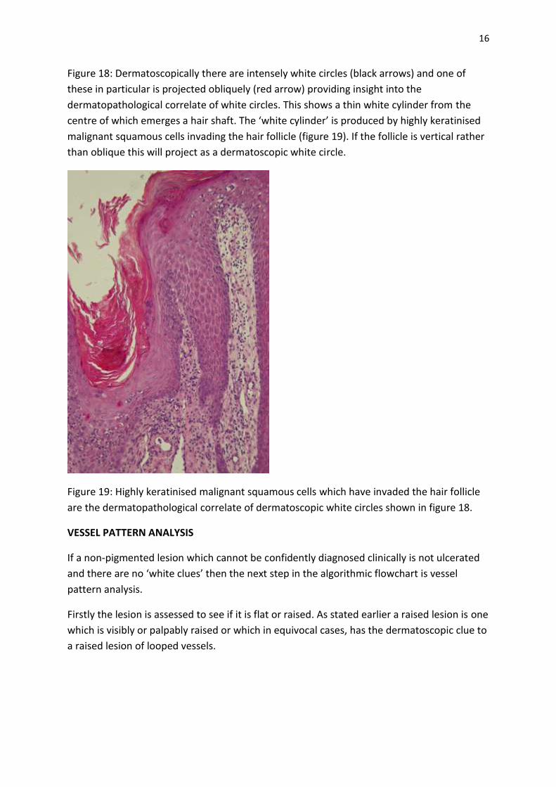
Figure 18: Dermatoscopically there are intensely white circles (black arrows) and one of
these in particular is projected obliquely (red arrow) providing insight into the
dermatopathological correlate of white circles. This shows a thin white cylinder from the
centre of which emerges a hair shaft. The ‘white cylinder’ is produced by highly keratinised
malignant squamous cells invading the hair follicle (figure 19). If the follicle is vertical rather
than oblique this will project as a dermatoscopic white circle.
Figure 19: Highly keratinised malignant squamous cells which have invaded the hair follicle
are the dermatopathological correlate of dermatoscopic white circles shown in figure 18.
VESSEL PATTERN ANALYSIS
If a non-pigmented lesion which cannot be confidently diagnosed clinically is not ulcerated
and there are no ‘white clues’ then the next step in the algorithmic flowchart is vessel
pattern analysis.
Firstly the lesion is assessed to see if it is flat or raised. As stated earlier a raised lesion is one
which is visibly or palpably raised or which in equivocal cases, has the dermatoscopic clue to
a raised lesion of looped vessels.

17
VESSEL PATTERN ANALYSIS OF FLAT LESIONS
Figure 20: Flowchart for the Prediction without Pigment algorithm for vessel pattern
analysis of flat non-pigmented lesions.
Firstly the lesion is assessed to determine whether the vessel pattern is monomorphous (a
pattern of one vessel type only) or polymorphous (two or more different patterns of
vessels). Pattern is assessed on groups of vessels rather than individual vessels. A pattern of
coiled vessels may have a few scattered dot vessels for example but it will still be a
monomorphous coiled pattern. A polymorphous pattern can be produced either by two or
more significant vessel patterns being present in different parts of the lesion or alternatively
by vessels of different structure occurring in significant quantities combined together
throughout the lesion (analogous to the speckled pattern in pigmented lesions).
If the pattern is polymorphous a biopsy is indicated.
a) If the pattern is polymorphous and this includes a pattern of dot vessels then biopsy
is indicated because this is a pattern frequently seen in melanomas.

18
Figure 21: A lightly pigmented lesion should be assessed according to pigment structures.
Chaos (asymmetry) plus the clue of grey structures mandates biopsy, but in this case vessel
pattern analysis permits the differential diagnosis to be narrowed down from melanoma,
BCC and pigmented IEC(SCC in-situ). There are two vessel patterns. Serpentine (centrally)
and dots. A pattern of dot vessels is not expected in a BCC (there may be a few isolated dots
but not a vast pattern) and serpentine vessels are not expected in a pigmented IEC. This is a
melanoma (Breslow thickness 0.55mm) (refer to the flowchart Figure 20).
b) If the pattern is polymorphous, not including a pattern of dots then a biopsy is also
indicated because the differential diagnosis includes BCC and IEC (SCC in-situ) .

19
Figure 22: The vessel pattern in this flat nonpigmented lesion is polymorphous. Coiled
helical, looped, curved and serpentine vessels are combined in a ‘speckled’ arrangement.
This does not include a pattern of dot vessels so melanoma is not in the differential
diagnosis. The differential diagnosis includes BCC and IEC. This is an IEC (SCC in-situ) and
ulceration has probably induced the polymorphous vessel pattern (refer to the flowchart
Figure 20).
If the pattern is monomorphous, then there is one pattern that is predictably benign (clods-
only) while all other patterns in flat lesions (dots, serpentine or coiled) have malignant
options in the differential diagnosis and must therefore be considered for biopsy.

20
Figure 23: This lesion is partly pigmented and assessment as a pigmented lesion discovers
chaos (asymmetry) and the clue of an eccentric structureless (pink) area therefore excision
biopsy was performed. If we ignore the pigmented reticular lines for the purpose of this
exercise and assess it as a non-pigmented lesion there is a monomorphous pattern of dot
vessels. The differential diagnosis includes melanocytic naevus, IEC (SCC in-situ) , verucca
and inflammation/psoriasis. This was a naevus (refer to the flowchart Figure 20).

21
Fugure 24: Dermatoscopically this flat nonpigmented lesion has a monomorphous pattern
of serpentine vessels. This is a BCC. (Refer to the flowchart Figure 20)
22
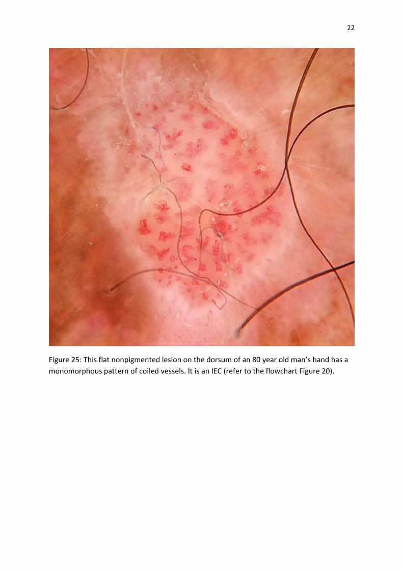
Figure 25: This flat nonpigmented lesion on the dorsum of an 80 year old man’s hand has a
monomorphous pattern of coiled vessels. It is an IEC (refer to the flowchart Figure 20).

23
Figure 26: This flat nonpigmented lesion on a 55 year old man’s chest was examined with a
dermatoscope (see figure 27)

24
Figure 27: Dermatoscopy revealed a pattern of clods-only. The clods were red and purple.
This lesion was assessed as benign – a haemangioma. For a clods-only pattern to be
interpreted as benign the clods must be red or purple and there must not be any vessels
seen within the clods. Although some of these clods were arguably blue, when blue/purple
clods occur in conjunction with red clods and there is absolutely no brown (melanin) colour
they are assessed as vascular rather than pigmented. This lesion was in fact biopsied due to
patient concern and was confirmed as a haemangioma (refer to the flowchart Figure 20).
VESSEL PATTERN ANALYSIS OF RAISED LESIONS
Figure 28: Flowchart for the Prediction without Pigment algorithm for vessel pattern
analysis of raised non pigmented lesions.
Raised lesions need to be assessed differently to flat lesions. It would be nice of the same
method of polymorphous versus monomorphous could be employed but this was not found
to be helpful in raised lesions.
Instead raised lesions are firstly assessed as to whether there is one of four different specific
vessel arrangements (radial, branched, serpiginous or centred). Two of these arrangements
are regarded as benign (serpiginous and centred) and the other two have malignant lesions
in the differential diagnosis and require consideration for biopsy.
a) If there is not one of the four designated specific vessel arrangements the
arrangement is described as “non-specific” and then if the pattern is clods-only the
pattern is regarded as benign (haemangioma). As with flat lesions the clods must be
red or purple and there must not be any vessels within the clods.
If the arrangement is non-specific and “not only clods” then a biopsy is indicated as
melanoma, BCC and SCC/KA are in the differential diagnosis as well as the benign
lesions SK and PG (pyogenic granuloma).

25
Figure 29: A raised non pigmented lesion inferior to the ear of a 76 year old man cannot be
confidently diagnosed as benign clinically.
Figure 30: Dermatoscopic image of the lesion shown in figure 29. The vessel arrangement is
non-specific (not radial, branched, serpiginous or centred). It is not a clod-only arrangement
therefore biopsy is indicated. This was an SCC (refer to the flowchart Figure 28).

26
Figure 31: Dermatoscopy image of a non-pigmented raised lesion which has a non-specific
vessel arrangement (not radial, branched, serpiginous or centred) and of the two options on
the flowchart for that category it is ‘only-clods’. This is a benign pattern consistent with the
diagnosis of haemangioma. Note that in a non-pigmented lesion in benign clods-only
pattern, the clods must be red or purple and there must be no vessels within the clods
(refer to the flowchart Figure 28).
b) If the vessel arrangement is either radial, branched, serpiginous or centred then the
lesions are assessed according to each of these specific arrangements as summarised
in figure 28.
Lesions with a radial pattern of vessels include SCC/KA and BCC with ulceration
Lesions with a branched vessel pattern include BCC, SCC/KA, Merkel cell carcinoma
and any raised cyst or neoplasm, benign or malignant.
The serpiginous vessel pattern is specific to clear cell acanthoma.

27
Figure 32: Dermatoscopy image of a non-pigmented raised lesion which has white clues
(surface keratin, white structureless areas and white circles) which take priority over vessel
pattern analysis and point to a diagnosis of SCC/KA. Ignoring the white clues for the purpose
of this exercise this lesion has a specific (radial) vessel arrangement and therefore requires a
biopsy. It is an SCC (refer to the flowchart Figure 28).

28
Figure 33: Dermatoscopy image of a non-pigmented raised lesion with a specific (branched)
vessel arrangement. This pattern requires biopsy which in this case confirmed that it was a
BCC. (Refer to the flowchart Figure 28).
29
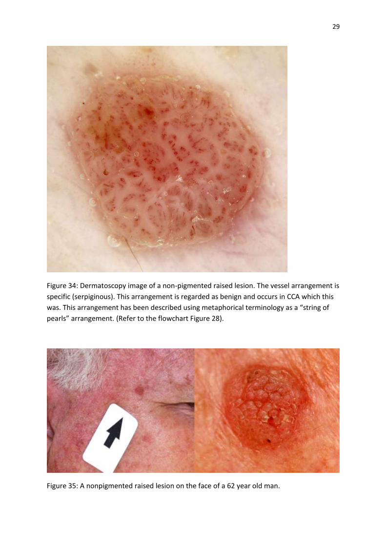
Figure 34: Dermatoscopy image of a non-pigmented raised lesion. The vessel arrangement is
specific (serpiginous). This arrangement is regarded as benign and occurs in CCA which this
was. This arrangement has been described using metaphorical terminology as a “string of
pearls” arrangement. (Refer to the flowchart Figure 28).
Figure 35: A nonpigmented raised lesion on the face of a 62 year old man.

30
Figure 36: Dermatoscopy of the lesion shown in figure 35 reveals that this raised
nonpigmented lesion has a specific (centred) vascular arrangement. This pattern is regarded
as benign. This presumed seborrhoeic keratosis was not excised. Note that a benign centred
pattern must consist of vessels centred in skin coloured clods. Vessels centred in red clods
can occur in melanoma!
Extreme caution is needed when diagnosing benign patterns
The vessel patterns regarded as benign (serpiginous, centred and (red) clods-only) should be
interpreted taking the clinical situation into consideration. No lesion which has a history of
growing continuously should be regarded as benign just because the vessel pattern is
centred, serpiginous or (red) clods-only, particularly if it is elevated and firm (EFG).
A centred pattern must have the vessels centred in skin-coloured clods (not
erythematous/pink or red clods).
A clods-only pattern should have only red/purple clods and there must be absolutely no
vessels seen within any of the clods.
31
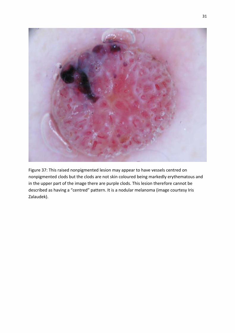
Figure 37: This raised nonpigmented lesion may appear to have vessels centred on
nonpigmented clods but the clods are not skin coloured being markedly erythematous and
in the upper part of the image there are purple clods. This lesion therefore cannot be
described as having a “centred” pattern. It is a nodular melanoma (image courtesy Iris
Zalaudek).

32
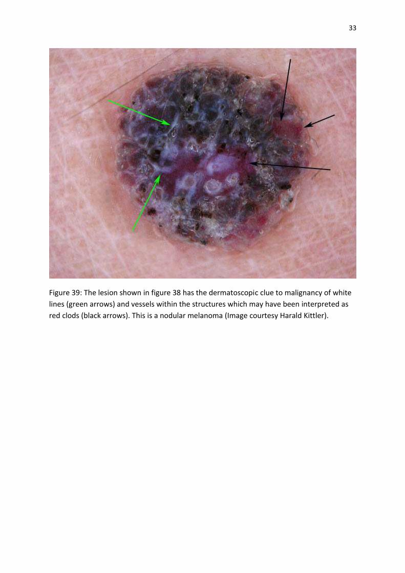
Figure 38: This raised lesion is not pigmented by melanin (when blue, purple or black colour
is associated with any brown colour it should be interpreted as being caused by melanin,
otherwise it should be interpreted as a vascular colour). Before interpreting this as a
pattern of “clods only” it should be noted that there are two things which disqualify it from
that classification. See the annotated image in figure 39 for an explanation.
33
Figure 39: The lesion shown in figure 38 has the dermatoscopic clue to malignancy of white
lines (green arrows) and vessels within the structures which may have been interpreted as
red clods (black arrows). This is a nodular melanoma (Image courtesy Harald Kittler).
34
REFERENCES
1. Chamberlain AJ, Fritschi L, Kelly JW. Nodular melanoma: patients’perceptions of presenting features and implications for earlier detection. J Am Acad Dermatol 2003;48:694–701.
2. Rosendahl C, Tschandl P, Cameron A, Kittler H. Diagnostic accuracy of
dermatoscopy for melanocytic and nonmelanocytic pigmented lesions. J. Am. Acad.
Dermatol. 2011 Jun;64(6):1068–73.
3. Zalaudek I, Argenziano G, Leinweber B, et al. Dermoscopy of Bowen’s disease.Br J Dermatol. 2004;150(6):1112-1116.
4. Bugatti L, Filosa G, De Angelis R. The specific dermoscopical criteria of Bowen’s
disease. J Eur Acad Dermatol Venereol. 2007;21(5):700-701.
5. Pan Y, Chamberlain AJ, Bailey M, Chong AH, Haskett M, Kelly JW. Dermatoscopy aids in the diagnosis of the solitary red scaly patch or plaque-features distinguishing superficial basal cell carcinoma, intraepidermal carcinoma, and psoriasis. J Am Acad Dermatol. 2008;59(2):268-274. 6. Felder S, Rabinovitz H, Oliviero M, Kopf A. Dermoscopic differentiation of a superficial basal cell carcinoma and squamous cell carcinoma in situ. Dermatol Surg. 2006;32(3):423-425. 7. Zalaudek I, Giacomel J, Argenziano G, et al. Dermoscopy of facial nonpigmented actinic keratosis. Br J Dermatol. 2006;155(5):951-956. 8. Cuellar F, Vilalta A, Puig S, Palou J, Salerni G, Malvehy J. New dermoscopic pattern in actinic keratosis and related conditions. Arch Dermatol. 2009;145(6):732. doi:10.1001/archdermatol.2009.86. 9. Kreusch J, Koch F. Incident light microscopic characterization of vascular patterns in skin tumors. Hautarzt. 1996;47(4):264-272. 10. Zalaudek I, Giacomel J, Schmid K, et al. Dermatoscopy of facial actinic keratosis, intraepidermal carcinoma, and invasive squamous cell carcinoma: a progression model. J Am Acad Dermatol. 2011;66(4):589-597. 11. Rosendahl C, Cameron A, Argenziano G, Zalaudek I, Tschandl P, Kittler H.
Dermoscopy of squamous cell carcinoma and keratoacanthoma. Arch Dermatol. 2012
Dec 1;148(12):1386–92.
12. Kittler H. Dermatoscopy: Introduction of a new algorithmic method based on pattern analysis for diagnosis of pigmented skin lesions. Dermatopathology: Practical & Conceptual. 2007;13(1):3. Available at: http://www.derm101.com.

35
13. Cameron A, Rosendahl C, Tschandl P, Riedl E, Kittler H. Dermatoscopy of pigmented Bowen’s disease. J. Am. Acad. Dermatol. 2010 Apr;62(4):597–604.
14. Kittler H, Riedl E, Rosendahl C, Cameron A. “Dermatoscopy of unpigmented lesions of the skin: a new classification of vessel morphology based on pattern analysis,” Dermatopathology: Practical & Conceptual 14, no. 4 (December 2008). 15. Kittler H, Rosendahl C, Cameron A, Tschandl P. Dermatoscopy.2011 facultas.wuv, Austria
Related Documents