The Revised Atlanta Classification of Acute Pancreatitis: Its Importance for the Radiologist and Its Effect on Treatment Volume 262: Number 3—March 2012 radiology.rsna.org Dr M Venkatesh

Revised Atlanta classification of Acute Pancreatitis
Aug 12, 2015
Welcome message from author
This document is posted to help you gain knowledge. Please leave a comment to let me know what you think about it! Share it to your friends and learn new things together.
Transcript
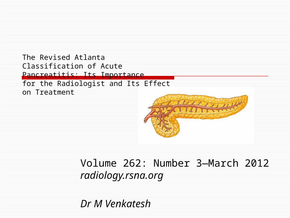
The Revised AtlantaClassification of AcutePancreatitis: Its Importancefor the Radiologist and Its Effecton Treatment
Volume 262: Number 3—March 2012 radiology.rsna.org
Dr M Venkatesh
Learning Objectives
Define acute pancreatitis in its early phase and later phase, and the persistent organ failure that can accompany its occurance.
List the various fluid collections encountered in acute pancreatitis as defined by the revised Atlantaclassification.

Identify the two phases of acute Pancreatitis, the parameters that determine care, and the treatment foran infected walled-off necrosis.
The modifications (a) address the clinical course and severity of disease
(b) divide acute pancreatitis into
interstitial edematous pancreatitis and necrotizing pancreatitis

(c) distinguish an early phase (1st week) and a late phase (after the 1st week), and
(d) emphasize systemic inflammatory response syndrome and multisystem organ failure

In the 1st week, only clinical parameters are important for treatment planning.
After the 1st week,morphologic criteria defined on the basis of computed tomographic findings are combined with clinical parameters to help determine care.
In 1992, the Atlanta classification for acute pancreatitis was introduced as a universally applicable classification system for the various manifestations of acute pancreatitis
It defined acute pancreatitis as an acute inflammatory process of the pancreas with variable involvement of other local tissues and remote organ systems.

1992 ATLANTA CLASSIFICATION

It was found that the definitions of severity and local complications of acute pancreatitis were not used consistently and that characterizationof severity based on presence of organ failure had limitations

The definition of necrotizing pancreatitis was determined to be inadequate because it included sterile and infected necrosis and did not distinguish between pancreatic and peripancreatic necrosis
The initial Atlanta classification system also did not include exact radiologic criteria for local complications and controversy developed over the natural course of pancreatic and peripancreatic fluid collections.

In 2008, a global consensus statement was developed that included broad and international participation of many experts in the field of pancreatitis and was led by the Acute Pancreatitis Classification Working Group

Precise description of pancreatic collections is particularly important, because treatment varies with collection type.
This revised classification is directly applicable only to adults (>18 years of age).
Radiologic imaging has become increasingly important in staging and treating acute pancreatitis
The revision of the Atlanta classification focuses heavily on morphologic criteria for defining the various manifestations of acute pancreatitis

REVISED ATLANTA CLASSIFICATION
Clinical Definition, Course,and Severity of Disease
Acute pancreatitis (regardless of presence or absence of chronic pancreatitis) is clinically defined by at least the two of three features
(a) abdominal pain suggestive of pancreatitis with the start of such pain considered to be the onset of acute pancreatitis
(b) serum amylase and lipase levels three or more times normal (imagingis to be used if the elevated values are<3 times normal)
(c) characteristic findings on CT, magnetic resonance (MR) imaging, or transabdominal ultrasonographic (US) studies.
Course and severity
Introduces two distinct phases of acute pancreatitis:
First or early phase that occurs within the 1st week of onset of disease
Second or late phase that takes place after the 1st week of onset

In the early phase, severity is entirely based on clinical parameters, because the need for treatment is determined primarily by presence or absence of organ failure

It is standard clinical practice within the first 3 days of admission of a patient with acute pancreatitis to record markers of severity

Hematocrit APACHE II, Ranson Scores Pulmonary complications on chest
radiograph, including pleural effusion; and serum levels of CRP)
Other severity markers CT severity index Modified CT severity index Other parameters of acute
pancreatic injury
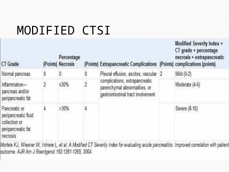
MODIFIED CTSI

Imaging-based MorphologicClassification
Contrast-enhanced CT is the primary tool for assessing the imaging-based criteria because it is widely available for these acutely ill patients and has a high degree of accuracy

The ideal time for assessing the complications with CT is after 72
hours from onset of symptoms. CT should be repeated when the
clinical picture drastically changes, such as with sudden onset of fever, decrease in hematocrit or sepsis.

Furthermore, in patients with their first episode of pancreatitis who are over 40 years of age and have no
identifiable cause for pancreatitis, contrast enhanced CT should be used to exclude a possible neoplasm
ROLE OF RADIOLOGIST
Address whether pancreatic necrosis is present, characterize pancreatic
parenchymal and extrapancreaticfluid collections, and describe thepresence of ascites, extrapancreatic findings such as gallstones, biliary dilatation,venous thrombosis, aneurysms

And contiguous inflammatory involvement of the gastrointestinal tract.
MR imaging is reserved for detection of choledocholithiasis not
visualized on contrast-enhanced CT images and to further characterize collections for the presence of nonliquefied material

Nonliquefied material refers to solid and semisolid components usually pancreatic and extra-pancreatic debris and necrotic fatty tissue and may appear on contrast-enhanced CT images as a homogeneous or heterogeneous fluid collection.

MR imaging has an important role in patients in whom contrast-enhanced CT is contraindicated
ERCP has no role in this morphologic imaging–based classification of acute pancreatitis
Morphologic Stages of Acute Pancreatitis
In the 1992 Atlanta classification, a distinction was made between interstitial pancreatitis and sterile or infected necrosis.
In the revised Atlanta classification,these two types are defined similarly as IEP and acute necrotizing pancreatitis
Necrotizing pancreatitis is furthersubdivided into -Parenchymal necrosis alone-Peripancreatic necrosis alone-Combined type (peripancreatic and parenchymal necrosis) with or without infection

It is important for the radiologist to adopt this new nomenclature so that imaging descriptions are standardized and communication with clinical and surgical colleagues is precise.
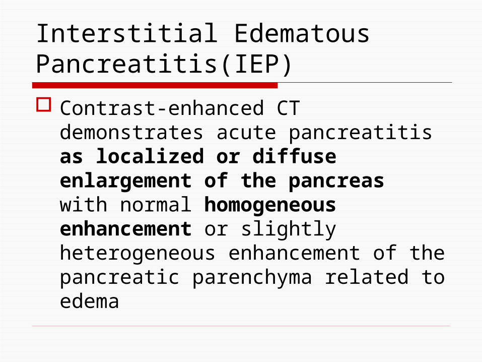
Interstitial Edematous Pancreatitis(IEP)
Contrast-enhanced CT demonstrates acute pancreatitis as localized or diffuse enlargement of the pancreas with normal homogeneous enhancement or slightly heterogeneous enhancement of the pancreatic parenchyma related to edema

Coronal CT image of Interstitial edematous pancreatitis (IEP) in a 34-year-old man. Pancreas (arrows) is heterogeneously enhanced, with indistinct margins due to inflammation of peripancreatic fat. Some stranding and minimal fluid are also present
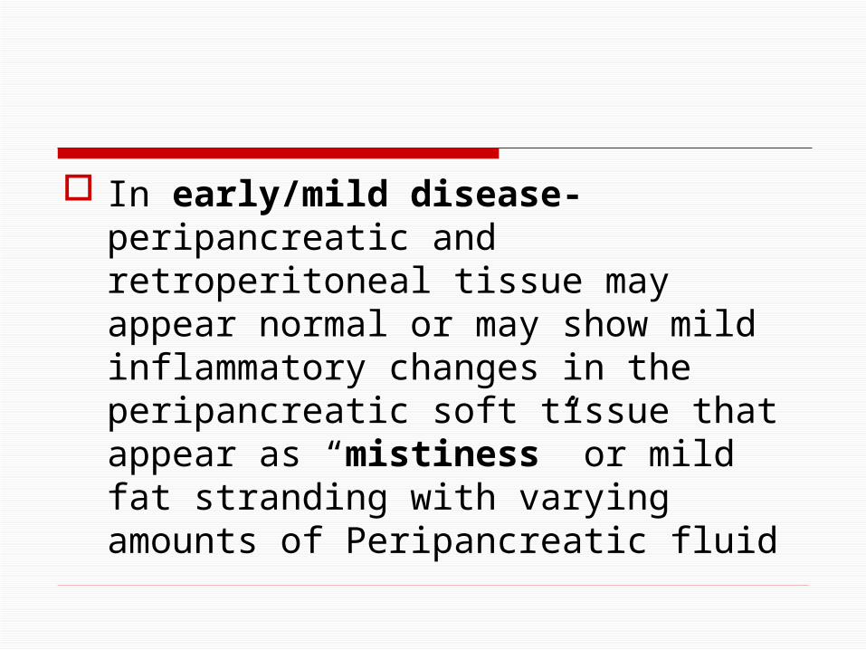
In early/mild disease- peripancreatic and retroperitoneal tissue may appear normal or may show mild inflammatory changes in the peripancreatic soft tissue that appear as “mistiness” or mild fat stranding with varying amounts of Peripancreatic fluid
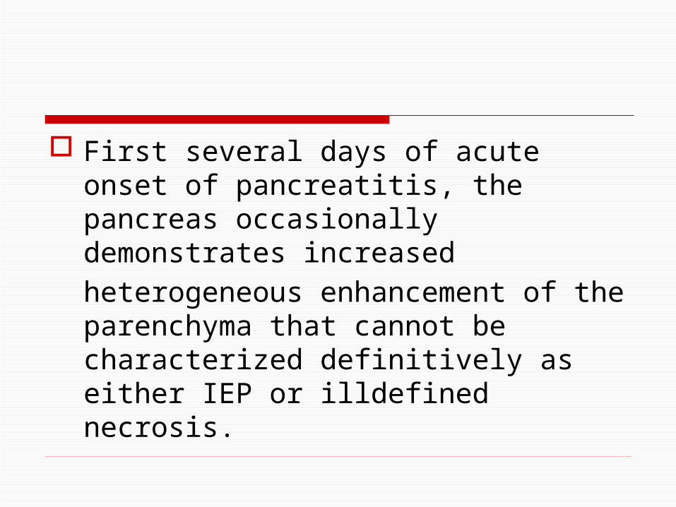
First several days of acute onset of pancreatitis, the pancreas occasionally demonstrates increasedheterogeneous enhancement of the parenchyma that cannot be characterized definitively as either IEP or illdefined necrosis.

Axial multidetector CT image of IEP in a 39-year-old man obtained 48 hours after onset of pain. Note focal heterogeneous low-attenuation area in pancreas body and neck (arrows). At this stage, the appearance could not be definitively characterized as IEP or patchy necrosis and was classified as indeterminate.Follow-up multidetector CT study did not show any necrosis.

With these findings, the presence or absence of pancreatic necrosis needs to be described initially as indeterminate.
CECT performed 5–7 days later permits definitive characterization.

Necrotizing Pancreatitis
Three forms of acute necrotizingpancreatitis, depending on location.
All three types can be sterile or infected Pancreatic parenchymal necrosis Peripancreatic necrosis alone Pancreatic parenchymal necrosis with peripancreatic necrosis
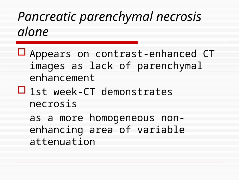
Pancreatic parenchymal necrosis alone
Appears on contrast-enhanced CT images as lack of parenchymal enhancement
1st week-CT demonstrates necrosisas a more homogeneous non-enhancing area of variable attenuation

Later in the course of the disease, as a more heterogeneous area.
Often the extent of parenchymal necrosis is divided into less than 30% 30%–50% greater than 50% of the gland
involved

Axial CT image in a 38-year-old man obtained 5 days after onset of symptoms. Tail and body of the pancreas are nonenhancing(arrows) and slightly heterogeneous in appearance.

On coronal reformation CT image obtained 4 weeks after onset, capsule (arrows) is evident and some heterogeneity (arrowheads) is seen within this collection, reflecting presence of nonliquefied material.

At times, areas of no or poorenhancement that are estimated to be less than 30% in the early phase may actually be findings of edema rather than necrosis.
A definitive diagnosis in thesepatients requires a follow-up study.

Peripancreatic necrosis alone
Difficult to confirm,its presence is diagnosed when heterogeneous areas of nonenhancement are visualized that contain nonliquefiedcomponents.
Peripancreatic necrosis is commonly located in the retroperitoneum and lesser sac.
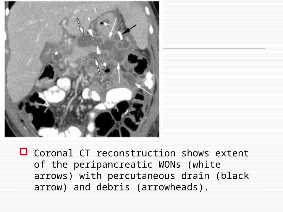
Coronal CT reconstruction shows extent of the peripancreatic WONs (white arrows) with percutaneous drain (black arrow) and debris (arrowheads).

The clinical importance of peripancreatic necrosis alone lies in the fact that patients with this condition have a better prognosis than do patients with pancreatic parenchymal necrosis
Pancreatic parenchymal necrosis with peripancreatic necrosis It is the most common type and can
be seen in 75%–80% of patients with acute necrotizing pancreatitis
Peripancreatic necrosis associatedwith full width necrosis of the pancreatic parenchyma may be connected to the main pancreatic duct
Pancreatic and Peripancreatic Collections
In the revised Atlanta classification, an important distinction is made between fluid and nonliquefied collections
The acute collections are referred to as either APFCs or as ANCs, depending on the absence or presence of necrosis respectively.
IEP can be associated with APFC and over time with pancreatic pseudocysts.
Necrotizing pancreatitis in its three forms can be associated with ANC and over time with WON.
All of these collections can be sterile or infected
APFCs-Acute peripancreatic fluid collections
Peripancreatic fluid collections without nonliquefied components
arising in patients with IEP during the first 4 weeks are referred to as
APFCs
APFCs conform to the anatomic boundaries of the retroperitoneum (especially the anterior pararenal fascia),
Are usually seen immediately next to the pancreas and have no discernable wall.
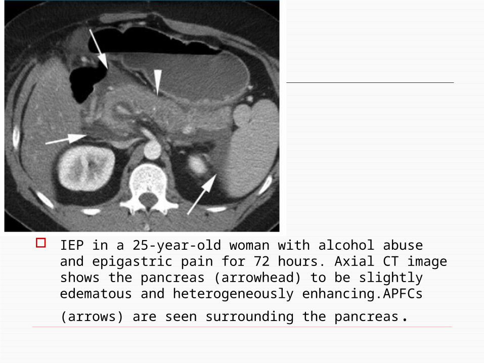
IEP in a 25-year-old woman with alcohol abuse and epigastric pain for 72 hours. Axial CT image shows the pancreas (arrowhead) to be slightly edematous and heterogeneously enhancing.APFCs (arrows) are seen
surrounding the pancreas.
Fluid collections in the pancreaticparenchyma should be diagnosedas necrosis and not as APFCs
Most APFCs are reabsorbed spontaneously within the first few weeks and do not become infected. Intervention at this stage is to be avoided

In the 1st week of acute pancreatitis,distinction between APFC and ANC may be difficult or impossible, because both collections may appear as areas of nonenhancement
If nonenhancing areas of variable attenuation are seen in these collections, the diagnosis of peri -pancreatic necrosis with nonliquefied components is suggested.

Pseudocyst
Within 4 weeks from onset of acute IEP, an APFC may gradually transition into a pseudocyst
On CECT images, pseudocysts can be diagnosed as well-circumscribed, round or oval peripancreatic fluid collections of homogeneously low attenuation surrounded by a well-defined enhancing wall (capsule consisting of fibrous or granulation tissue).

According to the revised Atlanta classification, pseudocysts contain no nonliquefied components within the fluid collection
In the rare event in which an APFC develops an enhancing capsule earlier than 4 weeks after onset of acute IEP, it should be characterized as a pseudocyst.
Demonstrating the presence or absence of communication of pseudocyst with the pancreatic duct may be important since it may help determine management
Persistent communication with the pancreatic duct can be shown oncontrast-enhanced CT images and curved planar reconstructions, but MRCP is usually more accurate.

Pancreatitis with pseudocyst in a 27-year-old woman. Coronal CT reconstruction obtained 5 weeks after acute episode shows pseudocyst(arrows) with well-defined rim representingthe capsule near the tail of the pancreas. Gastric folds are slightly thickened (arrowheads).

An infected pseudocyst is diagnosed on CT images by the presence of gas within the pseudocyst or in absence of gas by means of fine-needle aspiration (FNA) with Gram staining and culture for bacteria or fungal organisms

Pseudocyst in a 61-year-old man. (a) Coronal CT reconstruction shows pseudocyst (arrows) next to body of the pancreas with a well-defined capsule. The patient complained of pain in the midabdomen to left upper quadrant and early satiety. (b) Follow-up coronal CT reconstruction was obtained after stent (arrow) had been placed endoscopically through the stomach into the sterile collection. There is no residual collection next to the pancreas.

ANCs-Acute necrotic collections
In first 4 weeks after development of necrotizing pancreatitis a persistent collection is to be diagnosed as ANC
Contains both fluid and necrotic material of various amounts(some of which are loculated) and is to be distinguished from APFC.

In these ANCs, liquefaction of the necrotic tissue occurs gradually (usually within 2–6 weeks)
Within the 1st week, both APFCs and ANCs can manifest as homogeneous
nonenhancing areas. Usually, the distinction on contrast-enhanced CT images should become possible after the 1st week.
WON-Walled off necrosis
Over time (usually at or after 4 weeks), the ANC matures and develops thickened nonepithelializedwall between the necrosis and the adjacent tissue. This maturing collection is called a WON.
WON may involve pancreatic parenchymal tissue and the
peripancreatic tissue, peripancreatic tissue alone or pancreas alone.

Any apparent fluid collection that occupies or replaces portions of the pancreatic parenchyma should be called a WON after 4 weeks from onset of necrotizing pancreatitis.
In contradistinction to a pseudocyst, WON contains necrotic pancreatic parenchyma or necrotic fat.
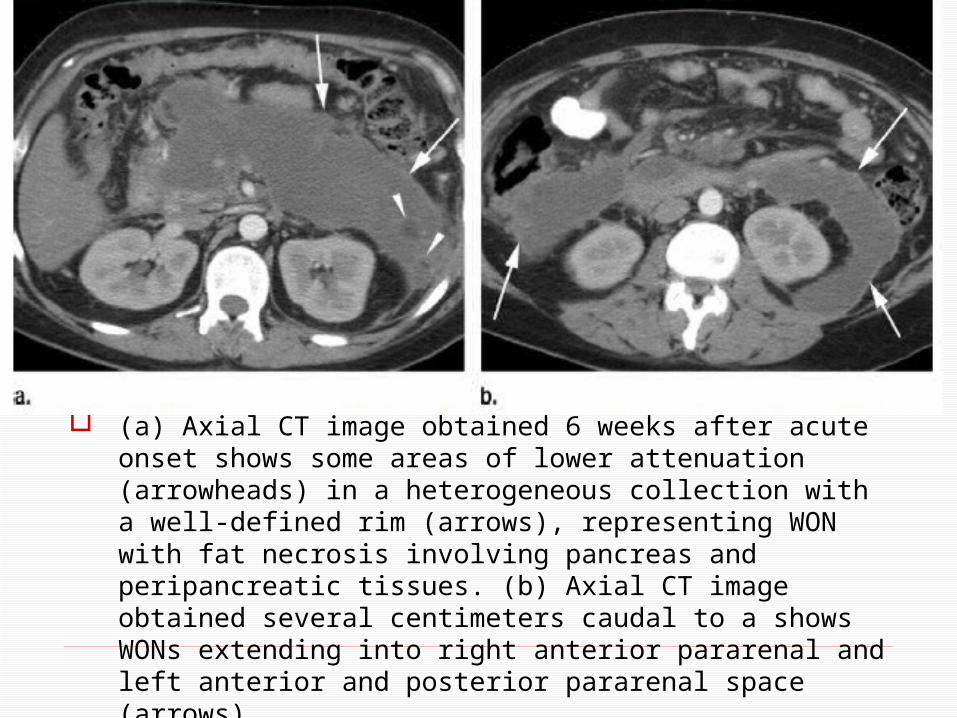
(a) Axial CT image obtained 6 weeks after acute onset shows some areas of lower attenuation (arrowheads) in a heterogeneous collection with a well-defined rim (arrows), representing WON with fat necrosis involving pancreas and peripancreatic tissues. (b) Axial CT image obtained several centimeters caudal to a shows WONs extending into right anterior pararenal and left anterior and posterior pararenal space (arrows).
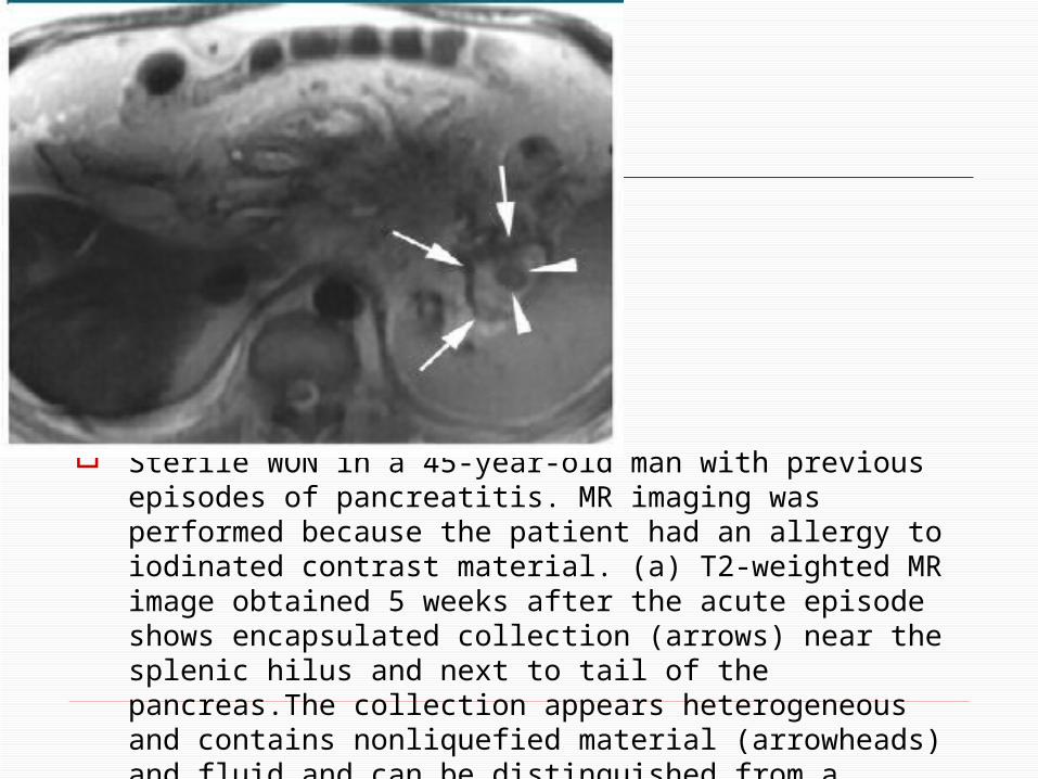
Sterile WON in a 45-year-old man with previous episodes of pancreatitis. MR imaging was performed because the patient had an allergy to iodinated contrast material. (a) T2-weighted MR image obtained 5 weeks after the acute episode shows encapsulated collection (arrows) near the splenic hilus and next to tail of the pancreas.The collection appears heterogeneous and contains nonliquefied material (arrowheads) and fluid and can be distinguished from a pseudocyst with fluid only
Most nonliquefied componentsneed to be removed by meansof a percutaneous image-guided approach, a laparoscopic or endoscopic procedure, or surgery.
A pseudocyst can be treated effectively by draining the fluid in most cases.
Therefore the distinction between a collection containing fluid only and a collection containing fluid and nonliquefied material is very important.

Complications of Acute Pancreatitis
Collections that contain nonliquefied material are more likely to become infected.
Distinction between a sterile and an infected collection is important because treatment and prognosis aredifferent
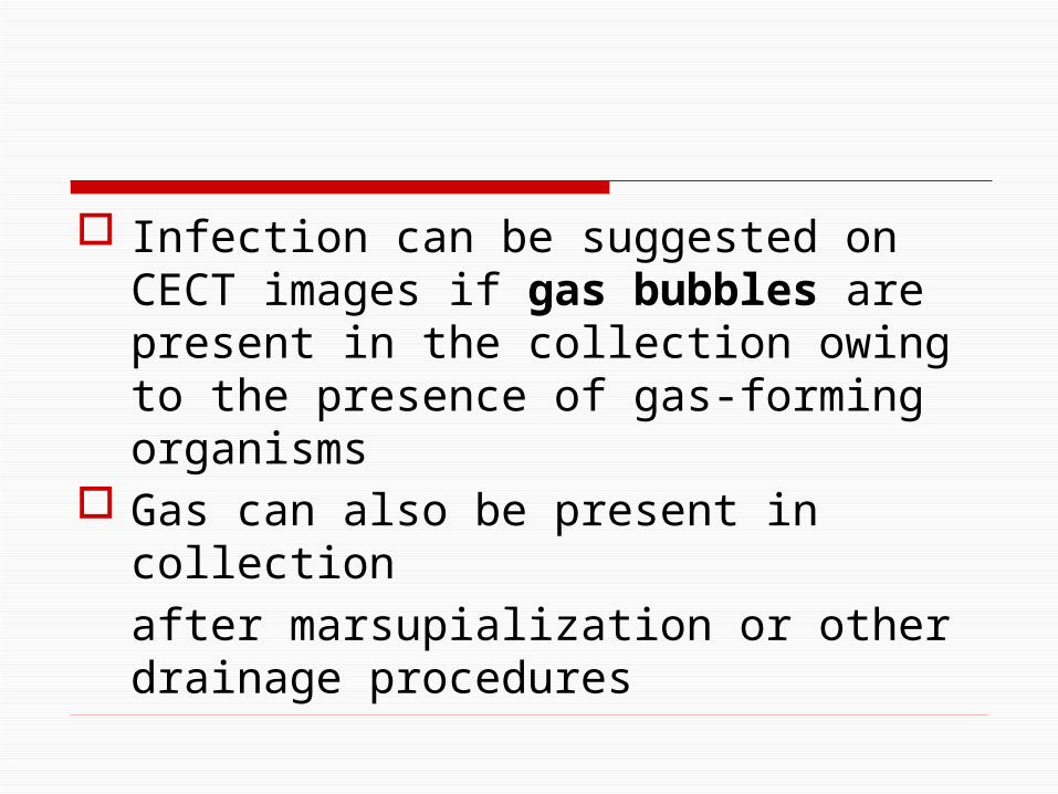
Infection can be suggested on CECT images if gas bubbles are present in the collection owing to the presence of gas-forming organisms
Gas can also be present in collectionafter marsupialization or other drainage procedures
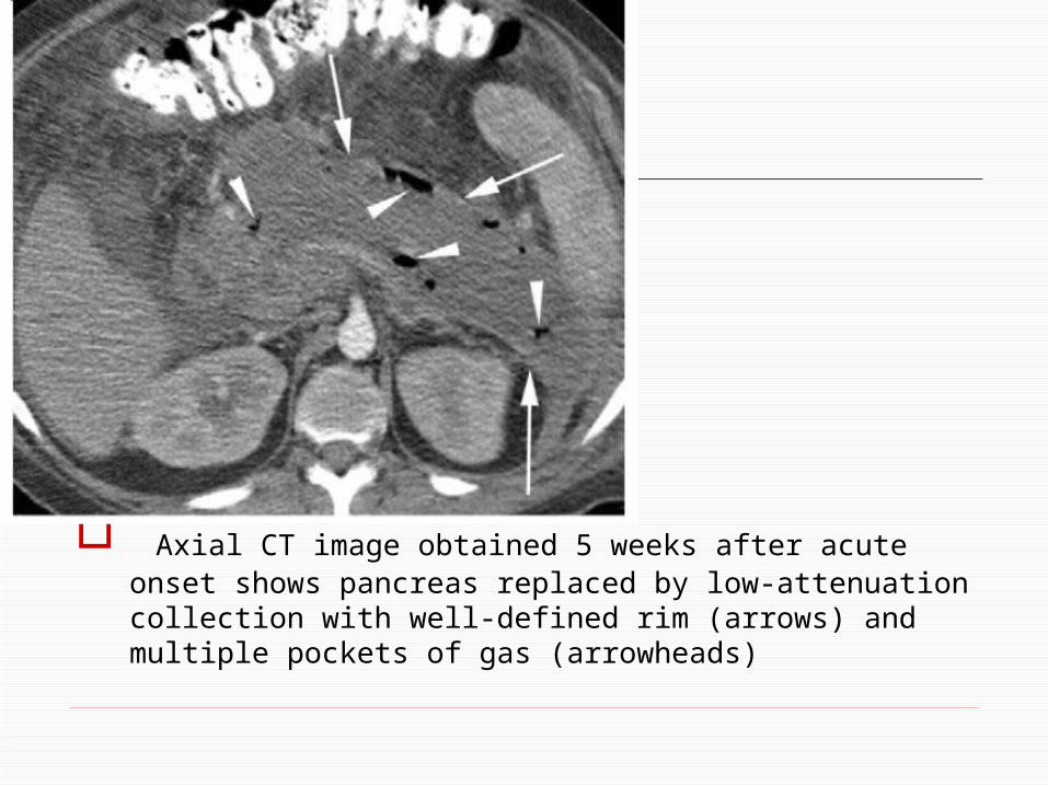
Axial CT image obtained 5 weeks after acute onset shows pancreas replaced by low-attenuation collection with well-defined rim (arrows) and multiple pockets of gas (arrowheads)

Axial CT image obtained 3 days after placement of percutaneous drainage catheters (arrows) shows large residual WON with air bubbles, indicative of incomplete drainage of an infected WON.

Treatment of IEP
IEP is usually self-limited, and supportive measures alone suffice. Most APFCs resolve spontaneously or mature into pseudocysts.
The majority of these pseudocysts disappear spontaneously over time and do not require any treatment.
About 25% become symptomatic or infected and necessitate drainage

Treatment of NecrotizingPancreatitis
No universally accepted treatmentalgorithm currently exists. The approach often is dictated by the expertise of the surgeon and the interventional radiologist
Image-guided drainage procedures proved to be effective alternatives to
surgery

Treatment of Sterile Pancreatic Necrosis
If the pancreatic fluid sample is sterile, the patient is diagnosed as having sterile necrosis.
Percutaneous drainage and surgey depending on the clinical severity.

Treatment of Infected Pancreatic Necrosis
Generally treated with surgical débridement and antibiotics
If a patient is too unstable for surgery, percutaneous catheter
drainage may help stabilize the patient

Interventional radiology also is called on for ancillary procedures.
Pseudoaneurysms or active bleeding related to acute pancreatitis are usually diagnosed on the basis ofcontrast-enhanced CT findings

Most commonly, coil embolization is used
Cases where coil embolization is too risky or not feasible, a covered stent can be placed.
Embolization may also be performed in selected instances of a bleeding vessel caused by pancreatitis.

Conclusions
The most important change in Atlanta classification is the categorization of the various pancreatic collections.
In acute IEP, collections that do not have an enhancing capsule are called APFCs; after development of a capsule, they are referred to aspseudocysts
In necrotizing pancreatitis,a collection without an enhancing capsule is called an ANC (usually in the first 4 weeks) and thereafter a WON, which has an enhancing capsule.
The most important distinction between collections in necrotizing pancreatitis and those associated with acute IEP is the presence of nonliquefied material in collections due to necrotizing pancreatitis.

In the early phase of pancreatitis,distinction between APFC and ANC by CT may be impossible and if clinically needed for treatment planning, MR imaging or US may be used to determine presence of nonliquefiedmaterial

The revised Atlanta classification system with CT helps guide management and monitor the success of treatment.

THANK YOU
Related Documents