Accepted manuscripts are peer-reviewed but have not been through the copyediting, formatting, or proofreading process. Copyright © 2018 the authors This Accepted Manuscript has not been copyedited and formatted. The final version may differ from this version. A link to any extended data will be provided when the final version is posted online. Research Articles: Cellular/Molecular α 2 δ-4 is required for the molecular and structural organization of rod and cone photoreceptor synapses Vasily Kerov 1 , Joseph G. Laird 2 , Mei-ling Joiner 1 , Sharmon Knecht 7 , Daniel Soh 1 , Jussara Hagen 1 , Sarah H. Gardner 2 , Wade Gutierrez 3 , Takeshi Yoshimatsu 7 , Sajag Bhattarai 4 , Teresa Puthussery 8 , Nikolai O. Artemyev 1 , Arlene V. Drack 4 , Rachel O. Wong 7 , Sheila A. Baker 2,4 and Amy Lee 1,5,6 1 Dept. of Molecular Physiology and Biophysics 2 Dept. of Biochemistry 3 Medical Scientist Training Program 4 Dept. of Ophthalmology and Institute for Vision Research 5 Dept. of Otolaryngology-Head and Neck Surgery 6 Dept. of Neurology, University of Iowa, Iowa City, 52242, USA 7 Dept. of Biological Structure, University of Washington, Seattle, WA 98195, USA 8 Casey Eye Institute, Oregon Health & Science University, Portland, OR 97239, USA DOI: 10.1523/JNEUROSCI.3818-16.2018 Received: 12 December 2016 Revised: 10 May 2018 Accepted: 31 May 2018 Published: 6 June 2018 Author contributions: V.K., M.-l.A.J., S.K., T.P., N.O.A., A.V.D., R.O.L.W., S. Baker, and A.L. designed research; V.K., J.L., M.-l.A.J., S.K., D.S., J.H., S.G., W.G., T.Y., S. Bhattarai, T.P., N.O.A., and S. Baker performed research; V.K., J.L., M.-l.A.J., S.K., J.H., W.G., T.Y., S. Bhattarai, T.P., N.O.A., A.V.D., R.O.L.W., and S. Baker analyzed data; S. Baker and A.L. wrote the paper. Conflict of Interest: The authors declare no competing financial interests. We thank William Paradee (U. Iowa Genome Editing Core) for aiding development of #2#-4 KO mice, Chantal Allamargot (U. Iowa Central Microscopy Research Facility (CMRF)) for processing of tissue samples for electron microscopy, David Wilson and John Ng (OHSU) for providing human retinal samples, and Jordan Breffle, Taylor Vogel, and Jacqueline Gayet-Primo for excellent technical assistance, Ed Parker for serial TEM assistance and Sharm Knecht for SBFSEM analysis, and Chris Johnson for assistance with measuring room luminescence. This work was supported by NIH grants: NS084190, DC009433 to AL; EY026817 to AL and SAB; EY020542, EY027054 to SAB; EY12682 to NOA; EY017168 to AVD; EY024265 to TP; EY010572 to Ophthalmology Core Facility at OHSU; EY017101 to ROW; EY01730 Vision Core grant (M. Neitz); RR018998 in support of the JEOL JEM Transmission electron microscope in the CMRF); an Unrestricted Grant from RPB to OHSU; a Wynn Institute Advisory Board grant (AVD); and a Carver Research Program of Excellence Award to AL. Correspondence: Amy Lee, UI Dept. of Molecular Physiology and Biophysics, 5318 PBDB, 169 Newton Rd., Iowa City, IA 52242; Phone: (319) 384-1762; e-mail: [email protected] or Sheila Baker, UI Dept. of Biochemistry, 4-712 BSB, Iowa City, IA 52242, Phone: (319) 353-4119; email: [email protected] Cite as: J. Neurosci ; 10.1523/JNEUROSCI.3818-16.2018 Alerts: Sign up at www.jneurosci.org/cgi/alerts to receive customized email alerts when the fully formatted version of this article is published.

Welcome message from author
This document is posted to help you gain knowledge. Please leave a comment to let me know what you think about it! Share it to your friends and learn new things together.
Transcript

Accepted manuscripts are peer-reviewed but have not been through the copyediting, formatting, or proofreading process.
Copyright © 2018 the authors
This Accepted Manuscript has not been copyedited and formatted. The final version may differ fromthis version. A link to any extended data will be provided when the final version is posted online.
Research Articles: Cellular/Molecular
α2δ-4 is required for the molecular and structural organization of rodand cone photoreceptor synapses
Vasily Kerov1, Joseph G. Laird2, Mei-ling Joiner1, Sharmon Knecht7, Daniel Soh1, Jussara Hagen1, Sarah
H. Gardner2, Wade Gutierrez3, Takeshi Yoshimatsu7, Sajag Bhattarai4, Teresa Puthussery8, Nikolai O.
Artemyev1, Arlene V. Drack4, Rachel O. Wong7, Sheila A. Baker2,4 and Amy Lee1,5,6
1Dept. of Molecular Physiology and Biophysics2Dept. of Biochemistry3Medical Scientist Training Program4Dept. of Ophthalmology and Institute for Vision Research5Dept. of Otolaryngology-Head and Neck Surgery6Dept. of Neurology, University of Iowa, Iowa City, 52242, USA7Dept. of Biological Structure, University of Washington, Seattle, WA 98195, USA8Casey Eye Institute, Oregon Health & Science University, Portland, OR 97239, USA
DOI: 10.1523/JNEUROSCI.3818-16.2018
Received: 12 December 2016
Revised: 10 May 2018
Accepted: 31 May 2018
Published: 6 June 2018
Author contributions: V.K., M.-l.A.J., S.K., T.P., N.O.A., A.V.D., R.O.L.W., S. Baker, and A.L. designed research; V.K., J.L.,M.-l.A.J., S.K., D.S., J.H., S.G., W.G., T.Y., S. Bhattarai, T.P., N.O.A., and S. Baker performed research; V.K., J.L., M.-l.A.J.,S.K., J.H., W.G., T.Y., S. Bhattarai, T.P., N.O.A., A.V.D., R.O.L.W., and S. Baker analyzed data; S. Baker and A.L. wrote thepaper.
Conflict of Interest: The authors declare no competing financial interests.
We thank William Paradee (U. Iowa Genome Editing Core) for aiding development of #2#-4 KO mice, Chantal Allamargot (U.Iowa Central Microscopy Research Facility (CMRF)) for processing of tissue samples for electron microscopy, David Wilsonand John Ng (OHSU) for providing human retinal samples, and Jordan Breffle, Taylor Vogel, and Jacqueline Gayet-Primofor excellent technical assistance, Ed Parker for serial TEM assistance and Sharm Knecht for SBFSEM analysis, and ChrisJohnson for assistance with measuring room luminescence. This work was supported by NIH grants: NS084190, DC009433to AL; EY026817 to AL and SAB; EY020542, EY027054 to SAB; EY12682 to NOA; EY017168 to AVD; EY024265 to TP;EY010572 to Ophthalmology Core Facility at OHSU; EY017101 to ROW; EY01730 Vision Core grant (M. Neitz); RR018998in support of the JEOL JEM Transmission electron microscope in the CMRF); an Unrestricted Grant from RPB to OHSU; aWynn Institute Advisory Board grant (AVD); and a Carver Research Program of Excellence Award to AL.
Correspondence: Amy Lee, UI Dept. of Molecular Physiology and Biophysics, 5318 PBDB, 169 Newton Rd., Iowa City, IA52242; Phone: (319) 384-1762; e-mail: [email protected] or Sheila Baker, UI Dept. of Biochemistry, 4-712 BSB, IowaCity, IA 52242, Phone: (319) 353-4119; email: [email protected]
Cite as: J. Neurosci ; 10.1523/JNEUROSCI.3818-16.2018
Alerts: Sign up at www.jneurosci.org/cgi/alerts to receive customized email alerts when the fully formatted version of thisarticle is published.
2 4 is required for the molecular and structural organization of rod and cone photoreceptor synapses Abbreviated title: 2 4 at the photoreceptor ribbon synapse Vasily Kerov1, Joseph G. Laird2, Mei-ling Joiner1, Sharmon Knecht7, Daniel Soh1, Jussara Hagen1, Sarah H. Gardner2, Wade Gutierrez,3 Takeshi Yoshimatsu7, Sajag Bhattarai4, Teresa Puthussery, 8,9 Nikolai O. Artemyev,1 Arlene V. Drack4, Rachel O. Wong7, Sheila A. Baker2,4, and Amy Lee1,5,6 1Dept. of Molecular Physiology and Biophysics; 2Dept. of Biochemistry; 3Medical Scientist Training Program; 4Dept. of Ophthalmology and Institute for Vision Research; 5Dept. of Otolaryngology-Head and Neck Surgery; 6Dept. of Neurology, University of Iowa, Iowa City, 52242, USA 7Dept. of Biological Structure, University of Washington, Seattle, WA 98195, USA 8Casey Eye Institute, Oregon Health & Science University, Portland, OR 97239, USA 9Present address: School of Optometry & Helen Wills Neuroscience Institute, University of California, Berkeley, Berkeley CA, 94720, USA Correspondence to: Amy Lee, UI Dept. of Molecular Physiology and Biophysics, 5318 PBDB, 169 Newton Rd., Iowa City, IA 52242; Phone: (319) 384-1762; e-mail: [email protected] or Sheila Baker, UI Dept. of Biochemistry, 4-712 BSB, Iowa City, IA 52242, Phone: (319) 353-4119; email: [email protected] # pages: 41 # figures: 10 # tables: 3 # words in Abstract: 230 # words in Introduction: 595 # words in Discussion: 1050 Conflict of Interest: The authors declare no competing financial interests. Acknowledgements: We thank William Paradee (U. Iowa Genome Editing Core) for aiding development of 2 KO mice, Chantal Allamargot (U. Iowa Central Microscopy Research Facility (CMRF)) for processing of tissue samples for electron microscopy, David Wilson and John Ng (OHSU) for providing human retinal samples, and Jordan Breffle, Taylor Vogel, and Jacqueline Gayet-Primo for excellent technical assistance, Ed Parker for serial TEM assistance and Sharm Knecht for SBFSEM analysis, and Chris Johnson for assistance with measuring room luminescence. This work was supported by NIH grants: NS084190, DC009433 to AL; EY026817 to AL and SAB; EY020542, EY027054 to SAB; EY12682 to NOA; EY017168 to AVD; EY024265 to TP; EY010572 to Ophthalmology Core Facility at OHSU; EY017101 to ROW; EY01730 Vision Core grant (M. Neitz); RR018998 in support of the JEOL JEM Transmission electron microscope in the CMRF); an Unrestricted Grant from RPB to OHSU; a Wynn Institute Advisory Board grant (AVD); and a Carver Research Program of Excellence Award to AL.

1
ABSTRACT
2 4 is an auxiliary subunit of voltage-gated Cav1.4 L-type channels that regulate the 1
development and mature exocytotic function of the photoreceptor ribbon synapse. In humans, 2
mutations in the CACNA2D4 gene encoding 2 4 cause heterogeneous forms of vision 3
impairment in humans, the underlying pathogenic mechanisms of which remain unclear. To 4
investigate the retinal function of 2 4, we used genome editing to generate an 2 4 knock-5
out ( 2 4 KO) mouse. In male and female 2 4 KO mice, rod spherules lack ribbons and 6
other synaptic hallmarks early in development. Although the molecular organization of cone 7
synapses is less affected than rod synapses, horizontal and cone bipolar processes extend 8
abnormally in the outer nuclear layer in 2 4 KO retina. In reconstructions of 2 4 KO cone 9
pedicles by serial block face scanning electron microscopy, ribbons appear normal except that 10
less than a third show the expected triadic organization of processes at ribbon sites. The severity 11
of the synaptic defects in 2 4 KO mice correlates with a progressive loss of Cav1.4 channels, 12
first in terminals of rods and later cones. Despite the absence of b-waves in electroretinograms, 13
visually guided behavior is evident in 2 4 KO mice and better under photopic than scotopic 14
conditions. We conclude that 2 4 plays an essential role in maintaining the structural and 15
functional integrity of rod and cone synapses, the disruption of which may contribute to visual 16
impairment in humans with CACNA2D4 mutations. 17
18
19
20
21
22
2
SIGNIFICANCE STATEMENT 23
In the retina, visual information is first communicated by the synapse formed between 24
photoreceptors and second-order neurons. The mechanisms that regulate the structural integrity 25
of this synapse are poorly understood. Here we demonstrate a role for 2 4, a subunit of 26
voltage-gated Ca2+ channels, in organizing the structure and function of photoreceptor synapses. 27
We find that presynaptic Ca2+ channels are progressively lost and that rod and cone synapses are 28
disrupted in mice that lack 2 4. Our results suggest that alterations in presynaptic Ca2+ 29
signaling and photoreceptor synapse structure may contribute to vision impairment in humans 30
with mutations in the CACNA2D4 gene encoding 2 4. 31
32
INTRODUCTION 33
In the synaptic terminals of rod and cone photoreceptors (PRs), Ca2+ influx through voltage-34
gated Cav1 L-type Ca2+ channels triggers the release of glutamate required for the transmission 35
of visual information through the retina (Schmitz and Witkovsky, 1997; Thoreson et al., 1997). 36
Of the multiple classes of Cav1 channels, Cav1.4 is crucial for PR synaptic transmission and 37
vision. Cav1.4 channels are concentrated beneath the synaptic ribbon – a structure specialized for 38
sustained exocytosis at the PR active zone (Mercer and Thoreson, 2011). Disruption of the 39
CACNA1F gene encoding the pore-forming 1 subunit (referred to hereafter as Cav1.4) blunts PR 40
synaptic transmission in mice (Mansergh et al., 2005; Chang et al., 2006). Moreover, numerous 41
mutations in CACNA1F cause human vision disorders including X-linked congenital stationary 42
night blindness type 2 (CSNB2; (Lodha et al., 2012)) and cone-rod dystrophy (Jalkanen et al., 43
2006; Hauke et al., 2013). 44

3
In addition to their exocytotic function at mature PR synapses, Cav1.4 channels regulate 45
the structure and molecular composition of the PR synapse. Cav1.4 knock-out (KO) mice lack 46
synaptic ribbons and invaginating postsynaptic bipolar and horizontal cell processes that are 47
characteristic of mature rod and cone synapses (Mansergh et al., 2005; Raven et al., 2008; Liu et 48
al., 2013; Zabouri and Haverkamp, 2013; Regus-Leidig et al., 2014). In mice harboring a CSNB2 49
mutation that greatly increases Cav1.4 Ca2+ influx, PR synapses are also lost (Knoflach et al., 50
2013; Liu et al., 2013; Regus-Leidig et al., 2014; Knoflach et al., 2015), indicating the 51
importance of regulated Cav1.4 signaling for the maintenance of PR synapse structure and 52
function. 53
Like other high-voltage activated Cav channels, Cav1.4 channels interact with auxiliary 54
and 2 subunits (Lee et al., 2015). Encoded by 4 different genes each, and 2 subunits 55
generally increase the cell-surface density of Cav channels and can alter their biophysical 56
properties (Dolphin, 2016). While genetic and immunohistochemical evidence supports a key 57
role for the 2 variant in Cav1.4 complexes in PR terminals (Ball et al., 2002; Ball et al., 2011; 58
Katiyar et al., 2015; Lee et al., 2015), the importance of specific 2 variants is less clear. 2 1, 59
3, and 4 have been detected in the outer plexiform layer (OPL) containing PR synapses (De 60
Sevilla Muller et al., 2013; Huang et al., 2013; Lee et al., 2015; Perez de Sevilla Muller et al., 61
2015), but mRNA for 2 4 far exceeds that of other 2 variants in mouse retina (Knoflach et 62
al., 2013). Moreover, mutations in the CACNA2D4 gene encoding 2 4 cause defects in cone-63
mediated vision in humans (Wycisk et al., 2006b; Ba-Abbad et al., 2015), and PR synapses are 64
structurally and functionally disrupted in mice with a truncating mutation in CACNA2D4 65
(Ruether et al., 2000; Wycisk et al., 2006a; Caputo et al., 2015). The mutation causes 66
upregulation of a short splice variant of 2 4 that cannot increase Cav1.4 current density like 67

4
the full-length 2 4 when heterologously coexpressed (Bacchi et al., 2015); it is not clear 68
whether the retinal defects in the mutant mice are due to loss-of function of the full-length 69
protein or the abnormal properties of the short splice variant. 70
Here, we used genome editing to abolish 2 4 expression in a new mouse line ( 2 4 71
KO). We find that 2 4 is required to maintain the presynaptic density of Cav1.4 channels in 72
rods and rod synapse structure, consistent with a report that was published while our study was 73
under review (Wang et al., 2017). Additionally, we define a crucial role for 2 4 in maintaining 74
the architecture of cone synapses and their complement of Cav1.4 channels, the loss of which 75
may explain the visual phenotypes in humans with CACNA2D4 mutations. 76
77
MATERIALS AND METHODS 78
Generation of 2 mice 79
All procedures using mice were approved by the University of Iowa Institutional Animal Care 80
and Use Committee. Guide RNAs (gRNAs) were generated by cloning sequences targeting exon 81
2 of the mouse CACNA2D4 gene into separate px335 vectors (Cong et al., 2013): gRNA1f (5’- 82
AACACGGTGACCAGATATTC) and gRNA1r (5’-CAGATTCCTGCCGAAGGTAT). The 83
resulting vectors expressed the gRNAs and a nickase version of Cas9 (Cas9n). Correct targeting 84
and nickase activity was confirmed by monitoring homology-guided repair of enhanced green 85
fluorescent protein (Mashiko et al., 2013) in HEK293T cells co-transfected with the gRNA 86
constructs and pEGxxFP plasmid containing exon 2 of CACNA2D4. gRNA constructs (2.5 ng/ul 87
each) were injected into pronuclei of mouse zygotes (C57Bl6 (RRID:IMSR_JAX:000664) x 88
C57Bl6/SJL(F1)(RRID:IMSR_JAX:100012)) by the University of Iowa Genome Editing Core 89
Facility. Genotyping of resulting mouse pups was done by PCR (forward primer 5’-90

5
TATGAGTTAGATGCCTGCTTTGC, reverse primer 5’-TCACTGACCTTCTGCAGCAGG). 91
Mice were backcrossed to the C57/Bl6 parental strain for 2-3 generations. Males and females 92
exhibited similar retinal and visual phenotypes and so both sexes were used for experiments. 93
C57Bl6 (RRID:IMSR_JAX:000664) used as wildtype “WT” controls, Cav1.4 KO 94
(RRID:IMSR_JAX:017761), and mGluR6 KO (nob3) (RRID:IMSR_JAX:016883) mice were 95
obtained from The Jackson Lab. 96
97
Antibodies 98
The characterization of rabbit polyclonal Cav1.4 antibodies was described previously (Liu et al., 99
2013). Rabbit polyclonal 2 antibodies were generated against a peptide corresponding to 100
amino acids 784–799 (KVSDRKFLTPEDEASI) of mouse CACNA2D4 (NP_001028554.2) by a 101
commercial source (Covance). Antiserum was subject to affinity purification by standard 102
protocols prior to use. The working concentrations of these and other antibodies are listed in 103
Table 1. 104
Western blotting 105
For western blots of transfected human embryonic kidney cells transformed with SV40 T-antigen 106
(HEK 293T; American Type Culture Collection catalog #CRL-3216, RRID: CVCL_0063), 107
cDNAs encoding hemagglutinin (HA) tagged 2 variants were used. HA- 2 1 (Genbank # 108
NM_012919) in pMT2 was a kind gift from A. Dolphin. HA- 2 2 was obtained from Addgene 109
(#58731) and recloned into pcDNA3.1. HA was cloned into 2 3 (in pMT2) between glycine 110
594 and lysine 595 (Genbank # NM_009785) using NEBuilder HiFi DNA Assembly cloning 111
system. The HA epitope was inserted into human 2 (in pcDNA 3.1) between leucine 505 112
and serine 506 (Genbank # NM_172364). All constructs were confirmed by DNA sequencing. 113

6
HEK293T cells were transfected with cDNAs encoding HA-tagged 2 2 2 or 114
2 using GenePORTER transfection reagent (# T201015, Genlantis). After 48 h, transfected 115
cells were processed for western blot. Lysates from transfected cells were prepared and subjected 116
to SDS-PAGE and western blotting as described (Liu et al., 2013). 117
For western blot analysis of retinal lysates, retinal tissue was harvested from 3, 5, or 21 118
week old mice, lysed in buffer (20 mM HEPES pH 7.4, 150 mM NaCl, 2 mM EGTA, 2 mM 119
EDTA, 1% n-Dodecyl -D-Maltoside) supplemented with protease inhibitor cocktail and PMSF 120
subjected to SDS-PAGE and transfered to PVDF membranes. Primary and secondary antibodies 121
used for western blot are listed in Table 1. For each experiment, the PVDF membrane was cut to 122
allow for probing with actin antibodies. For quantitative analysis, chemiluminenscent signals 123
were collected with an Odyssey Fc Imaging System and pixel intensity measured with median 124
background subtraction using Image Studio v5.2 software. Signals were normalized to that for 125
beta actin from the same blot. 126
127
Immunofluorescent labeling of mouse and primate retina 128
Mice at P10 or older were sacrificed with CO2 followed by decapitation or cervical dislocation. 129
At least 2 mice (male or female) were used for each genotype. Eyes were quickly removed and 130
incubated in 4% paraformaldehyde in 0.1 M phosphate buffer (PFA) for 15–60 min. The anterior 131
segments were removed, and the posterior eye cups were infiltrated with 30% sucrose on ice and 132
frozen in embedding media on dry ice. Frozen blocks were cut in 12 μm sections which were 133
collected on electrostatic slides and stored at -80 °C until use. Retinas from two adult (~13-15 134
yrs) female rhesus macaques (Macaca mulatta) and one adult (~7 yrs) cynomolgous macaque 135
(Macaca fascicularis) were obtained from the Tissue Distribution Program at the Oregon 136
7
National Primate Research Center from animals used for unrelated experiments. Adult normal 137
human retinal sections were obtained from surgical samples that were de-identified prior to 138
investigator receipt. Use of these samples was thus deemed non-human subject research by the 139
OHSU Institutional Review Board. Isolated primate retinas were fixed for 30 min in PFA and 140
infiltrated in graded sucrose solutions (10-30%) and frozen in embedding medium before 141
obtaining tissue sections (14 μm) on a cryostat. 142
For immunofluorescence, tissue sections were incubated in blocking buffer (10% normal 143
goat or donkey or horse serum and 0.5-1% Triton X-100 (X-100) diluted in phosphate-buffered 144
saline (PBS/X-100) for 30-60 min and then with primary antibodies for 1-24 h at room 145
temperature or at 4 °C. After rinsing 3 times with PBS/X-100, sections were incubated for 1 h in 146
darkness in secondary antibodies (in some experiments with addition of Hoechst stain (# H6024, 147
Sigma; 1:4000). All antibodies were diluted in PBS/X-100. For macaque and human tissues, 148
antigen retrieval was performed prior to immunohistochemistry as described previously (Lee et 149
al., 2015). Sections were rinsed 3 times, coverslipped, sealed with clear nail polish, and stored at 150
4 °C. Confocal microscopy was performed using a Fluoview 1000 confocal microscope with 151
60X or 63X oil-immersion objectives (Olympus) or a LSM710 confocal microscope with a Plan-152
Neofluar 63x/1.4 oil-immersion objective (Zeiss). 153
Quantitative analyses were performed on confocal images (maximum z-stack projections) 154
obtained from at least 3 animals per genotype. For quantitation of synapses, the number of 155
RIBEYE-labeled structures adjacent to mGluR6-labeling was divided by the total number of 156
RIBEYE-labeled structures. For quantitation of ribbons, confocal images were analyzed with 157
Image J software by drawing a line through the long axis of the ribbon, the length of which was 158
calibrated to the image scale bar. From each confocal image of retina labeled with RIBEYE and 159
8
PNA, 20 rod ribbons (not adjacent to PNA) were randomly selected for measurement by an 160
investigator blinded to genotype. The area of cone pedicles (PNA labeling) in the same images 161
was measured and cone ribbons were counted as particles after applying the watershed 162
algorithm. For quantitation of ectopic neurites and RIBEYE-labeling, neurites that extended 163
from the OPL beyond the first layer of nuclei of the ONL were counted in the entire retinal 164
section (sectioned through the optic nerve). For quantitation of Cav1.4 labeling, Cav1.4-labeled 165
structures with or without PNA staining were counted in confocal images. 166
167
Analysis of retinal layer thickness 168
For confocal analysis, retinal cryosections were prepared as described for immunofluorescence 169
but labeled with wheat germ agglutinin (WGA-Alexa 594) labeling was used to aid in 170
visualization of the outer segment and synaptic layers. For quantitation, images were analyzed 171
using Adobe Photoshop software. Hoechst labeled nuclei were used to define the boundaries of 172
each retinal layer, which was measured and calibrated to the image scale bar. 173
For optical coherence tomography (OCT), mice were anesthesitized with 174
ketamine/xylazine and kept warm on a heating pad. Tropicamide (1%) was used to dilate the 175
pupils and Genteal added to keep the eye lubricated. Retinal images were collected with a 176
Bioptigen spectral-domain imaging system (Bioptigen, Inc.) equipped with a mouse retina 177
objective, reference arm position set at 1264. Scan parameters were as follows: rectangluar (1.4 178
mm2) volume scans, 1000 A-scans/B-scan, 33 B-scans/volume, 3 frames/B-scan, and 1 volume. 179
The outer nuclear layer was measured using Adobe Photoshop software to draw a line 180
perpendicular to the layer from the outer limiting membrane to the outer plexiform layer the 181
length of which was calibrated to the image scale bar. 182

9
Electron microscopy 183
Eye tissue was processed as for immunofluorescence except eyes were fixed with 2.5% 184
glutaraldehyde in 0.1M cacodylate buffer (pH=7.2) for 1 hour at 22 °C and then were moved to 4 185
°C. Eyecups were then dissected into smaller pieces and processed using standard protocols for 186
embedding in epoxy resin (Eponate 812, Ted Pella). Retinas were oriented and cut into 70-80 nm 187
sections. Sections were contrasted with 5% uranyl acetate in water followed by Reynold’s lead 188
citrate, and then imaged using JEOL JEM 1230 electron microscope at the University of Iowa 189
Central Microscopy Research Facility. 190
Serial block face scanning-electron microscopy was performed on 2 KO mice and 191
wild-type littermates at P19-21. Retinal tissue was fixed in 4% glutaraldehyde in 0.1 M sodium 192
cacodolytate buffer for several hours. Thereafter, enbloc staining and tissue processing was 193
performed as described previously (Della Santina et al., 2016). Serial sections were obtained 194
every 50-65 nm, at 5 nm xy resolution using the 3View system (Zeiss/Gatan). TrackEM (NIH) 195
was used for visualization of the serial sections and to track individual ribbons across the 196
sections. 197
198
In Vivo Electroporation and Ca2+ imaging 199
In vivo electroporation was done essentially as described (Matsuda and Cepko, 2004). Briefly, 200
plasmid DNA (0.2-0.4 l in sterile PBS: 2 pcDNA3.1, ∼3 μg/μl; pRho-mKate2, ~1 μg/μl) 201
was injected into the subretinal space of newborn 2 KO pups through the cornea with a 202
Hamilton syringe with a 33-gauge blunt-ended needle. Tweezer-type electrodes were placed on 203
the sides of the head and five square pulses of 50-ms duration with 950-ms intervals were 204

10
applied by using a pulse generator (ECM830, BTX). Pups were raised to adulthood for 205
immunohistochemical analysis between the ages of P21 and P30. 206
For Ca2+ imaging experiments, WT, 2 KO, or Cav1.4 KO mice were electroporated 207
as described above but with pGP-CMV-GCaMP6s (Addgene) and pRho-synaptophysin-mRFP1 208
or pRho-mKate2 as electroporation markers. At P21-30, the retina was dissected in oxygenated 209
Ames Medium (A1420, SigmaAldrich) prepared as per manufacturer instructions (with 1.9 g of 210
sodium bicarbonate added) and gently flattened on a piece of nitrocellulose (BA85, Protran) with 211
PRs facing up. After sectioning with a tissue slicer (51425, Stoelting), the slices (200 m) were 212
placed into a chamber constantly perfused with oxygenated physiological saline (in mM: NaCl 213
119, NaHCO3 23, glucose 10, NaH2PO4 1.25, KCl 2.5, NaPyruvate 2, NaLactate 2, CaCl2 3, 214
MgCl2) and imaged on a Fluoview 1000 confocal microscope (Olympus) with a 40X water-215
immersion objective at 512 x 512 resolution with 4X digital zoom. Fluorescent signals in the 216
outer plexiform layer were measured by perfusing with high K+ solution (in mM: NaCl 84, 217
NaHCO3 23, glucose 10, NaH2PO4 1.25, KCl 40, NaPyruvate 2, NaLactate 2, CaCl2 3, MgCl2). 218
For each recording, 75-120 total frames (2.2 sec/frame) were obtained and analyzed in Image J. 219
Regions of interest (ROIs) representing rod terminals were selected based on their position in the 220
OPL and occasional connecting axon extending to a soma with typical rod morphology in the 221
ONL. The fluorescence intensity in these ROIs (14 pixels in diameter) within electroporated rod 222
terminals was recorded and analyzed as Max F/Fo, where Max F is the difference between the 223
maximum fluorescence signal (Max F) and the average fluorescence of the first five frames (Fo) 224
collected prior to the application of high K+ buffer. In some experiments, the tissue was 225
incubated for 10 min with isradipine (#I6658, SigmaAldrich; 2 M in physiological saline) prior 226
to application of the high K+ buffer also containing isradipine. 227
11
228
Electroretinography (ERG) 229
ERG recordings were obtained for 5-6 week old dark-adapted mice using the Espion E system 230
(Diagnosys LLC) as described previously (Liu et al., 2013). ERG responses were evoked in mice 231
by a series of flashes ranging from 0.0001 to 100 cd∙s/m2. Responses to 6 sweeps were averaged 232
for dim flashes up to 0.6 cd∙s/m2, 2 sweeps were averaged for 4 cd∙s/m2, and responses to 233
brighter flashes were recorded without averaging. Intersweep intervals for flashes with 234
increasing strength were increased from 10 to 60 s to allow full recovery from preceding flashes. 235
To record photopic ERGs, mice were exposed to a background light (30 cd•s /m2) for 3 min 236
before flash stimulation (3, 30, or 100 cd•s/m2). Six sweeps were averaged. 237
For flicker response assays, mice were dark-adapted overnight and anesthetized with an 238
intraperitoneal injection of ketamine (87.5 mg/kg) and xylazine (2.5 mg/kg). ERGs were 239
recorded simultaneously from the corneal surface of each eye after pupil dilation (1% 240
tropicamide) using gold ring electrodes (Diagnosys) referenced to a needle electrode (The 241
Electrode Store) placed on the back of the head. Another needle electrode placed in the tail 242
served as ground. A drop of hypromellose (0.3%) was placed on the corneal surface to ensure 243
electrical contact and to maintain corneal integrity. Body temperature was maintained at a 244
constant temperature of 38°C using the system's heating pad. All stimuli were presented in a 245
Ganzfeld (ColorDome; Diagnosys), and the mouse head and electrode positioning were 246
monitored on the camera attached to the system. Dim red light was used for illumination until 247
testing was completed. A flicker protocol as previously described (Tanimoto et al., 2016) with 248
some modification was used to obtain the recordings. Briefly, flashes of fixed luminance (3 cd•s 249

12
/m2) but varying frequencies (0.5- 30 Hz) were used during recording. No background 250
illumination was used and the responses were averaged 15 times. 251
252
Visually-guided behavioral assay 253
Mice (male and female,1-7 month-old mice) were trained to swim in a 4-foot diameter pool to a 254
high contrast visible escape platform measuring 4 inches in diameter, ~0.5 inch above the water 255
line. A series of 5 trials are conducted per day. After 4 days of training trials, test trials are 256
conducted over 6 days (for a total of 30 trials) and the average time to escape recorded. For 257
scotopic testing, the same animals tested under photopic conditions were dark-adapted overnight 258
prior to completing another 6 days of trials. 259
260
Experimental design and statistical analysis 261
Figure 1: Western blots of transfected cell lysates were performed at least 3 times and used 262
untransfected cells as controls. Western blots of mouse retinal lysate were performed at least 3 263
times using 4 retinas/genotype for each experiment. Immunofluorescence of WT and α2δ-4 KO 264
mouse retina with α2δ-4 antibodies and PNA was performed at least 3 times and involved 265
analysis of 4 retinal sections/experiment (2 mice/experiment). α2δ-4 immunolabeling was 266
analyzed in 21 sections from 3 macaque eyes in 3 experiments and in 15 sections in 3 human 267
eyes in 2 experiments. For these experiments, the α2δ-4 immunolabeling was deemed specific 268
based on its similarity with the labeling pattern in mouse retina and that it was never observed in 269
past negative control experiments when the same secondary antibodies were used alone. 270
Figure 2: Analysis of retinal layer thickness in fluorescently labeled retinal sections involved 60 271
measurements from 3 images for each of 3 animals per genotype. For OCT, 15-40 measurements 272
13
of retinal layer thickness were taken from 3 images collected from the center of the eye (near the 273
optic nerve) in 2-6 mice for each age group. 274
Figure 3: Analyses of rod ribbon length and RIBEYE:mGluR6 clusters were performed on 9 275
images taken from 3 retinal sections taken from each of 3 mice per genotype (8-9 retinal images 276
analyzed/mouse). 277
Figure 4: The number of neurites and RIBEYE puncta extending into the ONL was determined 278
in 2 retinal sections from each of 3 mice per genotype. 279
Figure 5/Table 2: Reconstructions of cone pedicles were performed from one retina each from 3 280
WT and 4 α2δ-4 KO mice at P19-21. 281
Figure 6/Table 3: The number of Cav1.4 puncta in the OPL was determined in 6 images taken 282
from 2 retinal sections from each of 3 mice per age group per genotype. 283
Figure 7: Western blots of mouse retinal lysate collected from animals at 5 weeks of age were 284
performed 6 times using 4 retinas/genotype for each experiment. 285
Figure 8: Ca2+ imaging measurements were made on 2-3 retinal slices from one retina from each 286
mouse (3-5 mice per genotype). 287
Figure 9: ERGs were performed on 5-7 animals per genotype. Flicker response assays were 288
performed on 4 WT, 4 Cav1.4 KO, and 9 α2δ-4 KO mice. 289
Figure 10: Swim assays were performed using 5 WT, 3 α2δ-4 HET, 6 α2δ-4 KO, 6 Cav1.4 KO, 290
and 5 mGluR6 KO mice around the same time of day by the same investigator. 291
Statistical differences were determined using GraphPad Prism software (version 6 or 7). 292
Statistical significance was defined using an alpha of 0.05. Normality was assessed by the 293
Shapiro-Wilk test. Parametric data were analyzed by ANOVA or t-test; non-parametric data 294
14
were analyzed by Kruskal Wallis test or Mann Whitney test. P-values are represented in graphs 295
as follows: n.s., p > 0.05; *, p ≤ 0.05; **, p ≤ 0.01; ***, p ≤ 0.001; ****, p ≤ 0.0001. 296
297
RESULTS 298
Generation of 2 4 KO mice and 2 4 antibodies 299
To prevent expression of all functionally relevant domains of 2 4, we introduced a stop codon 300
in exon 2 of the mouse CACNA2D4 gene encoding 2 4 using clustered regularly interspaced 301
short palindromic repeats (CRISPR) and CRISPR-associated protein 9n (Cong et al., 2013) 302
(Fig.1A). The presence of the mutant allele (c.360_408 del) in heterozygous and homozygous 303
mice was verified by genotyping (Fig.1B). The resulting homozygous mice were of normal body 304
weight and size, and showed no overt behavioral phenotypes. 305
To confirm the absence of 2 4 protein in the homozygous mice, we generated rabbit 306
polyclonal antibodies against an epitope in 2 4 that is not conserved in other 2 variants. 307
These antibodies were specific for the 2 4 isoform because they recognized a band of the 308
predicted molecular weight of the glycosylated form of 2 4 (~140 kDa) in western blots of 309
lysates of HEK293T cells transfected with hemagglutinin (HA)-tagged 2 4 but not with the 310
other three HA-tagged 2 variants; the expression of each was confirmed by detection with HA 311
antibodies (Fig.1C). The 2 4 antibodies detected a ~140 kDa band in retinal lysate from wild-312
type (WT) mice that was weaker in heterozygous (HET) mice and absent in homozygous 2 4 313
KO mice (Fig.1D). Additional bands were labeled, but were deemed nonspecific since they were 314
present in all 3 genotypes. In the the outer plexiform layer (OPL) of WT mice, immunolabeling 315
was associated with elongated structures resembling synaptic ribbons in terminals of rods and 316
cones, the latter of which were labeled with fluorescent peanut agglutinin (PNA; Fig.1E). Only 317
15
very weak punctate immunofluorescence was seen in the OPL of 2 4 KO mice (Fig.1E). The 318
pattern of 2 4 localization at rod and cone synapses in macaque and human retina was similar 319
to that in WT mouse retina (Fig.1F). Taken together, these results verify that 2 4 protein is not 320
expressed in the retina of our 2 4 KO mouse line and support the specificity of our 2 4 321
antibodies for western blot and immunofluorescence. Our findings also provide the first evidence 322
that 2 4 is localized in rod and cone terminals in primate retina, thus corroborating genetic 323
evidence for the importance of 2 4 in PR transmission in humans (Wycisk et al., 2006b). 324
325
2 O mice undergo a late onset retinal degeneration 326
We investigated the consequences of genetic silencing of 2 4 first by histology. In mature 327
2 4 KO mice at 2 months of age, the OPL was significantly thinner than in heterozygous 328
(HET) control littermates (47% thinner, 95% CI [26, 67]; adj. p = 0.0001; Fig.2A,B). There was 329
a trend towards thinning of the ONL at this age in 2 4 KO mice but it was not statistically 330
significant (18% thinner compared to HET, 95% CI [-3, 39]; adj. p = 0.95). The difference in the 331
thickness of the inner plexiform layer (IPL) of 2 4 KO mice compared to HET mice was also 332
not significant (11% thinner, 95% CI [-10, 32]; adj. p = 0.42), suggesting that bipolar cell 333
synapses with ganglion and amacrine cells are not grossly altered in the complete absence of 334
2 4. We also used optical coherence tomography to collect retinal images of living WT and 335
2 O mice. In 6-month old 2 O mice, the major layers of the retina were readily 336
observed and there were no gross disruptions in retinal structure compared to 8-month old WT 337
mice (Fig.2C). The average ONL thickness (57 m, 95% CI [56, 58]) was not significantly 338
different between WT mice from 2 to 11 months of age (mean = 3 m, 95% CI [-11.2,5.2]; 339

16
adj. p = 0.79). Compared to WT mice, the ONL was significantly thinner (~15%; mean = 8 340
m, 95% CI [-0.28,15.72]; adj. p = 0.04) in 6-month old but not 2-month old 2 O mice 341
(mean = 1 m, 95% CI [-9.63,7.63]; adj. p = 1.00; Fig.2D,E). These results show that 342
2 O mice undergo a mild retinal degeneration after 2 months of age. 343
344
PR synapses are abnormal in 2 4 KO mice 345
To determine if the thinning of the OPL in 2 O mice (Fig.2A,B) resulted from a loss of PR 346
synapses, we triple-labeled retinal sections with antibodies against various synaptic proteins and 347
PNA to label cone pedicles. We restricted analysis to mice at P21 to avoid confounding effects 348
of PR degeneration in older 2 O mice (i.e., > 2 month-old). Labeling for RIBEYE, the 349
major ribbon protein was more spherical than elongated in 2 4 KO than in WT retina (Fig.3A-350
C). In addition, labeling for the scaffolding protein PSD95, which lines the presynaptic 351
membrane of rods and cones, was strongly diminished in 2 4 KO retina (Fig.3A). 352
Postsynaptic proteins were also affected based on double-labeling with antibodies against 353
glutamate receptors on processes of horizontal cells (GluR2) and depolarizing (ON) bipolar cells 354
(mGluR6). Unlike in WT retina, punctate labeling for GluR2 and mGluR6 was generally not 355
clustered with RIBEYE-labeling in 2 4 KO except in PNA-positive cone pedicles (Fig.3B,C). 356
By transmission electron microscopy (TEM), ribbons were observed in cone pedicles 357
whereas only electron dense spheres were detected in rod spherules of 2 4 KO mice (Fig.3D). 358
Resembling structural intermediates of ribbons during assembly (Regus-Leidig et al., 2009) and 359
disassembly (Spiwoks-Becker et al., 2004), these spheres likely corresponded to the RIBEYE-360
labeled puncta in 2 4 KO retina (Fig.3A-C) and were significantly shorter in rod terminals 361
compared to RIBEYE-labeled ribbons in WT mice (mean = 1 m, 95% CI [0.93,1.12]; adj. p < 362
17
0.0001; Fig.3E). Rod ribbons were slightly but significantly shorter in HET than in WT mice 363
(mean = 0.23 m, 95% CI [0.13, 0.32]; adj. p = 0.007; Fig.3E), which may result from the 364
decreased levels of 2 4 protein in the retina of HET compared to WT mice (Fig.1D). To 365
estimate the number of synapses, we measured the fraction of total RIBEYE-labeled structures 366
that were adjacent to mGluR6 labeling. Compared to WT mice, this metric was significantly 367
lower in 2 4 KO rods (mean = 0.97, 95% CI [0.96,1]; p = 0.0022) but not in 2 4 KO 368
cones (mean = 0.16, 95% CI [-0.14, 0.5]; adj. p = 0.22; Fig.3F). Together, these results verify 369
that the absence of 2 4 impacts the presynaptic organization of rod photoreceptors. 370
371
Distinct alterations in rod and cone synapse structure in 2 4 KO mice 372
The outgrowth of processes from bipolar cells and horizontal cells from the OPL and 373
appearance of ectopic synapses in the ONL is common to mice with presynaptic defects in PRs 374
(Spiwoks-Becker et al., 2004). To determine if this is the case in 2 4 KO retina, we performed 375
double-labeling with antibodies against RIBEYE and calbindin or protein kinase C (PKC) to 376
mark horizontal cells and rod bipolar cells, respectively, or against secretagogin, a Ca2+ binding 377
protein expressed in most types of cone ON and OFF bipolar cells (Puthussery et al., 2010). At 378
both P21 and P52, calbindin-, PKC-, and secretagogin-labeled processes terminated in the OPL 379
in WT retina but extended into the ONL in 2 O retina (Fig.4A-D). Compared to WT mice, 380
there was a significant increase in the number of neurites immunolabeled for calbindin (median 381
= 188, 95% CI [154, 200], p = 0.0007 at P21; median = 186, 95% CI [137, 266], p = 0.002 at 382
P52; Fig.4E), PKC (median = 182, 95% CI [140, 218], p = 0.0002 at P21; median = 292, 383
95% CI [259, 310], p = 0.0007 at P52; Fig.4F), and secretagogin (median = 13, 95% CI [3, 384
22], p = 0.0002 at P21; median = 57, 95% CI [47, 59], p = 0.001 at P52; Fig.4G). Compared to 385

18
WT and HET mice, RIBEYE-labeled puncta were more numerous in the ONL of 2 O 386
(median = 63, 95% CI [52, 74]; adj p <0.0001; Fig.4H). Thus, remodeling of rod and cone 387
bipolar neurites occurs along with their presynaptic partners in 2 O retina. 388
Despite the presence of morphologically normal cone ribbons (Fig.3D), the sprouting of 389
cone bipolar neurites (Fig.4C,D,G) indicated a significant disruption of cone synapse 390
organization. To investigate this further, we analyzed cone synapses in retina from 2 O 391
and control mice by serial block face scanning EM. We traced the cone pedicles and all 392
processes apposed to the ribbons. In three-dimensional (3D) reconstructions of pedicles of both 393
WT and 2 O cones, the number of ribbons was not significantly different (mean = 10 in 394
each, p = 0.9 by t-test; Fig.5A). While ribbons in both WT and 2 O cone pedicles were 395
always associated with the pedicle membrane (i.e., not floating), the arrangement of postsynaptic 396
processes appeared different between genotypes (Fig.5B,C). In WT pedicles, ribbons were most 397
often observed at 'triad-like' structures where the ribbon aligned vertically at the active zone and 398
was flanked by three processes that invaginated into the cone pedicle at the base of the ribbon 399
(examples of two ribbon sites shown in Fig.5B,C, WT). There were no significant differences in 400
the number of ribbons or their positioning at synaptic triads in WT and HET cone pedicles 401
(Fig.5-1). In contrast, the fraction of ribbons that were present at synaptic triads was significantly 402
less in 2 O cones (0.28 ± 0.07) as compared to WT cones (0.74 ± 0.08, p = 0.008 by t-test; 403
Table 2). Often only two processes terminated in the pedicle right at the base of the ribbon in 404
2 O cone pedicles (Fig.5C). Similar results were obtained in 3-D reconstructions of 405
images obtained by TEM (data not shown). Although the processes apposed to any of the ribbons 406
could not be identified without further tracing to their cell of origin, they likely arose from 407
horizontal and bipolar cells based on our immunofluorescent detection of their processes 408

19
improperly extending into the ONL of 2 O retina (Fig.4A,C). Together, our results 409
revealed imperfect synaptic arrangements at 2 O cone synapse arrangements that is more 410
evident postsynaptically than presynaptically. 411
412
The levels of presynaptic Cav1.4 channels and synaptic proteins are reduced in 2 4 KO retina 413
2 proteins enhance the cell-surface trafficking and presynaptic abundance of Cav 414
channels (Hoppa et al., 2012; Cassidy et al., 2014). Considering that Cav1.4 channels are 415
required for the formation and maintenance of ribbons in rods and cones (Raven et al., 2008; Liu 416
et al., 2013; Zabouri and Haverkamp, 2013; Regus-Leidig et al., 2014), the less severe disruption 417
of ribbons in cones than in rods of 2 4 KO mice could reflect greater preservation of 418
presynaptic Cav1.4 channels in cone pedicles than in rod spherules. To test this, we analyzed the 419
distribution of synaptic Cav1.4 channels and ribbons in 2 4 KO mice by double-label 420
immunofluorescence with Cav1.4 antibodies and PNA at different postnatal ages using HET 421
littermates as controls. 422
As we described previously in WT retina (Liu et al., 2013), Cav1.4 labeling in HET retina 423
was associated with RIBEYE spheres at P10 (Fig.6A), and tightly colocalized with ribbons at 424
P14 and P21 (Fig.6B,C). In 2 4 KO retina, punctate Cav1.4 labeling was very sparse at P10 425
and P14 (Fig.6A,B). At P21, there was no difference in Cav1.4 labeling in 2 4 KO and the 426
background immunofluorescence seen in Cav1.4 KO retina (Fig.6C,D, Table 3). In rod terminals 427
of 2 4 KO mice, Cav1.4 labeling was greatest at P10 but <13% of that in HET mice at any age 428
examined. Cav1.4 labeling was also lost in 2 4 KO cones, but more slowly than in rods. The 429
number of Cav1.4-labeled cone terminals was significantly less in the OPL of 2 4 KO mice 430
than HET mice, and became undetectable with age (Fig.6A-C, Table 3). Electroporation of 431

20
cDNA encoding 2 4 in the retina of neonatal (P0) 2 4 KO mice rescued the loss of 432
presynaptic Cav1.4 channels and ribbons in rod terminals (Fig.6E). This approach restricts 433
expression of the exogenous 2 4 to rods since cones have differentiated at this age and 434
therefore resist transfection (Matsuda and Cepko, 2004). Nevertheless, these results show that at 435
rod synapses, 2 expression presynaptically is necessary for maintaining the density of 436
Cav1.4 channels and ribbon structure. 437
It should be noted that our Cav1.4 antibodies are likely of limited sensitivity in fixed 438
tissue since they require very weak fixation conditions in mouse retina (i.e., 4% 439
paraformaldehyde for 15 min). Therefore, our immunofluorescence analysis may under-report 440
the levels of Cav1.4 in 2 retina. Indeed, the antibodies detected a faint band on western 441
blots of retinal lysates from 2 4 KO mice at 5 weeks of age (Fig. 7A). The absence of this 442
band in Cav1.4 KO mice confirmed the specificity of the Cav1.4 signal. In semi-quantitative 443
analyses, the level of Cav1.4 in 2 4 KO retina was significantly reduced to only ~20% of that 444
in WT retina (mean = 1.708 AU, 95% CI [2.415, 1.002], adj p = 0.0001; Fig.7B). These 445
results indicate a severe loss of Cav1.4 channels from PR terminals in 2 4 KO retina. 446
Since key presynaptic proteins are disrupted in Cav1.4 KO PR terminals (Raven et al., 447
2008; Liu et al., 2013; Zabouri and Haverkamp, 2013; Regus-Leidig et al., 2014), we next tested 448
if the level of presynaptic PR proteins was similarly disturbed in 2 4 KO and Cav1.4 KO 449
retina by western blot. Consistent with our immunofluorescence analysis (Fig.3A,B), the level of 450
RIBEYE and PSD-95 protein in 2 4 KO compared to WT retina was reduced by ~50% by 451
western blot (Fig.7A,B). However, the levels of the active zone-associated structural protein 452
(CAST), and vesicle associated membrane protein-2 (VAMP2) were not significantly different 453
from WT (Fig.7A,B). Although the levels of CAST in Cav1.4 KO retina have not been 454
21
investigated, our findings of reduced levels of RIBEYE and PSD-95, but not VAMP2, in 2 4 455
KO retina are similar to what occurs in Cav1.4 KO retina (Raven et al., 2008; Liu et al., 2013; 456
Zabouri and Haverkamp, 2013; Regus-Leidig et al., 2014), and therefore may be a consequence 457
of downregulation of Cav1.4 in 2 4 KO retina. 458
To functionally confirm the reductions in the density of presynaptic Cav1.4 channels in 459
2 4 KO PR terminals, we measured Ca2+ signals by confocal imaging in rods of mice in which 460
the genetically encoded Ca2+ indicator, GCaMP6s, was expressed by electroporation. Under 461
basal conditions, GCaMP6s signals were only detected in cells considered to be rods based on 462
their morphology and localization of soma in the ONL and synaptic terminals in the OPL, and 463
occasionally visible axon connecting the two (Fig.8A). The level of baseline GCaMP6s 464
fluorescence varied between cells but was not significantly different between genotypes (p = 465
0.0548 by ANOVA, F(2, 12) = 3.735). This variability is unlikely to be a factor in 466
depolarization-evoked GCaMP6s signals, which are not affected by differences in GCaMP6s 467
expression levels (Lock et al., 2015). Depolarization with a high concentration of K+ caused 468
robust increases in GCaMP6s fluorescence in terminals that could be clearly distinguished from 469
the rod soma and axon by their size and position in the OPL (Fig.8A). The Ca2+ signals in the 470
WT rod terminals were likely mediated by Cav1.4 channels since they were blocked by the Cav1 471
antagonist isradipine (Fig.8B), reduced in 2 4 KO mice, and absent in Cav1.4 KO mice 472
(Fig.8C). The maximal change in GCaMP6s fluorescence normalized to baseline signal (Max 473
F/Fo) was significantly weaker in rod terminals of 2 4 KO mice (median = 0.16, 95% CI 474
[0.07, 0.31]) than in WT mice (median = 4.25, 95% CI [3.8, 4.76]; adj. p < 0.0001), but was 475
significantly greater than in Cav1.4 KO rods (median = 0.03, 95% CI [0.01,0.08]; adj. p = 0.043; 476
Fig.8C). These results demonstrate an essential role for 2 4 in maintaining functional Cav1.4 477
22
channels in PR synaptic terminals. 478
479
Electroretinograms are abnormal in 2 O mice 480
Given that the Cav1.4 function is reduced but not completely abolished in 2 4 KO PR 481
terminals, we expected abnormalities in PR synaptic transmission to be less severe than those 482
exhibited by Cav1.4 KO mice. We tested this in electroretinograms (ERGs) of 2-month old, dark-483
adapted mice in response to a single-flash luminance series. As shown previously (Mansergh et 484
al., 2005; Regus-Leidig et al., 2014), b-waves representing transmission from PRs to 485
depolarizing (ON) rod and cone bipolar cells were significantly reduced in Cav1.4 KO compared 486
to WT mice (mean = 408 V, 95% CI [383, 432]; adj. p <0.0001; Fig.10A,B). Although 487
slightly larger in HET mice than in WT mice (mean = -33 V, 95% CI [-57,-9]; adj. p = 488
0.003), b-wave amplitudes were strongly reduced in 2 O and were not significantly 489
different from those in Cav1.4 KO mice (mean = -6 V, 95% CI [-30, 19]; adj. p = 0.93; 490
Fig.9A,B). Also similar to Cav1.4 KO mice, there was a small but significant reduction in the 491
amplitude of the a-wave in 2 O mice as compared to WT (mean = 75 V, 95% CI [55, 492
95]; adj. p < 0.0001; Fig.9B). As the a-wave reflects light-dependent hyperpolarization of PRs, 493
these results are consistent with the trend towards thinning of the ONL in 2-month old 2 O 494
mice (Fig.2) but unlikely to account for the complete absence of b-waves in these mice. 495
An electronegative ERG may be a consequence of presynaptic or postsynaptic 496
dysfunction in ON (rod and cone) and/or OFF (cone) bipolar pathways. The source of the deficit 497
can be partially discriminated in flicker response assays in which retinal responses are evoked by 498
flashes of fixed luminance (3 cd•s /m2) but varying frequencies (0.5- 30 Hz). Responses in the 499
low frequency range reflect signaling principally by the primary rod pathway, in the mid-range 500
23
by the cone ON-bipolar pathway, and in the high-frequency range by the cone OFF-bipolar 501
pathway (Tanimoto et al., 2016). Compared to WT controls, flicker responses in 2 KO mice 502
were significantly weaker across all stimulus frequencies (mean = 122 V, 95% CI [107,137]; 503
adj. p < 0.0001). There was no difference in flicker responses in 2 KO and Cav1.4 KO mice 504
(mean = 9 V, 95% CI [-5,24]; adj. p = 0.26; Fig.9C,D). Therefore, both ON and OFF bipolar 505
pathways are impaired 2 KO mice, and to a similar extent as in Cav1.4 KO mice 506
507
Visual behavior is significantly affected in 2 O mice 508
ERGs reflect the properties of neuronal activity in the visual pathway, but not necessarily 509
whether vision is preserved. To determine if visual function is affected in 2 4 KO mice, we 510
used a behavioral task in which mice are trained to swim in a pool to a visible escape platform; 511
the latency to swim to the platform in subsequent trials reflects visual function (Prusky et al., 512
2000; Pang et al., 2006). Following an initial training period, WT, HET and 2 4 KO mice 513
learned to swim to the platform (Fig.10A,B). Consistent with ERG analyses (Fig.9A,B), there 514
was no difference in how fast WT and HET mice found the platform in normal room lighting 515
(photopic, luminance of 11.1 cd/m2) or in dim light (scotopic, luminance of 0.002 cd/m2; 516
Fig.10C). Under scotopic conditions where mice are dependent on the primary rod pathway, 517
2 4 KO mice performed significantly worse than WT and HET mice (mean = 21.8 sec, 95% 518
CI [15.3, 28.3]; adj p = 0.0001) but significantly better than Cav1.4 KO mice (mean = 21.7 sec, 519
95% CI [15.5, 27.9]; adj p = 0.0001). This was surprising given the similar abnormalities in 520
ERGs of 2 4 KO and Cav1.4 KO mice (Fig.9). Moreover, under photopic conditions where 521
both rod and cone pathways can operate, 2 4 KO mice behaved similarly to WT mice (mean 522
= -2.3 sec, 95% CI [-8.7, 4.2]; adj p = 0.78; Fig.10C). Again this was unexpected given the major 523

24
structural defects in cone synapses in 2 4 KO mice (Fig.5). 524
In trying to reconcile these inconsistencies, we considered that visual behavior could be 525
guided by alternate pathways in mutant mouse strains that show similar ERGs but differ in their 526
retinal and visual phenotypes (McCall and Gregg, 2008). For example, mice lacking expression 527
of mGluR6 (mGluR6 KO) perform normally in a visual learning assay despite the expectation 528
that light-evoked signaling through ON pathways should be strongly impaired (Masu et al., 529
1995). To assess the range of visual deficits uncovered by the swim assay, we compared the 530
swim times of 2 4 KO mice with those of mGluR6 KO mice. Similar to 2 4 KO mice, 531
mGluR6 KO mice exhibited impairment under scotopic but not photopic conditions (Fig.10C). 532
These results suggest that the swim assay can accurately report visual behavior associated with 533
complete loss of PR synapse structure and function (i.e., Cav1.4 KO) but not in mice with less 534
severe deficits in these parameters (e.g., 2 4 KO, mGluR6 KO). 535
536
537
DISCUSSION 538
Here, we show that 2 is required for maintaining the density of presynaptic Cav1.4 channels 539
in PRs and the molecular and structural organization of both rod and cone synapses. In 2 540
KO mice, ribbon abnormalities are greater and Cav1.4 channels are lost faster in terminals of 541
rods than in cones. These defects likely contribute to the lack of b-waves in ERGs and alterations 542
in visual behavior in 2 KO mice. Our results highlight the importance of the Cav1.4 channel 543
complex in the regulation of PR synapse structure and function. 544
545
A necessary role for 2 as a Cav1.4 channel subunit. Although all 2 s increase the current 546

25
density of Cav channels in heterologous expression systems (Bacchi et al., 2015; Dolphin, 2016), 547
their effect in regulating the trafficking of native Cav channels varies with channel- and cell-type. 548
In hippocampal neurons in culture, alterations in the expression of 2 influence the number of 549
presynaptic Cav2.1 channels and neurotransmitter release properties (Hoppa et al., 2012). At the 550
mouse inner hair cell synapse of 2 null mice, the presynaptic localization of Cav1.3 is 551
unchanged despite significant reductions in Cav1.3 current density (Fell et al., 2016). By contrast, 552
we find that 2 is essential for clustering of presynaptic Cav1.4 channels at PR ribbons. 553
2 is not absolutely required for the forward trafficking of Cav1.4 to the PR terminal since 554
Cav1.4 immunofluorescence is found in the OPL early in development (Fig.6, Table 3) and Ca2+ 555
signals are measurable in PR terminals of 2 KO retina (Fig.8C). Like other 2 variants 556
(Bourdin et al., 2015), 2 may enhance the stability of Cav1.4 channels in PR terminals by 557
suppressing their turnover. The reduced amounts of appropriate synaptic scaffolds such as PSD-558
95 in 2 KO retina (Figs.3A,7) may further limit retention of Cav1.4 channels in the 559
presynaptic membrane. The loss of Cav1.4 channels and correct positioning of other presynaptic 560
as well as postsynaptic proteins (e.g., mGluR6, Fig.3F) may contribute to the severe disruption 561
of ERG responses in 2 KO mice (Fig.9). 562
563
Defects in rod and cone synapse structure in 2 KO mice. Considering that the formation of 564
synaptic ribbons in PRs requires Cav1.4 (Raven et al., 2008; Liu et al., 2013; Zabouri and 565
Haverkamp, 2013; Regus-Leidig et al., 2014), the ribbon defects in 2 KO mice are not 566
surprising. Notably, the reductions in Cav1.4 protein and ribbon abnormalities in 2 KO mice 567
are identical to those in mice lacking expression of the Cav 2 subunit ( 2-KO). Like 2 , Cav 568
subunits increase the cell-surface density of Cav channels (reviewed in (Buraei and Yang, 2013)). 569

26
Thus, reductions in Cav1.4-mediated Ca2+ influx and subsequently weak exocytotic function may 570
stall ribbon morphogenesis in 2-KO and 2 KO rod terminals. In agreement with this 571
possibility, ribbon abnormalities are also seen in rods under conditions of artificially high intra-572
terminal Ca2+ buffering (Regus-Leidig et al., 2010) and in PRs that lack proteins involved in 573
neurotransmitter release (Dick et al., 2003; Reim et al., 2009). The partial sparing of cone 574
ribbons in 2-KO and 2 KO mice may result from cone-specific mechanisms that help 575
stabilize Cav1.4 channels at the developing active zone in the absence of 2 or 2 576
While our study was under review, Wang et al. (2017) characterized their own 2 KO 577
mouse line which had retinal phenotypes that are consistent with our observations (Wang et al., 578
2017). However, these authors did not find major abnormalities in cone synapse structure despite 579
evidence from ERGs and patch clamp recordings suggesting significant impairment of cone 580
synaptic transmission in their 2 KO mice (Wang et al., 2017). We discovered the structural 581
defect in 2 KO cone synapses by measuring cone bipolar sprouting (Fig.4C,D) and through 582
3D reconstructions by serial block face scanning EM (Fig.5). This latter strategy was particularly 583
informative: even though ribbons were normally distributed throughout the pedicle (Fig.5A), a 584
minority of the 2 KO cone ribbons were found within a synaptic triad (Table 2). Thus, 585
imperfect wiring of cone synapses may contribute to abnormalities in cone transmission through 586
ON and OFF bipolar pathways in our 2 KO mice (Fig.9) and in humans with 2 587
mutations producing similar cone ERG phenotypes (Ba-Abbad et al., 2015). In this context, it is 588
perhaps noteworthy that with minor exceptions (i.e., rod ribbon length, Fig.3E), WT and HET 589
mice were similar with respect to the PR synapse properties investigated here. The significant 590
differences noted in HET and 2 KO mice (Figs.2,5,5-1,6,7) may therefore parallel 591
phenotypic distinctions between heterozygous (unaffected) and homozygous individuals with 592
27
cone-rod dystrophy due to CACNA2D4 mutations (Wycisk et al., 2006b; Ba-Abbad et al., 2015). 593
A major finding of the Wang et al. study was that 2 interacts directly with the cell 594
adhesion molecule, ELFN1, and forms a tertiary, trans-synaptic complex with mGluR6. The loss 595
of rod synapses in both ELFN1 KO and 2 KO mice supports a role for the 2 ELFN1 596
interaction in rod synaptogenesis (Cao et al., 2015; Wang et al., 2017). However, ELFN1 is 597
found at rod synapses and not cone synapses (Cao et al., 2015). Thus, there must be an additional 598
mechanism by which 2 regulates cone synapse structure. Consistent with our study, Wang 599
et al. observed a profound reduction in presynaptic Cav1.4 channels in rods and cones, which we 600
propose is the primary cause for the loss of rod and cone synapses in 2 KO mice. First, PR 601
synapse defects correlate with the extent to which presynaptic Cav1.4 channels are lost. 602
Immunofluorescence for Cav1.4 is more strongly reduced in terminals of rods than cones at P10 603
an age when ribbons are readily found in cones and rarely in rods (Fig.6, Table 3). The sprouting 604
of rod and cone bipolar processes (Fig.4) also parallels the disappearance of Cav1.4 605
immunofluorescence from PR terminals in 2 KO (Fig.6, Table 3), and a similar sprouting 606
phenotype is seen in Cav1.4 KO mice (Raven et al., 2008; Liu et al., 2013; Zabouri and 607
Haverkamp, 2013). The requirement for Cav1.4 in PR synapse development is further 608
underscored by the findings of Wang et al. that electroporation of 2 in rods of Cav1.4 KO 609
mice does not rescue rod synapses (Wang et al., 2017). Finally, the loss of Cav1.4 channels in 610
2 KO mice should disrupt a presynaptic network of proteins required for rod and cone 611
synapse formation. These include bassoon and dystroglycan, the absence of which leads to 612
impairments in ribbon maturation (Dick et al., 2003) and proper connectivity with bipolar cell 613
processes (Omori et al., 2012). Collectively, the data support a model in which 2 maintains 614
the presynaptic density of Cav1.4 channels that serve as key organizers of rod and cone synapse 615

28
assembly. Understanding the role of Cav1.4 in this process remains an important challenge for 616
future studies. 617
618 619
REFERENCES 620 621
Ba-Abbad R, Arno G, Carss K, Stirrups K, Penkett CJ, Moore AT, Michaelides M, Raymond 622
FL, Webster AR, Holder GE (2015) Mutations in CACNA2D4 Cause Distinctive Retinal 623
Dysfunction in Humans. Ophthalmology 123:668-671. 624
Bacchi N, Messina A, Burtscher V, Dassi E, Provenzano G, Bozzi Y, Demontis GC, Koschak A, 625
Denti MA, Casarosa S (2015) A New Splicing Isoform of Cacna2d4 Mimicking the 626
Effects of c.2451insC Mutation in the Retina: Novel Molecular and Electrophysiological 627
Insights. Invest Ophthalmol Vis Sci 56:4846-4856. 628
Ball SL, McEnery MW, Yunker AM, Shin HS, Gregg RG (2011) Distribution of voltage gated 629
calcium channel beta subunits in the mouse retina. Brain Res 1412:1-8. 630
Ball SL, Powers PA, Shin HS, Morgans CW, Peachey NS, Gregg RG (2002) Role of the β2 631
subunit of voltage-dependent calcium channels in the retinal outer plexiform layer. Invest 632
Ophthalmol Vis Sci 43:1595-1603. 633
Bourdin B, Shakeri B, Tetreault MP, Sauve R, Lesage S, Parent L (2015) Functional 634
characterization of CaValpha2delta mutations associated with sudden cardiac death. J 635
Biol Chem 290:2854-2869. 636
Buraei Z, Yang J (2013) Structure and function of the beta subunit of voltage-gated Ca2+ 637
channels. Biochim Biophys Acta 1828:1530-1540. 638

29
Cao Y, Sarria I, Fehlhaber KE, Kamasawa N, Orlandi C, James KN, Hazen JL, Gardner MR, 639
Farzan M, Lee A, Baker S, Baldwin K, Sampath AP, Martemyanov KA (2015) 640
Mechanism for Selective Synaptic Wiring of Rod Photoreceptors into the Retinal 641
Circuitry and Its Role in Vision. Neuron 87:1248-1260. 642
Caputo A, Piano I, Demontis GC, Bacchi N, Casarosa S, Della Santina L, Gargini C (2015) 643
TMEM16A is associated with voltage-gated calcium channels in mouse retina and its 644
function is disrupted upon mutation of the auxiliary α2δ-4 subunit. Front Cell Neurosci 645
9:422. 646
Cassidy JS, Ferron L, Kadurin I, Pratt WS, Dolphin AC (2014) Functional exofacially tagged N-647
type calcium channels elucidate the interaction with auxiliary α2δ-1 subunits. Proc Natl 648
Acad Sci U S A 111:8979-8984. 649
Chang B, Heckenlively JR, Bayley PR, Brecha NC, Davisson MT, Hawes NL, Hirano AA, Hurd 650
RE, Ikeda A, Johnson BA, McCall MA, Morgans CW, Nusinowitz S, Peachey NS, Rice 651
DS, Vessey KA, Gregg RG (2006) The nob2 mouse, a null mutation in Cacna1f: 652
anatomical and functional abnormalities in the outer retina and their consequences on 653
ganglion cell visual responses. Vis Neurosci 23:11-24. 654
Cong L, Ran FA, Cox D, Lin S, Barretto R, Habib N, Hsu PD, Wu X, Jiang W, Marraffini LA, 655
Zhang F (2013) Multiplex genome engineering using CRISPR/Cas systems. Science 656
339:819-823. 657

30
De Sevilla Muller LP, Liu J, Solomon A, Rodriguez A, Brecha NC (2013) Expression of 658
voltage-gated calcium channel α2δ-4 subunits in the mouse and rat retina. J Comp Neurol 659
521:2486-2501. 660
Della Santina L, Kuo SP, Yoshimatsu T, Okawa H, Suzuki SC, Hoon M, Tsuboyama K, Rieke F, 661
Wong ROL (2016) Glutamatergic Monopolar Interneurons Provide a Novel Pathway of 662
Excitation in the Mouse Retina. Curr Biol 26:2070-2077. 663
Dick O, tom Dieck S, Altrock WD, Ammermuller J, Weiler R, Garner CC, Gundelfinger ED, 664
Brandstatter JH (2003) The presynaptic active zone protein bassoon is essential for 665
photoreceptor ribbon synapse formation in the retina. Neuron 37:775-786. 666
Dolphin AC (2016) Voltage-gated calcium channels and their auxiliary subunits: physiology and 667
pathophysiology and pharmacology. J Physiol 594:5369-5390. 668
Fell B, Eckrich S, Blum K, Eckrich T, Hecker D, Obermair GJ, Munkner S, Flockerzi V, Schick 669
B, Engel J (2016) α2δ−2 Controls the Function and Trans-Synaptic Coupling of Cav1.3 670
Channels in Mouse Inner Hair Cells and Is Essential for Normal Hearing. J Neurosci 671
36:11024-11036. 672
Hauke J, Schild A, Neugebauer A, Lappa A, Fricke J, Fauser S, Rosler S, Pannes A, Zarrinnam 673
D, Altmuller J, Motameny S, Nurnberg G, Nurnberg P, Hahnen E, Beck BB (2013) A 674
novel large in-frame deletion within the CACNA1F gene associates with a cone-rod 675
dystrophy 3-like phenotype. PLoS One 8:e76414. 676
Hoppa MB, Lana B, Margas W, Dolphin AC, Ryan TA (2012) α2δ expression sets presynaptic 677
calcium channel abundance and release probability. Nature 486:122-125. 678
31
Huang J, Zhou L, Wang H, Luo J, Zeng L, Xiong K, Chen D (2013) Distribution of 679
thrombospondins and their neuronal receptor alpha2delta1 in the rat retina. Exp Eye Res 680
111:36-49. 681
Jalkanen R, Mantyjarvi M, Tobias R, Isosomppi J, Sankila EM, Alitalo T, Bech-Hansen NT 682
(2006) X linked cone-rod dystrophy, CORDX3, is caused by a mutation in the 683
CACNA1F gene. J Med Genet 43:699-704. 684
Katiyar R, Weissgerber P, Roth E, Dorr J, Sothilingam V, Garcia Garrido M, Beck SC, Seeliger 685
MW, Beck A, Schmitz F, Flockerzi V (2015) Influence of the β2 Subunit of L-Type 686
Voltage-Gated Cav Channels on the Structural and Functional Development of 687
Photoreceptor Ribbon Synapses. Invest Ophthalmol Vis Sci 56:2312-2324. 688
Knoflach D, Schicker K, Glosmann M, Koschak A (2015) Gain-of-function nature of Cav1.4 L-689
type calcium channels alters firing properties of mouse retinal ganglion cells. Channels 690
9:298-306. 691
Knoflach D, Kerov V, Sartori SB, Obermair GJ, Schmuckermair C, Liu X, Sothilingam V, 692
Garcia Garrido M, Baker SA, Glosmann M, Schicker K, Seeliger M, Lee A, Koschak A 693
(2013) Cav1.4 IT mouse as model for vision impairment in human congenital stationary 694
night blindness type 2. Channels 7:503-513. 695
Lee A, Wang S, Williams B, Hagen J, Scheetz TE, Haeseleer F (2015) Characterization of 696
Cav1.4 complexes (α11.4, β2, and α2δ−4) in HEK293T cells and in the retina. J Biol 697
Chem 290:1505-1521. 698
32
Liu X, Kerov V, Haeseleer F, Majumder A, Artemyev N, Baker SA, Lee A (2013) Dysregulation 699
of Cav1.4 channels disrupts the maturation of photoreceptor synaptic ribbons in 700
congenital stationary night blindness type 2. Channels 7:514-523. 701
Lock JT, Parker I, Smith IF (2015) A comparison of fluorescent Ca(2)(+) indicators for imaging 702
local Ca(2)(+) signals in cultured cells. Cell calcium 58:638-648. 703
Lodha N, Loucks CM, Beaulieu C, Parboosingh JS, Bech-Hansen NT (2012) Congenital 704
stationary night blindness: mutation update and clinical variability. Adv Exp Med Biol 705
723:371-379. 706
Mansergh F, Orton NC, Vessey JP, Lalonde MR, Stell WK, Tremblay F, Barnes S, Rancourt DE, 707
Bech-Hansen NT (2005) Mutation of the calcium channel gene Cacna1f disrupts calcium 708
signaling, synaptic transmission and cellular organization in mouse retina. Hum Mol 709
Genet 14:3035-3046. 710
Mashiko D, Fujihara Y, Satouh Y, Miyata H, Isotani A, Ikawa M (2013) Generation of mutant 711
mice by pronuclear injection of circular plasmid expressing Cas9 and single guided RNA. 712
Sci Rep 3:3355. 713
Masu M, Iwakabe H, Tagawa Y, Miyoshi T, Yamashita M, Fukuda Y, Sasaki H, Hiroi K, 714
Nakamura Y, Shigemoto R, et al. (1995) Specific deficit of the ON response in visual 715
transmission by targeted disruption of the mGluR6 gene. Cell 80:757-765. 716
Matsuda T, Cepko CL (2004) Electroporation and RNA interference in the rodent retina in vivo 717
and in vitro. Proc Natl Acad Sci U S A 101:16-22. 718

33
McCall MA, Gregg RG (2008) Comparisons of structural and functional abnormalities in mouse 719
b-wave mutants. J Physiol 586:4385-4392. 720
Mercer AJ, Thoreson WB (2011) The dynamic architecture of photoreceptor ribbon synapses: 721
cytoskeletal, extracellular matrix, and intramembrane proteins. Vis Neurosci 28:453-471. 722
Omori Y, Araki F, Chaya T, Kajimura N, Irie S, Terada K, Muranishi Y, Tsujii T, Ueno S, 723
Koyasu T, Tamaki Y, Kondo M, Amano S, Furukawa T (2012) Presynaptic dystroglycan-724
pikachurin complex regulates the proper synaptic connection between retinal 725
photoreceptor and bipolar cells. J Neurosci 32:6126-6137. 726
Pang JJ, Chang B, Kumar A, Nusinowitz S, Noorwez SM, Li J, Rani A, Foster TC, Chiodo VA, 727
Doyle T, Li H, Malhotra R, Teusner JT, McDowell JH, Min SH, Li Q, Kaushal S, 728
Hauswirth WW (2006) Gene therapy restores vision-dependent behavior as well as 729
retinal structure and function in a mouse model of RPE65 Leber congenital amaurosis. 730
Mol Ther 13:565-572. 731
Perez de Sevilla Muller L, Sargoy A, Fernandez-Sanchez L, Rodriguez A, Liu J, Cuenca N, 732
Brecha N (2015) Expression and cellular localization of the voltage-gated calcium 733
channel α2δ−3 in the rodent retina. J Comp Neurol 523:1443-1460. 734
Prusky GT, West PW, Douglas RM (2000) Behavioral assessment of visual acuity in mice and 735
rats. Vision Res 40:2201-2209. 736
Puthussery T, Gayet-Primo J, Taylor WR (2010) Localization of the calcium-binding protein 737
secretagogin in cone bipolar cells of the mammalian retina. J Comp Neurol 518:513-525. 738

34
Raven MA, Orton NC, Nassar H, Williams GA, Stell WK, Jacobs GH, Bech-Hansen NT, Reese 739
BE (2008) Early afferent signaling in the outer plexiform layer regulates development of 740
horizontal cell morphology. J Comp Neurol 506:745-758. 741
Regus-Leidig H, Specht D, Tom Dieck S, Brandstatter JH (2010) Stability of active zone 742
components at the photoreceptor ribbon complex. Mol Vis 16:2690-2700. 743
Regus-Leidig H, Tom Dieck S, Specht D, Meyer L, Brandstatter JH (2009) Early steps in the 744
assembly of photoreceptor ribbon synapses in the mouse retina: the involvement of 745
precursor spheres. J Comp Neurol 512:814-824. 746
Regus-Leidig H, Atorf J, Feigenspan A, Kremers J, Maw MA, Brandstatter JH (2014) 747
Photoreceptor degeneration in two mouse models for congenital stationary night 748
blindness type 2. PLoS One 9:e86769. 749
Reim K, Regus-Leidig H, Ammermuller J, El-Kordi A, Radyushkin K, Ehrenreich H, 750
Brandstatter JH, Brose N (2009) Aberrant function and structure of retinal ribbon 751
synapses in the absence of complexin 3 and complexin 4. J Cell Sci 122:1352-1361. 752
Ruether K, Grosse J, Matthiessen E, Hoffmann K, Hartmann C (2000) Abnormalities of the 753
photoreceptor-bipolar cell synapse in a substrain of C57BL/10 mice. Invest Ophthalmol 754
Vis Sci 41:4039-4047. 755
Schmitz Y, Witkovsky P (1997) Dependence of photoreceptor glutamate release on a 756
dihydropyridine-sensitive calcium channel. Neuroscience 78:1209-1216. 757

35
Spiwoks-Becker I, Glas M, Lasarzik I, Vollrath L (2004) Mouse photoreceptor synaptic ribbons 758
lose and regain material in response to illumination changes. Eur J Neurosci 19:1559-759
1571. 760
Tanimoto N, Akula JD, Fulton AB, Weber BH, Seeliger MW (2016) Differentiation of murine 761
models of "negative ERG" by single and repetitive light stimuli. Doc Ophthalmol 762
132:101-109. 763
Thoreson WB, Nitzan R, Miller RF (1997) Reducing extracellular Cl- suppresses 764
dihydropyridine-sensitive Ca2+ currents and synaptic transmission in amphibian 765
photoreceptors. J Neurophysiol 77:2175-2190. 766
Wang Y, Fehlhaber KE, Sarria I, Cao Y, Ingram NT, Guerrero-Given D, Throesch B, Baldwin 767
K, Kamasawa N, Ohtsuka T, Sampath AP, Martemyanov KA (2017) The Auxiliary 768
Calcium Channel Subunit alpha2delta4 Is Required for Axonal Elaboration, Synaptic 769
Transmission, and Wiring of Rod Photoreceptors. Neuron 93:1359-1374 e1356. 770
Wycisk KA, Budde B, Feil S, Skosyrski S, Buzzi F, Neidhardt J, Glaus E, Nurnberg P, Ruether 771
K, Berger W (2006a) Structural and functional abnormalities of retinal ribbon synapses 772
due to Cacna2d4 mutation. Invest Ophthalmol Vis Sci 47:3523-3530. 773
Wycisk KA, Zeitz C, Feil S, Wittmer M, Forster U, Neidhardt J, Wissinger B, Zrenner E, Wilke 774
R, Kohl S, Berger W (2006b) Mutation in the auxiliary calcium-channel subunit 775
CACNA2D4 causes autosomal recessive cone dystrophy. Am J Hum Genet 79:973-977. 776
Zabouri N, Haverkamp S (2013) Calcium channel-dependent molecular maturation of 777
photoreceptor synapses. PLoS One 8:e63853. 778
36
779
FIGURE LEGENDS 780
Figure 1. Generation of 2 –4 KO mice. (A) Schematic illustrating domain structure of 2 –4 781
and strategy for interrupting exon 2 using the indicated guide RNA sequences (sgRNA 1f and 782
1r). The four chemosensory domains (1-4) are indicated in colored boxes. VWA (von Willebrand 783
A domain). Numbers beneath diagram indicate amino acids. Putative disulfide bonds (S-S) 784
between 2 and are shown above the diagram. The resulting amino sequence change 785
(underlined) and premature truncation is shown below. (B) PCR reaction with primers flanking 786
the deleted region in exon 2 using retinal genomic DNA from wild-type (WT), knock-out (KO), 787
and heterozygous (HET) mice. (C,D) Western blots probed with 2 –4 or HA antibodies of 788
lysates from HEK293T cells untransfected (control) or transfected with HA-tagged 2 variants 789
(C) or retina from WT, HET, or KO mice (D). Arrows and astericks represent specific and 790
nonspecific protein detection, respectively. (E,F) Confocal micrographs showing labeling with 791
2 –4 antibodies in WT and 2 –4 KO mouse retina at P21 (E) and macaque and human retina 792
(F). In E, mouse retina was double-labeled with fluorescent PNA to mark cone terminals. 2 –4 793
labeling is present at ribbons of rods (arrows) and cones (arrowheads). Scale bars, 2 m. 794
795
Figure 2. Photoreceptor synapse organization is disrupted in 2 –4 KO mice. (A) Sections of 796
2 –4 HET and 2 –4 KO retina at P21 stained with WGA, to label the membrane rich outer 797
segment and synaptic layers, and Hoechst to label the nuclei. OS, outer segment; IS, inner 798
segment; ONL, outer nuclear layer; OPL, outer plexiform layer; INL, inner nuclear layer; IPL, 799
inner plexiform layer; GC, ganglion cell layer. Scale bar, 20 m. (B) Quantification of the 800
thickness of retinal layers in histological sections processed in A. Data were normalized to HET 801
37
values. (C) OCT scans from WT (8-month old) and 2 –4 KO (2-month old) retinas. Position of 802
optic nerve is indicated. Scale bar, 100 m. (D) Pseudocolored OCT scans of WT and 2 –4 KO 803
retina at higher magnification. Brackets indicate ONL thickness defined by the boundaries of the 804
outer limiting membrane (OLM) and the OPL (arrows). (E) Quantification of ONL thickness as 805
measured in OCT scans in C,D in WT and 2 –4 KO retinas at the indicated ages. In B and D, 806
data represent mean ± SD. Significant differences were determined by one-way ANOVA with 807
Sidak’s post-hoc test in B and one-way ANOVA with Dunnett’s post-hoc test in E. 808
809
Figure 3. PR synapses are abnormal in 2 –4 KO mice. (A-C) Confocal micrographs of the OPL 810
of WT and 2 –4 KO retina at P21 triple-labeled with PNA and antibodies against RIBEYE and 811
PSD95 (A), GluR2 (B), or mGluR6 (C). RIBEYE labeling associated with rods (white arrows) 812
and cones (arrowheads) is indicated. Yellow arrows mark punctate RIBEYE labeling in 2 –4 813
KO retina. Insets show higher magnification of cone synapse labeling. Scale bar, 2 m. (D) 814
Electron micrographs showing ribbon sphere (arrowhead in left panel) found in rods, and ribbons 815
in cones (arrows in right panel) in 2 –4 KO retinas. Scale bar, 200 nm. (E-F) Quantitative 816
analyses of rod ribbon lengths (E) and fraction of total RIBEYE punctae adjacent to mGluR6 817
punctae in rods or cones (F). Significant differences were determined by one-way ANOVA with 818
Tukey’s post-hoc test in E and Mann Whitney in F. Data plotted as mean ± S.D. 819
820
821
Figure 4. Ectopic ribbons and sprouting of horizontal and bipolar cell processes in 2 –4 KO 822
retina. (A-D) Confocal micrographs of WT and 2 –4 KO retina labeled with PNA and 823
antibodies against RIBEYE and calbindin (A), PKC (B), or secretagogin (C,D) at P21 (A-C) or 824
38
P52 (D). Arrows indicate processes of horizontal cells (A), rod bipolar cells (B), and cone 825
bipolar cells (C,D) extending from the OPL. Scale bar, 5 m. (E-G) Quantification of the number 826
of neurites extending into the ONL from images. (H) Quantification of RIBEYE-labeled 827
structures in the ONL. In E-G, significant differences between WT and 2 –4 KO at P21 were 828
determined by Mann Whitney test; significant differences between all groups regardless of age 829
were determined by Kruskal Wallis test and Dunn’s post-hoc test. In H, data were analyzed by 830
one-way ANOVA and Tukey’s post-hoc test. Data are plotted as mean ± S.D. 831
832
Figure 5. Aberrant synaptic organization of cone synapses in 2 –4 KO mice determined by 833
serial block face scanning EM. (A) Examples of ribbons traced from sections through 3 cone 834
pedicles (c1-c3) from wildtype (WT) and 2 –4 KO (KO) retina. (B) Two image planes (top, 835
bottom panels) of a WT (left panels) or KO (right panels) cone pedicle each with 2 ribbons (R1, 836
R2). Processes that contact the cone at these ribbon sites are pseudocolored. Each process was 837
traced to some distance from the ribbon site to provide a 3D view of the synaptic arrangement at 838
the ribbon site. (C) Two 3D views (top, bottom panels) of the processes colored in (B) and their 839
associated ribbon (R1, R2). Left panels, In the WT pedicle, typical triad-like arrangements are 840
observed at both the ribbon (R1, R2) sites, as evidence by three processes converging at the 841
ribbon site. The identities of these processes are not confirmed owing to their partial 842
reconstructions. Similar results were obtained in reconstructions of HET cone pedicles (Fig.5-1). 843
Right panels, In the KO pedicle, triadic arrangement of processes at the ribbon sites was not 844
readily apparent. The R1 ribbon terminated opposite two processes (magenta, green), with a 845
third (blue) somewhat further away. The R2 ribbon was apposed to only two processes (orange, 846
purple), and did not appear ‘sandwiched’ by these two processes, as typically seen in WT triads. 847

39
848
Figure 6. Cav1.4 immunofluorescence declines faster in terminals of rods than cones in the OPL 849
of 2 –4 KO retina. (A-C) Confocal micrographs of the OPL of HET (upper panel) and 2 –4 850
KO (lower panel) or mouse retina labeled with PNA and antibodies against RIBEYE and Cav1.4 851
at the indicated postnatal ages; for clarity, the images in the right column are the same as those 852
on the left but shown without the RIBEYE channel. (D) Confocal micrograph of the OPL of 853
Cav1.4 KO mouse retina labeled with PNA and Cav1.4. Examples of cone terminals with (filled 854
arrowheads) or without (open arrowheads) clusters of Cav1.4 labeling are marked. Insets show 855
higher magnification views of the cone terminals in the area indicated by the dashed box. (E) 856
OPL of 2 –4 KO retina electroporated with 2 –4 and the electroporation marker mKate2, and 857
immunostained with RIBEYE and Cav1.4 antibodies. mKate2 labeled synaptic terminals are 858
encircled with yellow dotted lines. Arrows in B represent nonspecific staining. Quantitation of 859
Cav1.4 immunofluorescence in A-D is in Table 3. Scale bar, 2 m. 860
861
Figure 7. The levels of Cav1.4 and other presynaptic proteins are decreased in 2 –4 KO retina. 862
(A) Representative Western blots of retinal lysates from WT, 2 –4 KO, and Cav1.4 KO mice at 863
5 weeks of age. (B) Quantitative analysis of results in A. Signal intensity represents Western blot 864
signals normalized to that for -actin (B). Data represent mean ± SD. Significant differences 865
were determined by one-way ANOVA with Dunnett’s post-hoc test. 866
867
Figure 8. Presynaptic Ca2+ signals in rod spherules are diminished in 2 –4 KO mice. (A) 868
Representative images of GCaMP6s signals in the retina of electroporated WT, 2 –4 KO, and 869
Cav1.4 KO mice before (upper panels) and after (lower panels) exposure to high K+-containing 870

40
solution. Dashed line in upper panels marks the boundary of the OPL. Rod terminals serving as 871
ROIs (circled) were readily distinguished from rod soma (S). Rod axons (arrows) and 872
occasionally soma (*) showed increases in K+-evoked signals (lower panels). Scale bar, 5 m. 873
(B) Representative traces from WT mice corresponding to GCaMP6s fluorescence following 874
exposure to high K+. Arrow indicates initiation of 10-s perfusion with 40 mM K+ before (top) 875
and after (bottom) 10-min perfusion with isradipine (2 M). The color of the trace indicates an 876
individual ROI and is the same in the pre- and post-isradipine traces. (C) Top, representative 877
traces corresponding to changes in GCaMP6s fluorescence evoked as in (B) but in WT, 2 –4 878
KO, Cav1.4 KO mice. Bottom, maximal change in GCaMP6s fluorescence (Max F/Fo) in 879
response to high K+ in synaptic terminals of WT, 2 –4 KO, and Cav1.4 KO mice. Bars 880
represent mean ± S.D. Significant differences were determined by Kruskal-Wallis test with 881
Dunnett’s post-hoc test. 882
883
Figure 9. b-waves are undetectable in ERGs of 2 –4 KO mice. (A) Representative traces of 884
scotopic (top) and photopic (bottom) ERGs from WT, 2 –4 KO, and Cav1.4 KO mice. Flash 885
intensities shown at left in cd s/m². Photopic traces represent responses to flashes delivered in the 886
presence of background light of 30 cd s/m². (B) Box and whisker plots of a-wave and b-wave 887
amplitudes from WT, 2 –4 HET, 2 –4 KO, and Cav1.4 KO mice, n = 6 mice/genotype. 888
(C,D) Representative traces of flicker responses (C) and box and whisker plots of flicker 889
amplitudes plotted against flash frequency (D) in WT (n = 4), 2 –4 KO (n = 9), and Cav1.4 KO 890
(n = 4) mice. The dominant signaling pathway driving the response at each range of frequencies 891
separated by dashed lines is indicated. In B,D, data are plotted with boxes set at 25th – 75th, and 892
41
whiskers at 5th–95th, percentiles about the median, and analyzed by two-way ANOVA and 893
Dunnett’s or Tukey’s post-hoc tests. 894
Figure 10. Dim-light vision is compromised in 2 –4 KO mice. (A) Experimental paradigm for 895
visually guided swim assays. Training under photopic conditions consisted of 5 trials/day for 4 896
days. Test trials were subsequently done for 5 days in photopic conditions, followed by 5 days in 897
scotopic conditions. Only 5 trials were conducted per day to prevent exhaustion of the animals. 898
(B) Performance of animals during the training period. The time required to swim to the platform 899
is plotted against training day. Mice that did not find the platform within 30 s on days 1 and 2 or 900
45 s on days 3 and 4 were guided manually to the platform. Unlike the other genotypes which 901
learned to find the platform within the training period, Cav1.4 KO mice continued to require 902
assistance. (C) The average swim latency in 30 test trials is plotted for WT, 2 –4 HET, 2 –4 903
KO, Cav1.4 KO, and mGluR6 KO mice. Results are shown for photopic and scotopic conditions. 904
Significant differences were determined by 2-way ANOVA with Dunnett’s post-hoc test. Data in 905
B,C represent mean ± S.E.M. 906
907
908
909 910
911


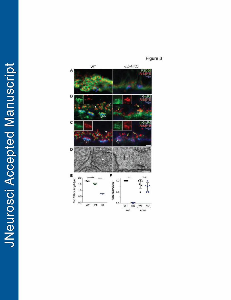
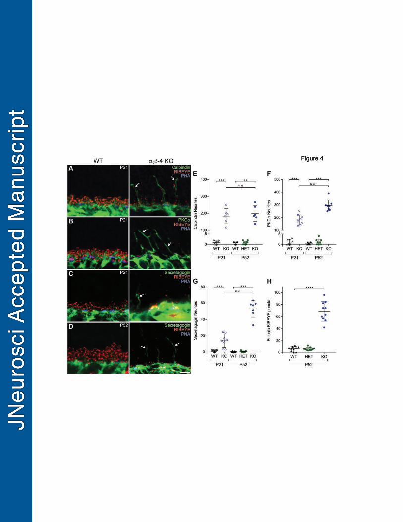


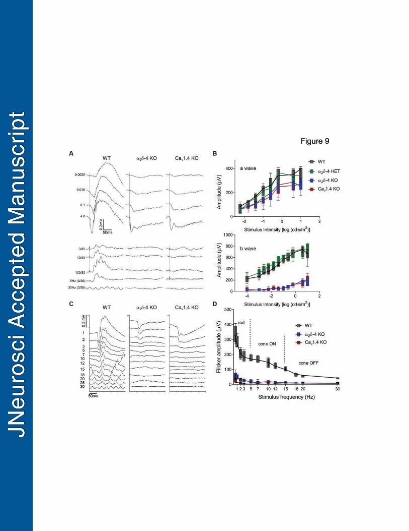


1
Table 1. Antibodies used for immunofluorescence or western blot (WB) Antibody Source Cat. # Conc.* RRID Rabbit anti-
2 In house (this paper) N/A 1:1000 NA (this study)
Rabbit anti-Cav1.4
In house (Liu et al., 2011)
N/A 1:1000-1:4000 AB_2650487
Rabbit anti-Ribeye
Synaptic Systems 192103 1:1000
AB_2086775
Mouse anti-Ribeye
BD bioscience 612044 1:1000 1:500 (WB)
AB_399431
Mouse anti-PSD95
ThermoFisher MA1-046 1:1000 AB_2092361
Mouse anti-PSD-95
Millipore 529541 1:1000 (WB) AB_565012
Rabbit anti- ERC 1b/2 (CAST)
Synaptic Systems 143003 1:2000 (WB) AB_887715
Rabbit anti-synaptobrevin2 (VAMP2)
Synaptic Systems 104202 1:5000 (WB) AB_887810
Rabbit anti-PKC-α
Santa Cruz SC208 1:500 AB_2168668
PNA-AF647 Invitrogen L32460 1:250 AB_2650486 Sheep anti-mGluR6
K. Martemyanov (Cao et al., 2011)
N/A 1:200 AB_2650490
Guinea pig anti-mGluR6
Neuromics GP13105 1:500 AB_2341540
Mouse anti-GluR2
Millipore MAB397 1:150 AB_11212990
Mouse anti-calbindin
Sigma C9848-100UL 1:400 AB_476894
Rabbit anti-secretagonin
Biovendor RD181120100 1:1000 AB_2034060
Mouse anti-HA
Covance MMS-101R 1:1000 (WB) AB_291263
Mouse anti- -actin
SigmaAldrich A2228 1:10 000 (WB) AB_476697
AF488 goat anti-mouse
ThermoFisher A-11001 1:500 AB_2534069
AF568 goat anti-mouse
ThermoFisher A11004 1:500 AB_2534072
AF647 goat anti-mouse
ThermoFisher A-21235 1:500 AB_2535804
AF488 goat anti-rabbit
ThermoFisher A11008 1:500 AB_143165
AF568 goat anti-rabbit
ThermoFisher A-11011 1:500 AB_143157
AF647 goat ThermoFisher A-21244 1:500 AB_2535812
2
anti-rabbit AF488 donkey anti-sheep
ThermoFisher A-11015 1:500 AB_2534082
AF647 donkey anti-sheep
ThermoFisher A-21448 1:500 AB_2535865
AF488 donkey anti-guinea pig
ThermoFisher A-11073 1:500 AB_2534117
Streptav-AF488
ThermoFisher S11223 1:500 AB_2336881
Biotin-conj. ThermoFisher 31820 1:500 AB_228340 HRP sheep anti-mouse IgG
GE Healthcare NA931 1:20000 (WB) AB_772210
HRP donkey anti-rabbit IgG
GE Healthcare NA934V 1:20000 (WB) AB_772206
AF594-WGA ThermoFisher W11262 1:1000 NA

1
Table 2. Serial block face scanning EM analysis of cone synaptic triads in WT and 2 4 KO mice
Genotype Cone number Ribbon + triad Total
WT
1 7 10
2 7 11
3 8* 9
2 –4 KO
1 2 13
2 4 10
3 1 6
4 5 12
Data were obtained from cone synapses reconstructed in the OPL of WT and 2 –4 KO at P19. “Ribbon+ triad” was scored when ribbon was vertically aligned at a junction between 2 processes with a third in between. “Total” represents the total number of ribbons in the reconstructed cone pedicle (n = 3 pedicles in WT, n = 4 pedicles in 2 –4 KO, one mouse each).*, one ribbon contacted 2 triads.

1
Table 3. Quantification of Cav1.4 immunofluorescence in the OPL
The number of Cav1.4-labeled structures in the OPL of HET, 2 4 KO, and Cav1.4 KO mice at the indicated ages were counted in confocal micrographs processed for immunofluorescence as in Fig.7. Cav1.4-labeling was assigned to rods or cones based on the presence or absence, respectively, of PNA fluorescence. Values represent median, 95% CI, n = number of animals. P-values were determined by Mann-Whitney test.
Age/ Genotype
Rod Cone Cav1.4-labeled
structures (Median, 95% CI)
n p-value Cav1.4-labeled
structures (Median, 95% CI)
n p-value
P10 HET 42.0 [29.8,52.3] 4
0.0048 3.0 [2,4] 15
0.024 2 4 KO 5.5 [3.5,7.2] 6 2.0 [1,3] 15
P14 HET 58.5 [54.4,74.3 6
0.0004 2.5 [2,3] 26
<0.0001 2 4 KO 2.0 [1.3,4.2] 9 1.0 [0,1] 29
P21 HET 82.0 [66.8,98.2] 4
0.0048 4.0 [3,5] 19
<0.0001 2 4 KO 0.5 [-0.19,1.5] 6 0 [0,0] 27
Cav1.4 KO 0 [0,0] 6 0.1818 0 [0,0] 15 >0.9999
Related Documents