© 2007 Nature Publishing Group The field of microtechnology is beginning to impact on microbiology. Its scale of size is well matched to the physical dimensions of most microorganisms, and micron-scale tools make it possible to manipulate individual cells, their immediate extracellular envi- ronments (referred to as the microenvironment) and ultimately, their shape and internal organization. This Review describes the intersection of microtechnology and microbiology, with a focus on soft lithography — a set of techniques that are particularly well suited for generating microscale or nanoscale structures in soft materials 1 . Soft lithography is a simple technique, and is available to most microbiologists either for their own use, or through shared facilities (public foundries) 2 . This Review is designed to bridge the divide between biologists who study microbial life, and physical sci- entists and engineers who make new materials and structures. New tools bring new capabilities to research. In microbiology, genetic techniques revolutionized our understanding of microorganisms by making it possi- ble to manipulate and study their genomes. We believe that the development of physical techniques, including those based on microstructures, will lead to tools that are complementary to genetics and genomics, including new techniques to isolate, manipulate, grow and study single cells and multicellular structures. A number of global problems in microbiology will benefit from the development of these tools, including: the evolutionary origin of cells; the cell as a dynamic, out-of-equilibrium system; intracellular organization and cellular replica- tion; mechanisms of communication between cells; the organization and characteristics of multicellular struc- tures; mechanisms of differentiation and behaviour; and new techniques for the isolation and culturing of microbial strains. Opportunities in microbiology To begin a discussion on the representative questions in microbiology that can be addressed using microfabricated tools, we have selected several broad areas of microbiol- ogy. In each section, we suggest areas in which new tools and capability are needed and suggest opportunities for the application of soft lithographic techniques. Structure and intracellular organization of bacteria. Included among the physical and chemical properties that characterize bacteria are: a specific shape and morphol- ogy, organized subcellular components, tightly regulated mechanisms of growth and division, machinery for trans- porting intracellular cargo (proteins, plasmids and DNA), and mechanisms of polarization 3–8 . The most fundamental questions regarding the structure of bacteria — how they produce, maintain and replicate their spatial organization — remain unresolved at the molecular level. The development of new techniques for manipulat- ing the shape of bacteria can complement experimental approaches using genetics and biochemistry. These techniques will make it possible to study the control of cell shape, the relationship between the shape of cells and the organization and function of intracellular machinery, the mechanical properties of cells and the role of physical forces in cellular differentiation. Soft lithography makes it possible to fabricate structures with length scales that match the intrinsic scale of bacterial cells. These techniques are just beginning to be exploited to control and study cell shape. Phenotypic behaviour. We now appreciate that solu- ble small molecules and peptides can cause bacterial cells to differentiate into various phenotypes. Many strains of bacteria use small molecules to coordinate and synchronize their behaviour 9 . Some strains grow *Department of Biochemistry, University of Wisconsin-Madison, 433 Babcock Drive, Madison, Wisconsin, 53706 USA. ‡ Department of Chemistry and Chemical Biology, Harvard University, 12 Oxford Street, Cambridge, Massachusetts, 02138 USA. Correspondence to D.B.W. and G.M.W. e-mails: [email protected]; gwhitesides@gmwgroup. harvard.edu doi:10.1038/nrmicro1616 Microtechnology The fabrication and application of materials, structures and systems with micron or submicron-scale features. Microenvironment The region sensed by a cell. The dimensions of this region are usually set by molecular contact, mass transport and diffusion, and range from a few nanometers to perhaps a millimeter. Soft lithography A set of techniques that makes microstructures by printing, moulding and embossing using a patterned, elastomeric stamp or mould, and/or a polymeric substrate. Microfabrication meets microbiology Douglas B. Weibel* ‡ , Willow R. DiLuzio ‡ and George M. Whitesides ‡ Abstract | This Review summarizes methods for constructing systems and structures at micron or submicron scales that have applications in microbiology. These tools make it possible to manipulate individual cells and their immediate extracellular environments and have the capability to transform the study of microbial physiology and behaviour. Because of their simplicity, low cost and use in microfabrication, we focus on the application of soft lithographic techniques to the study of microorganisms, and describe several key areas in microbiology in which the development of new microfabricated materials and tools can have a crucial role. REVIEWS NATURE REVIEWS | MICROBIOLOGY VOLUME 5 | MARCH 2007 | 209

Welcome message from author
This document is posted to help you gain knowledge. Please leave a comment to let me know what you think about it! Share it to your friends and learn new things together.
Transcript
© 2007 Nature Publishing Group
The field of microtechnology is beginning to impact on microbiology. Its scale of size is well matched to the physical dimensions of most microorganisms, and micron-scale tools make it possible to manipulate individual cells, their immediate extracellular envi-ronments (referred to as the microenvironment) and ultimately, their shape and internal organization. This Review describes the intersection of microtechnology and microbiology, with a focus on soft lithography — a set of techniques that are particularly well suited for generating microscale or nanoscale structures in soft materials1. Soft lithography is a simple technique, and is available to most microbiologists either for their own use, or through shared facilities (public foundries)2. This Review is designed to bridge the divide between biologists who study microbial life, and physical sci-entists and engineers who make new materials and structures.
New tools bring new capabilities to research. In microbiology, genetic techniques revolutionized our understanding of microorganisms by making it possi-ble to manipulate and study their genomes. We believe that the development of physical techniques, including those based on microstructures, will lead to tools that are complementary to genetics and genomics, including new techniques to isolate, manipulate, grow and study single cells and multicellular structures. A number of global problems in microbiology will benefit from the development of these tools, including: the evolutionary origin of cells; the cell as a dynamic, out-of-equilibrium system; intracellular organization and cellular replica-tion; mechanisms of communication between cells; the organization and characteristics of multicellular struc-tures; mechanisms of differentiation and behaviour; and new techniques for the isolation and culturing of microbial strains.
Opportunities in microbiologyTo begin a discussion on the representative questions in microbiology that can be addressed using microfabricated tools, we have selected several broad areas of microbiol-ogy. In each section, we suggest areas in which new tools and capability are needed and suggest opportunities for the application of soft lithographic techniques.
Structure and intracellular organization of bacteria. Included among the physical and chemical properties that characterize bacteria are: a specific shape and morphol-ogy, organized subcellular components, tightly regulated mechanisms of growth and division, machinery for trans-porting intracellular cargo (proteins, plasmids and DNA), and mechanisms of polarization3–8. The most fundamental questions regarding the structure of bacteria — how they produce, maintain and replicate their spatial organization — remain unresolved at the molecular level.
The development of new techniques for manipulat-ing the shape of bacteria can complement experimental approaches using genetics and biochemistry. These techniques will make it possible to study the control of cell shape, the relationship between the shape of cells and the organization and function of intracellular machinery, the mechanical properties of cells and the role of physical forces in cellular differentiation. Soft lithography makes it possible to fabricate structures with length scales that match the intrinsic scale of bacterial cells. These techniques are just beginning to be exploited to control and study cell shape.
Phenotypic behaviour. We now appreciate that solu-ble small molecules and peptides can cause bacterial cells to differentiate into various phenotypes. Many strains of bacteria use small molecules to coordinate and synchronize their behaviour9. Some strains grow
*Department of Biochemistry, University of Wisconsin-Madison, 433 Babcock Drive, Madison, Wisconsin, 53706 USA. ‡Department of Chemistry and Chemical Biology, Harvard University, 12 Oxford Street, Cambridge, Massachusetts, 02138 USA. Correspondence to D.B.W. and G.M.W. e-mails: [email protected]; [email protected]:10.1038/nrmicro1616
MicrotechnologyThe fabrication and application of materials, structures and systems with micron or submicron-scale features.
MicroenvironmentThe region sensed by a cell. The dimensions of this region are usually set by molecular contact, mass transport and diffusion, and range from a few nanometers to perhaps a millimeter.
Soft lithographyA set of techniques that makes microstructures by printing, moulding and embossing using a patterned, elastomeric stamp or mould, and/or a polymeric substrate.
Microfabrication meets microbiology Douglas B. Weibel*‡, Willow R. DiLuzio‡ and George M. Whitesides‡
Abstract | This Review summarizes methods for constructing systems and structures at micron or submicron scales that have applications in microbiology. These tools make it possible to manipulate individual cells and their immediate extracellular environments and have the capability to transform the study of microbial physiology and behaviour. Because of their simplicity, low cost and use in microfabrication, we focus on the application of soft lithographic techniques to the study of microorganisms, and describe several key areas in microbiology in which the development of new microfabricated materials and tools can have a crucial role.
R E V I E W S
NATURE REVIEWS | MICROBIOLOGY VOLUME 5 | MARCH 2007 | 209
Akshay
Highlight
Akshay
Highlight
© 2007 Nature Publishing Group
Soft materialA material (especially a polymer) that is pliable, compressible or elastic.
Public foundryA facility for the fabrication of microstructured materials and systems.
Elastomeric polymer A soft, compliant, rubber-like polymer.
MaskTypically a transparent substrate with a pattern on its surface defined in an opaque material (chrome metal or ink) used in photolithography.
PDMSPoly(dimethylsiloxane). An elastomeric silicone polymer that is commercially available and has properties that make it well suited to applications in microbiology.
Embossed structureA structure that is moulded in a surface in relief.
Bas-relief structureA structure that projects away from a surface.
Bas-relief masterRefers to the master copy and, in soft lithography, consists of patterns of a photoreactive polymer on the surface of a silicon wafer or glass slide.
PhotolithographyA process used to transfer a pattern from a mask onto a thin film of photosensitive polymer (photoresist) and then onto the surface of a substrate. Photolithography is commonly used in semiconductor fabrication to fabricate integrated circuits.
and organize into multicellular structures — colonies, biofilms, spores, mycelium and fruiting bodies — that have the characteristics of multicellular organisms; that is, the cells behave in a collective manner in which a division of cellular labour increases access to resources or provides protection and improves the probability of sur-vival against competitors10,11. Although recent advances have revolutionized our understanding of this area of microbiology, we are still just beginning to understand the chemical and physical basis of social interactions between cells.
New tools for controlling the chemical and physical microenvironment surrounding cells will enhance our understanding of the behaviour of bacteria. Related areas in eukaryotic biology have been studied using the techniques of soft lithography with excellent success12,13.
Cell culture. The global diversity of bacteria has been estimated at 107–109 species14. Only a tiny fraction of these species have been isolated and cultured using traditional microbiological techniques15. Remarkably, the core set of techniques used for microbial culture has changed little since their development by Koch, Beijerinck and Winogradsky in the nineteenth century; these techniques are, unfortunately, incompatible with the cultivation of most microorganisms.
A detailed understanding of the chemical and physi-cal environment surrounding cells, including the role of abiotic surfaces, stress, temperature and the dif-fusion of molecules (gas, nutrients, communication substances and metabolic waste) on their homeostasis and replication, will contribute to the development of new techniques for culturing microorganisms. Microfabricated structures that control the local microenvironment of microbial cells might improve culture technology, and contribute to general and medical microbiology. Networks of microchannels could eventually bypass the need for microbial cul-ture by making it possible to perform genetic and biochemical analyses on single cells that are part of environmental communities.
The biology of single cells. The field of microbiology was built largely on studies of the behaviour and bio-chemistry of populations of cells. Measurements based on ensemble averaging cannot characterize the dif-ferences between individual cells. We now appreciate that individual cells — even within a clonal population — can differ from each other genetically, biochemically and behaviourally; however, in many examples, we do not understand the significance of these differences16. Studies on the behaviour and genetics of individual microbial cells are clarifying our understanding of hori-zontal gene transfer, chemical signalling, pathogenesis, motility, chemotaxis, microbial viability and persistence, the interaction of cells with surfaces and, in general, the relationship between genetics, biochemistry and behav-iour17. The development of microstructures for isolating and studying single cells will improve our understand-ing of the structural, biochemical and genetic variation between cells.
Quantitative microbiology. Much of biology is based on qualitative data. In microbiology, many of the techniques that are used — for example, streaking cells and studying their structure and behaviour using optical microscopy — were not designed with reproducibility or quantifica-tion in mind. These methods make it difficult (or impos-sible) to collect data that is quantitative, time-dependent or spatially resolved in experiments in which individual cells, or collections of cells, are studied repeatedly under identical conditions. The development of techniques that facilitate the collection of statistically significant data will accelerate the transformation of microbiology from a science based on qualitative observation into one based on the quantitative analysis of data. One area in which microfabrication can have an important role is in the design and fabrication of structures on which thousands of parallel experiments with single cells — or small groups of cells — can be carried out under identi-cal conditions. Another area in which small structures can impact microbiology is the use of microchannels to transport, mix and deliver fluids to cells. The phys-ics of fluids at microscopic dimensions is predictable and makes it possible to design and carry out experi-ments under well defined and reproducible conditions. Bacterial chemotaxis is one of many areas of microbiol-ogy that can benefit from techniques that take advantage of the properties of fluids at small dimensions.
Systems biology is another emerging field that builds models of biomolecular interactions and networks in cells and organisms, and uses these models to describe biological processes18. It requires an understanding of the quantitative expression of genes under specific condi-tions, and of the intracellular location and concentration of metabolites and proteins, and is one field that has much to gain from advances in quantitative microbial biology. Microfabrication has the opportunity to play an important part in the development of new tools and techniques that allow quantitative measurements at the cellular level19.
New tools open new doorsMany of the techniques that are now used to isolate, purify and study microorganisms have not changed significantly since their initial development. Although new techniques for studying microorganisms have emerged, including mass spectrometry, capillary (gel) electrophoresis, and optical, electron and scanning probe microscopy, the use of these techniques still lags behind genetic and genomic approaches. We believe that new developments in physically based techniques will make it possible to study microorganisms at an unprecedented — even revolutionary — level of detail. Below we describe a set of tools for microfabrication, a key component in this impending revolution, known collectively as ‘soft lithography’. These tools have characteristics that make them particularly useful in addressing a number of the important research questions in microbiology.
Soft lithography and microfabricationSoft lithography refers to a collection of techniques for creating microstructures and nanostructures based on printing, moulding and embossing1. The techniques
R E V I E W S
210 | MARCH 2007 | VOLUME 5 www.nature.com/reviews/micro
Akshay
Highlight
Akshay
Highlight
Akshay
Highlight
Akshay
Highlight

© 2007 Nature Publishing Group
Silicon wafer
Deposit photoresist
Photoresist
PDMS
Silicon wafer
Master
a
e f
b
c d
UV light exposure;remove mask;dissolve photoresist
Pour on PDMS;cure (65°C); peel away PDMS
Mask
~500 nm – 500 µm
~500 nm – 500 µm
PDMS
Embossed microstructures
Add mask
CAD toolComputer aided design. A software program used by engineers and designers for drafting two- and three-dimensional structures.
PhotoresistA photoreactive polymer that undergoes chemical changes that lead to changes in physical properties (such as solubility) after exposure to ultraviolet light.
Microfluidic systemA set of channels that have micron-scale dimensions (typically between 5–500 µm), and are used to manipulate fluids.
were developed as an alternative to photolithography and electron-beam lithography, and share the name ‘soft lithography’ because they are all based on using a pat-terned elastomeric polymer as a mask, stamp or mould, to pattern ‘soft materials’ (for example, polymers, gels and organic monolayers, rather than silicon and glass). The tools of soft lithography are being used with increasing frequency in cell biology because of their simplicity, low cost and compatibility with cells2. This section of the Review introduces the core set of soft-lithographic tech-niques, and describes the characteristics that make them useful in microbiology. This section will not provide an exhaustive review on soft lithography nor will it describe other techniques for microfabrication; other reviews provide more detail on these and related subjects1,20,21.
An introduction to soft lithographyAll of the techniques in soft lithography use, as their central component, a layer of poly(dimethylsiloxane) (PDMS), or another polymer with similar character-istics, with embossed structures or bas-relief structures on the surface. The layer of PDMS is fabricated from a bas-relief master by embossing, and typically has fea-tures with lateral dimensions of 1–1000 µm and vertical dimensions between 100 nm and hundreds of microns
— the master is produced using photolithography. Y. Xia and G.M.W. have described the procedure for creat-ing microstructures in detail1; the relevant steps are summarized in FIG. 1.
A pattern is drawn using a CAD tool (computer-aided design). Patterns that have features with lateral dimen-sions >8 µm are printed on a transparency (a sheet of transparent polymer) using a high-resolution photoplot-ter22; the resulting transparency functions as a mask for photolithography. For designs with features that have lateral dimensions <8 µm, the pattern is transferred to a thin layer of metal (usually chrome) on a glass slide using a laser or an electron-beam mask writer. The mask is placed between a source of ultraviolet light and a thin layer of a photoreactive polymer referred to as a pho-toresist that has been coated on a silicon wafer or glass slide. Exposure to UV light transfers the pattern from the mask to the polymer; the unexposed photoresist is removed using an organic solvent, leaving behind the bas-relief structure in the polymer layer. A silicon wafer with polymeric photoresist structures patterned on its surface is typically referred to as the master copy, or simply the master. The height of the polymer structure is controlled by the thickness of the layer of photoresist that is initially spread on the surface of the wafer; the mask controls the shape and lateral dimensions. New approaches are being developed to reduce both the cost and number of steps required to produce masters; these techniques will make it possible to create microstruc-tures using equipment that is routinely available in many laboratories.
One useful characteristic of soft lithography is that hundreds of replicas of the inverse pattern can be pro-duced in PDMS from one master; this technique (replica moulding) is described in detail in the next section. To make PDMS replicas, the surface of the master is initially treated with a perfluoralkyl trichlorosilane to prevent the PDMS from sticking to the master. The liquid prepolymer of PDMS is mixed, degassed and poured on the surface of the master. After curing thermally (at 65 °C), the layer of PDMS is peeled away and contains the inverse of the original pattern embossed on the surface of the master.
Although each replica takes approximately two hours to prepare, the time can be reduced by curing the layer of PDMS at a temperature >65 °C. A useful characteristic of soft lithography is that it is possible to create prototype structures rapidly23. In many cases, the entire process — from a CAD design to a completed microfabricated structure in PDMS — requires less than twelve hours. The resulting topographically patterned layers of PDMS can be used as microchambers for isolating and cultur-ing cells, as stamps for patterning cells on surfaces, or as masters for replicating the structure in another material. The structures can also be sealed against flat surfaces and used as microfluidic systems. These applications and others are discussed in more detail in the next section.
PDMS has been the most widely used material for the applications of soft lithography in microbiology, and in biology in general, because of the following charac-teristics: it is soft, flexible, biocompatible, insulating, unreactive, transparent to ultraviolet and visible light,
Figure 1 | The fabrication of micropatterned slabs of PDMS. a–b | Photoresist is spin-coated on a silicon wafer. c | A mask is placed in contact with the layer of photoresist. d | The photoresist is illuminated with ultraviolet (UV) light through the mask. An organic solvent dissolves and removes photoresist that is not crosslinked. The master consists of a silicon wafer with features of photoresist in bas-relief. An expanded view of one of the microfabricated structures with its characteristic critical dimensions is shown. e | PDMS is poured on the master, cured thermally and peeled away. f | The resulting layer of PDMS has microstructures embossed in its surface. PDMS, poly(dimethylsiloxane).
R E V I E W S
NATURE REVIEWS | MICROBIOLOGY VOLUME 5 | MARCH 2007 | 211
Akshay
Highlight
Akshay
Highlight
Akshay
Highlight
Akshay
Highlight
Akshay
Highlight
Akshay
Highlight
Akshay
Highlight
Akshay
Highlight
Akshay
Highlight
Akshay
Highlight
Akshay
Highlight
Akshay
Highlight
Akshay
Highlight
Akshay
Highlight
Akshay
Highlight
Akshay
Highlight
© 2007 Nature Publishing Group
Spin coatingA process for depositing uniform layers of polymer on a substrate. Rotating the substrate at a high speed spreads the material uniformly over the surface. The viscosity of the material and the rotational velocity of the substrate control the thickness of the layer of material; surface tension flattens the surface of the spun film.
SAMsSelf assembled monolayers. Monolayer structures formed by the spontaneous self-assembly of alkanethiols on metal surfaces. In SAMs, the thiol groups are bonded covalently to the metal surface, and the non-covalent, intermolecular packing of the alkane chains causes the molecules to arrange into an ordered, two-dimensional crystal or liquid crystal.
permeable to gases and only moderately permeable to water2,24. The prepolymer of PDMS is commercially available, inexpensive, and easy to prepare. It is prepared by mixing two liquid components together, the base and curing agent, followed by degassing to remove air bubbles; the procedure typically takes less than 30 minutes.
Public foundries have been established at several universities (including Harvard University, Stanford University, California Institute of Technology and the University of Washington) to make the techniques of soft lithography available to the biology commu-nity. These foundries are sites for training users in the techniques of soft lithography, and they also provide the materials and tools of soft lithography to scientists who want to use the techniques but do not want to be trained in their fabrication; in this capacity, foundries can produce limited numbers of PDMS stamps, moulds or microfluidic systems. Users send their designs and specifications to a foundry and receive masters and rep-lica copies of the masters in PDMS for a reasonable price and without any capital investment in equipment.
In the following sections, we introduce several of the core techniques of soft lithography and describe recent applications of these techniques to the study of micro-organisms. Rather than providing a comprehensive review of the literature, we have chosen examples that illustrate the range of applications of these techniques to microbiology.
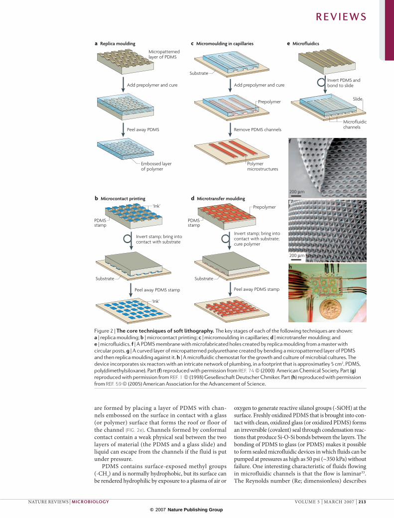
The tools of soft lithographyReplica moulding. Replica moulding is a technique for duplicating the shape, size and pattern of features on a master and provides a method of patterning a wider range of materials than is possible using photo-lithography1. In contrast to photolithography, replica moulding duplicates three-dimensional structures in a single step. A topographically patterned layer of PDMS typically functions as the mould in this technique, and is used to transfer the pattern to the surface of another polymer (FIG. 2a). A prepolymer is deposited on the PDMS mould by casting or spin coating, is cured, and then separated from the master by peeling them apart. This technique has been used to micro-pattern biocompatible polymers, including epoxies, polyurethanes, polyethylene glycol (PEG), agar and agarose. The ability to create micropatterned agar and agarose should be especially useful in microbiology since these materials are so widely used as substrates for isolating and culturing bacterial cells.
Microcontact printing. Microcontact printing transfers patterns from the surface of a topographically patterned PDMS stamp to the surface of a substrate1 (FIG. 2b). Various ‘inks’, including small biomolecules, proteins, polyelectro-lytes and suspensions of cells, can be patterned directly on surfaces using microcontact printing25–27. In this tech-nique, contact between the PDMS stamp and a substrate only transfers the ‘ink’ from the raised surface of the bas-relief features of the stamp to the substrate, and produces patterns with feature sizes as small as 100 nm over areas as large as ~1 m2. The elasticity of the PDMS stamp facilitates
conformal contact between the stamp and the substrate, and makes it possible to pattern non-planar surfaces (including porous, rough or curved surfaces).
Self-assembled monolayers (SAMs) are formed when PDMS stamps ‘inked’ with alkanethiols (SH-(CH2)n-X) are used to microcontact print on surfaces of gold, silver, palladium, platinum or other metals28. SAMs are structures produced by the spontaneous chemisorp-tion and self-organization of alkanethiols. Alkanethiols with long alkyl chains (n = 16 or 18) form hydrophobic monolayers, whereas those with different terminal func-tional groups (X) can form hydrophilic, hydrophobic or charged SAMs. Many synthetic methods are available for attaching ligands to functional groups (X) on SAMs28.
Another useful characteristic of microcontact print-ing is that it can be used to pattern surfaces with multiple SAMs. After a SAM of an alkanethiol is patterned on a gold-coated surface by microcontact printing, the sub-strate is dipped into a solution of a second alkanethiol to form a different SAM on bare regions of the metal surface that were not in contact with the stamp. This technique has been used to make islands of SAMs that adsorb proteins and cells, and which are surrounded by SAMs that resist the adsorption of biomaterial. Patterns of SAMs have been used to study the adhesion of mam-malian cells on surfaces2. PDMS stamps have also been used to create patterns of proteins, polyelectrolytes and other molecules on glass, polystyrene or silicon26,29. Agarose stamps made by replica moulding from PDMS moulds have been used to microcontact print patterns of proteins on glass substrates30.
Micromoulding in capillaries. Micromoulding in capil-laries (MIMIC) is a technique that uses a typographically patterned layer of PDMS in contact with a surface to form a network of microchannels1. The layer of PDMS is brought into conformal contact with a solid substrate, such as a glass slide, and liquid prepolymer is placed at the edge of the PDMS layer (FIG. 2c). The prepolymer is drawn into the channels by capillary action or by suction, and is subsequently cured to produce solid structures. By peel-ing the stamp away carefully, a pattern of micropatterned material is left on the substrate surface.
Microtransfer moulding. Microtransfer moulding is a technique for patterning materials in which a thin layer of a liquid prepolymer is applied to the patterned surface of a PDMS stamp1 (FIG. 2d). The excess prepolymer is removed by scraping the surface with a flat block of PDMS or by blowing N2 over the surface. The stamp, with its features filled with prepolymer, is brought into conformal contact with a surface, and the prepolymer is cured. When the stamp is peeled away, patterned microstructures remain on the surface of the substrate. The stamp can be refilled and used to pattern more substrates. This technique pro-duces three-dimensional structures with feature sizes as small as ~1 µm in a single step.
Microfluidics. Elastomeric polymers such as PDMS have been widely used for fabricating microfluidic channels and networks31,32. Microfluidic channels
R E V I E W S
212 | MARCH 2007 | VOLUME 5 www.nature.com/reviews/micro
Akshay
Highlight
Akshay
Highlight
Akshay
Highlight
Akshay
Highlight
Akshay
Highlight
Akshay
Highlight
Akshay
Highlight
Akshay
Highlight
Akshay
Highlight
Akshay
Highlight
Akshay
Highlight
© 2007 Nature Publishing Group
a Replica moulding
b Microcontact printing
c Micromoulding in capillaries e Microfluidics
d Microtransfer moulding
Micropatterned layer of PDMS
Add prepolymer and cure
Peel away PDMS
Add prepolymer and cure
Remove PDMS channels
Peel away PDMS stamp Peel away PDMS stamp
Embossed layerof polymer
Polymer microstructures
Prepolymer
Substrate
SubstrateSubstrate
Microfluidic channels
Slide
Invert PDMS andbond to slide
Invert stamp; bring intocontact with substrate
Invert stamp; bring intocontact with substrate;cure polymer
‘Ink’
‘Ink’
PDMS stamp
PDMS stamp
Prepolymer
200 µm
200 µm
f
g
h
are formed by placing a layer of PDMS with chan-nels embossed on the surface in contact with a glass (or polymer) surface that forms the roof or floor of the channel (FIG. 2e). Channels formed by conformal contact contain a weak physical seal between the two layers of material (the PDMS and a glass slide) and liquid can escape from the channels if the fluid is put under pressure.
PDMS contains surface-exposed methyl groups (-CH3) and is normally hydrophobic, but its surface can be rendered hydrophilic by exposure to a plasma of air or
oxygen to generate reactive silanol groups (-SiOH) at the surface. Freshly oxidized PDMS that is brought into con-tact with clean, oxidized glass (or oxidized PDMS) forms an irreversible (covalent) seal through condensation reac-tions that produce Si-O-Si bonds between the layers. The bonding of PDMS to glass (or PDMS) makes it possible to form sealed microfluidic devices in which fluids can be pumped at pressures as high as 50 psi (~350 kPa) without failure. One interesting characteristic of fluids flowing in microfluidic channels is that the flow is laminar33.The Reynolds number (Re; dimensionless) describes
Figure 2 | The core techniques of soft lithography. The key stages of each of the following techniques are shown: a | replica moulding; b | microcontact printing; c | micromoulding in capillaries; d | microtransfer moulding; and e | microfluidics. f | A PDMS membrane with microfabricated holes created by replica moulding from a master with circular posts. g | A curved layer of micropatterned polyurethane created by bending a micropatterned layer of PDMS and then replica moulding against it. h | A microfluidic chemostat for the growth and culture of microbial cultures. The device incorporates six reactors with an intricate network of plumbing, in a footprint that is approximatley 5 cm2. PDMS, poly(dimethylsiloxane). Part (f) reproduced with permission from REF. 74 © (2000) American Chemical Society. Part (g) reproduced with permission from REF. 1 © (1998) Geselleschaft Deutscher Chmiker. Part (h) reproduced with permission from REF. 59 © (2005) American Association for the Advancement of Science.
R E V I E W S
NATURE REVIEWS | MICROBIOLOGY VOLUME 5 | MARCH 2007 | 213
© 2007 Nature Publishing Group
a
b
c
d
e
Agarose stamp with positive features
Add suspension of cells
Invert stamp and print
Incubate
Agar surface
Stamp absorbs excess liquid
Cells deposited on stamp
Petri dish
Bacterial colony
1 mm
2 mm
2 mm
250 µm
f
g
h
i
the behaviour of fluids. It is defined by Re = ρuDh/µ, in which ρ is the density of fluid (g per cm3), u is the velocity of the fluid (cm s–1), Dh is the hydraulic diameter (cm) of the channel, and µ is the viscosity of the fluid (g per cm per s). When the Reynolds number is less than ~2,000, fluid flow is laminar; above this value the flow is turbulent. The flow of fluids in microchannels is typically laminar and the velocity of the fluid in a given region of the channel is constant in time. An important aspect of laminar flow in microfluidic channels is that adjacent streams of miscible liquids flow side-by-side, with mixing only by diffusion at the interface between the streams. Networks of branching and recombining microchannels can be used to produce stable gradients of biomolecules34–36. These gradients are perpendicular to the direction of flow, can be generated in solution and on surfaces, and have spatial and temporal stability. As mentioned above, the behaviour of fluids at the micro-scale level is predictable and this characteristic makes it
possible to reproduce experimental conditions that take advantage of liquids moving at a low Reynolds number.
Microfluidic valves. Several groups have used soft-litho-graphic techniques to develop valves for controlling the flow of fluids and the movement of cells and particles in microfluidic channels. The most frequently used exam-ple of this class of structures is the monolithic, pneumatic valves developed by Quake et al.37,38 Monolithic valves are based on two sets of PDMS microfluidic channels that are perpendicular to each other, and bonded together, one on top of the other. A thin layer of PDMS separates the two layers of channels. One set of channels forms the valves and the other set forms the microfluidic channels in which experiments are carried out. A pressure applied to the valve channels collapses the thin layer of PDMS into the microfluidic channels and controls the flow of fluids. These structures are being used in microbiological studies with increasing frequency.
Applications of soft lithography in microbiologyImmobilizing bacteria on surfaces. The immobilization and patterning of bacteria on surfaces provides new opportunities for sensing and detecting biomolecules using whole cells and for studying cell–cell interactions and interactions between cells and their surroundings.
SAMs have been used to immobilize cells on surfaces in the study of host–pathogen interactions. The covalent attachment of biological ligands to the terminal regions of SAMs provides a way of controlling the density of a ligand on a surface and, importantly, the attached ligands retain their activity. One recent example described the applica-tion of SAMs and optical tweezers to measure the force of adhesion between uropathogenic Escherichia coli cells expressing type I pili and a mannose-presenting SAM39.
The combination of microcontact printing and SAMs has been used to selectively immobilize bacterial cells on surfaces. For example, Rowan et al. used a PDMS stamp to microcontact print patterns of hydrophobic and reac-tive SAMs on gold to produce ‘enclosures’ that trapped cells of E. coli40. Several groups have used microcontact printing to capture and detect pathogenic organisms. One study used PDMS stamps to pattern antibodies on silicon substrates and then measured the binding of E. coli O157:H7 to the antibodies using optical diffraction41. Morhard and co-workers detected the binding of E. coli O157:H7 to antibodies that were covalently attached to patterned SAMs using optical methods42. Another group used microcontact printing to pattern antibodies that were subsequently used to capture cells of E. coli O157:H7 and Renibacterium salmoninarum43. More recently, Rozhok and co-workers fabricated patterns of surface-bound cells of a motile strain of E. coli by microcontact printing SAMs derivatized with anti-lipopolysaccharide antibodies or poly-l-lysine44.
Micromoulding in capillaries has also been used to pattern microbial cells onto surfaces. Suh et al. used the MIMIC process to selectively confine bacterial cells within PEG microstructures using virus–antibody inter-actions45. The authors fabricated PEG microstructures on the surface of silicon by micromoulding in PDMS
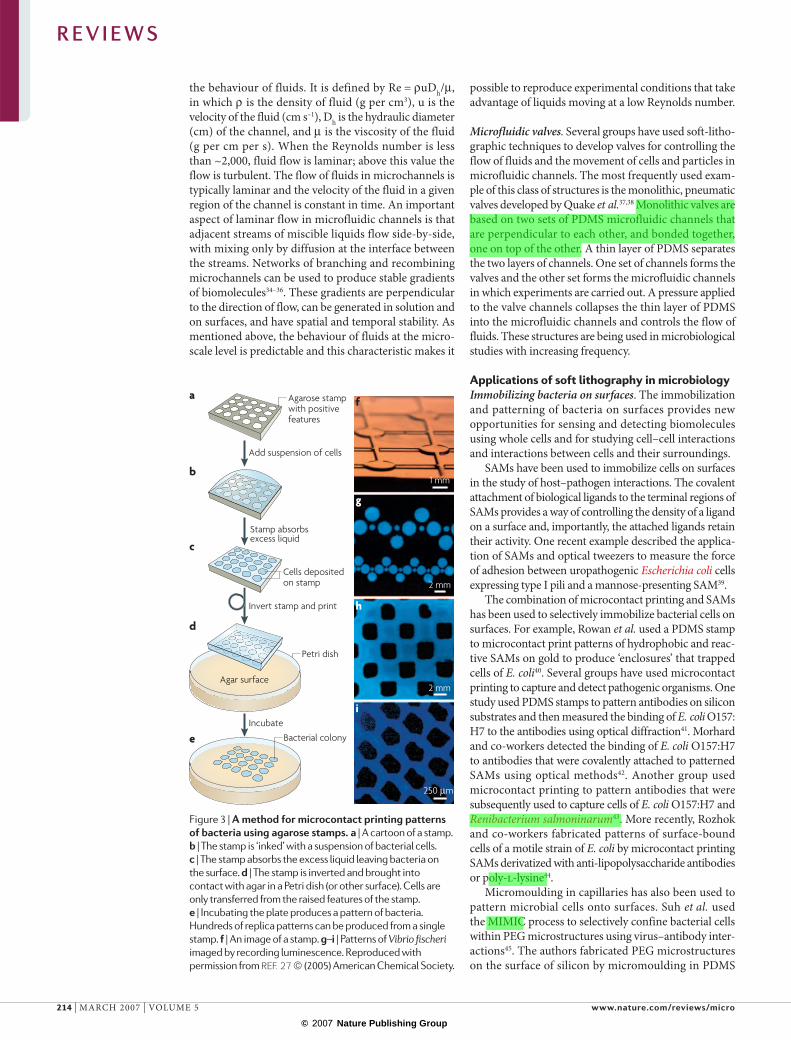
Figure 3 | A method for microcontact printing patterns of bacteria using agarose stamps. a | A cartoon of a stamp. b | The stamp is ‘inked’ with a suspension of bacterial cells. c | The stamp absorbs the excess liquid leaving bacteria on the surface. d | The stamp is inverted and brought into contact with agar in a Petri dish (or other surface). Cells are only transferred from the raised features of the stamp. e | Incubating the plate produces a pattern of bacteria. Hundreds of replica patterns can be produced from a single stamp. f | An image of a stamp. g–i | Patterns of Vibrio fischeri imaged by recording luminescence. Reproduced with permission from REF. 27 © (2005) American Chemical Society.
R E V I E W S
214 | MARCH 2007 | VOLUME 5 www.nature.com/reviews/micro
Akshay
Highlight
Akshay
Highlight
Akshay
Highlight
Akshay
Highlight
© 2007 Nature Publishing Group
a
b
c
d
Microchamber
Add cells
Inhibit septation;cells filament
Release cells into solution;cells retain their shape
Cell of normal length (E. coli)
e
f
g 10 µm
8 µm 3 µm
50 50 µm
(width of a channel)
HydrogelA low-density, crosslinked polymer network containing a high-volume fraction of water.
capillaries and modified the surrounding (bare) surface of silicon with P3 or P9 antibodies that bind to the sur-face of the M13 virus. Only E. coli cells that were infected with the M13 virus bound to regions of the substrate patterned with antibodies.
Patterning bacterial cells using hydrogels. Agar and agarose form hydrogels. This category of polymer has two characteristics that are particularly useful in microbiology: first, cells grown on their surface remain hydrated; and second, nutrients, gas and byproducts of metabolism diffuse through the polymer network of the gel. There are many other types of hydrogel that also have a use in microbiology. For example, Heo et al. filled PDMS channels with a solution of a photoreac-tive PEG prepolymer that contained E. coli cells, and exposed regions of the channel to UV light46. Exposure to the UV light resulted in the photopolymerization of the polymer, thereby encapsulating the E. coli cells within
PEG microstructures inside the channels. Recently, we developed a method for microcontact printing bacteria on agar plates, glass slides and nylon membranes using micropatterned agarose stamps that are formed using replica moulding27. The technique makes it possible to create patterns of bacteria on agar plates with control over the pattern, shape and spacing of features as small as 200 µm over areas as large as 50 cm2 (FIG. 3).
Microfluidics: motility, chemotaxis, quorum sensing and population dynamics. Microfluidics has many charac-teristics that make it useful for the study of cells31,47. We investigated the behaviour of swarmer cells of E. coli — a multiflagellated phenotype that is approximately 2–3 times the length of a swimming cell — confined in narrow microchannels in which the floor was agar, and the walls and ceiling were hydrophilic PDMS48. Hydrodynamic interactions between the cells and the agar floor caused the cells to swim along the bottom of the channel; the rotation of the cell body made the cells swim preferentially along the right-hand side of the microchannel. A similar principle is being used to sort motile bacteria based on their length as they swim along curved microchannels.
In another study, Mao et al. used laminar flow to cre-ate gradients of attractants and repellents in microfluidic channels as a system for studying bacterial chemotaxis49. The authors found that the microfluidic system was three orders of magnitude more sensitive than tradi-tional capillary-based chemotaxis assays and, using the system, they discovered that l-leucine is a bacterial chemoattractant at low concentrations and a repellent at high concentrations.
Bacteria can alter their expression of genes collec-tively when the density of cells reaches a certain level. This behaviour is referred to as quorum sensing, and it has a role in many forms of microbial behaviour, includ-ing bioluminescence, swarming motility, pathogenicity and biofilm formation50. As part of the quorum sensing process, bacterial cells communicate by secreting small, soluble molecules into the surrounding fluid. Park et al. studied the growth of E. coli and Vibrio harveyi in PDMS microfluidic devices and found that cells of E. coli accu-mulated in enclosed areas of a microfluidic maze (for example in dead-ends)51,52. The authors concluded that chemotaxis between cells is capable of producing the density of cells required for quorum sensing.
Microfluidic systems also have applications in study-ing bacterial population dynamics. In one recent study, a microstructured environment for creating heterogeneity in bacterial habitats was constructed that allowed the authors to study how bacteria adapted to regions of the landscape53. Although these structures were created using traditional methods of fabrication, the application of soft lithography to the fabrication of related structures is clear.
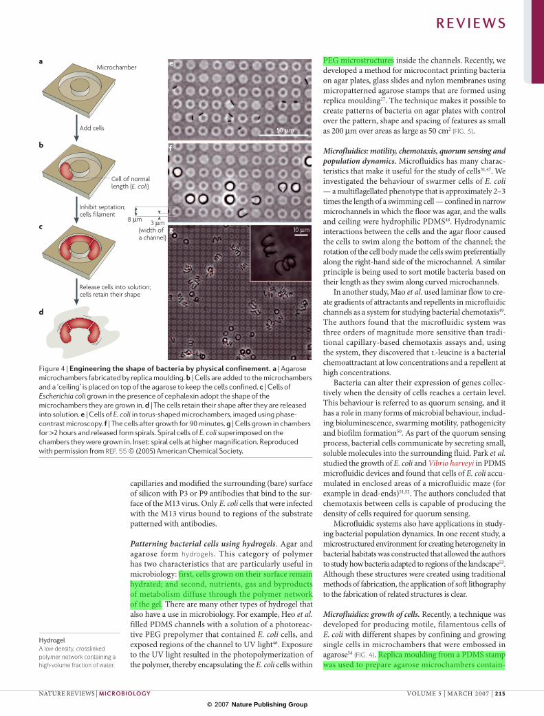
Microfluidics: growth of cells. Recently, a technique was developed for producing motile, filamentous cells of E. coli with different shapes by confining and growing single cells in microchambers that were embossed in agarose54 (FIG. 4). Replica moulding from a PDMS stamp was used to prepare agarose microchambers contain-
Figure 4 | Engineering the shape of bacteria by physical confinement. a | Agarose microchambers fabricated by replica moulding. b | Cells are added to the microchambers and a ‘ceiling’ is placed on top of the agarose to keep the cells confined. c | Cells of Escherichia coli grown in the presence of cephalexin adopt the shape of the microchambers they are grown in. d | The cells retain their shape after they are released into solution. e | Cells of E. coli in torus-shaped microchambers, imaged using phase-contrast microscopy. f | The cells after growth for 90 minutes. g | Cells grown in chambers for >2 hours and released form spirals. Spiral cells of E. coli superimposed on the chambers they were grown in. Inset: spiral cells at higher magnification. Reproduced with permission from REF. 55 © (2005) American Chemical Society.
R E V I E W S
NATURE REVIEWS | MICROBIOLOGY VOLUME 5 | MARCH 2007 | 215
Akshay
Highlight
Akshay
Highlight
Akshay
Highlight

© 2007 Nature Publishing Group
ing nutrients. The microchambers were seeded with cells by adding a drop of a suspension of E. coli to the surface of the micropatterned agarose mould and a thin piece of PDMS or agarose was placed on top. In the pres-ence of an antibiotic (cephalexin) that inhibits septation, the E. coli cells filamented and adopted the shape of the microchambers in which they grew. Indeed, the cells maintained their shape when they were released from the microchambers.
Bacterial persistence refers to the small percentage of a homogenous population of bacteria that survives after treatment with an antibiotic. Balaban and colleagues studied the persistence of single cells of E. coli confined in a flow cell consisting of microfluidic channels fabricated in PDMS55. From this analysis, the authors built a model of bacterial persistence based on their observations that persister cells were not affected by the antibiotic, were not genetically different from other bacterial cells and grew at much slower rates than normal cells.
When microorganisms grow in static cultures, the final density of cells is limited by their depletion of nutrients from their surrounding environment. In che-mostatic growth, a small number of cells are constantly removed from the culture and fresh nutrients are added continuously56. Cultures can achieve steady-state growth under these conditions. Several groups have designed and used ‘microbioreactors’ based on microfluidics for growing bacterial and yeast cells57–59. For example, Balagadde et al. developed a microfluidic chemostat for the long-term culture of small numbers of bacterial cells58. Each microchamber had a volume of 16 nL and was filled with liquid that was constantly recirculated; the chambers were periodically washed with solutions to pre-vent the formation of biofilms. Groisman and co-workers developed a microfluidic chemostat in which bacteria and yeast cells were confined in microchambers that were con-nected by channels that allowed the diffusion of nutrients but were impassable to cells59.
Microfluidics: harnessing the mechanical work produced by microbial cells. Several groups have demonstrated approaches for capturing the mechanical work produced by motile strains of microbial cells. Although these examples are exploratory in nature, the techniques could also find application in studying fundamental questions related to the structure and behaviour of microorgan-isms. In one example, researchers created a monolayer of bacterial cells on a surface (referred to as bacterial carpets) by adsorbing cells of Serratia marcescens onto a PDMS surface. They found that the motion of the bacterial flagella within the monolayer was coordinated and produced a flow of fluid above the surface of the polymer60. Kim and Breuer studied the mixing of two laminar streams of fluids in PDMS microfluidic channels using motile E. coli61. Hiratsuka et al. used microchan-nels to harness the motility of Mycoplasma cells to trans-port polystyrene beads — 500 nm in diameter — that were attached to the cells using streptavidin-biotin chemistry62. Recently the same group demonstrated the remarkable rotation of a freestanding object by biasing the coordinated movement of motile Mycoplasma cells
using a microchannel63. We have developed a method of moving microscale loads (1–6 µm diameter polystyrene beads) in PDMS microchannels using the unicellular photosynthetic algae Chlamydomonas reinhardtii as ‘micro-oxen’64. Using a combination of photochemistry and surface chemistry, we have demonstrated that motile microorganisms can be guided to pick up, transport and drop off polystyrene beads.
Microfluidics: molecular biology. Several groups have used microfluidic systems for genetic studies and for the specific detection of bacterial cells. Microfluidic platforms have several attractive characteristics for genetic investigations, including the consumption of small volumes of reagents and samples, the generation of small volumes of waste, low unit cost, short reaction times and the capability of analysing single cells.
An important step in many molecular biology tech-niques and in the detection of pathogens is the lysis of microorganisms to release DNA and other intracel-lular contents into solution. In one study, E. coli and Saccharomyces cerevisiae cells were lysed within PDMS microfluidic chambers that were fabricated on compact discs (CDs)65. The microfluidic chambers contained spherical beads that collided with cells when the CD was rotated around its horizontal axis (similar to a bead mill). In another study, researchers used optical tweezers in a PDMS microfluidic system to manipulate and sort cells of E. coli and a pulsed laser to punch holes in the cell wall66. This system has applications in the transformation of single microbial cells with DNA constructs.
The transformation of bacterial cells with foreign DNA inserted into plasmids is one of the most fundamen-tal techniques used in bacterial genetics. Using an array of microchambers in a microfluidic device fabricated in PDMS, Nagamine and co-workers carried out multiple, simultaneous transformations of E. coli cells with plasmid DNA67. Each chamber contained a different plasmid that had been immobilized on the floor of the chamber. The authors demonstrated that E. coli cells in the different chambers were successfully transformed with plasmid DNA by confirming the presence of new phenotypes.
Recent progress toward the miniaturization of PCR-based assays has been reviewed elsewhere68,69. Several groups are working on PCR in microfluidic devices fabricated using soft lithography, and although these systems are still too complex for routine use, this situa-tion should improve as microfluidic technology matures. A fully automated, microfluidic, PCR-based detection system for the detection of bacterial pathogens was recently described70. The device contains an integrated microprocessor, pumps, valves, thermocycler and a fluo-rescent detection system, and was capable of detecting approximately 104–107 cells of Listeria monocytogenes per hour. In addition, the automation of gene ligation, trans-formation and PCR in microfluidic systems fabricated in PDMS has also been described71,72. A recent paper by Ottesen and co-workers elegantly demonstrated the use of microfluidic PCR to amplify and analyse genes isolated from single cells of bacteria harvested from the midgut of termites73.
R E V I E W S
216 | MARCH 2007 | VOLUME 5 www.nature.com/reviews/micro
Akshay
Highlight
Akshay
Highlight
Akshay
Highlight
Akshay
Highlight
Akshay
Highlight
Akshay
Highlight
Akshay
Highlight

© 2007 Nature Publishing Group
OutlookMicrofabrication has much to offer microbiology, although the full potential of close and constructive collaboration between the fields is yet to be achieved. It is still unclear to physical scientists and engineers what the needs of microbiologists are in terms of materials and structures, and microbiologists are unsure as to the availability of structures and what types are possible. Soft lithography provides one bridge between these fields. The techniques of soft lithography are sufficiently easy for microbiologists to learn so that it is possible for them to experiment without a major investment in time and resources. Soft lithography makes it possible to fabricate biocompatible materials that have features with dimen-sions that match the intrinsic scale of microbial cells. The application of these techniques to specific problems by microbiologists can help guide the development of new techniques and materials by chemists, physicists and engineers.
We believe that microfabricated tools will bring new capability to microbiology, and especially to the growing field of quantitative microbiology, in which they will have an important role in furthering our
understanding of the physiology and behaviour of microorganisms. Materials for isolating and studying large numbers of cells in parallel, under identical condi-tions, has the potential to transform microbiology from a qualitative, observational field into a science that is based on statistically significant data. Microfabricated constraints offer methods for forcing cells to grow into shapes that are otherwise unachievable. The availability of microchannel systems of almost any arbitrary shape and complexity makes it straightforward to explore how microorganisms sense their world, and one another, at a new level of physical detail. In the broadest terms, microfabrication provides new and experimentally straightforward techniques that make it possible to examine microorganisms — either individually or at the population level — in the context of environments in which many of the parameters that are relevant to their behaviour are controlled.
Some tools are already available. The next step is the development of collaborations between fabricators and microbiologists that will lead to the development, refine-ment and application of new tools and the development of new science.
1. Xia, Y. & Whitesides, G. M. Soft lithography. Angewandte Chemie, International Edition 37, 550–575 (1998).This review provides an overview of the techniques of soft lithography and their application in the fabrication of microstructured and nanostructured materials.
2. Whitesides, G. M., Ostuni, E., Takayama, S., Jiang, X. & Ingber, D. E. Soft lithography in biology and biochemistry. Annu. Rev. Biomed. Eng. 3, 335–373 (2001).This review introduces several applications of microstructures in mammalian cell biology and provides examples of biological questions that might be studied with these materials
3. Moller-Jensen, J. & Loewe, J. Increasing complexity of the bacterial cytoskeleton. Curr. Opin. Cell Biol. 17, 75–81 (2005).
4. Bates, D. & Kleckner, N. Chromosome and replisome dynamics in E. coli: loss of sister cohesion triggers global chromosome movement and mediates chromosome segregation. Cell 121, 899–911 (2005).
5. Ryan, K. R. & Shapiro, L. Temporal and spatial regulation in prokaryotic cell cycle progression and development. Annu. Rev. Biochem. 72, 367–394 (2003).
6. Shapiro, L., McAdams, H. H. & Losick, R. Generating and exploiting polarity in Bacteria. Science 298, 1942–1946 (2002).
7. Lutkenhaus, J. Dynamic proteins in bacteria. Curr. Opin. Microbiol. 5, 548–552 (2002).
8. Margolin, W. Bacterial cell division: A moving MinE sweeper boggles the MinD. Curr. Biol. 11, R395–R398 (2001).
9. Bassler, B. L. & Losick, R. Bacterially speaking. Cell 125, 237–246 (2006).
10. Belas, R. in Bacteria as Multicellular Organisms (J. A. Shapiro and M. Dworkin)183–219 (1997).
11. Shapiro, J. A. Thinking about bacterial populations as multicellular organisms. Annu. Rev. Microbiol. 52, 81–104 (1998).
12. Chen, C. S., Jiang, X. & Whitesides, G. M. Microengineering the environment of mammalian cells in culture. MRS Bulletin 30, 194–201 (2005).
13. Jiang, X. & Whitesides, G. M. Engineering microtools in polymers to study cell biology. Eng. Life Sci. 3, 475–480 (2003).
14. Curtis, T. P., Sloan, W. T. & Scannell, J. W. Estimating prokaryotic diversity and its limits. Proc. Natl Acad. Sci. USA 99, 10494–10499 (2002).
15. Rappe, M. S. & Giovannoni, S. J. The uncultured microbial majority. Annu. Rev. Microbiol. 57, 369–394 (2003).
16. Elowitz, M. B., Levine, A. J., Siggia, E. D. & Swain, P. S. Stochastic gene expression in a single cell. Science 297, 1183–1186 (2002).
17. Brehm-Stecher, B. F. & Johnson, E. A. Single-cell microbiology: tools, technologies, and applications. Microbiol. Mol. Biol. Rev. 68, 538 (2004).
18. Kitano, H. Systems biology: a brief overview. Science 295, 1662–1664 (2002).
19. Breslauer, D. N., Lee, P. J. & Lee, L. P. Microfluidics-based systems biology. Mol. Biosyst. 2, 97–112 (2006).The authors describe applications of microfluidic structures to the study of systems biology.
20. Gates, B. D., Xu, Q., Love, J. C., Wolfe, D. B. & Whitesides, G. M. Unconventional nanofabrication. Annu. Rev. Materials Res. 34, 339–372 (2004).
21. Wolfe, D. B. & Whitesides, G. M. in Nanolithography and Patterning Techniques in Microelectronics (ed. D. Bucknall) 76–119 (Woodhead publishing limited, Cambridge UK, 2005).
22. Linder, V., Wu, H. K., Jiang, X. Y. & Whitesides, G. M. Rapid prototyping of 2D structures with feature sizes larger than 8 mu m. Anal. Chem. 75, 2522–2527 (2003).
23. Duffy, D. C., McDonald, J. C., Schueller, O. J. A. & Whitesides, G. M. Rapid prototyping of microfluidic systems in poly(dimethylsiloxane). Anal. Chem. 70, 4974–4984 (1998).
24. Lee, J. N., Jiang, X., Ryan, D. & Whitesides, G. M. Compatibility of mammalian cells on surfaces of poly(dimethylsiloxane). Langmuir 20, 11684–11691 (2004).
25. Quist, A. P., Pavlovic, E. & Oscarsson, S. Recent advances in microcontact printing. Anal. Bioanal. Chem. 381, 591–600 (2005).
26. Delamarche, E. Microcontact printing of proteins. Nanobiotechnol. 31–52 (2004).
27. Weibel, D. B. et al. Bacterial printing press that regenerates its ink: Contact-printing bacteria using hydrogel stamps. Langmuir 21, 6436–6442 (2005).The authors demonstrate a technique for stamping patterns of bacteria on surfaces that are relevant to the study of chemical and physical interactions between microbes.
28. Love, J. C., Estroff, L. A., Kriebel, J. K., Nuzzo, R. G. & Whitesides, G. M. Self-assembled monolayers of thiolates on metals as a form of nanotechnology. Chemical Reviews 105, 1103–1169 (2005).
29. Campbell, C. J., Smoukov, S. K., Bishop, K. J. M. & Grzybowski, B. A. Reactive surface micropatterning by wet stamping. Langmuir 21, 2637–2640 (2005).
30. Mayer, M., Yang, J., Gitlin, I., Gracias, D. H. & Whitesides, G. M. Micropatterned agarose gels for stamping arrays of proteins and gradients of proteins. Proteomics 4, 2366–2376 (2004).
31. Whitesides, G. M. The origins and the future of microfluidics. Nature 442, 368–373 (2006).This review is one of several articles in a Nature Insight section that provides an excellent overview of the history and applications of microfluidics.
32. McDonald, J. C. et al. Fabrication of microfluidic systems in poly(dimethylsiloxane). Electrophoresis 21, 27–40 (2000).
33. Squires, T. M. & Quake, S. R. Microfluidics: fluid physics at the nanoliter scale. Reviews of Modern Physics 77, 977–1026 (2005).
34. Jeon, N. L. et al. Generation of solution and surface gradients using microfluidic systems. Langmuir 16, 8311–8316 (2000).
35. Dertinger, S. K. W., Chiu, D. T., Jeon, N. L. & Whitesides, G. M. Generation of gradients having complex shapes using microfluidic networks. Anal. Chem. 79, 1240–1246 (2001).
36. Jiang, X. Y. et al. A general method for patterning gradients of biomolecules on surfaces using microfluidic networks. Anal. Chem. 77, 2338–2347 (2005).
37. Unger, M. A., Chou, H. P., Thorsen, T., Scherer, A. & Quake, S. R. Monolithic microfabricated valves and pumps by multilayer soft lithography. Science 288, 113–116 (2000).
38. Thorsen, T., Maerkl, S. J. & Quake, S. R. Microfluidic large-scale integration. Science 298, 580–584 (2002).
39. Liang, M. N. et al. Measuring the forces involved in polyvalent adhesion of uropathogenic Escherichia coli to mannose-presenting surfaces. Proc. Natl Acad. Sci. USA 97, 13092–13096 (2000).
40. Rowan, B., Wheeler, M. A. & Crooks, R. M. Patterning bacteria within hyperbranched polymer film templates. Langmuir 18, 9914–9917 (2002).
41. St John, P. M. et al. Diffraction-based cell detection using a microcontact printed antibody grating. Anal. Chem. 70, 1108–1111 (1998).
42. Morhard, F., Pipper, J., Dahint, R. & Grunze, M. Immobilization of antibodies in micropatterns for cell detection by optical diffraction. Sens Actuators B-Chem. 70, 232–242 (2000).
43. Howell, S. W., Inerowicz, H. D., Regnier, F. E. & Reifenberger, R. Patterned protein microarrays for bacterial detection. Langmuir 19, 436–439 (2003).
44. Rozhok, S. et al. Methods for fabricating microarrays of motile bacteria. Small 1, 445–451 (2005).
R E V I E W S
NATURE REVIEWS | MICROBIOLOGY VOLUME 5 | MARCH 2007 | 217

© 2007 Nature Publishing Group
45. Suh, K. Y., Khademhosseini, A., Yoo, P. J. & Langer, R. Patterning and separating infected bacteria using host–parasite and virus–antibody interactions. Biomed. Microdevices 6, 223–229 (2004).
46. Heo, J., Thomas, K. J., Seong, G. H. & Crooks, R. M. A microfluidic bioreactor based on hydrogel-entrapped E. coli: Cell viability, lysis, and intracellular enzyme reactions. Anal. Chem. 75, 22–26 (2003).
47. Beebe, D. J., Mensing, G. A. & Walker, G. M. Physics and applications of microfluidics in biology. Annu. Rev. Biomed. Eng. 4, 261–286 (2002).
48. DiLuzio, W. R. et al. Escherichia coli swim on the right-hand side. Nature 435, 1271–1274 (2005).The authors use microfluidics to study the interactions of motile cells of bacteria with surfaces. They observe that hydrodynamic interactions between cells and the walls of the channels cause the cells to swim preferentially on one side of the channel.
49. Mao, H. B., Cremer, P. S. & Manson, M. D. A sensitive, versatile microfluidic assay for bacterial chemotaxis. Proc. Natl Acad. Sci. USA 100, 5449–5454 (2003).
50. Miller, M. B. & Bassler, B. L. Quorum sensing in bacteria. Annu. Rev. Microbiol. 55, 165–199 (2001).
51. Park, S. et al. Motion to form a quorum. Science 301, 188 (2003).
52. Park, S. et al. Influence of topology on bacterial social interaction. Proc. Natl Acad. Sci. USA 100, 13910–13915 (2003).
53. Keymer, J. E., Galajda, P., Muldoon, C., Park, S. & Austin, R. H. Bacterial meta populations in nanofabricated landscapes. Proc. Natl Acad. Sci. USA 103, 17290–17295 (2006).Using microstructured and nanostructured materials the authors studied the interaction and adaptation of cells of bacteria in confined environments.
54. Takeuchi, S., DiLuzio, W. R., Weibel, D. B. & Whitesides, G. M. Controlling the shape of filamentous cells of Escherichia coli. Nano Letters 5, 1819–1823 (2005).The authors demonstrate an approach for engineering the shape of bacteria by growing filamentous cells in microfabricated compartments with a defined shape.
55. Balaban, N. Q., Merrin, J., Chait, R., Kowalik, L. & Leibler, S. Bacterial persistence as a phenotypic switch. Science 305, 1622–1625 (2004).
Using a microfluidic flow cell the authors confined individual cells of bacteria in channels and studied the response of ‘persister’ cells to treatment with antibiotics.
56. Novick, A. & Szilard, L. Description of the chemostat. Science 112, 715–716 (1950).
57. Zhang, Z. et al. Microchemostat-microbial continuous culture in a polymer-based, instrumented microbioreactor. Lab Chip 6, 906–913 (2006).
58. Balagadde, F. K., You, L. C., Hansen, C. L., Arnold, F. H. & Quake, S. R. Long-term monitoring of bacteria undergoing programmed population control in a microchemostat. Science 309, 137–140 (2005).
59. Groisman, A. et al. A microfluidic chemostat for experiments with bacterial and yeast cells. Nature Methods 2, 685–689 (2005).
60. Darnton, N., Turner, L., Breuer, K. & Berg, H. C. Moving fluid with bacterial carpets. Biophys. J. 86, 1863–1870 (2004).
61. Kim, M. J. & Breuer, K. S. Enhanced diffusion due to motile bacteria. Phys. Fluids 16, L78–L81 (2004).
62. Hiratsuka, Y., Miyata, M. & Uyeda, T. Q. P. Living microtransporter by uni-directional gliding of Mycoplasma along microtracks. Biochem. Biophys. Res. Commun. 331, 318–324 (2005).
63. Hiratsuka, Y., Miyata, M., Tada, T. & Uyeda, T. Q. P. A microrotary motor powered by bacteria. Proc. Natl Acad. Sci. USA 103, 13618–13623 (2006).
64. Weibel, D. B. et al. Microoxen: microorganisms to move microscale loads. Proc. Natl Acad. Sci. USA 102, 11963–11967 (2005).
65. Kim, J. et al. Cell lysis on a microfluidic CD (compact disc). Lab Chip 4, 516–522 (2004).
66. Enger, J., Goksoer, M., Ramser, K., Hagberg, P. & Hanstorp, D. Optical tweezers applied to a microfluidic system. Lab Chip 4, 196–200 (2004).
67. Nagamine, K. et al. On-chip transformation of bacteria. Anal. Chem. 77, 4278–4281 (2005).
68. Kricka, L. J. & Wilding, P. Microchip PCR. Anal. Bioanal. Chem. 377, 820–825 (2003).
69. Roper, M. G., Easley, C. J. & Landers, J. P. Advances in polymerase chain reaction on microfluidic chips. Anal. Chem. 77, 3887–3893 (2005).
70. Cady, N. C., Stelick, S., Kunnavakkam, M. V. & Batt, C. A. Real-time PCR detection of Listeria monocytogenes using an integrated microfluidics platform. Sens Actuators B-Chem. 107, 332–341 (2005).
71. Hong, J. W., Studer, V., Hang, G., Anderson, W. F. & Quake, S. R. A nanoliter-scale nucleic acid processor with parallel architecture. Nature Biotechnol. 22, 435–439 (2004).
72. Hong, J. W., Chen, Y., Anderson, W. F. & Quake, S. R. Molecular biology on a microfluidic chip. J. Phys. Condens. Matter 18, S691–S701 (2006).
73. Ottesen, E. A., Hong, J. W., Quake, S. R. & Leadbetter, J. R. Microfluidic digital PCR enables multigene analysis of individual environmental bacteria. Science 314, 1464–1467 (2006).
74. Ostuni, E., Kane, R., Chen, C., Ingber, D. & Whitesides, G. Patterning mammalian cells using elastomeric membranes. Langmuir 16, 7811–7819 (2000).
AcknowledgementsThis work was supported by grants from the National Institutes of Health and the Defense Advanced Research Projects Agency. We thank Rich Losick for reading the manu-script and providing insightful comments.
Competing interests statementThe authors declare no competing financial interests.
DATABASESThe following terms in this article are linked online to:Entrez Genome Project: http://www.ncbi.nlm.nih.gov/entrez/query.fcgi?db=genomeprjChlamydomonas reinhardtii | Escherichia coli | Listeria monocytogenes | Renibacterium salmoninarum | Saccharomyces cerevisiae | Serratia Marcescens | Vibrio harveyi
FURTHER INFORMATIONDouglas B. Weibel’s homepage: http://www.biochem.wisc.edu/weibel/index.htmlGeorge M. Whitesides’ homepage: http://gmwgroup.harvard.eduHarvard University Soft Lithography Foundry: http://www.cns.fas.harvard.eduStanford Soft Lithography Foundry: http://thebigone.stanford.edu/foundry/CalTech’s Foundry: http://www.kni.caltech.edu/foundry/University of Washington’s Foundry: http://depts.washington.edu/ntuf/facility/inventory.phpAccess to this links box is available online.
R E V I E W S
218 | MARCH 2007 | VOLUME 5 www.nature.com/reviews/micro
Related Documents