Hindawi Publishing Corporation ISRN Dermatology Volume 2013, Article ID 842359, 8 pages http://dx.doi.org/10.1155/2013/842359 Review Article The Role of Optical Radiations in Skin Cancer Fabrizio Ayala, Marco Palla, Rossella Di Trolio, Nicola Mozzillo, and Paolo A. Ascierto Department of Melanoma, National Cancer Institute Pascale Foundation, Via Mariano Semmola, 80131 Naples, Italy Correspondence should be addressed to Fabrizio Ayala; [email protected] Received 12 March 2013; Accepted 1 April 2013 Academic Editors: S. Husz and R. L. Konger Copyright © 2013 Fabrizio Ayala et al. is is an open access article distributed under the Creative Commons Attribution License, which permits unrestricted use, distribution, and reproduction in any medium, provided the original work is properly cited. Purpose. Electromagnetic radiation with wavelength in the range 100 nm to 1 mm is known as optical radiation and includes ultraviolet radiation, the visible spectrum, and infrared radiation. e deleterious short- and long-term biological effects of ultraviolet radiation, including melanoma and other skin cancers, are well recognized. Infrared radiation may also have damaging biological effects. Methods. e objective of this review was to assess the literature over the last 15 years and to summarize correlations between exposure to optical radiation and the risk of melanoma and other cancers. Results. ere is a clear correlation between exposure to UV radiation and the development of skin cancer. Most importantly, a strong association between artificial UV radiation exposure, for example, tanning devices, and the risk of melanoma and squamous cell carcinoma has been clearly demonstrated. ere is no clear evidence that exposure to IR and laser radiation may increase the risk of skin cancer, although negative health effects have been observed. Conclusions. Preventative strategies that involve provision of public information highlighting the risks associated with exposure to sunlight remain important. In addition, precautionary measures that discourage exposure to tanning appliances are required, as is legislation to prevent their use during childhood. 1. Introduction Electromagnetic radiation with wavelength () in the range 100 nm to 1 mm is known as optical radiation and includes ultraviolet radiation (UV; 100–400 nm), through the visible spectrum (380–780 nm) to infrared radiation (IR; 9780 nm– 1 mm) (Figure 1)[1]. UV radiation is subdivided into three regions: UVC (100–280 nm), UVB (280–315 nm), and UVA (315–400), with minimal superimposition over the visible band in the range of 380–400 nm. IR radiation is also further divided into IRA (780–1400 nm), IRB (1400–3000 nm), and IRC (3000 nm–1 mm). ese spectral bands, as defined by the International Commission on Illumination (CIE) in 1987 [2], represent the starting point for this consideration of the biologic effects of optical radiation. 1.1. Ultraviolet Radiation. Our planet is subjected to a solar radiation of about 1350 W/m 2 , although in reality only around 900 W/m 2 reaches the Earth’s surface because of the reflective effect of the stratosphere. Of this amount, the UV component constitutes a limited fraction (around 5%), since sunlight also consists of visible and infrared bands. e maximum UV radiation measured at ground level is 70 W/m 2 (4200 J/min) of UVA, 2.5 W/m 2 (150 J/min) of UVB, and almost no UVC [3]. To have a better understanding of the characteristics of UV radiations that reach the Earth’s surface, we should consider that at noon on a sunny day along the Mediterranean coast, the solar spectrum contains 95-96% UVA and 4-5% of UVB. e intensity of these radiations varies with time and location and is dependent on a range of factors, including hour of the day, season, latitude, altitude, weather, and degree of reflection. Taking these factors into account, the presumed dose of UV that reaches our body at a particular hour, day, and place can be evaluated as an index that is expressed on a scale of 1 to 10 [4]. is UV index is an indicator of the irradiance to the ground on a flat surface and can be used to predict how long people can safely stay exposed to the sun’s radiation without deleterious biological effects. e damaging effects of UV radiation from the sun can be both short term and long term. Some of the adverse effects that are apparent aſter just a few hours of sunlight exposure

Welcome message from author
This document is posted to help you gain knowledge. Please leave a comment to let me know what you think about it! Share it to your friends and learn new things together.
Transcript

Hindawi Publishing CorporationISRN DermatologyVolume 2013, Article ID 842359, 8 pageshttp://dx.doi.org/10.1155/2013/842359
Review ArticleThe Role of Optical Radiations in Skin Cancer
Fabrizio Ayala, Marco Palla, Rossella Di Trolio, Nicola Mozzillo, and Paolo A. Ascierto
Department of Melanoma, National Cancer Institute Pascale Foundation, Via Mariano Semmola, 80131 Naples, Italy
Correspondence should be addressed to Fabrizio Ayala; [email protected]
Received 12 March 2013; Accepted 1 April 2013
Academic Editors: S. Husz and R. L. Konger
Copyright © 2013 Fabrizio Ayala et al. This is an open access article distributed under the Creative Commons Attribution License,which permits unrestricted use, distribution, and reproduction in any medium, provided the original work is properly cited.
Purpose. Electromagnetic radiation with wavelength in the range 100 nm to 1mm is known as optical radiation and includesultraviolet radiation, the visible spectrum, and infrared radiation. The deleterious short- and long-term biological effects ofultraviolet radiation, including melanoma and other skin cancers, are well recognized. Infrared radiation may also have damagingbiological effects. Methods. The objective of this review was to assess the literature over the last 15 years and to summarizecorrelations between exposure to optical radiation and the risk of melanoma and other cancers. Results. There is a clear correlationbetween exposure to UV radiation and the development of skin cancer. Most importantly, a strong association between artificialUV radiation exposure, for example, tanning devices, and the risk of melanoma and squamous cell carcinoma has been clearlydemonstrated. There is no clear evidence that exposure to IR and laser radiation may increase the risk of skin cancer, althoughnegative health effects have been observed. Conclusions. Preventative strategies that involve provision of public informationhighlighting the risks associated with exposure to sunlight remain important. In addition, precautionary measures that discourageexposure to tanning appliances are required, as is legislation to prevent their use during childhood.
1. Introduction
Electromagnetic radiation with wavelength (𝜆) in the range100 nm to 1mm is known as optical radiation and includesultraviolet radiation (UV; 100–400 nm), through the visiblespectrum (380–780 nm) to infrared radiation (IR; 9780 nm–1mm) (Figure 1) [1]. UV radiation is subdivided into threeregions: UVC (100–280 nm), UVB (280–315 nm), and UVA(315–400), with minimal superimposition over the visibleband in the range of 380–400 nm. IR radiation is also furtherdivided into IRA (780–1400 nm), IRB (1400–3000 nm), andIRC (3000 nm–1mm). These spectral bands, as defined bythe International Commission on Illumination (CIE) in 1987[2], represent the starting point for this consideration of thebiologic effects of optical radiation.
1.1. Ultraviolet Radiation. Our planet is subjected to a solarradiation of about 1350W/m2, although in reality onlyaround 900W/m2 reaches the Earth’s surface because ofthe reflective effect of the stratosphere. Of this amount, theUV component constitutes a limited fraction (around 5%),
since sunlight also consists of visible and infrared bands.The maximum UV radiation measured at ground level is70W/m2 (4200 J/min) of UVA, 2.5W/m2(150 J/min) of UVB,and almost no UVC [3].
To have a better understanding of the characteristicsof UV radiations that reach the Earth’s surface, we shouldconsider that at noon on a sunny day along theMediterraneancoast, the solar spectrum contains 95-96% UVA and 4-5% ofUVB. The intensity of these radiations varies with time andlocation and is dependent on a range of factors, includinghour of the day, season, latitude, altitude, weather, and degreeof reflection. Taking these factors into account, the presumeddose of UV that reaches our body at a particular hour, day,and place can be evaluated as an index that is expressed ona scale of 1 to 10 [4]. This UV index is an indicator of theirradiance to the ground on a flat surface and can be used topredict how long people can safely stay exposed to the sun’sradiation without deleterious biological effects.
The damaging effects of UV radiation from the sun canbe both short term and long term. Some of the adverse effectsthat are apparent after just a few hours of sunlight exposure

2 ISRN Dermatology
UV VIS IR
UV
BBand CIE UVC UVA Visible IRA IRB IRC
𝜆 (nm) 100 200 300 400 780 1400 3000 106
Optical radiation
Figure 1: Wavelenghts of the main optical radiations.
(e.g., skin redness and burning) are due to the release of sub-stances that cause vasodilation and erythema. Long-termeffects include accelerated ageing (photoaging) of the skin,with a loss of elasticity and blotchy appearance, the onset ofvarious skin tumours, cataracts, and also immunodepressiveeffects.
Cutaneous absorption of UV radiation is limited tothe epidermis at wavelengths below 290 nm, while approxi-mately 10% reaches the dermis in the range of 290–320 nm.About 50% of UV radiation reaches the cutaneous layer atwavelengths higher than 320 nm, meaning UVA is able topenetrate deeper into the skin than UVB. Since UVA alsoconstitutes most of the UV spectrum reaching the Earth’ssurface, more UVA than UVB reaches the basal layers of theepidermiswhere keratinocytic stem cells andmelanocytes arelocated [5].
Many studies have confirmed the mutagenic property ofUV radiation [5–11]. UVB rays are carcinogenic agents thatare directly absorbed by DNA and cause direct damage. Thistypically includes the formation of cyclobutane pyrimidine(CPD) dimers and 6-4 photoproducts (6-4P). Mutationsinduced by UVB are conversions such as C→T and CC→TT, commonly named the “UVB fingerprint” or “UVBsignature.” UVB can also induce the formation of singletoxygen (O2−), an extremely reactive oxidative compound thatcan indirectly damage DNA [12]. A recent study also sug-gested that C→T conversion can be induced by UVA [13].
Unlike UVB, UVA is not absorbed by DNA and so hasno direct effect. Instead, UVA indirectly induces damageto DNA through the absorption of photons by other cellstructures (chromophores) and the subsequent formationof oxygen reactive species (singlet oxygen and hydrogenperoxide). These principally react with guanine, therebyinducing DNA mutations. This damage is characterized byT→G conversions, known as “UVA fingerprint” or “UVAsignature” mutations [14].
UVA and UVB cause cellular damage through differentmechanisms [15, 16], although both act on expression ofP53 and bcl-2 proteins that are involved in the regulation ofapoptosis induced byUV radiation [17–20]. In fact,mutationsin the P53 gene have beennoticed both in basal cell carcinoma(BCC) [21, 22] and in squamous cell carcinoma (SCC) [23,24]. Several studies have proven a pathogenetic correlationbetween UV radiation and skin cancer [25–29].
1.2. Infrared Radiation and Lasers. The International Com-mission on Nonionizing Radiation Protection (ICNIRP)
offers limited information on trends in human exposureto IR radiations [30]. In recent years though, new typesof IR heating devices, for example, dish warmers, infraredheating boxes (known as IR sauna) have been introduced fordomestic use. Very little is known about the biologic effects ofIR radiation, even though skin has considerable exposure toboth natural and artificial sources. Epidemiologic and clinicaldata suggest that IR radiation is involved in the process ofpremature skin ageing and carcinogenesis, indicating that IRexposure is not entirely safe [31, 32]. It is worth noting that thedamaging effects of RI radiation on crystalline lens throughthe action of heat in the iris are well recognized (e.g., in thehigh prevalence of cataracts observed in glass blowers).
The acronym L.A.S.E.R. (Light Amplification by Stimu-lated Emission of Radiation) refers to electromagnetic non-ionizing radiations. Unlike ionizing radiations, the energyof nonionizing ones is not sufficient to ionize atoms andmolecules by modifying bonds. However, they can breakchemical bonds by means of photochemical reactions.
2. Materials and Methods
Data in this review have been extracted from reports andstudies from the major international professional bodies andcommittees, including the International Commission onNonionizing Radiation Protection (ICNIRP), the Interna-tional Commission on Illumination (CIE), the World HealthOrganization (WHO), the INTERSUN Programme, theInternational Agency for Research on Cancer (IARC), theUS. Environmental ProtectionAgency SunWise Program, theNational Weather Service-Climate Prediction Center (NWS-CPC), theUnitedNations Environment Programme (UNEP),the World Meteorologic Organization (OMM), and the Elec-trotechnical International Committee.
These data have been complemented by a systematicliterature review, in which various combinations of keywordshave been used to search MEDLINE and identify relevantpublications. These keywords consisted of ultraviolet radia-tion, UVR, infrared radiation, IR, cancer, tumor, skin can-cer, melanoma, basal cell carcinoma, BCC, squamous cellcarcinoma, SCC, sun tanning, sunburn, solaria, sunlamp,sunbed, artificial UV, laser, Nd:YAG, diode, Alexandrite,carbon dioxide, ruby, erbium:YAG, pulsed dye, and argon.
3. Results
3.1. Solar Exposure. Table 1 summarizes the principal studiesthat have investigated the association between UV radiationand skin cancer [33–43]. In many epidemiological studies,exposure to UV solar radiations has been recognized as themain environmental or behavioural cause for the appearanceof melanoma. Although a dose-response type relationshipbetween UV radiation exposure and the risk of melanomais not always demonstrable because of confounding patient-specific variables, such as phototype and tendency to developmoles [41–44], excessive cumulative solar exposure (totallifetime hours) is well proven as the main causal factor in thepathogenesis ofmelanoma [45]. In particular, there is a strong

ISRN Dermatology 3
Table 1:Main published studies on association between solar UV exposure and the risk ofmelanoma, basal cell and squamous cell, carcinomaonset.
Reference Type of exposure Epidemiological indexOdds ratio (95% CI) Comment
MelanomaWhite et al. (1994) [33] Chronic 0.3 (0.16–0.59) Exposure aged 2–20 years
Mark Elwood and Jopson(1997) [34]
Chronic (occupational) 0.86 (0.77–0.96)Meta-analysis of 29 studiesIntermittent 1.71 (1.54–1.90)
Total 1.18 (1.02–1.38)
Autier and Dore (1998) [35]
>1 year tropical or subtropicalarea 4.3 (1.7–11.1) Exposure aged <10 years
>1 year tropical or subtropicalarea 4.1 (1.3–13.4) Exposure in adolescence or
adulthood
Walter et al. (1999) [36] Chronic 0.67 (0.52–0.85) Exposure aged <18 yearsIntermittent 1.67 (1.31–2.12)
Kaskel et al. (2001) [37] Chronic 0.3 (0.1–1.1) Exposure aged <12 yearsIntermittent 2.4 (1.2–4.9)
Whiteman et al. (2006) [38] Chronic 2.49 (1.12–5.54) Head and neckIntermittent 0.38 (0.17–0.83)
Kricker et al. (2007) [39]Chronic 1.03
Multiple versus single melanomaIntermittent (beach) 1.85Intermittent (recreational) 1.38
Nagore et al.(2010) [40] Chronic (<20 years) 0.6 (0.3–1.3) Age at diagnosis >60 yearsChronic (>20 years) 2.1 (1.1–4.0)
Basal cell (BCC) and squamous cell carcinoma (SCC)
Armstrong and Kricker(2001) [41]
BCC:
Meta-analysis
Chronic 1.19 (1.07–1.32)Intermittent 1.38 (1.24–1.54)Total 0.98 (0.68–1.41)SCC:Chronic 1.64 (1.26–2.13)Intermittent 0.91 (0.68–1.22)Total 1.53 (1.02–2.27)
Zanetti et al. (2006) [42]
BCC:Chronic (occupational) 1.2 (0.70–2.13)Intermittent 1.3 (0.72–2.39)Total 1.7 (0.97–3.03)SCC:Chronic (occupational) 2.2 (1.13–4.08)Intermittent 0.6 (0.29–1.21)Total 1.8 (0.95–3.32)
Han et al. (2006) [43] BCC total 1.95 (1.34–2.83)SCC total 1.97 (1.37–2.85)
correlation between the onset of melanoma and intenseintermittent exposures or number of sunburn episodes. Therelative risk of melanoma is significantly increased if theseevents happen during childhood and adolescence rather thanin adult life (odds ratio (OR) 4.3 [1.7–11.1]) [35].
The pattern of exposure to solar radiations is a key deter-mining factor in the occurrence of melanoma [46]. Moststudies confirm a direct correlation between recreationalor intermittent sun exposure and melanoma onset. Forinstance, in a meta-analysis of 29 case-control studies which
4 ISRN Dermatology
assessed incident melanoma, sun exposure, and sunburn,Mark Elwood and Jopson (1997) reported an OR of 1.71associated with intermittent solar exposure [34]. Conversely,an inverse correlation (i.e., a protective effect) was shownfor heavy occupational exposure (OR 0.86). Another meta-analysis that included 57 studies also showed a protectiveeffect of continuous exposure, especially in people who easilytan and rarely burn [47]. Indeed, many studies confirmthe protective effect of continuous solar exposure duringchildhood and adolescence, particularly in individuals withphototype III-IV [33, 37], while excessive exposures, espe-cially in people with fair skin and in the first 10 yearsof life, is associated with an increased risk of melanomain later life [41, 48, 49]. Interestingly, some studies haveindicated that artificially induced suntan, for example, beforeholidays/increased exposure to sunlight, has minimal to noprotection against DNA damage [50–52].
Recent epidemiological studies have also demonstratedthat solar UV radiations have a role in the onset of melanomaof the conjunctiva and iris (ocular melanoma), with patho-genetic mechanisms similar to those of cutaneous melanoma(OR 3.5 [1.2–8.9]) [53, 54]. The frequent confinement to thelower area of the iris confirms the role of UV radiations[55]. There is only a limited correlation between exposure toUV solar radiations and melanoma of the ciliary body andchoroid, ocular areas not directly exposed to sunlight (OR 1.1[0.7–1.6]) [56].
In addition to melanoma, both BCC and SCC are associ-ated with UV radiation exposure. BCC is doubtless the mostfrequent skin cancer and UV rays represent the main causeof its onset [57]. The pattern of intermittent solar exposureand high doses of UV during childhood are more apparentin patients affected by BCC [37, 44, 45], whereas SCC ismore strongly correlated with high doses of total or chronic-working exposures [44, 45].
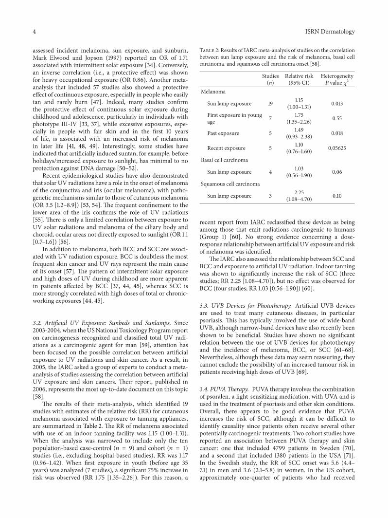
3.2. Artificial UV Exposure: Sunbeds and Sunlamps. Since2003-2004, when theUSNational Toxicology Program reporton carcinogenesis recognized and classified total UV radi-ations as a carcinogenic agent for man [59], attention hasbeen focused on the possible correlation between artificialexposure to UV radiations and skin cancer. As a result, in2005, the IARC asked a group of experts to conduct a meta-analysis of studies assessing the correlation between artificialUV exposure and skin cancers. Their report, published in2006, represents the most up-to-date document on this topic[58].
The results of their meta-analysis, which identified 19studies with estimates of the relative risk (RR) for cutaneousmelanoma associated with exposure to tanning appliances,are summarized in Table 2. The RR of melanoma associatedwith use of an indoor tanning facility was 1.15 (1.00–1.31).When the analysis was narrowed to include only the tenpopulation-based case-control (𝑛 = 9) and cohort (𝑛 = 1)studies (i.e., excluding hospital-based studies), RR was 1.17(0.96–1.42). When first exposure in youth (before age 35years) was analyzed (7 studies), a significant 75% increase inrisk was observed (RR 1.75 [1.35–2.26]). For this reason, a
Table 2: Results of IARCmeta-analysis of studies on the correlationbetween sun lamp exposure and the risk of melanoma, basal cellcarcinoma, and squamous cell carcinoma onset [58].
Studies(n)
Relative risk(95% CI)
HeterogeneityP value 𝜒2
Melanoma
Sun lamp exposure 19 1.15(1.00–1.31) 0.013
First exposure in youngage 7 1.75
(1.35–2.26) 0.55
Past exposure 5 1.49(0.93–2.38) 0.018
Recent exposure 5 1.10(0.76–1.60) 0,05625
Basal cell carcinoma
Sun lamp exposure 4 1.03(0.56–1.90) 0.06
Squamous cell carcinoma
Sun lamp exposure 3 2.25(1.08–4.70) 0.10
recent report from IARC reclassified these devices as beingamong those that emit radiations carcinogenic to humans(Group 1) [60]. No strong evidence concerning a dose-response relationship between artificial UV exposure and riskof melanoma was identified.
The IARC also assessed the relationship between SCC andBCC and exposure to artificial UV radiation. Indoor tanningwas shown to significantly increase the risk of SCC (threestudies; RR 2.25 [1.08–4.70]), but no effect was observed forBCC (four studies; RR 1.03 [0.56–1.90]) [60].
3.3. UVB Devices for Phototherapy. Artificial UVB devicesare used to treat many cutaneous diseases, in particularpsoriasis. This has typically involved the use of wide-bandUVB, although narrow-band devices have also recently beenshown to be beneficial. Studies have shown no significantrelation between the use of UVB devices for phototherapyand the incidence of melanoma, BCC, or SCC [61–68].Nevertheless, although these data may seem reassuring, theycannot exclude the possibility of an increased tumour risk inpatients receiving high doses of UVB [69].
3.4. PUVATherapy. PUVA therapy involves the combinationof psoralen, a light-sensitizing medication, with UVA and isused in the treatment of psoriasis and other skin conditions.Overall, there appears to be good evidence that PUVAincreases the risk of SCC, although it can be difficult toidentify causality since patients often receive several otherpotentially carcinogenic treatments. Two cohort studies havereported an association between PUVA therapy and skincancer: one that included 4799 patients in Sweden [70],and a second that included 1380 patients in the USA [71].In the Swedish study, the RR of SCC onset was 5.6 (4.4–7.1) in men and 3.6 (2.1–5.8) in women. In the US cohort,approximately one-quarter of patients who had received

ISRN Dermatology 5
more than 2000 J/cm2 developed SCC.Ameta-analysis by thesame group reported that patients exposed to high doses ofPUVA (more than 200 treatments or more than 2000 J/cm2)had a risk 14 times higher than those treated with less than100 sessions or exposed to less than 1000 J/cm2 [72]. Anotherrecent 30-year prospective study showed that receivingbetween 350 and 450 PUVA treatments had anRRof 6.0 (4.4–8.2) for SCCcomparedwith less than 50 treatments.However,even high-dose exposure did not increase BCC risk [73].
The risk of melanoma onset associated with PUVA ismore controversial.The American cohort study [71] reportedan increased risk of melanoma, with patients exposed tomore than 200 treatments compared to lower doses havingan almost threefold greater risk (RR 2.9 [1.3–6.4]). Moreover,this risk increased over time, with an incident relative riskof 5.0 (1.6–15.5) among patients after >15 years followup(versus <15 years). In the Swedish cohort, an increased riskfor melanoma was not observed. Since the Swedish studyis both larger and has a longer period of followup (onaverage 16 years), data obtained from this cohort are themoreconvincing.
3.5. Exposure to Infrared and Laser Radiations. As with UVradiation, extended exposure (15–20 years) to IR radiationcan induce actinic keratosis, a possible precursor to in situ orinvasive carcinomas. It is also known that prolonged exposureof the skin to heat induces particular changes known aswarmer erythema, heat dermatitis, or erythema ab igne.However, there are only limited data on the topic of IRradiation and cancers (in particular of the skin), with mostbeing case reports of tumours arising secondarily to erythemaab igne after many years [73–87].
At the present moment there are no studies that demon-strate potential carcinogenicity of laser devices, although thiscould depend on the relative rare occurrence of exposureto laser beams. There are case reports in the literatureconcerning malignant tumours arising from benign lesionsafter prolonged laser treatments. However, the possibility ofdiagnostic error before laser therapy cannot be excluded inthese rare cases [88–97].
4. Conclusions
Our review of the literature shows a clear association betweenUV radiation exposure and increased risk of melanoma andother skin cancers. However, there is no clear evidence of anyrelationship between skin cancer and IR or laser radiations.
Preventative strategies that include public informationcampaigns to highlight the risks associated with exposure tosunlight, including the degree of exposure that is consideredacceptable with regard to health and identifying patient typesmost at risk, remain important. The use of artificial tanningdevices requires special consideration. It is now clear thatthere is a strong association between these devices and therisk of melanoma and SCC. As such, precautionary measuresthat discourage exposure to tanning appliances, especiallyamong younger people, are required, as is legislation toprevent their use during childhood. Public health initiatives
comparative to those that have successfully targeted cigarettesmoking are now needed to limit recreational exposure toartificial UV sources.
References
[1] Sicurezza and Energia Multiservice, “Radiazioni Ottiche,”http://www.sicurezzaenergia.com/Sicurezza-sul-lavoro/Radi-azioni-ottiche.php.
[2] “International Commission on Illumination,” http://www.cieco.at
[3] World Health Organization, “Ultraviolet radiation and health,”http://www.who.int/uv/uv and health/en/index.html.
[4] World Health Organization, “Global solar UV index,” http://www.who.int/mediacentre/factsheets/fs305/en/index.html.
[5] N. S. Agar, G. M. Halliday, R. S. C. Barnetson, H. N. Anan-thaswamy, M. Wheeler, and A. M. Jones, “The basal layer inhuman squamous tumors harbors more UVA than UVB finger-print mutations: a role for UVA in human skin carcinogenesis,”Proceedings of the National Academy of Sciences of the UnitedStates of America, vol. 101, no. 14, pp. 4954–4959, 2004.
[6] C. Robert, B. Muel, A. Benoit, L. Dubertret, A. Sarasin, and A.Stary, “Cell survival and shuttle vector mutagenesis induced byultraviolet A and ultraviolet B radiation in a human cell line,”Journal of Investigative Dermatology, vol. 106, no. 4, pp. 721–728,1996.
[7] S. Pavel, N. P. M. Smit, H. Van Der Meulen et al., “Homozy-gous germline mutation of CDKN2A/p16 and glucose-6-phosphate dehydrogenase deficiency in a multiple melanomacase,”Melanoma Research, vol. 13, no. 2, pp. 171–178, 2003.
[8] G. P. Pfeifer, Y. H. You, and A. Besaratinia, “Mutations inducedby ultraviolet light,”Mutation Research, vol. 571, no. 1-2, pp. 19–31, 2005.
[9] G. M. Halliday, “Inflammation, gene mutation and photoim-munosuppression in response to UVR-induced oxidative dam-age contributes to photocarcinogenesis,”MutationResearch, vol.571, no. 1-2, pp. 107–120, 2005.
[10] G. M. Halliday, N. S. Agar, R. S. C. Barnetson, H. N. Anan-thaswamy, and A. M. Jones, “UV-A fingerprint mutations inhuman skin cancer,” Photochemistry and Photobiology, vol. 81,no. 1, pp. 3–8, 2005.
[11] A. J. Ridley, J. R. Whiteside, T. J. McMillan, and S. L. Allinson,“Cellular and sub-cellular responses to UVA in relation tocarcinogenesis,” International Journal of Radiation Biology, vol.85, no. 3, pp. 177–185, 2009.
[12] H. R. Griffiths, P.Mistry, K. E. Herbert, and J. Lunec, “Molecularand cellular effects of ultraviolet light-induced genotoxicity,”Critical Reviews in Clinical Laboratory Sciences, vol. 35, no. 3,pp. 189–237, 1998.
[13] T. M. Runger, “C→T transition mutations are not solely UVB-signature mutations, because they are also generated by UVA,”Journal of Investigative Dermatology, vol. 128, no. 9, pp. 2138–2140, 2008.
[14] E. A. Drobetsky, J. Turcotte, and A. Chateauneuf, “A role forultraviolet A in solar mutagenesis,” Proceedings of the NationalAcademy of Sciences of the United States of America, vol. 92, no.6, pp. 2350–2354, 1995.
[15] R. Nishigaki, H. Mitani, N. Tsuchida, and A. Shima, “Effectof cyclobutane pyrimidine dimers on apoptosis induced bydifferent wavelengths of UV,” Photochemistry and Photobiology,vol. 70, no. 2, pp. 228–235, 1999.

6 ISRN Dermatology
[16] H. Zhang and I. Rosdahl, “Ultraviolet A andBdifferently induceintracellular protein expression in human skin melanocytes: aspeculation of separate pathways in initiation of melanoma,”Carcinogenesis, vol. 24, no. 12, pp. 1929–1934, 2003.
[17] A. Woollons, P. H. Clingen, M. L. Price, C. F. Arlett, and M.H. L. Green, “Induction of mutagenic DNA damage in humanfibroblasts after exposure to artificial tanning lamps,” BritishJournal of Dermatology, vol. 137, no. 5, pp. 687–692, 1997.
[18] Y.Wang, B. Rosenstein, S. Goldwyn, X. Zhang,M. Lebwohl, andH. Wei, “Differential regulation of P53 and Bcl-2 expression byultraviolet A and B,” Journal of Investigative Dermatology, vol.111, no. 3, pp. 380–384, 1998.
[19] S. E. Whitmore, W. L. Morison, C. S. Potten, and C. Chadwick,“Tanning salon exposure and molecular alterations,” Journal ofthe American Academy of Dermatology, vol. 44, no. 5, pp. 775–780, 2001.
[20] A. E. Persson, D. W. Edstrom, H. Backvall et al., “The muta-genic effect of ultraviolet-A1 on human skin demonstrated bysequencing the p53 gene in single keratinocytes,” Photoderma-tology Photoimmunology and Photomedicine, vol. 18, no. 6, pp.287–293, 2002.
[21] M. Situm, M. Buljan, V. Bulat, Z. Bolanea, and D. Simic,“The role of UV radiation in the development of Basal cellcarcinoma,” Collegium Antropologicum, vol. 32, no. SUPPL. 2,pp. 167–170, 2008.
[22] M. R. Gailani, D. J. Leffell, A. Ziegler, E. G. Gross, D. E. Brash,and A. E. Bale, “Relationship between sunlight exposure anda key genetic alteration in basal cell carcinoma,” Journal of theNational Cancer Institute, vol. 88, no. 6, pp. 349–354, 1996.
[23] C. L. Benjamin, V. O. Melnikova, and H. N. Ananthaswamy,“P53 protein and pathogenesis of melanoma and nonmelanomaskin cancer,” Advances in Experimental Medicine and Biology,vol. 624, pp. 265–282, 2008.
[24] A. Pacifico and G. Leone, “Role of p53 and CDKN2A inac-tivation in human squamous cell carcinomas,” Journal ofBiomedicine and Biotechnology, vol. 2007, Article ID 43418, 5pages, 2007.
[25] T. M. Runger, “How different wavelengths of the ultravioletspectrum contribute to skin carcinogenesis: the role of cellulardamage responses,” Journal of Investigative Dermatology, vol.127, no. 9, pp. 2103–2105, 2007.
[26] J. Moan, A. C. Porojnicu, and A. Dahlback, “Ultraviolet radi-ation and malignant melanoma,” Advances in ExperimentalMedicine and Biology, vol. 624, pp. 104–116, 2008.
[27] D. C. Bennett, “Ultraviolet wavebands and melanoma initia-tion,” Pigment Cell and Melanoma Research, vol. 21, no. 5, pp.520–524, 2008.
[28] L. Marrot and J. R. Meunier, “Skin DNA photodamage and itsbiological consequences,” Journal of the American Academy ofDermatology, vol. 58, no. 5, pp. S139–S148, 2008.
[29] T. M. Runger and U. P. Kappes, “Mechanisms of mutation for-mation with long-wave ultraviolet light (UVA),” Photoderma-tology Photoimmunology and Photomedicine, vol. 24, no. 1, pp.2–10, 2008.
[30] International Commission on Non-Ionizing Radiation Protec-tion, “Guidelines on limits of exposure to laser radiation ofwavelengths between 180 nm and 1,000 nm,”Health Physics, vol.71, no. 5, pp. 804–814, 1996.
[31] L. H. Klingman and A. M. Klingman, “Reflection on heat,”British Journal of Dermatology, vol. 110, no. 3, pp. 369–375, 1984.
[32] J. S. Dover, T. J. Philips, and K. A. Arndt, “Cutaneous effects andtherapeutic uses of heat with emphasis on infrared radiation,”Journal of the American Academy of Dermatology, vol. 20, no. 2I, pp. 278–286, 1989.
[33] E.White, C. S. Kirkpatrick, and J. A.H. Lee, “Case-control studyof malignant melanoma in Washington State. I. Constitutionalfactors and sun exposure,” The American Journal of Epidemiol-ogy, vol. 139, no. 9, pp. 857–868, 1994.
[34] J. Mark Elwood and J. Jopson, “Melanoma and sun exposure: anoverview of published studies,” International Journal of Cancer,vol. 73, no. 2, pp. 198–203, 1997.
[35] P. Autier and J.-F. Dore, “Influence of sun exposures duringchildhood and during adulthood on melanoma risk,” Interna-tional Journal of Cancer, vol. 77, no. 4, pp. 533–537, 1998.
[36] S. D. Walter, W. D. King, and L. D. Marrett, “Association ofcutaneous malignant melanoma with intermittent exposure toultraviolet radiation: results of a case-control study in Ontario,Canada,” International Journal of Epidemiology, vol. 28, no. 3,pp. 418–427, 1999.
[37] P. Kaskel, S. Sander, M. Kron, P. Kind, R. U. Peter, and G.Krahn, “Outdoor activities in childhood: a protective factor forcutaneous melanoma? Results of a case-control study in 271matched pairs,” British Journal of Dermatology, vol. 145, no. 4,pp. 602–609, 2001.
[38] D. C. Whiteman, M. Stickley, P. Watt, M. C. Hughes, M. B.Davis, and A. C. Green, “Anatomic site, sun exposure, and riskof cutaneous melanoma,” Journal of Clinical Oncology, vol. 24,no. 19, pp. 3172–3177, 2006.
[39] A. Kricker, B. K. Armstrong, C. Goumas et al., “AmbientUV, personal sun exposure and risk of multiple primarymelanomas,” Cancer Causes and Control, vol. 18, no. 3, pp. 295–304, 2007.
[40] E. Nagore, L. Hueso, R. Botella-Estrada et al., “Smoking, sunexposure, number of nevi and previous neoplasias are risk fac-tors formelanoma in older patients (60 years and over),” Journalof the European Academy of Dermatology and Venereology, vol.24, no. 1, pp. 50–57, 2010.
[41] B. K. Armstrong and A. Kricker, “The epidemiology of UVinduced skin cancer,” Journal of Photochemistry and Photobiol-ogy B, vol. 63, no. 1–3, pp. 8–18, 2001.
[42] R. Zanetti, S. Rosso, C. Martinez et al., “Comparison of riskpatterns in carcinoma and melanoma of the skin in men: amulti-centre case-case-control study,” British Journal of Cancer,vol. 94, no. 5, pp. 743–751, 2006.
[43] J. Han, G. A. Colditz, and D. J. Hunter, “Risk factors for skincancers: a nested case-control study within the Nurses’ HealthStudy,” International Journal of Epidemiology, vol. 35, no. 6, pp.1514–1521, 2006.
[44] J. M. Elwood, R. P. Gallagher, G. B. Hill, and J. C. G. Pearson,“Cutaneous melanoma in relation to intermittent and constantsun exposure: the Western Canada melanoma study,” Interna-tional Journal of Cancer, vol. 35, no. 4, pp. 427–433, 1985.
[45] R. M. MacKie, “Incidence, risk factors and prevention ofmelanoma,” European Journal of Cancer, vol. 34, supplement 3,pp. S3–S4, 1998.
[46] B. K. Armstrong, “How sun exposure causes skin cancer: anepidemiological perspective,” in Prevention of Skin Cancer:Cancer Prevention, Cancer Causes, D. Hill, J. M. Elwood, andD. R. English, Eds., Kluwer Academic Publishers, Boston,Mass,USA, 2004.

ISRN Dermatology 7
[47] S. Gandini, F. Sera,M. S. Cattaruzza et al., “Meta-analysis of riskfactors for cutaneous melanoma: II. Sun exposure,” EuropeanJournal of Cancer, vol. 41, no. 1, pp. 45–60, 2005.
[48] P. Autier, G. Severi, R. Pedeux et al., “Number and size ofnevi are influenced by different sun exposure components:implications for the etiology of cutaneous melanoma (Belgium,Germany, France, Italy),”Cancer Causes and Control, vol. 14, no.5, pp. 453–459, 2003.
[49] D. C.Whiteman, C. A.Whiteman, andA. C. Green, “Childhoodsun exposure as a risk factor for melanoma: a systematic reviewof epidemiologic studies,” Cancer Causes and Control, vol. 12,no. 1, pp. 69–82, 2001.
[50] K. Hemminki, V. J. Bykov, J. A. Marcusson, P. Autier, R. Pedeux,and J. F. Dore, “Re: sunscreen use and duration of sun exposure:a double-blind, randomized trial,” Journal of the NationalCancer Institute, vol. 91, no. 23, pp. 2046–2047, 1999.
[51] V. J. Bykov, J. A. Marcusson, and K. Hemminki, “Protectiveeffects of tanning on cutaneous DNA damage in situ,” Derma-tology, vol. 202, no. 1, pp. 22–26, 2001.
[52] J. Ruegemer, B. Schuetz, K. Hermann, R. Hein, J. Ring, andD. Abeck, “UV-induced skin changes due to regular use ofcommercial sunbeds,”Photodermatology Photoimmunology andPhotomedicine, vol. 18, no. 5, pp. 223–227, 2002.
[53] D.-N. Hu, “Photobiology of ocular melanocytes andmelanoma,” Photochemistry and Photobiology, vol. 81, no.3, pp. 506–509, 2005.
[54] C.M. Vajdic, A. Kricker,M. Giblin et al., “Sun exposure predictsrisk of ocular melanoma in Australia,” International Journal ofCancer, vol. 101, no. 2, pp. 175–182, 2002.
[55] J. A. Shields and S. C. Shields, Intraocuiar Tumor: A Text andAtlas, WB Saunders, Philadelphia, Pa, USA, 1992.
[56] A. D. Singh, I. G. Rennie, S. Seregard, M. Giblin, and J. McKen-zie, “Sunlight exposure and pathogenesis of uveal melanoma,”Survey of Ophthalmology, vol. 49, no. 4, pp. 419–428, 2004.
[57] R. E. Neale, M. Davis, N. Pandeya, D. C. Whiteman, and A. C.Green, “Basal cell carcinoma on the trunk is associated withexcessive sun exposure,” Journal of the American Academy ofDermatology, vol. 56, no. 3, pp. 380–386, 2007.
[58] IARC, “Exposure to artificial UV radiation and skin can-cer/views and expert opinions of an IARCWorking Group thatmet in Lyon, France 27–29 June 2005,” International Agency forResearch on Cancer, 2006.
[59] National Toxicology Program, “Ultraviolet radiation relatedexposures: broad-spectrum ultraviolet (UV) radiation, UVA,UVB, UVC, solar radiation, and exposure to sunlamps andsunbeds,” Report on Carcinogens, vol. 10, pp. 250–254, 2002.
[60] F. El Ghissassi, R. Baan, K. Straif et al., “A review of humancarcinogens. Part D: radiation,”The lancet oncology, vol. 10, no.11, pp. 751–752, 2009.
[61] W. Z.Maughan, S. A.Muller, andH.O. Perry, “Incidence of skincancers in patients with atopic dermatitis treated with coal tar.A 25-year follow-up study,” Journal of the American Academy ofDermatology, vol. 3, no. 6, pp. 612–615, 1980.
[62] M. R. Pittelkow, H. O. Perry, S. A.Muller,W. Z.Maughan, and P.C. O’Brien, “Skin cancer in patients with psoriasis treated withcoal Tar. A 25-year follow-up study,” Archives of Dermatology,vol. 117, no. 8, pp. 465–468, 1981.
[63] K. M. Halprin, M. Comerford, and J. R. Taylor, “Cancer inpatients with psoriasis,” Journal of the American Academy ofDermatology, vol. 7, no. 5, pp. 633–638, 1982.
[64] O. Larko and G. Swanbeck, “Is UVB treatment of psoriasis safe?A study of extensively UVB-treated psoriasis patients comparedwith a matched control group,” Acta Dermato-Venereologica,vol. 62, supplement, no. 6, pp. 507–512, 1982.
[65] S. M. Bhate, G. R. Sharpe, J. M. Marks, S. Shuster, and W. M.Ross, “Prevalence of skin and other cancers in patients withpsoriasis,” Clinical and Experimental Dermatology, vol. 18, no.5, pp. 401–404, 1993.
[66] R. S. Stern andN. Laird, “The carcinogenic risk of treatments forsevere psoriasis. Photochemotherapy Follow-up Study,”Cancer,vol. 73, pp. 2759–2764, 1994.
[67] M. Weischer, A. Blum, F. Eberhard, M. Rocken, and M.Berneburg, “No evidence for increased skin cancer risk inpsoriasis patients treated with broadband or narrowbandUVB phototherapy: a first retrospective study,” Acta Dermato-Venereologica, vol. 84, no. 5, pp. 370–374, 2004.
[68] E. Lee, J. Koo, and T. Berger, “UVB phototherapy and skincancer risk: a review of the literature,” International Journal ofDermatology, vol. 44, no. 5, pp. 355–360, 2005.
[69] R. M. R. Hearn, A. C. Kerr, K. F. Rahim, J. Ferguson, and R.S. Dawe, “Incidence of skin cancers in 3867 patients treatedwith narrow-band ultraviolet B phototherapy,” British Journalof Dermatology, vol. 159, no. 4, pp. 931–935, 2008.
[70] B. Lindelof, B. Sigurgeirsson, E. Tegner et al., “PUVA andcancer risk: the Swedish follow-up study,” British Journal ofDermatology, vol. 141, no. 1, pp. 108–112, 1999.
[71] R. S. Stern, “The risk of melanoma in association with long-term exposure to PUVA,” Journal of the American Academy ofDermatology, vol. 44, no. 5, pp. 755–761, 2001.
[72] R. S. Stern and E. J. Lunder, “Risk of squamous cell carcinomaand methoxsalen (psoralen) and UV-A radiation (PUVA): ameta-analysis,” Archives of Dermatology, vol. 134, no. 12, pp.1582–1585, 1998.
[73] R. S. Stern, “PUVA Follow-Up Study. The risk of squamous celland basal cell cancer associated with psoralen and ultraviolet Atherapy: a 30-year prospective study,” Journal of the AmericanAcademy of Dermatology, vol. 66, no. 4, pp. 553–562, 2012.
[74] J. A. Bain, H. P. Rusch, and B. E. Kline, “The effect of temper-ature upon ultraviolet carcinogenesis with wavelength 2.800–3.400 A,” Cancer Research, vol. 3, pp. 610–612, 1943.
[75] G. A. G. Peterkin, “Malignant changes in erythema ab igne,”British Medical Journal, vol. 31, no. 2, pp. 1599–1602, 1955.
[76] R. G. Freeman and J. M. Knox, “Influence of temperature onultraviolet injury,”Archives of dermatology, vol. 89, pp. 858–864,1964.
[77] G. R. Finlayson, W. M. Sams Jr., and J. G. Smith, “Erythemaab igne: a histopathological study,” Journal of InvestigativeDermatology, vol. 46, no. 1, pp. 104–108, 1966.
[78] F. Cross, “On a turf (peat) fire cancer: malignant changesuperimposed on erythema ab igne,” Proceedings of the RoyalSociety of Medicine, vol. 60, no. 12, pp. 1307–1308, 1967.
[79] J. H. Arrington III and D. S. Lockman, “Thermal keratoses andsquamous cell carcinoma in situ associated with erythema abigne,” Archives of Dermatology, vol. 115, no. 10, pp. 1226–1228,1979.
[80] L. H. Kligman, “Intensification of ultraviolet-induced dermaldamage by infrared radiation,” Archives of DermatologicalResearch, vol. 272, no. 3-4, pp. 229–238, 1982.
[81] C. S. Jones, S. K. Tyring, P. C. Lee, and J. D. Fine, “Develop-ment of neuroendocrine (Merkel cell) carcinoma mixed withsquamous cell carcinoma in erythema ab igne,” Archives ofDermatology, vol. 124, no. 1, pp. 110–113, 1988.

8 ISRN Dermatology
[82] T. Akasaka and S. Kon, “Two cases of squamous cell carcinomaarising from erythema ab igne,” Nippon Hifuka Gakkai zasshi,vol. 99, no. 6, pp. 735–742, 1989.
[83] J. B. Hewitt, A. Sherif, K. M. Kerr, and L. Stankler, “Merkel celland squamous cell carcinomas arising in erythema ab igne,”British Journal of Dermatology, vol. 128, no. 5, pp. 591–592, 1993.
[84] M. V. Iacocca, J. L. Abernethy, C. M. Stefanato, A. E. Allan,and J. Bhawan, “Mixed Merkel cell carcinoma and squamouscell carcinoma of the skin,” Journal of the American Academy ofDermatology, vol. 39, no. 5, pp. 882–887, 1998.
[85] P. Boukamp, S. Popp, K. Bleuel, E. Tomakidi, A. Burkle, and N.E. Fusenig, “Tumorigenic conversion of immortal human skinkeratinocytes (HaCaT) by elevated temperature,”Oncogene, vol.18, no. 41, pp. 5638–5645, 1999.
[86] C.M. Rudolph, H. P. Soyer, P.Wolf, andH. Kerl, “Squamous cellcarcinoma arising in Erythema ab igne,”Hautarzt, vol. 51, no. 4,pp. 260–263, 2000.
[87] J. B. Wharton, D. J. Sheehan, and J. L. Lesher Jr., “Squamous cellcarcinoma in situ arising in the setting of erythema ab igne,”Journal of Drugs in Dermatology, vol. 7, no. 5, pp. 488–489, 2008.
[88] A. Boer, M. Wolter, and R. Kaufmann, “Pseudomelanomafollowing laser treatment or laser-treated melanoma?” Journalof the German Society of Dermatology, vol. 1, no. 1, pp. 47–50,2003.
[89] B. S. Kim, J. B. Lee, H. S. Jang, Y. W. Kwon, K. S. Kwon, and C.K. Oh, “Multiple basal cell carcinomas arising in a port-winestain with a remote history of therapeutic irradiation,” Journalof Dermatology, vol. 31, no. 10, pp. 820–823, 2004.
[90] L.Hedelund,M.Haedersdal,H. Egekvist,M.Heidenheim,H.C.Wulf, and T. Poulsen, “CO
2laser resurfacing and photocarcino-
genesis: an experimental study,” Lasers in Surgery andMedicine,vol. 35, no. 1, pp. 58–61, 2004.
[91] Z. F. Jasim, W. K. Woo, M. Y. Walsh, and J. M. Handley, “Mul-tifocal basal cell carcinoma developing in a facial port winestain treated with argon and pulsed dye laser: a possible role forprevious radiotherapy,” Dermatologic Surgery, vol. 30, no. 8, pp.1155–1157, 2004.
[92] Y.-C. Bae, Y.-S. Kang, C.-K. Oh, and S.-M. Hwang, “A report of12 cases of basal cell carcinoma arising in lesions following lasertherapy,” Annals of Plastic Surgery, vol. 54, no. 4, pp. 384–386,2005.
[93] S. S. Kelishadi, G. A. Wirth, and G. R. D. Evans, “Recalcitrantverrucous lesion: verrucous hyperplasia or epithelioma cunicu-latum (verrucous carcinoma),” Journal of theAmerican PodiatricMedical Association, vol. 96, no. 2, pp. 148–153, 2006.
[94] M.-W. Lee, S.-J. Ahn, J.-H. Choi, K.-C. Moon, and J.-K.Koh, “Pseudomelanoma following laser therapy,” Journal of theEuropean Academy of Dermatology and Venereology, vol. 20, no.3, pp. 342–344, 2006.
[95] C. Gottschaller, U. Hohenleutner, andM. Landthaler, “Metasta-sis of a malignant melanoma 2 years after carbon dioxide lasertreatment of a pigmented lesion: case report and review of theliterature,”Acta Dermato-Venereologica, vol. 86, no. 1, pp. 44–47,2006.
[96] J. I. Na, K. H. Cho, Y. G. Kim, and K. C. Park, “Angioblastomashowing aggravation after treatment with long-pulsed Nd:YAGlaser (1064 nm),” Pediatric Dermatology, vol. 24, no. 4, pp. 397–400, 2007.
[97] J. Dvorackova, J. Sterba, L. Ceganova et al., “Problems of suit-ability laser’s excision of pigmented dermal lesions: case reportof minimal deviation melanoma,” Ceskoslovenska patologie, vol.43, no. 2, pp. 64–67, 2007.

Submit your manuscripts athttp://www.hindawi.com
Stem CellsInternational
Hindawi Publishing Corporationhttp://www.hindawi.com Volume 2014
Hindawi Publishing Corporationhttp://www.hindawi.com Volume 2014
MEDIATORSINFLAMMATION
of
Hindawi Publishing Corporationhttp://www.hindawi.com Volume 2014
Behavioural Neurology
EndocrinologyInternational Journal of
Hindawi Publishing Corporationhttp://www.hindawi.com Volume 2014
Hindawi Publishing Corporationhttp://www.hindawi.com Volume 2014
Disease Markers
Hindawi Publishing Corporationhttp://www.hindawi.com Volume 2014
BioMed Research International
OncologyJournal of
Hindawi Publishing Corporationhttp://www.hindawi.com Volume 2014
Hindawi Publishing Corporationhttp://www.hindawi.com Volume 2014
Oxidative Medicine and Cellular Longevity
Hindawi Publishing Corporationhttp://www.hindawi.com Volume 2014
PPAR Research
The Scientific World JournalHindawi Publishing Corporation http://www.hindawi.com Volume 2014
Immunology ResearchHindawi Publishing Corporationhttp://www.hindawi.com Volume 2014
Journal of
ObesityJournal of
Hindawi Publishing Corporationhttp://www.hindawi.com Volume 2014
Hindawi Publishing Corporationhttp://www.hindawi.com Volume 2014
Computational and Mathematical Methods in Medicine
OphthalmologyJournal of
Hindawi Publishing Corporationhttp://www.hindawi.com Volume 2014
Diabetes ResearchJournal of
Hindawi Publishing Corporationhttp://www.hindawi.com Volume 2014
Hindawi Publishing Corporationhttp://www.hindawi.com Volume 2014
Research and TreatmentAIDS
Hindawi Publishing Corporationhttp://www.hindawi.com Volume 2014
Gastroenterology Research and Practice
Hindawi Publishing Corporationhttp://www.hindawi.com Volume 2014
Parkinson’s Disease
Evidence-Based Complementary and Alternative Medicine
Volume 2014Hindawi Publishing Corporationhttp://www.hindawi.com
Related Documents