Review Article The Relevance of the Renin-Angiotensin System in the Development of Drugs to Combat Preeclampsia Norikazu Ueki, 1,2 Satoru Takeda, 2 Daisuke Koya, 1,3 and Keizo Kanasaki 1,3 1 Department of Diabetology and Endocrinology, Kanazawa Medical University, Ishikawa 920-0293, Japan 2 Department of Obstetrics and Gynecology, Juntendo University, Tokyo 113-8431, Japan 3 Division of Anticipatory Molecular Food Science and Technology, Medical Research Institute, Kanazawa Medical University, Ishikawa 920-0293, Japan Correspondence should be addressed to Keizo Kanasaki; [email protected] Received 15 October 2014; Revised 28 March 2015; Accepted 28 March 2015 Academic Editor: Francesco Perticone Copyright © 2015 Norikazu Ueki et al. is is an open access article distributed under the Creative Commons Attribution License, which permits unrestricted use, distribution, and reproduction in any medium, provided the original work is properly cited. Preeclampsia is a hypertensive disorder that occurs during pregnancy. It has an unknown etiology and affects approximately 5–8% of pregnancies worldwide. e pathophysiology of preeclampsia is not yet known, and preeclampsia has been called “a disease of theories.” e central symptom of preeclampsia is hypertension. However, the etiology of the hypertension is unknown. In this review, we analyze the molecular mechanisms of preeclampsia with a particular focus on the pathogenesis of the hypertension in preeclampsia and its association with the renin-angiotensin system. In addition, we propose potential alternative strategies to target the renin-angiotensin system, which is enhanced during pregnancy. 1. Introduction Preeclampsia refers to a new onset of hypertension (systolic pressure ≥140 mmHg and/or diastolic pressure ≥90 mmHg on two occasions at least 4 hours apart) and either pro- teinuria (≥300 mg in 24 hours) or end-organ dysfunction (including thrombocytopenia, renal insufficiency, impaired liver function, pulmonary edema, and cerebral symptoms) aſter 20 weeks of gestation in a previously normotensive woman. Severe hypertension (systolic pressure ≥160 mmHg or diastolic pressure ≥110 mmHg on two occasions at least 4 hours apart while the patient is on bed rest) and symp- toms of end-organ injury comprise the severe end of the spectrum of the disease [1]. Worldwide, 10–15% of direct maternal deaths are associated with preeclampsia/eclampsia [2]. Preeclampsia not only is a hypertensive disorder, but also is associated with metabolic defects such as glucose intolerance and dyslipidemia. Preeclamptic women exhibit increased risks of cardiovascular disease (CVD) and type 2 diabetes later in life [3]. During oral glucose tolerance tests, preeclamptic women display higher insulin levels compared with those of normal pregnant women. Preeclampsia is also associated with insulin resistance, and serum free fatty acid levels in preeclamptic women appear to be higher [3, 4]. Insulin resistance, even despite normal glucose levels in early pregnancy, is also associated with preeclampsia onset in later gestational periods [4]. e mechanism explaining such metabolic defects in preeclampsia remains unclear. erefore, understanding the biology of preeclampsia is important to uncover metabolic defects in pregnancy, a condition associ- ated with potential harmful effects for both the baby and the mother. Despite such clinical significance, the pathophysiology that leads to preeclampsia remains unknown. erefore, pathophysiology-based therapies have not yet been estab- lished. Hypertension is the major symptom of preeclampsia. However, the molecular mechanisms underlying the onset of hypertension in preeclampsia have not been clearly estab- lished. In this review, we analyzed the molecular mechanisms of preeclampsia, with a particular focus on the molecular mechanisms of hypertension in preeclampsia. Hindawi Publishing Corporation International Journal of Endocrinology Volume 2015, Article ID 572713, 12 pages http://dx.doi.org/10.1155/2015/572713

Welcome message from author
This document is posted to help you gain knowledge. Please leave a comment to let me know what you think about it! Share it to your friends and learn new things together.
Transcript
Review ArticleThe Relevance of the Renin-Angiotensin System inthe Development of Drugs to Combat Preeclampsia
Norikazu Ueki,1,2 Satoru Takeda,2 Daisuke Koya,1,3 and Keizo Kanasaki1,3
1Department of Diabetology and Endocrinology, Kanazawa Medical University, Ishikawa 920-0293, Japan2Department of Obstetrics and Gynecology, Juntendo University, Tokyo 113-8431, Japan3Division of Anticipatory Molecular Food Science and Technology, Medical Research Institute, Kanazawa Medical University,Ishikawa 920-0293, Japan
Correspondence should be addressed to Keizo Kanasaki; [email protected]
Received 15 October 2014; Revised 28 March 2015; Accepted 28 March 2015
Academic Editor: Francesco Perticone
Copyright © 2015 Norikazu Ueki et al. This is an open access article distributed under the Creative Commons Attribution License,which permits unrestricted use, distribution, and reproduction in any medium, provided the original work is properly cited.
Preeclampsia is a hypertensive disorder that occurs during pregnancy. It has an unknown etiology and affects approximately 5–8%of pregnancies worldwide. The pathophysiology of preeclampsia is not yet known, and preeclampsia has been called “a disease oftheories.” The central symptom of preeclampsia is hypertension. However, the etiology of the hypertension is unknown. In thisreview, we analyze the molecular mechanisms of preeclampsia with a particular focus on the pathogenesis of the hypertension inpreeclampsia and its association with the renin-angiotensin system. In addition, we propose potential alternative strategies to targetthe renin-angiotensin system, which is enhanced during pregnancy.
1. Introduction
Preeclampsia refers to a new onset of hypertension (systolicpressure ≥140mmHg and/or diastolic pressure ≥90mmHgon two occasions at least 4 hours apart) and either pro-teinuria (≥300mg in 24 hours) or end-organ dysfunction(including thrombocytopenia, renal insufficiency, impairedliver function, pulmonary edema, and cerebral symptoms)after 20 weeks of gestation in a previously normotensivewoman. Severe hypertension (systolic pressure ≥160mmHgor diastolic pressure ≥110mmHg on two occasions at least4 hours apart while the patient is on bed rest) and symp-toms of end-organ injury comprise the severe end of thespectrum of the disease [1]. Worldwide, 10–15% of directmaternal deaths are associated with preeclampsia/eclampsia[2].
Preeclampsia not only is a hypertensive disorder, butalso is associated with metabolic defects such as glucoseintolerance and dyslipidemia. Preeclamptic women exhibitincreased risks of cardiovascular disease (CVD) and type 2diabetes later in life [3]. During oral glucose tolerance tests,
preeclamptic women display higher insulin levels comparedwith those of normal pregnant women. Preeclampsia is alsoassociated with insulin resistance, and serum free fatty acidlevels in preeclamptic women appear to be higher [3, 4].Insulin resistance, even despite normal glucose levels in earlypregnancy, is also associated with preeclampsia onset inlater gestational periods [4]. The mechanism explaining suchmetabolic defects in preeclampsia remains unclear.Therefore,understanding the biology of preeclampsia is important touncover metabolic defects in pregnancy, a condition associ-ated with potential harmful effects for both the baby and themother.
Despite such clinical significance, the pathophysiologythat leads to preeclampsia remains unknown. Therefore,pathophysiology-based therapies have not yet been estab-lished. Hypertension is the major symptom of preeclampsia.However, the molecular mechanisms underlying the onsetof hypertension in preeclampsia have not been clearly estab-lished. In this review, we analyzed themolecular mechanismsof preeclampsia, with a particular focus on the molecularmechanisms of hypertension in preeclampsia.
Hindawi Publishing CorporationInternational Journal of EndocrinologyVolume 2015, Article ID 572713, 12 pageshttp://dx.doi.org/10.1155/2015/572713
2 International Journal of Endocrinology
2. Hypertension in Preeclampsia
Using its worldwide definition, hypertension is the conditionof a systolic blood pressure ≥140mmHg or/and diastolicblood pressure≥90mmHg.Thepathogenesis of hypertensionduring pregnancy is not completely understood, and itlikely depends on the complex interplay between increasedangiotensin II (ATII) activity and mineralocorticoid excess[5], genetic factors [6], endothelial dysfunction [7], neu-rovascular anomalies [8], and increased sympathetic nervousactivity [9].
Clinically, hypertension is the most serious symptomaffecting maternal and neonatal health in preeclampsia. Innormal human pregnancy, the systolic and diastolic arterialblood pressures decrease slightly due to the reduced totalperipheral vascular resistance throughout the course of preg-nancy. Normal pregnant women are also known to increasetheir secretion of aldosterone and to be quite resistant tothe pressor effects of ATII [10]. The lowest pressures occurat approximately 28 weeks of gestation; thereafter, the bloodpressure fluctuates and tends to return to levels similarto those observed in nonpregnant women [10–14]. Indeed,during pregnancy, left ventricular function is increased asa result of a combination of increased preload, decreasedafterload and an increase in intrinsic myocardial contractilitythat is independent of the loading condition [15]. Plasmavolume expansion in pregnant women contributes to protectagainst placental hypoperfusion [16].
The renin-angiotensin system (RAS) plays an importantrole in maintaining normal blood pressure, and renin hasbeen recognized as a volume sensor. Low plasma reninactivity (PRA) is associated with plasma volume expansion innonpregnant individuals.The PRA in preeclamptic women islower comparedwith that of normal pregnant women [17, 18].However, such PRA suppression in preeclampsia is unlikelygiven that the hypertension in preeclampsia is associatedwithvolume-dependent hypertension.
Gant et al. published seminal reports regarding the role ofthe RAS in the pathogenesis of pregnancy-induced hyperten-sion [19]. In their report, they analyzed the pressor responsesto infused ATII that were required to achieve a 20mmHgrise in diastolic blood pressure in nonpregnant women and192 pregnant women. Of the latter, 120 women had normalblood pressure throughout the pregnancy, and 72 womensubsequently developed pregnancy-induced hypertension.The authors first found that the doses required for a pressorresponse in normal pregnant women were generally higherthan those in nonpregnant women (7.35 ± 0.67 ng/kg/min).The dose required in normal pregnancy increased toward 28weeks of gestation, and after 30weeks of gestation, the pressorresponse dose decreased (8.6 ± 0.76 ng/kg/min at 7–10 weeksof gestation, 14.9±1.1 ng/kg/min at 28weeks of gestation, and10.7 ± 0.71 ng/kg/min at 38 weeks of gestation). Comparedwith nonpregnant women, the amount of ATII requiredto raise the blood pressure in normal pregnant womenwas significantly higher. However, the women who laterdeveloped pregnancy-induced hypertension were sensitive tothe pressor effects of ATII comparedwith normotensive preg-nant women. Women who developed pregnancy-induced
hypertension also exhibited resistance to the pressor responseearly in their pregnancy (12.9 ± 1.1 ng/kg/min at 15–18 weeksof gestation). However, this resistance progressively declinedas the pregnancy progressed.
By 23–26 weeks of gestation, there was a clear differencein the pressor response between women with normal preg-nancy and women with pregnancy-induced hypertension.The mean required dose of ATII for a pressor responsein women who developed pregnancy-induced hypertensionprogressively declined compared with normotensive preg-nant women, and the difference was even greater comparedwith nonpregnant women. It is important that the sensitivityto ATII between 23 and 32 weeks of gestation was increasedin women who developed pregnancy-induced hypertension[19]. Hanssens et al. showed that the lowest ATII levels werefound in women with severe pregnancy-induced hyperten-sion [20].
Angiotensin-(1-7) is a bioactive component of the RASand that displays antagonistic actions of ATII by acting asa modulator of vascular tone and by releasing nitric oxide(NO) and prostaglandins. Angiotensin-(1-7) is generatedfrom ATII by angiotensin-converting enzyme 2 (ACE2) [21–23]. In human studies, plasma angiotensin-(1-7) increases innormal pregnancy and decreases in preeclampsia, and suchalternative profiles of ATII products may be relevant to thehypertension and metabolic defects in preeclampsia [21].
In contrast, several groups have suggested that hyperten-sive disorders result from the presence of agonistic autoan-tibodies (AAs) that bind to and activate the angiotensin IItype 1 (AT1) receptor [24]. Wallukat et al. initially describedthe presence of AT1-AAs in preeclampsia. Immunoglobulinsfrom the serum of women with preeclampsia stimulated theAT1 receptor and had agonistic activity, but immunoglob-ulins from normotensive pregnant women had no effect[25]. Immunoglobulin G (IgG) from preeclamptic womencontributed to the production of reactive oxygen speciesby stimulating nicotinamide-adenine dinucleotide phos-phate (NADPH) oxidase activity in vascular smooth musclecells and human trophoblasts [24]. AT1-AAs resulted inincreased soluble fms-like tyrosine kinase-1 (sFlt-1) and sol-uble endoglin (sEng) production from human trophoblastsand placental explants [26, 27]. Irani et al. showed that AT1-AAs induced apoptosis in the placentas of pregnant mice,human villous explants, and human trophoblast cells inculture. The AT1-AAs crossed the placenta and entered thefetal circulation. Finally, the authors showed that the AT1receptor antagonist losartan diminished intrauterine growthrestriction (IUGR) and placental apoptosis, which mightbe associated with AT1-AAs. The administration of agonis-tic AT1-AAs obtained from preeclamptic women inducedkey features of preeclampsia such as hypertension, protein-uria, glomerular endotheliosis, placental abnormalities, andembryonic defects in pregnant mice. These AT1-AA-inducedpreeclamptic-like symptoms in pregnant mice were attenu-ated by the AT1 receptor antagonist losartan or neutralizingpeptide against AT1-AAs [28]. Wenzel et al. showed that AT1-AAs induced ATII sensitivity in pregnant rats [29].
The evidence from the past studies clearly demon-strates that vasoconstrictive angiotensin receptor signaling
International Journal of Endocrinology 3
Angiotensin I
Angiotensin II
Angiotensin IIReceptor type I
Angiotensin-converting
enzyme
Renin
Angiotensin- converting
enzyme inhibitor
Angiotensin receptor
antagonist
Substrate peptides
Cleaved peptides
Angiotensinogen
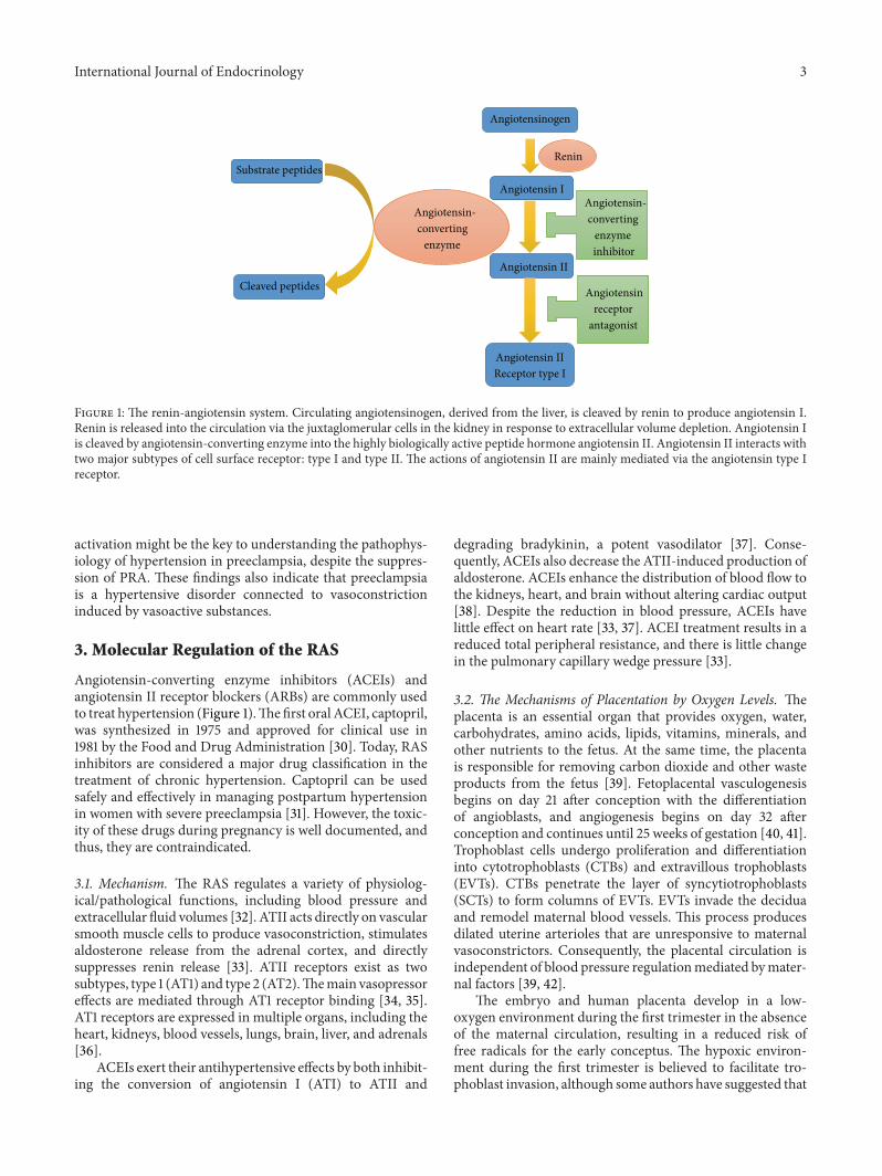
Figure 1: The renin-angiotensin system. Circulating angiotensinogen, derived from the liver, is cleaved by renin to produce angiotensin I.Renin is released into the circulation via the juxtaglomerular cells in the kidney in response to extracellular volume depletion. Angiotensin Iis cleaved by angiotensin-converting enzyme into the highly biologically active peptide hormone angiotensin II. Angiotensin II interacts withtwo major subtypes of cell surface receptor: type I and type II. The actions of angiotensin II are mainly mediated via the angiotensin type Ireceptor.
activation might be the key to understanding the pathophys-iology of hypertension in preeclampsia, despite the suppres-sion of PRA. These findings also indicate that preeclampsiais a hypertensive disorder connected to vasoconstrictioninduced by vasoactive substances.
3. Molecular Regulation of the RAS
Angiotensin-converting enzyme inhibitors (ACEIs) andangiotensin II receptor blockers (ARBs) are commonly usedto treat hypertension (Figure 1).Thefirst oral ACEI, captopril,was synthesized in 1975 and approved for clinical use in1981 by the Food and Drug Administration [30]. Today, RASinhibitors are considered a major drug classification in thetreatment of chronic hypertension. Captopril can be usedsafely and effectively in managing postpartum hypertensionin women with severe preeclampsia [31]. However, the toxic-ity of these drugs during pregnancy is well documented, andthus, they are contraindicated.
3.1. Mechanism. The RAS regulates a variety of physiolog-ical/pathological functions, including blood pressure andextracellular fluid volumes [32]. ATII acts directly on vascularsmooth muscle cells to produce vasoconstriction, stimulatesaldosterone release from the adrenal cortex, and directlysuppresses renin release [33]. ATII receptors exist as twosubtypes, type 1 (AT1) and type 2 (AT2).Themain vasopressoreffects are mediated through AT1 receptor binding [34, 35].AT1 receptors are expressed in multiple organs, including theheart, kidneys, blood vessels, lungs, brain, liver, and adrenals[36].
ACEIs exert their antihypertensive effects by both inhibit-ing the conversion of angiotensin I (ATI) to ATII and
degrading bradykinin, a potent vasodilator [37]. Conse-quently, ACEIs also decrease the ATII-induced production ofaldosterone. ACEIs enhance the distribution of blood flow tothe kidneys, heart, and brain without altering cardiac output[38]. Despite the reduction in blood pressure, ACEIs havelittle effect on heart rate [33, 37]. ACEI treatment results in areduced total peripheral resistance, and there is little changein the pulmonary capillary wedge pressure [33].
3.2. The Mechanisms of Placentation by Oxygen Levels. Theplacenta is an essential organ that provides oxygen, water,carbohydrates, amino acids, lipids, vitamins, minerals, andother nutrients to the fetus. At the same time, the placentais responsible for removing carbon dioxide and other wasteproducts from the fetus [39]. Fetoplacental vasculogenesisbegins on day 21 after conception with the differentiationof angioblasts, and angiogenesis begins on day 32 afterconception and continues until 25 weeks of gestation [40, 41].Trophoblast cells undergo proliferation and differentiationinto cytotrophoblasts (CTBs) and extravillous trophoblasts(EVTs). CTBs penetrate the layer of syncytiotrophoblasts(SCTs) to form columns of EVTs. EVTs invade the deciduaand remodel maternal blood vessels. This process producesdilated uterine arterioles that are unresponsive to maternalvasoconstrictors. Consequently, the placental circulation isindependent of blood pressure regulationmediated bymater-nal factors [39, 42].
The embryo and human placenta develop in a low-oxygen environment during the first trimester in the absenceof the maternal circulation, resulting in a reduced risk offree radicals for the early conceptus. The hypoxic environ-ment during the first trimester is believed to facilitate tro-phoblast invasion, although some authors have suggested that
4 International Journal of Endocrinology
Estradiol 17𝛽-Hydroxyestradiol
2-Methoxyestradiol
CYP450
COMT
HIF-1𝛼 suppression?Placental perfusion? Vascular homeostasis?
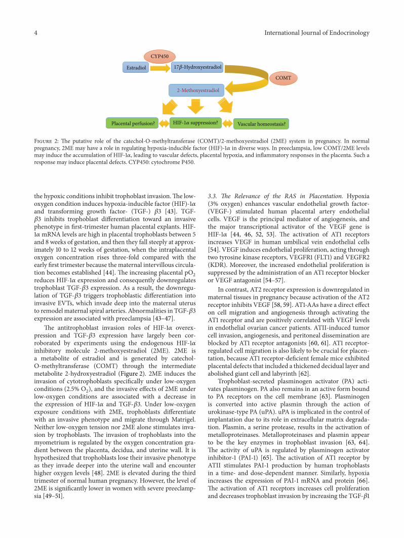
Figure 2: The putative role of the catechol-O-methyltransferase (COMT)/2-methoxyestradiol (2ME) system in pregnancy. In normalpregnancy, 2ME may have a role in regulating hypoxia-inducible factor (HIF)-1𝛼 in diverse ways. In preeclampsia, low COMT/2ME levelsmay induce the accumulation of HIF-1𝛼, leading to vascular defects, placental hypoxia, and inflammatory responses in the placenta. Such aresponse may induce placental defects. CYP450: cytochrome P450.
the hypoxic conditions inhibit trophoblast invasion.The low-oxygen condition induces hypoxia-inducible factor (HIF)-1𝛼and transforming growth factor- (TGF-) 𝛽3 [43]. TGF-𝛽3 inhibits trophoblast differentiation toward an invasivephenotype in first-trimester human placental explants. HIF-1𝛼mRNA levels are high in placental trophoblasts between 5and 8 weeks of gestation, and then they fall steeply at approx-imately 10 to 12 weeks of gestation, when the intraplacentaloxygen concentration rises three-fold compared with theearly first trimester because the maternal intervillous circula-tion becomes established [44]. The increasing placental pO
2
reduces HIF-1𝛼 expression and consequently downregulatestrophoblast TGF-𝛽3 expression. As a result, the downregu-lation of TGF-𝛽3 triggers trophoblastic differentiation intoinvasive EVTs, which invade deep into the maternal uterusto remodelmaternal spiral arteries. Abnormalities in TGF-𝛽3expression are associated with preeclampsia [43–47].
The antitrophoblast invasion roles of HIF-1𝛼 overex-pression and TGF-𝛽3 expression have largely been cor-roborated by experiments using the endogenous HIF-1𝛼inhibitory molecule 2-methoxyestradiol (2ME). 2ME isa metabolite of estradiol and is generated by catechol-O-methyltransferase (COMT) through the intermediatemetabolite 2-hydroxyestradiol (Figure 2). 2ME induces theinvasion of cytotrophoblasts specifically under low-oxygenconditions (2.5% O
2), and the invasive effects of 2ME under
low-oxygen conditions are associated with a decrease inthe expression of HIF-1𝛼 and TGF-𝛽3. Under low-oxygenexposure conditions with 2ME, trophoblasts differentiatewith an invasive phenotype and migrate through Matrigel.Neither low-oxygen tension nor 2ME alone stimulates inva-sion by trophoblasts. The invasion of trophoblasts into themyometrium is regulated by the oxygen concentration gra-dient between the placenta, decidua, and uterine wall. It ishypothesized that trophoblasts lose their invasive phenotypeas they invade deeper into the uterine wall and encounterhigher oxygen levels [48]. 2ME is elevated during the thirdtrimester of normal human pregnancy. However, the level of2ME is significantly lower in women with severe preeclamp-sia [49–51].
3.3. The Relevance of the RAS in Placentation. Hypoxia(3% oxygen) enhances vascular endothelial growth factor-(VEGF-) stimulated human placental artery endothelialcells. VEGF is the principal mediator of angiogenesis, andthe major transcriptional activator of the VEGF gene isHIF-1𝛼 [44, 46, 52, 53]. The activation of AT1 receptorsincreases VEGF in human umbilical vein endothelial cells[54]. VEGF induces endothelial proliferation, acting throughtwo tyrosine kinase receptors, VEGFR1 (FLT1) and VEGFR2(KDR). Moreover, the increased endothelial proliferation issuppressed by the administration of an AT1 receptor blockeror VEGF antagonist [54–57].
In contrast, AT2 receptor expression is downregulated inmaternal tissues in pregnancy because activation of the AT2receptor inhibits VEGF [58, 59]. AT1-AAs have a direct effecton cell migration and angiogenesis through activating theAT1 receptor and are positively correlated with VEGF levelsin endothelial ovarian cancer patients. ATII-induced tumorcell invasion, angiogenesis, and peritoneal dissemination areblocked by AT1 receptor antagonists [60, 61]. AT1 receptor-regulated cell migration is also likely to be crucial for placen-tation, because AT1 receptor-deficient female mice exhibitedplacental defects that included a thickened decidual layer andabolished giant cell and labyrinth [62].
Trophoblast-secreted plasminogen activator (PA) acti-vates plasminogen. PA also remains in an active form boundto PA receptors on the cell membrane [63]. Plasminogenis converted into active plasmin through the action ofurokinase-type PA (uPA). uPA is implicated in the control ofimplantation due to its role in extracellular matrix degrada-tion. Plasmin, a serine protease, results in the activation ofmetalloproteinases. Metalloproteinases and plasmin appearto be the key enzymes in trophoblast invasion [63, 64].The activity of uPA is regulated by plasminogen activatorinhibitor-1 (PAI-1) [65]. The activation of AT1 receptor byATII stimulates PAI-1 production by human trophoblastsin a time- and dose-dependent manner. Similarly, hypoxiaincreases the expression of PAI-1 mRNA and protein [66].The activation of AT1 receptors increases cell proliferationand decreases trophoblast invasion by increasing the TGF-𝛽1
International Journal of Endocrinology 5
and PAI-1 levels [65]. TGF-𝛽1 increases the synthesis of PAI-1[67, 68]. PAI-1 levels are significantly increased in the plasmaand placenta of preeclamptic women compared with normalpregnant women. Increased PAI-1 is associated with shallowtrophoblast invasion and hypercoagulation [65, 69]. AT1-AAsactivate AT1 receptors on human trophoblasts resulting inincreased expression of the PAI-1 protein. Then, AT1-AA-induced PAI-1 overproduction is blocked by the AT1 receptorantagonist losartan [65, 69–71].
3.4. The RAS and Fetal Development. The RAS is activeduring fetal development and has an important role incontrolling the umbilical-placental circulation, fetal bloodpressure, and cardiovascular function through ATII [72, 73].Animal studies also demonstrated that ATII induces cellgrowth through AT1 receptor, which is essential for thedevelopment of the fetal kidney [74].
In humans, all of the components of the RAS areexpressed as early as 5 weeks of gestation in human embryos[75–77]. One study found the expression of only AT2 recep-tors in the kidneys of 17–26 weeks of gestation in humanfetuses [78]. In humans, nephrogenesis begins in 5 weeks ofgestation and ceases by approximately 36 weeks of gestation,by which time approximately 1 million nephrons per kidneyare present. The fetus starts to produce urine by 9–12 weeksof gestation [79, 80]. The GFR increases progressively upto 34 to 36 weeks of gestation and matures rapidly in theearly postnatal period [81, 82]. In humans, mutations ingenes that encode renin, angiotensinogen, ACE, and AT1were found to be associated with the autosomal recessivedisease renal tubular dysgenesis. Renal tubular dysgenesiscauses oligohydramnios, which leads to the Potter sequenceand skull ossification defects [83].
In AT1 receptor-deficient pregnant mice, the numberof live newborns is significantly reduced by the placentalmalformation in 30% of all uteroplacental units. Within themalformed uteroplacental units, normal embryonic structureis not observed [62]. The human-angiotensinogen trans-genic female mice (hAG+/+) mated with the human-renintransgenic male mice (hRN+/+) display hypertension in latepregnancy due to secretion of human renin from the fetal sideinto thematernal circulation and exhibit placental abnormal-ities, maternal cardiac hypertrophy, proteinuria, and IUGR[84, 85]. However, hAG+/+ females lacking ATII receptortype 1a (mAT1a) mated with hRN+/+ male mice (pregnanthAG+/+/mAT1a−/−) have a normotensive phenotype, andthe placental abnormalities, maternal cardiac hypertrophy,and IUGR are ameliorated [85]. Similarly, pregnant hAG+/+mice administeredAT1 antagonist (5mg/kg/day) on gestationdays 18 and 19 exhibit improved hypertension and IUGR[85]. In brief, the lack of maternal AT1 might not affectfetal development while having a positive effect on maternalhypertension. ARB might therefore be an effective medica-tion for pregnancy-induced hypertension.
3.5. The Use of ACEIs or ARBs in the First Trimester. Cooperet al. studied a cohort of 29,507 infants enrolled in Tennessee
Medicaid whose mothers had no evidence of maternal dia-betes. The risk of major congenital anomalies among 209infants with exposure to ACEIs in the first trimester alonewas compared with the risk in 202 infants with exposure toother antihypertensive medications (excluding ARB) in thefirst trimester alone and 29,096 infants with no exposure toantihypertensive drugs at any time during pregnancy. Theyfound that 7.1% of the infants with exposure to ACEIs inthe first trimester alone had major congenital anomaliesafter adjusting for confounders, which was 2.7-fold higherthan infants with no exposure to antihypertensive drugs atany time during pregnancy. And they found a significantlyincreased risk of cardiovascular (risk ratio, 3.72; 95% con-fidence interval (CI), 1.89–7.30) and central nervous system(CNS; risk ratio, 4.39; 95% CI, 1.37–14.02) malformations[86]. However, some women with undiagnosed diabeteswere not excluded, and also prepregnancy body mass indexwas not controlled in this study, despite maternal diabetes,overweight, and obesity being associated with congenitalmalformations [87–89].
The association between maternal use of ACEIs or otherantihypertensive medications in the first trimester and con-genital anomalies among infants was explored in a recordlinkage study performed through the Swedish Medical BirthRegister [90]. In this study, women treated with antihy-pertensive medications had an increased risk of pretermdelivery, placental abruption, caesarean delivery, and laborinduction. Cardiovascular defects occurred with adjustedodds rate of 2.59 (95% CI, 1.92–3.51). However, the resultswere similar for the use of an ACEI or ARB during the firsttrimester comparedwith other antihypertensivemedications,particularly beta blockers [90].
Outcomes were studied in 91 pregnant women in whichthe mother took either an ACEI or ARB during earlypregnancy. Of the 71 pregnancies in women taking an ACEI,six babies (10.2%) had developmental anomalies: one smallventricular septal defect (1.4%), one mild sensorineural deaf-ness (1.4%), one mild microcephaly (1.4%), one hypospadias(1.4%), one umbilical hernia (1.4%), and one mild congenitalhypotonia (1.4%). In the 20 pregnancies (21 babies) in womenwho conceived while taking an ARB, two developmentaldefects were identified: one inguinal hernia (5%) and onecraniosynostosis with tower skull (5%). This study did notfind any convincing excess of congenital anomalies in womentaking an ACEI or ARB in early pregnancy [91].
Porta et al. reported the experience from the DIRECT(Diabetic Retinopathy and Candesartan Trials) study. Agroup of 615 women with type 1 diabetes and no retinopathyand 813 women with type 1 diabetes and mild to moder-ately severe nonproliferative retinopathy were randomizedto either candesartan (32mg/day) or placebo. A total of 42women taking candesartan and 45 taking a placebo becamepregnant and discontinued treatment 0 to 8 weeks after theirlast menstrual period. Pregnancy outcomes were found tobe similar for both groups, with no increase in the rate ofmalformation in neonates. There were two stillbirths andtwo “sick babies” in the candesartan group; there were onestillbirth, eight “sick babies,” and one ventricular septal defectin the placebo group. This study was a randomized trial,

6 International Journal of Endocrinology
and all of the women had diabetes and were normotensive.Although nowoman continued candesartan after eight weeksof gestation, the average length of time the women wereexposed to candesartan is not known [92].
Li et al. conducted a population-based retrospectivecohort study of 465,754 pregnant women in northernCalifor-nia and their live-born offspring. Women who used an ARBduring pregnancy were excluded from the study.The controlswere two cohorts composed of women who had diagnosesof hypertension but had not used any antihypertensive drugduring pregnancy and women who had neither a diagnosis ofhypertension nor a record of any prescription for an antihy-pertensive drug. After adjustment for maternal age, ethnicity,parity, and obesity, the rate of congenital heart defects amongthe offspring of women who used an ACEI in the firsttrimester (15/381 (3.9%)) was similar to the rate among theoffspring of women who used other antihypertensive drugs(28/1090 (2.6%)). This study found a similarly elevated riskof congenital heart defects in the offspring of women whohad used either ACEIs or other antihypertensive drugs orwho had hypertension and did not use antihypertensivedrugs during the first trimester of pregnancy (708/29735(2.4%)).The authors suggested that it is likely that underlyinghypertension increased the risk of congenital heart defects inthe offspring [93].
3.6. The Use of ACEIs or ARBs in the Second or ThirdTrimester. Using captopril (2.8–3.5mg/kg) in pregnant sheepduring late pregnancy (119–133 days of gestation—term is 147days) reduced the maternal blood pressure transiently for2 h. However, the fetal blood pressure remained reduced forup to 2 days [94]. The pressor response was evoked by anintravenous bolus of ATI (167–426 ng/kg). When the bloodpressure was again stable, captopril was again given to theewe. Basal blood pressures had fallen in all ewes and fetusesby 10min after captopril administration. The risk of stillbirthwas significantly elevated, and 7 of the 8 ewes producedstillborn lambs. The administration of captopril (3.3mg)to late-gestational-age rabbits (24–28 days of gestation—term is 31 days) resulted in a 37% stillbirth rate comparedwith 6% in saline control rabbits. The inhibition of AT IIsynthesis reduced uterine blood flow and increased fetalmortality [94]. Harewood et al. investigated fetal deaths wereassociated with the decrease of maternal blood pressure, andthe fetal mortality was likely secondary to the direct effectof enalapril (7.5mg/day) on the fetal RAS rather than to theeffects of placental ischemia. Postmortem examination of thedead fetuses revealed no fetal anomalies [95]. Some animalexperiments suggest that the use of ACEIs in the second orthird trimester leads to decreased uteroplacental blood flow,low birth weight, fetal hypotension, preterm delivery, andfetal death.
In human studies, the most commonly reported adverseeffects of ACEIs or ARBs taken in the second or thirdtrimester include suppressed fetal renal function and reducedurine output, which lead to oligohydramnios with fetalanuria, neonatal hypotension, renal failure, and hypocalvaria[96–99].
According to the above reports, fetal exposure to ACEIsorARBs in the first trimester only is not likely to be associatedwith fetal anomalies. However, fetal exposure to ACEIs orARBs during the second or third trimester is associatedwith fetal anomalies. ACEIs and ARBs should be avoided inwomen who are or may become pregnant.
4. Perspective: What Alternative TherapiesAre Available?
Theoretically, suppression of the RAS would be reasonablein preeclampsia treatment; however, due to the anomaliesreported with the use of drugs that suppress the RAS, wecannot recommend this class of drugs for the treatment ofpreeclamptic women. For alternative strategies to target theRAS, we have provided several ideas to combat preeclampsiathat are associated with potential RAS inhibition.
4.1. Rest and Physical Activity. Preeclampsia has beenbelieved as the condition associated with depletion of plasmavolume [100]. Placental perfusion is likely decreased by exer-cise and the bed rest in pregnant women displays increasedplacental perfusion, the hypothetical condition associatedwith the onset of preeclampsia [101]. Indeed the bed rest hasbeen considered in many pregnancies to prevent or treat avariety of conditions, such as spontaneous abortion, pretermlabor, fetal growth retardation, edema, chronic hypertension,and preeclampsia. However in the treatment of preeclampsia,the effects of bed rest are still questionable in its view [102].Also there might be risks associated with rest, such as deepvein thrombosis and pulmonary embolism [103]. In WHO,advice rest at home and strict bedrest are not recommendedfor prevention or improving pregnancy outcomes in womenwith preeclampsia [104].
Renin-angiotensin system is altered by physical exercise.In the patient with heart failure, physical exercise has beenshown to reduce sympathoexcitatory process by reducingoxidative stress, increasing nitric oxide (NO), and reducingATII [105]. Epidemiological data suggested that the physicalactivity either occupational or leisure-time was associatedwith the reduction in the incidence of preeclampsia [106].However, it is not concluded whether interventional physicalexercise is of benefit to the disease progression of preeclamp-sia since very limited information is only available in theliterature [106–108].
4.2. Antioxidants. Oxidative stress, which diminishes thebioavailability of NO, has been proposed to increase the riskof essential hypertension and preeclampsia [109, 110]. In ani-mal studies, ATII increases vascular superoxide production,which reduces the biologic activity of endothelium-derivedNO [111]. In humans, ATII blockade attenuates endothelium-dependent forearm vasodilation, and vitamin C improves theendothelial impairment [112]. Some authors have assessed theeffects of antioxidant supplementation with vitamins C andE on the risk of preeclampsia. Pregnant women between 9and 16 weeks of gestation or between 14 and 22 weeks ofgestation were randomly assigned to daily supplementation
International Journal of Endocrinology 7
with 1000mg of vitamin C and 400 IU of vitamin E orplacebo until delivery. Supplementation with vitamins C andE initiated at 9 to 16 weeks of gestation or at 14 and 22weeks of gestation until delivery did not reduce the rateof preeclampsia [110, 113]. Previous studies have suggestedthat low maternal serum 25-hydroxy vitamin D (25(OH)D)levels increase the risk of preeclampsia and that vitaminD supplementation lowers this risk [114]. Compared withnormotensive individuals with sufficient 25(OH)D levels(≥30.0 ng/mL), individuals with 25(OH)D insufficiency (15.0to 29.9 ng/mL) and deficiency (<15.0 ng/mL) had highercirculating ATII levels. Moreover, compared with individualswith sufficient vitamin D levels (145mL/min/1.73m2), thosewith vitamin D deficiency (115mL/min/1.73m2) had signif-icantly slowed renal plasma flow responses to infused ATII[115].
4.3. Anticoagulation Therapies. Preeclampsia is associatedwith thrombocytopenia, disseminated intravascular coagula-tion, and platelet aggregation. In addition, abnormal placen-tal development results in placental ischemia. In vitro, aspirinsuppresses ATII-mediated AT1R and VEGF expression inHMVECs [116]. Previous studies suggested that daily admin-istration of antiplatelet agents, particularly low-dose aspirinbeginning as early as the second trimester, had benefits forthe prevention of preeclampsia. No harmful effects wereidentified, but the long-term evidence is limited [117, 118].CLASP, a multicenter trial in which 9364 women wererandomly assigned 60mg aspirin daily or amatching placebo,suggested that low-dose aspirin did not prevent the incidenceof proteinuric preeclampsia [119]. Furthermore, Kyle et al.suggested a contrasting result and showed that the admin-istration of low-dose aspirin (60mg/day) did not preventpreeclampsia when initiated in 28 weeks of gestation in ATII-sensitive women [120]. In 2014, the US Preventive ServicesTask Force (USPSTF) recommended the use of low-doseaspirin (81mg/day) as a preventive medication after 12 weeksof gestation in women who are at high risk for preeclampsia[117].
4.4. COMT and 2ME. Kanasaki et al. reported that preg-nant mice deficient in COMT showed a preeclampsia-likephenotype resulting from the absence of 2ME, a naturalmetabolite of estradiol that is elevated during the thirdtrimester of normal human pregnancy [49, 51]. COMTis a catabolic enzyme involved in the degradation of anumber of bioactive molecules such as catecholamines andcatecholestrogens. Estradiol is metabolized by cytochromep450, and the resultant 17-hydroxyestradiol is a substrate forCOMT, which converts 17-hydroxyestradiol into 2ME as arate-limiting step in estrogen breakdown. The concentrationof maternal 2ME in the circulation immediately increasesduring normal pregnancy; however, the levels of COMT and2ME are significantly lower inwomenwith severe preeclamp-sia [49–51]. We know that hydralazine is widely used inthe treatment of preeclampsia. However, hydralazine inhibitsplacental COMT activity [121]; thus, physicians should becareful when using it.
2ME has been shown to ameliorate all preeclampsia-like features without toxicity in Comt(−/−) pregnant miceand to suppress placental hypoxia, HIF-1𝛼 expression, andincreased sFLT-1 expression [49]. Furthermore, 2ME maydirectly function as a vasodilator and inhibit vasospasm inpregnant women [122]. 2ME suppresses the AT1 receptorin vascular smooth muscle cells [123]. Finally, as describedabove, 2ME ameliorates the restricted trophoblast invasionin preeclampsia via suppression of HIF-1𝛼. 2ME (Panzem) isnow in development as an anticancer drug that induces HIF-1𝛼 suppression (phase II clinical trials) [124–126]. Therefore,2ME is available as a drug that is orally available. If 2ME issafe to use in pregnant women, it could be a therapeutic drugfor preeclampsia.
Alternatively, nutritional intervention also can enhancethe COMT-2ME system during pregnancy. Elevated homo-cysteine levels might be associated with an increased risk ofpreeclampsia, and homocysteine is converted into S-adenosylhomocysteine (SAH), a potent COMT inhibitor. Folic acidacts to remethylate homocysteine, converting homocysteineback into methionine [127–129]. Supplementation with folicacid during the second trimester is associated with a reducedrisk of preeclampsia [130]. COMT may be suppressed byvarious endogenous and exogenous molecules, includingpolychlorinated biphenols (PCBs), dioxin, mercury, andSAH.Therefore, epidemiologically, avoiding exposure to suchenvironmental COMT suppressors would be relevant to thedesign of any antipreeclampsia strategy.
5. Conclusions
The pathophysiology of preeclampsia is not completelyunderstood. Many hypotheses, theories, and models areassociatedwith preeclampsia,many of which have focused onthe abnormalities of angiogenesis. However, few investigatorshave tested whether the proposed hypertensive mechanismsare relevant to the pathophysiology of hypertension inpreeclamptic women. Careful attention should be paid whendesigning strategies to combat preeclampsia based on thepathogenesis known in both human disease and animalmodels. Further research would be required to obtain deeperinsight into the pathogenesis of preeclampsia by designingexperiments to test each hypothesis in relation to humandisease.
Conflict of Interests
The authors declare that there is no conflict of interestsregarding the publication of this paper.
References
[1] American College of Obstetricians and Gynecologists andTask Force on Hypertension in Pregnancy, “Hypertension inpregnancy. Report of the American College of Obstetriciansand Gynecologists’ Task Force on Hypertension in Pregnancy,”Obstetrics & Gynecology, vol. 122, no. 5, pp. 1122–1131, 2013.
[2] L. Duley, “The global impact of pre-eclampsia and eclampsia,”Seminars in Perinatology, vol. 33, no. 3, pp. 130–137, 2009.
8 International Journal of Endocrinology
[3] P. M. Villa, H. Laivuori, E. Kajantie, and R. Kaaja, “Free fattyacid profiles in preeclampsia,” Prostaglandins Leukotrienes andEssential Fatty Acids, vol. 81, no. 1, pp. 17–21, 2009.
[4] R. Kaaja, H. Laivuori, M. Laakso, M. J. Tikkanen, and O.Ylikorkala, “Evidence of a state of increased insulin resistance inpreeclampsia,” Metabolism: Clinical and Experimental, vol. 48,no. 7, pp. 892–896, 1999.
[5] I. S. MacKenzie and M. J. Brown, “Molecular and clinicalinvestigations in patients with low-renin hypertension,”Clinicaland Experimental Nephrology, vol. 13, no. 1, pp. 1–8, 2009.
[6] H. R. Taal, G. C. Verwoert, A. Demirkan et al., “Genome-wideprofiling of blood pressure in adults and children,” Hyperten-sion, vol. 59, no. 2, pp. 241–247, 2012.
[7] P. M. Vanhoutte, “Endothelium and control of vascular func-tion. State of the art lecture,”Hypertension, vol. 13, no. 6, part 2,pp. 658–667, 1989.
[8] R. Naraghi, H. Geiger, J. Crnac et al., “Posterior fossa neurovas-cular anomalies in essential hypertension,”The Lancet, vol. 344,no. 8935, pp. 1466–1470, 1994.
[9] S. Oparil, M. A. Zaman, and D. A. Calhoun, “Pathogenesis ofhypertension,” Annals of Internal Medicine, vol. 139, no. 9, pp.761–776, 2003.
[10] K. M. Jones, R. Lloyd-Jones, A. Riondel et al., “Aldosteronesecretion and metabolism in normal men and women and inpregnancy,” Acta Endocrinologica (Copenhagen), vol. 30, no. 3,pp. 321–342, 1959.
[11] K. L. Thornburg, S.-L. Jacobson, G. D. Giraud, and M. J.Morton, “Hemodynamic changes in pregnancy,” Seminars inPerinatology, vol. 24, no. 1, pp. 11–14, 2000.
[12] M. Wilson, A. A. Morganti, I. Zervoudakis et al., “Blood pres-sure, the renin-aldosterone system and sex steroids throughoutnormal pregnancy,” The American Journal of Medicine, vol. 68,no. 1, pp. 97–104, 1980.
[13] R. Abdul-Karim and N. S. Assali, “Pressor response toangiotonin in pregnant and nonpregnant women,” AmericanJournal of Obstetrics & Gynecology, vol. 82, no. 2, pp. 246–251,1961.
[14] D. Kumar, L. A. W. Feltham, and A. G. Gornall, “Aldosteroneexcretion and tissue electrolytes in normal pregnancy and pre-eclampsia,”The Lancet, vol. 273, no. 7072, pp. 541–545, 1959.
[15] G. J. Gilson, S. Samaan, M. H. Crawford, C. R. Qualls, and L.B. Curet, “Changes in hemodynamics, ventricular remodeling,and ventricular contractility during normal pregnancy: a lon-gitudinal study,” Obstetrics and Gynecology, vol. 89, no. 6, pp.957–962, 1997.
[16] W.Visser andH. C. S.Wallenburg, “Maternal and perinatal out-come of temporizing management in 254 consecutive patientswith severe pre-eclampsia remote from term,” European Journalof Obstetrics Gynecology and Reproductive Biology, vol. 63, no. 2,pp. 147–154, 1995.
[17] P. August, T. Lenz, K. L. Ales et al., “Longitudinal study of therenin-angiotensin-aldosterone system in hypertensive pregnantwomen: deviations related to the development of superimposedpreeclampsia,” American Journal of Obstetrics and Gynecology,vol. 163, no. 5, pp. 1612–1621, 1990.
[18] M. A. Brown, V. C. Zammit, D. A. Mitar, and J. A. Whitworth,“Renin-aldosterone relationships in pregnancy-induced hyper-tension,” The American Journal of Hypertension, vol. 5, no. 6,part 1, pp. 366–371, 1992.
[19] N. F. Gant, G. L. Daley, S. Chand, P. J. Whalley, and P. C. Mac-Donald, “A study of angiotensin II pressor response throughout
primigravid pregnancy,” Journal of Clinical Investigation, vol. 52,no. 11, pp. 2682–2689, 1973.
[20] M. Hanssens, M. J. N. C. Keirse, B. Spitz, and F. A. VanAssche, “Angiotensin II levels in hypertensive andnormotensivepregnancies,” British Journal of Obstetrics and Gynaecology, vol.98, no. 2, pp. 155–161, 1991.
[21] D. C. Merrill, M. Karoly, K. Chen et al., “Angiotensin-(1–7) innormal and preeclamptic pregnancy,” Endocrine, vol. 18, no. 3,pp. 239–245, 2002.
[22] K. B. Brosnihan, L. A. A. Neves, L. Anton, J. Joyner, G. Valdes,and D. C. Merrill, “Enhanced expression of Ang-(1-7) duringpregnancy,”Brazilian Journal ofMedical andBiological Research,vol. 37, no. 8, pp. 1255–1262, 2004.
[23] C. M. Ferrario, M. C. Chappell, E. A. Tallant, K. B. Brosnihan,and D. I. Diz, “Counterregulatory actions of angiotensin-(1–7),”Hypertension, vol. 30, no. 3, part 2, pp. 535–541, 1997.
[24] R. Dechend, C. Viedt, D. N. Muller et al., “AT1 receptor ago-nistic antibodies from preeclamptic patients stimulate NADPHoxidase,” Circulation, vol. 107, no. 12, pp. 1632–1639, 2003.
[25] G. Wallukat, V. Homuth, T. Fischer et al., “Patients withpreeclampsia develop agonistic autoantibodies against theangiotensin AT1 receptor,”The Journal of Clinical Investigation,vol. 103, no. 7, pp. 945–952, 1999.
[26] C. C. Zhou, S. Ahmad, T. Mi et al., “Autoantibody fromwomen with preeclampsia induces soluble Fms-like tyrosinekinase-1 production via angiotensin type 1 receptor and cal-cineurin/nuclear factor of activated T-cells signaling,” Hyper-tension, vol. 51, no. 4, pp. 1010–1019, 2008.
[27] C. C. Zhou, R. A. Irani, Y. Zhang et al., “Angiotensin recep-tor agonistic autoantibody-mediated tumor necrosis factor-𝛼induction contributes to increased soluble endoglin productionin preeclampsia,” Circulation, vol. 121, no. 3, pp. 436–444, 2010.
[28] R. A. Irani, Y. Zhang, S. C. Blackwell et al., “The detrimental roleof angiotensin receptor agonistic autoantibodies in intrauterinegrowth restriction seen in preeclampsia,”The Journal of Experi-mental Medicine, vol. 206, no. 12, pp. 2809–2822, 2009.
[29] K. Wenzel, A. Rajakumar, H. Haase et al., “Angiotensin II type1 receptor antibodies and increased angiotensin II sensitivity inpregnant rats,” Hypertension, vol. 58, no. 1, pp. 77–84, 2011.
[30] D. W. Cushman and M. A. Ondetti, “History of the designof captopril and related inhibitors of angiotensin convertingenzyme,” Hypertension, vol. 17, no. 4, pp. 589–592, 1991.
[31] M. M. Taslimi, A. R. Harbin, and A. Gonzalez-Ruiz, “Captoprilin severe preeclampsia,” Journal of the National Medical Associ-ation, vol. 83, no. 8, pp. 721–723, 1991.
[32] B. R. Carr and N. F. Gant, “The endocrinology of pregnancy-induced hypertension,” Clinics in Perinatology, vol. 10, no. 3, pp.737–761, 1983.
[33] G. H. Williams, “Converting-enzyme inhibitors in the treat-ment of hypertension,” The New England Journal of Medicine,vol. 319, no. 23, pp. 1517–1525, 1988.
[34] S. Alwan, J. E. Polifka, and J. M. Friedman, “Angiotensin IIreceptor antagonist treatment during pregnancy,” Birth DefectsResearch Part A: Clinical and Molecular Teratology, vol. 73, no.2, pp. 123–130, 2005.
[35] J. L. Izzo Jr. and M. R. Weir, “Angiotensin-converting enzymeinhibitors,” Journal of Clinical Hypertension, vol. 13, no. 9, pp.667–675, 2011.
[36] S. Kim and H. Iwao, “Molecular and cellular mechanismsof angiotensin II-mediated cardiovascular and renal diseases,”Pharmacological Reviews, vol. 52, no. 1, pp. 11–34, 2000.
International Journal of Endocrinology 9
[37] W. C. Mabie, J. R. Barton, and B. M. Sibai, “Adult respiratorydistress syndrome in pregnancy,” The American Journal ofObstetrics and Gynecology, vol. 167, no. 4, part 1, pp. 950–957,1992.
[38] M. Hanssens, M. J. N. C. Keirse, F. Vankelecom, and F.A. van Assche, “Fetal and neonatal effects of treatmentwith angiotensin-converting enzyme inhibitors in pregnancy,”Obstetrics and Gynecology, vol. 78, no. 1, pp. 128–135, 1991.
[39] N.M. Gude, C. T. Roberts, B. Kalionis, and R. G. King, “Growthand function of the normal human placenta,” ThrombosisResearch, vol. 114, no. 5-6, pp. 397–407, 2004.
[40] R. Demir, P. Kaufmann, M. Castellucci, T. Erbengi, and A.Kotowski, “Fetal vasculogenesis and angiogenesis in humanplacental villi,”ActaAnatomica, vol. 136, no. 3, pp. 190–203, 1989.
[41] P. Kaufmann, T. M. Mayhew, and D. S. Charnock-Jones,“Aspects of human fetoplacental vasculogenesis and angiogene-sis. II. Changes during normal pregnancy,” Placenta, vol. 25, no.2-3, pp. 114–126, 2004.
[42] T. Y. Khong, F. deWolf,W. B. Robertson, and I. Brosens, “Inade-quatematernal vascular response to placentation in pregnanciescomplicated by pre-eclampsia and by small-for-gestational ageinfants,” British Journal of Obstetrics and Gynaecology, vol. 93,no. 10, pp. 1049–1059, 1986.
[43] I. Caniggia, H. Mostachfi, J. Winter et al., “Hypoxia-induciblefactor-1 mediates the biological effects of oxygen on humantrophoblast differentiation through TGF𝛽3,” The Journal ofClinical Investigation, vol. 105, no. 5, pp. 577–587, 2000.
[44] E. Jauniaux, A. L.Watson, J. Hempstock, Y.-P. Bao, J. N. Skepper,and G. J. Burton, “Onset of maternal arterial blood flow andplacental oxidative stress: a possible factor in human earlypregnancy failure,”The American Journal of Pathology, vol. 157,no. 6, pp. 2111–2122, 2000.
[45] O. Genbacev, R. Joslin, C. H. Damsky, B. M. Polliotti, and S. J.Fisher, “Hypoxia alters early gestation human cytotrophoblastdifferentiation/invasion in vitro and models the placentaldefects that occur in preeclampsia,” The Journal of ClinicalInvestigation, vol. 97, no. 2, pp. 540–550, 1996.
[46] G. J. Burton and E. Jauniaux, “Placental oxidative stress:from miscarriage to preeclampsia,” Journal of the Society forGynecologic Investigation, vol. 11, no. 6, pp. 342–352, 2004.
[47] I. Caniggia, S. Grisaru-Gravnosky,M.Kuliszewsky,M. Post, andS. J. Lye, “Inhibition of TGF-𝛽3 restores the invasive capability ofextravillous trophoblasts in preeclamptic pregnancies,” Journalof Clinical Investigation, vol. 103, no. 12, pp. 1641–1650, 1999.
[48] S. B. Lee, A. P. Wong, K. Kanasaki et al., “Preeclampsia: 2-Methoxyestradiol induces cytotrophoblast invasion and vas-cular development specifically under hypoxic conditions,” TheAmerican Journal of Pathology, vol. 176, no. 2, pp. 710–720, 2010.
[49] K. Kanasaki, K. Palmsten, H. Sugimoto et al., “Deficiencyin catechol-O-methyltransferase and 2-methoxyoestradiol isassociated with pre-eclampsia,” Nature, vol. 453, no. 7198, pp.1117–1121, 2008.
[50] E. R. Barnea, N. J. MacLusky, A. H. DeCherney, and F. Naftolin,“Catechol-o-methyl transferase activity in the human termplacenta,”TheAmerican Journal of Perinatology, vol. 5, no. 2, pp.121–127, 1988.
[51] D. Berg, R. Sonsala, and E. Kuss, “Concentrations of 2-methoxyoestrogens in human serum measured by a het-erologous immunoassay with an 125I-labeled ligand,” ActaEndocrinologica, vol. 103, no. 2, pp. 282–288, 1983.
[52] D. Qian, H.-Y. Lin, H.-M. Wang et al., “Normoxic inductionof the hypoxic-inducible factor-1𝛼 by interleukin-1𝛽 involves
the extracellular signal-regulated kinase 1/2 pathway in normalhuman cytotrophoblast cells,” Biology of Reproduction, vol. 70,no. 6, pp. 1822–1827, 2004.
[53] G. L. Semenza, “HIF-1 and tumor progression: pathophysiologyand therapeutics,” Trends in Molecular Medicine, vol. 8, no. 4,supplement, pp. S62–S67, 2002.
[54] D. Herr, M. Rodewald, H. M. Fraser et al., “Regulation ofendothelial proliferation by the renin-angiotensin system inhuman umbilical vein endothelial cells,” Reproduction, vol. 136,no. 1, pp. 125–130, 2008.
[55] D. S. Charnock-Jones, P. Kaufmann, and T. M. Mayhew,“Aspects of human fetoplacental vasculogenesis and angiogen-esis. I. Molecular regulation,” Placenta, vol. 25, no. 2-3, pp. 103–113, 2004.
[56] C. Pupilli, L. Lasagni, P. Romagnani et al., “Angiotensin IIstimulates the synthesis and secretion of vascular permeabilityfactor/vascular endothelial growth factor in human mesangialcells,” Journal of the American Society of Nephrology, vol. 10, no.2, pp. 245–255, 1999.
[57] N. Ferrara and T. Davis-Smyth, “The biology of vascularendothelial growth factor,” Endocrine Reviews, vol. 18, no. 1, pp.4–25, 1997.
[58] R. Benndorf, R. H. Boger, S. Ergun, A. Steenpass, and T.Wieland, “Angiotensin II type 2 receptor inhibits vascularendothelial growth factor-induced migration and in vitro tubeformation of human endothelial cells,”Circulation Research, vol.93, no. 5, pp. 438–447, 2003.
[59] M. deGasparo, K. J. Catt, T. Inagami, J.W.Wright, and T. Unger,“International union of pharmacology. XXIII. The angiotensinII receptors,” Pharmacological Reviews, vol. 52, no. 3, pp. 415–472, 2000.
[60] T. Suganuma, K. Ino, K. Shibata et al., “Functional expression ofthe angiotensin II type 1 receptor in human ovarian carcinomacells and its blockade therapy resulting in suppression of tumorinvasion, angiogenesis, and peritoneal dissemination,” ClinicalCancer Research, vol. 11, no. 7, pp. 2686–2694, 2005.
[61] L. Song, S.-L. Zhang, K.-H. Bai et al., “Serum agonistic autoan-tibodies against type-1 angiotensin II receptor titer in patientswith epithelial ovarian cancer: a potential role in tumor cellmigration and angiogenesis,” Journal of Ovarian Research, vol.6, no. 1, article 22, 2013.
[62] T. Walther, A. Jank, S. Heringer-Walther, L.-C. Horn, and H.Stepan, “Angiotensin II type 1 receptor has impact on murineplacentation,” Placenta, vol. 29, no. 10, pp. 905–909, 2008.
[63] P. K. Lala and C. H. Graham, “Mechanisms of trophoblastinvasiveness and their control: the role of proteases and proteaseinhibitors,”Cancer andMetastasis Reviews, vol. 9, no. 4, pp. 369–379, 1990.
[64] F. Khamsi, D. T. Armstrong, and X. Zhang, “Expression ofurokinase-type plasminogen activator in human preimplanta-tion embryos,”Molecular Human Reproduction, vol. 2, no. 4, pp.273–276, 1996.
[65] Y. Xia, H. Y. Wen, and R. E. Kellems, “Angiotensin II inhibitshuman trophoblast invasion through AT1 receptor activation,”The Journal of Biological Chemistry, vol. 277, no. 27, pp. 24601–24608, 2002.
[66] T. E. Fitzpatrick and C. H. Graham, “Stimulation of plasmino-gen activator inhibitor-1 expression in immortalized humantrophoblast cells cultured under low levels of oxygen,” Exper-imental Cell Research, vol. 245, no. 1, pp. 155–162, 1998.
[67] H. W. Schnaper, J. B. Kopp, A. C. Poncelet et al., “Increasedexpression of extracellular matrix proteins and decreased
10 International Journal of Endocrinology
expression ofmatrix proteases after serial passage of glomerularmesangial cells,” Journal of Cell Science, vol. 109, part 10, pp.2521–2528, 1996.
[68] K. Kanasaki, D. Koya, T. Sugimoto, M. Isono, A. Kashiwagi,and M. Haneda, “N-acetyl-seryl-aspartyl-lysyl-proline inhibitsTGF-beta mediated plasminogen activator inhibitor-1 expres-sion via inhibition of Smad pathway in human mesangial cells,”Journal of the American Society of Nephrology, vol. 14, no. 4, pp.863–872, 2003.
[69] A. Estelles, J. Gilabert, S. Grancha et al., “Abnormal expressionof type 1 plasminogen activator inhibitor and tissue factor insevere preeclampsia,”Thrombosis & Haemostasis, vol. 79, no. 3,pp. 500–508, 1998.
[70] D. T. Dinh, A. G. Frauman, C. I. Johnston, and M. E. Fabiani,“Angiotensin receptors: distribution, signalling and function,”Clinical Science (London), vol. 100, no. 5, pp. 481–492, 2001.
[71] M. Araki-Taguchi, S. Nomura, K. Ino et al., “Angiotensin IImimics the hypoxic effect on regulating trophoblast prolifera-tion and differentiation in human placental explant cultures,”Life Sciences, vol. 82, no. 1-2, pp. 59–67, 2008.
[72] R. Soares de Moura and M. A. Cerqueira Lopes, “Effectsof captopril on the human foetal placental circulation: aninteraction with bradykinin and angiotensin I,” British Journalof Clinical Pharmacology, vol. 39, no. 5, pp. 497–501, 1995.
[73] S. Tabacova, “Mode of action: angiotensin-converting enzymeinhibition—developmental effects associated with exposure toACE inhibitors,” Critical Reviews in Toxicology, vol. 35, no. 8-9,pp. 747–755, 2005.
[74] T. Sekine, K.-I. Miura, K. Takahashi, and T. Igarashi, “Children’stoxicology from bench to bed—drug-induced Renal Injury (1):the toxic effects ofARB/ACEI on fetal kidney development,”TheJournal of Toxicological Sciences, vol. 34, no. 2, pp. SP245–SP250,2009.
[75] A. Quan, “Fetopathy associated with exposure to angiotensinconverting enzyme inhibitors and angiotensin receptor antago-nists,” Early HumanDevelopment, vol. 82, no. 1, pp. 23–28, 2006.
[76] M. C. Gubler and C. Antignac, “Renin-angiotensin system inkidney development: renal tubular dysgenesis,” Kidney Interna-tional, vol. 77, no. 5, pp. 400–406, 2010.
[77] S. Schutz, J. M. le Moullec, P. Corvol, and J.-M. Gasc, “Earlyexpression of all the components of the renin-angiotensin-system in human development,” The American Journal ofPathology, vol. 149, no. 6, pp. 2067–2079, 1996.
[78] H.-J. Grone, M. Simon, and E. Fuchs, “Autoradiographic char-acterization of angiotensin receptor subtypes in fetal and adulthuman kidney,” The American Journal of Physiology—RenalFluid and Electrolyte Physiology, vol. 262, no. 2, part 2, pp. F326–F331, 1992.
[79] M. F. Schreuder, R. R. Bueters, M. C. Huigen, F. G. M. Russel,R. Masereeuw, and L. P. van Den Heuvel, “Effect of drugs onrenal development,” Clinical Journal of the American Society ofNephrology, vol. 6, no. 1, pp. 212–217, 2011.
[80] J. E. Polifka, “Is there an embryopathy associated withfirst-trimester exposure to angiotensin-converting enzymeinhibitors and angiotensin receptor antagonists? A criticalreview of the evidence,” Birth Defects Research, Part A—Clinicaland Molecular Teratology, vol. 94, no. 8, pp. 576–598, 2012.
[81] J. P. Guignard, “Effect of drugs on the immature kidney,”Advances in Nephrology from the Necker Hospital, vol. 22, pp.193–211, 1993.
[82] M. Puddu, V. Fanos, F. Podda, and M. Zaffanello, “The kid-ney from prenatal to adult life: perinatal programming and
reduction of number of nephrons during development,” TheAmerican Journal of Nephrology, vol. 30, no. 2, pp. 162–170, 2009.
[83] M. Lacoste, Y. Cai, L. Guicharnaud et al., “Renal tubular dysge-nesis, a not uncommon autosomal recessive disorder leading tooligohydramnios: role of the renin-angiotensin system,” Journalof the American Society of Nephrology, vol. 17, no. 8, pp. 2253–2263, 2006.
[84] E. Takimoto, J. Ishida, F. Sugiyama, H.Horiguchi, K.Murakami,and A. Fukamizu, “Hypertension induced in pregnant miceby placental renin and maternal angiotensinogen,” Science, vol.274, no. 5289, pp. 995–998, 1996.
[85] T. Saito, J. Ishida, E. Takimoto-Ohnishi et al., “An essentialrole for angiotensin II type 1a receptor in pregnancy-associatedhypertensionwith intrauterine growth retardation.,”TheFASEBJournal, vol. 18, no. 2, pp. 388–390, 2004.
[86] W. O. Cooper, S. Hernandez-Diaz, P. G. Arbogast et al., “Majorcongenital malformations after first-trimester exposure to ACEinhibitors,” The New England Journal of Medicine, vol. 354, no.23, pp. 2443–2451, 2006.
[87] K. J. Stothard, P.W.G. Tennant, R. Bell, and J. Rankin, “Maternaloverweight and obesity and the risk of congenital anomalies: asystematic review and meta-analysis,” Journal of the AmericanMedical Association, vol. 301, no. 6, pp. 636–650, 2009.
[88] D. Racusin, B. Stevens, G. Campbell, and K. M. Aagaard,“Obesity and the risk and detection of fetal malformations,”Seminars in Perinatology, vol. 36, no. 3, pp. 213–221, 2012.
[89] J. S. Sheffield, E. L. Butler-Koster, B. M. Casey, D. D. McIntire,and K. J. Leveno, “Maternal diabetes mellitus and infantmalformations,” Obstetrics and Gynecology, vol. 100, no. 5, part1, pp. 925–930, 2002.
[90] R. Lennestal, P. O. Olausson, and B. Kallen, “Maternal useof antihypertensive drugs in early pregnancy and deliveryoutcome, notablythe presence of congenital heart defects in theinfants,” European Journal of Clinical Pharmacology, vol. 65, no.6, pp. 615–625, 2009.
[91] V. J. Karthikeyan, R. E. Ferner, S. Baghdadi, D. A. Lane, G. Y.H. Lip, and D. G. Beevers, “Are angiotensin-converting enzymeinhibitors and angiotensin receptor blockers safe in pregnancy:a report of ninety-one pregnancies,” Journal of Hypertension,vol. 29, no. 2, pp. 396–399, 2011.
[92] M. Porta, J. W. Hainer, S. O. Jansson et al., “Exposure tocandesartan during the first trimester of pregnancy in type1 diabetes: experience from the placebo-controlled diabeticretinopathy candesartan trials,” Diabetologia, vol. 54, no. 6, pp.1298–1303, 2011.
[93] D.-K. Li, C. Yang, S. Andrade, V. Tavares, and J. R. Fer-ber, “Maternal exposure to angiotensin converting enzymeinhibitors in the first trimester and risk of malformations inoffspring: a retrospective cohort study,” British Medical Journal,vol. 343, no. 7829, article d5931, 2011.
[94] F. B. Pipkin, E. M. Symonds, and S. R. Turner, “The effect ofcaptopril (SQ14,225) upon mother and fetus in the chronicallycannulated ewe and in the pregnant rabbit,” Journal of Physiol-ogy, vol. 323, pp. 415–422, 1982.
[95] W. J. Harewood, A. F. Phippard, G. G. Duggin, J. S. Horvath,and D. J. Tiller, “Fetotoxicity of angiotensin-converting enzymeinhibition in primate pregnancy: a prospective, placebo-controlled study in baboons (Papio hamadryas),” AmericanJournal of Obstetrics & Gynecology, vol. 171, no. 3, pp. 633–642,1994.
International Journal of Endocrinology 11
[96] H. S. Buttar, “An overview of the influence of ACE inhibitors onfetal-placental circulation and perinatal development,”Molecu-lar and Cellular Biochemistry, vol. 176, no. 1-2, pp. 61–71, 1997.
[97] M. Barr, “Teratogen update: angiotensin-converting enzymeinhibitors,” Teratology, vol. 50, no. 6, pp. 399–409, 1994.
[98] M. Barr Jr. and M. M. Cohen Jr., “ACE Inhibitor fetopathy andhypocalvaria: the kidney-skull connection,” Teratology, vol. 44,no. 5, pp. 485–495, 1991.
[99] F. W. Rosa, L. A. Bosco, C. F. Graham, J. B. Milstien, M. Dreis,and J. Creamer, “Neonatal anuria with maternal angiotensin-converting enzyme inhibition,” Obstetrics and Gynecology, vol.74, no. 3, part 1, pp. 371–374, 1989.
[100] I. Cope, “Plasma and blood volume changes in pregnanciescomplicated by pre-eclampsia,” The Journal of Obstetrics andGynaecology of the British Commonwealth, vol. 68, pp. 413–416,1961.
[101] D. D.Mathews, T. P. Shuttleworth, and E. F. B. Hamilton, “Mod-ern trends in management of non-albuminuric hypertension inlate pregnancy,” BritishMedical Journal, vol. 2, no. 6137, pp. 623–625, 1978.
[102] S. Meher, E. Abalos, and G. Carroli, “Bed rest with or withouthospitalisation for hypertension during pregnancy,” CochraneDatabase of Systematic Reviews, no. 4, Article ID CD003514,2005.
[103] S. Colman-Brochu, “Deep vein thrombosis in pregnancy,”MCNThe American Journal of Maternal/Child Nursing, vol. 29, no. 3,pp. 186–192, 2004.
[104] World Health Organization, WHO Recommendations for Pre-vention and Treatment of Pre-Eclampsia and Eclampsia, 2011.
[105] I. H. Zucker, H. D. Schultz, K. P. Patel, and H. Wang, “Modu-lation of angiotensin II signaling following exercise training inheart failure,” The American Journal of Physiology—Heart andCirculatory Physiology, 2015.
[106] T. L. Weissgerber, L. A. Wolfe, and G. A. L. Davies, “The role ofregular physical activity in preeclampsia prevention,” Medicineand Science in Sports and Exercise, vol. 36, no. 12, pp. 2024–2031,2004.
[107] M. D. Avery, A. S. Leon, and R. A. Kopher, “Effects of a partiallyhome-based exercise program for women with gestationaldiabetes,” Obstetrics and Gynecology, vol. 89, no. 1, pp. 10–15,1997.
[108] S. Yeo, N. M. Steele, M.-C. Chang, S. M. Leclaire, D. L.Ronis, and R. Hayashi, “Effect of exercise on blood pressure inpregnant women with a high risk of gestational hypertensivedisorders,” Journal of Reproductive Medicine for the Obstetricianand Gynecologist, vol. 45, no. 4, pp. 293–298, 2000.
[109] G. Zalba, G. S. Jose, M. U. Moreno et al., “Oxidative stress inarterial hypertension: role of NAD(P)H oxidase,”Hypertension,vol. 38, no. 6, pp. 1395–1399, 2001.
[110] A. R. Rumbold, C. A. Crowther, R. R.Haslam,G. A.Dekker, andJ. S. Robinson, “Vitamins C and E and the risks of preeclampsiaand perinatal complications,” The New England Journal ofMedicine, vol. 354, no. 17, pp. 1796–1806, 2006.
[111] S. Rajagopalan, S. Kurz, T. Munzel et al., “Angiotensin II-mediated hypertension in the rat increases vascular superoxideproduction via membrane NADH/NADPH oxidase activation:contribution to alterations of vasomotor tone,” The Journal ofClinical Investigation, vol. 97, no. 8, pp. 1916–1923, 1996.
[112] Y. Hirooka, K. Eshima, S. Setoguchi, T. Kishi, K. Egashira, andA. Takeshita, “Vitamin C improves attenuated angiotensin II-induced endothelium-dependent vasodilation in human fore-arm vessels,”Hypertension Research, vol. 26, no. 12, pp. 953–959,2003.
[113] J. M. Roberts, L. Myatt, C. Y. Spong et al., “Vitamins C and E toprevent complications of pregnancy-associated hypertension,”TheNew England Journal of Medicine, vol. 362, no. 14, pp. 1282–1291, 2010.
[114] E. Hypponen, A. Cavadino, D. Williams et al., “Vitamin dand pre-eclampsia: original data, systematic review and meta-analysis,” Annals of Nutrition and Metabolism, vol. 63, no. 4, pp.331–340, 2014.
[115] J. P. Forman, J. S. Williams, and N. D. L. Fisher, “Plasma25-hydroxyvitamin D and regulation of the renin-angiotensinsystem in humans,” Hypertension, vol. 55, no. 5, pp. 1283–1288,2010.
[116] S. Mitra, X. Wang, M. Khaidakov et al., “Aspirin downregulatesangiotensin type 1 receptor transcription implications in capil-lary formation from endothelial cells,” Journal of CardiovascularPharmacology, vol. 60, no. 2, pp. 187–192, 2012.
[117] J. T. Henderson, E. P. Whitlock, E. O’Connor, C. A. Senger,J. H. Thompson, and M. G. Rowland, “Low-dose aspirin forprevention of morbidity and mortality from preeclampsia: asystematic evidence review for theU.S. Preventive Services TaskForce,”Annals of InternalMedicine, vol. 160, no. 10, pp. 695–703,2014.
[118] L. M. Askie, L. Duley, D. J. Henderson-Smart, and L. A. Stewart,“Antiplatelet agents for prevention of pre-eclampsia: a meta-analysis of individual patient data,” The Lancet, vol. 369, no.9575, pp. 1791–1798, 2007.
[119] G. Beroyz, R. Casale, A. Farreiros et al., “CLASP: a randomisedtrial of low-dose aspirin for the prevention and treatment of pre-eclampsia among 9364 pregnant women,” The Lancet, vol. 343,no. 8898, pp. 619–629, 1994.
[120] P. M. Kyle, D. Buckley, J. Kissane, M. De Swiet, and C. W. G.Redman, “The angiotensin sensitivity test and low-dose aspirinare ineffective methods to predict and prevent hypertensivedisorders in nulliparous pregnancy,” The American Journal ofObstetrics and Gynecology, vol. 173, no. 3, part 1, pp. 865–872,1995.
[121] E. R. Barnea, H. Fakih, G. Oelsner, S. Walner, A. H. DeCherney,andF.Naftolin, “Effect of antihypertensive drugs on catechol-O-methyltransferase and monoamine oxidase activity in humanterm placental explants,” Gynecologic and Obstetric Investiga-tion, vol. 21, no. 3, pp. 124–130, 1986.
[122] J. Yan, C. Chen, J. Lei et al., “2-methoxyestradiol reduces cere-bral vasospasm after 48 hours of experimental subarachnoidhemorrhage in rats,” Experimental Neurology, vol. 202, no. 2, pp.348–356, 2006.
[123] S. Koganti, R. Snyder, and T. Thekkumkara, “Pharmacologiceffects of 2-methoxyestradiol on angiotensin type 1 receptordown-regulation in rat liver epithelial and aortic smoothmusclecells,” Gender Medicine, vol. 9, no. 2, pp. 76–93, 2012.
[124] N. J. Lakhani, M. A. Sarkar, J. Venitz, and W. D. Figg, “2-Methoxyestradiol, a promising anticancer agent,” Pharma-cotherapy, vol. 23, no. 2, pp. 165–172, 2003.
[125] C. Sweeney, G. Liu, C. Yiannoutsos et al., “A phase II mul-ticenter, randomized, double-blind, safety trial assessing thepharmacokinetics, pharmacodynamics, and efficacy of oral2-methoxyestradiol capsules in hormone-refractory prostate
12 International Journal of Endocrinology
cancer,” Clinical Cancer Research, vol. 11, no. 18, pp. 6625–6633,2005.
[126] A. J. Tevaarwerk, K.D.Holen,D. B. Alberti et al., “Phase I trial of2-methoxyestradioI NanoCrystal dispersion in advanced solidmalignancies,” Clinical Cancer Research, vol. 15, no. 4, pp. 1460–1465, 2009.
[127] E. Mantovani, F. Filippini, R. Bortolus, and M. Franchi, “Folicacid supplementation and preterm birth: results from observa-tional studies,” BioMed Research International, vol. 2014, ArticleID 481914, 8 pages, 2014.
[128] Z. Li, R. Ye, L. Zhang, H. Li, J. Liu, and A. Ren, “Folicacid supplementation during early pregnancy and the risk ofgestational hypertension and preeclampsia,” Hypertension, vol.61, no. 4, pp. 873–879, 2013.
[129] A. L.Miller, “Themethionine-homocysteine cycle and its effectson cognitive diseases,” Alternative Medicine Review, vol. 8, no.1, pp. 7–19, 2003.
[130] S.W.Wen, X.-K. Chen,M. Rodger et al., “Folic acid supplemen-tation in early second trimester and the risk of preeclampsia,”American Journal of Obstetrics and Gynecology, vol. 198, no. 1,pp. 45.e1–45.e7, 2008.

Submit your manuscripts athttp://www.hindawi.com
Stem CellsInternational
Hindawi Publishing Corporationhttp://www.hindawi.com Volume 2014
Hindawi Publishing Corporationhttp://www.hindawi.com Volume 2014
MEDIATORSINFLAMMATION
of
Hindawi Publishing Corporationhttp://www.hindawi.com Volume 2014
Behavioural Neurology
EndocrinologyInternational Journal of
Hindawi Publishing Corporationhttp://www.hindawi.com Volume 2014
Hindawi Publishing Corporationhttp://www.hindawi.com Volume 2014
Disease Markers
Hindawi Publishing Corporationhttp://www.hindawi.com Volume 2014
BioMed Research International
OncologyJournal of
Hindawi Publishing Corporationhttp://www.hindawi.com Volume 2014
Hindawi Publishing Corporationhttp://www.hindawi.com Volume 2014
Oxidative Medicine and Cellular Longevity
Hindawi Publishing Corporationhttp://www.hindawi.com Volume 2014
PPAR Research
The Scientific World JournalHindawi Publishing Corporation http://www.hindawi.com Volume 2014
Immunology ResearchHindawi Publishing Corporationhttp://www.hindawi.com Volume 2014
Journal of
ObesityJournal of
Hindawi Publishing Corporationhttp://www.hindawi.com Volume 2014
Hindawi Publishing Corporationhttp://www.hindawi.com Volume 2014
Computational and Mathematical Methods in Medicine
OphthalmologyJournal of
Hindawi Publishing Corporationhttp://www.hindawi.com Volume 2014
Diabetes ResearchJournal of
Hindawi Publishing Corporationhttp://www.hindawi.com Volume 2014
Hindawi Publishing Corporationhttp://www.hindawi.com Volume 2014
Research and TreatmentAIDS
Hindawi Publishing Corporationhttp://www.hindawi.com Volume 2014
Gastroenterology Research and Practice
Hindawi Publishing Corporationhttp://www.hindawi.com Volume 2014
Parkinson’s Disease
Evidence-Based Complementary and Alternative Medicine
Volume 2014Hindawi Publishing Corporationhttp://www.hindawi.com
Related Documents