Hindawi Publishing Corporation Oxidative Medicine and Cellular Longevity Volume 2013, Article ID 690545, 11 pages http://dx.doi.org/10.1155/2013/690545 Review Article Regulation of NF-B-Induced Inflammatory Signaling by Lipid Peroxidation-Derived Aldehydes Umesh C. S. Yadav and Kota V. Ramana Department of Biochemistry and Molecular Biology, University of Texas Medical Branch, Galveston, TX 77555, USA Correspondence should be addressed to Kota V. Ramana; [email protected] Received 21 February 2013; Accepted 22 March 2013 Academic Editor: Sharad S. Singhal Copyright © 2013 U. C. S. Yadav and K. V. Ramana. is is an open access article distributed under the Creative Commons Attribution License, which permits unrestricted use, distribution, and reproduction in any medium, provided the original work is properly cited. Oxidative stress plays a critical role in the pathophysiology of a wide range of diseases including cancer. is view has broadened significantly with the recent discoveries that reactive oxygen species initiated lipid peroxidation leads to the formation of potentially toxic lipid aldehyde species such as 4-hydroxy-trans-2-nonenal (HNE), acrolein, and malondialdehyde which activate various signaling intermediates that regulate cellular activity and dysfunction via a process called redox signaling. e lipid aldehyde species formed during synchronized enzymatic pathways result in the posttranslational modification of proteins and DNA leading to cytotoxicity and genotoxicty. Among the lipid aldehyde species, HNE has been widely accepted as a most toxic and abundant lipid aldehyde generated during lipid peroxidation. HNE and its glutathione conjugates have been shown to regulate redox-sensitive transcription factors such as NF-B and AP-1 via signaling through various protein kinase cascades. Activation of redox-sensitive transcription factors and their nuclear localization leads to transcriptional induction of several genes responsible for cell survival, differentiation, and death. In this review, we describe the mechanisms by which the lipid aldehydes transduce activation of NF-B signaling pathways that may help to develop therapeutic strategies for the prevention of a number of inflammatory diseases. 1. Introduction Lipid peroxidation-derived aldehydes (LDAs) have been implicated in a number of oxidative stress-induced inflam- matory pathologies such as diabetes, metabolic syndrome, vascular and neural degeneration, liver and kidney toxicity, cancer, retinopathy of prematurity, aging, and ischemia [1– 12]. LDAs such as malondialdehyde (MDA), 4-hydroxy-2- nonenal (HNE), and acrolein are generated upon degradation of lipid peroxides subsequent to free radical-induced perox- idation of membrane lipids, particularly the polyunsaturated fatty acids, in the biological membranes [13, 14]. Arachidonic acid present in the biological membranes is predominantly susceptible to free radical attacks due to the presence of unsaturated bonds and is the primary source of LDAs. LDAs are relatively more stable as compared to free radicals such as oxygen and hydroxyl free radicals and act as highly reactive electrophilic molecules [13]. Quantitatively, while HNE and MDA are the most abundant aldehydes formed subsequent to lipid peroxidation, acrolein is the most reactive one. However, LDAs in general, have a tendency to react readily with the nucleophiles including thiols and amines containing cellular macromolecules such as proteins, and nucleic acids leading to cellular damage and accumulation of chemically altered macromolecules [15, 16]. In various disease states conjugates of LDAs with proteins and nucleic acids have been identified; for example, HNE-protein adducts were detected in mitotic, necrotic, and apoptotic cells in brain tumor tissues [17]. LDAs can act as toxic secondary messengers to propagate redox signals leading to cellular and tissue injury [18–20]. HNE generated from the peroxidation of arachidonic acid is highly toxic and the most abundant LDA in the living tissue reaching a reported concentration of up to 10 nanomol/g tissue [13]. Its in-situ concentration plays a key role in the cell growth, death, and differentiation. HNE has been shown to exert marked biological effects by affecting and altering cellular signaling events, modifying and damaging protein and DNA, eventually leading to cytotoxicity and pathogenesis

Welcome message from author
This document is posted to help you gain knowledge. Please leave a comment to let me know what you think about it! Share it to your friends and learn new things together.
Transcript

Hindawi Publishing CorporationOxidative Medicine and Cellular LongevityVolume 2013, Article ID 690545, 11 pageshttp://dx.doi.org/10.1155/2013/690545
Review ArticleRegulation of NF-𝜅B-Induced Inflammatory Signaling byLipid Peroxidation-Derived Aldehydes
Umesh C. S. Yadav and Kota V. Ramana
Department of Biochemistry and Molecular Biology, University of Texas Medical Branch, Galveston, TX 77555, USA
Correspondence should be addressed to Kota V. Ramana; [email protected]
Received 21 February 2013; Accepted 22 March 2013
Academic Editor: Sharad S. Singhal
Copyright © 2013 U. C. S. Yadav and K. V. Ramana. This is an open access article distributed under the Creative CommonsAttribution License, which permits unrestricted use, distribution, and reproduction in any medium, provided the original work isproperly cited.
Oxidative stress plays a critical role in the pathophysiology of a wide range of diseases including cancer. This view has broadenedsignificantly with the recent discoveries that reactive oxygen species initiated lipid peroxidation leads to the formation of potentiallytoxic lipid aldehyde species such as 4-hydroxy-trans-2-nonenal (HNE), acrolein, and malondialdehyde which activate varioussignaling intermediates that regulate cellular activity and dysfunction via a process called redox signaling. The lipid aldehydespecies formed during synchronized enzymatic pathways result in the posttranslational modification of proteins and DNA leadingto cytotoxicity and genotoxicty. Among the lipid aldehyde species, HNE has been widely accepted as a most toxic and abundantlipid aldehyde generated during lipid peroxidation. HNE and its glutathione conjugates have been shown to regulate redox-sensitivetranscription factors such as NF-𝜅B and AP-1 via signaling through various protein kinase cascades. Activation of redox-sensitivetranscription factors and their nuclear localization leads to transcriptional induction of several genes responsible for cell survival,differentiation, and death. In this review, we describe the mechanisms by which the lipid aldehydes transduce activation of NF-𝜅Bsignaling pathways that may help to develop therapeutic strategies for the prevention of a number of inflammatory diseases.
1. Introduction
Lipid peroxidation-derived aldehydes (LDAs) have beenimplicated in a number of oxidative stress-induced inflam-matory pathologies such as diabetes, metabolic syndrome,vascular and neural degeneration, liver and kidney toxicity,cancer, retinopathy of prematurity, aging, and ischemia [1–12]. LDAs such as malondialdehyde (MDA), 4-hydroxy-2-nonenal (HNE), and acrolein are generated upon degradationof lipid peroxides subsequent to free radical-induced perox-idation of membrane lipids, particularly the polyunsaturatedfatty acids, in the biological membranes [13, 14]. Arachidonicacid present in the biological membranes is predominantlysusceptible to free radical attacks due to the presence ofunsaturated bonds and is the primary source of LDAs. LDAsare relatively more stable as compared to free radicals such asoxygen and hydroxyl free radicals and act as highly reactiveelectrophilic molecules [13]. Quantitatively, while HNE andMDA are themost abundant aldehydes formed subsequent to
lipid peroxidation, acrolein is themost reactive one.However,LDAs in general, have a tendency to react readily with thenucleophiles including thiols and amines containing cellularmacromolecules such as proteins, and nucleic acids leadingto cellular damage and accumulation of chemically alteredmacromolecules [15, 16]. In various disease states conjugatesof LDAs with proteins and nucleic acids have been identified;for example, HNE-protein adducts were detected in mitotic,necrotic, and apoptotic cells in brain tumor tissues [17]. LDAscan act as toxic secondary messengers to propagate redoxsignals leading to cellular and tissue injury [18–20].
HNEgenerated from the peroxidation of arachidonic acidis highly toxic and themost abundant LDA in the living tissuereaching a reported concentration of up to 10 nanomol/gtissue [13]. Its in-situ concentration plays a key role in thecell growth, death, and differentiation. HNE has been shownto exert marked biological effects by affecting and alteringcellular signaling events, modifying and damaging proteinandDNA, eventually leading to cytotoxicity and pathogenesis

2 Oxidative Medicine and Cellular Longevity
LH +∙OH L∙
+ H2O
L∙+ O
2 LOO∙
LOO∙+ LH LOOH + L∙
L∙+ L∙ L-L
L∙+ vitamin E
(1) Initiation:(2) Propagation:
(3) Termination:Vitamin E∙
+ LH
Figure 1: Three reaction steps in free radical initiated lipid (LH)—peroxidation leading to the formation of reactive lipid molecules(LOO⋅) that form lipid-derived aldehydes such as HNE, acrolein,MDA, and HHE.
[18, 19]. Similar effects have been reported for acrolein as well[21–23]. In this review, we have discussed the mechanismof production of LDAs and their roles in regulating theinflammatory signals that activate redox-sensitive transcrip-tion factors such as NF-𝜅B.
2. Oxidative Stress and Lipid PeroxidationIn aerobes, varieties of highly reactive chemical entities areformed as the by-product of the oxygen utilization which iscollectively termed as reactive oxygen species (ROS) [24, 25].The ROS include superoxide anion (O
2
−
), hydroxy radical(OH⋅), nitric oxide radical (NO⋅), and their by-products (e.g.,hydrogen peroxide, H
2
O2
). A constant flux of ROS causedby acute or chronic inflammatory diseases or environmentalstresses leads to a state ofmoderately increased levels of intra-cellular ROS resulting in a condition referred to as oxidativestress [26]. The eukaryotic cells are evolutionarily evolved tomodulate the oxidant levels in a highly efficient manner bymaintaining sufficient antioxidant levels, induction of newgene expression and protein modification, and tightly regu-lating their redox status within a narrow range [27]. However,in the event of persistent exposure to the oxidants and othertoxic agents, excessive ROS are produced that are capableof causing oxidative damage to biomacromolecules such asperoxidation of membrane lipids, oxidation of amino acidside chains (especially cysteine), formation of protein-proteincross-links, oxidation of polypeptide backbones resultingin protein fragmentation, DNA damage, and DNA strandbreaks [28, 29]. Out of all these events, the formation of lipidperoxidation products is highly damaging because it leadsto widespread free radical reactions besides compromisingthe membrane integrity leading to loss of cell function andeventually results in severe cytotoxicity thatmay either lead touncontrolled cell growth (neoplasia) or cell death (apoptosis)[30, 31].
The process of lipid peroxidation includes several chem-ical reactions or steps such as initiation, propagation, andtermination (Figure 1) [32].The first step of lipid peroxidationthat is, initiation, includes hydrogen atom abstraction byfree radicals such as hydroxyl (⋅OH), alkoxyl (RO⋅), peroxyl(ROO⋅), and HO
2
⋅. The initial reaction of ⋅OH with polyun-saturated fatty acids produces a lipid radical (L⋅), which inturn reacts with the molecular oxygen to form a lipid peroxylradical (LOO⋅) in the propagation reaction.The LOO⋅ speciesthen acquire a hydrogen atom from the neighboring fattyacid molecule and produce a lipid hydroperoxide (LOOH)and simultaneously generate a second lipid radical [32]. TheLOOH can further be cleaved by reduced metals, such as
Fe++, forming a lipid alkoxyl radical (LO⋅). In addition,LOOH may also break down into the reactive aldehydeproducts or LDAs including MDA, HNE, ONE, 4-HHE,and acrolein in the presence of reduced metals or ascorbate[13, 33, 34]. A chain reaction sets in which both alkoxyl andperoxyl radicals stimulate lipid peroxidation by abstractingadditional hydrogen atoms from neighboring lipid molecules[33–35]. This results in the disruption of major chunk ofcell membrane lipids that disturbs the assembly of cellmembrane causing alterations in membrane fluidity andpermeability, alterations of ion transport, and suppressionof metabolic processes [36]. The final and third step is thetermination which involves the formation of a hydroperoxidewhich is achieved by reaction of a peroxyl radical with 𝛼-tocopherol, a lipophilic chain-breakingmolecule found in thecell membrane. Termination could also be achieved whena lipid radical (L⋅) reacts with lipid peroxide (LOO⋅) orwhen two peroxide molecules combine together and result innonreactive species LOOL or hydroxylated derivative (LOH),respectively, which are relatively stable. Some of the lipidperoxides could also react with the membrane proteins thatresult in the termination [37, 38].The propagation reaction isself-sustaining and continues unabated until the substrate isconsumed or the termination reaction sets in by antioxidantsor free radical quenchers [38]. Thus, being a self-sustainingprocess, lipid peroxidation amplifies the effects of the originalfree radical and results in extensive tissue damage.
Lipid peroxidation, an indicator of oxidative stress incells and tissues, is a well-defined mechanism of cell injury.Peroxidation of membrane lipids has been shown to generatetoxic and unstable biomolecules such as reactive carbonylcompounds including LDAs. These highly reactive elec-trophiles readily react with cellular proteins and nucleic acids,and also activate signaling cascade molecules and activatetranscription factors thus causing inflammation (Figure 2).The cells have elaborate mechanisms to handle the excessivelevels of LDAs which include enzymes that detoxify theLDA by reducing them to respective alcohols, for example,aldose reductase (AR) and aldehyde reductase or by oxidizingthem to acids, for example, aldehyde dehydrogenase. Mostof the unsaturated LDAs such as HNE and acrolein arealso metabolized through forming GS-LDAs. Some of theglutathione-S-transferase (GST) isozymes such as humanGSTA4-4 and GST5-8 have been shown to significantlycatalyze the conjugation of HNE. Several studies indicatethat regulation of enzymatic activity of GSTs could modulatethe cytotoxicity associated with the HNE [39–41]. Further,LDAs also readily react with cellular glutathione (GSH)forming GS-LDA conjugates which further get reduced intoGS-lipid alcohols and transported out of the cells. Recently,our laboratory has extensively presented evidence that AR-catalyzed GS-LDAs could mediate the activation of redox-sensitive transcription factors.
3. NF-𝜅B and Cell Signaling
NF-𝜅B is a family of transcription factors that regulate expres-sion of numerous genes and play important roles in immune

Oxidative Medicine and Cellular Longevity 3
and stress responses, inflammation, and apoptosis [42–45].There are five known proteins which come together as sub-units to constitute the transcription factor NF-𝜅B. These aresubunits p50 (derived from p105), p52 (derived from p100),p65 (RelA), c-Rel, and RelB. These subunits, ubiquitouslyexpressed in mammalian cells and highly conserved acrossthe species, can either form homodimer or heterodimer toform biologically active molecule of NF-𝜅B, which translo-cates to the nucleus upon phosphorylation and transcribesvarious genes [46]. Activation of NF-𝜅B via canonicalor noncanonical pathways is an important mechanism toregulate the body’s immune and inflammatory responses[47]. Various pathogens, oxidants, cytokines, chemokines,and growth factors either via specific receptors or generaloxidative stress induce the molecular signals that eventuallylead to activation of a redox-sensitive transcription factorNF-𝜅B [48]. Once activated either via canonical or noncanonicalpathway, NF-𝜅B enters nucleus where it binds to the kBDNA binding sequence and transcribes various genes forcytokines, chemokines, and other inflammatorymarkers.Theexpression of a large number of genes involved in apoptosis,cell growth, survival, differentiation, and immune responseis regulated by NF-𝜅B, which is associated with an arrayof diseases such as autoimmune, cancer, and inflammatorydiseases.
4. Lipid Aldehydes in NF-𝜅B MediatedCell Signaling
Increasing evidences suggest that aldehyde molecules gen-erated endogenously during ROS-induced process of lipidperoxidation are involved in most of the pathophysiologicaleffects associated with oxidative stress in cells and tissues[49, 50]. Though previously held view implied that lipid per-oxidation products only elicit damage, more recently evolvedparadigm suggests that their impact could bemore varied anddependent upon factors including the species, concentrationand the protein targets involved [51–53]. Increasing numberof studies now implicate LDAs in regulating oxidant-inducedcellular signaling [54–56]. The lipid aldehydes regulate cel-lular functions by interacting with the specific proteinscovalently and non-covalently [57–59]. For example, proteinadducts are formed through the reaction of lipid aldehydeswith nucleophilic protein constituents, including amino acidresidues such as cysteine, lysine, and histidine, resulting inSchiff base formation [60, 61]. Michael addition is anothermechanism of protein adduction, where thiolate groups ofcysteine residues react with electrophilic carbons presentin 𝛼-𝛽 unsaturated carbonyls. The most simple oxidationproducts containing this reactive group, resulting from 𝛽-scission, include 4-HNE and acrolein. Please refer to fewreviews available on the covalent reactivity of𝛼-𝛽unsaturatedcarbonyls with cellular biomolecules [57–61] for detailedinformation.
LDAs have been shown to regulate various signalingpathways initiated by cytokines, chemokines, and growthfactors. HNEhas been shown to regulatemany PKC isozymesdepending upon its in situ concentration. For example, in rat
External stimuli, injury, infection
Recruitment of immune cells, monocytes, macrophages,
neutrophils
Cell membrane
Lipid peroxidation
DNA damage/mutationDNA strand break
Altered repairDNA adducts
8-oxo-dG
Protein damage,tyrosin kinase
activation
RNA adducts
Lipid aldehydes MDA, HNE
Eicosanoids, prostanoids
Activation of signaling
kinases and TF Protein damage, kinase and TF
activation
Synthesis and release of inflammatory cytokines, chemokines, growth factors leading to inflammation, cell growth, cell death and trans-
differentiation, remodeling.
Pathogenesis: cancer, alzheimer's, asthma, diabetic and cardiovascular complications
respiratory burstROS (OH∗ , O−∗
2) formation by
Figure 2: Contribution of lipid peroxidation-derived aldehydes invarious disease complications.
hepatocytes low concentrations of HNE (0.1–1 uM) activatePKC beta1 and beta2 isozymes while higher concentrationsof HNE (1–10 uM) inhibit PKC beta isozymes [62]. Further,PKC delta activity was inhibited by low concentrations ofHNE (0.1 uM) and increased by high concentration of HNE(>1 uM; [63]). Although it is not clear how PKC isozymesare differentially regulated by HNE, it is possible that HNEcould target upstream signals of PKC such as PLC and DAGwhich in turn may activate PKC isozymes. In addition toPKC, HNE can also regulate other kinases such as MAPK,ERK, and JNK indirectlyby activating upstream kinases ordirectly by interacting with kinase active domains. Parola etal. [64] indicated that HNE could directly form conjugatewith JNK which is responsible for histone modificationand subsequent nuclear translocation. On the other hand,Song et al. [65] reported that HNE could activate JNK via

4 Oxidative Medicine and Cellular Longevity
activating an upstream kinase called stress-activated proteinkinase kinase-1 (SPKK1). Similarly, HNE activates ERK viaactivating MEK1/2 and P38MAPK via activating MKK3/6[66, 67]. However, it is not clear howHNE activates upstreamkinases of ERK andMAPK. HNE could also activate receptortyrosine kinases such as EGFR and PDGFR through directconjugation [68].
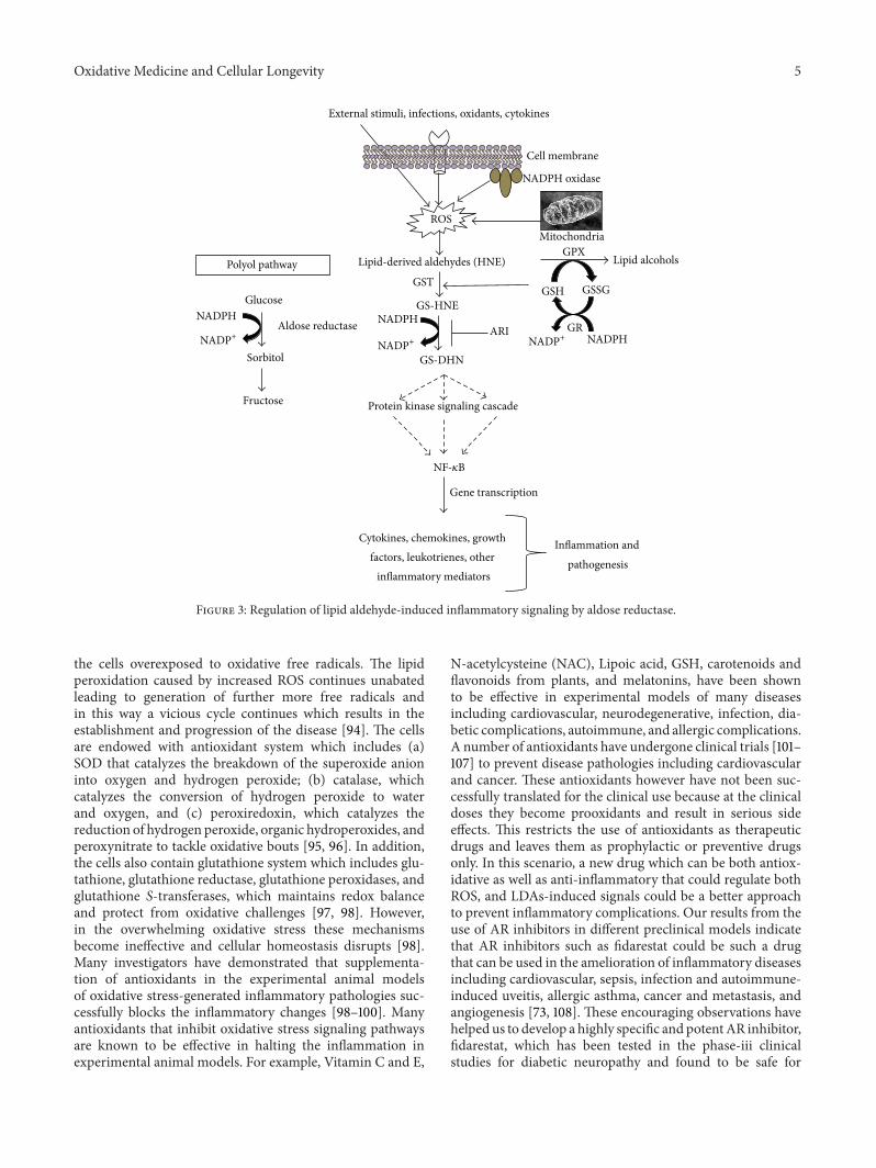
The cell signaling pathways activated by LDAs includenoncovalent mechanisms that involve binding to a pro-tein receptor and covalent mechanisms that modify proteinkinases directly. The direct interaction of LDAs with proteinkinases such as SRC and PLC could change cellular calciumsignaling and alter in-situ cation mobilization leading toactivation of the caspases involved in cell death. Vatsyayanet al. [6] have shown that HNE besides being cytotoxic, atlower doses it triggers phosphorylation of epidermal growthfactor receptor (EGFR) and activation of its downstreamsignaling components ERK1/2 and AKT which are knownto be involved in cell proliferation. Similarly, Chaudharyet al. [69] concluded that HNE could evoke signaling fordefense mechanisms to self-regulate its toxicity and simul-taneously may affect multiple signaling pathways throughits interactions with membrane receptors and transcriptionfactors/repressors [57]. Dwivedi et al. [70] suggested thatHNE has concentration-dependent opposing effects to celldeath and growth. This report indicates that constitutivelevels of HNE are needed for normal cell functions and lowlevels of HNE promote proliferative machinery while highlevels promote apoptotic signaling. Further, they suggestedthat HNE can modulate ligand-independent signaling bymembrane receptors such as EGFR or Fas (CD95) andmay act as a sensor of external stimuli for eliciting stress-response, suggesting a key role of HNE in cellular signaling[70]. Furthermore, our laboratory has presented numerousevidences [71–73] that GS-LDAswhen enzymatically reducedby AR, become important secondary messengers to activatesignaling molecules that eventually leads to activation of NF-𝜅B via yet unknown mechanism (Figure 3). The regulationof NF-𝜅B activation is the key for LDAs to modulate variousinflammatory pathologies. Since NF-𝜅B transcribes variousproinflammatory cytokines, growth factors, cell survival pro-teins, structural proteins, and apoptotic proteins, regulationof intracellular concentrations of LDAs could be novel strat-egy to prevent inflammatory complications. Indeed, recentstudies indicate that enzymes that regulate LDAs generationor participate in their metabolism could actually preventpathological effects of LDA-induced inflammatory complica-tions [2–5, 73].
5. Activation of NF-𝜅B under Oxidative Stress
Increased oxidative stress is a hallmark of inflammatorydiseases such as those caused by infections, xenobiotics,environmental pollutants, and those of autoimmune etiology,and ROS is an essential mediator of intracellular signalingunder a variety of conditions [74–79]. Several lines of evi-dence suggest that ROS mediates the activation of redox-sensitive transcription factors such as NF-𝜅B and AP1, whichin turn stimulate the expression of an array of inflammatory
cytokines and chemokines genes [80, 81]. These evidenceslargely suggest the use of a variety of antioxidants that inhibitNF-𝜅B activation and also antioxidant enzymes which areoverexpressed as a counter mechanism to bring down theNF-𝜅B activation. Excessive and unrestrained production ofinflammatory mediators causes cytotoxicity in an autocrineand paracrine manner. Among various redox-sensitive tran-scription factors such as NF-𝜅B, AP1, CREB, ERF2, NFAT,and ATF2 that are activated by increased ROS levels, NF-𝜅Bhas been extensively studied and is implicated inmany oxida-tive stress-induced inflammatory diseases [45–49]. Activa-tion of NF-𝜅B under oxidative stress has been noticed ina number of inflammatory complications. However, howexactly ROS activates various protein kinases upstream toNF-𝜅B is not known clearly. In the preceding section, wepresented evidence that suggests that ROS-generated HNEand other LDAs may directly or indirectly activate upstreamkinases including tyrosine receptor kinases [6, 68].
We and others have also presentedmany evidences whichimplicate ROS-induced lipid peroxidation products such aslipid aldehydes in the activation of signaling cascade thateventually activates NF-𝜅B [70, 73, 82, 83]. Indeed, ROS-induced lipid peroxidation has been proposed to be majorcontributor in the pathophysiology of many inflammatorydisorders. HNE, generated by a number of oxidative stressconditions inducing etiologies including bacterial infection,xenobiotics, environmental pollutants, and autoimmune dis-orders, is one of the highly abundant and toxic lipid aldehydes[84]. Acrolein is another highly reactive species generatedboth exo- and endogenously. Depending upon their respec-tive concentrations both HNE and acrolein elicit phenotyp-ically varied outcomes. Both are known to activate variousupstream kinases including MAPK and PKC [71, 82, 85–89],thus enzymes that either regulate or metabolize HNE couldbe mediators of oxidative stress signals. Modification of mul-tiple cytoskeletal proteins and the activation of MAPKs andROS-sensitive transcription factors by HNE metabolizingenzymes have been demonstrated by various investigators [71,82]. Further, growth factors and cytokine-induced increasedgeneration of ROS could be an essential step for cell growthbecause overexpression of antioxidants such as catalase andsuper oxide dismutase (SOD) or treatment with antioxidantssuch as N-acetylcysteine (NAC) are known to diminishgrowth factor and cytokine-stimulated cell growth or death[90, 91]. Many studies have indeed indicated that ROSthemselves can act as toxic messengers that activate NF-𝜅Band affect the cellular functions of growth factors, cytokinesand other molecules [92, 93].These evidences clearly indicatethat various stimulants and oxidants activate redox-sensitivetranscription factors including NF-𝜅B by generating ROSwhich in turn generate other secondary messengers suchas lipid aldehydes that phosphorylate upstream signalingkinases.
6. Modulation of Oxidative Stress and LipidAldehydes Signals by Antioxidants
In oxidative stress-induced inflammatory diseases, antioxi-dant status of the tissues undergoes severe alteration leaving
Oxidative Medicine and Cellular Longevity 5
GS-HNE
Lipid-derived aldehydes (HNE)
GS-DHN
ARIAldose reductase
GST
NADPH
Cytokines, chemokines, growth factors, leukotrienes, other
inflammatory mediators
Inflammation and pathogenesis
Protein kinase signaling cascade
Gene transcription
NADPH
Polyol pathway
Glucose
Sorbitol
Fructose
External stimuli, infections, oxidants, cytokines
NADPH oxidase
ROS
Cell membrane
Mitochondria
Lipid alcoholsGPX
GSH GSSG
NADPHGR
NADP+NADP+
NADP+
NF-𝜅B
Figure 3: Regulation of lipid aldehyde-induced inflammatory signaling by aldose reductase.
the cells overexposed to oxidative free radicals. The lipidperoxidation caused by increased ROS continues unabatedleading to generation of further more free radicals andin this way a vicious cycle continues which results in theestablishment and progression of the disease [94]. The cellsare endowed with antioxidant system which includes (a)SOD that catalyzes the breakdown of the superoxide anioninto oxygen and hydrogen peroxide; (b) catalase, whichcatalyzes the conversion of hydrogen peroxide to waterand oxygen, and (c) peroxiredoxin, which catalyzes thereduction of hydrogen peroxide, organic hydroperoxides, andperoxynitrate to tackle oxidative bouts [95, 96]. In addition,the cells also contain glutathione system which includes glu-tathione, glutathione reductase, glutathione peroxidases, andglutathione S-transferases, which maintains redox balanceand protect from oxidative challenges [97, 98]. However,in the overwhelming oxidative stress these mechanismsbecome ineffective and cellular homeostasis disrupts [98].Many investigators have demonstrated that supplementa-tion of antioxidants in the experimental animal modelsof oxidative stress-generated inflammatory pathologies suc-cessfully blocks the inflammatory changes [98–100]. Manyantioxidants that inhibit oxidative stress signaling pathwaysare known to be effective in halting the inflammation inexperimental animal models. For example, Vitamin C and E,
N-acetylcysteine (NAC), Lipoic acid, GSH, carotenoids andflavonoids from plants, and melatonins, have been shownto be effective in experimental models of many diseasesincluding cardiovascular, neurodegenerative, infection, dia-betic complications, autoimmune, and allergic complications.A number of antioxidants have undergone clinical trials [101–107] to prevent disease pathologies including cardiovascularand cancer. These antioxidants however have not been suc-cessfully translated for the clinical use because at the clinicaldoses they become prooxidants and result in serious sideeffects. This restricts the use of antioxidants as therapeuticdrugs and leaves them as prophylactic or preventive drugsonly. In this scenario, a new drug which can be both antiox-idative as well as anti-inflammatory that could regulate bothROS, and LDAs-induced signals could be a better approachto prevent inflammatory complications. Our results from theuse of AR inhibitors in different preclinical models indicatethat AR inhibitors such as fidarestat could be such a drugthat can be used in the amelioration of inflammatory diseasesincluding cardiovascular, sepsis, infection and autoimmune-induced uveitis, allergic asthma, cancer and metastasis, andangiogenesis [73, 108]. These encouraging observations havehelped us to develop a highly specific and potentAR inhibitor,fidarestat, which has been tested in the phase-iii clinicalstudies for diabetic neuropathy and found to be safe for
6 Oxidative Medicine and Cellular Longevity
human use, as a potential antioxidative, anti-inflammatory,antiangiogenic, antimitogenic, and chemopreventive drug forpreventing inflammatory diseases such as allergic asthma,colon cancer, and uveitis.
7. Role of AR in Mediation ofLipid Aldehydes Signals
It is well known that AR is overexpressed under oxidativestress initiated by various cytokines, growth factors, bacterialendotoxins such as lipopolysaccharides (LPS), and high glu-cose. Overexpression of AR increases the turnover of ROS-generated LDAs [73, 108].TheLDAs readily reactwith cellularGSH and form GSH-LDA conjugates which are excellent ARsubstrates [109, 110]. Since ROS is known to mediate and reg-ulate intracellular signaling under diverse conditions, some ofthe effects of ROS can bemediated by ROS-derived LDAs andtheir GSH conjugates. We have systematically investigatedthe involvement of lipid aldehydes and their GSH conjugatesin the mediation of signaling cascades that play importantrole in the pathophysiology of a number of diseases. Theresults from our studies indicate that reduction of LDAs andtheir GSH conjugates by AR is essential for transductionof cytotoxic signals [82–84]. This was firmly confirmed byvarious evidences including that AR has poor affinity forglucose (Km of 50–100mM), and in normal conditions onlya small percentage (3%) of glucose is metabolized by AR[111]. Further, the kinetic and structural properties of ARare unlike those of other glucose-metabolizing enzymes [112,113]. The low Km for catalysis of carbohydrate reduction isprobably due to high hydrophobicity of the substrate bindingdomain of AR which essentially prevents efficient reductionof glucose by AR and suggests that hydrophobic aldehydescould likely be the preferred substrates. Indeed, we haveunequivocally demonstrated that AR efficiently catalyzeslipid aldehydes and their GSH conjugates [109, 110]. Forexample, recombinant humanAR has been shown to catalyzethe reduction of a large series of saturated and unsaturatedaldehydes with 1000-fold higher efficiency when comparedto glucose [109–112]. Medium- to long-chain (C-6 to C-18)aldehydes, which are generated during lipid peroxidation,are most efficiently catalyzed by AR [109]. In particular, thecatalytic site of AR has more affinity towards GS-aldehydeconjugates than parent aldehydes. This is confirmed by site-directed mutagenesis studies showing that AR active site hasamino acid residues which efficiently bind with glutathionemoiety [109]. Further, molecular modeling of AR-GSH ana-log crystal structure confirmed that AR has a specific GS-aldehyde binding site at its catalytic site [112]. Thus, theseobservations firmly support that physiological role of ARcould be the reduction of LDAs besides glucose metabolismin polyol pathway.
Since LDAs are known to modulate the cellular functionby regulating the oxidative stress signals mediated by NF-𝜅B and AP1 [114–118], we postulated that AR regulatescellular functions by modulating oxidative stress signals.Our claim is supported by the studies demonstrating that
AR inhibition prevents HNE-, growth factor-, and cytokine-induced cytotoxicity in a variety of cultured cells [73, 108].Further, our studies also demonstrated that inhibition ofAR prevents endotoxin-, allergen-, cytokine-, and growthfactor-induced activation of NF-𝜅B signals (Figure 3). In allthese conditions, increased ROS levels, lipid peroxidation,and subsequent formation of lipid aldehydes play amajor rolein disrupting the cellular homeostasis.Thus, our observationspresent a basis for the novel role of AR in the pathophysiologyof various inflammatory disease processesmediated by LDAs.
Besides the novel role implicating AR in mediatingsignaling of the ROS-derived lipid aldehydes in inflamma-tory pathologies, the exact role of AR in the mediation ofcellular signaling is not yet clear. How AR-catalyzed productof GS-LDAs activates PKC and PLC isozymes still needsto be investigated. Nevertheless, we have shown that AR-catalyzed reduced product of GS-DHN activates variouskinases including PLC, PKC, and PI3K in cultured cells.Activation of these kinases eventually results in the activationof redox-sensitive transcription factors which transcribevarious inflammatory genes responsible for disease progres-sion. The involvement of AR-catalyzed reaction products ineliciting oxidative stress-induced cytotoxicity and inflamma-tion is more obvious given the fact that inhibition of ARprevents the increased synthesis of inflammatory cytokinesand chemokines by various oxidant stimuli such as LPS,cytokines, growth factors, and high glucose. This is furthersubstantiated by our demonstration that HNE, GS-HNE,and AR-catalyzed reduced product of GS-LDAs, that is, GS-DHN promote, VSMC growth in vitro [71]. AR inhibitionby pharmacological inhibitors or ablation by siRNA preventsHNE- and GS-HNE-induced VSMC proliferation but hasno effect on the GS-DHN-induced changes. These studiesthus suggest that the reduced forms of lipid-aldehyde glu-tathione conjugates (such as GS-DHN) could be involved inthe oxidative stress-induced inflammatory signaling. Furtherstudies are required to investigate the exact mechanism ofGS-DHN-induced activation of upstream kinases that resultsin transcription of inflammatory genes. Nevertheless, ourobservations have opened up a new area of research inunderstanding the role of AR in the mediation of oxidativestress signaling in a number of inflammatory diseases.
8. Current and Future Developments
Oxidative stress-induced generation of lipid aldehydes hasbeen observed in many inflammatory complications includ-ing cardiovascular disorders, bacterial sepsis, cancer, andasthma, which present enormous clinical challenges world-wide. Many researchers have presented the immense impor-tance of this aspect of pathophysiology, and therefore there isan urgent need for development of new therapeutic strategiestargeting the intervention in lipid aldehyde-mediated inflam-matory signals in these diseases. However, a more preciseelucidation of lipid aldehyde-induced inflammatory signalingis crucial for understanding the pathophysiology of multiplediseases including infections, atherosclerosis, autoimmune,and cancer. Based on these elucidations, a better therapeutic

Oxidative Medicine and Cellular Longevity 7
intervention could be developed to contain the inflammatoryresponses in patients. Our extensive research during recentyears has identified that oxidative stress-induced LDAs andtheir GS-conjugates catalyzed by AR play a major rolein the mediation of NF-𝜅B-induced inflammatory signalsvia PLC/PKC/IKK/MAPK pathways. We have demonstratedthat in experimental animal models, inhibition of GS-LDAmetabolizing enzymes, specifically AR, prevents inflamma-tory diseases such as uveitis, sepsis, colon cancer, atheroscle-rosis, and allergic asthma. These results have delineated anovel mechanism regulating inflammation and have laidfoundation for future studies that could result in clinicalapplication of AR inhibitors. Further, a better understandingof the role ofAR-catalyzed LDAs and theirGSH-conjugates inthe signalosome and inflammasome signaling pathways willbetter reveal underlying pathophysiological events in variousinflammatory diseases.
Abbreviations
AR: Aldose reductaseARI: AR inhibitorROS: Reactive oxygen speciesLDAs: Lipid peroxidation-derived aldehydesMDA: malondialdehydeHNE: 4-hydroxynonenalGSH: Reduced glutathioneGS-LDAs: Glutathione-lipid-derived aldehydesGS-HNE: Glutathione-HNE conjugateGS-DHN: Glutathione-1,4-dihydroxynonene conjugateLPS: LipopolysacharidesNADPH: Reduced nicotinamide adenine dinucleotide
phosphatePKC: Protein kinase CPLC: Phospholipase CMAPK: Mitogen-activated protein kinaseNF-𝜅B: Nuclear factor kappa BAP-1: Activator protein 1CREB: cAMP response element-binding proteinVSMC: Vascular smooth muscle cellsSOD: Superoxide dismutaseNAC: N-acetylcysteineAMD: Age-related macular degenerationEGFR: Epidermal growth factor receptor.
References
[1] N. J. Pillon, M. L. Croze, R. E. Vella, L. Soulere, M. Lagarde, andC.O. Soulage, “The lipid peroxidation by-product 4-hydroxy-2-nonenal (4-HNE) induces insulin resistance in skeletal musclethrough both carbonyl and oxidative stress,” Endocrinology, vol.153, no. 5, pp. 2099–2111, 2012.
[2] M. P. Mattson, “Roles of the lipid peroxidation product 4-hydroxynonenal in obesity, the metabolic syndrome, and asso-ciated vascular and neurodegenerative disorders,” ExperimentalGerontology, vol. 44, no. 10, pp. 625–633, 2009.
[3] C. S. Kang, H. W. Kim, B. K. Kim et al., “Hepatotoxicity andnephrotoxicity produced by 4-hydroxy-2-nonenal (4-HNE)following 4-week oral administration to sprague-dawley rats,”
Journal of Toxicology and Environmental Health A, vol. 74, no.12, pp. 779–789, 2011.
[4] P. Karihtala, S. Kauppila, U. Puistola, and A. Jukkola-Vu-orinen, “Divergent behaviour of oxidative stress markers 8-hydroxydeoxyguanosine (8-OHdG) and 4-hydroxy-2-nonenal(HNE) in breast carcinogenesis,” Histopathology, vol. 58, no. 6,pp. 854–862, 2011.
[5] K. Uno, K. Kato, G. Kusaka, N. Asano, K. Iijima, and T.Shimosegawa, “The balance between 4-hydroxynonenal andintrinsic glutathione/glutathione S-transferase A4 system maybe critical for the epidermal growth factor receptor phos-phorylation of human esophageal squamous cell carcinomas,”Molecular Carcinogenesis, vol. 50, no. 10, pp. 781–790, 2011.
[6] R. Vatsyayan, P. Chaudhary, A. Sharma et al., “Role of 4-hydroxynonenal in epidermal growth factor receptor-mediatedsignaling in retinal pigment epithelial cells,” Experimental EyeResearch, vol. 92, no. 2, pp. 147–154, 2011.
[7] S. Qin and G. A. Rodrigues, “Differential roles of AMPK𝛼1and AMPK𝛼2 in regulating 4-HNE-induced RPE cell death andpermeability,” Experimental Eye Research, vol. 91, no. 6, pp. 818–824, 2010.
[8] M. Singh, T. N. Dang, M. Arseneault, and C. Ramassamy, “Roleof by-products of lipid oxidation in Alzheimer’s disease brain: afocus on acrolein,” Journal of Alzheimer’s Disease, vol. 21, no. 3,pp. 741–756, 2010.
[9] D. A. Butterfield, M. L. Bader Lange, and R. Sultana, “Involve-ments of the lipid peroxidation product, HNE, in the patho-genesis and progression of Alzheimer’s disease,” Biochimica etBiophysica Acta, vol. 1801, no. 8, pp. 924–929, 2010.
[10] W. Volkel, T. Sicilia, A. Pahler et al., “Increased brain lev-els of 4-hydroxy-2-nonenal glutathione conjugates in severeAlzheimer’s disease,” Neurochemistry International, vol. 48, no.8, pp. 679–686, 2006.
[11] M. Fukai, T. Hayashi, R. Yokota et al., “Lipid peroxidationduring ischemia depends on ischemia time in warm ischemiaand reperfusion of rat liver,” Free Radical Biology and Medicine,vol. 38, no. 10, pp. 1372–1381, 2005.
[12] E. McCracken, D. I. Graham, M. Nilsen, J. Stewart, J. A. R.Nicoll, and K. Horsburgh, “4-hydroxynonenal immunoreactiv-ity is increased in human hippocampus after global ischemia,”Brain Pathology, vol. 11, no. 4, pp. 414–421, 2001.
[13] H. Esterbauer, R. J. Schaur, and H. Zollner, “Chemistry andbiochemistry of 4-hydroxynonenal, malonaldehyde and relatedaldehydes,” Free Radical Biology and Medicine, vol. 11, no. 1, pp.81–128, 1991.
[14] A. Loidl-Stahlhofen, K. Hannemann, and G. Spiteller, “Gener-ation of 𝛼-hydroxyaldehydic compounds in the course of lipidperoxidation,” Biochimica et Biophysica Acta, vol. 1213, no. 2, pp.140–148, 1994.
[15] R. M. LoPachin, T. Gavin, D. R. Petersen, and D. S. Barber,“Molecular mechanisms of 4-hydroxy-2-nonenal and acroleintoxicity: nucleophilic targets and adduct formation,” ChemicalResearch in Toxicology, vol. 22, no. 9, pp. 1499–1508, 2009.
[16] A. T. Jacobs and L. J. Marnett, “Systems analysis of proteinmodification and cellular responses induced by electrophilestress,” Accounts of Chemical Research, vol. 43, no. 5, pp. 673–683, 2010.
[17] G. Juric-Sekhar, K. Zarkovic, G. Waeg, A. Cipak, and N.Zarkovic, “Distribution of 4-hydroxynonenal-protein conju-gates as a marker of lipid peroxidation and parameter ofmalignancy in astrocytic and ependymal tumors of the brain,”Tumori, vol. 95, no. 6, pp. 762–768, 2009.
8 Oxidative Medicine and Cellular Longevity
[18] U. C. S. Yadav, K. V. Ramana, Y. C. Awasthi, and S. K.Srivastava, “Glutathione level regulates HNE-induced genotox-icity in human erythroleukemia cells,” Toxicology and AppliedPharmacology, vol. 227, no. 2, pp. 257–264, 2008.
[19] N. Zarkovic, K. Zarkovic, R. J. Schaur et al., “4-hydroxynonenalas a second messenger of free radicals and growth modifyingfactor,” Life Sciences, vol. 65, no. 18-19, pp. 1901–1904, 1999.
[20] C. A. Thompson and P. C. Burcham, “Genome-wide transcrip-tional responses to acrolein,” Chemical Research in Toxicology,vol. 21, no. 12, pp. 2245–2256, 2008.
[21] Y. S. Park and N. Taniguchi, “Acrolein induces inflammatoryresponse underlying endothelial dysfunction: a risk factor foratherosclerosis,” Annals of the New York Academy of Sciences,vol. 1126, pp. 185–189, 2008.
[22] C. C. Wu, C. W. Hsieh, P. H. Lai, J. B. Lin, Y. C. Liu, andB. S. Wung, “Upregulation of endothelial heme oxygenase-1 expression through the activation of the JNK pathway bysublethal concentrations of acrolein,” Toxicology and AppliedPharmacology, vol. 214, no. 3, pp. 244–252, 2006.
[23] J. Roy, P. Pallepati, A. Bettaieb, A. Tanel, and D. A. Averill-Bates, “Acrolein induces a cellular stress response and triggersmitochondrial apoptosis in A549 cells,” Chemico-BiologicalInteractions, vol. 181, no. 2, pp. 154–167, 2009.
[24] J. D. Lambeth, “NOX enzymes and the biology of reactiveoxygen,” Nature Reviews Immunology, vol. 4, no. 4, pp. 181–189,2004.
[25] K. Bedard and K. H. Krause, “The NOX family of ROS-generatingNADPHoxidases: physiology and pathophysiology,”Physiological Reviews, vol. 87, no. 1, pp. 245–313, 2007.
[26] K. Datta, S. Sinha, and P. Chattopadhyay, “Reactive oxygenspecies in health and disease,”NationalMedical Journal of India,vol. 13, no. 6, pp. 304–310, 2000.
[27] M. Valko, D. Leibfritz, J. Moncol, M. T. D. Cronin, M. Mazur,and J. Telser, “Free radicals and antioxidants in normal physi-ological functions and human disease,” International Journal ofBiochemistry and Cell Biology, vol. 39, no. 1, pp. 44–84, 2007.
[28] M. Vuillaume, “Reduced oxygen species, mutation, inductionand cancer initiation,”Mutation Research, vol. 186, no. 1, pp. 43–72, 1987.
[29] B. S. Berlett and E. R. Stadtman, “Protein oxidation in aging,disease, and oxidative stress,” Journal of Biological Chemistry,vol. 272, no. 33, pp. 20313–20316, 1997.
[30] M. L. Circu and T. Y. Aw, “Reactive oxygen species, cellu-lar redox systems, and apoptosis,” Free Radical Biology andMedicine, vol. 48, no. 6, pp. 749–762, 2010.
[31] H. Bartsch and J. Nair, “Oxidative stress and lipid peroxidation-derived DNA-lesions in inflammation driven carcinogenesis,”Cancer Detection and Prevention, vol. 28, no. 6, pp. 385–391,2004.
[32] A. Catala, “An overview of lipid peroxidation with emphasis inouter segments of photoreceptors and the chemiluminescenceassay,” International Journal of Biochemistry and Cell Biology,vol. 38, no. 9, pp. 1482–1495, 2006.
[33] M. Parola, G. Bellomo, G. Robino, G. Barrera, and M. U.Dianzani, “4-hydroxynonenal as a biological signal: molecularbasis and pathophysiological implications,” Antioxidants andRedox Signaling, vol. 1, no. 3, pp. 255–284, 1999.
[34] K. Uchida, “Current status of acrolein as a lipid peroxidationproduct,” Trends in Cardiovascular Medicine, vol. 9, no. 5, pp.109–113, 1999.
[35] G. R. Buettner, “The pecking order of free radicals and antioxi-dants: lipid peroxidation,𝛼-tocopherol, and ascorbate,”Archivesof Biochemistry and Biophysics, vol. 300, no. 2, pp. 535–543, 1993.
[36] S. Nigam and T. Schewe, “Phospholipase A2s and lipid peroxi-dation,”Biochimica et Biophysica Acta, vol. 1488, no. 1-2, pp. 167–181, 2000.
[37] H. Rubbo, S. Parthasarathy, S. Barnes, M. Kirk, B. Kalyanar-aman, and B. A. Freeman, “Nitric oxide inhibition oflipoxygenase-dependent liposome and low- density lipoproteinoxidation: termination of radical chain propagation reactionsand formation of nitrogen-containing oxidized lipid deriv-atives,” Archives of Biochemistry and Biophysics, vol. 324, no. 1,pp. 15–25, 1995.
[38] S. Jongberg, C. U. Carlsen, and L. H. Skibsted, “Peptidesas antioxidants and carbonyl quenchers in biological modelsystems,” Free Radical Research, vol. 43, no. 10, pp. 932–942,2009.
[39] R. Sharma, D. Brown, S. Awasthi et al., “Transfection with 4-hy-droxynonenal-metabolizing glutathione S-transferase isozymesleads to phenotypic transformation and immortalization ofadherent cells,” European Journal of Biochemistry, vol. 271, no.9, pp. 1690–1701, 2004.
[40] J. Z. Cheng, S. S. Singhal, A. Sharma et al., “Transfection ofmGSTA4 in HL-60 cells protects against 4-hydroxynonenal-induced apoptosis by inhibiting JNK-mediated signaling,”Archives of Biochemistry and Biophysics, vol. 392, no. 2, pp. 197–207, 2001.
[41] S. K. Srivastava, S. S. Singhal, S. Awasthi, S. Pikula, N. H.Ansari, andY.C.Awasthi, “A glutathione S-transferases isozyme(bGST 5.8) involved in the metabolism of 4-hydroxy-2-trans-nonenal is localized in bovine lens epithelium,” ExperimentalEye Research, vol. 63, no. 3, pp. 329–337, 1996.
[42] M. Comporti, “Lipid peroxidation and cellular damage in toxicliver injury,” Laboratory Investigation, vol. 53, no. 6, pp. 599–623,1985.
[43] P. A. Baeuerle and T. Henkel, “Function and activation of NF-kappa B in the immune system,”Annual Review of Immunology,vol. 12, pp. 141–179, 1994.
[44] P. A. Baeuerle and D. Baltimore, “Nf-𝜅B: ten years after,” Cell,vol. 87, no. 1, pp. 13–20, 1996.
[45] P. J. Barnes and M. Karin, “Nuclear factor-𝜅B—a pivotaltranscription factor in chronic inflammatory diseases,” NewEngland Journal ofMedicine, vol. 336, no. 15, pp. 1066–1071, 1997.
[46] S. Ghosh, M. J. May, and E. B. Kopp, “NF-𝜅B and Rel proteins:evolutionarily conserved mediators of immune responses,”Annual Review of Immunology, vol. 16, pp. 225–260, 1998.
[47] W. C. Sha, “Regulation of immune responses by NF-𝜅B/Reltranscription factors,” Journal of ExperimentalMedicine, vol. 187,no. 2, pp. 143–146, 1998.
[48] M. Grossmann, Y. Nakamura, R. Grumont, and S. Gerondakis,“New insights into the roles of ReL/NF-𝜅B transcription factorsin immune function, hemopoiesis and human disease,” Interna-tional Journal of Biochemistry and Cell Biology, vol. 31, no. 10, pp.1209–1219, 1999.
[49] G. Poli, F. Biasi, E. Chiaprotto, M. U. Dianzani, A. de Luca, andH. Esterbauer, “Lipid peroxidation in human diseases: evidenceof red cell oxidative stress after circulatory shock,” Free RadicalBiology and Medicine, vol. 6, no. 2, pp. 167–170, 1989.
[50] M. Polak and Z. Zagorski, “Lipid peroxidation in diabeticretinopathy,” Annales Universitatis Mariae Curie-Sklodowska.Sectio D: Medicina, vol. 59, no. 1, pp. 434–437, 2004.

Oxidative Medicine and Cellular Longevity 9
[51] A. Higdon, A. R. Diers, J. Y. Oh, A. Landar, and V. M. Darley-Usmar, “Cell signalling by reactive lipid species: new conceptsand molecular mechanisms,” Biochemical Journal, vol. 442, no.3, pp. 453–464, 2012.
[52] J. Ruef, M. Moser, C. Bode, W. Kubler, and M. S. Runge,“4-hydroxynonenal induces apoptosis, NF-𝜅B-activation andformation of 8-isoprostane in vascular smooth muscle cells,”Basic Research in Cardiology, vol. 96, no. 2, pp. 143–150, 2001.
[53] S. P. Faux and P. J. Howden, “Possible role of lipid peroxidationin the induction of NF-kappa B and AP-1 in RFL-6 cells bycrocidolite asbestos: evidence following protection by vitaminE,” Environmental Health Perspectives, vol. 105, 5, pp. 1127–1130,1997.
[54] A. L. Levonen, A. Landar, A. Ramachandran et al., “Cellularmechanisms of redox cell signalling: role of cysteine mod-ification in controlling antioxidant defences in response toelectrophilic lipid oxidation products,” Biochemical Journal, vol.378, part 2, pp. 373–382, 2004.
[55] E. K. Ceaser, D. R. Moellering, S. Shiva et al., “Mechanismsof signal transduction mediated by oxidized lipids: the roleof the electrophile-responsive proteome,” Biochemical SocietyTransactions, vol. 32, part 1, pp. 151–155, 2004.
[56] J. W. Zmijewski, A. Landar, N. Watanabe, D. A. Dickinson,N. Noguchi, and V. M. Darley-Usmar, “Cell signalling byoxidized lipids and the role of reactive oxygen species in theendothelium,” Biochemical Society Transactions, vol. 33, part 6,pp. 1385–1389, 2005.
[57] D. A. Dickinson, V. M. Darley-Usmar, and A. Landar, “Thecovalent advantage: a new paradigm for cell signaling by thiolreactive lipid oxidation products,” in Redox Proteomics: FromProtein Modifications to Cellular Dysfunction and Diseases, I.Dalle-Donne, A. Scalone, and D. A. Butterfield, Eds., pp. 345–367, John Wiley & Sons, Indianapolis, Ind, USA, 2006.
[58] K. Stamatakis and D. Perez-Sala, “Prostanoids with cyclopen-tenone structure as tools for the characterization of electrophiliclipid-protein interactomes,” Annals of the New York Academy ofSciences, vol. 1091, pp. 548–570, 2006.
[59] J. A. Doorn and D. R. Petersen, “Covalent modification ofamino acid nucleophiles by the lipid peroxidation products 4-hydroxy-2-nonenal and 4-oxo-2-nonenal,” Chemical Researchin Toxicology, vol. 15, no. 11, pp. 1445–1450, 2002.
[60] K. Uchida, “4-hydroxy-2-nonenal: a product and mediator ofoxidative stress,” Progress in Lipid Research, vol. 42, no. 4, pp.318–343, 2003.
[61] A. L. Isom, S. Barnes, L. Wilson, M. Kirk, L. Coward, and V.Darley-Usmar, “Modification of cytochrome c by 4-hydroxy-2-nonenal: evidence for histidine, lysine, and arginine-aldehydeadducts,” Journal of the American Society for Mass Spectrometry,vol. 15, no. 8, pp. 1136–1147, 2004.
[62] E. Chiarpotto, C. Domenicotti, D. Paola et al., “Regulation of rathepatocyte protein kinase C 𝛽 isoenzymes by the lipid peroxi-dation product 4-hydroxy-2,3-nonenal: a signaling pathway tomodulate vesicular transport of glycoproteins,”Hepatology, vol.29, no. 5, pp. 1565–1572, 1999.
[63] M. Nitti, C. Domenicotti, C. D’Abramo et al., “Activation ofPKC-𝛽 isoforms mediates HNE-induced MCP-1 release bymacrophages,” Biochemical and Biophysical Research Commu-nications, vol. 294, no. 3, pp. 547–552, 2002.
[64] M. Parola, G. Robino, F. Marra et al., “HNE interacts directlywith JNK isoforms in human hepatic stellate cells,” Journal ofClinical Investigation, vol. 102, no. 11, pp. 1942–1950, 1998.
[65] B. J. Song, Y. Soh, M. A. Bae, J. E. Pie, J. Wan, and K. S. Jeong,“Apoptosis of PC12 cells by 4-hydroxy-2-nonenal is mediatedthrough selective activation of the c-Jun N-Terminal proteinkinase pathway,” Chemico-Biological Interactions, vol. 130–132,pp. 943–954, 2001.
[66] N. Shibata, Y. Kato, Y. Inose et al., “4-hydroxy-2-nonenalupregulates and phosphorylates cytosolic phospholipase A2in cultured Ra2 microglial cells via MAPK pathways,” Neu-ropathology, vol. 31, no. 2, pp. 122–128, 2011.
[67] S. J. Lee, C. E. Kim, M. R. Yun et al., “4-hydroxynonenalenhances MMP-9 production in murine macrophages via 5-lipoxygenase-mediated activation of ERK and p38 MAPK,”Toxicology and Applied Pharmacology, vol. 242, no. 2, pp. 191–198, 2010.
[68] S. Saito, G. D. Frank, M. Mifune et al., “Ligand-independenttrans-activation of the platelet-derived growth factor receptorby reactive oxygen species requires protein kinase C-𝛿 and c-Src,” Journal of Biological Chemistry, vol. 277, no. 47, pp. 44695–44700, 2002.
[69] P. Chaudhary, R. Sharma, A. Sharma et al., “Mechanisms of 4-hydroxy-2-nonenal induced pro- and anti-apoptotic signaling,”Biochemistry, vol. 49, no. 29, pp. 6263–6275, 2010.
[70] S. Dwivedi, A. Sharma, B. Patrick, R. Sharma, and Y. C. Awasthi,“Role of 4-hydroxynonenal and its metabolites in signaling,”Redox Report, vol. 12, no. 1-2, pp. 4–10, 2007.
[71] K. V. Ramana, A. Bhatnagar, S. Srivastava et al., “Mitogenicresponses of vascular smoothmuscle cells to lipid peroxidation-derived aldehyde 4-hydroxy-trans-2-nonenal (HNE): role ofaldose reductase-catalyzed reduction of the HNE-glutathioneconjugates in regulating cell growth,” Journal of BiologicalChemistry, vol. 281, no. 26, pp. 17652–17660, 2006.
[72] K. V. Ramana, M. S. Willis, M. D. White et al., “Endotoxin-induced cardiomyopathy and systemic inflammation in mice isprevented by aldose reductase inhibition,” Circulation, vol. 114,no. 17, pp. 1838–1846, 2006.
[73] K. V. Ramana, “Aldose reductase: new insights for an oldenzyme,”BioMolecular Concepts, vol. 2, no. 1-2, pp. 103–114, 2011.
[74] H. J. Forman, M. Maiorino, and F. Ursini, “Signaling functionsof reactive oxygen species,” Biochemistry, vol. 49, no. 5, pp. 835–842, 2010.
[75] J. Segovia, A. Sabbah, V. Mgbemena et al., “TLR2/MyD88/NF-𝜅B pathway, reactive oxygen species, potassium efflux activatesNLRP3/ASC inflammasome during respiratory syncytial virusinfection,” PLoS One, vol. 7, no. 1, Article ID e29695, 2012.
[76] S. Mena, A. Ortega, and J. M. Estrela, “Oxidative stressin environmental-induced carcinogenesis,” Mutation Research,vol. 674, no. 1-2, pp. 36–44, 2009.
[77] A.Auerbach andM. L.Hernandez, “The effect of environmentaloxidative stress on airway inflammation,” Current Opinion inAllergy and Clinical Immunology, vol. 12, no. 2, pp. 133–139, 2012.
[78] K. J. Chuang, C. C. Chan, T. C. Su, C. T. Lee, and C. S.Tang, “The effect of urban air pollution on inflammation,oxidative stress, coagulation, and autonomic dysfunction inyoung adults,”American Journal of Respiratory and Critical CareMedicine, vol. 176, no. 4, pp. 370–376, 2007.
[79] E. Profumo, B. Buttari, and R. Rigano, “Oxidative stress incardiovascular inflammation: its involvement in autoimmuneresponses,” International Journal of Inflammation, vol. 2011,Article ID 295705, 6 pages, 2011.
[80] V. J. Thannickal and B. L. Fanburg, “Reactive oxygen species incell signaling,” American Journal of Physiology—Lung CellularandMolecular Physiology, vol. 279, no. 6, pp. L1005–L1028, 2000.

10 Oxidative Medicine and Cellular Longevity
[81] P. D. Ray, B. W. Huang, and Y. Tsuji, “Reactive oxygen species(ROS) homeostasis and redox regulation in cellular signaling,”Cellular Signalling, vol. 24, no. 5, pp. 981–990, 2012.
[82] K. V. Ramana, A. A. Fadl, R. Tammali, A. B. M. Reddy, A.K. Chopra, and S. K. Srivastava, “Aldose reductase mediatesthe lipopolysaccharide-induced release of inflammatory medi-ators in RAW264.7 murine macrophages,” Journal of BiologicalChemistry, vol. 281, no. 44, pp. 33019–33029, 2006.
[83] R. Tammali, K. V. Ramana, S. S. Singhal, S. Awasthi, and S. K.Srivastava, “Aldose reductase regulates growth factor-inducedcyclooxygenase-2 expression and prostaglandin E2 productionin human colon cancer cells,” Cancer Research, vol. 66, no. 19,pp. 9705–9713, 2006.
[84] S. K. Srivastava,U.C. Yadav,A. B. Reddy et al., “Aldose reductaseinhibition suppresses oxidative stress-induced inflammatorydisorders,” Chemico-Biological Interactions, vol. 191, no. 1–3, pp.330–338, 2011.
[85] S. J. Lee, C. E. Kim, K. W. Seo, and C. D. Kim, “HNE-induced5-LO expression is regulated by NF-𝜅B/ERK and Sp1/p38MAPK pathways via EGF receptor in murine macrophages,”Cardiovascular Research, vol. 88, no. 2, pp. 352–359, 2010.
[86] S. J. Lee, K. W. Seo, M. R. Yun et al., “4-hydroxynonenalenhances MMP-2 production in vascular smooth muscle cellsvia mitochondrial ROS-mediated activation of the Akt/NF-𝜅Bsignaling pathways,” Free Radical Biology and Medicine, vol. 45,no. 10, pp. 1487–1492, 2008.
[87] H. Zhang andH. J. Forman, “Acrolein induces heme oxygenase-1 through PKC-𝛿 and PI3K in human bronchial epithelial cells,”American Journal of Respiratory Cell andMolecular Biology, vol.38, no. 4, pp. 483–490, 2008.
[88] A. Tanel and D. A. Averill-Bates, “P38 and ERK mitogen-activated protein kinasesmediate acrolein-induced apoptosis inChinese hamster ovary cells,” Cellular Signalling, vol. 19, no. 5,pp. 968–977, 2007.
[89] H. Zhang and H. J. Forman, “Signaling pathways involvedin phase II gene induction by 𝛼, Β-unsaturated aldehydes,”Toxicology and Industrial Health, vol. 25, no. 4-5, pp. 269–278,2009.
[90] Y. Q. Fu, F. Fang, Z. Y. Lu, F. W. Kuang, and F. Xu, “N-acetylcysteine protects alveolar epithelial cells from hydrogenperoxideinduced apoptosis through scavenging reactive oxygenspecies and suppressing c-JunN-terminal kinase,” ExperimentalLung Research, vol. 36, no. 6, pp. 352–361, 2010.
[91] S. Umar, J. Zargan, K. Umar, S. Ahmad, C. K. Katiyar, and H.A. Khan, “Modulation of the oxidative stress and inflammatorycytokine response by thymoquinone in the collagen inducedarthritis inWistar rats,”Chemico-Biological Interactions, vol. 197,no. 1, pp. 40–46, 2012.
[92] J. M. Muller, R. A. Rupec, and P. A. Baeuerle, “Study of generegulation by NF-𝜅B and AP-1 in response to reactive oxygenintermediates,”Methods, vol. 11, no. 3, pp. 301–312, 1997.
[93] M. Meyer, H. L. Pahl, and P. A. Baeuerle, “Regulation ofthe transcription factors NF-𝜅B and AP-1 by redox changes,”Chemico-Biological Interactions, vol. 91, no. 2-3, pp. 91–100, 1994.
[94] H. Bartsch and J. Nair, “Chronic inflammation and oxidativestress in the genesis and perpetuation of cancer: role of lipidperoxidation, DNA damage, and repair,” Langenbeck’s Archivesof Surgery, vol. 391, no. 5, pp. 499–510, 2006.
[95] P. Amstad and P. Cerutti, “Genetic modulation of the cellularantioxidant defense capacity,” Environmental Health Perspec-tives, vol. 88, pp. 77–82, 1990.
[96] P. Cerutti, R. Ghosh, Y. Oya, and P. Amstad, “The role ofthe cellular antioxidant defense in oxidant carcinogenesis,” En-vironmental Health Perspectives, vol. 102, no. 10, pp. 123–129,1994.
[97] J. H. Limon-Pacheco and M. E. Gonsebatt, “The glutathionesystem and its regulation by neurohormone melatonin in thecentral nervous system,” Central Nervous System Agents inMedicinal Chemistry, vol. 10, no. 4, pp. 287–297, 2010.
[98] N. Ballatori, S. M. Krance, S. Notenboom, S. Shi, K. Tieu, and C.L. Hammond, “Glutathione dysregulation and the etiology andprogression of human diseases,” Biological Chemistry, vol. 390,no. 3, pp. 191–214, 2009.
[99] M. Valko, D. Leibfritz, J. Moncol, M. T. D. Cronin, M. Mazur,and J. Telser, “Free radicals and antioxidants in normal physi-ological functions and human disease,” International Journal ofBiochemistry and Cell Biology, vol. 39, no. 1, pp. 44–84, 2007.
[100] N. Tailor and M. Sharma, “Antioxidant hybrid compounds: apromising therapeutic intervention in oxidative stress induceddiseases,” Mini-Reviews in Medicinal Chemistry, vol. 13, no. 2,pp. 280–297, 2013.
[101] M. H. Hopkins, V. Fedirko, D. P. Jones, P. D. Terry, andR. M. Bostick, “Antioxidant micronutrients and biomarkersof oxidative stress and inflammation in colorectal adenomapatients: results from a randomized, controlled clinical trial,”Cancer Epidemiology Biomarkers and Prevention, vol. 19, no. 3,pp. 850–858, 2010.
[102] Z. Mazloom, N. Hejazi, M. H. Dabbaghmanesh, H. R.Tabatabaei, A. Ahmadi, and H. Ansar, “Effect of vitamin Csupplementation on postprandial oxidative stress and lipidprofile in type 2 diabetic patients,” Pakistan Journal of BiologicalSciences, vol. 14, no. 19, pp. 900–904, 2011.
[103] M.Goodman,R.M.Bostick,O.Kucuk, andD. P. Jones, “Clinicaltrials of antioxidants as cancer prevention agents: past, present,and future,” Free Radical Biology andMedicine, vol. 51, no. 5, pp.1068–1084, 2011.
[104] E. C. Ienco, A. LoGerfo, C. Carlesi et al., “Oxidative stresstreatment for clinical trials in neurodegenerative diseases,”Journal of Alzheimer’s Disease, vol. 24, no. 2, pp. 111–126, 2011.
[105] E. R. Greenberg, J. A. Baron, T. D. Tosteson et al., “A clinical trialof antioxidant vitamins to prevent colorectal adenoma,” NewEngland Journal of Medicine, vol. 331, no. 3, pp. 141–147, 1994.
[106] M. C. Mathew, A. M. Ervin, J. Tao, and R. M. Davis, “Antiox-idant vitamin supplementation for preventing and slowingthe progression of age-related cataract,” Cochrane Database ofSystematic Reviews, vol. 6, Article ID CD004567, 2012.
[107] U. C. S. Yadav, N. M. Kalariya, and K. V. Ramana, “Emergingrole of antioxidants in the protection of uveitis complications,”Current Medicinal Chemistry, vol. 18, no. 6, pp. 931–942, 2011.
[108] S. K. Srivastava, K. V. Ramana, andA. Bhatnagar, “Role of aldosereductase and oxidative damage in diabetes and the consequentpotential for therapeutic options,” Endocrine Reviews, vol. 26,no. 3, pp. 380–392, 2005.
[109] K. V. Ramana, B. L. Dixit, S. Srivastava, G. K. Balendiran,S. K. Srivastava, and A. Bhatnagar, “Selective recognition ofglutathiolated aldehydes by aldose reductase,” Biochemistry, vol.39, no. 40, pp. 12172–12180, 2000.
[110] B. L. Dixit, G. K. Balendiran, S. J. Watowich et al., “Kineticand structural characterization of the glutathione-binding siteof aldose reductase,” Journal of Biological Chemistry, vol. 275,no. 28, pp. 21587–21595, 2000.
[111] A. D. Morrison, R. S. Clements Jr., S. B. Travis, F. Oski, andA. I. Winegrad, “Glucose utilization by the polyol pathway

Oxidative Medicine and Cellular Longevity 11
in human erythrocytes,” Biochemical and Biophysical ResearchCommunications, vol. 40, no. 1, pp. 199–205, 1970.
[112] R. Singh, M. A. White, K. V. Ramana et al., “Structure ofa glutathione conjugate bound to the active site of aldosereductase,” Proteins, vol. 64, no. 1, pp. 101–110, 2006.
[113] K. V. Ramana, B. L. Dixit, S. Srivastava et al., “Characterizationof the glutathione binding site of aldose reductase,” Chemico-Biological Interactions, vol. 130–132, pp. 537–548, 2001.
[114] H. J. Forman, “Reactive oxygen species and 𝛼,𝛽-unsaturatedaldehydes as secondmessengers in signal transduction,” Annalsof the New York Academy of Sciences, vol. 1203, pp. 35–44, 2010.
[115] H. J. Forman, J. M. Fukuto, T. Miller, H. Zhang, A. Rinna,and S. Levy, “The chemistry of cell signaling by reactiveoxygen and nitrogen species and 4-hydroxynonenal,” Archivesof Biochemistry and Biophysics, vol. 477, no. 2, pp. 183–195, 2008.
[116] K. Uchida, “Lipid peroxidation and redox-sensitive signalingpathways,”Current Atherosclerosis Reports, vol. 9, no. 3, pp. 216–221, 2007.
[117] K. Uchida, “Role of reactive aldehyde in cardiovascular dis-eases,” Free Radical Biology and Medicine, vol. 28, no. 12, pp.1685–1696, 2000.
[118] O. Kutuk and H. Basaga, “Apoptosis signalling by 4-hy- drox-ynonenal: a role for JNK-c-Jun/AP-1 pathway,” Redox Report,vol. 12, no. 1-2, pp. 30–34, 2007.

Submit your manuscripts athttp://www.hindawi.com
Stem CellsInternational
Hindawi Publishing Corporationhttp://www.hindawi.com Volume 2014
Hindawi Publishing Corporationhttp://www.hindawi.com Volume 2014
MEDIATORSINFLAMMATION
of
Hindawi Publishing Corporationhttp://www.hindawi.com Volume 2014
Behavioural Neurology
EndocrinologyInternational Journal of
Hindawi Publishing Corporationhttp://www.hindawi.com Volume 2014
Hindawi Publishing Corporationhttp://www.hindawi.com Volume 2014
Disease Markers
Hindawi Publishing Corporationhttp://www.hindawi.com Volume 2014
BioMed Research International
OncologyJournal of
Hindawi Publishing Corporationhttp://www.hindawi.com Volume 2014
Hindawi Publishing Corporationhttp://www.hindawi.com Volume 2014
Oxidative Medicine and Cellular Longevity
Hindawi Publishing Corporationhttp://www.hindawi.com Volume 2014
PPAR Research
The Scientific World JournalHindawi Publishing Corporation http://www.hindawi.com Volume 2014
Immunology ResearchHindawi Publishing Corporationhttp://www.hindawi.com Volume 2014
Journal of
ObesityJournal of
Hindawi Publishing Corporationhttp://www.hindawi.com Volume 2014
Hindawi Publishing Corporationhttp://www.hindawi.com Volume 2014
Computational and Mathematical Methods in Medicine
OphthalmologyJournal of
Hindawi Publishing Corporationhttp://www.hindawi.com Volume 2014
Diabetes ResearchJournal of
Hindawi Publishing Corporationhttp://www.hindawi.com Volume 2014
Hindawi Publishing Corporationhttp://www.hindawi.com Volume 2014
Research and TreatmentAIDS
Hindawi Publishing Corporationhttp://www.hindawi.com Volume 2014
Gastroenterology Research and Practice
Hindawi Publishing Corporationhttp://www.hindawi.com Volume 2014
Parkinson’s Disease
Evidence-Based Complementary and Alternative Medicine
Volume 2014Hindawi Publishing Corporationhttp://www.hindawi.com
Related Documents