. 31 Incidence of foreign bodies affection, and its treatment in ruminants referred to AL-Muthanna Veterinary Hospital Ali Abbas Ajeel 1* ; Zainb Hasan Hadi 1 Department of surgery/ College of Veterinary Medicine/ Al Muthanna University/ Iraq ARTICLE INFO __________________ Received: 12.12.2019 Revised: 12.01. 2020 Accepted: 17.01.2020 Publish online: 27.02.2020 ____________________ *Corresponding author: Ali Abbas Ajeel Email address: [email protected] ____________________ Abstract This study was intended to study the incidence of foreign bodies' affection and its treatment in ruminants. The study was included twenty-six (12 cows, 5 ewes, and 9 does) clinical cases referred to AL- Muthanna Veterinary Hospital between October 2018 and April 2019. The average age of ewe and doe was 1- 2 years, while the age of cow was between 4-5 years. The restraint of animals depends upon the used operative site and animal species. The surgery was performed both in standing and lying down positions. Sheep and goats are restrained in right lateral recumbency with both forelegs and both hind legs tied separately in the ventrolateral and low-flank approaches. However, the operation was down in standing position with left flank in cows. The sedation was done, but the animals kept in the anxious condition. A 2% Xylazine Hydrochloride 0.03 to 0.1 mg/kg IM, was used as a sedative for the cow. A local anesthetic linear infiltration of the flank with 2% Lidocaine Hydrochloride was done for all cows. The foreign bodies were found in the rumen and reticulum associated with traumatic reticuloperitonitis. A various large number of non-metallic foreign bodies (15-20 kg) were removed from the cow's rumen (polyethylene bags, nylon ropes, electrical wires, clothes, leather pieces, socks, glasses, bones), while in does and ewes the weight of foreign bodies was (4-6kg). Metallic objects (springs, nails, coins, wire, spoons, hair clips, screws) were also found in some cases. However, metallic objects in five cows were found penetrating the wall of the reticulum, resulting in traumatic reticuloperitonitis. In some cases, nails were entangled in the reticular mucosa and remained hanging inside the reticulum. Most of these objects were located in the anteroventral part of the reticulum. In conclusion, various large numbers of foreign bodies were detected in rumen and reticulum in this study. Moreover, some of these foreign bodies were penetrating, while others were found accumulated in the rumen. A great economic significance was associated with these cases and led to reduced production and productivity of affected animals. Ingestion of metallic and non- metallic foreign bodies by cattle was the most important not only because of its mortality and morbidity but also it contributes a lot to animal output and the most of the non-metallic foreign bodies lodged in rumen while metallic foreign bodies lodged in the reticulum. To Cite this article: Ali Abbas Ajeel; Zainb Hasan Hadi. (2019). Incidence of foreign bodies affection, and its treatment in ruminants referred to AL-Muthanna Veterinary Hospital. (2019). MRVSA. 8 (3): 31-41. Doi: http://dx.doi.org/10.22428/mrvsa-2019-0083-04. ________________________________________________ Keywords: Does, Foreign bodies, cow, sheep, AlMuthanna, traumatic reticuloperitonitis ,Veterinary Hospital. REVIEW ARTICLE Mirror of Research in Veterinary Sciences and Animals MRVSA/ Open Access DOAJ

Welcome message from author
This document is posted to help you gain knowledge. Please leave a comment to let me know what you think about it! Share it to your friends and learn new things together.
Transcript

Ali Abbas Ajeel & Zainb Hasan Hadi. (2019); 8 (3):31-41 Mirror of Research in Veterinary Sciences and Animals
31
Incidence of foreign bodies affection, and its treatment in ruminants referred to AL-Muthanna Veterinary Hospital
Ali Abbas Ajeel 1*; Zainb Hasan Hadi
1 Department of surgery/ College of Veterinary Medicine/ Al Muthanna University/ Iraq
ARTICLE INFO __________________ Received: 12.12.2019 Revised: 12.01. 2020 Accepted: 17.01.2020 Publish online: 27.02.2020 ____________________ *Corresponding author: Ali Abbas Ajeel Email address: [email protected] ____________________ Abstract This study was intended to
study the incidence of foreign bodies' affection and its treatment in ruminants. The study was included twenty-six (12 cows, 5 ewes, and 9 does) clinical cases referred to AL-Muthanna Veterinary Hospital between October 2018 and April 2019. The average age of ewe and doe was 1-2 years, while the age of cow was between 4-5 years.
The restraint of animals depends upon the used operative site and animal species. The surgery was performed both in standing and lying down positions. Sheep and goats are restrained in right lateral recumbency with both forelegs and both hind legs
tied separately in the ventrolateral and low-flank approaches. However, the operation was down in standing position with left flank in cows. The sedation was done, but the animals kept in the anxious condition. A 2% Xylazine Hydrochloride 0.03 to 0.1 mg/kg IM, was used as a sedative for the cow. A local anesthetic linear infiltration of the flank with 2% Lidocaine Hydrochloride was done for all cows. The foreign bodies were found in the rumen and reticulum associated with traumatic reticuloperitonitis. A various large number of non-metallic foreign bodies (15-20 kg) were removed from the cow's rumen (polyethylene bags, nylon ropes, electrical wires, clothes, leather pieces, socks, glasses, bones), while in does and ewes the weight of foreign bodies was (4-6kg). Metallic objects (springs, nails, coins, wire, spoons, hair clips, screws) were also found in some cases. However, metallic objects in five cows were found penetrating the wall of the reticulum, resulting in traumatic reticuloperitonitis. In some cases, nails were entangled in the reticular mucosa and remained hanging inside the reticulum. Most of these objects were located in the anteroventral part of the reticulum.
In conclusion, various large numbers of foreign bodies were detected in rumen and reticulum in this study. Moreover, some of these foreign bodies were penetrating, while others were found accumulated in the rumen. A great economic significance was associated with these cases and led to reduced production and productivity of affected animals. Ingestion of metallic and non- metallic foreign bodies by cattle was the most important not only because of its mortality and morbidity but also it contributes a lot to animal output and the most of the non-metallic foreign bodies lodged in rumen while metallic foreign bodies lodged in the reticulum.
To Cite this article: Ali Abbas Ajeel; Zainb Hasan Hadi. (2019). Incidence of foreign bodies affection, and its treatment in ruminants referred to AL-Muthanna Veterinary Hospital. (2019). MRVSA. 8 (3): 31-41. Doi: http://dx.doi.org/10.22428/mrvsa-2019-0083-04. ________________________________________________ Keywords: Does, Foreign bodies, cow, sheep, AlMuthanna, traumatic reticuloperitonitis ,Veterinary Hospital.
REVIEW ARTICLE Mirror of Research in Veterinary Sciences and Animals MRVSA/ Open Access DOAJ

Ali Abbas Ajeel & Zainb Hasan Hadi. (2019); 8 (3):31-41 Mirror of Research in Veterinary Sciences and Animals
32
Introduction Cattle commonly ingest foreign objects because they do not discriminate against foreign bodies in the feed and do not completely masticate feed before swallowing(Kahn, 2005). Foreign body syndrome of bovine is still a matter of concern in different veterinary practices all over the world (Aref and Abdel-Hakiem, 2013; Sileshi et al., 2013). (Misk et al., 2001). The bovine species do not have highly sensitive prehensile organs such as lips and tongue that discriminate a sense of taste. As a consequence, cattle kept in farm yards stables or at other sites close to human, mechanical activities are prone to swallow metallic objects such as nails and pieces of wires that have been carelessly left in their feeding areas (Desiye and Mersha, 2012). Entrance and migration of foreign objects through the body tissues lead to many complications that differ according to the nature of the foreign body and the way of its entrance into the tissues (Calfe and Manning, 2002). Radiographic diagnosis of metallic foreign bodies is a helpful diagnostic technique ( Hunt et al., 2004). Ingestion of foreign body in cattle is resulted in a condition of great economic importance and causes severe loss of production and high mortality rate. The ingestion of foreign body is mainly related to nutritional deficiencies and feeding management and cause a various problem in the different organ of the animal, mainly in rumen and reticulum. The problem that is caused varies with the duration that the foreign body has been present, the location of the foreign body, the degree of obstruction that is caused as well as problems associated with the material of the foreign body. The disease of rumen and reticulum has great economic importance because of severe losses on the productivity of the animals, sometimes leading to the death of the animals (Radostits et al., 2007). Ingestion of foreign body in cattle is resulted in a condition of great economic importance and causes severe loss of production and a high mortality rate (Radostits et al., 2007). Traumatic reticuloperitonitis (TRP) is one of the most frequently occurring digestive diseases of bovines, which has drawn the attention of animal health professionals over the past years (Mousavi et al., 2007). The ingestion of foreign body is mainly related to nutritional deficiencies and feeding management and cause a various problem in the different organ of the animal, mainly in rumen and reticulum (Jones et al., 1997). The overall clinical symptoms of TRP are anorexia, recurrent tympany, fever, tachypnoea, and lordosis with abducted elbows. However, the observed signs are dependent upon the site of reticular perforation and lesions caused by the foreign body, as visceral and muscular contractions promote migration within the body. Nevertheless, non-metallic foreign bodies in the reticulo-rumen cause recurrent rumen tympany in adult dairy cattle (Vanitha et al,. 2009). In practice, an initial diagnosis of TRP is mainly based upon clinical signs and laboratory findings, while it is confirmed by radiography and ultrasonography (Saleh et al., 2008; Abdelaal et al., 2009; Aref and Abdel-Hakiem, 2013). The diagnosis depending on clinical findings, clinical examination, and metal detection. Depression, anorexia, restlessness, tympany, ruminal atony, the decline in milk production, off feed, reduced dung quantity evacuation, dehydration, distended left paralumbar fossa, impacted rumen, constipated faces in the rectum (Rahel, 2011). Laparoscopy in cattle is a promising tool for clinical diagnosis and treatment. Wither Test by pinching withers to cause
Ali Abbas Ajeel & Zainb Hasan Hadi. (2019); 8 (3):31-41 Mirror of Research in Veterinary Sciences and Animals
33
depression of back and eliciting grunt is an effective diagnostic tool. In small ruminants, foreign body syndrome can be diagnosed by palpation on both sides of the abdomen. Also, ultrasonography, radiography, and hematological findings can aid in diagnosis (Pitroda et al., 2010; Turkar et al., 2010). There are different treatment methods for foreign bodes and depending of its location and conditions . In case of penetrating foreign body the conservatives (medical) treatment and rumenotomy is the first choice. The conservative therapy comprises immunization of the animal by the administration of antimicrobial for the inflammation for 3-5days (Radostits et al., 2007). A magnet administered orally falls into the cranial sac of the rumen, but normal ruminal contraction usually brings the magnet to the reticulum and foreign bodies still partially in the lumen of the reticulum that have injured the reticular wall are attracted to and fixed to the magnets, thus preventing their migration from continuing and most times returning the foreign body into the lumen of reticulum (Roman and Hiwot, 2010). In case of non-penetrating foreign body, the emptying the rumen by rumenotomy is considered as a rapid and quick method of relieving the problem of the animals. Rumenotomy, along with transplantation of fresh ruminal cud, is the best technique for restoration of ruminal function at a fluid level for ruminal impaction due to plastics in cattle (Boodur et al., 2010). Review of literature revealed a scarce publication regarding the incidence of foreign bodies affection in Al-Muthanna province. Consequently, this study was designed to investigate the incidence of foreign bodies affection and its treatment in ruminants referred to AL-Muthanna Veterinary Hospital. Materials and methods Animals The study was carried out on twenty-six clinical cases of ruminants at AL-Muthanna Veterinary Hospital (12 cows, 9 does, and 5 ewes) between October 2018 and April 2019. The average age of cow, ewes and does was 4-5 years and 1-2 years respectively. Preoperative preparation and restraint All animals were administrated with sufficient fluid replacements, antibiotics. Commonly, a mixture of penicillin streptomycin at a dose rate of 30,000 IU/kg for the penicillin and 10 mg/kg IM streptomycin for 5 days. The surgery was performed with the animal, either standing or lying down. Restrained in right lateral recumbency was applied in the cases of sheep and goats with both forelegs and both hindlegs tied separately in the left flank approaches. While all cow cases, operation was performed with standing position. Approach Two surgical approaches have been used for the rumenotomy: 1. Standing in the cow (left paralumbar fossa (left flank approach).
Ali Abbas Ajeel & Zainb Hasan Hadi. (2019); 8 (3):31-41 Mirror of Research in Veterinary Sciences and Animals
34
2. Lateral recumbency in ewe and doe (ventrolateral and low-flank approach). Surgical preparation The site of operation was clipped, shaved, washed and washed with povidone-iodine as antiseptic. Anaesthesia All animals were Sedated, but it was kept in anxious condition. Xylazine Hydrochloride 0.03 to 0.1 mg/kg IM (Knight, 1980), is the most widely used sedative in bovine practice. Techniques for local anaesthesia using Lidocaine Hydrochloride 2% solution with linear subcutaneous infiltration. Surgical technique A skin incision of 15 - 20 cm long in doe and ewe while 20-25 cm were taken at the respective site of the incision after checking the bleeding points, subcutaneous tissues, and muscles were incised ( Figure.1 ).
Figure.1: Shows the skin and subcutaneous tissues and muscles incision in the cow. The peritoneum was incised, and omentum was pushed anteriorly. Every attempt was made to exteriorize the rumen outside the surgical wound (Figure.2). In doe and ewe, the incised rumen was pulled out and was grasped firmly on either side by the assistant until the foreign bodies were taken out the rumen. In the cow rumen fixed with Weingart's ruminal ring (Figure.3), then incised and impacted masses (15-20 kg polythene bags and metals) were taken out from the rumen (Figure.4) and (Figure.5), after the complete evacuation of these foreign bodies one cow needed rumen cud. In doe and ewe cases, the impacted masses were ( 4-6 kg polythene), and there were no metal foreign

Ali Abbas Ajeel & Zainb Hasan Hadi. (2019); 8 (3):31-41 Mirror of Research in Veterinary Sciences and Animals
35
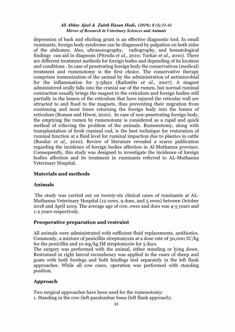
bodies). The incised edges of rumen were cleaned from blood clots and any debris with normal saline, two-layers closure was used for rumen suturing. Continuous inverting suture patterns, such as the Shmedin and Cushing were used because they provide a tight seal, minimize suture exposure, and promote healing (Figure.6). The abdominal wall usually requires two to three layers of closure (Figure.7). The peritoneum and transverse muscles were usually closed in one layer, using absorbable suture material (e.g., 2 chromic catgut) in a simple continuous pattern. The internal and external abdominal oblique muscles were closed together using absorbable suture material (e.g., 2 chromic gut) in a simple continuous pattern. The skin was closed using a horizontal mattress interrupted sutures using 2 silk.
Figure. 2: Shows the exteriorize the rumen outside the surgical wound
Figure. 3: Shows the rumen fixed with Weingart's ruminal ring

Ali Abbas Ajeel & Zainb Hasan Hadi. (2019); 8 (3):31-41 Mirror of Research in Veterinary Sciences and Animals
36
Figure, 4: Shows the impacted masses taken out from the rumen
Figure. 5: Shows the metal foreign bodies were taken out from reticulum through the rumen
Figure. 6: Shows two-layer closure were used for rumen suturing (Shmedin and Cushing)
Ali Abbas Ajeel & Zainb Hasan Hadi. (2019); 8 (3):31-41 Mirror of Research in Veterinary Sciences and Animals
37
Figure.7: Shows the abdominal wall closure
Post-operative care The success of the operation depends upon the post-operative care. Antibiotics and anti-inflammatory drugs are given for 5 days. The operative site cleaned daily with oxy-spray. Decreased food and water through first-week post-operation. The sutures can be removed 8-10days post-operative days. Results and Discussion Foreign bodies in the cow's rumen and reticulum is associated with traumatic reticuloperitonitis. In this study , a large number of non-metallic foreign bodies (15-20 kg) were removed from the cow's rumen (polyethylene bags, nylon ropes, electrical wires, clothes, leather pieces, socks, glasses, bones), while in does and ewes the weight of foreign bodies reached (4-6kg). Metallic objects (springs, nails, coins, wire, spoons, hair clips, screws) were also found. However, metallic objects in five cows were found penetrating the wall of the reticulum, resulting in traumatic reticuloperitonitis. In some cases, nails were entangled in the reticular mucosa and remained hanging inside the reticulum. Most of these objects were located in the anteroventral part of the reticulum. Tripathi et al., (2010) reported that indiscriminate feeding habit of animal coupled with insufficient feeding by the owner is forcing the animal to eat indigestible foreign material. Lack of dietary discrimination mostly in cattle, buffaloes, and to less extent in sheep and goats is leading to the ingestion of foreign materials causing the ruminal impaction. The ingested polythene hinders the process of fermentation and mixing of the contents leading to indigestion. The polythene and other plastic material do not degrade in the rumen \reticulum and remain

Ali Abbas Ajeel & Zainb Hasan Hadi. (2019); 8 (3):31-41 Mirror of Research in Veterinary Sciences and Animals
38
as causing hindrance in the orifice; this whole process also affects the rumen micro flora leading to indigestion of feed (Athar et al., 2010). Boodur et al., ( 2010) reported that the removal of plastics from the rumen is not enough, but the restoration of the normal rumen ecology is important for the speedy recovery of animals. Mohammed, (2004) reported that soft foreign bodies cause life-threatening adverse effects as grazing land is polluted with plastics, hoof, wool, hair, posing a major problem for grazing animals. Jana Debaris and Jana Mousami, (2006) reported that congestion of the ruminal mucosae and ruminitis might be due to the chemical reaction of polywastes or due to rubbing on the ruminal wall. Moreover, Ghurashal, 2009; Mushonga et al., 2015 reported that indigestible foreign bodies in ruminants are frequently encountered during hot, dry seasons and in desert areas. Poor husbandry management practices such as inadequate supplementation of minerals, vitamins, and forages, especially in dry seasons, may predispose ruminants to ingestion of indigestible foreign bodies as a result of pica. Improper waste management practices increase the chances of contamination or pollution of grazing lands with indigestible foreign bodies which is a big problem in extensive grazed ruminants (Ghurashi et al., 2009; Negash et al., 2015). Indigestible foreign bodies, once ingested, may cause anorexia and interference with the flow of ingesta and absorption of Volatile Fatty Acids (VFA), which results in weight loss (Mozaffari et al., 2009; Habasha and Yassein, 2014; Berrie et al., 2015). Anorexia may result due to the presence of the foreign body itself in the rumen occupying most of the rumen leaving little space for food (Baillie and Anzuino, 2006; Mozaffari et al., 2009). In conclusion, the results of this study approved the occurrence of various types of penetrated and non-penetrated foreign bodies in the rumen and reticulum of cows, sheep and goat. The study also approved the economic importance associated with reduced production and productivity of the affected animals. The authors recommend that animal owners should be educated about hard ware diseases and adequate minerals and vitamins should be included in animals feed, moreover, the animals should be reared and grazed away from areas of building construction. The owners should use magnets to screen for metallic foreign bodies in animals feed and encourage to put magnet pills into the rumen to all heifers and young bulls. It is very important that veterinarians should be trained to diagnose, treat and do surgical interventions for hardware disease. References • Abdelaal A, Floeck M, El Maghawry S, Baumgartner W. (2009).
Clinical and ultrasonographic differences between cattle and buffalo with various sequelae of traumatic reticuloperitonitis. Veterinarni Medicina 9, 399–406.
• Aref NM and Abdel-Hakiem MA. (2013). Clinical and diagnostic methods for evaluation of sharp foreign body syndrome in buffaloes. 6 (9): 586-591.

Ali Abbas Ajeel & Zainb Hasan Hadi. (2019); 8 (3):31-41 Mirror of Research in Veterinary Sciences and Animals
39
• Athar H, Mohindra J, SIingh KandSingh T. (2010). Indian polyvet119(2):180-183.
• Baillie S, Anzuino K. (2006). Hairballs as a cause of anorexia in Angora goats. Goat Vet Soc J 22: 53- 55.
• Berrie K, Tadesse E, Mossie B, Anteneh B (2015). Study on rumen and
reticulum foreign body in slaughtered cattle at Gondar Elfora abattoir. World J Biol Med Sci 2: 133-150.
• Boodur P, Sivaprakash BV, Kasaralivar VR, and Dilip D. (2010).
Intas Polyvet 11 (2):184-188. • Boodur P, Sivaprakash BV, Kasaralivar VR and Dilip D. (2010).
Indian poly vet11 (2):184-188. • Calfee T and Manning T O. (2002). Non healing subcutaneous wounds
in the cats and proposed surgical managements techniques. Clin. Tech. Small Anim.Pract. 17(4): 162-167.
• Desiye T and Mersha C. (2012). Study on Rumen and Reticulum Foreign
Bodies in Cattle Slaughtered at Jimma Municipal Abattoir, South West Ethiopia. American-Eurasian Journal of Scientific Research, 7(4): 160-167.
• Ghurashi MAH, Seri HI, Bakheit AH, Ashwag EAM. (2009). Effect
of surgical removal of foreign body from goat’s rumen with special reference to the prevalence of foreign body in goats in southern Darfur. AJBAS 3: 664-668.
• Habasha FG, Yassein SN. (2014). Advance techniques in traumatic
reticuloperitonitis diagnosis: review. AlQadisiya J Vet Med Sci 13: 50-57. • Hunt GB, Worth A and Marhevsky A. (2004). Migration of wooden
skewer foreign bodies from the gastrointestinal tract in eight dogs. J.Small Anim. Pract. 45 (7): 362-364.
• Jana Debaris and Jana Mourami. (2006). XII Annual conference of
IAAVR and round Table conference on Rumionogy11:81-85. • Jones TC, Hunt RD and King NW. (1997). Veterinary pathology. 6 ed.
USA, pp: 1060-1061. • Kahn MC. (2005). The Merck Veterinary Manual 8 ed, Merck and Co., INC.
White house station, USA, pp: 98: 185-192. • Knight AP. (1980). Xylazine. J Am Vet Med Assoc. 176:454–5.
Ali Abbas Ajeel & Zainb Hasan Hadi. (2019); 8 (3):31-41 Mirror of Research in Veterinary Sciences and Animals
40
• Misk NA, Semieka MA and Ali S El- M.(2001). Varieties and sequellae of ingested foreign bodies in buffaloes and cattle. Assiut Vet. Med. J. 46 (91): 250-273.
• Mohammad HA. (2004). M.V.ScThesis Sudan veterinary science and techonology.
• Mousavi G, Hassanpour A, AmoghliTabrizi B, Rezaie A (2007).
Electrocardiographic changes in buffalo with Traumatic reticuloperitonitis. Italy Journal Animal Science 6, 1029–1031.
• Mozaffari AA, Olomi MM, Vosough D. (2009). Unusual and severe
ruminal impaction in a goat-kid: clinical and radiological findings. IJVS 4: 115-119.
• Mushonga B, Habarugira G, Musabyemungu A, Udahemuka JC,
Jaja FI, Pepe D (2015). Investigations of foreign bodies in the forestomach of cattle at Ngoma slaughterhouse, Rwanda. J S Afr Vet Assoc 86: 1233.
• Negash S, Sibhat B, Sheferaw D. (2015). Post-mortem study on
indigestible foreign bodies in the rumen and reticulum of ruminants, Eastern Ethiopia. Onderstepoort J Vet Res 82: 881.
• Pitroda AH, Tiwari PK, Melrajudendar Patil DB and Parikh V.
(2010) Intas Polyvet 11(2):251- 252. • Radostits OM, Blood CC, Hinchclif KW and Constable PD. (2007).
Veterinary medicine a text book of disease of cattle, horse, sleep, pig and goat. 10th ed England, London. Saunders Elsevier, pp: 112- 522.
• Rahel M. (2011). Study on fore stomach foreign body Science, in cattle
Slaughtered Hawassa Municipal Abattoir. Ethiopia, DVM thesis Gondar University, Faculty of Veterinary Medicine, Gondar, Ethiopia.
• Roman T and Hiwot Y. (2010). Occurrence of rumen fibrinogen for the
diagnosis of traumatic foreign bodies in Sheep and Goat slaughtered at Addis Ababa Municipal Abattoir. Ethiopia Veterinary Journal, 14(1): 91-100.
• Saleh M, Rateb H, Misk N (2008). Comparison of blood serum proteins
in water buffalo with traumatic reticuloperitonitis and sequellae. Research in Veterinary Science 85, 208–213.
• Sileshi N, Ramaswamy V, Chandrashekhar U and Raja N. (2013).
Studies on Foreign Body Ingestion and their Related Complications in Ruminants Associated with Inappropriate Solid Waste Disposal in Gondar Town, North West Ethiopia. 5 (2): 67-74.
Ali Abbas Ajeel & Zainb Hasan Hadi. (2019); 8 (3):31-41 Mirror of Research in Veterinary Sciences and Animals
41
• Tripathi A K, Soodan JS, keeshawa RS and Shamod Kumar. (2010). Indian poly vet. 11(2):197-198.
• Turkar S, Sharma A k, Dhaliwal PS and Gopinathan A. (2010). Intas
Polyvet 11(2):191 – 193. • Vanitha V, Nambi AP, Gowri B and Kavitha S. (2010). Rumen
impaction in cattle with indigestible foreign bodies’. Veterinary and Animal Science. 6: 138-140.
Related Documents