REVIEW ARTICLE Sarcoidosis of the skin – A dermatological puzzle: important differential diagnostic aspects and guidelines for clinical and histopathological recognition G Tchernev, †, * JW Patterson, ‡ P Nenoff, § LC Horn – † Department of Dermatology, Venereology and Dermatosurgery, MVZ Kirchheim, Steingaustraße 13, 73230 Kirchheim unter Teck, Germany ‡ Division of Surgical Pathology and Cytopathology Room 3018, University of Virginia Hospital, 1215 Lee Street, Charlottesville, VA, USA § Laboratory for Medical Microbiology, Partnership Dr Ju ¨ rgen Herrmann, Prof Dr Pietro Nenoff & Dr Constanze Kru ¨ ger, Straße des Friedens 8, D-04579 Mo ¨ lbis, Germany – Division of Breast, Gynecologic and Perinatal Pathology, Institute of Pathology, University of Leipzig, Liebigstrasse 26, Leipzig, 04103, Germany *Correspondence: G Tchernev. E-mail: [email protected] Abstract Sarcoidosis of the skin may have an extremely heterogeneous clinical presentation, so that the definitions of ‘great imitator’ and ‘clinical chameleon’ have long been used. There is, in fact, a large group of skin diseases that can enter the differential diagnosis with cutaneous sarcoid manifestations, either clinically or/and pathologically. As the clinical consequences and the prognosis of these groups of diseases are often very different, it is important to correctly plan the diagnostic workup. The diagnostic process in this case often presents a challenge as no single test is sufficiently specific, so that a certain diagnosis can be only made in the presence of a compatible clinical and radiographic picture, along with histopathological evidence of non-necrotizing, epithelioid cell granulomas, and exclusion of other potential aetiologies. For practical reasons, four main groups of skin conditions capable of mimicking sarcoidosis can be identified: (i) transmissible, infectious diseases; (ii) allergic and immunological manifestations of various aetiologies; (iii) granulomatous diseases of various aetiologies; and (iv) lymphomas and pseudolymphomas. The aim of this article is to describe the main clinical and histopathological findings of such disease entities, and to discuss the role of those features (morphological, pathological and laboratory) that can help distinguish them from sarcoidosis of the skin. Received: 6 January 2009; Accepted 23 June 2009 Keywords differential diagnosis, Leishmania, leprosy, lupus, sarcoidosis Conflicts of interest None declared. Introduction Skin sarcoidosis is a disease of undetermined aetiology. 1,2 Although it is a relatively frequent disease for Afro-Americans liv- ing on the territory of the Caribbean region, as well as for Swedes, Irishmen and other ethnic groups, the reasons of its phenotypic manifestations are poorly known. 1,3–5 Besides the skin, sarcoidosis can affect any other organ and is capable of imitating a variety of diseases; consequently, in dermatology it is often called ‘The Great Imitator’ 1,6 or a ‘clinical chameleon’. 1 The clinical morphology of cutaneous sarcoidosis may vary within a wide range. Conse- quently, to diagnose it is not always easy (Table 1). Hence, the conclusion can be reached that, in case of suspicion of some cutaneous forms of sarcoidosis, the histopathology should play an important role in the diagnosis. The microscopic findings in cases of sarcoidosis include a characteristic, non-caseating gran- ulomatous reaction (Fig. 1a–d) or the non-specific reaction of ery- thema nodosum. 1,7,8 Having in mind, however, that many diseases show sarcoidal granulomatous reactions but have a completely dif- ferent pathogenesis, the application of certain auxiliary diagnostic methods is often necessary. 9,10 Sarcoidosis as a systemic disease can involve any organ or tissue, even though lung and intrathoracic lymph nodes are the most ª 2009 The Authors JEADV 2010, 24, 125–137 Journal compilation ª 2009 European Academy of Dermatology and Venereology DOI: 10.1111/j.1468-3083.2009.03396.x JEADV

Welcome message from author
This document is posted to help you gain knowledge. Please leave a comment to let me know what you think about it! Share it to your friends and learn new things together.
Transcript

REVIEW ARTICLE
Sarcoidosis of the skin – A dermatological puzzle: importantdifferential diagnostic aspects and guidelines for clinical andhistopathological recognition
G Tchernev,†,* JW Patterson,‡ P Nenoff,§ LC Horn–
†Department of Dermatology, Venereology and Dermatosurgery, MVZ Kirchheim, Steingaustraße 13, 73230 Kirchheim unter
Teck, Germany‡Division of Surgical Pathology and Cytopathology Room 3018, University of Virginia Hospital, 1215 Lee Street, Charlottesville, VA, USA§Laboratory for Medical Microbiology, Partnership Dr Jurgen Herrmann, Prof Dr Pietro Nenoff & Dr Constanze Kruger, Straße des
Friedens 8, D-04579 Molbis, Germany–Division of Breast, Gynecologic and Perinatal Pathology, Institute of Pathology, University of Leipzig, Liebigstrasse 26, Leipzig, 04103,
Germany
*Correspondence: G Tchernev. E-mail: [email protected]
AbstractSarcoidosis of the skin may have an extremely heterogeneous clinical presentation, so that the definitions of ‘great
imitator’ and ‘clinical chameleon’ have long been used. There is, in fact, a large group of skin diseases that can
enter the differential diagnosis with cutaneous sarcoid manifestations, either clinically or/and pathologically. As the
clinical consequences and the prognosis of these groups of diseases are often very different, it is important to
correctly plan the diagnostic workup. The diagnostic process in this case often presents a challenge as no single
test is sufficiently specific, so that a certain diagnosis can be only made in the presence of a compatible clinical
and radiographic picture, along with histopathological evidence of non-necrotizing, epithelioid cell granulomas, and
exclusion of other potential aetiologies. For practical reasons, four main groups of skin conditions capable of
mimicking sarcoidosis can be identified: (i) transmissible, infectious diseases; (ii) allergic and immunological
manifestations of various aetiologies; (iii) granulomatous diseases of various aetiologies; and (iv) lymphomas and
pseudolymphomas. The aim of this article is to describe the main clinical and histopathological findings of such
disease entities, and to discuss the role of those features (morphological, pathological and laboratory) that can
help distinguish them from sarcoidosis of the skin.
Received: 6 January 2009; Accepted 23 June 2009
Keywordsdifferential diagnosis, Leishmania, leprosy, lupus, sarcoidosis
Conflicts of interestNone declared.
IntroductionSkin sarcoidosis is a disease of undetermined aetiology.1,2
Although it is a relatively frequent disease for Afro-Americans liv-
ing on the territory of the Caribbean region, as well as for Swedes,
Irishmen and other ethnic groups, the reasons of its phenotypic
manifestations are poorly known.1,3–5 Besides the skin, sarcoidosis
can affect any other organ and is capable of imitating a variety of
diseases; consequently, in dermatology it is often called ‘The Great
Imitator’1,6 or a ‘clinical chameleon’.1 The clinical morphology of
cutaneous sarcoidosis may vary within a wide range. Conse-
quently, to diagnose it is not always easy (Table 1).
Hence, the conclusion can be reached that, in case of suspicion
of some cutaneous forms of sarcoidosis, the histopathology should
play an important role in the diagnosis. The microscopic findings
in cases of sarcoidosis include a characteristic, non-caseating gran-
ulomatous reaction (Fig. 1a–d) or the non-specific reaction of ery-
thema nodosum.1,7,8 Having in mind, however, that many diseases
show sarcoidal granulomatous reactions but have a completely dif-
ferent pathogenesis, the application of certain auxiliary diagnostic
methods is often necessary.9,10
Sarcoidosis as a systemic disease can involve any organ or tissue,
even though lung and intrathoracic lymph nodes are the most
ª 2009 The Authors
JEADV 2010, 24, 125–137 Journal compilation ª 2009 European Academy of Dermatology and Venereology
DOI: 10.1111/j.1468-3083.2009.03396.x JEADV

frequently affected.1,3,11 The diagnostic process is in this case often
a challenge as no single test is sufficiently specific, so that a certain
diagnosis can be only made in the presence of a compatible clinical
and radiographic picture, along with histopathological evidence of
non-necrotizing, epithelioid cell granulomas, and exclusion of
other potential aetiologies (Fig. 1a–d).1
In this scenario, the recognition of skin lesions may be crucial
as they provide both a visible clue to the diagnosis, and an easily
accessible site for histopathological confirmation of the clinical
suspicion.1,4,7,12 Cutaneous involvement occurs in up to one third
of sarcoid patients, and may be extremely heterogeneous in terms
of morphology, extent, evolution (self-limited vs. chronic forms),
clinical relevance and response to treatment.1,8,9,13,14 Most authors
tend to divide cutaneous manifestation of sarcoidosis into specific
and non-specific types, the latter (i.e. erythema nodosum) occur-
ring in association with systemic sarcoidosis but lacking the spe-
cific, non-necrotizing granulomas on biopsy.10,15–17
From a practical point of view, a dermatologist can face two
possible situations, leading to different pathways, in the manage-
ment of sarcoidosis patients:
1) A patient with unknown sarcoidosis seeks medical attention
for the presence of skin lesions. In such a case, the dermatologist is
the one who has the chance to raise the clinical suspicion, which is
sometimes difficult as the clinical lesions may assume a wide
array of morphologies. Histopathology plays a key role in this
case, but the pathologist cannot diagnose sarcoidosis in isolation,
Table 1 Cutaneous lesions able to mimic sarcoidosis of the skin either clinically or/and pathologically
Morphological mimickers of cutaneous sarcoidosis Histological mimickers of cutaneous sarcoidosis
Group 1Diseases of transmissiblenature
Erythema induratum Bazin, papulonecrotic tuberculid,tuberculosis cutis luposa, leprosy lesions, secondary andtertiary syphilids, tubero-serpiginous syphilids, tuberoulcero-serpiginous syphilids, dermatophytosis,leishmaniasis cutis, bacillary angiomatosis
Lichen scrophulosorum, tuberculosis cutis miliarisdisseminate, tuberculosis fungosa serpiginosa,tuberculosis miliaris ulcerosa mucosae et cutis,tuberculosis cutis luposa, leprosy lesions,leishmaniasis cutis (not always)
Group 2Allergic and immunologicalmanifestations of vagueaetiologies
Atopic dermatitis, psoriasis vulgaris, erythema nodosum,pernions, erythema annulare centrifugum, chronic discoidlupus erythematosus, subacute cutaneous lupuserythematosus
–
Group 3Granulomatous diseases ofunclear entity
Granuloma annulare, lipoidic necrobiosis, lichen nitidus,necrobiotic xanthogranuloma, lupoid rosacea, perioraldermatitis, facial eosinophilic granuloma, lupus miliarisdisseminatus faciei
Granulomatosis disciformis MiescherLupoid rosacea
Group 4Pseudolymphomas andlymphomas
Lymphoma and pseudolymphoma of B- and T-cellulartype
–
Group 5Other/rare forms of cutaneoussarcoidosis
Lichenoid sarcoidosis, keloidal sarcoidosis –
Figure 1 (a) Cutaneous sarcoidosis (haematoxylin and eosin (H&E) staining). Low-power view with dermal infiltration of nests and
clusters of non-caseating epitheloid granulomas surrounded by only weak inflammation. (b) Cutaneous sarcoidosis (H&E staining).High-power view with multinucleated giant cells and histiocytic cells. (c) Pulmonal sarcoidosis (H&E staining). Low-power view of
a transbronchial biopsy with non-caseating epitheloid granuloma in between a bronchus and pulmonal artery with weak accompany-
ing inflammation. (d) Sarcoidosis within a lymph node (H&E staining). Low-power view representing numerous non-caseatingepitheloid granulomas.
ª 2009 The Authors
JEADV 2010, 24, 125–137 Journal compilation ª 2009 European Academy of Dermatology and Venereology
126 Tchernev et al.
as sarcoidal granulomas have no unique pathological features to
differentiate them from other granulomas (statement on sarcoido-
sis AJRCCM 1999; 160: 736–755). A multidisciplinary approach
aimed towards determining the likelihood of a diagnosis of sar-
coidosis (after careful evaluation of clinical, radiological and path-
ological findings) and establishing a follow-up programme is
recommended for patients with suggestive skin biopsy findings
(statement on sarcoidosis AJRCCM 1999; 160: 736–755). A fol-
low-up programme will be of particular importance for those
patients in whom the skin involvement precedes the systemic
manifestations of the disease.
2) A patient with known sarcoidosis is referred for the diagnos-
tic evaluation of a skin lesion/s. In this case, the dermatologist is
asked to determine the nature of the lesion, and to discuss with
the referring physician the therapeutic options.
The aim of this article is to highlight the main clinical and his-
topathological differential diagnoses of cutaneous sarcoidosis, and
to discuss the role of auxiliary methods that may contribute to the
solution of the diagnostic dilemma. The clinical morphology of
lesions in dermatology is not always decisive, so a careful evalua-
tion of all parameters is frequently necessary.1,15
This article focuses on four basic categories of disease, some of
which imitate sarcoidosis of the skin both clinically and histopath-
ologically. These are: transmissible diseases, allergic and immuno-
logical diseases, granulomatous diseases of uncertain aetiology,
and lymphomas or pseudolymphomas. The role of several impor-
tant auxiliary methods contributing to diagnostic precision is also
discussed.
Aetiopathogenetic aspectsThe aetiopathogenetic factors in sarcoidosis are, at present, not
completely clear.1 A key role has been attributed to different viro-
logical, bacterial and chemical agents.16
The importance of human herpesvirus 8 (HHV-8) in cutaneous
sarcoidosis has yet to be clarified.18,19 Interferon therapy and the
hepatitis-C viral infections also probably play some role regarding
the manifestations of the disease, and the crucial factor is felt to be
the disturbance of internal homeostasis and consequent antigenic
mimicry.1,20 A possible role for Epstein–Barr or Coxsackie B
viruses has been discussed but not proven.21
Similarly, bacteria and related infectious agents, including Propi-
onibacterium acnes, Yersinia enterocolitica, Chlamydia pneumophila,
Borrelia burgdorferi and Mycoplasma species, have been implicated.
However, their roles as epigenetic factors, or factors influencing
the phenotypic manifestations of cutaneous sarcoidosis, are still
disputable.21 The detection of mycobacterial DNA in sarcoid
lesions in some patients is surprising, and imposes the obligatory
exclusion of any active, systemic or post-primary cutaneous forms
of tuberculosis.21–24 It is clearly important that these and other
infectious diseases be excluded, as there would be potential wors-
ening of the patient’s general status where corticosteroids or im-
munomodulators are to be employed as forms of therapy.
In addition, the importance of some metals in the development
of sarcoidosis, such as beryllium, zirconium, titanium and alumin-
ium, is not clear.1
Group 1: Diseases of transmissible nature
Many cutaneous lesions from other diseases are able to mimic
sarcoidosis of the skin either clinically or/and pathologically
(Table 1).
This group includes eight basic diseases, each of which may
have different clinical manifestations, that can imitate cutaneous
sarcoid lesions.25,26 A correct diagnosis is crucial to avoid dissemi-
nation in the skin area and/or involvement of distant organs or
tissues.
1. Sarcoidosis vs. tuberculosis
There are different forms of cutaneous tubercular reactions, and
each of them can mimic certain ‘sarcoid clinical variants’.
1.1 Hyperergic forms of skin tuberculosis
Lichen scrofulosorum can, to some extent, mimic small dissemi-
nating nodular sarcoidosis.27
The papulonecrotic tuberculid can resemble a disseminated
ulcerative form of sarcoidosis in patients with weakened T-cellular
immunity.
Erythema induratum Bazin is a hyperergic form of post-primary
tuberculosis, where the lesions are located in the area of the dorsal
part of the shin.1 There is no clinical difference between this disease
and the ulcerative localized form of sarcoid. Sarcoidosis of the skin,
however, only rarely occurs in this area. Women predisposed to
acrocyanosis and cutis marmorata livedoides/telangiectatica
congenita are also predisposed to sarcoidosis of the skin.1,28,29 For
diagnosis, it is important to determine not only the histopathologi-
cal picture, but also to detect mycobacterial DNA by the polymer-
ase chain reaction (PCR) method in lesional tissue.1
The tuberculin reaction in patients with hyperergic form of
sarcoidosis is strongly positive, while the Ziehl–Neelsen staining is
negative, to (sometimes) slightly positive.1,29
The histopathology of any one of the hyperergic forms differs
from the histopathology of sarcoidosis. In the case of erythema
induratum Bazin, the histopathological correlate is granulomatous
lobular panniculitis, frequently accompanied by vasculitis.1
In cases of papulonecrotic tuberculid, lymphocytic vasculitis
predominates, accompanied by tuberculoid infiltrates.1,30
Lichen scrofulosorum shows Langhans-type giant cells,
tuberculoid infiltrates and necrosis.31,32 It is important for the
patients that a blood test for interferon be performed, in order to
find/exclude any active systemic form of tuberculosis (pulmonary
or gastrointestinal) with further appropriate use of scheduled
polychemotherapy.
A distinction from polyarteritis nodosa, which can be associated
with cryoglobulinaemia, paraproteinaemia, Streptococcus infection
or tumours, is also important.
ª 2009 The Authors
JEADV 2010, 24, 125–137 Journal compilation ª 2009 European Academy of Dermatology and Venereology
Sarcoidosis of the skin: a clinical and histopathological chameleon 127
1.2 Anergic forms of skin tuberculosis
The anergic forms of tuberculosis can also mimic disseminated or
localized cutaneous forms of sarcoidosis. These forms are scarce
and mainly diagnosed in Third World countries. It is difficult to
find and diagnose such cases in Europe, although sporadic cases
have been described in Bulgaria and Romania.
Variant forms include tuberculosis cutis miliaris disseminata,
tuberculosis fungosa serpiginosa and tuberculosis miliaris ulcerosa
mucosae et cutis. These occur within the framework of inoculation
in cases of active tuberculosis (gastrointestinal or pulmonary) in
adults having a weakened immune system or in young children
whose immune protective mechanisms are underdeveloped.29,31–34
Clinically, tuberculosis cutis miliaris disseminata does not differ
from the small-papular disseminated ulcerative, the benign miliary
lupoid or the small disseminated nodular forms of sarcoidosis.35
However, there are few clinical similarities between the other two
forms and the different cutaneous forms of sarcoidosis.
An important feature of the disseminated forms of tuberculosis
is the patient’s generally debilitated status, as well as low-grade
fevers and night sweats, which are not observed in localized and
disseminated forms of cutaneous sarcoidosis.
The tuberculin reaction in the anergic forms is negative, while
PCR-DNA in lesional skin is strongly positive,34 as is staining for
organisms using the Ziehl–Neelsen method.31,32,34,36,37 The
histological analysis shows tuberculoid granulomas with, or more
frequently, without caseous necrosis. The distinction from
sarcoidosis is straightforward if the above-mentioned diagnostic
panels are observed.
1.3 Allergic forms of cutaneous tuberculosis
To classify the lesions, the allergic forms of cutaneous sarcoidosis
including lupus vulgaris (or tuberculosis cutis luposa) are the most
problematic (Fig. 2). This is a post-primary form of tuberculosis
that can be developed not only per continuitatem, at a distant site
from the primary focus of infection, but also after subsequent
exogenous inoculation.38 The tuberculin test is positive, and
Ziehl–Neelsen staining is often negative.39 Lesions resemble those of
the subcutaneous nodular exudative or plaque-like forms of
sarcoidosis. Caseous granulomatous reactions and tuberculoid
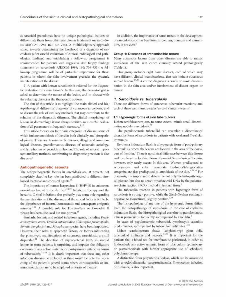
granulomas can be found.40 By diascopy, small lupus nodules are
observed in the form of ‘apple or orange jelly’ types (Fig. 2).1,39,41
Once the diagnosis is proven, the use of polychemotherapy is
recommended, as the risk of malignant transformation and
mutilating (lupus vulgaris mutilans) symptoms is high.38,39
The atypical mycobacterioses, including those due to Mycobac-
terium marinum, M. abscessus and M. ulcerans, are less commonly
included in the differential diagnosis of sarcoidosis, but they must
not be completely excluded.42,43 The morphology of any single
lesion must be carefully observed. Newly generated keloidal
formations with perilesional localization must be histologically
analysed.
2. Sarcoidosis vs. leprosy
Sarcoidosis of the skin vs. lepromatous leprosy and
tuberculoid leprosy
From a differential-diagnostic point of view, consideration should
be given to forms of lepromatous leprosy and tuberculoid leprosy.
This disease was known in the past under the names of Hansen or
Zaraath disease (biblical name of the disease).44 Transmission
takes place directly from ill persons or by direct contact with, or
consumption of, underdone species of armadillos infected by
Mycobacterium leprae.
Today, this disease can be transmitted through the nine-banded
armadillo in the State of Louisiana, USA. It is observed among certain
at-risk groups, including hunters and illegal meat middlemen.44,45
About 10–15 million people suffer from leprosy worldwide. In
2006, the WHO had reported approximately 300 000 new leprosy
infections. The incubational period lasts between 3 and 20 years,
and its clinical manifestations are largely determined by the status
of a patient’s T-cell immunity.40
An obvious key feature in distinguishing lesions caused by
mycobacteria from sarcoidosis is that Ziehl–Neelsen and Fite–Far-
aco staining of sarcoid skin lesions is negative, while those,
induced by Mycobacterium leprae and Mycobacterium tuberculosis
are often positive.40,44,45 The differentiation of Mycobacterium
leprae from Mycobacterium tuberculosis can be achieved by apply-
ing silver nitrate; Mycobacterium leprae shows light, while Myco-
bacterium tuberculosis shows dark, staining with this method.1,46
The most sensitive but not always accessible method to demon-
strate Mycobacterium leprae in lesional (nasal) mucosa is through
detection of mycobacterial DNA by PCR methods.46
Figure 2 Erythematous plaque-like form of lupus vulgaris.
Under pressure through a magnifying glass, the characteristic
‘apple jelly’ lupoid aspect is observed.1
ª 2009 The Authors
JEADV 2010, 24, 125–137 Journal compilation ª 2009 European Academy of Dermatology and Venereology
128 Tchernev et al.
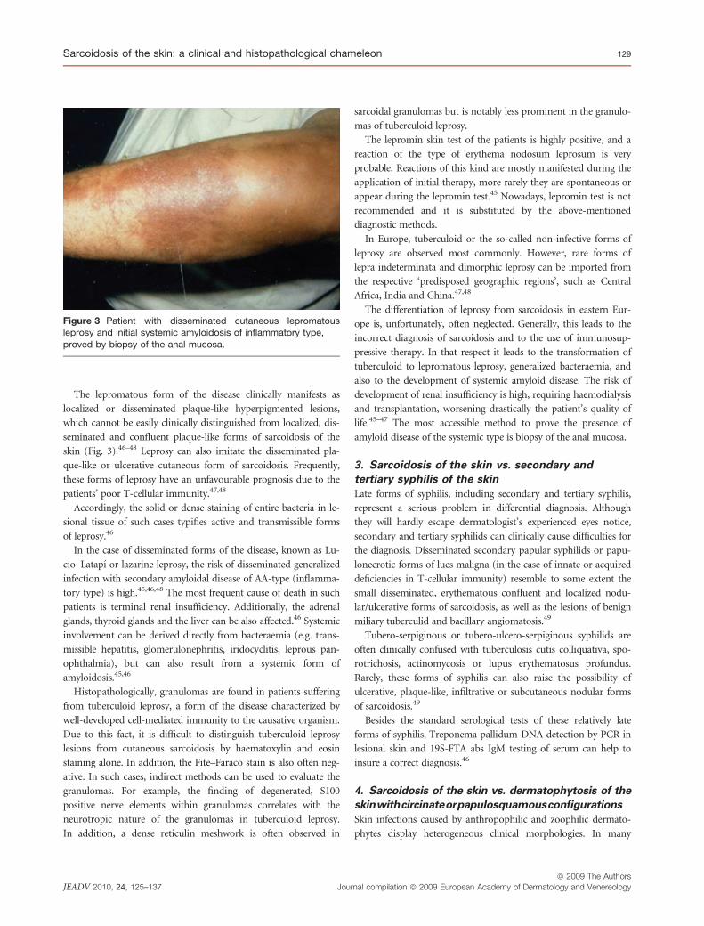
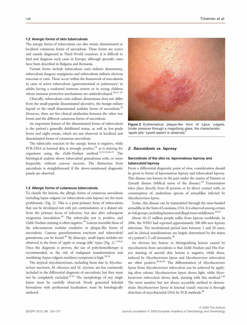
The lepromatous form of the disease clinically manifests as
localized or disseminated plaque-like hyperpigmented lesions,
which cannot be easily clinically distinguished from localized, dis-
seminated and confluent plaque-like forms of sarcoidosis of the
skin (Fig. 3).46–48 Leprosy can also imitate the disseminated pla-
que-like or ulcerative cutaneous form of sarcoidosis. Frequently,
these forms of leprosy have an unfavourable prognosis due to the
patients’ poor T-cellular immunity.47,48
Accordingly, the solid or dense staining of entire bacteria in le-
sional tissue of such cases typifies active and transmissible forms
of leprosy.46
In the case of disseminated forms of the disease, known as Lu-
cio–Latapı or lazarine leprosy, the risk of disseminated generalized
infection with secondary amyloidal disease of AA-type (inflamma-
tory type) is high.45,46,48 The most frequent cause of death in such
patients is terminal renal insufficiency. Additionally, the adrenal
glands, thyroid glands and the liver can be also affected.46 Systemic
involvement can be derived directly from bacteraemia (e.g. trans-
missible hepatitis, glomerulonephritis, iridocyclitis, leprous pan-
ophthalmia), but can also result from a systemic form of
amyloidosis.45,46
Histopathologically, granulomas are found in patients suffering
from tuberculoid leprosy, a form of the disease characterized by
well-developed cell-mediated immunity to the causative organism.
Due to this fact, it is difficult to distinguish tuberculoid leprosy
lesions from cutaneous sarcoidosis by haematoxylin and eosin
staining alone. In addition, the Fite–Faraco stain is also often neg-
ative. In such cases, indirect methods can be used to evaluate the
granulomas. For example, the finding of degenerated, S100
positive nerve elements within granulomas correlates with the
neurotropic nature of the granulomas in tuberculoid leprosy.
In addition, a dense reticulin meshwork is often observed in
sarcoidal granulomas but is notably less prominent in the granulo-
mas of tuberculoid leprosy.
The lepromin skin test of the patients is highly positive, and a
reaction of the type of erythema nodosum leprosum is very
probable. Reactions of this kind are mostly manifested during the
application of initial therapy, more rarely they are spontaneous or
appear during the lepromin test.45 Nowadays, lepromin test is not
recommended and it is substituted by the above-mentioned
diagnostic methods.
In Europe, tuberculoid or the so-called non-infective forms of
leprosy are observed most commonly. However, rare forms of
lepra indeterminata and dimorphic leprosy can be imported from
the respective ‘predisposed geographic regions’, such as Central
Africa, India and China.47,48
The differentiation of leprosy from sarcoidosis in eastern Eur-
ope is, unfortunately, often neglected. Generally, this leads to the
incorrect diagnosis of sarcoidosis and to the use of immunosup-
pressive therapy. In that respect it leads to the transformation of
tuberculoid to lepromatous leprosy, generalized bacteraemia, and
also to the development of systemic amyloid disease. The risk of
development of renal insufficiency is high, requiring haemodialysis
and transplantation, worsening drastically the patient’s quality of
life.45–47 The most accessible method to prove the presence of
amyloid disease of the systemic type is biopsy of the anal mucosa.
3. Sarcoidosis of the skin vs. secondary and
tertiary syphilis of the skin
Late forms of syphilis, including secondary and tertiary syphilis,
represent a serious problem in differential diagnosis. Although
they will hardly escape dermatologist’s experienced eyes notice,
secondary and tertiary syphilids can clinically cause difficulties for
the diagnosis. Disseminated secondary papular syphilids or papu-
lonecrotic forms of lues maligna (in the case of innate or acquired
deficiencies in T-cellular immunity) resemble to some extent the
small disseminated, erythematous confluent and localized nodu-
lar/ulcerative forms of sarcoidosis, as well as the lesions of benign
miliary tuberculid and bacillary angiomatosis.49
Tubero-serpiginous or tubero-ulcero-serpiginous syphilids are
often clinically confused with tuberculosis cutis colliquativa, spo-
rotrichosis, actinomycosis or lupus erythematosus profundus.
Rarely, these forms of syphilis can also raise the possibility of
ulcerative, plaque-like, infiltrative or subcutaneous nodular forms
of sarcoidosis.49
Besides the standard serological tests of these relatively late
forms of syphilis, Treponema pallidum-DNA detection by PCR in
lesional skin and 19S-FTA abs IgM testing of serum can help to
insure a correct diagnosis.46
4. Sarcoidosis of the skin vs. dermatophytosis of the
skinwithcircinateorpapulosquamousconfigurations
Skin infections caused by anthropophilic and zoophilic dermato-
phytes display heterogeneous clinical morphologies. In many
Figure 3 Patient with disseminated cutaneous lepromatousleprosy and initial systemic amyloidosis of inflammatory type,
proved by biopsy of the anal mucosa.
ª 2009 The Authors
JEADV 2010, 24, 125–137 Journal compilation ª 2009 European Academy of Dermatology and Venereology
Sarcoidosis of the skin: a clinical and histopathological chameleon 129
cases of tinea corporis (profunda), papulosquamous or annular/
polycyclic configurations prevail.50 It is not always possible to dis-
tinguish these lesions from acrally localized ichthyosiform, conflu-
ent erythematous or circinate annular forms of sarcoidosis.1,51
Important auxiliary methods helping to clarify the origin of lesions
include study of skin scrapings (potassium hydrochloride or calco-
fluor preparations), mycological culture, and PAS or Groccot
staining of tissue samples. Trichophyton rubrum, Trichophyton vio-
laceum, Trichophyton verrucosum, Trichophyton interdigitale (for-
merly Trichophyton mentagrophytes) and Epidermophyton
floccosum are considered to be the most frequent aetiological agents.50
5. Sarcoidosis of the skin vs. hyperergic and
anergic forms of leishmaniasis of the skin
5.1 Normoergic reactions
Localized forms of leishmaniasis cutis are the most frequent
(Fig. 4).52 The organisms are transmitted by Phlebotomus vectors
that are capable of freely penetrating through widely used mosquito
nettings. These vectors transmit Leishmania organisms in their pro-
mastigote form, about 10–15 microns long and 2–3 microns wide.
The amastigote form of leishmaniasis settles in the human body
after biting. The clinical manifestations of leishmaniasis cutis
cannot always be distinguished from cutaneous sarcoidosis, even
in locations where leishmaniasis is common.52,53 To prove that
patients have resided in regions endemic for leishmaniasis, such as
Mexico, the Amazon, Ethiopia, India, Nepal, China, Brazil, Bolivia
(South Europe and North Africa) and the territory of the former
socialist republics (USSR), is often problematic.53,54 Intact immu-
nity is most frequent among patients with localized skin lesions of
leishmaniasis.52–54 The affected persons show positive Montenegro
reactions, positive Giemsa staining and from high to average high
titres of antibodies in the serum.55 Isolation on special culture
media is recommended. The main agents of the lesions are
Leishmania minor, Leishmania major, Leishmania infantum and
Leishmania aethiopica, known also as Old World leishmaniasis.
5.2 Hyperergic form of reaction
Both hyperergic and anergic generalized forms of leishmaniasis
create more frequent differential diagnostic problems.56,57
The hyperergic recurring or persisting form clinically resembles
small disseminated nodular sarcoidosis, lichen scrofulosorum and
dermatophytid.57
This imposes the application of the respective additional ‘diag-
nostic panels’ in order to exclude the aforesaid diseases. The intracu-
taneous leishmania antigen test (Montenegro test) is strongly
positive. The titre of circulating serum antibodies is high, and
Giemsa staining can be both positive and negative.52–54 Isolation on
special selective media, such as Novy-McNeal-Nicolle/Adler med-
ium is possible and recommended.52,53 The amplification test, on
hamsters or humans, is not routinely practised nowadays, but it pro-
vokes a disseminated generalized form of leishmaniasis in rodents. If
the diagnosis is not clear and a generalized skin reaction is present,
the amplification test can significantly facilitate the diagnosis.
5.3 Anergic forms of reaction
The anergic forms of leishmaniasis are chiefly caused by
Leishmania aethiopica and Leishmania brasiliensis, and their
cutaneous manifestation is most frequently, the diffuse cutaneous
form of leishmaniasis.52
Clinically, one observes non-ulcerating nodules and plaques
tending towards confluence and the formation of small satellite
nodules in the immediate vicinity of the primary lesions. The
Montenegro reaction is negative, Giemsa staining is strongly posi-
tive, the titre of the antibodies in the serum is low to negative, and
the amplification test and the isolation of leishmania in culture are
positive.52,57,58 Clinically, the lesions resemble the plaque-like
confluent disseminating or localized forms of sarcoidosis or the
lazarine form of leprosy of the Lucio–Latapı type.57,58 The histopa-
thology is not typical and shows lymphocytes, histiocytes (macro-
phages), granulocytes, plasma-cell infiltrates and, partially,
tuberculoid granulomas.
In the presence of differential diagnostic difficulties in some of
the localized forms, observed in Brazil or Mexico and other
regions (due to infections by Leishmania aethiopica or L. brasiliensis –
so-called espundia), the diagnosis can be made more precise by
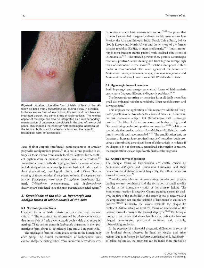
Figure 4 Localized ulcerative form of leishmaniasis of the skin
following bites from Phlebotomus sp. during a stay in Ethiopia.In the ulcerative form of sarcoidosis, the lesions do not have an
indurated border. The same is true of leishmaniasis. The keloidal
aspect of the edge can also be interpreted as a rare secondary
manifestation of cutaneous sarcoidosis in the area of new or oldscars. This imposes the need for histopathological appraisal of
the lesions, both to exclude leishmaniasis and the ‘specific
histological form’ of sarcoidosis.
ª 2009 The Authors
JEADV 2010, 24, 125–137 Journal compilation ª 2009 European Academy of Dermatology and Venereology
130 Tchernev et al.

the additional application of monoclonal antibodies against
amastigotes in lesional tissue52,53.
6. Sarcoidosis of the skin vs. bacillary angiomatosis
Bacillary angiomatosis can be difficult to differentiate from small
papular localized or disseminated sarcoidosis. Its distinction from
localized papular, partially ulcero-necrotic syphilids is also prob-
lematic. The aetiological agent is Rochalimaea (formerly Bartonella)
quintana or Rochalimaea henselae.59 Demonstration of these bacteria
is accomplished by its cultivation in special media or, directly, by
Warthin–Starry staining.60 An important clinical feature is the angi-
omatous nature of the nodules. The most specific and certain proof
that the infection is present is based on PCR detection of bacterial
DNA in lesional skin.59–61
Group 2: Allergic and immunological manifestations of
diverse aetiologies
Sarcoidosis of the skin vs. diseases of immunological
and infectious-allergic nature: differential diagnosis
Diseases of immunological and infectious-allergic nature can
induce localized or generalized sarcoid-like cutaneous lesions.1
They include atopic dermatitis, psoriasis vulgaris, erythema nodo-
sum, perniosis and erythema annulare centrifugum.1 Sarcoid-like
cutaneous lesions are also clinically observed in certain forms of
lupus erythematosus.
2.1 Localized/disseminated sarcoidosis of the skin vs.
psoriasis vulgaris of chronic stationary plaque-like and
erythrodermic types. The isolated subungual form of
sarcoidosis vs. psoriasis
The cutaneous form of sarcoidosis was described in 1877 for the
first time by the surgeon Hutchinson as a ‘papillary form of psori-
asis’.16 The erythrodermic form of sarcoidosis is extremely rare
and histopathologically shows the specific sarcoidal granulomas.1
The differentiation from psoriasis is important. Attention must be
paid to the anamnesis and the genetic disposition of the patients,
as well as to the evolution of lesions over time.1
The geographical or polycyclic annular form of psoriasis is rela-
tively easy to distinguish from the polycyclic annular or circinate
skin lesions of sarcoidosis. However, when lesions reach a large
size, tend towards confluence or develop infiltrative features,
biopsy is recommended.
Isolated forms of sarcoidosis with subungual localization and
onycholysis are very rare. Sarcoidal granulomas with epitheloid
and giant cells are found histopathologically. The occasional dem-
onstration of the subungual form of sarcoidosis in patients with
psoriasis would be extremely unlikely, but possible.
2.2 Sarcoidosis of the skin vs. atopic dermatitis
As noted above, the cutaneous form of sarcoidosis can rarely man-
ifest as erythroderma. It must therefore be distinguished not only
from erythrodermic psoriasis but also from atopic dermatitis with
erythroderma.1 The details provided by the patient’s history, the
clinical picture, the family history and the presence of type 1
hypersensitivity to certain airborne and alimentary allergens, are
very important to establishing a correct diagnosis.1 To this end, it
is recommended that one obtain the respective skin and blood
tests. The determination of total IgE in serum is obligatory if ato-
pic dermatitis is suspected.1,16 Histopathologically, epidermal
spongosis is found, and sarcoidal granulomas are absent.
2.3 Sarcoidosis of the skin vs. erythema annulare
centrifugum vs. erythema nodosum vs. chilblains
(perniosis)
The circirnate small-papular and large plaque-like disseminated,
partially annular forms from erythema annulare centrifugum,
could be wrongly interpreted as sarcoidosis of the skin.62 The aeti-
ology of this poly-aetiological, probably allergic reaction to infec-
tion is not completely clear.63,64 It is observed in various
malignancies, as well as in streptococcal infections, active tubercu-
losis, genital or anal candidosis, fungal infections of the skin (der-
matophytosis), lumbricosis, food allergies, paraproteinaemias, as a
reaction to medications, and in lupus erythematosus.65,66
The annular, polycyclic aspect of the lesions suggests the possi-
bility of an annular granulomatous disease.
The histopathological picture in erythema annulare centrifugum
is that of a dense, superficial and deep dermal, perivascular infil-
trate (sometimes described as ‘coat-sleeved’) comprised mainly of
lymphocytes but also including macrophages (histiocytes) and
occasional eosinophils.65,66 Significant thickening of the vascular
endothelium is observed. The epitheloid and giant cells so charac-
teristic for the ‘specific lesions’ of cutaneous sarcoidosis are not
observed.62
Erythema nodosum plays an important role in the diagnosis of
sarcoidosis. Most dermatologists consider it as a ‘non-specific his-
tological and clinical form’ of cutaneous sarcoidosis.1
It can also represent a cutaneous manifestation of sarcoidosis in
the form of the so-called Lofgren syndrome. Besides the presence
of erythema nodosum, bilateral hilar adenopathy and pains in the
joints are characteristic (immunological version of erythema
nodosum).67,68
The cutaneous findings in Crohn’s disease and chronic ulcera-
tive colitis are identical.
However, erythema nodosum can also result from exposure to
medications (contraceptives, salicylates, sulfonamides) or infec-
tious agents. Infections due to Toxoplasma, Streptococcus pyogenes,
Yersinia enterocolitica, dermatophytes and infections by Chlamydia
trachomatis serotype L1–L3 have been described as possible trig-
gers of erythema nodosum.67–69 Erythema nodosum lesions are
also observed in cat scratch disease.
Perniosis (chilblains) is the result of functional peripheral
vascular disease in patients with impaired digital circulation.70
Patients with acrocyanosis and cutis marmorata are predisposed.70,71
ª 2009 The Authors
JEADV 2010, 24, 125–137 Journal compilation ª 2009 European Academy of Dermatology and Venereology
Sarcoidosis of the skin: a clinical and histopathological chameleon 131

Bullous and ulcerative forms of chilblains are rare, but they have
been described in the literature.72 Histopathologically, fibrosing
inflammation in the corium, vascular dilatation, necrosis and/or
subepidermal blistering, and infiltrates composed of lymphocytes
and macrophages are observed.72 The occurrence of chilblains in the
legs can mimic the erythema nodosum that accompanies Lofgren
syndrome.
When present on the dorsal part of the fingers, chilblain lupus
should also be considered.71,72 From the differential diagnostic
point of view, chilblains must be differentiated from other forms
of vasculopathy.72 The determination of antithrombin III, protein
C, factor V Leiden mutation, antipospholipid antibodies with
their respective subfractions (anticardiolipin and antipospho-
lipid), lupus anticoagulant, cryoglobulins, cryofibrinogen and
paraproteins is indispensable.
2.4 Sarcoidosis of the skin vs. chronic discoid lupus
erythematosus
According to Otto Braun Falco, the chronic discoid form of lupus
erythematosus is characterized by three basic clinical features:
atrophy, erythema and keratosis.73 It is often localized to the facial
area, the scalp and the lower neck. In time, it can lead to
mutilating scars.73
Ulcerative lesions in the nasal area are not rare. In the initial
stage, when keratoses are absent, the lesions can be similar to the
ulcerative form of sarcoidosis or to ulcerating lupus pernio.
Regarding the cicatrizing forms of alopecia, in addition to
chronic discoid lupus erythematosus and lichen planopilaris,
sarcoidosis of the scalp must not be excluded.1,74
Early forms of chronic discoid lupus erythematosus emphasize
the clinically erythematous nature of the lesions.
2.5 Sarcoidosis of the skin vs. subacute cutaneous
lupus erythematosus
The subacute cutaneous form of lupus erythematosus shows a sim-
ilar clinical picture to sarcoidosis, whether papulosquamous,
purely erythematous or polycyclic/annular.73,75,76 This frequently
creates serious differential diagnostic problems.
The papulosquamous forms of sarcoidosis, already known since
Hutchinson’s time (1877), are difficult to discern.1,73 They were at
one time known as ‘papillary psoriasis’ of the skin.1 The polycyclic/
annular and erythematous forms of subacute cutaneous lupus eryth-
ematosus are mostly symmetrically localized in the area of the back
and lower neck.73 Clinically, the differentiation of the circinate forms
of sarcoidosis from those in subacute cutaneous lupus erythemato-
sus, as well as from granuloma annulare and erythema annulare cen-
trifugum, is difficult and sometimes impossible.73 It is necessary and
even indispensable to take skin biopsy and to perform serological
tests and comprehensive diagnostics. The purpose is to exclude sys-
temic involvement in patients with cutaneous lupus erythematosus.
The subacute form of lupus erythematosus can also manifest
as erythema in the area of the nasolabial folds or adjacent
skin.75,76 Lesions may be confined to the tip of the nose. In early
stages, atrophy and keratosis are absent, and this makes the dif-
ferentiation from lupus pernio in young women more difficult
(Fig. 5).
In some patients with cutaneous lupus, acrocyanosis in the
nasal area leads to an incorrect diagnosis, such as granulosis rubra
nasi, erythematotelangiectatic rosacea or Raynaud’s phenomenon
or disease.1
Group 3: Sarcoidosis of the skin vs. granulomatous
diseases of unclear aetiology
The variegated palette of this group includes diverse diseases whose
aetiopathogenesis is still a mystery. It comprises granuloma
annulare, necrobiosis lipoidica, rheumatoid nodules, lichen nitidus,
necrobiotic xanthogranuloma (Fig. 6), granuloma faciale, lupoid
rosacea, lupoid perioral dermatitis and the pseudotuberculoid
response, lupus miliaris disseminatus faciei.
3.1 Sarcoidosis of the skin vs. granuloma annulare
The localized form of granuloma annulare can show some resemblance
(due to its circinate configuration) to the polycyclic/annular form
Figure 5 Lupus pernio in a young female patient. Erythemas of
the nasal area, particularly in young women, should raise thepossibility of lupus pernio in addition to the cutaneous form of
lupus erythematosus.
ª 2009 The Authors
JEADV 2010, 24, 125–137 Journal compilation ª 2009 European Academy of Dermatology and Venereology
132 Tchernev et al.

of sarcoidosis. The infiltrative character of the lesions is common to
both diseases. In addition, the erythematous disseminated form of
sarcoidosis of the skin is often compared to the disseminated form
ofgranulomaannulare.77
Perforating localized and generalized granuloma annulare can
imitate the ulcerative localized or generalized forms of sarcoidosis
of the skin.78,79
In the case of granuloma annulare, necrobiosis (degeneration of
collagen, non-typical for sarcoid lesions), palisaded arrangements
of macrophages (histiocytes) and perivascular lymphocytic infil-
trates are observed.77 The epithelioid and giant cells characteristic
for sarcoidosis are usually not observed.
3.2 Sarcoidosis of the skin vs. necrobiosis lipoidica
Necrobiosis lipoidica is a granulomatous cutaneous disease that
shows diverse clinical presentations.80 Patients with diabetes mell-
itus and beta-lipoproteinaemia of the familial type are generally
predisposed to this disease.80
The localized form of this disease manifests unilaterally or bilat-
erally over the pretibial regions. This form shows plaque-like
lesions that tend towards ulceration. In these cases, the differentia-
tion from granuloma annulare and sarcoidosis is made both clini-
cally and histopathologically. Strongly expressed necrobiosis, with
palisaded arrangements of histiocytic infiltrates and giant cells, is
observed in the dermis.
Examplesof disseminatedmacular necrobiosis lipoidica andforms
localizedtotheforeheadandscalparediagnosticallyproblematic.81,82
The differentiation of disseminated erythematous sarcoidosis
from chronic and progressive granulomatosis disciformis of Mie-
scher is also difficult. In the case of this cutaneous variant, patients
do not have diabetes mellitus or systemic beta-lipoproteinaemia.
Tuberculoid granulomas are found histopathologically. To differ-
entiate this condition from certain cutaneous forms of tuberculosis
is not always easy.
3.3 Sarcoidosis of the skin vs. rheumatoid nodules
Rheumatoid nodules are observed in approximately 20% of
patients suffering from rheumatoid arthritis.83,84 They are localized
not only to cutaneous and subcutaneous tissues, but can also be
found in the heart, lungs, pericardium, myocardium, larynx, eyes,
nose and peritoneum.83,84
In the case of nodules with subcutaneous localization, the sub-
cutaneous nodular form of sarcoidosis is also a diagnostic possibil-
ity. For this reason, biopsy studies are recommended, as well as a
complex appraisal of all existing serological, microbiological and
clinical findings.
3.4 Sarcoidosis of the skin vs. lichen nitidus
Lichen nitidus is a granulomatous disease of unknown aetiology.
Clinically it manifests in the form of small lichenoid papules in the
genital skin or elsewhere.85,86 On clinical grounds, the small papu-
lar disseminated form of sarcoidosis could be considered. Micro-
scopically, lymphohistiocytic infiltrates and giant cells are found in
lichen nitidus, often confined to a single dermal papilla. Necrobio-
sis or tuberculoid infiltrates are absent.85 The prognosis for the
patient is good.
Due to their typical morphology and frequent localization to
the genital area, these lesions should seldom be clinically confused
with the small papular form of sarcoidosis.
3.5 Sarcoidosis of the skin vs. necrobiotic
xanthogranuloma
Necrobiotic xanthogranuloma affects adult patients.87 Lively
debates are held with respect to its association with paraproteina-
emia, Hodgkin’s disease and infections by human T-cell lympho-
tropic-1 viruses.87,88 The assertions of different authors in the
literature are controversial.87-89 Besides the skin, other organs can
also be affected.
The cutaneous manifestations of this disease can affect the face,
neck, thorax and proximal portions of the extremities, and consist
of plaques and nodules that frequently ulcerate.87,88 In the differ-
ential diagnosis, one must consider plaque-like or ulcerative-
plaque-like forms of cutaneous sarcoidosis.
The histopathology of a typical lesion shows pronounced
necrobiosis, xanthogranulomatous infiltrates, epithelioid and foam
Figure 6 Necrobiotic xanthogranuloma is rarely diagnosed
clinically. The lesions have a plaque-like configuration thatsuggests the possibility of plaque-like cutaneous sarcoidosis or
granuloma faciale. Depending on the morphology of the lesions,
the differential diagnosis may be even more extensive.
ª 2009 The Authors
JEADV 2010, 24, 125–137 Journal compilation ª 2009 European Academy of Dermatology and Venereology
Sarcoidosis of the skin: a clinical and histopathological chameleon 133

cells, and giant cells of Touton and foreign-body types.89 Lipid vacu-
oles, not typical for sarcoidosis, are also found.89
3.6 Sarcoidosis of the skin vs. lupoid rosacea
Some specific forms of rosacea, such as erythematous and papular
rosacea, rosacea conglobata and lupoid rosacea, can be clinically
interpreted as small papular or plaque-like forms of sarcoido-
sis.90,91
Other considerations include lupus pernio or the butterfly ery-
thema of systemic lupus erythematosus. Routine histopathology,
direct and indirect immunofluorescence, and determination of
several immunological parameters, such as ANA, ENA, comple-
ment components C3 and C4, dsDNA antibodies, Sm-Ag, anti-
phospholipid antibodies and U1-RNP-Ag, are obligatory.
3.7 Sarcoidosis of the skin vs. lupoid perioral dermatitis/
perioral dermatitis with atypical localization
The lupoid form of perioral dermatitis is characterized by succu-
lent single papules and/or papular plaques that, if pressed and seen
through a magnifying glass, demonstrate lupoid infiltration and
histopathologically show tuberculoid granulomas.92,93
It is not clear if the equivalent of this disease is lupus miliaris dis-
seminatus faciei. The different localization of the two diseases would
be one argument against this thesis. It is important to exclude the
post-primary and active systemic forms of cutaneous tuberculosis.
The small papular lesions of perioral dermatitis, together with
their partial periocular localization, can be confused with the local-
ized papular form of sarcoidosis, leading to differential diagnostic
problems.
In the case of perioral dermatitis with periocular localization,
histopathological study of a skin biopsy is indispensable. The dif-
ferentiation from lichen nitidus and severe forms of seborrheic
dermatitis is also very important.
3.8 Sarcoidosis of the skin vs. granuloma faciale
Granuloma faciale is a rare skin disease and its origin is not yet
clear. It clinically manifests in the form of confluent, brownish-yel-
low to orange infiltrative plaques.94,95 This lesional morphology
makes differentiation from lupus vulgaris, sarcoidosis of the skin
and leprosy difficult. Histopathologically, eosinophilic and neutro-
philic leucocytes, adipocytes, macrophages and plasma cells are
observed, separated from the overlying epidermis by a grenz zone
of uninvolved collagen.94,95 The specific microscopic findings facil-
itate the differentiation from sarcoidosis.
3.9 Sarcoidosis of the skin vs. granulomatous reactions –
pseudotuberculoid response/lupus miliaris disseminatus
faciei
Pseudotuberculids, such as lupus miliaris disseminatus faciei, are
not clinically different from small papular sarcoidosis. A slightly
lichenoid aspect to the lesions also can be noted. Histopatho-
logically, they show tuberculoid granulomas, and no Mycobacterium
tuberculosis-DNA can be detected by PCR in lesional skin.96,97 Ziehl
Neelsen staining is negative, and the tuberculin test can be slightly
positive to negative.96,97 Systemic tuberculosis generally cannot be
demonstrated. Radiology of the lungs, blood testing for interferon
or attempts to isolate Mycobacterium tuberculosis from morning
urine or by aspiration of gastric juice are recommended.1
Group 4: Sarcoidosis of the skin vs. pseudolymphomas
or lymphomas
Lymphomas and pseudolymphomas can play an important role in
the differential diagnosis of sarcoidosis of the skin. The correct
clinical identification of the lesions is not always possible, and this
necessitates the use of various auxiliary techniques.
4.1 Sarcoidosis of the skin vs. pseudolymphomas
Sometimes it is difficult to distinguish pseudolymphomas of the
skin from the true lymphomas, as well as from certain forms of
sarcoidosis. The pseudolymphomas and lymphomas of the skin
have both B- and T-cell characteristics.98 T-cell forms of lym-
phoma and pseudolymphoma are the more frequent.98
One of the most frequent forms of pseudolymphoma is lymph-
adenosis benigna cutis or so-called lymphocytoma of B-cell type.98
The majority of lymphocytomas has to be considered as Lyme
borreliosis at stage 1. Clinically, lymphadenosis benigna cutis man-
ifests as solitary or disseminated small papules or as a localized lu-
poid form. The latter shows lupoid infiltrates under pressure
through a magnifying glass, which inevitably suggests the possibil-
ity of lupus vulgaris.
Its differentiation from the small disseminated, solitary small-
papular and plaque-like forms of sarcoidosis is also difficult.99 It is
important to perform histopathological and immunohistochemical
studies, and also to apply certain molecular biological techniques in
order to determine the possible clonality of the infiltrate.98,99
Pseudolymphomas of the B-cell type are problematic when the
distribution of cutaneous infiltrates is symmetrical. Sometimes
these are wrongly interpreted as the rare Brocq–Pautrier angiolu-
poid variant of sarcoidosis, which has a chronic, infiltrative appear-
ance. This form frequently relapses after discontinuation of therapy
with local corticosteroids.1 Under pressure through a magnifying
glass, the characteristic ‘apple jelly’ lupoid infiltrate is observed.1
True cutaneous B-cell lymphomas are rare, but when they occur
they commonly present as solitary nodules or plaques.9
In the case of persistent arthropod reactions, angiolymphoid
hyperplasia with eosinophilia, and lymphocytic infiltration of the
skin of Jessner–Kanof, an acute erythematous form of sarcoidosis
could be considered.
In cases of drug-induced erythroderma, the acute, generalized
erythematous form of sarcoidosis would be quite possible.1 How-
ever, epidermotropism is absent from the infiltrates in lesional tis-
sue. Therefore, histopathology is indispensable. A determination
regarding possible clonality of the infiltrate provides key clinical
information.
ª 2009 The Authors
JEADV 2010, 24, 125–137 Journal compilation ª 2009 European Academy of Dermatology and Venereology
134 Tchernev et al.
4.2 Sarcoidosis of the skin vs. lymphomas
The initial stages of T-cell lymphomas can frequently lead to dif-
ferential diagnostic problems in the distinction from the localized
erythematous form of sarcoidosis.
During the stages of suberythroderma and erythroderma of T-
cell lymphomas, there is little likelihood of confusion with the dis-
seminated confluent erythematous form of sarcoidosis. However,
it is not impossible.1
In the case of lymphomas at initial stage: Ia/Ib, small papules or
plaques with variable localization are observed.100,101 Histopatho-
logical and immunohistochemical testing, as well as determination
of clonality, is of prime importance and contribute to the differen-
tiation of T-cell lymphomas from disseminated confluent ery-
thematous and localized cutaneous forms of sarcoidosis.100,101
Epidermotropism is not characteristic of B-cell lymphomas.
They are mostly located in the dermis and subcutis in the form of
nodules of violet to vermillion color.102 They would not be con-
fused with the plaque-like form of sarcoidosis.1
5. Other rare forms of sarcoidosis of the skin
Sarcoidosis can arise in the area of old scars, and therefore the
clinical differentiation from uncomplicated scars may be practi-
cally impossible.103 With chronicity, the lesions gradually acquire a
lichenoid gloss and brownish colour.
Lichenoid versions of sarcoidosis have also been described in
the literature.104,105 Persistence and/or alteration in colour of old
hypertrophic scars, or the appearance of spontaneous keloids,
should bring to mind the possibility of a rare form of cutaneous
sarcoidosis.106 In such cases, histopathological testing is indispens-
able. The exclusion of systemic involvement in those with cutane-
ous sarcoidosis is also essential.
Conclusions1. The polymorphic clinical picture of patients affected by sar-
coidosis frequently is the reason lesions are interpreted as rosa-
cea, perioral dermatitis, granuloma annulare, etc.103,107 This
way, the potential for incorrect diagnosis and therapy is pres-
ent, resulting in progression of cutaneous to systemic disease.
Some authors think that, in 20–30% of cases, cutaneous sar-
coidosis precedes systemic involvement.1
2. In the course of time, with persisting inflammation, patients
develop systemic amyloidosis of inflammatory type, leading to
sedimentation of SAA (AA) protein in the tissues and
organs.108,109 SAA- protein is synthesized in the liver and
belongs to the alpha-1-globulin fraction. It is precipitated in the
liver, adrenal glands, gastrointestinal tract, central nervous sys-
tem and kidneys.108,109 One of the major complications, arising
because of the precipitation of SAA amyloid in the kidneys, is
the development of terminal renal insufficiency. To prove the
SAA-type of amyloidosis, it is necessary to perform a rectal
and/or cutaneous biopsy.109
3. It is also important to know if, under the clinical and histo-
pathological guise of sarcoidosis, there is not in fact a disease
of transmissible nature, such as tuberculosis, leprosy or leish-
maniasis. Where this possibility disregarded, and therapy
using local and/or systemic immunomodulators/immuno-
suppressants employed, progression and dissemination of
the infection could be the result.
4. The clarification of the clinical morphology of the lesions
and of the sarcoid-like cutaneous lesions is extremely diffi-
cult. The cutaneous form of sarcoidosis may rightfully be
called a ‘clinical chameleon’.
This review offers a simplified method of making the diagnosis,
by dividing the differential diagnosis into four basic groups,
including the respective subgroups.
In this way, using the morphology of the lesions, the physician
should be better able to narrow the possible differential diagnoses.
AcknowledgmentsNo sources of funding were used to assist in the preparation
of this review. The author has no conflicts of interest that are
directly relevant to the content of this review.
References1 Tchernev G. Cutaneous sarcoidosis: The Great Imitator:
etiopathogenesis, morphology, differential diagnosis, and clinical
management. Am J Clin Dermatol 2006; 7: 375–382.
2 Newman LS, Rose CS, Maier LA. Sarcoidosis. N Engl J 1997, 17 (Apr
24), 1224–1234.
3 Rybicki BA, Major M, Popovich J Jr et al. Racial differences in
sarcoidosis incidence: a 5-year study in a health maintenance
organization. Am J Epidemiol 1997; 145: 234–241.
4 McNicol MW, Luce PJ. Sarcoidosis in a racially mixed community.
J R Coll Rev Physicians Lond 1985; 19: 179–183.
5 Scharkoff T. Epidemiologie der Sarkoidose. Pneumologie 1993, 10
(Oct), 588–592.
6 Kuznitsky E, Bittorf A. Boecksches Sarkoid mit Beteiligung innerer
Organe. Munch Med Wochenschr 1915, 1349–1353.
7 Baughman RP, Lower EE, du Bois EM. Sarcoidosis. Lancet 2003; 361:
1111–1118.
8 Chesnutt AN. Enigmas in sarcoidosis. West J Med 1995; 162: 519–526.
9 Samtsov AV. Cutaneous sarcoidosis. Int J Dermatol 1992; 31:
385–391.
10 Sharma OP. Sarcoidosis of the skin. In: Freedberg IM, Fitzpatrick TB,
eds. Fitzpatrick’s Dermatology in General Medicine, 5th edn. McGraw-
Hill, New York, 1999: 2099–2106.
11 Reich JM. What is sarcoidosis? Chest 2003; 124: 367–371.
12 Katta R. Cutaneous sarcoidosis: a dermatologic masquerader. Am Fam
Physician 2002; 65: 1581–1584.
13 Besnier E. Lupus pernio de la face; synovites fongueuses (scrofulo-
tuberculeuses) symetriques des extremites superieures. Ann Dermatol
Syphiligraphi 1889 (2. Aufl.), 333–336.
14 Schaumann J. Etude sur le lupus pernio et ses rapports avec les
sarcoides et la tuberculose. Ann Dermatologie et de Syphiligraphie.
Masson, Paris 6.1916–1917, 357–373.
15 Shetty A, Gedalia A. Sarcoidosis [online]. Available from URL: http://
www.emedicine.com/ped/topic2043.htm [Accessed 2006 Nov 16].
16 Braun-Falco O, Plewig G, Wolff HH, Burgdorf WHC, Landthaler
M. Granulomatise Erkrankungen. In: Goerdt S, ed. Dermatologie
und Venerologie 5 Auflage, Springer, Heidelberg, 2005: 523–536.
ª 2009 The Authors
JEADV 2010, 24, 125–137 Journal compilation ª 2009 European Academy of Dermatology and Venereology
Sarcoidosis of the skin: a clinical and histopathological chameleon 135

17 Johns CJ, Michele TM. The clinical management of sarcoidosis: a 50-
year experience at the Johns Hopkins Hospital. Medicine (Baltimore)
1999; 78: 65–111.
18 Di Gennaro G, Ganzonieri V, Schioppa O et al. Discordant HHV8
detection in a young HIV-negative patient with Kaposi’s sarcoma and
sarcoidosis. Clin Infect Dis 2001; 32: 1100–1102.
19 Di Alberti L, Piatelli A, Artese L et al. Human herpes virus 8 variants
in sarcoid tissues. Lancet 1997; 350: 1655–1661.
20 Ramos-Casals M, Mana J, Nardi N et al. Sarcoidosis in patients with
chronic hepatitis C virus infection: analysis of 68 cases. Medicine
(Baltimore) 2005; 84: 69–80.
21 Braun-Falco O, Plewig G, Wolf HH. Granulomatose Erkrankungen
unbekannter Ursache. In: Smolle J, Kerl H, eds. Dermatologie und
Venerologie, 4th edn. Walter de Gruyter, Berlin, 1997: 1231–1238.
22 Fite E, Fernandez-Figueras MT, Prats R et al. High prevalence of
Mycobacterium tuberculosis DNA in biopsies from sarcoidosis patients
from Catalonia, Spain. Respiration 2006; 73: 20–26.
23 Mankiewicz E. Die Bedeutung lysogener Mykobakterien fur die
Atiologie der Sarkoidose. Arch Klin Exp Dermatol 1966; 227: 63–77.
24 Mitchel IC, Turk JL, Mitchel DN. Detection of mycobacterial rRNA in
sarcoidosis with liquid-phase hybridisation. Lancet 1992; 339: 1015–
1017.
25 Newman LS, Rose CS, Maier LA. Sarcoidosis. N Engl J Med 1997; 336:
1224–1234.
26 Thomas KW, Hunninghake GW. Sarcoidosis. JAMA 2003; 289: 3300–
3303.
27 Graham-Brown RAC, Sarkany I. Lichen scrophulorosum with
tuberculous dactylitis. Br J Dermatol 1980; 103: 561–564.
28 Hassoun PM. Erythema induratum and active pulmonary tuberculosis.
Am J Med 1988; 84: 784–785.
29 Feldman RA. Primary mycobacterial skin infection: a summary. Int J
Dermatol 1974; 13: 353–356.
30 Morrison JGL, Fourie ED. The papulonecrotic tuberculide. From
Arthus reaction to lupus vulgaris. Br J Dermatol 1974; 91: 263–270.
31 Grange JM. Mycobacteria and the skin. A review. Int J Dermatol 1982;
21: 497–503.
32 Sehgal VN, Srivastava G, Khurana VK et al. An appraisal of
epidemiologic, clinical, bacteriologic, histopathologic and
immunologic parameters in cutaneous tuberculosis. Int J Dermatol
1987; 26: 521–526.
33 Brown FS, Anderson RH, Burnett JW. Cutaneous tuberculosis. J Am
Acad Dermatol 1982; 6: 101–106.
34 Nenoff P, Rytter M, Schubert S et al. Multilocular inoculation
tuberculosis of the skin after stay in Africa. Detection of mycobacterial
DNA using polymerase chain reaction. Br J Dermatol 2000; 143: 226–228.
35 Boeck CPM. Multiple benign sarcoid of the skin. J Cutan
Genitourinary Dis 1899; 17: 543–550.
36 Fisher JR. Miliary tuberculosis with unusual cutaneous manifestations.
J Am Acad Dermatol 1977; 238: 241–242.
37 Pereira CA, Webber B, Orson JM. Primary tuberculous complex of
the skin. J Am Med Assoc 1976; 235: 942.
38 Bork K. Disseminierte lichenoide Form des Lupus vulgaris. Hautarzt
1985; 36: 694–696.
39 Brauninger W, Bork K, Hoede N. Tumorformiger Lupus vulgaris.
Hautarzt 1981; 32: 321–323.
40 Ridley DS, Jopling WH. Classification of leprosy according to
immunity. A five-group system. Int J Leprosy 1966; 34: 255–273.
41 Marcoval J, Servitje O, Moreno A et al. Lupus vulgaris. J Am Acad
Dermatol 1992; 26: 404–407.
42 Janner M, Reinel D, Kuhlwein A. Infektion mit Mycobacterium
marinum aus einem Aquarium. Hautarzt 1983; 34: 635–637.
43 Nenoff P, Uhlemann R, Grunewald T, Nenning H, Grunewald S,
Paasch U. Atypische Mykobakteriose der Haut durch Mycobacterium
abscessus bei einer immunkompetenten Frau. Hautarzt 2007; 58:
1051–1057.
44 Brazin SA. Leprosy (Hansen’s disease). Otolaryngol Clin North Am
1982; 15: 597–611.
45 Jopling WH, McDougall AC (eds) Handbook of Leprosy. Heinemann,
Oxford, 1988.
46 Braun-Falco O, Plewig G, Wolf HH. Bakterielle Erkrankungen. Kapitel
4. Hauttuberkulosen. In: Braun-Falco O, Plewig G, Wolf HH, eds.
Dermatologie und Venerologie, 4th edn. Springer Verlag, Berlin, 1997:
219–250.
47 Modlin RL, Rea TH. Leprosy: new insight into an ancient disease. J
Am Acad Dermatol 1987; 17: 1–13.
48 Canizares O, Haman RRM (eds). Clinical Tropical Dermatology, 2nd
edn. Blackwell, Oxford, 1992.
49 Albertini JG, Tyler W. Miller of Ulcerative sarcoidosis: case report and
review of the literature. Arch Dermatol 1997; 133: 215–219.
50 Nenoff P, Mugge C, Herrmann J, Keller U. Tinea faciei incognito due
to Trichophyton rubrum as a result of autoinoculation from
onychomycosis. Mycoses 2007; 50(Suppl. 2): 20–25.
51 Cather JC, Cohen PR. Ichthyosiform sarcoidosis. J Am Acad Dermatol
1999; 40: 862–865.
52 Harms G, Bienzle U 1993 Leishmaniose. In: Lang E (Hrsg).
Tropenmedizin in Klinik und Praxis. Thieme, Stuttgart, S 37–53.
53 Berger RS, Perez-Figaredo RA, Spielvogel RL. Leishmaniasis. The
touch preparation as a rapid means of diagnosis. J Am Acad Dermatol
1987; 16: 1096–1105.
54 Sanguenza OP, Sanguenza JM, Stiller MJ et al. Mucocutaneous
leishmaniasis: a clinicopathologic classification. J Am Acad Dermatol
1993; 9: 437–443.
55 Allain DS, Kagan IG. A direct agglutination test for Leishmaniasis.
Am J Trop Med Hyg 1975; 24: 232–236.
56 Strick RA, Borok M, Gasiorowski HC. Recurrent cutaneous
leishmaniasis. J Am Acad Dermatol 1983; 9: 437–443.
57 Bryceson A. Tropical dermatology. Cutaneous leishmaniasis. Br J
Dermatol 1976; 94: 223–226.
58 Lin CS, Wang WJ, Wong CK et al. Cutaneous leishmaniasis. Clinical,
histopathologic, and electron microscopic studies. Int J Dermatol 1986;
25: 511–515.
59 Koehler JE, Quinn FD, Berger TG et al. Isolation of Rochalimaea
species from cutaneous lesions of bacillary angiomatosis. N Engl J Med
1992; 327: 1625–1631.
60 Lucey D, Dolan MJ, Moss CW et al. Relapsing illness due to Rochalimaea
henselae in immunocompetent hosts: implication for therapy and new
epidemiological association. Clin Infect Dis 1992; (14): 638–688.
61 Plettenberg A, Tronnier M, Kreusch J et al. Bazillare Angiomatose.
Hautarzt 1993; 46: 39–43.
62 Bressler CS, Jones RE Jr. Erythema annulare centrifugum. J Am Acad
Dermatol 1981; 4: 597–602.
63 Hendricks A, Lu C, Elfenbein GJ et al. Erythema annulare
centrifugum associated with ascariasis. Arch Dermatol 1981; 117: 582–
585.
64 Herzberg JJ, Seelman K. Erythema annulare centrifugum bei akuter
Leukose. Arch Klin Exp Dermatol 1953; 195: 434–446.
65 Jilson D. Allergic confirmation that some cases of erythema annulare
centrifugum are dermatophytids. Arch Dermatol Syphiol 1954; 70:
355–359.
66 Mahood JM. Erythema annulare centrifugum. A review of 24 cases
with special reference to its association with underlying disease. Clin
Exp Dermatol 1983; 8: 383–387.
67 Lofgren S. Erythema nodosum. Studies on etiology and pathogenesis
in 185 adult cases. Acta Med Scand 1952; 174(Suppl.): 1–197.
68 James DG, Thompson AD, Wilcox A. Erythema nodosum as a
manifestation of sarcoidosis. Lancet 1956; ii: 218–221.
69 Lofgren S, Lundback H. The bilateral hilar lymphoma syndrome. Acta
Med Scand 1952; 142: 259–264.
70 Coskey RJ, Mehregan AH. Shoe boot pernio. Arch Dermatol 1974;
109: 56–57.
ª 2009 The Authors
JEADV 2010, 24, 125–137 Journal compilation ª 2009 European Academy of Dermatology and Venereology
136 Tchernev et al.
71 Dana AS, Rex IH, Samitz MH. The hunting reaction. Arch Dermatol
1969; 99: 441–450.
72 Herman EW, Kezis JS, Silvers DN. A distinctive variant of pernio.
Clinical and histopathologic study of nine cases. Arch Dermatol 1981;
117: 26–28.
73 Wolff K 1983 Lupus erythematosus: Klinische Variationsbreite und
Diagnostik. In: Braun-Falco O, Burg G (Hrsg). Fortschritte der
Praktischen Dermatologie und Venerologie, Bd X. Springer, Berlin, S 214–
219.
74 Katta R, Nelson B, Chen D et al. Sarcoidosis of the scalp: a case series
and review of the literature. J Am Acad Dermatol 2000; 42: 690–692.
75 Sontheimer RD, Thomas JR, Gilliam JN. Subacute cutaneous lupus
erythematoses subset. Arch Dermatol 1979; 121: 327–330.
76 Ruzicka T, Bieber T, Meurer M. Subakuter kutaner Lupus
erythematosus: Klinik, Immunologie und Therapie. Wien Klin
Wochenschr 1987; 99: 802–807.
77 Wolff HH, Maciejewski W. The ultrastructure of granuloma annulare.
Arch Dermatol Res 1977; 259: 225–234.
78 Czarnecki N, Hitner H. Disseminiertes perforierendes Granuloma
annulare. Hautarzt 1979; 30: 295–298.
79 Dicken CH, Carrington SG, Winkelmann RK. Generalized granuloma
annulare. Arch Dermatol 1969; 99: 556–563.
80 Huntley AC. The cutaneous manifestations of diabetes mellitus. J Am
Acad Dermatol 1982; 7: 427–455.
81 Miescher G, Leder M. Granulomatosis disciformis chronica et
progressiva. Dermatologica 1848; 97: 25–34.
82 Kavanagh GM, Noveli M, Hartog M et al. Necrobiosis lipoidica-
involvement of atypical sites. Clin Exp Dermatol 1993; 18: 543–544.
83 Lowney ED, Simons HM. Rheumatoid’ nodules of the skin. Arch
Dermatol 1962; 88: 853–858.
84 Bennett GA, Zeller JW, Bauer W. Subcutaneous nodules of
rheumatoid arthritis and rheumatic fever. A pathologic study. Arch
Pathol 1940; 30: 70–89.
85 Clausen J, Jacobsen FK, Brandrup F. Lichen nitidus: electron
microscopic and immunofluorescent studies. Acta Derm Venereol
(Stockh) 1982; 62: 15–19.
86 Pinkus F. Uber eine neue knotchenformige Hauteruption: Lichen
nitidus. Arch Dermatol Syph 1907; 85: 11–36.
87 Finan MC, Winkelmann RK. Necrobiotic xanthogranuloma with
paraproteinemia: a review of 22 cases. Medicine 1986; 65: 376–388.
88 Reeder CB, Connolly SM, Winkelmann RK. The evolution of
Hodgkin’s disease and necrobiotic xanthogranuloma syndrome. Mayo
Clin Proc 1991; 66: 1222–1224.
89 Finan MC, Winkelmann RK. Histopathology of necrobiotic
xanthogranuloma with paraproteinemia. J Cutan Pathol 1987; 14: 92–
99.
90 Wilkin JK. Rosacea. A review. Int J Dermatol 1983; 22: 393–400.
91 Haneke E. Klinik und Therapie der Rosacea. In: Macher E, Knop J,
Czarnetzki BM (Hrsg). Jahrbuch der Dermatologie. Regensburg und
Biermann, Munster, 1986: S 151–164.
92 Marghescu S. Lupoide Form der Rosacea-artigen Dermatitis. Hautarzt
1988; 39: 382–383.
93 Mihan R, Ayres S, Jr. Perioral dermatitis. Arch Dermatol 1964; 89:
803–805.
94 Buchner SA, Koch B, Itin P et al. Granuloma faciale. Zur klinisch
histologischen Variationsbreite der Befunde bei funf Patienten.
Hautarzt 1988; 39: 217–222.
95 Pedace FJ, Perry HO. Granuloma faciale: a clinical and histological
review. Arch Dermatol 1966; 94: 387–395.
96 Miescher G. Rosacea und Rosacea ahnliche Tuberculide. Dermatologica
1943; (88): 150–170.
97 Lewandowsky F. Uber rosaceaahnliche Tuberkulide des Gesichtes.
Korresp-Blt Schweiz Arz 1917; (47): 1280–1282.
98 Sterry W. Kriterien zur Differenzierung von Pseudolymphomen und
malignen Lymphomen der Haut. Z Hautkr 1984; 61: 705–708.
99 Slater DN. Diagnostic difficulties in ‘non mycotic’ cutaneous
lymphoproliferative diseases. Histopathol 1992; 21: 203–213.
100 Tchernev G. Primary cutaneous CD30+ anaplastic large T-cell
lymphoma. Modern Med (Bulgaria) 2007; 5–6: 46–59.
101 Tchernev G. Kutanes anaplastisches CD30+ T-Zell-Lymphom: eine
große Herausforderung aus dermatologischer Sicht. Derm Praktische
Dermatol 2008; 1: 35–46.
102 Tchernev G, Sheitanov IV. Primary cutaneous diffuse large B-cell
lymphoma, leg type. Bulgarian Rheumatol 2006; 3: 44–46.
103 Krasowska D, Schwartz RA, Wojnowska D, Mackiewicz B, Czelej D.
Polymorphous cutaneous and chronic multisystem sarcoidosis. Acta
Dermatovenereol Alp Panonica Adriat 2008; 17: 26–30.
104 Garrido-Ruiz MC, Enguita-Valls AB, de Arriba MG, Vanachlocha F,
Peralto JL. Lichenoid sarcoidosis: a case with clinical and
histopathopathological lichenoid features. Am J Dermatopathol 2008;
30: 271–273.
105 Tsuboi H, Yonemoto K, Katsuoka K. A 14-year old girl with lichenoid
sarcoidosis successfully treated with tacrolimus. J Dermatol 2006; 33:
344–348.
106 Selim A, Ehrsam E, Attassi MB, Khachemoune A. Scar sarcoidosis: a
case report and brief review. Cutis 2006; 78: 418–422.
107 Fernandez-Faith E, McDonell J. Cutaneous sarcoidosis: differential
diagnosis. Clin Dermatol 2007; 25: 276–287.
108 Wright JR, Calkins E. Clinical-pathologic differentiation of
common amyloid syndromes. Medicine (Baltimore) 1981; 60:
329–436.
109 Scheinberg MA. Immunology of amyloid diseases: a review. Semin
Arthritis Rheum 1977; 133–148.
ª 2009 The Authors
JEADV 2010, 24, 125–137 Journal compilation ª 2009 European Academy of Dermatology and Venereology
Sarcoidosis of the skin: a clinical and histopathological chameleon 137
Related Documents


