MOL #30726 1 Reversion of structure-activity relationships of antitumor platinum complexes by acetoxime but not hydroxylamine ligands Stefanie Zorbas-Seifried, Michael A. Jakupec, Nikolay V. Kukushkin, Michael Groessl, Christian Hartinger, Olga Semenova, Haralabos Zorbas, Vadim Yu. Kukushkin, Bernhard K. Keppler Department of Biology, St.Petersburg State University, St.Petersburg, Russian Federation (N.V.K.) Institute of Inorganic Chemistry, University of Vienna, Vienna, Austria (M.A.J., M.G., C.H., B.K.K.) Institute of Inorganic Chemistry/Materials Chemistry, University of Vienna, Vienna, Austria (O.S.) Max-Planck Institute of Biochemistry, Martinsried, Germany (S.Z.-S., H.Z.) Department of Chemistry, St.Petersburg State University, Stary Petergof, Russian Federation (V.Yu.K.) Molecular Pharmacology Fast Forward. Published on October 18, 2006 as doi:10.1124/mol.106.030726 Copyright 2006 by the American Society for Pharmacology and Experimental Therapeutics. This article has not been copyedited and formatted. The final version may differ from this version. Molecular Pharmacology Fast Forward. Published on October 18, 2006 as DOI: 10.1124/mol.106.030726 at ASPET Journals on January 12, 2019 molpharm.aspetjournals.org Downloaded from

Welcome message from author
This document is posted to help you gain knowledge. Please leave a comment to let me know what you think about it! Share it to your friends and learn new things together.
Transcript

MOL #30726
1
Reversion of structure-activity relationships of antitumor platinum
complexes by acetoxime but not hydroxylamine ligands
Stefanie Zorbas-Seifried, Michael A. Jakupec, Nikolay V. Kukushkin, Michael Groessl,
Christian Hartinger, Olga Semenova, Haralabos Zorbas, Vadim Yu. Kukushkin, Bernhard K.
Keppler
Department of Biology, St.Petersburg State University, St.Petersburg, Russian Federation
(N.V.K.)
Institute of Inorganic Chemistry, University of Vienna, Vienna, Austria (M.A.J., M.G., C.H.,
B.K.K.)
Institute of Inorganic Chemistry/Materials Chemistry, University of Vienna, Vienna, Austria
(O.S.)
Max-Planck Institute of Biochemistry, Martinsried, Germany (S.Z.-S., H.Z.)
Department of Chemistry, St.Petersburg State University, Stary Petergof, Russian Federation
(V.Yu.K.)
Molecular Pharmacology Fast Forward. Published on October 18, 2006 as doi:10.1124/mol.106.030726
Copyright 2006 by the American Society for Pharmacology and Experimental Therapeutics.
This article has not been copyedited and formatted. The final version may differ from this version.Molecular Pharmacology Fast Forward. Published on October 18, 2006 as DOI: 10.1124/mol.106.030726
at ASPE
T Journals on January 12, 2019
molpharm
.aspetjournals.orgD
ownloaded from
MOL #30726
2
Running Title
Platinum complexes with acetoxime or hydroxylamine ligands
Corresponding Author
Bernhard K. Keppler, Institute of Inorganic Chemistry, University of Vienna, Waehringer
Strasse 42, 1090 Vienna, Austria, Phone +43 1 4277 52600, Fax +43 1 4277 52680, E-Mail
Number of text pages: 24
Number of tables: 2
Number of figures: 8
Number of references: 39
Number of words in the Abstract: 243
Number of words in the Introduction: 707
Number of words in the Discussion: 1485
Abbreviations
BGE, back ground electrolyte; CE, capillary electrophoresis; CZE, capillary zone
electrophoresis; DMSO, dimethyl sulfoxide; EDTA, ethylenediaminetetraacetic acid; EtBr,
ethidium bromide; dGMP, 2’-deoxyguanosine 5’-monophosphate; IC50, 50% inhibitory
concentration; ICL, interstrand cross-link; MEM, Mimimal Essential Medium; MTT, 3-(4,5-
dimethyl-2-thiazolyl)-2,5-diphenyl-2H-tetrazolium bromide; SDS, sodium dodecylsulfate
This article has not been copyedited and formatted. The final version may differ from this version.Molecular Pharmacology Fast Forward. Published on October 18, 2006 as DOI: 10.1124/mol.106.030726
at ASPE
T Journals on January 12, 2019
molpharm
.aspetjournals.orgD
ownloaded from

MOL #30726
3
Abstract
The presence of cis-configured exchangeable ligands has long been considered a prerequisite
for antitumor activity of platinum complexes, but over the past years several examples
violating this structure-activity relationship have been recognized. We report here on studies
with the geometric isomers of [PtCl2(acetoxime)2], 1 (cis) and 2 (trans), as well as
[PtCl2(hydroxylamine)2], 3 (cis) and 4 (trans). We found that 2 (trans) is 16 times more
cytotoxic than 1 (cis) and equally cytotoxic as cisplatin in cisplatin-sensitive ovarian
carcinoma cells (CH1). Moreover, 2 (trans) is 15 times more cytotoxic than both cisplatin and
1 (cis) in intrinsically cisplatin-resistant colon carcinoma cells (SW480). Thus, compound
2 (trans) represents a novel type of active platinum(II) complexes of the trans geometry,
whereas the hydroxylamine-containing complexes conform to the classic structure-activity
relationships. The reactivity of the compounds toward dGMP and DNA as well as their
capacity of altering the structure of dsDNA and forming interstrand cross-links were studied
by capillary electrophoresis and gel electrophoresis. The slow binding of 2 (trans) to dGMP
(τ½ = 50 h vs. 8.9 h in the case of cisplatin), the low reactivity toward DNA, the comparatively
small impact on DNA secondary structure and the lack of detectable interstrand cross-linking
suggest a mode of action fundamentally different from that of cisplatin. Implications of our
findings for the minimal structural requirements (e. g. planarity around the nitrogen donor
atom and/or ramified aliphatic moiety attached to the latter) of active trans-configured
platinum complexes are discussed.
This article has not been copyedited and formatted. The final version may differ from this version.Molecular Pharmacology Fast Forward. Published on October 18, 2006 as DOI: 10.1124/mol.106.030726
at ASPE
T Journals on January 12, 2019
molpharm
.aspetjournals.orgD
ownloaded from

MOL #30726
4
The resounding success of cisplatin in tumor therapy, in particular of testicular cancer, has set
off tremendous efforts to produce other platinum drugs with comparable therapeutic value but
devoid of its shortcomings (Wong and Giandomenico, 1999; Jakupec et al., 2003). Despite
the achievements of carboplatin and oxaliplatin, chances to bring about considerable advances
with complexes following the classic structure-activity relationships seem to become
gradually exhausted, forcing investigators to focus their efforts on non-classic structures that
might open up new avenues.
The classic structure-activity relationships, as inferred from cisplatin/transplatin and related
complexes, implied that the presence of two monodentate or one bidentate exchangeable
ligand(s) coordinated in the cis geometry is an essential prerequisite for antitumor activity
(Cleare and Hoeschele, 1973). The pharmacological inactivity of transplatin had been
attributed primarily to its inability to induce those DNA adducts that predominate in the case
of cisplatin, i. e. intrastrand cross-links between adjacent purine bases, with a variety of
consequences such as a different impact on DNA secondary structure, lower capacity of
inhibiting replication and transcription, faster repair and the lack of recognition by high
mobility group (HMG) domain proteins (Jamieson and Lippard, 1999).
These assumptions have turned out to be too simplistic, since several exceptions from what
had appeared as a rule have been recognized over the past years and repeatedly reviewed
(e. g. Pérez et al., 2000; Natile and Coluccia, 2004). However, no general criterion for
considering a trans complex active has been used. Cytotoxicity higher than or at least equal to
that of the corresponding cis isomer and/or that of cisplatin has mostly been taken as
sufficient. Each of the following classes of active trans complexes recognized so far includes
at least one representative with proved antitumor activity in an in vivo model: (i) platinum(II)
complexes with aromatic N-heterocyclic ligands such as thiazole or quinoline (Farrell, 1996);
(ii) platinum(II) complexes with one or two iminoether ligands (Coluccia et al., 1995); (iii)
platinum(IV) complexes with one ammine and one aliphatic amine ligand (Kelland et al.,
This article has not been copyedited and formatted. The final version may differ from this version.Molecular Pharmacology Fast Forward. Published on October 18, 2006 as DOI: 10.1124/mol.106.030726
at ASPE
T Journals on January 12, 2019
molpharm
.aspetjournals.orgD
ownloaded from

MOL #30726
5
1995); (iv) asymmetric platinum complexes with one branched aliphatic amine such as
isopropylamine and another, non-bulky amine ligand (Pérez et al., 2003); and (v) cationic and
neutral platinum(II) complexes with cycloaliphatic amines such as piperidine or piperazine
(Najajreh et al., 2006). Platinum(II) complexes with cyclic ligands mimicking iminoethers
(Intini et al., 2004) and with acetimine ligands (Boccarelli et al., 2006) have been reported as
a further classes, based on cytotoxicity data only.
Compounds of all these classes lack cross-resistance to cisplatin in cellular models of
acquired cisplatin resistance (Farrell et al., 1992; Kelland et al., 1995; Coluccia et al., 1999;
Pérez et al., 2003; Najajreh et al., 2006). Furthermore, some of these compounds display a
cytotoxicity profile that hardly correlates with that of cisplatin in the cell line panel of the NCI
comprising cells from a wide variety of malignancies (Farrell, 1996), and some even proved
to be active in in vivo models with intrinsic or acquired resistance to cisplatin (Kelland et al.,
1995; Coluccia et al., 1999), raising the hope that an antineoplastic drug with a different
clinical activity profile may emerge from these non-classic platinum agents.
Altered kinetics of DNA binding as compared to cisplatin and specific differences in DNA
adduct patterns such as increased numbers and variant forms of interstrand cross-links, the
formation of stable monofunctional DNA adducts and DNA-protein cross-links have been put
forward as tentative explanations for the unexpected activity of these compounds. However,
apparently none of these characteristics can be generalized to all active trans complexes, and
subtle differences in adduct structure seem to result in a different cellular processing and
different downstream effects leading to the manifestation of cytotoxicity (Brabec and
Kasparkova, 2005).
We report herein on a novel type of platinum(II) complexes of which the trans isomer, trans-
[PtCl2(acetoxime)2], 2 (trans) (Figure 1), displays a high cytotoxicity, whereas complexes of
the type [PtCl2(hydroxylamine)2] are shown to conform to the classic structure-activity
relationships. Data on the reactivity of these complexes toward dGMP and the impact on
This article has not been copyedited and formatted. The final version may differ from this version.Molecular Pharmacology Fast Forward. Published on October 18, 2006 as DOI: 10.1124/mol.106.030726
at ASPE
T Journals on January 12, 2019
molpharm
.aspetjournals.orgD
ownloaded from

MOL #30726
6
DNA secondary structure suggest that, if compound 2 (trans) exerts its biological effects by
targeting DNA, this interaction differs markedly from that of cisplatin. Structural
considerations regarding trans-[PtCl2(acetoxime)2] contribute to the understanding of the
structural requirements for active trans-platinum complexes in general.
This article has not been copyedited and formatted. The final version may differ from this version.Molecular Pharmacology Fast Forward. Published on October 18, 2006 as DOI: 10.1124/mol.106.030726
at ASPE
T Journals on January 12, 2019
molpharm
.aspetjournals.orgD
ownloaded from
MOL #30726
7
Materials and Methods
Syntheses. cis-[Bis(acetoxime)dichloroplatinum(II)], 1 (cis), was prepared from K2[PtCl4]
and 2 equivalents acetoxime in water, while trans-[bis(acetoxime)dichloroplatinum(II)],
2 (trans), via solid-state thermal isomerization of 1 (cis) (Kukushkin et al., 2004). Before
experiments, the complexes were recrystallized twice from hot (80–90 °C) water and a boiling
acetone/water mixture (3 : 4 in v/v), respectively. cis-
[Dichlorobis(hydroxylamine)platinum(II)], 3 (cis), was prepared from Li2[PtCl4] and
NH2OH·HCl in the presence of lithium acetate in water (Stetsenko et al., 1989), while trans-
[dichlorobis(hydroxylamine)platinum(II)], 4 (trans), by heating of [Pt(NH2OH)4](OH)2 in
0.1 M HCl (Alexander, 1900); both complexes were recrystallized from 0.1 M HCl.
Cell lines and culture conditions. Human CH1 (ovarian carcinoma) and SW480 (colon
carcinoma) cells were kindly provided by Lloyd R. Kelland (CRC Centre for Cancer
Therapeutics, Institute of Cancer Research, Sutton, UK) and Brigitte Marian (Institute of
Cancer Research, Medical University of Vienna, Austria), respectively. Cells were grown in
75 cm² culture flasks (Iwaki/Asahi Technoglass, Gyouda, Japan) as adherent monolayer
cultures in complete culture medium, i. e. Minimal Essential Medium (MEM) supplemented
with 10% heat-inactivated fetal bovine serum, 1 mM sodium pyruvate, 2 mM L-glutamine and
1% non-essential amino acids (100×) (all purchased from Gibco/Invitrogen, Paisley, UK).
Cultures were maintained at 37 °C in a humidified atmosphere containing 5% CO2.
Cytotoxicity in cancer cell lines. Cytotoxicity was determined by means of a colorimetric
microculture assay (MTT assay, MTT = 3-(4,5-dimethyl-2-thiazolyl)-2,5-diphenyl-2H-
tetrazolium bromide). CH1 and SW480 cells were harvested from culture flasks by
trypsinization and seeded into 96-well microculture plates (Iwaki/Asahi Technoglass,
This article has not been copyedited and formatted. The final version may differ from this version.Molecular Pharmacology Fast Forward. Published on October 18, 2006 as DOI: 10.1124/mol.106.030726
at ASPE
T Journals on January 12, 2019
molpharm
.aspetjournals.orgD
ownloaded from

MOL #30726
8
Gyouda, Japan) in cell densities of 2.5 × 10³ and 3 × 10³ cells/well, respectively, in order to
ensure exponential growth throughout drug exposure. After a 24 h pre-incubation, cells were
exposed to serial dilutions of the test compounds in 200 µL/well complete culture medium for
96 hours. At the end of exposure, drug solutions were replaced by 150 µL/well RPMI 1640
culture medium (supplemented with 10% heat-inactivated fetal bovine serum and 2 mM L-
glutamine) plus 20 µL/well MTT solution in phosphate-buffered saline (5 mg/ml). After
incubation for 4 hours, the medium/MTT mixtures were removed, and the formazan crystals
formed by the mitochondrial dehydrogenase activity of vital cells were dissolved in 150 µL
DMSO per well. Optical densities at 550 nm were measured with a microplate reader (Tecan
Spectra Classic). The quantity of vital cells was expressed in terms of T/C values by
comparison to untreated control microcultures, and 50% inhibitory concentrations (IC50) were
calculated from concentration-effect curves by interpolation. Evaluation is based on means
from at least three independent experiments, each comprising at least six microcultures per
concentration level.
Chemicals, electrolytes and samples for capillary zone electrophoresis. Sodium
hydroxide, sodium dihydrogenphosphate, dGMP (2’ deoxyguanosine 5’-monophosphate
disodium salt hydrate) and HEPES (N-[2-hydroxyethyl]piperazine-N'-[2-ethane-sulfonic
acid]) were of analytical grade and obtained from Fluka (Buchs, Switzerland). Disodium
hydrogenphosphate was purchased from Riedel-de Haen (Seelze, Germany). High purity
water used throughout this work was obtained from a Millipore Synergy 185 UV Ultrapure
Water system (Molsheim, France).
For incubation, a 20 mM HEPES buffer at physiological pH (7.4) and 37 °C was chosen.
Since HEPES absorbs in the UV range, a different buffer had to be used as background
electrolyte (BGE) for the electrophoretic separations – a 20 mM phosphate buffer (pH 7.4)
was utilized for this purpose. The incubation buffer and BGE were passed through a 0.45 µm
This article has not been copyedited and formatted. The final version may differ from this version.Molecular Pharmacology Fast Forward. Published on October 18, 2006 as DOI: 10.1124/mol.106.030726
at ASPE
T Journals on January 12, 2019
molpharm
.aspetjournals.orgD
ownloaded from
MOL #30726
9
disposable membrane filter (Sartorius, Goettingen, Germany) before being injected
hydrodynamically into the CZE system.
The platinum complexes were dissolved in the dGMP-containing incubation buffer,
constituting a drug-to-dGMP ratio of 1:2. Due to poor solubility, an initial concentration of
0.2 mM was chosen for 2 (trans) compared to 0.5 mM for the other compounds.
Studies on dGMP binding by capillary zone electrophoresis. CZE experiments were
performed on a HP3D CE system (Agilent, Waldbronn, Germany) equipped with an on-
column diode-array detector. For all measurements, uncoated fused silica capillaries of 50 cm
total length (50 µm ID, 42 cm effective length) were used (Polymicro Technologies, Phoenix,
AZ, USA). Capillary and sample tray were thermostated at 37 °C, injections were performed
by applying a pressure of 10 mbar for 15 s, and a constant voltage of 15 kV was used for all
separations (the resulting current was about 25 µA). Detection was carried out at 200 nm and
254 nm. Prior to first use, the capillary was flushed with 0.1 M HCl, water, 1 M NaOH, and
again with water for 10 min each and then equilibrated with the BGE for 10 min. Before each
injection, the capillary was purged with 0.1 M NaOH and water for 2 min each and finally
conditioned with the BGE for 3 min.
The rate of binding to dGMP was measured by monitoring the decrease of the peak area
response corresponding to the dGMP signal. The peak areas were normalized using the area
of the incubation buffer signal as an internal standard. The kinetic series was repeated at least
four times for each of the compounds.
In order to find an equation that most closely describes the behavior and character of kinetic
curves and fits the experimental data, regression analysis was undertaken (natural logarithm
of the dGMP concentration, i.e. its peak area, vs. time). Schematically, the first stage of
reaction can be expressed as follows:
[PtX2Y2] + dGMP � [PtX2Y(dGMP)] + Y
This article has not been copyedited and formatted. The final version may differ from this version.Molecular Pharmacology Fast Forward. Published on October 18, 2006 as DOI: 10.1124/mol.106.030726
at ASPE
T Journals on January 12, 2019
molpharm
.aspetjournals.orgD
ownloaded from
MOL #30726
10
or
A + B � C + D (1)
Second stage of the reaction:
[PtX2Y(dGMP)] + dGMP � [PtX2(dGMP)2] + Y
or
C + B � D + E (2)
The rate of the chemical reaction is determined by the slowest stage of the whole process. For
bimolecular reactions, as written in eq. (1), the rate of the reaction can be expressed as
-[ ][ ]td
Bd=k1[A][B] (3)
for its first stage, and for the second stage (assuming that [C] > [B], and pseudo first order
consequently):
-[ ][ ]td
Bd= k’2[B] (4)
where k1 is the rate constant of the first stage, and k’2 is a pseudo rate constant of the second
stage of the reaction.
Preliminary estimations of the rate constants for the both stages have discovered that the rate
constant of the first stage is much higher than the rate constant of the second stage. This
means that the rate constant of the second stage determines the rate of the complete reaction,
and in the following speculations we define k’2 as the pseudo rate constant for the whole
process (kbind). Pseudo rate constants were calculated from fitted curves, half-lives were
determined graphically.
This article has not been copyedited and formatted. The final version may differ from this version.Molecular Pharmacology Fast Forward. Published on October 18, 2006 as DOI: 10.1124/mol.106.030726
at ASPE
T Journals on January 12, 2019
molpharm
.aspetjournals.orgD
ownloaded from
MOL #30726
11
Starting materials for DNA interaction studies. For all examinations, a stock solution of
the investigated compounds was prepared in doubly distilled water and stored immediately at
–20 °C. Plasmid pTZ18u (2860 bp) was from Biorad (Munich, Germany). Plasmid P5
(3016 bp) was a gift from Dr. M. Ried (CRELUX GmbH, Munich/Martinsried). The plasmids
were transformed in XL1 blue cells, isolated and purified according to standard procedures
and dissolved in TE buffer. Restriction endonuclease PvuII and the Klenow fragment of DNA
polymerase I were purchased from New England Biolabs (Ipswich, MA, USA). Restricition
endonuclease EcoRI and molecular weight marker GeneRuler 50 bp DNA Ladder were from
Fermentas (Burlington, Ontario, Canada). All radioactive products were purchased from
Amersham Biosciences (Piscataway, NJ, USA).
Changes in DNA secondary structure and DNA modification degree. Plasmid P5 was
cleaved with EcoRI and PvuII to generate a linear double-stranded 177 bp fragment. The
fragment was eluted from an agarose gel after electrophoretic separation and 3'-end-labeled
by the Klenow fragment of DNA polymerase I and [α-32P]dATP.
For each time point of the kinetics analysis, 1 µg plasmid pTZ18u and 1.6 fmoles
radioactively endlabeled 177 bp fragment and either one of the compounds at a final
concentration of 60 µM were incubated separately in 40 µL 0.1× TE buffer (1 mM Tris-HCl,
pH 7.5, 0.1 mM EDTA) at 37 °C. For detection of changes in DNA secondary structure, 5 µL
5× “blue juice" sample buffer (final: 2.5% glycerol, 0.5% SDS, 10 mM EDTA, 0.025%
bromphenol blue, 0.025% xylene cyanol) were added to a 20 µL aliquot of a specific time
point. The reaction products were separated immediately in a 1% agarose gel in TBE buffer at
3 V/cm. The gel was stained with 0.2 µg/mL EtBr in 1× TBE, illuminated by UV light and
photographed using a gel documentation system from Vilber Lourmat (Torcy Z.I. Sud,
France). To visualize the DNA modification degree, 10 µl aliquots of each time point were
This article has not been copyedited and formatted. The final version may differ from this version.Molecular Pharmacology Fast Forward. Published on October 18, 2006 as DOI: 10.1124/mol.106.030726
at ASPE
T Journals on January 12, 2019
molpharm
.aspetjournals.orgD
ownloaded from

MOL #30726
12
mixed with 2.5 µL 5× “blue-juice" sample buffer. The samples were analyzed in a 4%
polyacrylamide gel in 1× TBE buffer, 0.1% SDS at 15 V/cm. After electrophoresis, the gel
was fixed in 7% acetic acid, 4% glycerol for 20 min and dried for 2 h at 65°C under vacuum.
The gel was exposed to an X-ray film overnight at –70 °C. Analyses of DNA secondary
structure and of DNA modification degree were performed at least 3 times with virtually
identical results.
Interstrand cross-link (ICL) assay. In order to analyze the ability of examined complexes to
form ICLs, 0.8 fmoles radioactively endlabeled 177 bp fragment (see above) were incubated
in 0.1× TE buffer as described above at a final concentration of 60 µM and a final volume of
20 µL per sample. After incubation, all samples were instantly evaporated to complete
dryness in a speed vac and resuspended in 10 µL loading dye (98% formamide, 10 mM
EDTA, 0.025% xylene cyanol, 0.025% bromphenol blue), heated for 3 min at 95°C and
chilled in ice. The reaction products were separated in a denaturing 4% polyacrylamide gel,
7 M urea, 1× TBE, at 10 V/cm for 1 h. After fixing the gel in 7% acetic acid and 4% glycerol
for 20 min and drying for 2 h at 65 °C under vacuum, it was exposed to an X-ray film at –
70 °C for an appropriate time. ICL assays were repeated at least two times.
This article has not been copyedited and formatted. The final version may differ from this version.Molecular Pharmacology Fast Forward. Published on October 18, 2006 as DOI: 10.1124/mol.106.030726
at ASPE
T Journals on January 12, 2019
molpharm
.aspetjournals.orgD
ownloaded from

MOL #30726
13
Results
Cytotoxicity. The cytotoxic potencies of the acetoxime and hydroxylamine platinum
complexes were compared with those of cisplatin and transplatin in the cisplatin-sensitive
ovarian carcinoma cell line CH1 and the inherently cisplatin-resistant colon carcinoma cell
line SW480 by means of the colorimetric MTT assay. IC50 values are listed in Table 1, and
complete concentration-effect curves are depicted in Figure 2.
In accord with ample evidence from the literature, transplatin is much less cytotoxic than
cisplatin, though the difference between their potencies is much less pronounced in SW480
cells than in CH1 cells, which differ tremendously in their cisplatin sensitivity. In sharp
contrast, the acetoxime complex 2 (trans) is roughly 15 times more potent than the
corresponding geometric isomer 1 (cis) in both cell lines. In CH1 cells, the cytotoxicity of
2 (trans) is comparable to that of cisplatin, while it is even more potent than cisplatin in
SW480 cells by an order of magnitude, indicating that the mechanisms causing the inherent
cisplatin resistance of the latter cell line do not affect the activity of 2 (trans).
In the case of the hydroxylamine complexes, 3 (cis) is superior to 4 (trans), concordant with
the classic structure-activity relationships derived from the cisplatin/transplatin couple. The
differences between the cytotoxic potencies of 3 (cis) and 4 (trans) are similar to those
between cisplatin and transplatin, their IC50 values being shifted to higher concentrations,
however. In contrast to acetoxime complex 1 (cis), hydroxylamine complex 3 (cis) closely
parallels cisplatin insofar as a certain fraction of SW480 cells (up to 10%) resists rather high
concentrations (3–12 times the respective IC50), resulting in a characteristic shoulder in the
concentration-effect curves (Figure 2), suggesting a yet unidentified resistance mechanism for
both compounds.
This article has not been copyedited and formatted. The final version may differ from this version.Molecular Pharmacology Fast Forward. Published on October 18, 2006 as DOI: 10.1124/mol.106.030726
at ASPE
T Journals on January 12, 2019
molpharm
.aspetjournals.orgD
ownloaded from

MOL #30726
14
Binding behavior toward dGMP. Capillary electrophoresis (CE) has often been applied to
the analysis of platinum group complexes as well as their interaction with biomolecules over
the recent years (Hartinger et al., 2003; Timerbaev et al., 2006). DNA is considered the
critical target for platinum complexes, and competitive studies including all four nucleobases
confirmed guanine (and to a lesser extent adenine) as the preferred binding partner for the
metal complexes – adduct formation takes place mainly via the N7 of the nucleobase (Martin,
1999). Therefore, it was reasonable to compare the binding behavior of the complexes
included in this study toward the model compound dGMP.
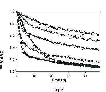
Reactivity decreases in the following order (based on the pseudo rate constants and the half-
life of the dGMP peak): 3 (cis) > 4 (trans) > cisplatin > transplatin > 2 (trans) > 1 (cis)
(Table 2). When comparing the binding kinetics of cis- and transplatin, it can be seen that
transplatin reacts slightly faster than its cis analogue during the first hours of incubation.
Nevertheless, the reaction speed decreases, as the amount of trans-bound dGMP increases,
indicating that the attachment of a second dGMP to the cis isomer is kinetically favored
(Figure 3). This might be due to faster exchange of the second chloro ligand in cisplatin with
water, because aquation is considered a prerequisite for adduct formation (Martin et al., 1999;
Zenker et al., 2000).
On the contrary, the acetoxime-containing complexes 1 (cis) and 2 (trans) show a different
behavior: The trans isomer binds faster to dGMP than the cis form not only in the beginning
but throughout the whole period of incubation. However, due to higher hydrolytic stability,
binding progresses at a much slower rate as compared to the other compounds (Table 2). In
the case of hydroxylamine-containing complexes 3 (cis) and 4 (trans), a similar observation
as for cis- and transplatin can be made: Binding of one equivalent of dGMP progresses
slightly faster for the trans compound, whereas the cis form shows stronger interaction toward
a second dGMP, as reflected by the pseudo rate constant of the overall process.
This article has not been copyedited and formatted. The final version may differ from this version.Molecular Pharmacology Fast Forward. Published on October 18, 2006 as DOI: 10.1124/mol.106.030726
at ASPE
T Journals on January 12, 2019
molpharm
.aspetjournals.orgD
ownloaded from
MOL #30726
15
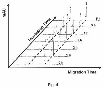
In general, due to the complex nature of the reactions taking place in the sample
simultaneously (aquation, oligomerization, dGMP binding), not all minor peaks in the
electropherograms could be assigned to an exact structure. Anyhow, detection at 254 nm,
analysis of the spectral patterns and migration times of the major peaks enabled us to clearly
distinguish dGMP adducts (Figure 4) and therefore also determine pseudo rate constants and
half-lives (Table 2).
Alterations of DNA secondary structure and reactivity with DNA. In order to examine the
alterations of DNA secondary structure for the investigated compounds, kinetic studies of
either one of the complexes with plasmid DNA were performed.
It is largely documented that platinum-based complexes can untwist, locally melt and/or bend
dsDNA, depending on the kind of the specific adducts formed on DNA (Lepre and Lippard,
1990); for example, monofunctional or intercalating adducts may untwist dsDNA, whereas
bifunctional adducts (intra- as well as interstrand cross-links), in addition, bend DNA.
Conversely, analyzing the DNA secondary structure may provide valuable clues about the
kind of the DNA adducts. Changes of DNA secondary structures can be easily monitored by
evaluating the electrophoretic migration pattern of a circular dsDNA plasmid in neutral
agarose gels. Adducts that untwist dsDNA effect a slower migration of the negatively
"supercoiled form" (sc) of the plasmid due to partial relief of the torsional stress and
consequent relaxing of the compact sc form; a faster migration of the nicked, "open circular"
form (oc) of a plasmid, on the other hand, is consistent with adducts that compact or
apparently "condense" dsDNA (Cohen et al., 1979).
Hence, in order to examine the time-dependent alterations of DNA secondary structure for the
investigated compounds, kinetic studies of the complexes with plasmid DNA were performed.
However, since different DNA-interacting drugs may induce secondary structure changes of
different magnitude, monitoring the kinetics of secondary structures does not necessarily
This article has not been copyedited and formatted. The final version may differ from this version.Molecular Pharmacology Fast Forward. Published on October 18, 2006 as DOI: 10.1124/mol.106.030726
at ASPE
T Journals on January 12, 2019
molpharm
.aspetjournals.orgD
ownloaded from

MOL #30726
16
reflect the degree of DNA modification or reactivity of the drug. For this reason, to directly
visualize the extent of DNA modification, we performed an additional direct control of
reactivity. A linear, radioactively end-labeled 177 bp dsDNA fragment was included in each
reaction and its migration analyzed in a neutral polyacrylamide gel. The modification of the
linear dsDNA fragment leads to increased molecular weight and to additional positive charges
on DNA resulting in upward shifting of the fragment in the gel analysis, which reflects the
modification degree of all DNA in the reaction. Consequently, by this set up it is possible to
visualize both induced alterations of DNA structure and the reactivity of complexes at the
same time.
Figure 5 shows the electrophoretic pattern of plasmid DNA incubated with 60 µM compound
1 (cis) or 2 (trans) at 1 h time intervals for up to 7 h. Both compounds effect relaxation of the
sc form and mobilization of the oc form of the plasmid. These differences in plasmid
migration are consistent with induced untwisting and apparent DNA condensation,
respectively. The DNA condensation may be caused by bending, i.e. rigid, directed but offset
bends or flexible hinge joints (see Discussion).
Compound 2 (trans) (Figure 5C) is evidently more inefficient than compound 1 (cis) (Figure
5A) in inducing changes in DNA secondary structure. This may be due to either slower
kinetics or to a weaker extent of structural changes at individual adducts (or both). However,
the moderate shifting of the radioactively end-labeled dsDNA fragment in gel analysis shown
in Figure 5D as compared to compound 1 (cis) (Figure 5B), reflecting a smaller DNA
modification degree, reveals that the inefficacy of inducing changes in DNA secondary
structure may be rather due to slower kinetics. Hence, the two isomers likely form adducts of
comparable average impact on the DNA structure, albeit with different speed.
Besides the general ability of compound 1 (cis) to react with and induce changes in secondary
structures of DNA faster than compound 2 (trans), an additional difference became visible.
By comparing the mobilization of the oc forms of the plasmid at nearly equivalent global
This article has not been copyedited and formatted. The final version may differ from this version.Molecular Pharmacology Fast Forward. Published on October 18, 2006 as DOI: 10.1124/mol.106.030726
at ASPE
T Journals on January 12, 2019
molpharm
.aspetjournals.orgD
ownloaded from

MOL #30726
17
untwisting degrees (lane 8 in Figure 5C vs. lane 5 in Figure 5A), it is obvious that bending (as
defined above) induced by compound 2 (trans) is less pronounced. This might indicate that, if
this bending is due to closure to bifunctional adducts of compound 2 (trans), this reaction is
also kinetically impaired compared to compound 1 (cis).
The results of the interaction of plasmid DNA and a radioactively end-labeled dsDNA
fragment with complexes of the hydroxylamine type, 3 (cis) and 4 (trans), are shown in
Figure 6. Both complexes displayed a much higher ability to induce changes in DNA
secondary structure than the acetoxime compounds. Beyond 1 h or 2 h of incubation,
respectively, compounds 3 (cis) and 4 (trans) untwisted the plasmid to positive supercoils.
The marked formation of adducts with plasmid DNA was accompanied by a distinctive
shifting of the included DNA fragment for both compounds shown in Figures 6B and 6D. In
addition, the 4 (trans) isomer showed a different migration behavior of the open circular form
of the plasmid, analogous to the 2 (trans) isomer of the acetoxime type.
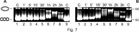
As a control, plasmid DNA was also incubated with cisplatin and transplatin. As expected,
both compounds effected relaxation of the sc form and mobilization of the oc form of the
plasmid, whereas transplatin showed a slightly higher efficiency to induce secondary
structures as contrasted to cisplatin (Figure 7). This parallels the known higher reactivity of
transplatin against DNA (Farrell et al., 1992). In accordance to the trans isomers 2 (trans) and
4 (trans), it is apparent that, at corresponding untwisting extent, the mobilization of the oc
form of the plasmid caused by transplatin was less pronounced than that caused by cisplatin.
With both substrates, dGMP and DNA fragment, the results show coincidence regarding the
comparative reactivities between each pair, i. e. 3 (cis)/4 (trans) prevail over
cisplatin/transplatin which beat 1 (cis)/2 (trans). Likewise, the reactivity order within the pair
3 (cis)/4 (trans) is matching between dGMP and DNA. However, the reactivity order within
the other two pairs is reversed depending on the substrate. Besides possible, inevitable
imprecision of the results with the DNA fragment, we suggest that the DNA results reflect
This article has not been copyedited and formatted. The final version may differ from this version.Molecular Pharmacology Fast Forward. Published on October 18, 2006 as DOI: 10.1124/mol.106.030726
at ASPE
T Journals on January 12, 2019
molpharm
.aspetjournals.orgD
ownloaded from
MOL #30726
18
rather faithfully the reactivity of the compounds. Investigations of reactivity with dGMP may
not mirror the rate of formation of DNA adducts in a representative way, because the rate of
formation of dGMP adducts is governed by the complete translational and rotational freedom
of the soluble substrate dGMP in contrast to the sterically constrained and spatially well
defined target bases in DNA.
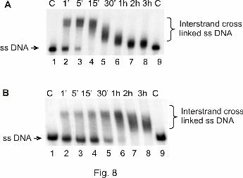
Formation of interstrand cross-links. It is known that intrastrand as well as interstrand
cross-links (ICLs) can bend DNA. To examine whether the investigated compounds can form
interstrand cross-links, a radioactively 3'-end-labeled 177 bp DNA fragment was incubated
with either one of the investigated compounds, and the reaction products were analyzed in a
denaturing urea-polyacrylamide gel. On the basis of this set up, the former double-stranded
DNA molecule appears single-stranded at lower regions of the gel when no ICLs are being
formed. If the investigated complex is able to form ICLs, a new distinct band with lower
mobility is visible in the gel, representing a former double-stranded DNA molecule with a
minimum of one interstrand cross-link not able to be separated in a denaturing polyacryamide
gel.
Figure 8 shows the results of the reaction of the hydroxylamine platinum complexes 3 (cis)
and 4 (trans) with linear dsDNA after separation in a denaturing urea-polyacrylamide gel.
Both complexes showed a clear increase of ICL formation over time. Beyond 15 min for
compound 3 (cis) or 1 h for compound 4 (trans), all DNA molecules contained at least one
ICL, displayed in discrete upward shifts of DNA. In general, further incubation led to faster
migration of the ICL-connected DNA strands. This might be due to an increasing
compactness of the DNA strands containing more ICLs, therefore mimicking the form and
migration properties of linear dsDNA.
This article has not been copyedited and formatted. The final version may differ from this version.Molecular Pharmacology Fast Forward. Published on October 18, 2006 as DOI: 10.1124/mol.106.030726
at ASPE
T Journals on January 12, 2019
molpharm
.aspetjournals.orgD
ownloaded from
MOL #30726
19
As contrasted to the clear formation of interstrand cross-links induced by compounds 3 (cis)
and 4 (trans), the acetoxime platinum complexes 1 (cis) and 2 (trans) showed no formation of
ICLs whatsoever for the investigated time points (1–7 h) (data not shown).
This article has not been copyedited and formatted. The final version may differ from this version.Molecular Pharmacology Fast Forward. Published on October 18, 2006 as DOI: 10.1124/mol.106.030726
at ASPE
T Journals on January 12, 2019
molpharm
.aspetjournals.orgD
ownloaded from

MOL #30726
20
Discussion
We have discovered that trans-[PtCl2(acetoxime)2], 2 (trans), is a new unconventional
platinum compound violating the classic structure-activity relationship, insofar as its
cytotoxicity is comparable with cisplatin in the ovarian carcinoma cell line CH1 and even
superior to cisplatin by one order of magnitude in the rather cisplatin-insensitive colon
carcinoma cell line SW480, indicating a potential of overcoming primary cisplatin resistance.
This again emphasizes the great relevance of active trans complexes for the development of
new platinum-based anticancer drugs, since non-cross-resistance to cisplatin is common to
most of these compounds. The observation that 2 (trans) is 15–16 times more cytotoxic than
1 (cis) is striking, since the cis counterparts of active trans complexes investigated by other
authors are not substantially less active (e. g. Farrell et al., 1992; Farrell, 1996), indicating
that the substitution of ammine by appropriate ligands usually activates the trans geometry
without severely impairing the activity of the cis congener. Hence, this is, to our knowledge,
the first successful reversion of the structure-activity relationship of the cis and trans
geometry.
A synopsis with the active trans-platinum complexes reported by other authors (see
Introduction) reveals that 2 (trans) shares with both iminoether and acetimine complexes the
azomethine moiety C=N and the planarity around the nitrogen donor atom resulting from its
sp2 hybridization. This also applies to N-heterocyclic complexes, but the involvement of the
nitrogen donor atom in an aromatic ring system strongly distinguishes them from the former.
Furthermore, the acetoxime ligand shares with the branched aliphatic amine and the acetimine
type of ligands the ramification of the alkyl residue at the proximate carbon atom. The
minimal structural requirement for an active trans-platinum complex, as inferred from this
synopsis, is the presence of at least one of the following characteristics: (i) an sp2-hybridized
This article has not been copyedited and formatted. The final version may differ from this version.Molecular Pharmacology Fast Forward. Published on October 18, 2006 as DOI: 10.1124/mol.106.030726
at ASPE
T Journals on January 12, 2019
molpharm
.aspetjournals.orgD
ownloaded from
MOL #30726
21
nitrogen donor atom (ii) a branched aliphatic chain attached to the nitrogen donor atom or (iii)
a nitrogen donor atom integrated into a cycloaliphatic amine.
While the acetoxime complex 2 (trans) resembles the acetimine complexes of Natile and co-
workers (Boccarelli et al., 2006) in two crucial respects, i. e. the planar azomethine and the
branched aliphatic moiety, it differs from them (and from all other examples of active trans-
platinum complexes) by the formal substitution of the nitrogen-bound hydrogen by hydroxyl
groups. Since this renders the compound a stronger H-bonding donor than conventional amine
complexes, an involvement of hydrogen bonding in the DNA interactions, e. g. in stabilizing
monofunctional adducts, should be considered. Moreover, the OH acidity of the metal-bound
acetoximes is significant (pKa1 6–7) (Kukushkin et al., 1996), but although this acidity
constitutes a major difference to other active trans-platinum complexes, the sole presence of a
hydroxyl group bound to the nitrogen donor atom is neither sufficient nor essential for activity
of the trans isomer, as can be inferred from the classic structure-activity relationship of the
[PtCl2(hydroxylamine)2] couple, 3 (cis) and 4 (trans).
The reactivity of the compounds has been investigated by monitoring the reaction with dGMP
and with a DNA fragment. Although 2 (trans) was the least reactive with DNA, it was the
most cytotoxic of the investigated compounds. The fact that slowly reacting compounds
display a rather strong cytotoxicity or, inversely, that strongly reacting compounds may be
devoid of biologic activity is striking but not new or astonishing. For instance, it has been
repeatedly shown that transplatin reacts about 2.5-fold more efficiently than cisplatin with
both calf thymus and plasmid DNA (see e. g. Farrell et al., 1992), yet without favorable
impact on its cytotoxicity. Likewise, the cytotoxity of trans-
dichlorobis(E-iminoether)platinum(II), trans-EE, which is closely related to 2 (trans), is
comparable to cisplatin in the P388 leukemia system (Coluccia et al., 1995), although
cisplatin displays significantly faster reaction kinetics with calf thymus DNA than trans-EE
(Coluccia et al., 1995; Žaludová et al., 1997). It may be that efficient reaction of a compound
This article has not been copyedited and formatted. The final version may differ from this version.Molecular Pharmacology Fast Forward. Published on October 18, 2006 as DOI: 10.1124/mol.106.030726
at ASPE
T Journals on January 12, 2019
molpharm
.aspetjournals.orgD
ownloaded from

MOL #30726
22
even with the cellular DNA cannot bring about increased cytotoxicity, if its adducts are
rapidly removed by repair systems. Instead, formation of repair-resistant adducts that actively
lead to programmed cell death is critical for the activity of the compounds (Zorbas and
Keppler, 2005). Hence, the cytotoxicity of active trans isomers in general and, in particular,
the isomer 2 (trans) investigated in this study may, therefore, rely on the formation of
particularly potent adducts.
In fact, the cytotoxic power of platinum complexes has been associated with adducts that
induce particular secondary structures of DNA (Eastman, 1999; Kartalou and Essigmann,
2001). We found that our novel compounds were able to induce changes of DNA structure,
namely visible DNA relaxation and DNA condensation. Relaxation of the scDNA was
obviously effected by local untwisting at the sites of adducts. In accord to numerous
investigations of platinum compounds, the detected untwisting is consistent with formation of
monofunctional adducts at purine nucleobases. Hence, the compounds of both types
(hydroxylamine and acetoxime) may form monofunctional adducts. Condensed circular DNA
modified with platinum complexes, first described for cisplatin and transplatin by Cohen et al.
(Cohen et al., 1979), was recognized by Bellon et al. as caused by multiple, rigid or flexible
bends not in phase with the DNA periodicity leading to apparent diminished diameter of
circular DNA (Bellon et al., 1991; Bellon and Lippard, 1990). We cannot distinguish between
the two variants of bending, stable or flexible, in this study. Bending may be caused by
bifunctional adducts, either intra- (Takahara et al., 1995) or interstrand (Huang et al., 1995)
cross-links. In addition, some compounds with the trans geometry have been reported to
effect bending by monofunctional coordination (Kasparkova et al., 2003; Novakova et al.,
2003; Zakovska et al., 1998), although, at least for the trans compounds with heterocyclic
ligands, (stacking) interactions of these ligand(s) with DNA were also discussed, which might
give rise to "pseudobifunctional" adducts (Zakovska et al., 1998). Hence, compounds of the
hydroxylamine type might have caused bending by any kind of adducts. On the other hand,
This article has not been copyedited and formatted. The final version may differ from this version.Molecular Pharmacology Fast Forward. Published on October 18, 2006 as DOI: 10.1124/mol.106.030726
at ASPE
T Journals on January 12, 2019
molpharm
.aspetjournals.orgD
ownloaded from
MOL #30726
23
since the acetoxime compounds lacked the capacity of forming ICLs, bending with these
compounds cannot have been effected by this type of cross-links, but rather by bifunctional
intrastrand cross-links or, in the case of 2 (trans), even monofunctional adducts.
We observed that the induced condensation of the oc form was less pronounced with all
investigated trans isomers than with all investigated cis isomers at comparable global
untwisting, best visible with the pair 1 (cis)/2 (trans) (Figure 5). If bifunctional adducts were
the cause of bending, this might be an indication of a slower reaction of the second platinum
valence of the trans isomers as compared to the cis isomers. Interestingly though, the same
observation has been made with trans- vs. cis-EE (see Fig. 5 in Žaludová et al., 1997), i. e.
with a structurally similar, also active trans compound that is known to cause bending by
abundant monofunctional adducts (Novakova et al., 2003). By analogy hence, it is tempting to
speculate, that in our case bending might have been effected by monofunctional adducts as
well. This, however, will by subject of future studies.
Which structural feature might constitute the high cytotoxicity of our active compounds?
Intrastrand cross-links, like the 1,2-d(GpG) adduct of cisplatin, which displays unique
structural features of which the main characteristic is a rigid, directed bend of 30–35 degrees
into the major groove of dsDNA (Jamieson and Lippard, 1999), may be important cytotoxic
lesions. On the other hand, ICLs, although minor adducts in general (e. g. ~2% of all cisplatin
adducts), have never been excluded as possible lethal lesions of platinum complexes. In fact,
ICLs may be equally important cytotoxic adducts under certain conditions like intrastrand
adducts (Aloyz et al., 2002; Zdraveski et al., 2000). 3 (cis) and 4 (trans) readily form ICLs.
Since 3 (cis) displays a rather fairly high cytotoxicity, we cannot exclude that, in this case,
ICLs may have contributed to the biologic effect. However, ICLs are certainly not sufficient
for cytotoxicity, as 4 (trans), which shows comparable ICL formation kinetics like 3 (cis),
was quite inactive. In contrast, we could not detect any ICLs for the time period investigated
with the acetoxime compounds, particularly with the active 2 (trans) compound. Therefore,
This article has not been copyedited and formatted. The final version may differ from this version.Molecular Pharmacology Fast Forward. Published on October 18, 2006 as DOI: 10.1124/mol.106.030726
at ASPE
T Journals on January 12, 2019
molpharm
.aspetjournals.orgD
ownloaded from

MOL #30726
24
ICLs seem definitely to be not necessary for the superb cytotoxic activity of the 2 (trans).
Interestingly, the highly cytotoxic and antitumoral trans-EE was also found to have a very
small DNA interstrand cross-linking efficacy (Coluccia et al., 1995; Žaludová et al., 1997).
If monofunctional adducts constitute a major fraction of the 2 (trans) lesions, they may
contribute significantly to cytotoxicity as well. As was shown for monofunctional adducts of
trans-EE (Novakova et al., 2003), proteins like histone H1 may be readily captured by the
available valence of the platinum giving rise to ternary DNA-drug-protein complexes. Such
complexes inhibit in vitro DNA polymerization and, most importantly, removal of adducts by
the NER system. Resultant prolonged persistence of such adducts may facilitate in vivo the
onset of cell death mechanisms. Further investigations will evaluate this possibility for our
compounds.
Acknowledgements
We (S. Z.-S. and H. Z.) express our gratitude to Prof. Dieter Oesterhelt, Director of the Max-
Planck Institute of Biochemistry, Martinsried, Department of Membrane Biochemistry, for
continuous and generous support.
This article has not been copyedited and formatted. The final version may differ from this version.Molecular Pharmacology Fast Forward. Published on October 18, 2006 as DOI: 10.1124/mol.106.030726
at ASPE
T Journals on January 12, 2019
molpharm
.aspetjournals.orgD
ownloaded from

MOL #30726
25
References
Alexander H (1900) Lieb Ann 311:120.
Aloyz R, Xu ZY, Bello V, Bergeron J, Han FY, Yan Y, Malapetsa A, Alaoui-Jamali MA,
Duncan AM and Panasci L (2002) Regulation of cisplatin resistance and homologous
recombinational repair by the TFIIH subunit XPD. Cancer Res 62:5457–5462.
Bellon SF and Lippard SJ (1990) Bending studies of DNA site-specifically modified by
cisplatin, trans-diamminedichloroplatinum(II) and cis-[Pt(NH3)2(N3-cytosine)Cl]+.
Biophys Chem 35:179–188.
Bellon SF, Coleman JH and Lippard SJ (1991) DNA unwinding produced by site-specific
intrastrand cross-links of the antitumor drug cis-diamminedichloroplatinum(II).
Biochemistry 30:8026–8035.
Boccarelli A, Intini FP, Sasanelli R, Sivo MF, Coluccia M and Natile G (2006) Synthesis and
in vitro antitumor activity of platinum acetonimine complexes. J Med Chem 49:829–37.
Brabec V and Kasparkova J (2005) DNA interactions of platinum anticancer drugs. Recent
advances and mechanisms of action, in Metal Compounds in Cancer Chemotherapy
(Pérez JM, Fuertes MA and Alonso C eds) pp 187–218, Research Signpost, Kerala.
Cleare MJ and Hoeschele JD (1973) Studies on the antitumor activity of group VIII transition
metal complexes. Part I. Platinum(II) complexes. Bioinorg Chem 2:187–210.
Cohen GL, Bauer WR, Barton JK and Lippard SJ (1979) Binding of cis- and trans-
dichlorodiammineplatinum(II) to DNA: evidence for unwinding and shortening of the
double helix. Science 203:1014–1016.
Coluccia M, Boccarelli A, Mariggio MA, Cardellicchio N, Caputo P, Intini FP and Natile G
(1995) Platinum(II) complexes containing iminoethers: a trans platinum antitumour
agent. Chem-Biol Interact 98:251–266.
This article has not been copyedited and formatted. The final version may differ from this version.Molecular Pharmacology Fast Forward. Published on October 18, 2006 as DOI: 10.1124/mol.106.030726
at ASPE
T Journals on January 12, 2019
molpharm
.aspetjournals.orgD
ownloaded from
MOL #30726
26
Coluccia M, Nassi A, Boccarelli A, Giordano D, Cardellicchio N, Locker D, Leng M, Sivo
M, Intini FP and Natile G (1999) In vitro and in vivo antitumour activity and cellular
pharmacological properties of new platinum-iminoether complexes with different
configuration at the iminoether ligands. J Inorg Biochem 77:31–35.
Eastman A (1999) The mechanism of action of cisplatin: From adducts to apoptosis, in
Cisplatin. Chemistry and Biochemistry of a Leading Anticancer Drug (Lippert B ed) pp
111–134, Wiley-VCH, Weinheim, Germany.
Farrell N, Kelland LR, Roberts JD and Van Beusichem M (1992) Activation of the trans
geometry in platinum antitumor complexes: a survey of the cytotoxicity of trans
complexes containing planar ligands in murine L1210 and human tumor panels and
studies on their mechanism of action. Cancer Res 52:5065–5072.
Farrell N (1996) Current status of structure-activity relationships of platinum anticancer
drugs: activation of the trans geometry, in Interactions of Metal Ions with Nucleotides,
Nucleic Acids, and Their Constituents (Sigel A and Sigel H eds) (Vol 32 of Metal Ions in
Biological Systems) pp 603–639, M. Dekker, New York.
Hartinger C, Timerbaev AR and Keppler BK (2003) Capillary electrophoresis in anti-cancer
metallodrug research: advances and future challenges. Electrophoresis 24:2023–2037.
Huang H, Zhu L, Reid BR, Drobny GP and Hopkins PB (1995) Solution structure of a
cisplatin-induced DNA interstrand cross-link. Science 270:1842–1845.
Intini FP, Boccarelli A, Francia VC, Pacifico C, Sivo MF, Natile G, Giordano D, De Rinaldis
P and Coluccia M (2004) Platinum complexes with imino ethers or cyclic ligands
mimicking imino ethers: synthesis, in vitro antitumour activity, and DNA interaction
properties. J Biol Inorg Chem 9:768–780.
Jakupec MJ, Galanski M and Keppler BK (2003) Tumour-inhibiting platinum complexes –
state of the art and future perspectives. Rev Physiol Biochem Pharmacol 146:1–53.
This article has not been copyedited and formatted. The final version may differ from this version.Molecular Pharmacology Fast Forward. Published on October 18, 2006 as DOI: 10.1124/mol.106.030726
at ASPE
T Journals on January 12, 2019
molpharm
.aspetjournals.orgD
ownloaded from
MOL #30726
27
Jamieson ER and Lippard SJ (1999) Structure, recognition, and processing of cisplatin–DNA
adducts. Chem Rev 99:2467–2498.
Kartalou M and Essigmann JM (2001) Recognition of cisplatin adducts by cellular proteins.
Mutat Res 478:1–21.
Kasparkova J, Novakova O, Farrell N and Brabec V (2003) DNA binding by antitumor trans-
[PtCl2(NH3)(thiazole)]. Protein recognition and nucleotide excision repair of
monofunctional adducts. Biochemistry 42:792–800.
Kelland LR, Barnard CFJ, Evans IG, Murrer BA, Theobald BRC, Wyer SB, Goddard PM,
Jones M, Valenti M, Bryant A, Rogers PM and Harrap KR (1995) Synthesis and in vitro
and in vivo antitumor activity of a series of trans platinum antitumor complexes. J Med
Chem 38:3016–3024.
Kukushkin VY, Tudela D and Pombeiro AJL (1996) Metal-ion assisted reactions of oximes
and reactivity of oxime-containing metal complexes. Coord Chem Rev 156:333–62.
Kukushkin VY, Izotova YA and Tudela D (2004) Platinum(II) complexes of propanone
oxime. Inorg Synth 34:81–85.
Lepre CA and Lippard SJ (1990) Interaction of platinum antitumor compounds with DNA, in
Nucleic Acids and Molecular Biology (Eckstein F, Lilley DMJ eds) pp 9–38, Springer,
Berlin.
Martin RB (1999) Platinum complexes: hydrolysis and binding to N(7) and N(1) of purines,
in Cisplatin. Chemistry and biochemistry of a leading anticancer drug (Lippert B ed) pp
183–205, Verlag Helvetica Chimica Acta, Zürich, and Wiley-VCH, Weinheim.
Najajreh Y, Khazanov E, Jawbry S, Ardeli-Tzaraf Y, Pérez JM, Kasparkova J, Brabec V and
Barenholz Y, Gibson D (2006) Cationic nonsymmetric transplatinum complexes with
piperidinopiperidine ligands. Preparation, characterization, in vitro cytotoxicity, in vivo
toxicity, and anticancer efficacy studies. J Med Chem 49:4665–4673.
This article has not been copyedited and formatted. The final version may differ from this version.Molecular Pharmacology Fast Forward. Published on October 18, 2006 as DOI: 10.1124/mol.106.030726
at ASPE
T Journals on January 12, 2019
molpharm
.aspetjournals.orgD
ownloaded from

MOL #30726
28
Natile G and Coluccia M (2004) Antitumor active trans-platinum compounds, in Metal
Complexes in Tumor Diagnosis and as Anticancer Agents (Sigel A and Sigel H eds) (Vol
42 of Metal Ions in Biological Systems) pp 209–250, M. Dekker, New York.
Novakova O, Kasparkova J, Malina J, Natile G and Brabec V (2003) DNA–protein cross-
linking by trans-[PtCl2(E-iminoether)2]. A concept for activation of the trans geometry in
platinum antitumor complexes. Nucleic Acids Res 31:6450–6460.
Pérez JM, Fuertes MA, Alonso C and Navarro-Ranninger C (2000) Current status of the
development of trans-platinum antitumor drugs. Crit Rev Oncol Hematol 35:109–120.
Pérez JM, Kelland LR, Montero EI, Boxall FE, Fuertes MA, Alonso C and Navarro-
Ranninger C (2003) Antitumor and cellular pharmacological properties of a novel
platinum(IV) complex: trans-[PtCl2(OH)2(dimethylamine)(isopropylamine)]. Mol
Pharmacol 63:933–944.
Stetsenko AI, Adamov OM, Dmitrieva ES, Prokhoda EF, Budnikova TI and Dankovskaya
NV (1989) A method for preparation of cis-dichlorobis(hydroxylamine)platinum(II).
Patent USSR 1561488.
Takahara PM, Rosenzweig AC, Frederick CA and Lippard SJ (1995) Crystal structure of
double-stranded DNA containing the major adduct of the anticancer drug cisplatin.
Nature 377:649–652.
Timerbaev AR, Hartinger CG, Aleksenko SS and Keppler BK (2006) Interactions of
antitumor metallodrugs with serum proteins: advances in characterization using modern
analytical methodology. Chem Rev 106:2224–2248.
Wong E and Giandomenico CM (1999) Current status of platinum-based antitumor drugs.
Chem Rev 99:2451–2466.
Zakovska A, Novakova O, Balcarova Z, Bierbach U, Farrell N and Brabec V (1998) DNA
interactions of antitumor trans-[PtCl2(NH3)(quinoline)]. Eur J Biochem 254:547–557.
This article has not been copyedited and formatted. The final version may differ from this version.Molecular Pharmacology Fast Forward. Published on October 18, 2006 as DOI: 10.1124/mol.106.030726
at ASPE
T Journals on January 12, 2019
molpharm
.aspetjournals.orgD
ownloaded from
MOL #30726
29
Žaludová R, Žákovská A, Kašpárkova J, Balcarová Z, Vrána O, Coluccia M, Natile G and
Brabec V (1997) DNA modifications by antitumor trans-[PtCl2(E-iminoether)2]. Mol
Pharmacol 52:354–361.
Zdraveski ZZ, Mello JA, Marinus MG and Essigmann JM (2000) Multiple pathways of
recombination define cellular responses to cisplatin. Chem Biol 7:39–50.
Zenker A, Galanski M,. Bereuter T, Keppler BK and Lindner W (2000) Time-dependent
interactions of platinum(II) complexes with 5’-GMP under simulated physiological
conditions studied by capillary electrophoresis. J Biol Inorg Chem 5:498–504.
Zorbas H and Keppler BK (2005) Cisplatin damage: are DNA repair proteins saviors or
traitors to the cell? ChemBioChem 6:1157–1166.
This article has not been copyedited and formatted. The final version may differ from this version.Molecular Pharmacology Fast Forward. Published on October 18, 2006 as DOI: 10.1124/mol.106.030726
at ASPE
T Journals on January 12, 2019
molpharm
.aspetjournals.orgD
ownloaded from

MOL #30726
30
Footnote:
a) Financial support for this study was provided by the FFG – Austrian Research Promotion
Agency (811591), the Austrian Council for Research and Technology Development
(IS526001), the FWF – Austrian Science Fund (P16186-NO3, P18123-N11, P16192-NO3,
P14519-CHE), COST D20, Faustus Forschung Translational Drug Development AG Vienna
(M.A.J., M.G., C.G.H., and B.K.K.), the Austrian Science Foundation under project No.
P14519-CHE (O.S.) and the Russian Fund for Basic Research (grants 05-03-32140, 06-03-
90901, and 06-03-32065) (V.Yu.K.). S. Z.-S. is a recipient of a Ph.D. fellowship of the Max-
Planck Society.
This article has not been copyedited and formatted. The final version may differ from this version.Molecular Pharmacology Fast Forward. Published on October 18, 2006 as DOI: 10.1124/mol.106.030726
at ASPE
T Journals on January 12, 2019
molpharm
.aspetjournals.orgD
ownloaded from

MOL #30726
31
Legends for Figures
Figure 1. Structures of acetoxime platinum complexes 1 (cis) and 2 (trans), hydroxylamine
platinum complexes 3 (cis) and 4 (trans), cisplatin and transplatin.
Figure 2. Concentration-effect curves of acetoxime platinum complexes 1 (cis) and 2 (trans)
(upper panels) and hydroxylamine platinum complexes 3 (cis) and 4 (trans) (lower panels) as
compared to cisplatin and transplatin in the human cancer cell lines CH1 (left) and SW480
(right). Values were obtained by the MTT assay and are the means ± standard deviations from
at least three independent experiments.
Figure 3. Time courses of the dGMP binding reaction of all studied substances. The solid
lines correspond to cisplatin ( ) and transplatin ( ), the long dashes to
acetoxime complexes 1 (cis) ( ) and 2 (trans) ( ), and the short dashes to
hydroxylamine complexes 3 (cis) ( ) and 4 (trans) ( ).
Figure 4. Monitoring of the dGMP-binding reaction of compound 3 (cis). Separation voltage:
15 kV. Detection wavelength: 254 nm. Other conditions: see experimental section. Peak
identification: 1, monoadduct; 2, bisadduct; 3, dGMP.
Figure 5. Interaction of acetoxime platinum complexes 1 (cis) (A, B) and 2 (trans) (C, D)
with dsDNA. Products of plasmid pTZ18u (A, C) and of a linear, radioactively labeled 177 bp
DNA fragment (B, D) after incubation with 60 µM compound 1 (cis) and compound 2 (trans),
respectively, for the indicated time points (lanes 2–8) and electrophoretic separation; A, C:
1% agarose gel stained with EtBr; B, D: autoradiograph of a dried, neutral 4% polyacrylamide
gel. C (lanes 1), control DNA, not incubated; C (lanes 9), control DNA, incubated for 7 hours.
This article has not been copyedited and formatted. The final version may differ from this version.Molecular Pharmacology Fast Forward. Published on October 18, 2006 as DOI: 10.1124/mol.106.030726
at ASPE
T Journals on January 12, 2019
molpharm
.aspetjournals.orgD
ownloaded from

MOL #30726
32
Figure 6. Interaction of hydroxylamine platinum complexes 3 (cis) (A, B) and 4 (trans) (C,
D) with dsDNA. Products of plasmid pTZ18u (A, C) and of a linear, radioactively labeled
177 bp DNA fragment (B, D) after incubation with 60 µM compound 3 (cis) and compound,
4 (trans), respectively, for the indicated time points (lanes 2–8) and electrophoretic
separation; A, C: 1% agarose gel stained with EtBr; B, D: autoradiograph of a dried, neutral
4% polyacrylamide gel. C (lanes 1), control DNA, not incubated; C (lanes 9), control DNA,
incubated for 3 hours.
Figure 7. Products of plasmid DNA after incubation with 60 µM cisplatin (A) and transplatin
(B), respectively, for the indicated time points (lanes 2–8) and electrophoretic separation in
1% agarose gel stained with EtBr. C (lanes 1), control DNA, not incubated; C (lanes 9),
control DNA, incubated for 3 hours.
Figure 8. Formation of ICLs by hydroxylamine platinum complexes 3 (cis) and 4 (trans).
Products of a linear, radioactively labeled 177 bp DNA fragment after incubation with 60 µM
compound 3 (cis) (A) and compound 4 (trans) (B), respectively, for the indicated time points
(lanes 2–8) and electrophoretic separation in a denaturing 4% polyacrylamide gel; the panels
depict the autoradiograph of the dried gels. C (lanes 1), control DNA, not incubated; C (lanes
9), control DNA, incubated for 3 hours.
This article has not been copyedited and formatted. The final version may differ from this version.Molecular Pharmacology Fast Forward. Published on October 18, 2006 as DOI: 10.1124/mol.106.030726
at ASPE
T Journals on January 12, 2019
molpharm
.aspetjournals.orgD
ownloaded from

MOL #30726
33
Table 1. Cytotoxicity of acetoxime platinum complexes 1 (cis) and 2 (trans) and
hydroxylamine platinum complexes 3 (cis) and 4 (trans) as compared to cisplatin and
transplatin in two human cancer cell lines
IC50 (µM)a
Compound CH1 SW480
1 (cis) 2.7 ± 0.7 3.4 ± 0.3
2 (trans) 0.17 ± 0.09 0.22 ± 0.05
3 (cis) 0.68 ± 0.23 12 ± 1
4 (trans) 51 ± 15 90 ± 3
cisplatin 0.14 ± 0.03 3.3 ± 0.4
transplatin 15 ± 2 19 ± 3
a 50% inhibitory concentrations in CH1 and SW480 cells in the MTT assay. Values are the
means ± standard deviations obtained from at least three independent experiments.
This article has not been copyedited and formatted. The final version may differ from this version.Molecular Pharmacology Fast Forward. Published on October 18, 2006 as DOI: 10.1124/mol.106.030726
at ASPE
T Journals on January 12, 2019
molpharm
.aspetjournals.orgD
ownloaded from
MOL #30726
34
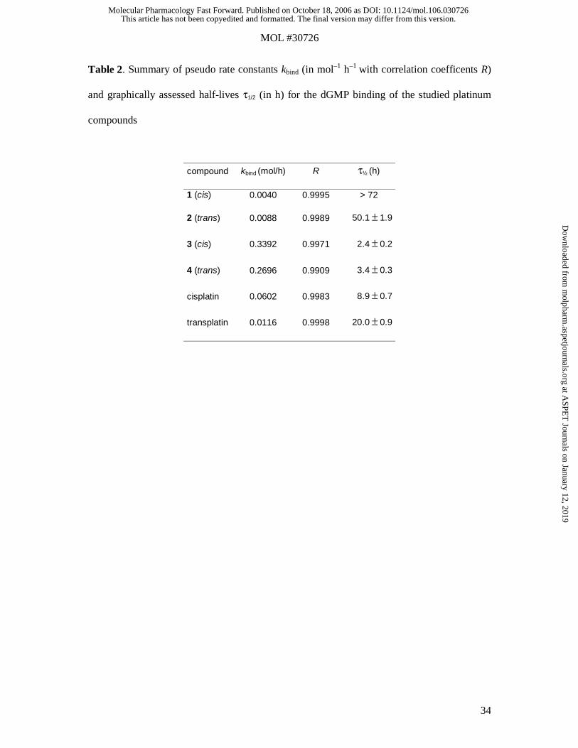
Table 2. Summary of pseudo rate constants kbind (in mol–1 h–1 with correlation coefficents R)
and graphically assessed half-lives τ1/2 (in h) for the dGMP binding of the studied platinum
compounds
compound kbind (mol/h) R τ½ (h)
1 (cis) 0.0040 0.9995 > 72
2 (trans) 0.0088 0.9989 50.1 ± 1.9
3 (cis) 0.3392 0.9971 2.4 ± 0.2
4 (trans) 0.2696 0.9909 3.4 ± 0.3
cisplatin 0.0602 0.9983 8.9 ± 0.7
transplatin 0.0116 0.9998 20.0 ± 0.9
This article has not been copyedited and formatted. The final version may differ from this version.Molecular Pharmacology Fast Forward. Published on October 18, 2006 as DOI: 10.1124/mol.106.030726
at ASPE
T Journals on January 12, 2019
molpharm
.aspetjournals.orgD
ownloaded from

This article has not been copyedited and formatted. The final version may differ from this version.Molecular Pharmacology Fast Forward. Published on October 18, 2006 as DOI: 10.1124/mol.106.030726
at ASPE
T Journals on January 12, 2019
molpharm
.aspetjournals.orgD
ownloaded from

This article has not been copyedited and formatted. The final version may differ from this version.Molecular Pharmacology Fast Forward. Published on October 18, 2006 as DOI: 10.1124/mol.106.030726
at ASPE
T Journals on January 12, 2019
molpharm
.aspetjournals.orgD
ownloaded from
This article has not been copyedited and formatted. The final version may differ from this version.Molecular Pharmacology Fast Forward. Published on October 18, 2006 as DOI: 10.1124/mol.106.030726
at ASPE
T Journals on January 12, 2019
molpharm
.aspetjournals.orgD
ownloaded from
This article has not been copyedited and formatted. The final version may differ from this version.Molecular Pharmacology Fast Forward. Published on October 18, 2006 as DOI: 10.1124/mol.106.030726
at ASPE
T Journals on January 12, 2019
molpharm
.aspetjournals.orgD
ownloaded from

This article has not been copyedited and formatted. The final version may differ from this version.Molecular Pharmacology Fast Forward. Published on October 18, 2006 as DOI: 10.1124/mol.106.030726
at ASPE
T Journals on January 12, 2019
molpharm
.aspetjournals.orgD
ownloaded from

This article has not been copyedited and formatted. The final version may differ from this version.Molecular Pharmacology Fast Forward. Published on October 18, 2006 as DOI: 10.1124/mol.106.030726
at ASPE
T Journals on January 12, 2019
molpharm
.aspetjournals.orgD
ownloaded from
This article has not been copyedited and formatted. The final version may differ from this version.Molecular Pharmacology Fast Forward. Published on October 18, 2006 as DOI: 10.1124/mol.106.030726
at ASPE
T Journals on January 12, 2019
molpharm
.aspetjournals.orgD
ownloaded from
This article has not been copyedited and formatted. The final version may differ from this version.Molecular Pharmacology Fast Forward. Published on October 18, 2006 as DOI: 10.1124/mol.106.030726
at ASPE
T Journals on January 12, 2019
molpharm
.aspetjournals.orgD
ownloaded from
Related Documents