Reverse blot hybridization assay Sepsis-ID test for simultaneous identification of pathogens and antimicrobial resistance genes of mecA, van A and van B from blood culture bottles Soon Deok Park The Graduate School Yonsei University Department of Biomedical Laboratory Science

Welcome message from author
This document is posted to help you gain knowledge. Please leave a comment to let me know what you think about it! Share it to your friends and learn new things together.
Transcript
Reverse blot hybridization assay Sepsis-ID test
for simultaneous identification of pathogens and
antimicrobial resistance genes of mecA, vanA and
vanB from blood culture bottles
Soon Deok Park
The Graduate School
Yonsei University
Department of Biomedical
Laboratory Science
Reverse blot hybridization assay Sepsis-ID test
for simultaneous identification of pathogens and
antimicrobial resistance genes of mecA, vanA and
vanB from blood culture bottles
Soon Deok Park
The Graduate School
Yonsei University
Department of Biomedical
Laboratory Science
Reverse blot hybridization assay Sepsis-ID test
for simultaneous identification of pathogens and
antimicrobial resistance genes of mecA, vanA and
vanB from blood culture bottles
A Dissertation
Submitted to the Department of Biomedical Laboratory Science
and the Graduate School of Yonsei University
in partial fulfillment of the
requirements for the degree of
Doctor of Philosophy
Soon Deok Park
December 2013
This certifies that the doctoral dissertation of Soon Deok Parkis approved.
Thesis Supervisor: Jong Bae Kim
Thesis Committee Member: Yong Serk Park
Thesis Committee Member: Hyeyoung Lee
Thesis Committee Member: Ki-Jong Rhee
Thesis Committee Member: Young Uh
The Graduate SchoolYonsei UniversityDecember 2013

- v -
ACKNOWLEDGEMENT
Writing a thesis is like running a marathon which requires a great deal ofpatience, diligence and carefully made plans. Tremendous help from great numberof people has placed me at the finish line of my long journey. It is a pleasure toconvey my gratitude to them all in my humble acknowledgement. First of all, Iwould like to express the deepest appreciation to professors Jongbae Kim, whocontinually inspired and enriched my growth as a student, Young Uh, whocontinually and convincingly conveyed a spirit of adventure in regard to researchand excitement in regard to teaching, and Hyeyoung Lee for providing me with agood environment and facilities for experiment to complete this study. Also Iwould like to thank professors, Yongserk Park and Ki-Jong Rhee for their crucialand valuable advice and using their precious time to read this thesis and givingout critical comments, and Taeue Kim, Yoonsuk Kim and Boyoung Jeon forproviding me with a good advice. I am grateful for the lab members in hospitaland school for technical assistance and maintenance of the tools used in thisstudy. Without their guidance and persistent help this dissertation would not havebeen possible. An honorable mention goes to our families for their understandingand supports on me in completing the thesis. I am forever indebted to myhusband, Jesun Lee for his understanding, endless patience and encouragementwhen it is most required. Words fail me to express my appreciation and love formy sons, Sangyun and Sangmin. Finally, I would like to thank everyone who wasimportant to the successful realization of thesis, as well as expressing my apologythat I could not mention personally one by one.
Soon Deok ParkDecember, 2013
- vi -
TABLE OF CONTENTS
LIST OF FIGURES -------------------------------------------------------------------------------------------------------------- ix
LIST OF TABLES ----------------------------------------------------------------------------------------------------------------- x
ABBREVIATIONS ----------------------------------------------------------------------------------------------------------------- xii
ABSTRACT IN ENGLISH -------------------------------------------------------------------------------------------- xv
I. INTRODUCTION ---------------------------------------------------------------------------------------------- 1
II. MATERIALS AND METHODS ------------------------------------------------------- 13
1. Preparation of oligonucleotide probes for REBA Sepsis-ID
test ---------------------------------------------------------------------------------------------------------------- 13
2. Bacterial strains and clinical specimens ------------------------------------- 15
2-1. Optimization of REBA Sepsis-ID test -------------------------------- 15
2-2. Evaluation of REBA Sepsis-ID test ----------------------------------- 19
3. DNA preparation ----------------------------------------------------------------------------------- 22
4. PCR amplification for blood culture positive bottles ---------- 23
5. Real-time PCR TaqMan assay for blood culture negative
- vii -
bottles ----------------------------------------------------------------------------------------------------------- 24
6. Reverse blot hybridization assay (REBA) --------------------------------- 25
7. Data analysis ------------------------------------------------------------------------------------------- 26
. RESULTS
1. Preparation for optimization of REBA Sepsis-ID test --------- 27
1-1. The PCR amplification for blood culture positive bottles
-------------------------------------------------------------------------------------------------------------- 27
1-2. The real-time PCR for blood culture negative bottles
-------------------------------------------------------------------------------------------------------------- 27
1-3. The determination of REBA sensitivity using reference
strains ------------------------------------------------------------------------------------------------ 30
2. Optimization of REBA Sepsis-ID test ------------------------------------ 32
2-1. Optimization of REBA Sepsis-ID test with reference
strains ----------------------------------------------------------------------------------------------- 32
2-2. Optimization of REBA Sepsis-ID test with clinical
isolates --------------------------------------------------------------------------------------------- 34
3. Evaluation of REBA Sepsis-ID test --------------------------------------------- 38

- viii -
3-1. Preliminary evaluation of REBA Sepsis-ID test with
100 blood culture positive bottles -------------------------------------- 38
3-2. Preliminary evaluation of REBA Sepsis-ID test with
200 blood culture negative bottles ------------------------------------- 41
3-3. Evaluation of REBA Sepsis-ID test with 300 blood
culture positive bottles ------------------------------------------------------------- 43
3-4. Evaluation of REBA Sepsis-ID test with 1,100 blood
culture negative bottles ----------------------------------------------------------- 52
3-5. Total concordance rates of 1,400 blood culture bottles
-------------------------------------------------------------------------------------------------------------- 55
. DISCUSSION ------------------------------------------------------------------------------------------------- 57
REFERENCES ---------------------------------------------------------------------------------------------------------------------------- 67
ABSTRACT IN KOREAN (국문요약) ---------------------------------------------------------------- 85

- ix -
LIST OF FIGURES
Figure 1. Schematic illustration for detection of pathogens from blood culture
positive bottles between culture methods and REBA Sepsis-ID test
------------------------------------------------------------------------------------------------------------------------------------------------ 21
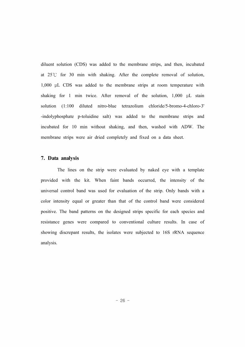
Figure 2. Example of PCR results in blood culture positive bottles ------------------- 28
Figure 3. Real-time PCR TaqMan assay for blood culture negative bottles
------------------------------------------------------------------------------------------------------------------------------------------------ 29
Figure 4. The determination of REBA sensitivity using reference strains -------- 31
Figure 5. Typical REBA Sepsis-ID test results obtained using reference strains
------------------------------------------------------------------------------------------------------------------------------------------------ 33
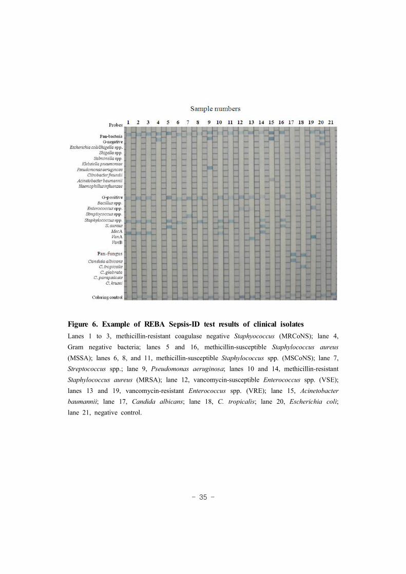
Figure 6. Example of REBA Sepsis-ID test results of clinical isolates ------------ 35
Figure 7. The sensitivity of REBA Sepsis-ID test in blood culture positive
bottles according to the blood suspension volumes ------------------------------------ 51
Figure 8. Schematic illustration for detection of pathogens from blood bottles
by blood cultures with REBA Sepsis-ID test ------------------------------------------------ 66
- x -
LIST OF TABLES
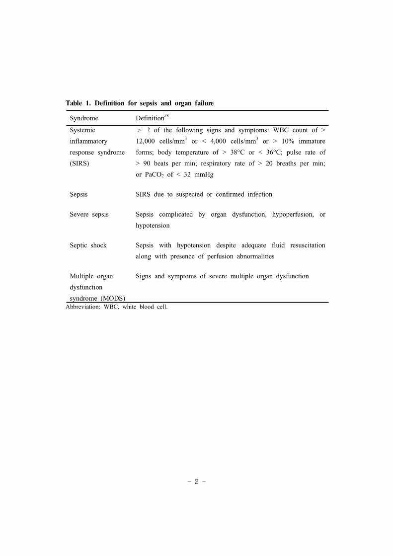
Table 1. Definition for sepsis and organ failure ------------------------------------------------------------- 2
Table 2. Selection of pathogens and antimicrobial resistance genes detected by
REBA Sepsis-ID test -------------------------------------------------------------------------------------------------- 12
Table 3. List of primers and probes used in this study ------------------------------------------- 14
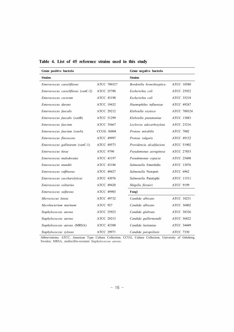
Table 4. List of 45 reference strains used in this study ------------------------------------------ 16
Table 5. The distribution of 118 clinical isolates from various specimens ----- 17
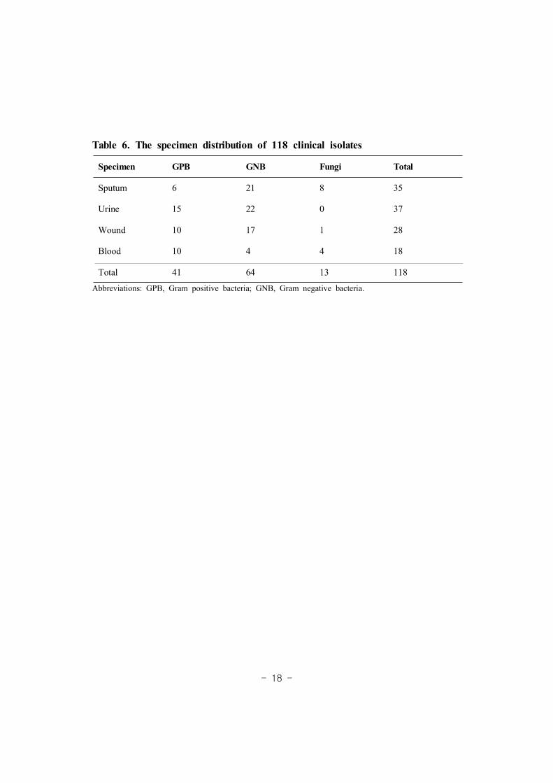
Table 6. The specimen distribution of 118 clinical isolates ------------------------------------ 18
Table 7. Comparison between culture and REBA Sepsis-ID test in 118 clinical
isolates -------------------------------------------------------------------------------------------------------------------------------- 36
Table 8. Comparison between culture and REBA Sepsis-ID test of 100 blood
culture positive bottles in preliminary evaluation ------------------------------------------ 39
Table 9. Comparison between REBA Sepsis-ID test and sequence analysis of 5
real-time PCR positive samples among 200 blood culture negative
bottles --------------------------------------------------------------------------------------------------------------------------------- 42
Table 10. The distribution of monomicrobial isolates and comparison results
between blood cultures and REBA Sepsis-ID test from 300 blood
culture positive bottles ------------------------------------------------------------------------------------------------ 46
- xi -
Table 11. The total concordance rates of 300 blood culture positive bottles
between conventional methods and REBA Sepsis-ID test ----------------------- 48
Table 12. Comparison between conventional methods and REBA Sepsis-ID test
of 12 polymicrobial isolates among 300 blood culture positive bottles
------------------------------------------------------------------------------------------------------------------------------------------------- 49
Table 13. The discrepant results between conventional methods and REBA
Sepsis-ID test in 300 blood culture positive bottles ----------------------------------- 50
Table 14. Comparison between REBA Sepsis-ID test, culture results and
sequence analysis of 26 real-time PCR positive samples among 1,100
blood culture negative bottles --------------------------------------------------------------------------------- 53
Table 15. The concordance rates of 1,400 blood culture bottles between blood
culture and REBA Sepsis-ID test ------------------------------------------------------------------------- 56
- xii -
ABBREVIATIONS
ADW : autoclaved distilled water
BC-NB : blood culture negative bottles
BC-PB : blood culture positive bottles
BSI : bloodstream infections
CAN : Candida
CDS : conjugate diluent solution
CLSI : Clinical and Laboratory Standards Institute
CMBCS : continuous monitoring blood culture system
CoNS : coagulase-negative staphylococci
CT : cycle threshold
DIC : disseminated intravascular coagulation
DNI : delta neutrophil index
ELISA : enzyme-linked immunosorbent assay
EtBr : ethidium bromide
GNB : Gram negative bacteria
- xiii -
GPB : Gram positive bacteria
IE : infective endocarditis
ITS : internal transcribed sequence
LB : Luria-Bertani
MALDI-TOF/MS : matrix assisted laser desorption ionization-time of flight/mass spectrometry
mecA : methicillin-resistance gene A
MODS : multiple organ dysfunction syndrome
MRSA : methicillin-resistant Staphylococcus aureus
NA : nucleic acid
NBT/BCIP : nitro-blue tetrazolium chloride/5-bromo-4-chloro-3'-indolyphosphate p-toluidine salt
NCBI/BLAST : National Center for Biotechnology Information/ BasicLocal Alignment Search Tool
PCA : plating count agar
PNA-FISH : peptide nucleic acid-fluorescence in situ hybridization
R2A : Reasoner's 2A agar
RBC : red blood cell
REBA : reverse blot hybridization assay
- xiv -
RFLP : restriction fragment length polymorphism
SIRS : systemic inflammatory response syndrome
spp. : species
SPS : sodium polyanethol sulfonate
TAE : Tris base, acetic acid and EDTA
TAT : turnaround time
vanA, vanB : vancomycin-resistance gene A, B
VRE : vancomycin-resistant enterococci
WBC : white blood cell
WSCH : Wonju Severance Christian Hospital
- xv -
ABSTRACT
Reverse blot hybridization assay Sepsis-ID test
for simultaneous identification of pathogens and
antimicrobial resistance genes of mecA, vanA and
vanB from blood culture bottles
Soon Deok Park
Department of Biomedical Laboratory Science
The Graduate School, Yonsei University
The rapid and accurate identification of pathogens in blood with sepsis
is crucial for prompt initiation of appropriate therapy to decrease related
morbidity and mortality rates. The aim of this study was to evaluate diagnostic
performance of newly designed Reverse blot hybridization assay Sepsis
-Identification test (REBA Sepsis-ID test) for rapid diagnosis of bacteremia and
- xvi -
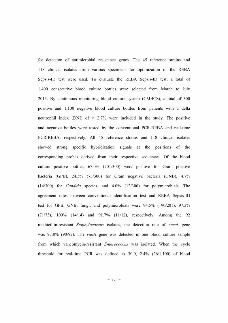
for detection of antimicrobial resistance genes. The 45 reference strains and
118 clinical isolates from various specimens for optimization of the REBA
Sepsis-ID test were used. To evaluate the REBA Sepsis-ID test, a total of
1,400 consecutive blood culture bottles were selected from March to July
2013. By continuous monitoring blood culture system (CMBCS), a total of 300
positive and 1,100 negative blood culture bottles from patients with a delta
neutrophil index (DNI) of > 2.7% were included in the study. The positive
and negative bottles were tested by the conventional PCR-REBA and real-time
PCR-REBA, respectively. All 45 reference strains and 118 clinical isolates
showed strong specific hybridization signals at the positions of the
corresponding probes derived from their respective sequences. Of the blood
culture positive bottles, 67.0% (201/300) were positive for Gram positive
bacteria (GPB), 24.3% (73/300) for Gram negative bacteria (GNB), 4.7%
(14/300) for Candida species, and 4.0% (12/300) for polymicrobials. The
agreement rates between conventional identification test and REBA Sepsis-ID
test for GPB, GNB, fungi, and polymicrobials were 94.5% (190/201), 97.3%
(71/73), 100% (14/14) and 91.7% (11/12), respectively. Among the 92
methicillin-resistant Staphylococcus isolates, the detection rate of mecA gene
was 97.8% (90/92). The vanA gene was detected in one blood culture sample
from which vancomycin-resistant Enterococcus was isolated. When the cycle
threshold for real-time PCR was defined as 30.0, 2.4% (26/1,100) of blood

- xvii -
culture negative samples tested were positive by real-time PCR. The results of
26 samples showed discordance by conventional test, real-time PCR-REBA, and
16S rRNA sequence analysis. The REBA Sepsis-ID test could be useful to
simultaneously and rapidly detect causative agents and antimicrobial resistance
genes, such as mecA and vanA or vanB, in blood culture positive samples.
The application of REBA Sepsis-ID test with culture methods in clinical
microbiology laboratories would reduce the time for detection of pathogens
responsible for sepsis, presumably resulting in reduction of patient mortality
rates.
Key words: reverse blot hybridization assay, blood cultures, delta neutrophil
index, real-time PCR, mecA

- 1 -
. INTRODUCTION
Bloodstream infections (BSI) are associated with high morbidity and
mortality rates, with a mortality rate ranging from 20% to 70% in the
world.2,7,22,26 Recently, sepsis and sepsis-associated shock complicated by BSI
are the 10th leading cause of death in the United States, accounting for 6% of
all deaths (Table 1).52 In Europe, an estimated 135,000 patients die each year
of sepsis-associated complications, with an overall incidence of 3 sepsis cases
per 1,000 individuals.57
The different forms of sepsis are always associated with bacteremia (or
fungemia); on the other hand, bacteremia and fungemia do not always cause
the syndrome of sepsis. However, if they are not properly controlled, they may
be associated with the development of sepsis, a clinical syndrome related to an
infectious process with important alterations in the inflammatory response and
coagulation. The clinical spectrum of sepsis ranges from systemic inflammatory
response syndrome (SIRS) to multiple organ dysfunction syndrome (MODS)
and the midpoints are sepsis, severe sepsis, and septic shock (Table 1).58
Blood cultures based on detection of viable microorganisms in the
blood are the current gold standard of BSI diagnosis.92 The accurate
identification of blood isolates is the mainstay of the proper management of
BSI. Blood cultures, which are used to detect viable pathogens, have the
- 2 -
Table 1. Definition for sepsis and organ failure
Abbreviation: WBC, white blood cell.
Syndrome Definition58
Systemicinflammatoryresponse syndrome(SIRS)
2 of the following signs and symptoms: WBC count of >12,000 cells/mm3 or < 4,000 cells/mm3 or > 10% immatureforms; body temperature of > 38°C or < 36°C; pulse rate of> 90 beats per min; respiratory rate of > 20 breaths per min;or PaCO2 of < 32 mmHg
Sepsis SIRS due to suspected or confirmed infection
Severe sepsis Sepsis complicated by organ dysfunction, hypoperfusion, orhypotension
Septic shock Sepsis with hypotension despite adequate fluid resuscitationalong with presence of perfusion abnormalities
Multiple organdysfunctionsyndrome (MODS)
Signs and symptoms of severe multiple organ dysfunction

- 3 -
advantage of allowing evaluation of antimicrobial susceptibility. This
characteristic has still not been substituted by any other techniques available to
date. This aspect is important, as several studies have shown that inadequate
antimicrobial therapy is an independent risk factor of mortality or
microbiological failure in severely ill patients with life-threatening
infections.41,54,56,96
Patients with sepsis, defined as a clinical infection resulting in a
systemic inflammatory response, are not always culture positive, with only
about one-third overall having positive cultures. Although blood cultures are
currently performed with continuous monitoring blood culture system
(CMBCS), several factors may still reduce the overall sensitivity of blood
cultures.
Firstly, the blood volume is the most important factor influencing
blood culture diagnostic yield.32,84 Several studies confirmed that the rate of
isolation from blood cultures in both of adults5,11,43,64 and pediatric patients42,45
increases with the quantity of blood submitted. This is particularly important
for pediatric patients, for whom it is not always possible to draw a sufficient
volume of blood. Clinical and Laboratory Standards Institute (CLSI)15
recommended the collection of two to three blood culture sets, an aerobic
bottle and an anaerobic bottle per each suspected BSI patient, collecting 20 to
30 mL of blood per each set.15 According to a research study by Ilstrup and
- 4 -
Washington,40 a larger volume of blood significantly increases the number of
positive cultures. They demonstrated that the detection yields of 20 mL and 30
mL blood volumes were 38% and 62% greater than the yields of 10 mL
respectively. Nevertheless, a high percentage of health care professionals do not
know the optimal amount of blood recommended for blood culture even
though blood culture volume remains the most important determinant factor for
blood culture positivity. These findings raise an important quality assurance
issue. Future quality assurance initiatives should be considered to ensure proper
collection of volume for blood cultures.23 The Wonju Severance Christian
Hospital (WSCH) in Korea has been using the blood volume measuring system
for blood culture quality improvement.81
Secondly, another important variable influencing diagnostic performance
is the time taken from blood withdrawal to the loading of blood culture
bottles into the instrument. Blood bottles should be loaded immediately into
the CMBCS in order to minimize the time to detection.78,79
Thirdly, culture methods can fail to identify the fastidious organisms
that are difficult or impossible to culture and also be confounded if antibiotics
are administered before the blood is collected. Blood cultures are reported to
be negative in more than 50% of the cases where true bacterial or fungal
sepsis is believed to exist.18,27 An intrinsic limitation of blood culture method
is their low sensitivity to slow growing and fastidious organisms such as
- 5 -
Bartonella species (spp.), Francisella tularensis, Mycoplasma spp., several
molds, and Nocardia spp.. Other uncultivable pathogens such as Rickettsia
spp., Coxiella burnetii, Chlamydophila pneumoniae, and Tropheryma whipplei
are better diagnosed by immunodiagnostic or molecular techniques.79,87 Many of
these organisms may be responsible for infective endocarditis, the diagnosis of
which is based upon the detection of vegetations on the cardiac valves and
positive blood cultures.24
Lastly, a major factor to reduce the sensitivity of blood cultures is the
time required to complete the process, which ranges from 3 to 5 days or
more. This delay can endanger the patient if ineffective therapy is given for
antibiotic-resistant organisms and antibiotic-resistance may be encouraged if
unnecessary antibiotics are given for sensitive organisms.75,87 For patients with
septic shock, Kumar et al.51 have reported that each hour of delay in effective
therapy is associated with a 7.6% decrease in survival. The survival rate of
severe sepsis goes from approximately 80% if effective therapy is given within
the first hour of the onset of symptoms to less than 10% if effective therapy
is not given by 24 h. Therefore, various serological biomarkers such as
C-reactive protein, procalcitonin, interleukin 6, interleukin 8, and delta
neutrophil index (DNI) have been used to help rapid identification of patients
with sepsis.34,37,46,72 Especially, for DNI, modern technological advances have
led to automated cell analyzers to provide information on leukocyte
- 6 -
differentials based on cytochemical myeloperoxidase reaction and nuclear
lobularity of the white blood cells, it reflects the fraction of circulating
immature granulocytes.35,43,50 DNI has been reported to be significantly
associated with disseminated intravascular coagulation (DIC) scores, positive
blood culture rate, and mortality in patients with suspected sepsis. High levels
of DNI may help to identify patients with an impending risk of developing
severe sepsis/septic shock.70
Clinically, antibiotic treatment of bacterial infections depends on the
species of bacteria, with the differentiation between Gram positive bacteria
(GPB), Gram negative bacteria (GNB), or fungi. Empirical antibiotic therapy is
maintained until culture results are reported. The most common isolates in
blood cultures were coagulase-negative staphylococci (CoNS) followed by
Staphylococcus aureus, Enterococcus spp. and Candida spp.. The isolation
frequency of enteric Gram-negative pathogens in decreasing order, in general,
figured lower on the ranks of organisms and included Escherichia coli,
Klebsiella spp., Pseudomonas aeruginosa and Enterobacter spp.. Other studies
confirmed the emergence of Gram-positive pathogens, specifically CoNS, S.
aureus and Enterococcus spp. as the dominant organisms in nosocomial
BSI.20,30,62,90,91 This rise in the frequency of CoNS BSI isolates has occurred
concurrently with the increased use of invasive intravascular catheters.83
The decreasing turnaround time (TAT) for positive blood culture

- 7 -
results is important because appropriate antimicrobial agents can be selected
immediately, unnecessary treatment of likely contaminants and antibiotic
exposure can be avoided, and expenditure can be decreased. Timely detection,
distinction of S. aureus from CoNS and methicillin-susceptibility results have
great therapeutic, prognostic and economic significance.3,8,17 Methicillin-resistant
S. aureus (MRSA) and vancomycin-resistant enterococci (VRE) are responsible
for a large proportion of infections in communities and hospitals. Bacteremia
caused by MRSA and VRE is one of the most serious infectious diseases, not
only because of its increasing frequency, but also because of its resistance to
treatment. The morbidity and mortality by MRSA and VRE are high compared
with methicillin-susceptible S. aureus and vancomycin-susceptible
enterococci.18,59
The incidence of BSI caused by Candida spp. has increased five- to
ten-fold, as a result, it has become the 4th leading cause of nosocomial
infection. Among Candida spp., Candida tropicalis and Candida parapsilosis
were found to be the most frequently isolated non-albicans species from the
bloodstream.31 Surveillance for candidemia or bacteremia is necessary to detect
trends in species distribution and antifungal resistance. As described above,
conventional identification and susceptibility testing have several limitations,
including lack of speed and sensitivity. Therefore, developing a rapid, sensitive,
and specific method is important for identifying bacterial and fungal pathogens
- 8 -
to promote more timely, appropriate, and accurate antimicrobial therapy.95
Recently, nucleic acid (NA)-based assays have been provided an early
and accurate diagnosis of diseases caused by bacterial pathogens in blood and
have improved the rate of microbial detection.21,88 NA-based assays applied to
sepsis can be divided into two main categories; detection and identification of
pathogens from blood culture bottles and for detection and identification of
pathogens directly from blood, serum, or plasma samples. Three types of
targets are described: assays targeting species- or genus-specific genes,
broad-range assays targeting conserved sequences in the bacterial or fungal
genome such as the pan-bacterial 16S, 5S, and 23S ribosomal RNA (rRNA)
genes, and the pan-fungal 18S, 5.8S, and 28S rDNA.71 Polymerase chain
reaction (PCR) techniques based on the automated fluorescence detection of
amplicons are usually more robust, less labor-intensive, and less contamination
than conventional PCR techniques.47,73 However, real-time PCR techniques are
certainly more expensive than conventional PCR techniques, and some of the
commercially available platforms are still too small with insufficient
throughput. Therefore, most of clinical laboratories are not willing to undertake
these changes.76 Recently, a non-NA-based technique, matrix-assisted laser
desorption-ionization time-of flight mass spectrometry (MALDI-TOF MS) is
being investigated for its use in the identification of bacteria or fungi by
determining its proteomic profiles,86 bacterial virulence factors,9 and antibiotic

- 9 -
resistance markers25 directly from blood cultures. This method has the main
advantage of allowing a definitive identification of isolated microorganisms in
only a few minutes. However, it could not be applied directly to biological
samples due to insufficient microbial cells for analysis. Therefore, its
application in the diagnosis of BSI directly from blood samples is not
foreseeable in the near future. Moreover, the high cost of equipment precludes
their routine use in the clinical laboratory.
Several NA-based commercial assays that target a panel of the most
relevant bacterial and fungal bloodstream pathogens have been developed.
Among the commercial assays applied for positive blood cultures, the most
studied is peptide nucleic acid fluorescence in situ hybridization
(PNA-FISH)-based assays (AdvanDx, Woburn, MA),28,36 which targeting the
rRNA genes of a limited number of bacterial and targeting the rDNA of
Candida spp.. The Hyplex BloodScreen (BAG, Lich, Germany), a multiplex
PCR assay by hybridization in an enzyme-linked immunosorbent assay
(ELISA)-like format,93 and Prove-it Sepsis (Mobidiag, Helsinki, Finland), which
was the first microarray-based assay designed for the diagnosis of sepsis.33
Among the assays for detection and identification of pathogens directly
on blood samples, SepsiTest (Molzym, Bremen, Germany) is a broad-range
PCR based assay targeting the 16S rRNA genes of bacteria and the 18S
rDNA of fungi,68 Vyoo (SIRS-Lab, Jena, Germany), a multiplex PCR-based
- 10 -
assay and the LightCycler SeptiFast Test (Roche Molecular Systems,
Branchburg, NJ) is, to date, the only multiplex real-time PCR assay available
for the diagnosis of sepsis. It is capable of detecting genetic material
belonging to several bacterial and fungal pathogens, representing approximately
90% of the species responsible for nosocomial bacteremia.55 However, no real
advantage was observed for patients with suspected infective endocarditis (IE)13
because several IE-related bacterial species are not included in the above-
described commercial assays, and the sensitivity of the assays may not be
sufficient to detect the low-grade bacteremia. Moreover, limitations of the
current commercial assays include its very high cost and the lack of
information on antimicrobial susceptibility.
The reverse blot hybridization assay (REBA) is affordable in many
practical settings since it is relatively simple and inexpensive as well as
informative as DNA microarray and readily applicable to small clinical
laboratories.14 Moreover, it does not require expensive instrumentation, and
provides rapid results when compared with conventional culture and alternative
molecular methods like sequencing and restriction fragment length
polymorphism (RFLP) analysis. Thus, this method could become a powerful
and reliable tool for the identification of common pathogens.49 Future research
should focus on the direct identification of bacterial DNA in samples that are
suspected on clinical evidence to contain pathogens, with particular reference to
- 11 -
samples that yield negative culture results.89
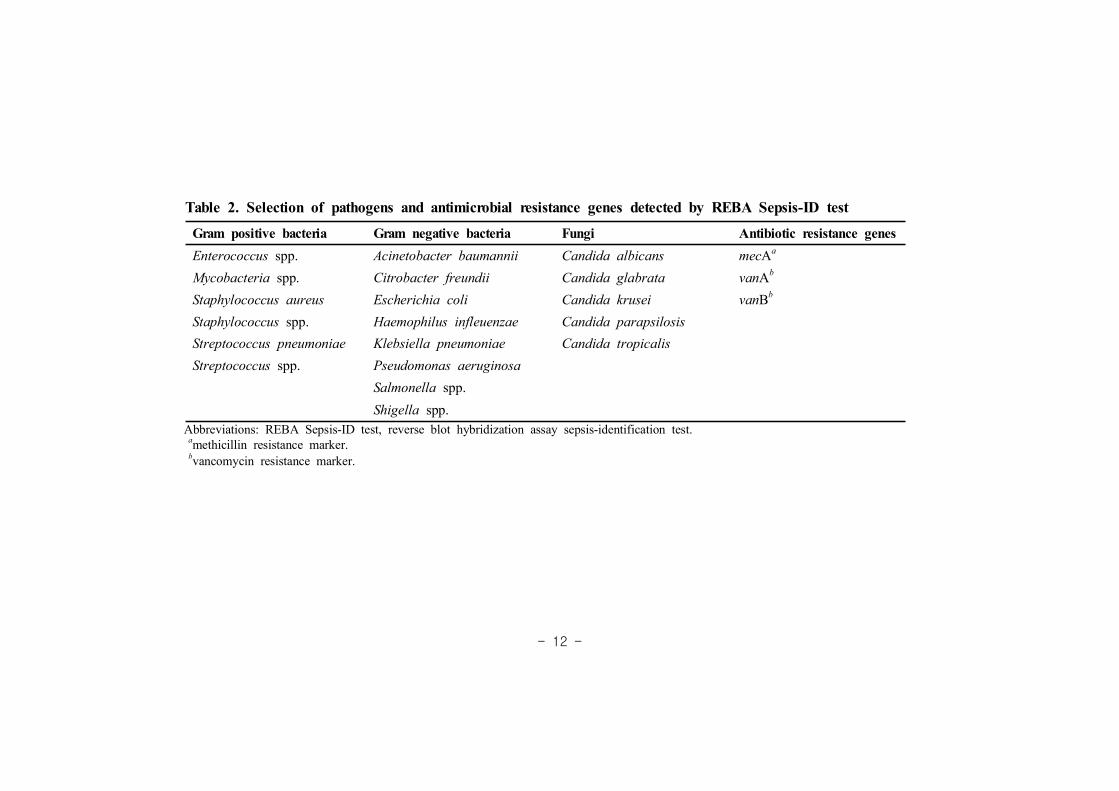
Therefore, REBA Sepsis-ID test for the rapid diagnosis of
sepsis-causing agents was newly designed in this study. The membrane of
REBA comprises for DNA probes for GPB including Staphylococcus aureus,
Staphylococcus spp., Streptococcus pneumoniae, Streptococcus spp.,
Enterococcus spp., and Mycobacterium spp., and DNA probes for GNB
including E. coli, Klebsiella pneumoniae, Salmonella spp., Shigella spp.,
Citrobacter freundii, Haemophilus influenzae, P. aeruginosa, and Acinetobacter
baumannii. Moreover, it comprises probes of Candida including Candida
albicans, C. tropicalis, Candida glabrata, C. parapsilosis, and Candida krusei
and antimicrobial resistance genes including mecA, vanA and vanB (Table 2).
The aims of this study were; 1) to develop and validate the REBA
Sepsis-ID test with reference strains and clinical isolates, 2) to evaluate the
assay with blood culture positive bottles for rapid detection of pathogens and
negative bottles for detecting slow-growing and fastidious organisms 3) to
determine whether it can be used as a routine diagnostic tool.
- 12 -
Table 2. Selection of pathogens and antimicrobial resistance genes detected by REBA Sepsis-ID test
Abbreviations: REBA Sepsis-ID test, reverse blot hybridization assay sepsis-identification test.amethicillin resistance marker.bvancomycin resistance marker.
Gram positive bacteria Gram negative bacteria Fungi Antibiotic resistance genesEnterococcus spp. Acinetobacter baumannii Candida albicans mecAa
Mycobacteria spp. Citrobacter freundii Candida glabrata vanAb
Staphylococcus aureus Escherichia coli Candida krusei vanBb
Staphylococcus spp. Haemophilus infleuenzae Candida parapsilosisStreptococcus pneumoniae Klebsiella pneumoniae Candida tropicalisStreptococcus spp. Pseudomonas aeruginosa
Salmonella spp.Shigella spp.

- 13 -
II. MATERIALS AND METHODS
1. Preparation of oligonucleotide probes for REBA Sepsis-ID test
To maximize the sensitivities and specificities of the species- or
genus-specific primers and probes, sequences from the most variable regions
within the bacterial 16S ribosomal DNA (16S rDNA) for GPB and GNB, 18S
5.8S internal transcribed sequence (ITS) for fungi, the nuc gene for S.
aureus, the mecA gene for methicillin-resistant Staphylococcus spp. (MRS) and
the vanA and vanB genes for VRE, were selected by multiple alignment using
ClustalW (http://www.cmbi.kun.nl/bioinf/tools/clustalw.shtml). The target
sequences for PCR primers and REBA probes used for the genus and species
of bacteria and fungi, and antimicrobial resistance detection are listed in Table
3. The sequences of the primers and probes were compared by National
Center for Biotechnology Information Basic Local Alignment Search Tool
(NCBI BLAST) (Blastn) to determine their sequence homologies. The selection
of oligonucleotide probes was designed to avoid self-complementarity of more
than three nucleotides per probe. After evaluation of the probes, sixteen
bacterial-, six fungal-, and three antibiotic resistance probes were finally
selected for the REBA Sepsis-ID test (Table 3).

- 14 -
Table 3. List of primers and probes used in this studyTarget Primer/probe names* Nucleotide sequences (5´-3´)†
16S rRNA F-16S TAAYACATGCAAGTCGARCG
R-16S TGGCACGDAGTTRGCCGKKGCTT
Staphylococcus aureus F-Saur AGCGATTGATGGTGATACGGT
R-Saur ATGCACTTGCTTCAGGACCA
mecA F-mecA GGTGTTGGTGAAGATATACCAAGTG
R-mecA GAAAGGATCTGTACTGGGTTAATCAT
vanA F-VanA TCAATAGCGCGGACGAATTG
R-VanA GCGGGAACGGTTATAACTGCGTTT
vanB F-VanB TACCTACCCTGTCTTTGTGAAGCC
R-VanB GCTGCTTCTATCGCAGCGTTTAGT
Fungus F-Fung AACGCANMTTGCRCYCHHTG
R-Fung CAGCGGGTADYCCYACCTGA
Bacteria p-panBact AGYGGCGGACGGGTGAGTAA
Gram positive bacteria p-GrPo TGAGTAACACGTGGGYAACC
Enterococcus spp. p-Ent CCATCAGARGGGGATAACACTT
Mycobacterium spp. p-Mycob TGGTGSAAAGCTTTTGCGGT
Staphylococcus aureus p-Saur TTGGTTGATACACCTGAAACAAAG
Staphylococcus spp. p-Stap CACGTRGCTAACCTACCTATAAGACTG
Streptococcus pneumoniae p-Stpneum TCAGTGTCGCTGTTTTAGCAGAT
Streptococcus spp. p-Str CGCGTAGGTAACCTGCCTGGTA
Gram negative bacteria p-GrNg ATGTCTGGGAAACTGCCTGATG
Acinetobacter baumannii p-Abaum AGCTTGCTACCGGACCTAGC
Citrobacter freundii P-Cfreun TAGCACAGAGGAGCTTGCTCCTTG
Escherichia coli p-Ecoli AAAGGGAGTAAAGTTAATACCTTTGCTCA
Haemophilus influenzae p-Hinfl CGTATTATCGGAAGATGAAAGTGC
Klebsiella pneumoniae p-KPneum AAAAAAAGGTTAATAACCTCATCGATTGAC
Pseudomonas aeruginosa p-Paer ATACGTCCTGAGGGAGAAAGTG
Salmonella spp. p-Salm CGGAAGCCTCCGCTAATTTGAT
Shigella spp. p-Shig AGTTCAGTAAGATGGTTGTGCGCA
Fungus p-Fung TGCGTTCAARRAYTCGATGA
Candida albicans p-Calb AATAGTGGTAAGGCGGGATC
Candida glabrata p-Cgrab AGCGCAAGCTTCTCTATTAATCTG
Candida krusei p-Ckrus AGCGGAGCGGACGACGTGTA
Candida parapsilosis p-Cpara AGGCGGAGTATAAACTAATGGATAGGT
Candida tropicalis p-Ctrp ACGTGGAAACTTATTTTAAGCGA
mecA p-MecA AGCTGATTCAGGTTACGGACAAGGT
vanA p-VanA TCGTATTCATCAGGAAGTCGAGCC
vanB p-VanB TCGTCCTTTGGCGTAACCAA* F means forward primer, R means reverse primer, p means probe.† Y, C or T; R, A or G; D, A or T or G; K, T or G; N, A or C or G or T; M, A or C; H, A or T or C; Y, C or T; S, Cor G.

- 15 -
2. Bacterial strains and clinical specimens
2-1. Optimization of REBA Sepsis-ID test
For optimization of the REBA Sepsis-ID test, 45 reference strains
(Table 4) and 118 isolates from various clinical specimens were used (Table
5). The isolates from same patients were excluded in this study. The clinical
isolates were obtained from December 2011 through January 2012 at WSCH.
The 118 clinical isolates were consisted of 41 GPB, 64 GNB, and 13 fungi
from specimens of sputum, urine, wound and blood (Table 6). All strains were
subcultured on sheep blood agar and MacConkey agar (BD Diagnostic
Systems, Sparks, MD, USA) then incubated at 35 under CO2 conditions
overnight. The identification of isolates from blood cultures was performed
using MicroScan® (Siemens Healthcare Diagnostics, Sacramento, CA, USA)
panels: PID2, overnight POS COMBO (PBC28) and overnight NEG COMBO
(NC44). For identification of isolates from the other specimens, MicroScan®
was used for GPB and Microplate method85 was used for GNB. For
identification for Candida spp. from all specimens, VITEK-2 YST ID CARD
(bioMérieux, Hazelwood, MO, USA) was used. The antimicrobial susceptibility
tests for methicillin and vancomycin in GPB were determined using the
MicroScan® POS COMBO (PBC28), and those of the GNB and fungi were
determined according to the guidelines of the CLSI.16
- 16 -
Table 4. List of 45 reference strains used in this study
Abbreviations: ATCC, American Type Culture Collection; CCUG, Culture Collection, University of Göteborg,Sweden; MRSA, methicillin-resistant Staphylococcus aureus.
Gram positive bacteria Gram negative bacteria
Strains Strains
Enterococcus casseliflavus ATCC 700327 Bordetella bronchiseptica ATCC 10580
Enterococcus casseliflavus (vanC-2) ATCC 25788 Escherichia coli ATCC 25922
Enterococcus cecorum ATCC 43198 Escherichia coli ATCC 35218
Enterococcus durans ATCC 19432 Haemophilus influenzae ATCC 49247
Enterococcus faecalis ATCC 29212 Klebsiella oxytoca ATCC 700324
Enterococcus faecalis (vanB) ATCC 51299 Klebsiella pneumoniae ATCC 13883
Enterococcus faecium ATCC 35667 Leclercia adecarboxylata ATCC 23216
Enterococcus faecium (vanA) CCUG 36804 Proteus mirabilis ATCC 7002
Enterococcus flavescens ATCC 49997 Proteus vulgaris ATCC 49132
Enterococcus gallinarum (vanC-1) ATCC 49573 Providencia alcalifaciens ATCC 51902
Enterococcus hirae ATCC 9790 Pseudomonas aeruginosa ATCC 27853
Enterococcus malodoratus ATCC 43197 Pseudomonas cepacia ATCC 25608
Enterococcus mundtii ATCC 43186 Salmonella Enteritidis ATCC 13076
Enterococcus raffinosus ATCC 49427 Salmonella Newport ATCC 6962
Enterococcus saccharolyticus ATCC 43076 Salmonella Paratyphi ATCC 11511
Enterococcus solitarius ATCC 49428 Shigella flexneri ATCC 9199
Enterococcus sulfureus ATCC 49903 Fungi
Micrococcus luteus ATCC 49732 Candida albicans ATCC 10231
Mycobacterium marinum ATCC 927 Candida albicans ATCC 36802
Staphylococcus aureus ATCC 25923 Candida glabrata ATCC 38326
Staphylococcus aureus ATCC 29213 Candida guillermondii ATCC 56822
Staphylococcus aureus (MRSA) ATCC 43300 Candida lusitaniae ATCC 34449
Staphylococcus xylosus ATCC 29971 Candida parapsilosis ATCC 7330
- 17 -
Table 5. The distribution of 118 clinical isolates from various specimensGram positive bacteria (n=41) Antimicrobial resistance Gram negative bacteria (n=64) Fungi (n=13)
Staphylococcus aureus (12) methicillin resistance (9) Escherichia coli (16) Candida albicans (5)
Staphylococcus epidermidis (4) methicillin resistance (4) Klebsiella pneumoniae (14) Candida glabrata (3)
Staphylococcus haemolyticus (3) methicillin resistance (3) Pseudomonas aeruginosa (13) Candida tropicalis (2)
Staphylococcus capitis (1) methicillin resistance (1) Acinetobacter baumannii (11) Candida parapsilosis (1)
Streptococcus mitis (2) Aeromonas spp. (1) Saccharomyces cerevisiae (2)
Streptococcus agalactiae (1) Citrobacter freundii (1)
Streptococcus parasanguis (1) Enterobacter asburiae (1)
Streptococcus pyogenes (1) Enterobacter cloacae (1)
Streptococcus salivarius (1) Haemophilus influenzae (1)
Enterococcus faecium (10) vancomycin resistance (7) Moraxella catarrhalis (1)
Enterococcus faecalis (4) Morganella morgannii (1)
Corynebacterium spp. (1) Proteus mirabilis (1)
Providencia rettgeri (1)
Serratia marcescens (1)
- 18 -
Table 6. The specimen distribution of 118 clinical isolates
Abbreviations: GPB, Gram positive bacteria; GNB, Gram negative bacteria.
Specimen GPB GNB Fungi Total
Sputum 6 21 8 35
Urine 15 22 0 37
Wound 10 17 1 28
Blood 10 4 4 18
Total 41 64 13 118
- 19 -
2-2. Evaluation of REBA Sepsis-ID test
For evaluation of the REBA Sepsis-ID test, a total of 400 positive
blood culture bottles (100 for preliminary study and 300 for evaluation) by
BACTEC FX (BD, Sparks, MD, USA) or BacT/ALERT 3D (bioMérieux,
Marcy, France) were collected at WSCH and enrolled from January to July,
2013. Only one positive blood culture per patient was used to avoid
redundancy of enrolled samples. Blood culture bottles were eligible for study
enrolment if they showed a positive signal (defined as BC-PB) by CMBCS
and DNI value was over 2.7%.80 After they were removed from the
instrument, 1,000 L aliquot of the culture-broth mixture was aseptically takenμ
with a needle syringe. The aliquot was divided, 500 L of blood suspensionsμ
were used for Gram stain, and subcultured on sheep blood agar and
MacConkey agar and then incubated at 35 for 24-48 h under 5% CO2
incubator. The other 500 L of blood suspensions was stored at -20 untilμ
DNA extraction. Identification and antimicrobial susceptibility test were
performed using MicroScan system (Siemens Healthcare Diagnostics).
A total of 1,300 (200 for preliminary study and 1,100 for evaluation)
blood culture bottles with negative signal (defined as BC-NB) by CMBCS
after 5 days incubation with DNI > 2.7% were selected during study period.
One negative culture bottle per patient was selected and enrolled. After they
were removed from the instrument, 500 L of blood suspensions were takenμ
- 20 -
and stored at -20 until DNA extraction. All BC-NB incubated at 35
incubator until PCR results were obtained. As for BC-NB and real-time PCR
positive, 1,000 L of blood suspension from culture bottles inoculated on twoμ
sheep blood, chocolate and Sabouraud dextrose agar, and then, incubated at 3
5 in a 5% CO2 and O2 incubator for 5 days, respectively. Another 1,000 Lμ
of blood suspension inoculated on plating count agar media (PCA),6 and
Reasoner’s 2A (R2A) agar media77 for slow-growing organisms which may be
suppressed by faster-growing bacteria, and then further incubated at 20 in a
O2 incubator. Moreover, Luria-Bertani (LB)10 and brain heart infusion (BHI)
broth, were added for growth of the bacteria in blood. Isolated colonies from
the media and the bottles revealing positive by real-time PCR confirmed by
sequence analysis of conserved regions of the 16S rRNA and the amplicons
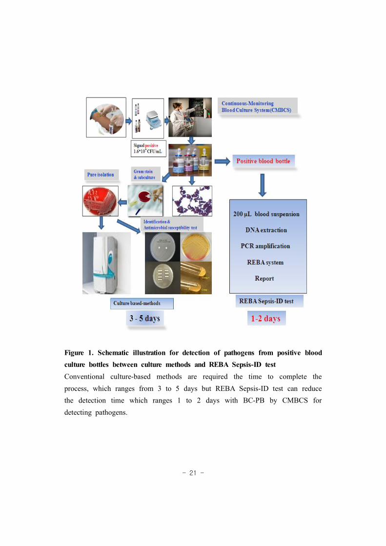
were sequenced by Xenotech Company (Daejeon, Republic of Korea). Figure 1
shows schematic illustration for detection of pathogens from positive blood
culture bottles between culture methods and REBA Sepsis-ID test. Conventional
blood culture method is the time required to complete the process, which
ranges from 3 to 5 days for detecting pathogens from samples with suspected
sepsis but molecular-based method can decrease the detection time with BC-PB
by CMBCS, which ranges 1 to 2 days.
- 21 -
Figure 1. Schematic illustration for detection of pathogens from positive bloodculture bottles between culture methods and REBA Sepsis-ID testConventional culture-based methods are required the time to complete theprocess, which ranges from 3 to 5 days but REBA Sepsis-ID test can reducethe detection time which ranges 1 to 2 days with BC-PB by CMBCS fordetecting pathogens.

- 22 -
3. DNA Preparation
To prepare DNA templates for optimizing the membrane, one or two
colonies of reference strains and clinical isolates were mixed with 1,000 Lμ of
autoclaved distilled water (ADW) and the supernatant was removed after
centrifugation at 13,000 rpm for 5 min. One hundred Lμ of DNA extraction
solution (M&D, Wonju, Republic of Korea) added with the pellet and boiled
for 15 min. After centrifugation at 13,000 rpm for 10 min, the supernatant
was used as the DNA template.
To optimize DNA extraction method from blood specimens, the
preliminary method for preparing DNA template from 100 positive and 200
negative blood culture samples were used as follows; 500 Lμ aliquot of the
blood suspensions was mixed with 1,000 Lμ of ADW and kept at -70
deepfreezer for 5 min and melted in order to lyse red blood cells (RBCs).
The supernatant was removed after centrifugation at 13,000 rpm for 5 min.
The pellet was washed with 1,000 Lμ of ADW again, then centrifuged at the
same conditions. One thousand Lμ of PBS buffer (pH 8.0) was added to the
pellet and centrifuged then the pellet was washed with ADW repeatedly until
RBCs were removed. The pellet was resuspended in DNA extraction solution
as described above for the clinical isolates.
To prepare DNA template from 300 BC-PB samples and 1,100 BC-NB
samples for evaluating the REBA Sepsis-ID test, DNA extraction method was

- 23 -
used as follows; 200 L aliquot of the blood materials was mixed with 1,000μ
L of erythrocyte lysis buffer (ELB) (Sigma, St. Louis, MO, USA) at roomμ
temperature for 10 min in order to lyse RBCs. The supernatant was removed
after centrifugation at 13,000 rpm for 5 min. The pellet was washed with
1,000 L of ELB to remove RBCs completely, and then, it was centrifugedμ
under the same condition. One hundred L of ELB were added to the pellet,μ
and it was frozen and thawed twice. One hundred L of DNA extractionμ
solution (M&D) was added to the mixture, and then boiled for 15 min. After
centrifugation at 13,000 rpm for 10 min, the supernatant was used as the
DNA template.
4. PCR amplification for blood culture positive bottles
PCR was performed using a 50 L of reaction mixture (Daejeon,μ
Republic of Korea) containing 2X master mix, 1X primer mix, and 5 L ofμ
sample DNA. ADW was added to give a final volume of 50 L. For PCRμ
amplification, primer mix set I [16S rRNA (GNB (470 bp), GPB(250 bp)), S.
pneumoniae (120 bp), S. aureus (120 bp), and mecA (120 bp)] and set II
[Fungus (230 bp), vanA (250 bp), and vanB (100 bp)] were used. For PCR
amplification, the first 10 PCR cycles comprised initial denaturation at 95°C
for 30 s, followed by annealing and extension at 65°C for 30 s. These 10
cycles were followed by 40 cycles of denaturation at 95°C for 30 s, followed
- 24 -
by annealing and extension at 60°C for 30 s. After the final cycle, samples
were maintained at 72°C for 7 min to complete the synthesis of all strands. In
all PCR assays, ADW was added to the PCR mixture in place of bacterial
DNA as a negative control. Following amplification, 5 L of each PCRμ
product was electrophoresed on a 2.0% agarose gel to confirm successful
amplification. The gel was stained with 0.05 mg/mL ethidium bromide (EtBr)
solution, visualized with an Agaro-power system (Bioneer), and photographed
with the Gel Doc EQ system (Bio-Rad, Hercules, CA, USA).
5. Real-time PCR TaqMan assay for blood culture negative bottles
For evaluation of BC-NB, real-time PCR TaqMan assay was carried
out with the Real-GP (for GPB), -GN (for GNB) and -CAN (for fungi) probes
in PCR assay kits (M&D), and a CFX-96 real-time PCR system (Bio-rad,
Hercules, CA, USA) and an ABI 7500 FAST instrument (Applied Biosystem,
Foster City, CA, USA) were used for the thermo-cycling and fluorescence
detection. The real-time PCR amplification was performed in a total volume of
25 L that contained 12.5 L of 2X Thunderbird probe qPCR mix (Toyobo,μ μ
Osaka, Japan), 5 L of primer and TaqMan probe mixture, 5 L of templateμ μ
DNA, and sterile distilled water was added to give a final volume of 25 Lμ
for each sample. Positive and negative controls were included throughout the
procedure. No-template controls with sterile distilled water instead of template
- 25 -
DNA were incorporated in each run under the following conditions: 95°C for
3 min and 40 cycles of 95°C for 20 s and 60°C for 40 s in single real-time
PCR. The bacterial load was quantified by determining the cycle threshold
(CT), the number of PCR cycles required for the fluorescence to exceed a
value significantly higher than the background fluorescence. When CT value
was below 30.0, the sample was considered to be positive. Suspected positive
samples were subjected to REBA and 16S rRNA sequence analysis.
6. Reverse blot hybridization assay (REBA)
The REBA was performed according to the standard protocol provided
by the manufacturer. The membrane strips contain a set of immobilized
oligonucleotides, which are responsible for specific binding of the appropriate
biotinylated amplicons derived from the PCR amplification. After a colorimetric
staining procedure, the resulting band pattern can be evaluated by naked eye.
PCR products (40 L) were mixed with 40 L of denaturing reagent (providedμ μ
with the kit) for 5 min. The membrane strips were put to every trough and
added with 1,000 L hybridization buffer. PCR amplicons were added to theμ
membrane strips and hybridization performed at 55 for 30 min with shaking
(80 rpm), followed by two washing steps with prewarmed washing buffer for
10 min with shaking. For colorimetric detection of hybridized amplicons, 1,000
L streptavidin-conjugated alkaline phosphatase (1/2,000 diluted with conjugateμ
- 26 -
diluent solution (CDS) was added to the membrane strips, and then, incubated
at 25 for 30 min with shaking. After the complete removal of solution,
1,000 L CDS was added to the membrane strips at room temperature withμ
shaking for 1 min twice. After removal of the solution, 1,000 L stainμ
solution (1:100 diluted nitro-blue tetrazolium chloride/5-bromo-4-chloro-3'
-indolyphosphate p-toluidine salt) was added to the membrane strips and
incubated for 10 min without shaking, and then, washed with ADW. The
membrane strips were air dried completely and fixed on a data sheet.
7. Data analysis
The lines on the strip were evaluated by naked eye with a template
provided with the kit. When faint bands occurred, the intensity of the
universal control band was used for evaluation of the strip. Only bands with a
color intensity equal or greater than that of the control band were considered
positive. The band patterns on the designed strips specific for each species and
resistance genes were compared to conventional culture results. In case of
showing discrepant results, the isolates were subjected to 16S rRNA sequence
analysis.

- 27 -
III. RESULTS
1. Preparation for optimization of REBA Sepsis-ID test
1-1. The PCR amplification for blood culture positive bottles
The PCR amplification results for the REBA Sepsis-ID test in BC-PB
are shown in Figure 2. The PCR products for the 16S rRNA genes of GPB
and GNB, 18S 5.8S internal transcribed sequence (ITS) of fungi, the nuc
gene of S. aureus, the mecA gene of methicillin-resistance, and the vanA and
vanB genes of vancomycin-resistance were of the expected sizes as 16S PCR
(470 bp, 250 bp), S. aureus (120 bp), mecA (120 bp). vanA (250 bp), vanB
(100 bp), fungus (230 bp), respectively.
1-2. The real-time PCR TaqMan assay for blood culture negative
samples
For accurate evaluation of BC-NB, real-time PCR TaqMan assay,
Real-GP, -GN, -CAN probes were used in this study. The CT values of the
positive controls ranged from 11.0 to 20.94 (Figure 3). The CT value for
which real-time PCR was defined as positive was less than 30.0.
- 28 -
Figure 2. Example of PCR results in blood culture positive bottlesLanes M, 100-bp DNA ladder (Bioneer, Daejeon, Republic of Korea); lane 1, 16SPCR (470 bp, 250 bp); lane 2, S. aureus (120 bp); lane 3, mecA (120 bp); lane4, vanA (250 bp); lane 5, vanB (100 bp); lane 6, fungus (230 bp); lane 7, 16S-S.aureus-mecA mix; lane 8, fungus-vanA; lane 9, fungus-vanB; lane 10, fungus;lane 11, fungus-vanA-vanB.

- 29 -
Figure 3. Real-time PCR TaqMan assay for blood culture negative bottlesThe CT values of positive samples in Gram positive and negative bacteria rangedfrom 11.0 to 20.94. The CT value for which real-time PCR was defined aspositive was less than 30.0. in this study.
- 30 -
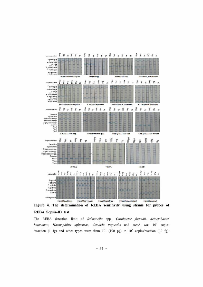
1-3. The determination of REBA sensitivity using reference strains
The detection limit of REBA was determined using a serially diluted
plasmid DNA which inserted genomic DNA PCR products of strains used to
REBA probes. A series of 10-fold dilutions from 100 pg to 1 fg (107-102
copies/reaction) were repeated five times with each different REBA kit. The
interpretation of detection limit was that the result of process by five times
was obtained all 102 copies/reaction then final value of detection limit was 102
copies/reaction but the result was 102-103 copies/reaction once of five times
then the result of the type was regarded as 102-103 copies/reaction (10 fg).
The minimum detection limit of the REBA ranged from 100 pg to 1 fg for
each species and all probes did not show any cross-reactions with others. As a
result, the REBA detection limit of Salmonella spp., Citrobacter freundii,
Acinetobacter baumannii, Haemophilus influenzae, Candida tropicalis and mecA
was 102 copies/reaction (1 fg) and other types were from 107 (100 pg) to 103
copies/reaction (10 fg) (Figure 4).
- 31 -
Figure 4. The determination of REBA sensitivity using strains for probes of
REBA Sepsis-ID test
The REBA detection limit of Salmonella spp., Citrobacter freundii, Acinetobacter
baumannii, Haemophilus influenzae, Candida tropicalis and mecA was 102 copies
/reaction (1 fg) and other types were from 107 (100 pg) to 103 copies/reaction (10 fg).
- 32 -
2. Optimization of REBA Sepsis-ID test
2-1. Optimization of REBA Sepsis-ID test with reference strains
Each amplified target DNA from reference strains including GPB,
GNB and fungi was spotted to REBA membrane strips with the selected
probes to validate the usefulness of REBA Sepsis-ID test. The example of
hybridization results of reference strains are shown in Figure 5. The amplified
PCR products reacted with universal probes (including the pan-bacteria probe,
Gram-positive probe, Gram-negative probe, and fungi probe) and genus- and
species- specific probes. The target DNAs hybridized strongly to the probes
derived from their targets and showed no cross-reactivity. All of the reference
strains in this study showed strong specific hybridization signals at the
positions of the corresponding probes derived from their respective sequences,
suggesting that sepsis-causing microbial pathogens could be specifically
detected and identified.

- 33 -
Figure 5. Typical REBA Sepsis-ID test results obtained using reference strainsLane 1, Klebsiella oxytoca ATCC 700324; lane 2, E. coli ATCC 25922; lane 3, Shigellaflexneri ATCC 9199; lane 4, Salmonella Enteritidis ATCC 13076; lane 5, K. pneumoniaeATCC 13883; lane 6, Pseudomonas aeruginosa ATCC 27853; lane 7, Citrobacter freundii;lane 8, Acinetobacter baumannii; lane 9, Haemophilus infleunzae ATCC 49247; lane 10,Mycobacterium marinum ATCC 927; lane 11, Streptococcus agalactiae; lane 12, S.pneumoniae ATCC 49619; lane 13, Staphylococcus xylosus ATCC 29971; lane 14,MRCoNS; lane 15, S. aureus (MRSA) ATCC 43300; lane 16, Enterococcus faecium(vanA) CCUG 36804; lane 17, E. faecalis (vanB) ATCC 51299; lane 18, Candidaalbicans ATCC 36802; lane 19, C. tropicalis; lane 20, C. glabrata ATCC 38326; lane 21,C. parapsilosis ATCC 7330; lane 22 C. kruzei ATCC 6258.
- 34 -
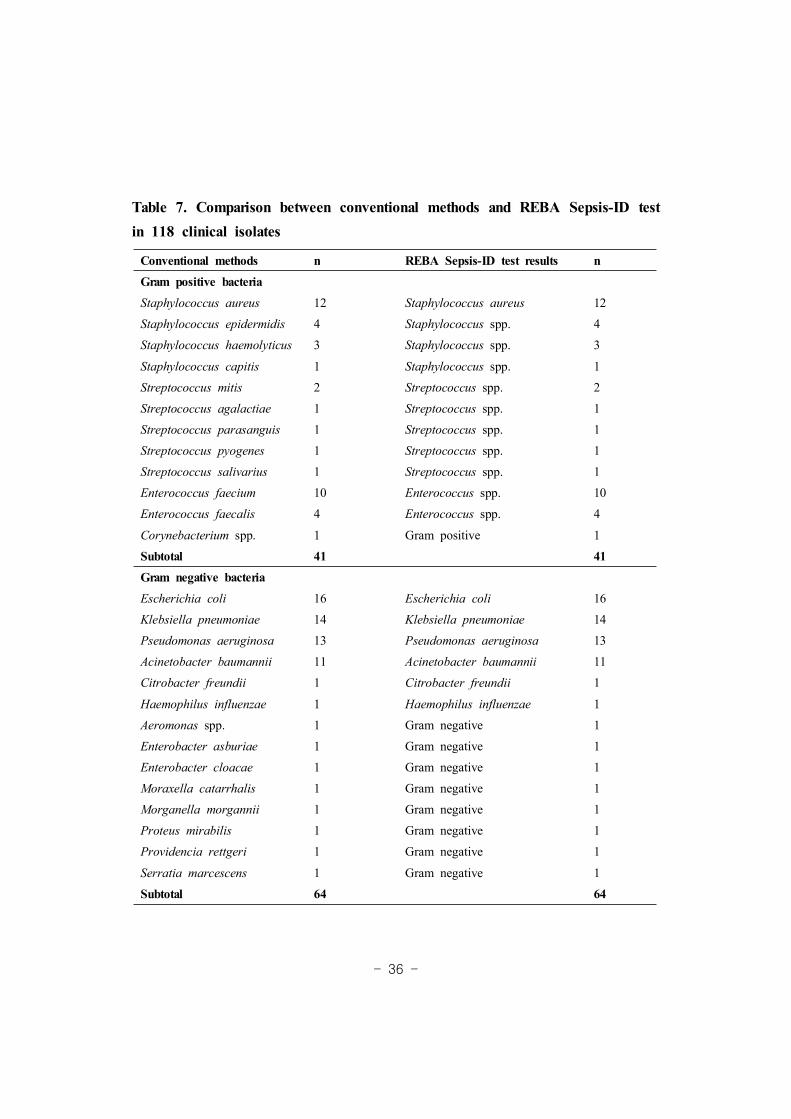
2-2. Optimization of REBA Sepsis-ID test with clinical isolates
One hundred eighteen clinical isolates (GPB 41 isolates, GNB 64,
fungi 13) that had been identified by conventional methods were analyzed by
REBA Sepsis-ID test. All DNAs of 118 clinical isolates hybridized at the
position of the corresponding universal probes (pan-bacteria, GPB, GNB, and
fungi) respectively. The REBA Sepsis-ID test identified the following clinical
isolates at the genus and/or species level: S. aureus, Staphylococcus spp., S.
pneumoniae, Streptococcus spp., Enterococcus spp., Candida spp., E. coli, K.
pneumoniae, P. aeruginosa, A. baumannii, H. influenzae, C. freundii, C.
albicans, C. tropicalis, C. glabrata, and C. parapsilosis. Nine of 12 S. aureus
and all 8 coagulase-negative staphylococci (CoNS) strains were resistant to
oxacillin ( 4 g/mL), and 7 of 14μ Enterococcus spp. were resistant to
vancomycin ( 32 g/mL) by CLSI recommended broth microdilution test.μ
All antimicrobial-resistant clinical isolates, including MRSA, methicillin-resistant
CoNS (MRCoNS), and VRE hybridized with resistance gene probes such as
mecA and vanA (Figure 6 and Table 7).
- 35 -
Figure 6. Example of REBA Sepsis-ID test results of clinical isolatesLanes 1 to 3, methicillin-resistant coagulase negative Staphyococcus (MRCoNS); lane 4,Gram negative bacteria; lanes 5 and 16, methicillin-susceptible Staphylococcus aureus(MSSA); lanes 6, 8, and 11, methicillin-susceptible Staphylococcus spp. (MSCoNS); lane 7,Streptococcus spp.; lane 9, Pseudomonas aeruginosa; lanes 10 and 14, methicillin-resistantStaphylococcus aureus (MRSA); lane 12, vancomycin-susceptible Enterococcus spp. (VSE);lanes 13 and 19, vancomycin-resistant Enterococcus spp. (VRE); lane 15, Acinetobacterbaumannii; lane 17, Candida albicans; lane 18, C. tropicalis; lane 20, Escherichia coli;lane 21, negative control.
- 36 -
Table 7. Comparison between conventional methods and REBA Sepsis-ID testin 118 clinical isolates
Conventional methods n REBA Sepsis-ID test results nGram positive bacteriaStaphylococcus aureus 12 Staphylococcus aureus 12
Staphylococcus epidermidis 4 Staphylococcus spp. 4
Staphylococcus haemolyticus 3 Staphylococcus spp. 3
Staphylococcus capitis 1 Staphylococcus spp. 1
Streptococcus mitis 2 Streptococcus spp. 2
Streptococcus agalactiae 1 Streptococcus spp. 1
Streptococcus parasanguis 1 Streptococcus spp. 1
Streptococcus pyogenes 1 Streptococcus spp. 1
Streptococcus salivarius 1 Streptococcus spp. 1
Enterococcus faecium 10 Enterococcus spp. 10
Enterococcus faecalis 4 Enterococcus spp. 4
Corynebacterium spp. 1 Gram positive 1
Subtotal 41 41Gram negative bacteriaEscherichia coli 16 Escherichia coli 16
Klebsiella pneumoniae 14 Klebsiella pneumoniae 14
Pseudomonas aeruginosa 13 Pseudomonas aeruginosa 13
Acinetobacter baumannii 11 Acinetobacter baumannii 11
Citrobacter freundii 1 Citrobacter freundii 1
Haemophilus influenzae 1 Haemophilus influenzae 1
Aeromonas spp. 1 Gram negative 1
Enterobacter asburiae 1 Gram negative 1
Enterobacter cloacae 1 Gram negative 1
Moraxella catarrhalis 1 Gram negative 1
Morganella morgannii 1 Gram negative 1
Proteus mirabilis 1 Gram negative 1
Providencia rettgeri 1 Gram negative 1
Serratia marcescens 1 Gram negative 1
Subtotal 64 64
- 37 -
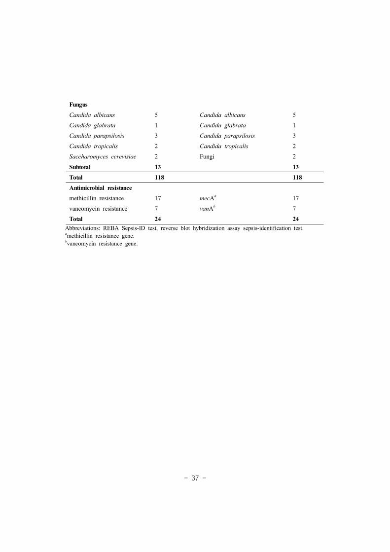
Abbreviations: REBA Sepsis-ID test, reverse blot hybridization assay sepsis-identification test.amethicillin resistance gene.bvancomycin resistance gene.
FungusCandida albicans 5 Candida albicans 5
Candida glabrata 1 Candida glabrata 1
Candida parapsilosis 3 Candida parapsilosis 3
Candida tropicalis 2 Candida tropicalis 2
Saccharomyces cerevisiae 2 Fungi 2
Subtotal 13 13Total 118 118Antimicrobial resistancemethicillin resistance 17 mecAa 17
vancomycin resistance 7 vanAb 7
Total 24 24

- 38 -
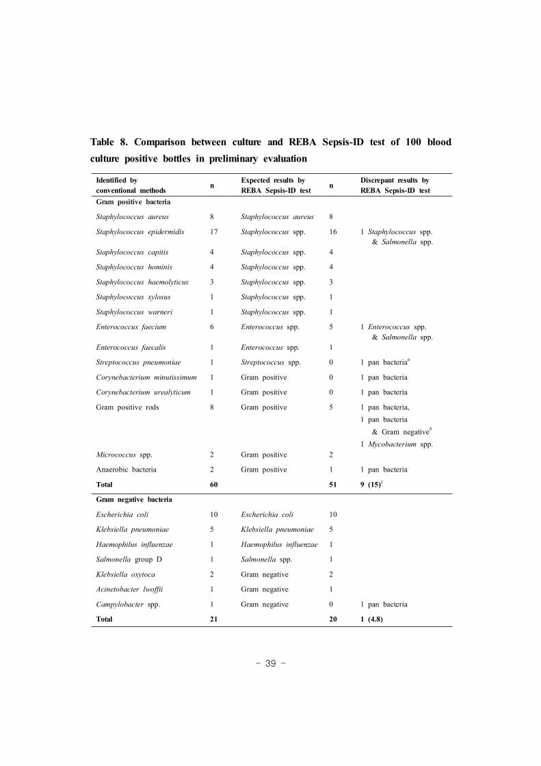
3. Evaluation of REBA Sepsis-ID test
3-1. Preliminary evaluation of REBA Sepsis-ID test with 100 blood
culture positive bottles
Comparison between blood cultures and REBA Sepsis-ID test in the
preliminary evaluation of 100 BC-PB is shown in Table 8. A total of 100
BC-PB, 5 were negative by Gram stain and 5 days incubation and 95 blood
culture true positives, 60 GPB, 21 GNB, 11 fungi, and 3 polymicrobial
isolates were obtained. A total of 35 antimicrobial-resistant isolates (33
vancomycin and 2 vancomycin resistance) were used in this study. According
to the results of REBA Sepsis-ID test comparing with culture results, 85.0%
(51/60) of GPB, 95.2% (20/21) of GNB, 90.9% (10/11) of fungi, and 66.7%
(2/3) of polymicrobial isolates showed agreement. Thirty one (88.6%) of 35
antimicrobial-resistant isolates were identified correctly resistance genes such as
mecA or vanA. Among GPB, except S. aureus, most of Gram positive rod
isolates showed discrepancy by this assay. Only Campylobacter spp. isolate
among the 21 GNB was not identified as expected result by REBA Sepsis-ID
test and one C. albicans of the 11 Candida spp. was not identified correctly.
Of 4 polymicrobial bacteremia cases, just one case showed discrepancy
between blood cultures and REBA Sepsis-ID test. Four antimicrobial-resistant
isolates, 3 MRCoNS and 1 VRE were not detected resistance genes by REBA
Sepsis-ID test in preliminary evaluation.
- 39 -
Table 8. Comparison between culture and REBA Sepsis-ID test of 100 bloodculture positive bottles in preliminary evaluation
Identified byconventional methods n
Expected results byREBA Sepsis-ID test n
Discrepant results byREBA Sepsis-ID test
Gram positive bacteria
Staphylococcus aureus 8 Staphylococcus aureus 8
Staphylococcus epidermidis 17 Staphylococcus spp. 16 1 Staphylococcus spp.& Salmonella spp.
Staphylococcus capitis 4 Staphylococcus spp. 4
Staphylococcus hominis 4 Staphylococcus spp. 4
Staphylococcus haemolyticus 3 Staphylococcus spp. 3
Staphylococcus xylosus 1 Staphylococcus spp. 1
Staphylococcus warneri 1 Staphylococcus spp. 1
Enterococcus faecium 6 Enterococcus spp. 5 1 Enterococcus spp.& Salmonella spp.
Enterococcus faecalis 1 Enterococcus spp. 1
Streptococcus pneumoniae 1 Streptococcus spp. 0 1 pan bacteriaa
Corynebacterium minutissimum 1 Gram positive 0 1 pan bacteria
Corynebacterium urealyticum 1 Gram positive 0 1 pan bacteria
Gram positive rods 8 Gram positive 5 1 pan bacteria,1 pan bacteria
& Gram negativeb
1 Mycobacterium spp.Micrococcus spp. 2 Gram positive 2
Anaerobic bacteria 2 Gram positive 1 1 pan bacteria
Total 60 51 9 (15)c
Gram negative bacteria
Escherichia coli 10 Escherichia coli 10
Klebsiella pneumoniae 5 Klebsiella pneumoniae 5
Haemophilus influenzae 1 Haemophilus influenzae 1
Salmonella group D 1 Salmonella spp. 1
Klebsiella oxytoca 2 Gram negative 2
Acinetobacter lwoffii 1 Gram negative 1
Campylobacter spp. 1 Gram negative 0 1 pan bacteria
Total 21 20 1 (4.8)
- 40 -
Abbreviation: REBA Sepsis-ID test, reverse blot hybridization assay sepsis-identification test; mecA,methicillin-resistance gene A; vanA, vancomycin-resistance gene A; vanB, vancomycin-resistance gene B.aUniversal 16S rRNA probe.b16S rRNA Gram negative probe.cDiscrepant rates between blood cultures and REBA Sepsis-ID test.
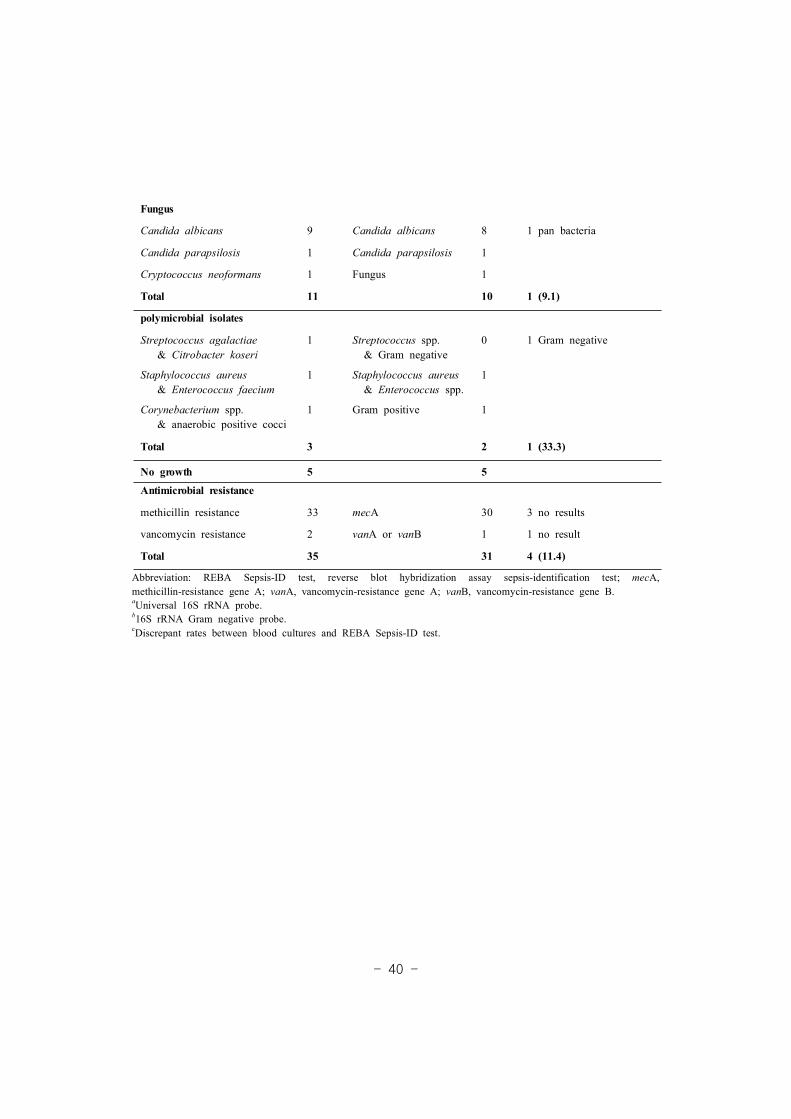
Fungus
Candida albicans 9 Candida albicans 8 1 pan bacteria
Candida parapsilosis 1 Candida parapsilosis 1
Cryptococcus neoformans 1 Fungus 1
Total 11 10 1 (9.1)
polymicrobial isolates
Streptococcus agalactiae& Citrobacter koseri
1 Streptococcus spp.& Gram negative
0 1 Gram negative
Staphylococcus aureus& Enterococcus faecium
1 Staphylococcus aureus& Enterococcus spp.
1
Corynebacterium spp.& anaerobic positive cocci
1 Gram positive 1
Total 3 2 1 (33.3)
No growth 5 5Antimicrobial resistance
methicillin resistance 33 mecA 30 3 no results
vancomycin resistance 2 vanA or vanB 1 1 no result
Total 35 31 4 (11.4)
- 41 -
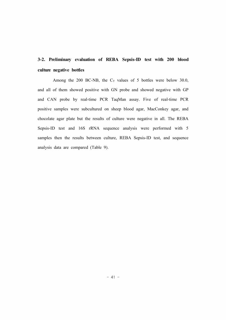
3-2. Preliminary evaluation of REBA Sepsis-ID test with 200 blood
culture negative bottles
Among the 200 BC-NB, the CT values of 5 bottles were below 30.0,
and all of them showed positive with GN probe and showed negative with GP
and CAN probe by real-time PCR TaqMan assay. Five of real-time PCR
positive samples were subcultured on sheep blood agar, MacConkey agar, and
chocolate agar plate but the results of culture were negative in all. The REBA
Sepsis-ID test and 16S rRNA sequence analysis were performed with 5
samples then the results between culture, REBA Sepsis-ID test, and sequence
analysis data are compared (Table 9).

- 42 -
Table 9. Comparison between REBA Sepsis-ID test and sequence analysis of 5real-time PCR positive samples among 200 blood culture negative bottles
Abbreviations: REBA Sepsis-ID test, reverse blot hybridization assay sepsis-identification test; GN, 16S rRNAGram-negative probe; GP, 16S rRNA Gram-positive probe; Pan, bacterial 16S rRNA probe; Fun, 18S 5.8Sinternal transcribed sequence (ITS) probe.aThe cut off value for positive by real-time PCR was below 30.0 in this study.bSamples subcultured on sheep blood agar, MacConkey agar, and chocolate agar plate.cUndetermined value or CT value was over 30.0.
Real-time PCR(CT value)a REBA Sepsis-ID test Cultureb results
16S rRNA sequenceanalysis resultsGN GP CAN
28.25 NDc ND Pan No growth Janthinobacterium spp.
29.05 ND ND Pan, GN, Fun No growth Proteobacterium
27.01 ND ND Pan No growth Proteobacterium
20.48 ND ND Pan, GN No growth Duganella spp.
22.79 ND ND Pan, GN No growth Pseudomonas spp.

- 43 -
3-3. Evaluation of REBA Sepsis-ID test with 300 blood culture
positive bottles
3-3-1. Monomicrobial blood cultures
Of the 300 BC-PB, 288 contained a single organism as determined by
the culture method. Of the monomicrobial samples, 69.8% (201/288) contained
GPB including 27 with S. aureus, 103 with CoNS, 5 with S. pneumoniae, 22
with Streptococccus spp., 13 with Enterococcus spp., 27 other GPB, and 4
anaerobic GPB. An additional 25.3% (73/288) contained GNB including 35
with E. coli, 6 with A. baumannii, 5 with P. aeruginosa, 11 with K.
pneumoniae, 1 with C. freundii, 14 with other GNB and 1 with anaerobic
GNB. A total of 4.9% (14/288) contained Candida spp. including 7 with C.
albicans, 4 with C. parapsilosis, 2 with C. glabrata and 1 with C. tropicalis.
The distribution of strains and comparison results between blood cultures and
REBA Sepsis-ID test of the 300 BC-PB are shown in Table 10 and Table 11.
Among 130 Staphylococcus spp., 2 of 27 S. aureus and 3 of 103
Staphylococcus spp. showed discrepancy between blood cultures and REBA
Sepsis-ID test. A total of 5 S. pneumoniae isolates were perfectly identified
and one of 22 Streptococcus spp. was not correctly identified to genus level.
Two Streptococcus spp. cases, one bacteria isolated by blood cultures was
identified as two isolates by REBA Sepsis-ID test. All 13 Enterococcus spp.
showed concordance between two methods, and one, Enterococcus avium
- 44 -
monobacteremia by blood cultures was detected with C. freundii by REBA
Sepsis-ID test. The probes of other 31 GPB including 10 Corynebacterium
spp., 9 Bacillus spp., 8 Micrococcus spp., and 4 anaerobic GPB were not
included on the REBA membrane and thus showed hybridization bands with
Gram-positive probes except 2 cases. Finally, a total of 201 GPB, 190 (94.5%)
showed concordance and 8 were not perfectly concordant between blood
cultures and REBA Sepsis-ID test. Three monobacteremia cases were detected
one isolate more by REBA Sepsis-ID test than by blood cultures. Seventy-one
(97.3%) of 73 GNB were correctly concordant, but one E. coli isolate was not
identified by REBA Sepsis-ID test. Enterobacter aerogenes monobacteremia
case, A. baumannii was additionally detected by REBA Sepsis-ID test. All 14
(100%) Candida species including 7 C. albicans, 1 C. tropicalis, 4 C.
parapsilosis and 2 C. glabrata were perfectly identified by REBA Sepsis-ID
test.
3-3-2. Identification of mecA, vanA, and vanB as markers of
antimicrobial resistance
The REBA Sepsis-ID test identifies the mecA gene which serves as an
indicator of resistance to methicillin in Staphylococcus spp. and the vanA and
vanB genes which serve as markers of resistance to vancomycin in
Enterococcus spp.. Ninety (97.8%) out of the 92 blood culture samples with

- 45 -
methicillin-resistant Staphylococcus isolates tested were mecA positive, mecA
was not detected in two methicillin-resistant Staphylococcus saprophyticus
isolates. No mecA genes were identified by REBA Sepsis-ID test in 44
methicillin-susceptable Staphylococcus spp.. A vanA gene was detected in one
blood sample from which vancomycin-resistant Enterococcus was isolated. The
sensitivity and specificity of REBA Sepsis-ID test for antimicrobial resistance
isolates were 97.9% (91/93) and 100%, respectively (Table 10).
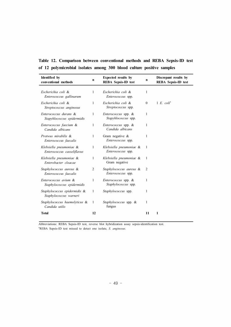
3-3-3. Polymicrobial blood samples
Of the 12 polymicrobial bacteremia samples, 11 cases were concordant,
91.7% (11/12) between blood cultures and REBA Sepsis-ID test. One E. coli
and S. anginosus case isolated by blood cultures, only E. coli was detected by
REBA Sepsis-ID test (Table 12).
3-3-4. The total concordance rates and discrepant results of 300 blood
culture positive bottles
The blood culture bottles showing discrepant results between blood
cultures and REBA Sepsis-ID test were performed 16S rRNA sequence
analysis. Most of discrepant cases were identified as uncultured bacterium by
sequence analysis, but 4 monobacteremia cases by blood cultures were detected
as two isolates by REBA Sepsis-ID test and sequence analysis (Table 13).

- 46 -
Table 10. The distribution of monomicrobial isolates and comparison resultsbetween culture method and REBA Sepsis-ID test of 300 blood culturepositive bottles
Identified byconventional methods
nExpected results byREBA Sepsis-ID test
n Discrepant results byREBA Sepsis-ID test
Gram positive bacteria
Staphylococcus aureus 27 S. aureus 25 1 no resulta,1 Staphylococcus spp.b
Staphylococcus epidermidis 46 Staphylococcus spp. 45 1 Gram positiveb
Staphylococcus hominis 23 Staphylococcus spp. 23
Staphylococcus capitis 21 Staphylococcus spp. 21
Staphylococcus haemolyticus 9 Staphylococcus spp. 9
Staphylococcus saprophyticus 2 Staphylococcus spp. 1 1 pan bacteriab
Staphylococcus intermedius 1 Staphylococcus spp. 1
Staphylococcus chleiferi 1 Staphylococcus spp. 0 1 pan bacteriab
Streptococcus pneumoniae 5 S. pneumoniae 5
Streptococcus mitis 6 Streptococcus spp. 5 1 Streptococcus spp.& S. aureusc
Streptococcus agalatiae 5 Streptococcus spp. 5
Streptococcus salivarius 3 Streptococcus spp. 3
Streptococcus anginosus 2 Streptococcus spp. 2
Streptococcus pyogenes 2 Streptococcus spp. 2
Streptococcus sanguinis 2 Streptococcus spp. 1 1 pan bacteriab
Streptococcus bovis 1 Streptococcus spp. 1
Streptococcus dysgalactiae 1 Streptococcus spp. 0 1 Streptococcus spp.& S. aureusc
Enterococcus faecium 6 Enterococcus spp. 6
Enterococcus faecalis 5 Enterococcus spp. 5
Enterococcus avium 1 Enterococcus spp. 0 1 Enterococcus spp.& C. freundiic
Enterococcus gallinarum 1 Enterococcus spp. 1
Corynebacterium spp. 10 Gram positive 10
Bacillus spp.e 9 Gram positive 7 2 Gram positive& Streptococcus spp.
Micrococcus spp. 8 Gram positive 8
Anaerobic positive cocci 4 Gram positive 4
Subtotal 201 190 11

- 47 -
Abbreviations: REBA Sepsis-ID test, reverse blot hybridization assay sepsis-identification test.aOne was not amplified and no result by REBA Sepsis-ID test.bUncultured bacterium was identified by 16S rRNA sequence analysis.cREBA Sepsis-ID test results showed concordance with 16S rRNA sequence analysis.dTwo S. saprophyticus were not identified mecA gene by REBA Sepsis-ID test.eBacillus spp. not anthracis.fIt showed weak hybridization band with E. coli probe.
Gram negative bacteria
Escherichia coli 35 E. coli 34 1 Gram negativef
Klebsiella pneumoniae 11 K. pneumoniae 11
Acinetobacter baumannii 6 A. baumannii 6
Pseudomonas aeruginosa 5 P. aeruginosa 5
Citrobacter freundii 1 C. freundii 1
Aeromonas hydrophila 3 Gram negative 3
Enterobacter cloacae 2 Gram negative 2
Morganella morganii 2 Gram negative 2
Chryseobacterium indologenes 1 Gram negative 1
Delftia acidovorans 1 Gram negative 1
Enterobacter aerogenes 1 Gram negative 0 1 Gram negative& A. baumanniic
Klebsiella oxytoca 1 Gram negative 1
Moraxella catarhalis 1 Gram negative 1
Ochrobactrum anthropi 1 Gram negative 1
Serratia marcescens 1 Gram negative 1
Anaerobic negative bacilli 1 Gram negative 1
Subtotal 73 71 2
Candida
Candida albicans 7 C. albicans 7
Candida parapsilosis 4 C. parapsilosis 4
Candida glabrata 2 C. glabrata 2
Candida tropicalis 1 C. tropicalis 1
subtotal 14 14
Antimicrobial resistance
methicillin resistance 92 mecA 90 2 no detectiond
vancomycin resistance 1 vanA or vanB 1
Total 93 91

- 48 -
Table 11. The total concordance rates of 300 blood culture positive bottlesbetween conventional methods and REBA Sepsis-ID test
Abbreviation: REBA Sepsis-ID test, reverse blot hybridization assay sepsis-identification test.*Number of isolates.
PathogensConventionalmethods
REBA Sepsis-ID testConcordant Discrepant
% ofConcordance
Gram positive bacteria 201* 190 11 94.5
Gram negative bacteria 73 71 2 97.3
Fungus 14 14 0 100
Polymicrobial 12 11 1 91.7
Antimicrobial resistance 93 91 2 97.8
- 49 -
Table 12. Comparison between conventional methods and REBA Sepsis-ID testof 12 polymicrobial isolates among 300 blood culture positive samples
Abbreviations: REBA Sepsis-ID test, reverse blot hybridization assay sepsis-identification test.aREBA Sepsis-ID test missed to detect one isolate, S. anginosus.
Identified byconventional methods n Expected results by
REBA Sepsis-ID test n Discrepant results byREBA Sepsis-ID test
Escherichia coli &Enterococcus gallinarum
1 Escherichia coli &Enterococcus spp.
1
Escherichia coli &Streptococcus anginosus
1 Escherichia coli &Streptococcus spp.
0 1 E. colia
Enterococcus durans &Stapyhlococcus epidermidis
1 Enterococcus spp. &Stapyhlococcus spp.
1
Enterococcus faecium &Candida albicans
1 Enterococcus spp. &Candida albicans
1
Protwus mirabilis &Enterococcus faecalis
1 Gram negative &Enterococcus spp.
1
Klebsiella pneumoniae &Enterococcus casseliflavus
1 Klebsiella pneumoniae &Enterococcus spp.
1
Klebsiella pneumoniae &Enterobacter cloacae
1 Klebsiella pneumoniae &Gram negative
1
Staphylococcus aureus &Enterococcus faecalis
2 Staphylococcus aureus &Enterococcus spp.
2
Enterococcus avium &Staphylococcus epidermidis
1 Enterococcus spp. &Staphylococcus spp.
1
Staphylococcus epidermidis &Staphylococcus warneri
1 Staphylococcus spp. 1
Staphylococcus haemolyticus &Candida utilis
1 Staphylococcus spp. &fungus
1
Total 12 11 1

- 50 -
Table 13. The discrepant results between conventional methods and REBA Sepsis-ID test in 300 blood culturepositive bottles
Abbreviations: REBA Sepsis-ID test, reverse blot hybridization assay sepsis-identification test.
Identified byconventional methods
No.Discrepant results byREBA Sepsis-ID test
16S rRNA sequence analysis
Staphylococcus aureus 2 1 no result, 1 Staphylococcus spp. No test & Uncultured bacterium
Staphylococcus chleiferi 1 1 pan bacteria Uncultured bacterium
Staphylococcus epidermidis 1 1 Gram positive Uncultured bacterium
Staphylococcus saprophyticus 1 1 pan bacteria Uncultured bacterium
Streptococcus dysgalactiae 1 1 Streptococcus spp. & Staphylococcus aureus Streptococcus dysgalactiae & Staphylococcus aureus
Streptococcus mitis 1 1 Streptococcus spp. & Staphylococcus aureus Streptococcus mitis & Staphylococcus aureus
Enterococcus avium 1 1 Enterococcus spp. & Citrobacter freundii Enterococcus avium & Citrobacter freundii
Enterobacter aerogenes 1 1 Gram negative & Acinetobacter baumannii Enterobacter aerogenes & Acinetobacter baumannii
Streptococcus sanguinis 1 1 pan bacteria Uncultured bacterium
Bacillus spp. 2 2 Gram positive & Streptococcus spp. Uncultured bacteriumEscherichia coli 1 1 Gram negative No test
Escherichia coli& Streptococcus anginosus
1 1 Escherichia coli No test
Subtotal 14
methicillin resistance 2 2 no detection No test
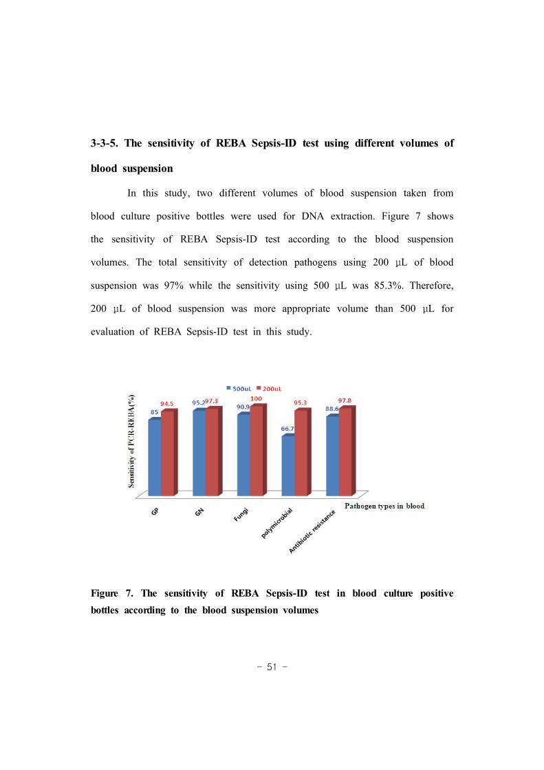
- 51 -
3-3-5. The sensitivity of REBA Sepsis-ID test using different volumes of
blood suspension
In this study, two different volumes of blood suspension taken from
blood culture positive bottles were used for DNA extraction. Figure 7 shows
the sensitivity of REBA Sepsis-ID test according to the blood suspension
volumes. The total sensitivity of detection pathogens using 200 L of bloodμ
suspension was 97% while the sensitivity using 500 L was 85.3%. Therefore,μ
200 L of blood suspension was more appropriate volume than 500 Lμ μ for
evaluation of REBA Sepsis-ID test in this study.
Figure 7. The sensitivity of REBA Sepsis-ID test in blood culture positivebottles according to the blood suspension volumes

- 52 -
3-4. Evaluation of REBA Sepsis-ID test with 1,100 blood culture
negative bottles
A total of 1,100 BC-NB, 97.6% (1,074/1,100) had CT values over
30.0. The remaining 2.4% (26/1,100) had CT values below 30.0, including 4
with values < 25.0, 1 from 26.0 to < 27.0, and 21 samples with CT values in
the range of 28.0 30.0. Of these 26 samples, 18 (69.2%) were positive with
GN probe, 5 (19.2%) were positive with GP probe and 3 (11.6%) were
positive with CAN probe by real-time PCR TaqMan assay. Colonies from
additional media and 26 real-time PCR positive samples were performed 16S
rRNA sequence analysis. Most of GP and GN probe positive cases hybridized
weakly to pan-bacteria and 3 positive cases of CAN probe hybridized weakly
to 2 C. tropicalis and 1 C. tropicalis plus C. parapsilosis probes by REBA
Sepsis-ID test. The two CAN probe positive cases, C. tropicalis were
concordant between REBA Sepsis-ID test and sequence analysis. A total of 26
samples, only one case showed concordance and 25 samples showed
discordance by conventional test, real-time PCR-REBA, and 16S rRNA
sequence analysis (Table 3).
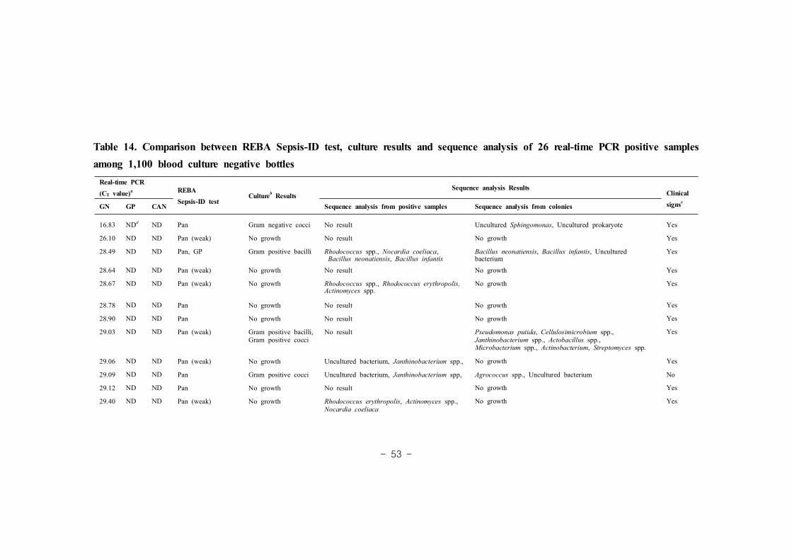
- 53 -
Table 14. Comparison between REBA Sepsis-ID test, culture results and sequence analysis of 26 real-time PCR positive samplesamong 1,100 blood culture negative bottlesReal-time PCR(CT value)a REBA
Sepsis-ID testCultureb Results
Sequence analysis ResultsClinicalsignscGN GP CAN Sequence analysis from positive samples Sequence analysis from colonies
16.83 NDd ND Pan Gram negative cocci No result Uncultured Sphingomonas, Uncultured prokaryote Yes
26.10 ND ND Pan (weak) No growth No result No growth Yes
28.49 ND ND Pan, GP Gram positive bacilli Rhodococcus spp., Nocardia coeliaca,Bacillus neonatiensis, Bacillus infantis
Bacillus neonatiensis, Bacillus infantis, Unculturedbacterium
Yes
28.64 ND ND Pan (weak) No growth No result No growth Yes
28.67 ND ND Pan (weak) No growth Rhodococcus spp., Rhodococcus erythropolis,Actinomyces spp.
No growth Yes
28.78 ND ND Pan No growth No result No growth Yes
28.90 ND ND Pan No growth No result No growth Yes
29.03 ND ND Pan (weak) Gram positive bacilli,Gram positive cocci
No result Pseudomonas putida, Cellulosimicrobium spp.,Janthinobacterium spp., Actobacillus spp.,Microbacterium spp., Actinobacterium, Streptomyces spp.
Yes
29.06 ND ND Pan (weak) No growth Uncultured bacterium, Janthinobacterium spp., No growth Yes
29.09 ND ND Pan Gram positive cocci Uncultured bacterium, Janthinobacterium spp, Agrococcus spp., Uncultured bacterium No
29.12 ND ND Pan No growth No result No growth Yes
29.40 ND ND Pan (weak) No growth Rhodococcus erythropolis, Actinomyces spp.,Nocardia coeliaca
No growth Yes

- 54 -
Abbreviations: REBA Sepsis-ID test, reverse blot hybridization assay sepsis-identification test; GN, 16S rRNA Gram-negative primers; GP, 16S rRNA Gram-positive primers; Pan, bacterial 16S rRNAprobe.aThe CT value for positive by real-time PCR was below 30.0 in this study.bSamples grew on Sheep blood, MacConkey, chocolate, R2A or Plating agar.cClinical signs follow definition of systemic inflammatory response syndrome.dUndetermined result or cut off value was over 30.0 by real-time PCR.
29.45 ND ND Pan (weak) No growth No result No growth Yes
29.67 ND ND Pan (weak) Gram positive cocci Rhodococcus spp., Rhodococcus erythropolis,Actinomyces spp., Nocardia coeliaca
Staphylococcus hominis, Uncultured bacterium Yes
29.80 ND ND Pan (weak) No growth No result No growth Yes
29.82 ND ND Pan (weak) Gram positive bacilli No result Bacillus megaterium, Bacillus aryabhattai Yes
29.85 ND ND Pan (weak) Gram positive bacilli No result Bacillus acidiceler, Bacillus luciferensis, Bacillus spp. No
29.91 ND ND Pan (weak) No growth Janthinobacterium spp., Uncultured bacterium No growth No
ND 17.08 ND Staphylococcus spp. Gram positive cocci Staphylococcus epidermidis Staphylococcus epidermidis Yes
ND 23.83 ND Pan No growth No result No growth Yes
ND 24.26 ND Staphylococcus spp. No growth Staphylococcus warneri No growth Yes
ND 26.01 ND Pan (weak) No growth Actinomyces spp. No growth Yes
ND 28.91 ND Pan (weak) No growth No result No growth Yes
ND ND 29.17 Candida tropicalis(weak)
No growth Candida tropicalis No growth Yes
ND ND 29.21 Candida tropicalis& C. parapsilosis(weak)
No growth Uncultured fungus No growth Yes
ND ND 29.85 Candida tropicalis(weak)
No growth Candida tropicalis No growth Yes
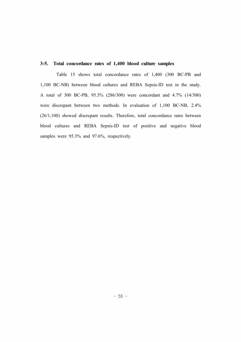
- 55 -
3-5. Total concordance rates of 1,400 blood culture samples
Table 15 shows total concordance rates of 1,400 (300 BC-PB and
1,100 BC-NB) between blood cultures and REBA Sepsis-ID test in the study.
A total of 300 BC-PB, 95.3% (286/300) were concordant and 4.7% (14/300)
were discrepant between two methods. In evaluation of 1,100 BC-NB, 2.4%
(26/1,100) showed discrepant results. Therefore, total concordance rates between
blood cultures and REBA Sepsis-ID test of positive and negative blood
samples were 95.3% and 97.6%, respectively.
- 56 -
Abbreviation: REBA Sepsis-ID test, reverse blot hybridization assay sepsis-identification test;BC-PB, blood culture positive bottles; BC-NB, blood culture negative bottles
Table 15. The concordance rates of 1,400 blood culture samples betweenblood cultures and REBA Sepsis-ID test
Blood culturesREBA Sepsis-ID test
Total no.% ofconcordance ratesconcordant discrepant
BC-PB 286 14 300 95.3
BC-NB 1,074 26 1,100 97.6

- 57 -
IV. DISCUSSION
Antimicrobial therapy for bacteremic patient depends primarily on the
species of bacteria, with differentiation between Gram positive bacteria, Gram
negative bacteria, or fungi after obtaining by Gram staining. Munson et al.69
reported that, as far as antimicrobial management is concerned, the most
important information provided by the clinical microbiology laboratory is the
first telephone call reporting positive blood culture and Gram stain results
because definitive identification and antibiotic susceptibilities are usually not
available until 24 to 48 h later. Therapy is usually empiric, involving use of a
broad-spectrum antibiotic until culture results are reported. Therefore, rapid
identification pathogens and detection of resistance genes in patients with
sepsis are important. The molecular approach would be advantageous when a
quick diagnosis needs to be made, in particular when the infectious agent is
believed to be fastidious or fungi,61,65 or when blood cultures failed to identify
the causative agent. Most of molecular-based methods have been performed on
positive blood bottle samples and some have been successful with direct blood
samples, serum, or buffy coat specimens.
In this study, a molecular-based method, REBA Sepsis-ID test was
newly designed for rapid detection of important sepsis-relevant pathogens and
antimicrobial resistance genes. The system targeted bacteria and fungi that
cause bloodstream infections and it was designed primers capable of producing
- 58 -
PCR products from common microorganisms causing bacteremia and a protocol
for direct application to blood culture suspensions. The REBA Sepsis-ID test
system can distinguish Gram positive bacteria, Gram negative bacteria, and
fungi using pan-probes, and identify bacteria and fungi using 6 genus-specific
probes and 13 species-specific probes. Moreover, this assay can simultaneously
identify antimicrobial resistance genes such as mecA and van genes.
The present system was evaluated using reference strains and clinical
isolates, as well as directly 300 instrument-positive and 1,100 instrument
-negative blood culture bottles. Even though the interpretation of the results
given by molecular tools must be made very carefully and problems may arise
the risk of contamination from the detection of DNA rather than living
pathogens, this assay was successful in identifying all 45 reference strains and
118 clinical isolates to the genus level and even to the species level.
Moreover, the concordance rates between blood cultures and REBA Sepsis-ID
test of 300 BC-PB for GPB, GNB, fungi, and mecA gene were 94.5%, 97.3%,
100% and 97.8%, respectively. Steindor et al.82 have reported that correct
identification percentages using the PCR-based DNA strip assay GenoType BC
for GPB, GNB and mecA gene were 96.1%, 89.9% and 92.9%, respectively.
The present results in positive cultures showed that the overall concordance
rate of REBA Sepsis-ID test with the culture method was high at 97.3%,
which was similar to other reports.44,60 The assay was dependent on successful
- 59 -
DNA extraction, amplification of bacterial DNA directly from positive blood
cultures, and the sensitivity and specificity of the oligonucleotide array. The
DNA extraction and achieving accurate PCR results from blood culture samples
are difficult because of presence of sodium polyanetholsulfonate (SPS), several
blood components including heme, hematin, hemoglobin, lactoferrin, and IgG
can interfere with PCR.1,4,53,92,94 Effective methods for the removal of
amplification inhibitors have been developed but often require significant
manipulation of the sample, increasing the processing time and the chance of
contamination.29 Another source of amplification failure may be occurred when
the amounts of bacteria in the liquid culture are too small or higher number.74
In this study, firstly, when 500 uL blood suspension was used for
DNA extraction, PCR amplification failed in some samples. It revealed that the
reason was too much DNA content in a bottle. After blood material volume
was reduced then competent results were obtained (Figure 7). An optimal
sample preparation procedure should efficiently break very resistant bacterial
cell walls for the DNA released from cells. An additional factor concerning
with the relatively high number of PCR negative in Gram positive bacteria is
the lower efficiency of DNA release for mechanically robust wall.74 However,
in this study, good results were obtained in DNA extraction from Gram
positive bacteria and especially, all 5 S. pneumoniae were 100% sensitive with
REBA Sepsis-ID test even though differentiation from mitis (viridians) group

- 60 -
streptococci is difficult due to close genetic relationship. Millar et al.66
investigated several commercial and in-house extraction methods to isolate
bacterial and fungal DNA from positive blood bottles. In this study described
here own in-house method was used, additional freeze and thaw steps,66 to
enhance the lysis of bacterial cell walls and fungi, especially Candida spp..
Although the developed assay could clearly discriminate the important
organisms causing bacteremia, 4.7% (14/300) for identification of pathogens
including mixed infection and 2.2% (2/93) for detecting antibiotic genes were
not concordant with conventional methods. However, if common skin flora
such as CoNS, Propionibacterium acnes, Bacillus spp., and Corynebacterium
spp. are classified as contaminants in this study, only 3% (9/300) failed to
detect the pathogens by REBA Sepsis-ID test compared with the culture
methods. Of the 14 discrepant results, one S. aureus sample failed PCR
amplification with negative PCR results. Most of discrepant pathogens excluded
samples of which PCR amplification failed may be due to inhibition or too
small amounts of bacteria in the culture bottle.
Cultures containing multiple organisms can be a challenge for direct
detection methodologies. The molecular tests that detect a limited number of
targets may be reported only one of the constituents and this can be
misleading in the case of cultures containing multiple organisms in a
polymicrobial culture. Buchan et al.12 reported that an agreement between
- 61 -
reference culture and microarray-based nucleic acid test for polymicrobial
cultures was only 72%. The reasons that polymicrobial case was not detected
with the assay might be due to technical limitations of the test such as signal
interference between multiple probes on the assay, and undetectable bacterial
concentration of minor constituents of the polymicrobial culture may be
another possible causes. Nevertheless, REBA Sepsis-ID test was successful in
identifying mixture organisms of 11 among 12 cases, 91.7% sensitivity, in
polymicrobial bacteremias.
Four cases, single bacteria isolated by blood cultures were identified
existing two isolates by REBA Sepsis-ID test and confirmed the results
correctly by 16S rRNA sequence analysis. However, in two cases,
Streptococcus mitis and Streptococcus dysgalactiae, the exact reason of
detecting with S. aureus by REBA Sepsis-ID test was not clarified with
clinical data. C. freundii monobacteremia by culture was detected with A.
baumannii by REBA Sepsis-ID test and reported additionally following the day
after by culture. It may suggest that REBA Sepsis-ID test could able to detect
the presence of small amount bacteria, which cannot be grown in culture and
has practical benefits in the clinical setting, such as for the patient who is on
empirical antibiotic treatment before blood obtained.
Of 92 MRCoNS by conventional method, mecA gene was not detected
in two (2.2%) S. saprophyticus but correctly detected as having the mecA gene
- 62 -
in all MRSA by REBA Sepsis-ID test. Two mecA-negative S. saprophyticus
isolates that would be considered methicillin resistant were false positive
because the CLSI interpretive criteria for the detection of mecA-mediated
resistance in S. saprophyticus may over estimate resistance.38 No mecA genes
were detected by REBA Sepsis-ID test in methicillin-susceptable Staphylococcus
spp. in this study.
For evaluation of BC-NB, same DNA extraction method as positive
and real-time PCR were used. In this study, when CT values considered to be
positive were below 30.0, 26 cases of 1,100 samples showed positive by
real-time PCR TaqMan assay. However, most of the cases showed weak
hybridization bands with pan-bacteria probes even though positive and negative
control bands indicated successful PCR amplification. Moreover, 13 of 26
cases might be not true positive by real-time PCR because the results were
not obtained by sequence analysis. It might originate from the amplification of
free DNA released from nonviable bacteria. Colonies isolated from subcultured
media may also be a contamination because sequence analysis results between
blood suspension and colonies were not concordant.
The reason of using the same volume for negative bottles, 200 L asμ
positive, was to know whether able to detect bacteria by REBA Sepsis-ID test
with small amount blood volumes and prevent to interfere PCR due to high
levels of human DNA when using high blood volumes. Matsuda et al.63
- 63 -
reported that positive rate using 500 L blood material from negative bloodμ
culture bottles was 10.5% by PCR method and Steindor et al.82 evaluated the
PCR-based DNA strip assay GenoType BC with blood culture positive bottles,
then emphasized that using the assay needs the presence of abundant bacteria
in blood. Kocoglu et al.48 reported that 2.6% was positive by reculture on the
media among 904 instrument-negative bottles, not PCR-based methods and
concluded that subculture is valuable in negative bottles, especially in only one
set of blood cultures is taken.
In this study, when using below 30.0 CT values for positive by
real-time PCR TaqMan assay, the positive rate of negative bottles was low
and the results between culture, REBA Sepsis-ID test, and 16S rRNA sequence
analysis were not concordant. It suggests that redesigning may be necessitated
with more specific primers, and needed a higher sample volume for evaluation
of negative bottles. On the other hand, it may be that recent CMBCS has
been becoming more sensitive to detect of bacteria in blood bottles, therefore,
subculture with negative bottles after 5 days incubation needs not.39,67
Therefore, the REBA Sepsis-ID test assay may be more useful for detecting of
pathogens in positive bottles than negative.
Nahm et al.70 reported that DNI was to be strongly correlated with
manual immature granulocyte count in sepsis patients, and Seok et al.80
described that the cut off value for DNI for predicting sepsis was 2.7%.
- 64 -
However, in this study, significant in cremental trend was not observed in
DNI between positive and negative bottles with suspected sepsis patients (data
not shown).
The newly designed REBA Sepsis-ID test in this study has certain
advantages that can simultaneously identify of bacteria, fungi and antimicrobial
resistant genes within a few hours of receipt of the blood culture samples and
has a advantage over other commercial diagnostic assays for sepsis. For
example, the SeptiFast Test (Roche) does not include probes for antibiotic
resistance, while the VYOO® test (SIRS-Lab) does contain probes for
resistance to vancomycin but it does not test for resistance to methicillin.97
Moreover, the discriminatory power of this assay can be continually extended
by adding oligonucleotides to the membrane without significantly increasing
complexity or cost.
In conclusion, REBA Sepsis-ID test is a rapid, accurate,
molecular-based method in positive blood bottles even though there was a
limitation for evaluation with negative cultures such as existing small amount
or nonviable bacteria after exposure to antibiotics. In order to be successfully
implemented in a routine microbiology laboratory, the procedure should be
fast, reliable, cost-effective, and simple to perform. This present system took
within 1 day to perform so it would be easy to adopt in normal daily
routines. If the real-time PCR can applicate for detecting pathogens in blood

- 65 -
culture positive bottles or direct blood samples prior to culture, processing time
can be completed in less than 4 hours, enables same day analysis and
reporting of results to physicians. Moreover, automation of the REBA system
is possible, which should make the system even more attractive for routine
diagnostic laboratories (Figure 8). Although the REBA Sepsis-ID test will not
absolutely replace conventional culture method, but it will provide rapid
clinical information relevant to patients due to rapid detection of important
pathogens and antimicrobial resistance genes. In the end, the application of
REBA Sepsis-ID test with culture methods in clinical microbiology laboratories
would reduce the time for detection of pathogens responsible for sepsis,
presumably resulting in reduction of patient mortality rates.
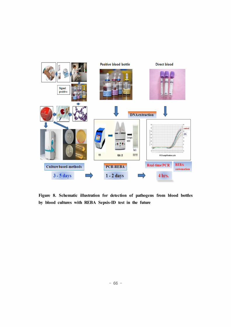
- 66 -
Figure 8. Schematic illustration for detection of pathogens from blood bottlesby blood cultures with REBA Sepsis-ID test in the future
- 67 -
REFERENCES
1. Akane A, Matsubara K, Nakamura H, Takahashi S, Kimura K. Identification
of the heme compound copurified with deoxyribonucleic acid (DNA) from
bloodstains, a major inhibitor of polymerase chain reaction (PCR) amplification.
J Forensic Sci. 1994. 39:362-372.
2. Angus DC, Linde-Zwirble WT, Lidicker J, Clermont G, Carcillo J, Pinsky
MR. Epidemiology of severe sepsis in the United States: analysis of incidence,
outcome, and associated costs of care. Crit Care Med. 2001. 29:1303-1310.
3. Annane D, Bellissant E, Cavaillon JM. Septic shock. Lancet. 2005. 365:
63-78.
4. Ansari-Lari MA, Kickler TS, Borowitz MJ. Immature granulocyte
measurement using the Sysmex XE-2100. Relationship to infection and sepsis.
Am J Clin Pathol. 2003. 120:795-799.
5. Arpi M, Bentzon MW, Jensen J, Frederiksen W. Importance of blood
volume cultured in the detection of bacteremia. Eur J Clin Microbiol Infect
Dis. 1989. 8:838-842.
6. Atlas RM. Handbook of microbiological media. London CRC Press. 2004.
- 68 -
pp.1390.
7. Barnato AE, Alexander SL, Linde-Zwirble WT, Angus DC. Racial variation
in the incidence, care, and outcomes of severe sepsis: analysis of population,
patient, and hospital characteristics. Am J Respir Crit Care Med. 2008.
177:279-284.
8. Beekmann SE, Diekema DJ, Chapin KC & Doern GV. Effects of rapid
detection of bloodstream infections on length of hospitalization and hospital
charges. J Clin Microbiol. 2003. 41:3119-3125.
9. Bernardo K, Fleer S, Pakulat N, Krut O, Hünger F, Krönke M. Identification
of Staphylococcus aureus exotoxins by combined sodium dodecyl sulfate gel
electrophoresis and matrix-assisted laser desorption/ionization-time of flight
mass spectrometry. Proteomics. 2002. 2:740-746.
10. Bertani G. Studies on lysogenesis. The mode of phase liberation by
lysogenic Escherichia coli. J Bacteriol. 1951. 62:293-300.
11. Bouza E, Sousa D, Rodríguez-Créixems M, Lechuz JG, Muñoz P. Is the
volume of blood cultured still a significant factor in the diagnosis of
bloodstream infections? J Clin Microbiol. 2007. 45:2765-2769.
12. Buchan BW, Ginocchio CC, Manii R, Cavagnolo R, Pancholi P, Swyers
- 69 -
L, Thomson RB Jr, Anderson C, Kaul K, Ledeboer NA. Multiplex
identification of gram-positive bacteria and resistance determinants directly from
positive blood culture broths: evaluation of an automated microarray-based
nucleic acid test. PLoS Med. 2013. 10:e1001478.
13. Casalta JP, Gouriet F, Roux V, Thuny F, Habib G, Raoult D. Evaluation
of the LightCycler SeptiFast test in the rapid etiologic diagnostic of infectious
endocarditis. Eur J Clin Microbiol Infect Dis. 2009. 28:569-573.
14. Chiang YC, Yang CY, Li C, Ho YC, Lin CK, Tsen HY. Identification of
Bacillus spp., Escherichia coli, Salmonella spp., Staphylococcus spp. and Vibrio
spp. with 16S ribosomal DNA-based oligonucleotide array hybridization. Int J
Food Microbiol. 2006. 107:131-137.
15. CLSI. Principles and procedures for blood cultures. Approved guideline
(M47-A), vol. 27, no. 17. Clinical and Laboratory Standards Institute, Wayne,
PA. 2007.
16. Clinical and Laboratory Standards Institute. Performance standards for
antimicrobial susceptibility testing. Twenty-first informational supplement.
Document M100-S21. Wayne PA CLSI. 2011.
17. Cosgrove SE, Sakoulas G, Perencevich EN, Schwaber MJ, Karchmer A W.
- 70 -
Carmeli Y. Comparison of mortality associated with methicillin-resistant and
methicillin-susceptible Staphylococcus aureus bacteremia: a meta-analysis. Clin
Infect Dis. 2003. 36:53-59.
18. Dellinger RP, Levy MM, Carlet JM et al. Dellinger RP, Levy MM, Carlet
JM, Bion J, Parker MM, Jaeschke R, Reinhart K, Angus DC, Brun-Buisson C,
Beale R, Calandra T, Dhainaut JF, Gerlach H, Harvey M, Marini JJ, Marshall
J, Ranieri M, Ramsay G, Sevransky J, Thompson BT, Townsend S, Vender
JS, Zimmerman JL, Vincent JL. Surviving Sepsis Campaign: international
guidelines for management of severe sepsis and septic shock. Crit Care Med.
2008. 36:296-327.
19. DiazGranados CA, Zimmer SM, Klein M, Jernigan JA. Comparison of
mortality associated with vancomycin-resistant and vancomycin-susceptible
enterococcal bloodstream infections: a meta-analysis. Clin Infect Dis. 2005.
41:327-333.
20. Diekema DJ, Pfaller MA, Jones RN; SENTRY Participants Group.
Age-related trends in pathogen frequency and antimicrobial susceptibility of
bloodstream isolates in North America: SENTRY Antimicrobial Surveillance
Program, 1997-2000. Int J Antimicrob Agents. 2002. 20:412-418.
21. Dierkers C, Ehrenstein B, Siebig S, Linde HJ, Reischl U, Salzberger B.
Clinical impact of a commercially available multiplex PCR system for rapid

- 71 -
detection of pathogens in patients with presumed sepsis. BMC Infect Dis.
2009. 9:126.
22. Dombrovskiy VY, Martin AA, Sunderram J, Paz HL. Rapid increase in
hospitalization and mortality rates for severe sepsis in the United States: a
trend analysis from 1993 to 2003. Crit Care Med. 2007. 35:1244-1250.
23. Donnino MW, Goyal N, Terlecki TM, Donnino KF, Miller JB, Otero R
M, Howell MD. Inadequate blood volume collected for culture: a survey of
health care professionals. Mayo Clin Proc. 2007. 82:1069-1072.
24. Durack DT, Lukes AS, Bright DK. New criteria for diagnosis of infective
endocarditis: utilization of specific echocardiographic findings. Duke
Endocarditis Service. Am J Med. 1994. 96:200-209.
25. Edwards-Jones V, Claydon MA, Evason DJ, Walker J, Fox AJ, Gordon
DB. Rapid discrimination between methicillin-sensitive and methicillin-resistant
Staphylococcus aureus by intact cell mass spectrometry. J Med Microbiol.
2000. 49:295-300.
26. Esper AM, Moss M, Lewis CA, Nisbet R, Mannino DM, Martin GS. The
role of infection and comorbidity: factors that influence disparities in sepsis.
Crit Care Med. 2006. 34:2576-2582.

- 72 -
27. Fenollar F, Raoult D. Molecular diagnosis of bloodstream infections caused
by non-cultivable bacteria. Int J Antimicrob Agents. 2007. Suppl 1:S7-S15.
28. Forrest GN, Mankes K, Jabra-Rizk MA, Weekes E, Johnson JK, Lincalis
DP, Venezia RA. Peptide nucleic acid fluorescence in situ hybridization-based
identification of Candida albicans and its impact on mortality and antifungal
therapy costs. J Clin Microbiol. 2006. 44:3381-3383.
29. Fredricks DN, Relman DA. Improved amplification of microbial DNA from
blood cultures by removal of the PCR inhibitor sodium polyanetholsulfonate. J
Clin Microbiol. 1998. 36:2810-2816.
30. Gatell JM, Trilla A, Latorre X, Almela M, Mensa J, Moreno A, Miro JM,
Martinez JA, Jimenez de Anta MT, Soriano E. Nosocomial bacteremia in a
large Spanish teaching hospital: analysis of factors influencing prognosis. Rev
Infect Dis. 1988. 10:203-210.
31. Goldani LZ, Mário PS. Candida tropicalis fungemia in a tertiary care
hospital. J Infect. 2003. 46:155-160.
32. Hall MM, Ilstrup DM, Washington JA 2nd. Effect of volume of blood
cultured on detection of bacteremia. J Clin Microbiol. 1976. 3:643-645.

- 73 -
33. Hansen WL, Beuving J, Bruggeman CA, Wolffs PF. Molecular probes for
diagnosis of 260 clinically relevant bacterial infections in blood cultures. J Clin
Microbiol. 2010. 48:4432-4438.
34. Harbarth S, Holeckova K, Froidevaux C, Pittet D, Ricou B, Grau GE,
Vadas L, Pugin J; Geneva Sepsis Network. Diagnostic value of procalcitonin,
interleukin-6, and interleukin-8 in critically ill patients admitted with suspected
sepsis. Am J Respir Crit Care Med. 2001. 164:396-402.
35. Harris N, Jou JM, Devoto G, Lotz J, Pappas J, Wranovics D, Wilkinson
M, Fletcher SR, Kratz A. Performance evaluation of the ADVIA 2120
hematology analyzer: an international multicenter clinical trial. Lab Hematol.
2005. 11:62-70.
36. Hensley DM, Tapia R, Encina Y. An evaluation of the Advandx
Staphylococcus aureus/CNS PNA FISH assay. Clin Lab Sci. 2009. 22:30-33.
37. Herzum I, Renz H. Inflammatory markers in SIRS, sepsis and septic
shock. Curr Med Chem. 2008. 15:581-587.
38. Higashide M, Kuroda M, Ohkawa S, Ohta T. Evaluation of a cefoxitin
disk diffusion test of mecA-positive methicillin resistant Staphylococcus
saprophyticus. Int J Antimicrob Agents. 2006. 27:500-504.
- 74 -
39. Huang AH, Yan JJ, Wu JJ. Comparison of five days versus seven days of
incubation for detection of positive blood cultures by the Bactec 9240 system.
Eur J Clin Microbiol Infect Dis. 1998. 17:637-641.
40. Ilstrup DM, Washington JA 2nd, The Importance of Volume of Blood
Cultures in the Detection of Bacteremia and Fungemia. Diagn Microbiol Infect
Dis. 1983. 1:107-110.
41. Iregui M, Ward S, Sherman G, Fraser VJ, Kollef MH. Clinical importance
of delays in the initiation of appropriate antibiotic treatment for
ventilator-associated pneumonia. Chest. 2002. 122:262-268.
42. Isaacman DJ, Karasic RB, Reynolds EA, Kost SI. Effect of number of
blood cultures and volume of blood on detection of bacteremia in children. J
Pediatr. 1996. 128:190-195.
43. Jonsson B, Nyberg A, Henning C. Theoretical aspects of detection of
bacteraemia as a function of the volume of blood cultured. APMIS. 1993.
101:595-601.
44. Jordan JA, Durso MB. Real-time polymerase chain reaction for detecting
bacterial DNA directly from blood of neonates being evaluated for sepsis. J
Mol Diagn. 2005. 7:575-581.
- 75 -
45. Kaditis AG, Marcaigh ASO, Rhodes KH, Weaver AL, Henry NK. Yield of
positive blood cultures in pediatric oncology patients by a new method of
blood culture collection. Pediatr Infect Dis J. 1996. 15:615-620.
46. Kibe S, Adams K, Barlow G. Diagnostic and prognostic biomarkers of
sepsis in critical care. J Antimicrob Chemother. 2011. Suppl 2:ii33-40.
47. Klouche M, Schröder U. Rapid methods for diagnosis of bloodstream
infections. Clin Chem Lab Med. 2008. 46:888-908.
48. Kocoglu ME, Bayram A, Balci I. Evaluation of negative results of
BacT/Alert 3D automated blood culture system. J Microbiol. 2005. 43:257-259.
49. Kong F, Gilbert GL. Multiplex PCR-based reverse line blot hybridization
assay (mPCR RLB) a practical epidemiological and diagnostic tool. Nat
Protocols. 2007. 1:2668-2680.
50. Kratz A, Maloum K, O'Malley C, Zini G, Rocco V, Zelmanovic D, Kling
G. Enumeration of nucleated red blood cells with the ADVIA 2120
Hematology System: an International Multicenter Clinical Trial. Lab Hematol.
2006. 12:63-70.
51. Kumar A, Roberts D, Wood KE et al. Kumar A, Roberts D, Wood KE,
- 76 -
Light B, Parrillo JE, Sharma S, Suppes R, Feinstein D, Zanotti S, Taiberg L,
Gurka D, Kumar A, Cheang M. Duration of hypotension before initiation of
effective antimicrobial therapy is the critical determinant of survival in human
septic shock. Crit Care Med. 2006. 34:1589-1596.
52. Kung H, Hoyert DL, Xu J, Murphy S. Deaths: final data for 2005. Natl
Vital Stat Rep. 2008. 56:1-120.
53. Lam NY, Rainer TH, Chiu RW, Lo YM. EDTA is a better anticoagulant
than heparin or citrate for delayed blood processing for plasma DNA analysis.
Clin Chem. 2004. 50:256-257.
54. Larche, J, Azoulay E, Fieux F, Mesnard L, Moreau D, Thiery G, Darmon
M, Le Gall JR, Schlemmer B. Improved survival of critically ill cancer
patients with septic shock. Intensive Care Med. 2003. 29:1688-1695.
55. Lehmann LE, Hunfeld KP, Emrich T, Haberhausen G, Wissing H, Hoeft
A, Stüber F. A multiplex real-time PCR assay for rapid detection and
differentiation of 25 bacterial and fungal pathogens from whole blood samples.
Med Microbiol Immunol. 2008. 197:313-324.
56. Leibovici L, Shraga I, Drucker M, Konigsberger H, Samra Z, Pitlik SD.
The benefit of appropriate empirical antibiotic treatment in patients with

- 77 -
bloodstream infection. J Intern Med. 1998. 244:379-386.
57. Lever A, Mackenzie I. Sepsis: definition, epidemiology, and diagnosis.
BMJ. 2007. 335:879-883.
58. Levy MM, Fink MP, Marshall JC, Abraham E, Angus D, Cook D, Cohen
J, Opal SM, Vincent JL, Ramsay G. 2001 SCCM/ ESICM/ACCP/ATS/SIS
International Sepsis Definitions Conference. Crit Care Med. 2003.
31:1250-1256.
59. Liu C, Bayer A, Cosgrove SE, Daum RS, Fridkin SK, Gorwitz RJ, Kaplan
SL, Karchmer AW, Levine DP, Murray BE, Rybak M, Talan DA, Chambers
HF. Clinical practice guidelines by the Infectious Diseases Society of America
for the treatment of methicillin-resistant Staphylococcus infections in adults and
children: executive summary. Clin Infect Dis. 2011. 52:285-292.
60. Liu Y, Han JX, Huang HY, Zhu B. Development and evaluation of 16S
rDNA microarray for detecting bacterial pathogens in cerebrospinal fluid. Exp
Biol Med. 2005. 230:587-591.
61. Mallon PG, Millar BC, Moore JE, Murphy PG, McClurg RB, Chew EW,
Crowe MJ. Molecular identification of Acinetobacter spp. in a patient with
culture-nagative endocarditis. Clin Microbiol Infect. 2000. 6:277-278.
- 78 -
62. Martin MA, Pfaller MA, Wenzel RP. Coagulase-negative staphylococcal
bacteremia. Mortality and hospital stay. Ann Intern Med. 1989. 110:9-16.
63. Matsuda K, Iwaki KK, Garcia-Gomez J, Hoffman J, Inderlied CB, Mason
WH, Iwaki Y. Bacterial identification by 16S rRNA gene PCR-hybridization as
a supplement to negative culture results. J Clin Microbiol. 2011. 49:2031-2034.
64. Mermel LA, Maki DG. Detection of bacteremia in adults: consequences of
culturing an inadequate volume of blood. Ann Intern Med. 1993. 119:270-272.
65. Millar BC, Moore JE, Mallon PG, Crowe MJ, McClurg RB, Raoult D,
Earle J, Hone R, Murphy PG. Molecular diagnosis of endocarditis-a new
Duke’s criterion. Eur Heart J. 2001. 33:673-680.
66. Millar BC, Jiru X, Moore JE, Earle JA. A simple and sensitive method to
extract bacterial, yeast and fungal DNA from blood culture material. J
Microbiol Methods. 2000. 42:139-147.
67. Mirrett S, Reller LB, Petti CA, Woods CW, Vazirani B, Sivadas R,
Weinstein MP. Controlled clinical comparison of BacT/ALERT standard aerobic
medium with BACTEC standard aerobic medium for culturing blood. J Clin
Microbiol. 2003. 41:2391-2394.
- 79 -
68. Mühl H, Kochem AJ, Disqué C, Sakka SG. Activity and DNA
contamination of commercial polymerase chain reaction reagents for the
universal 16S rDNA real-time polymerase chain reaction detection of bacterial
pathogens in blood. Diagn Microbiol Infec Dis. 2010. 66:41-49.
69. Munson EL, Diekema DJ, Beekmann SE, Chapin KC, Doern GV.
Detection and treatment of bloodstream infection: laboratory reporting and
antimicrobial management. J Clin Microbiol. 2003. 41:495-497.
70. Nahm CH, Choi JW, Lee J. Delta neutrophil index in automated immature
granulocyte counts for assessing disease severity of patients with sepsis. Ann
Clin Lab Sci. 2008. 38:241-246.
71. Mancini N, Carletti S, Ghidoli N, Cichero P, Burioni R, Clementi M. The
era of molecular and other non-culture-based methods in diagnosis of sepsis.
Clin Microbiol Rev. 2010. 23:235-251.
72. Phua J, Koay ES, Lee KH. Lactate, procalcitonin, and amino-terminal
pro-B type natriuretic peptide versus cytokine measurements and clinical
severity scores for prognostication in septic shock. Shock. 2008. 29:328-333.
73. Procop GW. Molecular diagnostics for the detection and characterization of
microbial pathogens. Clin Infect Dis. 2007. 45:S99-S111.

- 80 -
74. Rantakokko-Jalava K, Jalava J. Optimal DNA isolation method for
detection of bacteria in specimens by broad-range PCR. J Clin Microbiol.
2002. 40:4211-4217.
75. Rao PS, Shivananda PG. Bacteraemia due to non-fermenting gram-negative
bacilli in immunocompromised patients. Indian J Med Microbiol. 1993.
11:95-99.
76. Raoult D, Fournier PE, Drancourt M. What does the future hold for
clinical microbiology? Nat. Rev Microbiol. 2004. 2:151-159.
77. Reasoner DJ, Blannon JC, Geldreich EE. Rapid seven hour fecal coliform
test. Appl Environ Microbiol. 1979. 38:229-236.
78. Sautter RL, Bills AR, Lang DL, Ruschell G, Heiter BJ, Bourbeau PP.
Effects of delayed-entry conditions on the recovery and detection of
microorganisms from BacT/ALERT and BACTEC blood culture bottles. J Clin
Microbiol. 2006. 44:1245-1249.
79. Schwetz I, Hinrichs G, Reisinger EC, Krejs GJ, Olschewski H, Krause R.
Delayed processing of blood samples influences time to positivity of blood
cultures and results of Gram stain-acridine orange leukocyte Cytospin test. J
Clin Microbiol. 2007. 45:2691-2694.

- 81 -
80. Seok Y, Choi JR, Kim J, Kim YK, Lee J, Song J, Kim SJ, Lee KA.
Delta neutrophil index: A promising diagnostic and prognostic marker for
sepsis. Shock. 2012. 37:242-246.
81. Shim H, Kim KS, Uh Y, Seo DM, Kim HY, Yoon YR. The Development
and Evaluation of Blood Volume Measuring System for Blood Culture Quality
Improvement. J Test Eval. 2012. 40:521-526.
82. Steindor M, Weizenegger M, Harrison N, Hirschl AM, Schweickert B,
Göbel UB, Mackenzie CR. Use of a commercial PCR-based line blot method
for identification of bacterial pathogens and the mecA and van genes from
BacT/Alert blood culture bottles. J Clin Microbiol. 2012. 50:157-159.
83. Templeton A, Schlegel M, Fleisch F, Rettenmund G, Schöbi B, Henz S,
Eich G. Multilumen central venous catheters increase risk for catheter-related
bloodstream infection: prospective surveillance study. Infection. 2008.
36:322-327.
84. Tenney JH, Reller LB, Mirrett S, Wang WL, Weinstein MP. Controlled
evaluation of the volume of blood cultured in detection of bacteremia and
fungemia. J Clin Microbiol. 1982. 15:558-561.
85. Uh Y, Son JS, Hwang GY, Jang IH, Yoon KJ, Seo DM. Microplate

- 82 -
identification system of Enterobacteriacae. Korean J Clin Microbiol. 1999.
2:135-143.
86. van Baar BL. Characterisation of bacteria by matrix-assisted laser
desorption/ionisation and electrospray mass spectrometry. FEMS Microbiol Rev.
2000. 24:193-219.
87. Veenu. Sikka R, Arora DR. Isolation and susceptibility pattern of
non-fermenting Gram-negative bacilli from clinical samples. Indian J Med
Microbiol. 1999. 17:14-18.
88. Wallet F, Nseir S, Baumann L, Herwegh S, Sendid B, Boulo M,
Roussel-Delvallez M, Durocher AV, Courcol RJ. Preliminary clinical study
using a multiplex real-time PCR test for the detection of bacterial and fungal
DNA directly in blood. Clin Microbiol Infect. 2010. 16:774-779.
89. Wang Y, Kong F, Gilbert GL, Brown M, Gao W, Yu S, Yang Y. Use of
a multiplex PCR-based reverse line blot (mPCR/RLB) hybridization assay for
the rapid identification of bacterial pathogens. Clin Microbiol Infect. 2008.
14:155-160.
90. Warren DK, Zack JE, Elward AM, Cox MJ, Fraser VJ. Nosocomial
primary bloodstream infections in intensive care unit patients in a nonteaching
community medical center: a 21-month prospective study. Clin Infect Dis.
- 83 -
2001. 33:1329-1335.
91. Weinstein MP, Murphy JR, Reller LB, Lichtenstein KA. The clinical
significance of positive blood cultures: a comprehensive analysis of 500
episodes of bacteremia and fungemia in adults. II. Clinical observations, with
special reference to factors influencing prognosis. Rev Infect Dis. 1983.
5:54-70.
92. Weinstein MP, Towns ML, Quartey SM, Mirrett S, Reimer LG, Parmigiani
G, Reller LB. The clinical significance of positive blood cultures in the 1990s:
a prospective comprehensive evaluation of the microbiology, epidemiology, and
outcome of bacteremia and fungemia in adults. Clin Infect Dis. 1997.
24:584-602.
93. Wellinghausen N, Wirths B, Essig A, Wassill L. Evaluation of the Hyplex
BloodScreen multiplex PCR-enzyme-linked immunosorbent assay system for
direct identification of gram-positive cocci and gram-negative bacilli from
positive blood cultures. J Clin Microbiol. 2004. 42:3147-3152.
94. Wilson ML. Outpatient blood cultures: progress and unanswered questions.
Eur J Clin Microbiol Infect Dis. 2004. 23:879-880.
95. Yoo SM, Choi JY, Yun JK, Choi JK, Shin SY, Lee K, Kim JM, Lee S
- 84 -
Y. DNA microarray-based identification of bacterial and fungal pathogens in
blood stream infections. Mol Cell Probes. 2010. 24:44-52.
96. Zaragoza R, Artero A, Camarena JJ, Sancho S, Gonzalez R, Nogueira JM.
The influence of inadequate empirical antimicrobial treatment on patients with
bloodstream infections in an intensive care unit. Clin Microbiol Infect. 2003.
9:412-418.
97. Zhang K, McClure JA, Elsayed S, Louie T, Conly JM. Novel multiplex
PCR assay for characterization and concomitant subtying of staphylococcal
cassette chromosome mec types I to V in 283 methicillin-resistant
Staphylococcus aureus. J Clin Microbiol. 2005. 43:5026-5033.

- 85 -
국 문 요 약
를 이용한Reverse blot hybridization assay Sepsis-ID test
패혈증 환자 혈액 배양병 내의 원인균과
mecA, vanA, van 항생제 내성 유전자의 동시 검출B
연세대학교 일반대학원
임상병리학과
박순덕
패혈증 환자의 혈액에서 원인균을 빠르고 정확하게 검출하는 것은 적당한 치료
를 빠르게 함으로써 이로 인한 사망률과 이환율을 줄이는 데 매우 중요하다 이번 연구.
에서는 혈류 감염의 원인이 되는 병인균과 항생제 내성 유전자를 동시에 신속하게
동정할 수 있는 새로운 Reverse blot hybridization assay Sepsis-Identification test
를 제작하고 임상검체로 민감도와 특이도를 평가하였다 이(REBA Sepsis-ID test) .
의 최적화를 위해서 표준균주 임상 분리 균주로 검사하였고 의 평가kit 45 , 118 kit
는 임상 검체로 년 월부터 월 사이에 수집된 혈액배양 장비 양성 검체2013 3 7 300
와 음성 검체를 대상으로 실시하였다 임상평가를 위한 전체 검체는1,100 . 1,400
의 일종인 수치 이상인 검체를 선sepsis biomarker delta neutrophil index (DNI) 2.7%
별하였다 배양 양성병은 전통적 법 배양 음성병은 법 실시 후. PCR , real-time PCR

- 86 -
를 시행하였다 전체 배양 양성병 중에는 그람 양성균 그람REBA . 300 201 (67.0%),
음성균 진균 다중감염이 포함되었고73 (24.3%), 14 (4.7%), 12 (4.0%) 92
내성균주와 내성균주를 대상으로 를methicillin 1 vancomycin REBA Sepsis-ID test
평가하였다 이 의 최적화를 위해 실시한 표준균주와 임상균주 평가에서. kit 45 118
는 혈액배양과의 결과 비교에서 일치율을 보였다 민감도를 위한 배양100% . 300
양성병 평가에서 전통적 혈액 배양과 간의 일치율은 그람 양REBA Sepsis-ID test
성균 그람 음성균 진균 다중감염 의 결과를 보였다 전94.5%, 97.3%, 100%, 91.7% .
체 내성 균주 중 에서methicillin REBA Sepsis-ID test mec 유전자가A 97.8% (90/92)
검출되었고 내성 균주에서1 vancomycin vanA 유전자가 검출되었다 혈액배양 음.
성병 평가에서는 양성real-time PCR CT 값을 이하로 했을 때30 , 2.4% (26/1,100)
양성 결과를 보였다 양성 검체를 전통적 방법. Real-time PCR 26 , real-time PCR,
염기 서열 분석법으로 비교하였을 때 방법간의 결과가 일치하지 않은16S rRNA
것으로 나타났다 결론적으로 는 혈액배양 양성 검체에서 혈류. REBA Sepsis-ID test
감염의 원인균과 항생제 내성 유전자를 동시에 신속하고 정확하게 검출 할 수 있
는 유용한 방법으로 임상 검사실에서 전통적 혈액 배양법과 같이 사용한다면 검출
시간 단축으로 환자에게 빠르고 적절한 치료를 함으로써 환자 사망률을 줄이는데
도움을 줄 수 있을 것으로 사료된다.
핵심이 되는 말 혈액배양 혈: polymerase chain reaction, reverse, blot, hybridization, ,
류 감염 항균제 내성 유전자, , mecA, vanA, vanB
Related Documents