Annu. Rev. Plant Biol. 2002. 53:109–30 DOI: 10.1146/annurev.arplant.53.091701.153921 Copyright c 2002 by Annual Reviews. All rights reserved REVEALING THE MOLECULAR SECRETS OF MARINE DIATOMS Angela Falciatore and Chris Bowler Laboratory of Molecular Plant Biology, Stazione Zoologica “A. Dohrn,” Villa Comunale, I-80121 Naples, Italy; e-mail: [email protected] Key Words iron, marine algae, photoperception, quorum sensing, silica ■ Abstract Diatoms are unicellular photosynthetic eukaryotes that contribute close to one quarter of global primary productivity. In spite of their ecological success in the world’s oceans, very little information is available at the molecular level about their bio- logy. Their most well-known characteristic is the ability to generate a highly ornamen- ted silica cell wall, which made them very popular study organisms for microscopists in the last century. Recent advances, such as the development of a range of molecular tools, are now allowing the dissection of diatom biology, e.g., for understanding the molecular and cellular basis of bioinorganic pattern formation of their cell walls and for eluci- dating key aspects of diatom ecophysiology. Making diatoms accessible to genomics technologies will potentiate greatly these efforts and may lead to the use of diatoms to construct submicrometer-scale silica structures for the nanotechnology industry. CONTENTS DIATOMS AND ECOSYSTEM EARTH ................................... 109 DIATOM BIOLOGY ................................................... 111 General Characteristics ............................................... 111 Cell Division and Cell Wall Biogenesis .................................. 112 Diatom Sex ........................................................ 113 Diatom Photosynthesis ............................................... 114 PERCEPTION OF ENVIRONMENTAL SIGNALS .......................... 115 Nutrients ........................................................... 116 Light .............................................................. 117 Neighbor Perception ................................................. 119 DIATOM PHYLOGENY ............................................... 120 TOWARD A DIATOM MODEL SPECIES ................................. 121 DIATOMS AND ECOSYSTEM EARTH The origin of life and the subsequent changes in oceanic productivity that followed the evolution of photosynthesis have profoundly influenced the geochemistry of the Earth for the past three billion years (29). In the contemporary ocean, marine 1040-2519/02/0601-0109$14.00 109

Welcome message from author
This document is posted to help you gain knowledge. Please leave a comment to let me know what you think about it! Share it to your friends and learn new things together.
Transcript
5 Apr 2002 10:17 AR AR156-05.tex AR156-05.SGM LaTeX2e(2001/05/10)P1: IKH10.1146/annurev.arplant.53.091701.153921
Annu. Rev. Plant Biol. 2002. 53:109–30DOI: 10.1146/annurev.arplant.53.091701.153921
Copyright c© 2002 by Annual Reviews. All rights reserved
REVEALING THE MOLECULAR SECRETS OF
MARINE DIATOMS
Angela Falciatore and Chris BowlerLaboratory of Molecular Plant Biology, Stazione Zoologica “A. Dohrn,” Villa Comunale,I-80121 Naples, Italy; e-mail: [email protected]
Key Words iron, marine algae, photoperception, quorum sensing, silica
■ Abstract Diatoms are unicellular photosynthetic eukaryotes that contribute closeto one quarter of global primary productivity. In spite of their ecological success in theworld’s oceans, very little information is available at the molecular level about their bio-logy. Their most well-known characteristic is the ability to generate a highly ornamen-ted silica cell wall, which made them very popular study organisms for microscopists inthe last century. Recent advances, such as the development of a range of molecular tools,are now allowing the dissection of diatom biology, e.g., for understanding the molecularand cellular basis of bioinorganic pattern formation of their cell walls and for eluci-dating key aspects of diatom ecophysiology. Making diatoms accessible to genomicstechnologies will potentiate greatly these efforts and may lead to the use of diatoms toconstruct submicrometer-scale silica structures for the nanotechnology industry.
CONTENTS
DIATOMS AND ECOSYSTEM EARTH . . . . . . . . . . . . . . . . . . . . . . . . . . . . . . . . . . . 109DIATOM BIOLOGY . . . . . . . . . . . . . . . . . . . . . . . . . . . . . . . . . . . . . . . . . . . . . . . . . . . 111
General Characteristics. . . . . . . . . . . . . . . . . . . . . . . . . . . . . . . . . . . . . . . . . . . . . . . 111Cell Division and Cell Wall Biogenesis. . . . . . . . . . . . . . . . . . . . . . . . . . . . . . . . . . 112Diatom Sex . . . . . . . . . . . . . . . . . . . . . . . . . . . . . . . . . . . . . . . . . . . . . . . . . . . . . . . . 113Diatom Photosynthesis. . . . . . . . . . . . . . . . . . . . . . . . . . . . . . . . . . . . . . . . . . . . . . . 114
PERCEPTION OF ENVIRONMENTAL SIGNALS. . . . . . . . . . . . . . . . . . . . . . . . . . 115Nutrients. . . . . . . . . . . . . . . . . . . . . . . . . . . . . . . . . . . . . . . . . . . . . . . . . . . . . . . . . . . 116Light . . . . . . . . . . . . . . . . . . . . . . . . . . . . . . . . . . . . . . . . . . . . . . . . . . . . . . . . . . . . . . 117Neighbor Perception. . . . . . . . . . . . . . . . . . . . . . . . . . . . . . . . . . . . . . . . . . . . . . . . . 119
DIATOM PHYLOGENY . . . . . . . . . . . . . . . . . . . . . . . . . . . . . . . . . . . . . . . . . . . . . . . 120TOWARD A DIATOM MODEL SPECIES . . . . . . . . . . . . . . . . . . . . . . . . . . . . . . . . . 121
DIATOMS AND ECOSYSTEM EARTH
The origin of life and the subsequent changes in oceanic productivity that followedthe evolution of photosynthesis have profoundly influenced the geochemistryof the Earth for the past three billion years (29). In the contemporary ocean, marine
1040-2519/02/0601-0109$14.00 109
5 Apr 2002 10:17 AR AR156-05.tex AR156-05.SGM LaTeX2e(2001/05/10)P1: IKH
110 FALCIATORE ¥ BOWLER
phytoplankton, made up of photosynthetic bacteria such as prochlorophytes andcyanobacteria, and eukaryotic microalgae, such as chromophytes (brown algae),rhodophytes (red algae), and chlorophytes (green algae), represent the major con-tributors of marine carbon fixation. In some regions of the ocean, these organismscan fix approximately the same amount of carbon, a few grams per square meterper day, as a terrestrial forest (82). Today, the oceans cover 70% of the Earth’ssurface, and on a global scale they are thought to contribute approximately onehalf of the total primary productivity of the planet.
Diatoms are a group of unicellular chromophyte algae that colonize the oceansdown to depths to which photosynthetically available radiation can penetrate. Theyare thought to be the most important group of eukaryotic phytoplankton, respon-sible for approximately 40% of marine primary productivity (29). They also playa key role in the biogeochemical cycling of silica (84) owing to their requirementfor this mineral for cell wall biogenesis.
There are well over 250 genera of extant diatoms, with perhaps as many as100,000 species (71, 88) ranging across three orders of magnitude in size (aboutas many as land plants) and exhibiting a remarkable variety of shapes. The well-studied small-celled species (5–50µm) tend to be most abundant at the beginningof spring and autumn, when nutrients are not limiting and when light intensity andday length are optimal for diatom photosynthesis. When nutrients run out they willoften aggregate into flocs that sink quickly out of the photic zone. The giant diatoms(which can reach 2–5 mm in size) are ubiquitous in all oceans, and their abundanceshows less seasonal variability. Their silica cell walls predominate in the sedimentsof the ocean floor, thus making them serious players in ocean biogeochemistryover geologically significant timescales (45). Besides planktonic diatoms, whichare found in all open water masses, there are many benthic forms, growing onsediments or attached to rocks or macroalgae, and some species can also be foundin soil (59). Diatoms also constitute a large proportion of the algae associatedwith sea ice in the Antarctic and Arctic. Furthermore, in warm oligotrophic seas itis possible to find symbioses between nitrogen-fixing bacteria and cyanobacteriaand diatoms (91). In these areas the fixation of nitrogen by these endosymbiontscontributes a significant amount of nitrogen to the ecosystem.
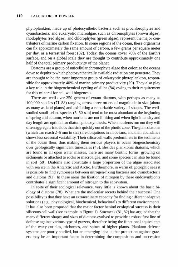
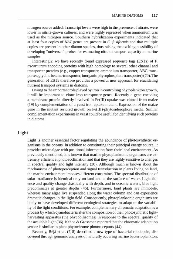
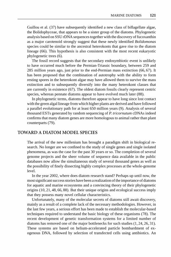
In spite of their ecological relevance, very little is known about the basic bi-ology of diatoms (78). What are the molecular secrets behind their success? Onepossibility is that they have an extraordinary capacity for finding different adaptivesolutions (e.g., physiological, biochemical, behavioral) to different environments.It has also been proposed that the major factor behind ecological success is theirsiliceous cell wall (see example in Figure 1). Smetacek (81, 82) has argued that themany different shapes and sizes of diatoms evolved to provide a robust first line ofdefense against various type of grazers, therefore being the functional equivalentsof the waxy cuticles, trichomes, and spines of higher plants. Plankton defensesystems are poorly studied, but an emerging idea is that protection against graz-ers may be an important factor in determining the composition and succession
10 Apr 2002 10:7 AR AR156-05.tex AR156-05.SGM LaTeX2e(2001/05/10)P1: IKH
MARINE DIATOMS 111
Figure 1 Electron micrograph of the diatomMastogloia binotata(Grun.) cl. at 5800times magnification. Photo courtesy of Keigo Osada.
of phytoplankton. If this is the case plankton evolution may be ruled as much byprotection as by short-term adaptations or competition (82).
DIATOM BIOLOGY
General Characteristics
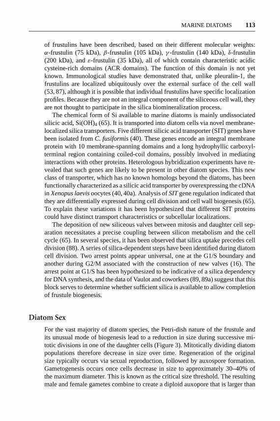
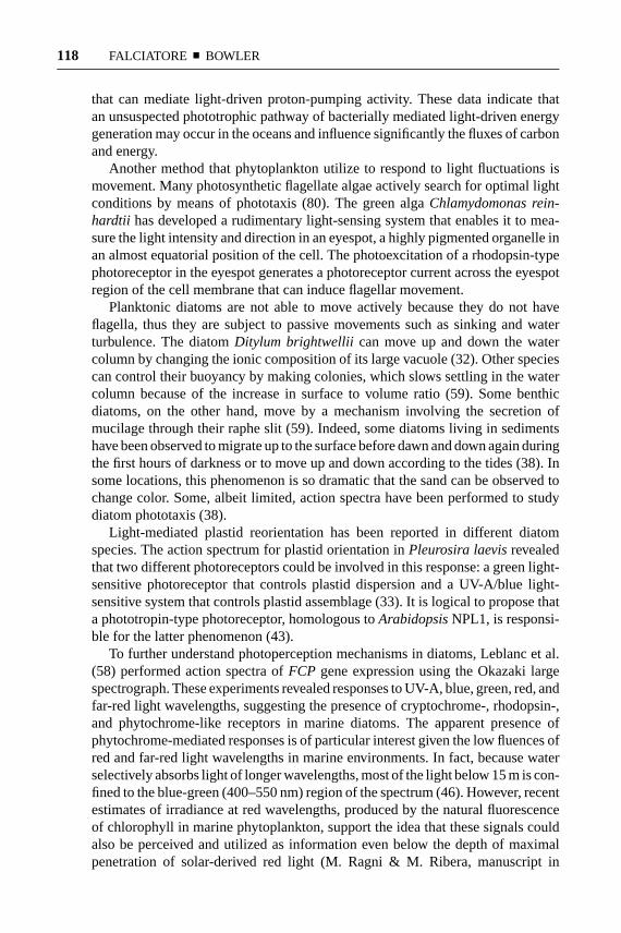
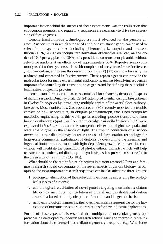
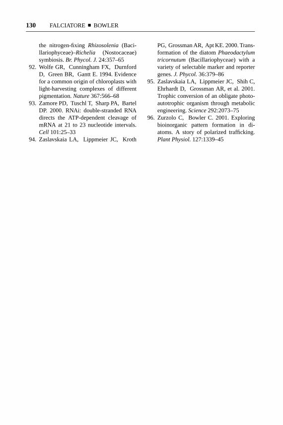
Diatoms are Bacillariophycea within the division Heterokontophyta (also knownas Stramenopiles). Their most characteristic feature is the ability to generate ahighly patterned external wall composed of amorphous silica [(SiO2)n(H2O)],known as the frustule (Figure 1; Figure 2). This is constructed of two almost equalhalves, with the smaller fitting into the larger like a Petri dish (Figure 2). Thelarger of the two halves is denoted the epitheca, and the inner one is denotedthe hypotheca. Each theca is typically composed of two parts: the valve (whichforms the larger outer surface) and a girdle (circular bands of silica attached to theedge of the valve) (Figure 2). The siliceous material of the frustule is laid downby largely unknown mechanisms in highly regular patterns that leave the wallbeautifully ornamented. Pattern design is faithfully reproduced from generationto generation, implicating a strict genetic control of the process. The precision ofthis nanoscale architecture far exceeds the capabilities of present-day materialsscience engineering, indicating that the understanding of the process will one daybe exploitable in nanotechnological applications (15a, 63, 70, 73).
Diatoms are generally classified into two major groups depending on the sym-metry of their frustules (88). Centric diatoms are radially symmetrical, whereaspennate diatoms are elongated and bilaterally symmetrical. The former group tends

10 Apr 2002 10:9 AR AR156-05.tex AR156-05.SGM LaTeX2e(2001/05/10)P1: IKH
112 FALCIATORE ¥ BOWLER
to be planktonic, whereas the latter are benthic, living on sediments or other sur-faces. Some of the pennate diatoms are able to glide along surfaces, owing to thepresence of a crevice (known as raphe) within one or both of the frustules throughwhich mucilage is secreted to aid movement (Figure 2).
Cell Division and Cell Wall Biogenesis
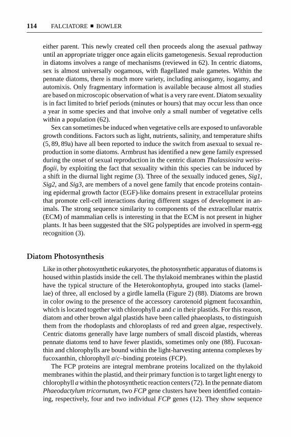
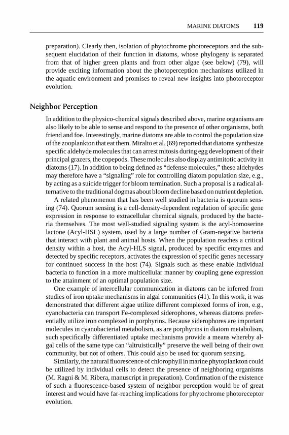
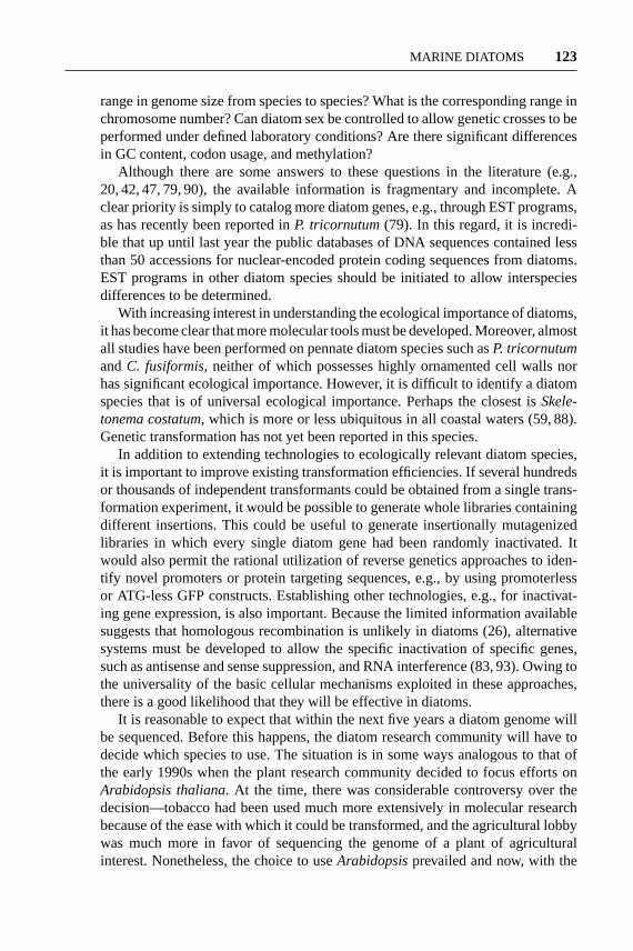
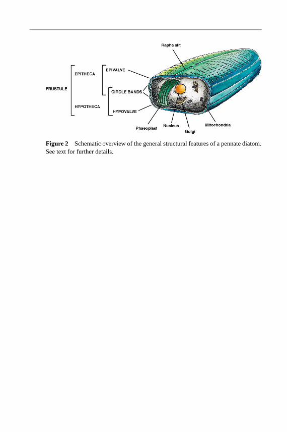
As far as is known, vegetative diatom cells are diploid. The normal asexual methodof reproduction is by division of one cell into two, with each valve of the parent cellbecoming an epitheca of the daughter cell (75). Each daughter cell must thereforegenerate a new hypotheca (Figure 3). The process of frustule formation is onlypartially understood and is largely based on microscopical observations. Priorto cell division, the cell elongates, pushing the epitheca away slightly from thehypotheca, and the nucleus divides by an “open” mitosis. After the protoplast hasdivided in two by invagination of the plasma membrane, each daughter cell mustgenerate a new hypotheca. Remarkably, this structure, which must cover one halfof the cell, is commonly generated by the polarized generation of a huge vesicleknown as the silica deposition vesicle (SDV). Hypotheca biogenesis involves thelaying down of a precise silica lattice work followed by its coating with an organicmatrix that prevents its dissolution. Once generated, the entire structure is thenexocytosed, after which the two daughter cells can separate. This whole processis not well understood but has been reviewed recently (96).
Analysis of frustule composition has revealed the presence of specific organiccomponents in addition to silica. Modified peptides known as silaffins togetherwith putrescine-derived polyamines bind the silica scaffold extremely tightly andcan only be removed following complete solubilization of silica with anhydroushydrogen fluoride. Both the silaffins and polyamines can promote silica precipita-tion in vitro, generating a network of nanospheres with diameters between 100 nmand 1µm, depending on the molecules used (51, 52). It is therefore possible thatsuch components generate the basic building blocks for silica cell wall formation.Cylindrotheca fusiformiscontains two major types of silaffins, denoted silaffin-1A(4 kDa) and silaffin-1B (8 kDa). Both are proteolytically derived from the productof a single gene, denotedSil1.
Hydrogen fluoride-extractable material also contains other proteinaceous frac-tions of high molecular mass, denoted pleuralins (pleuralin-1 was formerly knownas HEP200) (53, 54). Immunolocalization experiments have revealed that pleuralin-1 is specifically localized to the terminal girdle band (known as pleural band) ofthe epitheca but that it is also targeted to the pleural band of the hypotheca of a cellthat has mitotically divided but not yet generated a hypotheca (54). It is thereforea terminal differentiation marker for the epitheca. A small gene family encodespleuralin proteins in theC. fusiformisgenome, which are localized together (53).All encode proteins with characteristic repeat domains.
Calcium-binding glycoproteins known as frustulins (49, 50) have been local-ized to the outer coating of diatom cell walls (87). To date, five different types
5 Apr 2002 10:17 AR AR156-05.tex AR156-05.SGM LaTeX2e(2001/05/10)P1: IKH
MARINE DIATOMS 113
of frustulins have been described, based on their different molecular weights:α-frustulin (75 kDa),β-frustulin (105 kDa),γ -frustulin (140 kDa),δ-frustulin(200 kDa), andε-frustulin (35 kDa), all of which contain characteristic acidiccysteine-rich domains (ACR domains). The function of this domain is not yetknown. Immunological studies have demonstrated that, unlike pleuralin-1, thefrustulins are localized ubiquitously over the external surface of the cell wall(53, 87), although it is possible that individual frustulins have specific localizationprofiles. Because they are not an integral component of the siliceous cell wall, theyare not thought to participate in the silica biomineralization process.
The chemical form of Si available to marine diatoms is mainly undissociatedsilicic acid, Si(OH)4 (65). It is transported into diatom cells via novel membrane-localized silica transporters. Five different silicic acid transporter (SIT) genes havebeen isolated fromC. fusiformis(40). These genes encode an integral membraneprotein with 10 membrane-spanning domains and a long hydrophyllic carboxyl-terminal region containing coiled-coil domains, possibly involved in mediatinginteractions with other proteins. Heterologous hybridization experiments have re-vealed that such genes are likely to be present in other diatom species. This newclass of transporter, which has no known homologs beyond the diatoms, has beenfunctionally characterized as a silicic acid transporter by overexpressing the cDNAin Xenopus laevisoocytes (40, 40a). Analysis ofSITgene regulation indicated thatthey are differentially expressed during cell division and cell wall biogenesis (65).To explain these variations it has been hypothesized that different SIT proteinscould have distinct transport characteristics or subcellular localizations.
The deposition of new siliceous valves between mitosis and daughter cell sep-aration necessitates a precise coupling between silicon metabolism and the cellcycle (65). In several species, it has been observed that silica uptake precedes celldivision (88). A series of silica-dependent steps have been identified during diatomcell division. Two arrest points appear universal, one at the G1/S boundary andanother during G2/M associated with the construction of new valves (16). Thearrest point at G1/S has been hypothesized to be indicative of a silica dependencyfor DNA synthesis, and the data of Vaulot and coworkers (89, 89a) suggest that thisblock serves to determine whether sufficient silica is available to allow completionof frustule biogenesis.
Diatom Sex
For the vast majority of diatom species, the Petri-dish nature of the frustule andits unusual mode of biogenesis lead to a reduction in size during successive mi-totic divisions in one of the daughter cells (Figure 3). Mitotically dividing diatompopulations therefore decrease in size over time. Regeneration of the originalsize typically occurs via sexual reproduction, followed by auxospore formation.Gametogenesis occurs once cells decrease in size to approximately 30–40% ofthe maximum diameter. This is known as the critical size threshold. The resultingmale and female gametes combine to create a diploid auxopore that is larger than
5 Apr 2002 10:17 AR AR156-05.tex AR156-05.SGM LaTeX2e(2001/05/10)P1: IKH
114 FALCIATORE ¥ BOWLER
either parent. This newly created cell then proceeds along the asexual pathwayuntil an appropriate trigger once again elicits gametogenesis. Sexual reproductionin diatoms involves a range of mechanisms (reviewed in 62). In centric diatoms,sex is almost universally oogamous, with flagellated male gametes. Within thepennate diatoms, there is much more variety, including anisogamy, isogamy, andautomixis. Only fragmentary information is available because almost all studiesare based on microscopic observation of what is a very rare event. Diatom sexualityis in fact limited to brief periods (minutes or hours) that may occur less than oncea year in some species and that involve only a small number of vegetative cellswithin a population (62).
Sex can sometimes be induced when vegetative cells are exposed to unfavorablegrowth conditions. Factors such as light, nutrients, salinity, and temperature shifts(5, 89, 89a) have all been reported to induce the switch from asexual to sexual re-production in some diatoms. Armbrust has identified a new gene family expressedduring the onset of sexual reproduction in the centric diatomThalassiosira weiss-flogii, by exploiting the fact that sexuality within this species can be induced bya shift in the diurnal light regime (3). Three of the sexually induced genes,Sig1,Sig2, andSig3, are members of a novel gene family that encode proteins contain-ing epidermal growth factor (EGF)-like domains present in extracellular proteinsthat promote cell-cell interactions during different stages of development in an-imals. The strong sequence similarity to components of the extracellular matrix(ECM) of mammalian cells is interesting in that the ECM is not present in higherplants. It has been suggested that the SIG polypeptides are involved in sperm-eggrecognition (3).
Diatom Photosynthesis
Like in other photosynthetic eukaryotes, the photosynthetic apparatus of diatoms ishoused within plastids inside the cell. The thylakoid membranes within the plastidhave the typical structure of the Heterokontophyta, grouped into stacks (lamel-lae) of three, all enclosed by a girdle lamella (Figure 2) (88). Diatoms are brownin color owing to the presence of the accessory carotenoid pigment fucoxanthin,which is located together with chlorophylla andc in their plastids. For this reason,diatom and other brown algal plastids have been called phaeoplasts, to distinguishthem from the rhodoplasts and chloroplasts of red and green algae, respectively.Centric diatoms generally have large numbers of small discoid plastids, whereaspennate diatoms tend to have fewer plastids, sometimes only one (88). Fucoxan-thin and chlorophylls are bound within the light-harvesting antenna complexes byfucoxanthin, chlorophylla/c–binding proteins (FCP).
The FCP proteins are integral membrane proteins localized on the thylakoidmembranes within the plastid, and their primary function is to target light energy tochlorophyllawithin the photosynthetic reaction centers (72). In the pennate diatomPhaeodactylum tricornutum, two FCPgene clusters have been identified contain-ing, respectively, four and two individualFCP genes (12). They show sequence
5 Apr 2002 10:17 AR AR156-05.tex AR156-05.SGM LaTeX2e(2001/05/10)P1: IKH
MARINE DIATOMS 115
similarity to the chlorophylla/b–binding protein genes (CAB) of plants and greenalgae. Like CAB proteins, diatom FCP proteins are encoded in the nucleus. Al-though they are functionally and structurally strongly related to higher plant CABproteins, the transport mechanisms that target them to the diatom plastid are verydifferent, owing to the fact that diatom plastids are enclosed within four membranesrather than two membranes as in land plants (see Diatom Phylogeny below). TheN-terminal translocation sequences of immature FCP proteins are in fact bipartite.One of these is likely to be utilized for translocation through the outer membrane,whereas the other (a more conventional plastid transit peptide) is utilized for trans-port through the inner two membranes (11). The former sequence resembles an en-doplasmic reticulum signal peptide and, indeed, is capable of cotranslational importand processing by microsomal membranes. The process of plastid protein targetingand import has been more thoroughly studied for a related presequence from theγ -subunit of the plastid ATP synthase from the centric diatomOdontella sinensis(55).
In T. weissflogii, semiquantitative RT-PCR analysis ofFCP gene expressionrevealed that transcript levels decrease during prolonged darkness and are highlyinduced following a subsequent shift to white light (58). InDunaliella tertiolecta,a marine green alga, a shift from high to low irradiance leads to a rapid inductionof CAB gene expression (57). It has been proposed that this regulation allowsincreased production of light-harvesting pigment complexes in conditions of lowlight intensity and that redox signaling from the chloroplast to the nucleus controlsthe process (25). It will be interesting to determine whether such mechanismsalso exist in diatoms and whether they contribute to the ability of diatoms tophotosynthesize at optimal levels over a wide range of light intensities (30).
One potentially significant observation that has recently been made is that di-atoms may be capable of C4 photosynthesis (77). This is a specialized form ofphotosynthesis that is restricted to a few land plants, such as sugar cane and maize,and that allows a more efficient utilization of available CO2. The report by Rein-felder and colleagues is the first description of C4 photosynthesis in a marinemicroalga, and the data suggest that C4 carbon metabolism may be confined tothe cytoplasm, away from the RUBISCO-dependent reactions within the plastid.If shown to be a universal feature of diatoms, C4 photosynthesis may provide afurther explanation for their ecological success in the world’s oceans, in that it pro-vides a means of storing carbon for use in less favorable conditions. Identificationof the genes encoding key enzymes of C4 metabolism will provide the moleculartools for exploring the universality of this type of photosynthesis in diatoms.
PERCEPTION OF ENVIRONMENTAL SIGNALS
Living organisms are constantly bombarded with information to which they mustreact. In plant cells, both external and internal signals are amplified and communi-cated by complex signal transduction pathways, most of which are initiated by theactivation of receptor proteins (85). The marine environment potentially contains
5 Apr 2002 10:17 AR AR156-05.tex AR156-05.SGM LaTeX2e(2001/05/10)P1: IKH
116 FALCIATORE ¥ BOWLER
a great number of physicochemical signals that can be utilized to control organ-ismal adaptive responses to changing local conditions. The importance of molec-ular sensing of environmental signals in diatoms has recently been examined bystudying calcium-dependent signal transduction inP. tricornutum(28). Transgenicdiatom cells containing the calcium-sensitive photoprotein aequorin were gener-ated to follow changes in calcium homeostasis in response to a range of relevantenvironmental stimuli. The results revealed sensing systems for detecting and re-sponding to fluid motion, osmotic stress, and nutrient limitation. In particular,an exquisitely sensitive calcium-dependent signaling mechanism was induced byiron, a key nutrient controlling diatom abundance in the ocean (see below). Basedon our knowledge of calcium signaling in other organisms, the physiological re-sponses of diatoms to environmental changes are therefore likely to be regulatedby sense-process-respond chains involving specific receptors and feedback mech-anisms, whose activity is determined by the previous history of the cell. The furthercharacterization of signal transduction pathways that tune cellular metabolism toambient light and nutrient levels will be an important step for understanding themolecular mechanisms that contribute to the ecological success of diatoms.
Nutrients
External nutrient concentrations are key regulators of phytoplankton growth. In themarine environment, nutrients such as nitrate, silicate, and phosphate are extremelyimportant, and strong evidence also implicates dissolved iron as being a limitingresource for phytoplankton growth in many regions of the oceans (see 29 andreferences therein). The “iron hypothesis” has been tested and supported throughin situ iron fertilization experiments in the equatorial Pacific Ocean and elsewhere(15, 18, 64), which resulted in a clear and unambiguous physiological response tothe addition of iron—a massive phytoplankton bloom of predominantly diatoms.Subsequently, some effort has been dedicated to developing methods to determineiron deficiency in phytoplankton in situ. Flavodoxin is the first such example of amolecular marker for studying diatom cell physiology in their natural context (56).Because the appearance of this protein correlates with iron deficiency, flavodoxinhas been used as an immunological probe to map the degree of iron stress in naturalpopulations.
To better understand some of the nutrient-sensing mechanisms in marine algae,it will be necessary to clone genes encoding nutrient transporters. Besides thepreviously described silicic acid transporters (SIT), Hildebrand & Dahlin recentlyreported the cloning and initial characterization of the first genes encoding nitratetransporters (NAT) in a marine organism, from the diatomC. fusiformis(39).The NAT proteins are predicted to have 12 membrane-spanning domains withhydrophyllic amino- and carboxy-termini that are located in the cytoplasm. Theproteins show significant homology to the nitrate transporter family NRT2 ofAspergillus nidulans, but no similarity to the NTR1 class ofArabidopsis. NATgene expression inC. fusiformiswas sensitive to both the level and the type of
5 Apr 2002 10:17 AR AR156-05.tex AR156-05.SGM LaTeX2e(2001/05/10)P1: IKH
MARINE DIATOMS 117
nitrogen source added: Transcript levels were high in the presence of nitrate, werelower in nitrite-grown cultures, and were highly repressed when ammonium wasused as the nitrogen source. Southern hybridization experiments indicated thatat least four copies ofNAT genes are present inC. fusiformisand that multiplecopies are present in other diatom species, thus raising the exciting possibility ofdeveloping “universal” probes for estimating nitrate transport capacity in marinesamples.
Interestingly, we have recently found expressed sequence tags (ESTs) ofP.tricornutumencoding proteins with high homology to several other channel andtransporter proteins (e.g., copper transporter, ammonium transporter, ABC trans-porter, glycine betaine transporter, inorganic phyrophosphate transporter) (79). Thegeneration of ESTs therefore provides a powerful new approach for elucidatingnutrient transport systems in diatoms.
Owing to the important role played by iron in controlling phytoplankton growth,it will be important to clone iron transporter genes. Recently a gene encodinga membrane protein directly involved in Fe(III) uptake was cloned from maize(19) by complementation of a yeast iron uptake mutant. Expression of the maizegene in the mutant restored growth on Fe(III)-phytosiderophore media. Similarcomplementation experiments in yeast could be useful for identifying such proteinsin diatoms.
Light
Light is another essential factor regulating the abundance of photosynthetic or-ganisms in the oceans. In addition to constituting their principal energy source, itprovides microalgae with positional information from their local environment. Aspreviously mentioned, it is known that marine phytoplanktonic organisms are ex-tremely efficient at photoacclimation and that they are highly sensitive to changesin spectral quality and light intensity (30). Although much is known about themechanisms of photoperception and signal transduction in plants living on land,the marine environment imposes different constraints. The spectral distribution ofsolar irradiance is identical only on land and at the surface of water. Light flu-ence and quality change drastically with depth, and in oceanic waters, blue lightpredominates at greater depths (46). Furthermore, land plants are immobile,whereas many algae live suspended along the water column and can experiencedramatic changes in the light field. Consequently, phytoplanktonic organisms arelikely to have developed different ecological strategies to adapt to the variabil-ity of the light conditions. For example, complementary chromatic adaptation is aprocess by which cyanobacteria alter the composition of their photosynthetic light-harvesting apparatus (the phycobilisomes) in response to the spectral quality ofthe available light (36). Kehoe & Grossman reported that the chromatic adaptationsensor is similar to plant phytochrome photoreceptors (44).
Recently, Beja et al. (7, 8) described a new type of bacterial rhodopsin, dis-covered through genomic analyses of naturally occuring marine bacterioplankton,
5 Apr 2002 10:17 AR AR156-05.tex AR156-05.SGM LaTeX2e(2001/05/10)P1: IKH
118 FALCIATORE ¥ BOWLER
that can mediate light-driven proton-pumping activity. These data indicate thatan unsuspected phototrophic pathway of bacterially mediated light-driven energygeneration may occur in the oceans and influence significantly the fluxes of carbonand energy.
Another method that phytoplankton utilize to respond to light fluctuations ismovement. Many photosynthetic flagellate algae actively search for optimal lightconditions by means of phototaxis (80). The green algaChlamydomonas rein-hardtii has developed a rudimentary light-sensing system that enables it to mea-sure the light intensity and direction in an eyespot, a highly pigmented organelle inan almost equatorial position of the cell. The photoexcitation of a rhodopsin-typephotoreceptor in the eyespot generates a photoreceptor current across the eyespotregion of the cell membrane that can induce flagellar movement.
Planktonic diatoms are not able to move actively because they do not haveflagella, thus they are subject to passive movements such as sinking and waterturbulence. The diatomDitylum brightwellii can move up and down the watercolumn by changing the ionic composition of its large vacuole (32). Other speciescan control their buoyancy by making colonies, which slows settling in the watercolumn because of the increase in surface to volume ratio (59). Some benthicdiatoms, on the other hand, move by a mechanism involving the secretion ofmucilage through their raphe slit (59). Indeed, some diatoms living in sedimentshave been observed to migrate up to the surface before dawn and down again duringthe first hours of darkness or to move up and down according to the tides (38). Insome locations, this phenomenon is so dramatic that the sand can be observed tochange color. Some, albeit limited, action spectra have been performed to studydiatom phototaxis (38).
Light-mediated plastid reorientation has been reported in different diatomspecies. The action spectrum for plastid orientation inPleurosira laevisrevealedthat two different photoreceptors could be involved in this response: a green light-sensitive photoreceptor that controls plastid dispersion and a UV-A/blue light-sensitive system that controls plastid assemblage (33). It is logical to propose thata phototropin-type photoreceptor, homologous toArabidopsisNPL1, is responsi-ble for the latter phenomenon (43).
To further understand photoperception mechanisms in diatoms, Leblanc et al.(58) performed action spectra ofFCP gene expression using the Okazaki largespectrograph. These experiments revealed responses to UV-A, blue, green, red, andfar-red light wavelengths, suggesting the presence of cryptochrome-, rhodopsin-,and phytochrome-like receptors in marine diatoms. The apparent presence ofphytochrome-mediated responses is of particular interest given the low fluences ofred and far-red light wavelengths in marine environments. In fact, because waterselectively absorbs light of longer wavelengths, most of the light below 15 m is con-fined to the blue-green (400–550 nm) region of the spectrum (46). However, recentestimates of irradiance at red wavelengths, produced by the natural fluorescenceof chlorophyll in marine phytoplankton, support the idea that these signals couldalso be perceived and utilized as information even below the depth of maximalpenetration of solar-derived red light (M. Ragni & M. Ribera, manuscript in
5 Apr 2002 10:17 AR AR156-05.tex AR156-05.SGM LaTeX2e(2001/05/10)P1: IKH
MARINE DIATOMS 119
preparation). Clearly then, isolation of phytochrome photoreceptors and the sub-sequent elucidation of their function in diatoms, whose phylogeny is separatedfrom that of higher green plants and from other algae (see below) (79), willprovide exciting information about the photoperception mechanisms utilized inthe aquatic environment and promises to reveal new insights into photoreceptorevolution.
Neighbor Perception
In addition to the physico-chemical signals described above, marine organisms arealso likely to be able to sense and respond to the presence of other organisms, bothfriend and foe. Interestingly, marine diatoms are able to control the population sizeof the zooplankton that eat them. Miralto et al. (69) reported that diatoms synthesizespecific aldehyde molecules that can arrest mitosis during egg development of theirprincipal grazers, the copepods. These molecules also display antimitotic activity indiatoms (17). In addition to being defined as “defense molecules,” these aldehydesmay therefore have a “signaling” role for controlling diatom population size, e.g.,by acting as a suicide trigger for bloom termination. Such a proposal is a radical al-ternative to the traditional dogmas about bloom decline based on nutrient depletion.
A related phenomenon that has been well studied in bacteria is quorum sens-ing (74). Quorum sensing is a cell-density-dependent regulation of specific geneexpression in response to extracellular chemical signals, produced by the bacte-ria themselves. The most well-studied signaling system is the acyl-homoserinelactone (Acyl-HSL) system, used by a large number of Gram-negative bacteriathat interact with plant and animal hosts. When the population reaches a criticaldensity within a host, the Acyl-HLS signal, produced by specific enzymes anddetected by specific receptors, activates the expression of specific genes necessaryfor continued success in the host (74). Signals such as these enable individualbacteria to function in a more multicellular manner by coupling gene expressionto the attainment of an optimal population size.
One example of intercellular communication in diatoms can be inferred fromstudies of iron uptake mechanisms in algal communities (41). In this work, it wasdemonstrated that different algae utilize different complexed forms of iron, e.g.,cyanobacteria can transport Fe-complexed siderophores, whereas diatoms prefer-entially utilize iron complexed in porphyrins. Because siderophores are importantmolecules in cyanobacterial metabolism, as are porphyrins in diatom metabolism,such specifically differentiated uptake mechanisms provide a means whereby al-gal cells of the same type can “altruistically” preserve the well being of their owncommunity, but not of others. This could also be used for quorum sensing.
Similarly, the natural fluorescence of chlorophyll in marine phytoplankton couldbe utilized by individual cells to detect the presence of neighboring organisms(M. Ragni & M. Ribera, manuscript in preparation). Confirmation of the existenceof such a fluorescence-based system of neighbor perception would be of greatinterest and would have far-reaching implications for phytochrome photoreceptorevolution.
5 Apr 2002 10:17 AR AR156-05.tex AR156-05.SGM LaTeX2e(2001/05/10)P1: IKH
120 FALCIATORE ¥ BOWLER
DIATOM PHYLOGENY
The plastids of all photosynthetic organisms are likely to have arisen at least1.5 billion years ago from the engulfment of a photosynthetic bacterium by aunicellular eukaryotic heterotroph (86, 88). Analysis of the sequences of plastidgenomes from a range of eukaryotic algae and from higher and lower plants clearlysupports the hypothesis that all photosynthetic eukaryotes are derived from a singleendosymbiotic event involving a photosynthetic bacterium highly similar to extantcyanobacteria (9, 21, 48, 76). Functional analysis of the photosynthetic apparatusfrom different algae also supports this view (e.g., 92). Hence, all plastids appearto have a monophyletic origin.
Current knowledge suggests that the initial endosymbiotic event gave rise even-tually to two major plastid lineages: chloroplasts and rhodoplasts. Green algae, aswell as their descendants, the higher plants, contain chloroplasts that are charac-terized by the presence of stacked thylakoid membranes and the use of chlorophylla andb for light harvesting. Red algae, on the other hand, contain rhodoplasts,which utilize chlorophylla and phycobilisomes for the capture of light energy.
Chromophyte algae, such as diatoms, differ fundamentally from the majorityof photosynthetic eukaryotes: Whereas the plastids of red algae, green algae, andplants are normally surrounded by two membranes, diatom plastids have four mem-branes. It is therefore believed that diatoms and related chromophyte algae arosefollowing a secondary endosymbiotic event in which a eukaryotic alga was en-gulfed by a second eukaryotic heterotroph (21, 34). In such a scenario, a flagellatehost contributed the nucleus, endomembranes, and mitochondria to the chimera,whereas the photosynthetic endosymbiont provided its plastid and plasma mem-brane (that perhaps became the inner three membranes of the plastid). Such ahypothesis is supported by the finding of a second nucleus (the nucleomorph)between the outer and inner two membranes in some chromophytes, presumablyderived from the nucleus of the ancestral algal endosymbiont (21). The nucleo-morph of the chromophyteGuillardia thecawas recently sequenced (22). Thegenome is highly reduced (0.55 Mb) and lacks almost all genes for metabolicfunctions, which have been transferred to the nucleus of the secondary host. Thereare, however, 30 genes for plastid-localized proteins, and all these genes are essen-tial for plastid function, presumably explaining why the nucleomorph has persistedduring evolution.
Analysis of plastid ribosomal RNAs as well as comparisons of plastid genomesstrongly support the hypothesis that diatom plastids were derived from a secondaryendosymbiosis involving an alga from the rhodoplast lineage (10, 21, 48). How-ever, diatoms have not retained phycobilins for light harvesting and instead usechlorophylla andc together with the brown carotenoid fucoxanthin (chlorophyllb is never found in diatoms).
The ancestral heterotrophic host that paired with the red alga to give rise to thephaeoplast lineage is believed to have been a small unicellular flagellate belongingto the Oomycetes (lower fungi) and similar toCafeteria roenbergensis(66, 88).
5 Apr 2002 10:17 AR AR156-05.tex AR156-05.SGM LaTeX2e(2001/05/10)P1: IKH
MARINE DIATOMS 121
Guillou et al. (37) have subsequently identified a new class of biflagellate algae,the Bolidophyceae, that appears to be a sister group of the diatoms. Phylogeneticanalysis based on SSU rDNA sequences together with the discovery of fucoxanthinas a major carotenoid strongly suggest that these newly identifiedBolidomonasspecies could be similar to the ancestral heterokonts that gave rise to the diatomlineage (66). This hypothesis is also consistent with the most recent eukaryoticphylogenetic trees (6).
The fossil record suggests that the secondary endosymbiotic event is unlikelyto have occurred much before the Permian-Triassic boundary, between 259 and285 million years ago, just prior to the end-Permian mass extinction (66, 67). Ithas been proposed that the combination of autotrophy with the ability to formresting spores in the heterokont algae may have allowed them to survive the massextinction and to subsequently diversify into the many heterokont classes thatare currently in existence (67). The oldest diatom fossils clearly represent centricspecies, whereas pennate diatoms appear to have evolved much later (88).
In phylogenetic terms, diatoms therefore appear to have long since lost contactwith the green algal lineage from which higher plants are derived and have followeda parallel evolutionary path for at least 650 million years (9). Analysis of severalthousand ESTs generated by random sequencing ofP. tricornutumcDNAs indeedconfirms that many diatom genes are more homologous to animal rather than plantcounterparts (79).
TOWARD A DIATOM MODEL SPECIES
The arrival of the new millenium has brought a paradigm shift in biological re-search. No longer are we confined to the study of single genes and single isolatedphenomena, as was the case for the past 30 years or so. The completion of severalgenome projects and the sheer volume of sequence data available in the publicdatabases now allow the simultaneous study of several thousand genes as well asthe possibility of finely dissecting highly complex processes at the whole-genomelevel.
In the year 2002, where does diatom research stand? Perhaps up until now, themost significant success stories have been a realization of the importance of diatomsfor aquatic and marine ecosystems and a convincing theory of their phylogeneticorigins (10, 21, 48, 66, 88). But their unique origins and ecological success implythat they possess many novel cellular characteristics.
Unfortunately, many of the molecular secrets of diatoms still await discovery,mainly as a result of a complete lack of the necessary methodologies. However, inthe last few years, a serious effort has been made to establish the molecular-basedtechniques required to understand the basic biology of these organisms (78). Therecent development of genetic transformation systems for a limited number ofdiatoms has removed one of the major bottlenecks for such studies (1, 24, 26, 31).These systems are based on helium-accelerated particle bombardment of ex-ogenous DNA, followed by selection of transfected cells using antibiotics. An
5 Apr 2002 10:17 AR AR156-05.tex AR156-05.SGM LaTeX2e(2001/05/10)P1: IKH
122 FALCIATORE ¥ BOWLER
important factor behind the success of these experiments was the realization thatendogenous promoter and regulatory sequences are necessary to drive the expres-sion of foreign genes.
Genetic transformation technologies are most advanced for the pennate di-atomP. tricornutumin which a range of antibiotic resistance genes can be used toselect for transgenic clones, including phleomycin, kanamycin, and nourseo-thricin (1, 26, 94). Even though transformation efficiencies are low, on the or-der of 10−6 perµg plasmid DNA, it is possible to co-transform plasmids withoutselectable markers at an efficiency of approximately 60%. Reporter genes com-monly used in other systems such as chloramphenicol acetyl transferase, luciferase,β-glucuronidase, and green fluorescent protein (GFP) (27) can now be easily in-troduced and expressed inP. tricornutum. These reporter genes can provide themolecular tools for many experimental applications, such as identifying sequencesimportant for controlling the transcription of genes and for defining the subcellularlocalization of specific proteins.
Genetic transformation is also an essential tool for enhancing the applied aspectsof diatom research. Dunahay et al. (23, 24) attempted to enhance lipid biosynthesisin Cyclotella crypticaby introducing multiple copies of the acetyl CoA carboxy-lase gene. Most significantly, Zaslavskaia et al. (95) recently reported the trophicconversion ofP. tricornutum, an obligate photoautotroph, into a heterotroph bymetabolic engineering. In this work, genes encoding glucose transporters fromhuman erythrocytes (glut1) or from the microalgaChlorella kessleri(hup1) wereexpressed inP. tricornutum, and the transgenic cells exhibited glucose uptake andwere able to grow in the absence of light. The trophic conversion ofP. tricor-nutumand other diatoms may increase the use of fermentation technology forlarge-scale commercial exploitation of diatoms by overcoming the financial andlogistical limitations associated with light-dependent growth. Moreover, this con-version will facilitate the generation of photosynthetic mutants, which will helpresearchers to understand diatom photosynthesis, as has proved so successful inthe green algaC. reinhardtii (35, 38a).
What should be the major future objectives in diatom research? First and fore-most, research should concentrate on the novel aspects of diatom biology. In ouropinion the most important research objectives can be classified into three groups:
1. ecological: elucidation of the molecular mechanisms underlying the ecolog-ical success of diatoms;
2. cell biological: elucidation of novel protein targeting mechanisms; diatomlife cycles, including the regulation of critical size thresholds and diatomsex; silica-based bioinorganic pattern formation and its genetic basis;
3. nanotechnological: harnessing the novel mechanisms responsible for the fab-rication of micrometer-scale silica structures for new industrial applications.
For all of these aspects it is essential that multiparallel molecular genetic ap-proaches be developed to underpin research efforts. First and foremost, more in-formation about the characteristics of diatom genomes is required: e.g., What is the
5 Apr 2002 10:17 AR AR156-05.tex AR156-05.SGM LaTeX2e(2001/05/10)P1: IKH
MARINE DIATOMS 123
range in genome size from species to species? What is the corresponding range inchromosome number? Can diatom sex be controlled to allow genetic crosses to beperformed under defined laboratory conditions? Are there significant differencesin GC content, codon usage, and methylation?
Although there are some answers to these questions in the literature (e.g.,20, 42, 47, 79, 90), the available information is fragmentary and incomplete. Aclear priority is simply to catalog more diatom genes, e.g., through EST programs,as has recently been reported inP. tricornutum(79). In this regard, it is incredi-ble that up until last year the public databases of DNA sequences contained lessthan 50 accessions for nuclear-encoded protein coding sequences from diatoms.EST programs in other diatom species should be initiated to allow interspeciesdifferences to be determined.
With increasing interest in understanding the ecological importance of diatoms,it has become clear that more molecular tools must be developed. Moreover, almostall studies have been performed on pennate diatom species such asP. tricornutumandC. fusiformis, neither of which possesses highly ornamented cell walls norhas significant ecological importance. However, it is difficult to identify a diatomspecies that is of universal ecological importance. Perhaps the closest isSkele-tonema costatum, which is more or less ubiquitous in all coastal waters (59, 88).Genetic transformation has not yet been reported in this species.
In addition to extending technologies to ecologically relevant diatom species,it is important to improve existing transformation efficiencies. If several hundredsor thousands of independent transformants could be obtained from a single trans-formation experiment, it would be possible to generate whole libraries containingdifferent insertions. This could be useful to generate insertionally mutagenizedlibraries in which every single diatom gene had been randomly inactivated. Itwould also permit the rational utilization of reverse genetics approaches to iden-tify novel promoters or protein targeting sequences, e.g., by using promoterlessor ATG-less GFP constructs. Establishing other technologies, e.g., for inactivat-ing gene expression, is also important. Because the limited information availablesuggests that homologous recombination is unlikely in diatoms (26), alternativesystems must be developed to allow the specific inactivation of specific genes,such as antisense and sense suppression, and RNA interference (83, 93). Owing tothe universality of the basic cellular mechanisms exploited in these approaches,there is a good likelihood that they will be effective in diatoms.
It is reasonable to expect that within the next five years a diatom genome willbe sequenced. Before this happens, the diatom research community will have todecide which species to use. The situation is in some ways analogous to that ofthe early 1990s when the plant research community decided to focus efforts onArabidopsis thaliana. At the time, there was considerable controversy over thedecision—tobacco had been used much more extensively in molecular researchbecause of the ease with which it could be transformed, and the agricultural lobbywas much more in favor of sequencing the genome of a plant of agriculturalinterest. Nonetheless, the choice to useArabidopsisprevailed and now, with the
10 Apr 2002 10:9 AR AR156-05.tex AR156-05.SGM LaTeX2e(2001/05/10)P1: IKH
124 FALCIATORE ¥ BOWLER
availability of its complete genome sequence (2) as well as powerful functionalgenomics platforms in several research centers worldwide, it is clear that the rightdecision was made.
Which diatom species should be chosen for genome-level research? Essentialrequirements include:
1) ease of growth under defined laboratory conditions,
2) ease of genetic transformation,
3) small genome size,
4) availability of genetic-based methodologies (isolation of mutants, a genomemap, sexual crosses),
5) knowledge of basic biochemistry.
Currently, no single diatom species can fulfill these criteria and represent, at thesame time, a good model from an ecological point of view. Those that come closestareP. tricornutumandC. fusiformis, at least for criteria 1, 2, and 5. Both have alsobeen reasonably well studied in terms of ecology and physiology. However, sexhas not been described in either of them. The genome size ofP. tricornutumwasmeasured as between 55 and 100 Mb (20, 90), the smallest of any diatom examinedin previous work (90). Subsequent measurements by us (79) and by M. Hildebrand(personal communication) have found theP. tricornutumgenome to be more onthe order of 15 Mb, slightly higher than that of the yeastSaccharomyces cerevisiae,whose sequence was completed in 1997 (68), whereas theC. fusiformisgenomeis approximately 60 Mb. It is important to note that several thousand ESTs havebeen generated fromP. tricornutum, and its genome properties have been char-acterized in much more detail than any other diatom (79). Its genome displaysvery low levels of methylation, normally associated with transcriptionally inactiveDNA, suggesting that its genome is gene dense with only low amounts of non-coding “junk” DNA (42). It would therefore appear to be the best choice for a se-quencing initiative. Furthermore, recent research in our laboratory using transgenicmarker genes suggests that it is possible to perform sexual crosses with this species(O. Malakhova & C. Bowler, unpublished observations).
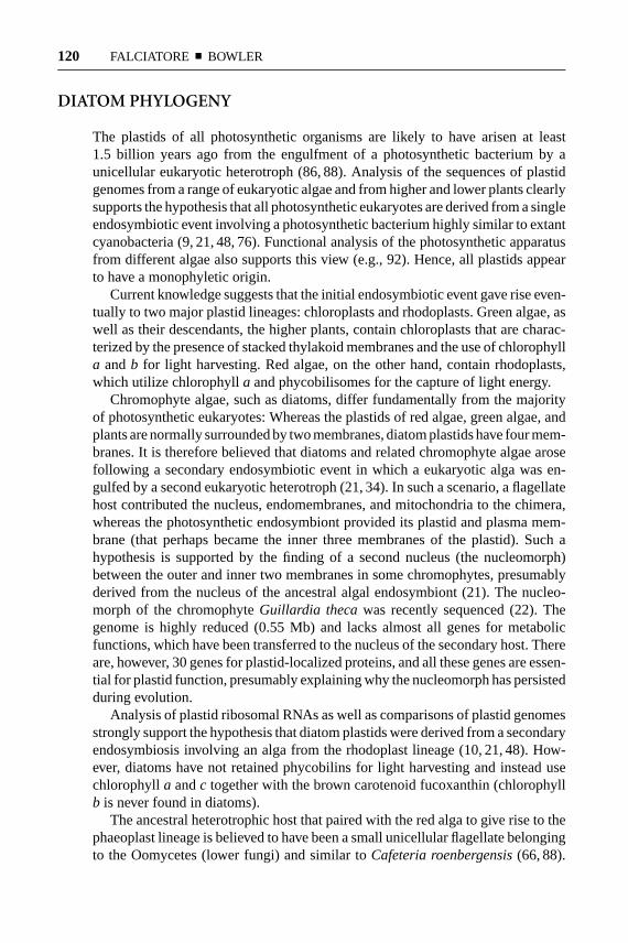
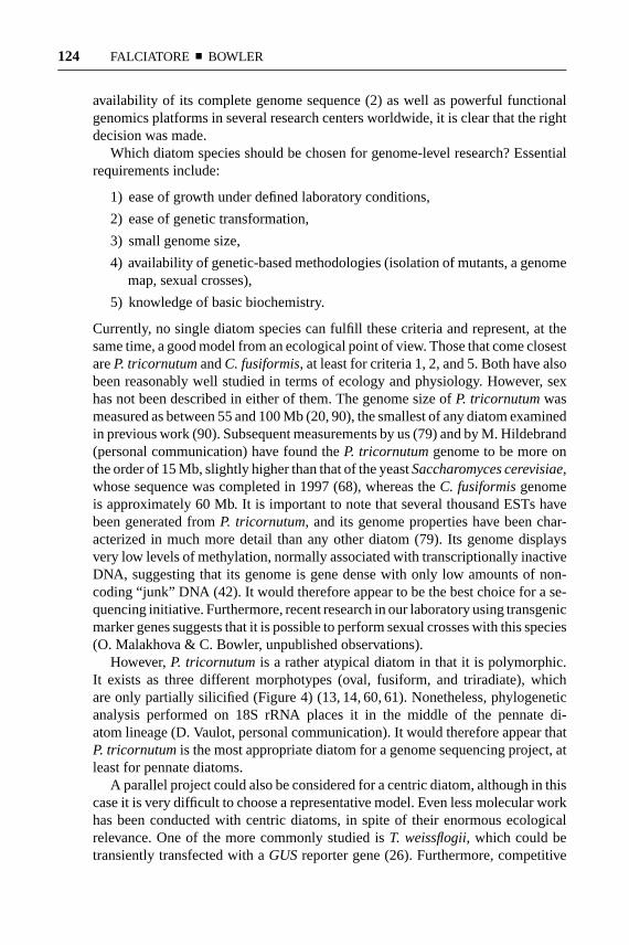
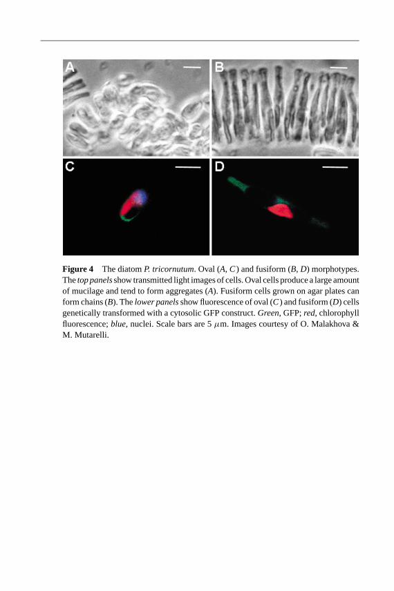
However,P. tricornutumis a rather atypical diatom in that it is polymorphic.It exists as three different morphotypes (oval, fusiform, and triradiate), whichare only partially silicified (Figure 4) (13, 14, 60, 61). Nonetheless, phylogeneticanalysis performed on 18S rRNA places it in the middle of the pennate di-atom lineage (D. Vaulot, personal communication). It would therefore appear thatP. tricornutumis the most appropriate diatom for a genome sequencing project, atleast for pennate diatoms.
A parallel project could also be considered for a centric diatom, although in thiscase it is very difficult to choose a representative model. Even less molecular workhas been conducted with centric diatoms, in spite of their enormous ecologicalrelevance. One of the more commonly studied isT. weissflogii, which could betransiently transfected with aGUSreporter gene (26). Furthermore, competitive
5 Apr 2002 10:17 AR AR156-05.tex AR156-05.SGM LaTeX2e(2001/05/10)P1: IKH
MARINE DIATOMS 125
semiquantitative RT-PCR techniques have been optimized for gene expressionstudies with this organism (58). At this time, the only centric diatom that canbe stably transformed isC. cryptica (24). Armbrust recently characterized thestructural features of the eight known nuclear genes ofT. weissflogii, with thegoal of furthering the potential of this diatom for molecular studies (4). However,the development of molecular tools for this diatom is just beginning, and thetime might be premature to determine if it could be the best centric diatom fora genome sequence project. As an alternative, some researchers have proposedThalassiosira pseudonana, which appears to have a genome size similar to that ofP. tricornutum(M. Hildebrand, personal comunication). At press time, this diatomwas on the list of organisms to be sequenced by the U.S. Department of Energy(http//www.er.doc.gov/production/ober/EPR/migcont.html).
In conclusion, although diatom research has made some important advances inrecent years, it is clear that radical measures are required to make it accessible tothe enormously powerful genomic and postgenomic research platforms. Given thenovelty and potential applicability of certain aspects of diatom ecology and cellbiology, this is surprising. However, now that a range of molecular technologiesare in place, we hope that more researchers will be attracted to this field so thatprogress can be accelerated.
ACKNOWLEDGMENTS
We thank all the numerous colleagues who have helped us to understand diatombiology over the past years. We are especially grateful to Mark Hildebrand andDaniel Vaulot for sharing unpublished information and to Ian Nettleton for theillustrations. We apologize that, owing to size restrictions, it has not been possibleto discuss all the work of our colleagues. Finally, this review is dedicated to thememory of Gaetano Salvatore, without whom our research on diatoms would neverhave begun.
Visit the Annual Reviews home page at www.annualreviews.org
LITERATURE CITED
1. Apt KE, Kroth-Pancic PG, GrossmanAR. 1997. Stable nuclear transformationof the diatomPhaeodactylum tricornu-tum. Mol. Gen. Genet.252:572–79
2. Arabidopsis Genome Initiative. 2000.Analysis of the genome sequence of theflowering plantArabidopsis thaliana. Na-ture408:796–815
3. Armbrust EV. 1999. Identification of anew gene family expressed during the on-set of sexual reproduction in the centric di-
atomThalassiosira weissflogii. Appl. Env.Microbiol. 65:3121–28
4. Armbrust EV. 2000. Structural featuresof nuclear genes in the centric diatomThalassiosira weissflogii (Bacillario-phyceae).J. Phycol.36:942–46
5. Armbrust EV, Chisholm SW. 1990. Roleof light and the cell cycle on the inductionof spermatogenesis in a centric diatom.J.Phycol.26:470–78
6. Baldauf SL, Roger AJ, Wenk-Siefert I,
5 Apr 2002 10:17 AR AR156-05.tex AR156-05.SGM LaTeX2e(2001/05/10)P1: IKH
126 FALCIATORE ¥ BOWLER
Doolittle WF. 2000. A kingdom-levelphylogeny of eukaryotes based on com-bined protein data.Science290:972–77
7. Beja O, Aravind L, Koonin EV, SuzukiMT, Hadd A, et al. 2000. Bacterial rho-dopsin: evidence for a new type of pho-totrophy in the sea.Science289:1902–6
8. Beja O, Spudich EN, Spudich JL, LeclercM, DeLong EF. 2001. Proteorhodopsinphototrophy in the ocean.Nature 411:786–89
9. Besendahl A, Qiu YL, Lee J, Palmer JD,Bhattacharya D. 2000. The cyanobacte-rial origin and vertical transmission of theplastid tRNA(Leu) group-I intron.Curr.Genet.37:12–23
10. Bhattacharya D, Medlin L. 1995. Thephylogeny of plastids: a review based oncomparisons of small-subunit ribosomalRNA coding regions.J. Phycol.31:489–98
11. Bhaya D, Grossman A. 1991. Targetingproteins to diatom plastids involves trans-port through an endoplasmic reticulum.Mol. Gen. Genet.229:400–4
12. Bhaya D, Grossman AR. 1993. Charac-terization of gene clusters encoding the fu-coxanthin chlorophyll proteins of the di-atomPhaeodactylum tricornutum. Nucl.Acids Res.21:4458–66
13. Borowitzka MA, Chiappino ML, VolcaniBE. 1977. Ultrastructure of a chain-forming diatomPhaeodactylum tricornu-tum. J. Phycol.13:162–70
14. Borowitzka MA, Volcani BE. 1978. Thepolymorphic diatomPhaeodactylum tri-cornutum: ultrastructure of its morpho-types.J. Phycol.14:10–21
15. Boyd PW, Watson AJ, Law CS, AbrahamER, Trull T. 2000. A mesoscale phyto-plankton bloom in the polar SouthernOcean stimulated by iron fertilization.Na-ture407:695–702
15a. Brott LL, Nait RR, Pikas DJ, KirkpatrickSM, Tomiln DW, et al. 2001. Ultrafastholographic nanopatterning of biocat-alytically formed silica.Nature 413:29–93
16. Brzezinski MA, Olson RJ, Chisholm SW.1990. Silicon availability and cell-cycleprogression in marine diatoms.Mar. Ecol.Prog. Ser.67:83–96
17. Casotti R, Mazza S, Ianora A, Miralto A.2001. Strategies to reduce mortality inmarine and freshwater phytoplankton.ASLO Aquat. Sci. Meet. Spec. Sess., Al-buquerque, New Mex.
18. Coale KH, Johnson KS, Fitzwater SE,Gordon RM, Tanner S, et al. 1996. A mas-sive phytoplankton bloom induced by anecosystem-scale iron fertilization experi-ment in the equatorial Pacific Ocean.Na-ture383:495–501
19. Curie C, Panaviene Z, Loulergue C, Del-laporta SL, Briat JF, et al. 2001. Maizeyellow stripe1encodes a membrane pro-tein directly involved in Fe(III) uptake.Nature409:346–49
20. Darley WM. 1968. Deoxyribonucleic acidcontent of the three cell types ofPhaeo-dactylum tricornutumBohlin. J. Phycol.4:219–20
21. Delwiche CF, Palmer JD. 1997. The ori-gin of plastids and their spread via sec-ondary symbiosis. InOrigins of Algae andTheir Plastids, ed. D Bhattacharya, pp.53–86. Vienna/New York: Springer Ver-lag
22. Douglas S, Zauner S, Fraunholz M, Bea-ton M, Penny S, et al. 2001. The highlyreduced genome of an enslaved algal nu-cleus.Nature410:1091–96
23. Dunahay TG, Jarvis EE, Dais SS, Roes-sler PG. 1996. Manipulation of microalgallipid production using genetic engineer-ing. Appl. Biochem. Biotech.57/58:223–31
24. Dunahay TG, Jarvis EE, Roessler PG.1995. Genetic transformation of the di-atomsCyclotella crypticaand Naviculasaprophila. J. Phycol.31:1004–12
25. Escoubas J-M, Lomas M, LaRoche J,Falkowski PG. 1995. Light intensity regu-lation of cabgene transcription is signaledby the redox state of plastoquinone pool.Proc. Natl. Acad. Sci. USA92:10237–41
5 Apr 2002 10:17 AR AR156-05.tex AR156-05.SGM LaTeX2e(2001/05/10)P1: IKH
MARINE DIATOMS 127
26. Falciatore A, Casotti R, Leblanc C, Ab-rescia C, Bowler C. 1999. Transformationof nonselectable reporter genes in marinediatoms.Mar. Biotech.1:239–51
27. Falciatore A, Formiggini F, Bowler C.2001. Reporter genes and in vivo imag-ing. In Molecular Plant Biology: A Prac-tical Approach, ed. P Gilmartin, C Bowler,2:265–83. Oxford, UK: Oxford Univ.Press
28. Falciatore A, Ribera D’Alcal`a M, CrootP, Bowler C. 2000. Perception of environ-mental signals by a marine diatom.Sci-ence288:2363–66
29. Falkowski PG, Barber RT, Smetacek V.1998. Biogeochemical controls and feed-backs on ocean primary production.Sci-ence281:200–6
30. Falkowski PG, LaRoche J. 1991. Accli-mation to spectral irradiance in algae.J.Phycol.27:8–14
31. Fischer H, Robl I, Sumper M, Kr¨oger N.1999. Targeting and covalent modifica-tion of cell wall and membrane proteinsheterologously expressed in the diatomCylindrotheca fusiformis (Bacillario-phyceae).J. Phycol.35:113–20
32. Fisher AE, Harrison PJ. 1996. Does car-bohydrate content affect the sinking rateof marine diatoms?J. Phycol.32:360–65
33. Furukawa T, Watanabe M, Shihira-Ishikawa I. 1998. Green- and blue-light-mediated chloroplast migration in thecentric diatomPleurosira laevis. Proto-plasma203:214–20
34. Gibbs SP. 1981. The chloroplasts of somealgal groups may have evolved from en-dosymbiotic eukaryotic algae.Ann. NewYork Acad. Sci.361:193–208
35. Grossman AR. 2000.Chlamydomonasreinhardtii and photosynthesis: geneticsto genomics.Curr. Opin. Plant Biol.3:132–37
36. Grossman AR, Schaefer MR, ChiangGG, Collier JL. 1993. Environmental ef-fects on the light-harvesting complex ofcyanobacteria.J. Bacteriol.175:575–82
37. Guillou L, Chretiennot-Dinet MJ, Medlin
LK, Claustre H, Loiseaux–de Goer S,et al. 1999.Bolidomonas: a new genuswith two species belonging to a new algalclass, the Bolidophyceae (Heterokonta).J. Phycol.35:368–81
38. Harper MA. 1977. Movements. InTheBiology of Diatoms, ed. D Werner, pp.224–49. Berkeley, CA: Univ. Calif. Press
38a. Harris EH. 2000.Chlamydomonasas amodel organism.Annu. Rev. Plant Phys-iol. Plant Mol. Biol.52:363–406
39. Hildebrand M, Dahlin K. 2000. Nitratetransporter genes from the diatomCylin-drotheca fusiformis(Bacillariophyceae):mRNA levels controlled by nitrogensource and by the cell cycle.J. Phycol.36:702–13
40. Hildebrand M, Dahlin K, Volcani BE.1998. Characterization of a silicon trans-porter gene family inCylindrotheca fusi-formis: sequences, expression analysis,and identification of homologs in other di-atoms.Mol. Gen. Genet.260:480–86
40a. Hildebrand M, Volcani BE, GassmannW, Schroeder JI. 1997. A gene family ofsilicon transporters.Nature385:68–89
41. Hutchins DA, Witter AE, Butler A, Lu-ther GW III. 1999. Competition amongmarine phytoplankton for different che-lated iron species.Nature400:858–61
42. Jarvis EE, Dunahay TG, Brown LM.1992. DNA nucleoside composition andmethylation in several species of microal-gae.J. Phycol.28:356–62
43. Kagawa T, Sakai T, Suetsugu N, OikawaK, Ishiguro S, et al. 2001.ArabidopsisNPL1: a phototropin homolog controllingthe chloroplast high-light avoidance re-sponse.Science291:2138–41
44. Kehoe DM, Grossman AR. 1996. Simi-larity of a chromatic adaptation sensor tophytochrome and ethylene receptors.Sci-ence273:1409–12
45. Kemp AES, Pike J, Pearce RB, LangeCB. 2000. The “fall dump”—a new per-spective on the role of a “shade flora” inthe annual cycle of diatom production andexport flux.Deep-Sea Res.47:2129–54
5 Apr 2002 10:17 AR AR156-05.tex AR156-05.SGM LaTeX2e(2001/05/10)P1: IKH
128 FALCIATORE ¥ BOWLER
46. Kirk JTO. 1983.Light and Photosynthesisin Aquatic Ecosystems. Cambridge, UK:Cambridge Univ. Press
47. Kociolek JP, Stoermer EF. 1989. Chro-mosome numbers in diatoms: a review.Diatom Res.4:47–54
48. Kowallik KV. 1992. Origin and Evolu-tion of Plastids from Chlorophyll-a+ c-Containing Algae: Suggested AncestralRelationships to Red and Green AlgalPlastids, ed. RA Lewin. New York/London: Chapman Hall
49. Kroger N, Bergsdorf C, Sumper M. 1994.A new calcium-binding glycoprotein fam-ily constitutes a major diatom cell wallcomponent.EMBO J.13:4676–83
50. Kroger N, Bergsdorf C, Sumper M. 1996.Frustulins: domain conservation in a pro-tein family associated with diatom cellwalls.Eur. J. Biochem.239:259–64
51. Kroger N, Deutzmann R, Bergsdorf C,Sumper M. 2000. Species-specific poly-amines from diatoms control silica mor-phology.Proc. Natl. Acad. Sci. USA97:14133–38
52. Kroger N, Deutzmann R, Sumper M.1999. Polycationic peptides from diatombiosilica that direct silica nanosphere for-mation.Science286:1129–32
53. Kroger N, Lehmann G, Rachel R, Sum-per M. 1997. Characterization of a 200-kDa diatom protein that is specifically as-sociated with a silica-based substructureof the cell wall.Eur. J. Biochem.250:99–105
54. Kroger N, Wetherbee R. 2000. Pleuralinsare involved in theca differentiation in thediatom Cylindrotheca fusiformis. Protist151:263–73
55. Lang M, Apt KE, Kroth PG. 1998. Pro-tein transport into “complex” diatom plas-tids utilizes two different targeting sig-nals.J. Biol. Chem.273:30973–78
56. LaRoche J, Boyd PW, McKay RML,Geider RJ. 1996. Flavodoxin as an in situmarker for iron stress in phytoplankton.Nature382:802–5
57. LaRoche J, Mortain-Bertrand A, Falkow-
ski PG. 1991. Light intensity–inducedchanges incab mRNA and light-har-vesting complex II apoprotein levels in theunicellular chlorophyteDunaliella terti-olecta. Plant Physiol.97:147–53
58. Leblanc C, Falciatore A, Bowler C. 1999.Semi-quantitative RT-PCR analysis ofphotoregulated gene expression in marinediatoms.Plant Mol. Biol.40:1031–44
59. Lee RE. 1999. Heterokontophyta, Bacil-lariophyceae. InPhycology, ed. RE Lee,pp. 415–58. Cambridge, UK: CambridgeUniv. Press
60. Lewin JC. 1958. The taxonomic positionof Phaeodactylum tricornutum. J. Gen.Microbiol. 18:427–32
61. Lewin JC, Lewin RA, Philpott DE. 1958.Observations onPhaeodactylum tricor-nutum. J. Gen. Microbiol.18:418–26
62. Mann DG. 1993. Patterns of sexual re-production in diatoms.Hydrobiologia269/270:11–20
63. Mann S, Ozin GA. 1996. Synthesis ofinorganic materials with complex form.Nature382:313–18
64. Martin JH, Coale KH, Johnson KS,Fitzwater SE, Gordon RM, et al. 1994.Testing the iron hypothesis in ecosystemsof the equatorial Pacific Ocean.Nature371:123–29
65. Martin-Jezequel V, Hildebrand M, Brze-zinski MA. 2000. Silicon metabolism indiatoms: implications for growth.J. Phy-col. 36:821–40
66. Medlin LK, Kooistra WCH, SchmidA-MM. 2000. A review of the evolutionof the diatoms—a total approach usingmolecules, morphology and geology. InThe Origin and Early Evolution of theDiatoms: Fossil, Molecular and Biogeo-graphical Approaches, ed. A Witkowski, JSieminska, pp. 13–35. Cracow: W SzaferInst. Bot., Pol. Acad. Sci.
67. Medlin LK, Kooistra WHCF, GersondeR, Sims PA, Wellbrock U. 1997. Is theorigin of the diatoms related to the end-Permian mass extinction?Nova Hedwigia65:1–11
5 Apr 2002 10:17 AR AR156-05.tex AR156-05.SGM LaTeX2e(2001/05/10)P1: IKH
MARINE DIATOMS 129
68. Mewes HW, Albermann K, Bahr M,Frishman D, Gleissner A, et al. 1997.Overview of the yeast genome.Nature387 (Suppl.):7–65
69. Miralto A, Barone G, Romano G, PouletSA, Ianora A, et al. 1999. The insidiouseffect of diatoms on copepod reproduc-tion. Nature402:173–76
70. Morse DE. 1999. Silicon biotechnology:harnessing biological silica production toconstruct new materials.Trends Biotech-nol. 17:230–32
71. Norton TA, Melkonian M, Andersen RA.1996. Algal biodiversity.Phycologia35:308–26
72. Owens TG. 1986. Light-harvesting func-tion in the diatomPhaeodactylum tricor-nutum. Plant Physiol.80:732–38
73. Parkinson J, Gordon R. 1999. Beyondmicromachining: the potential of diatoms.Trends Biotechnol.17:190–96
74. Parsek M, Greenberg EP. 2000. Acyl-homoserine lactone quorum sensingin Gram-negative bacteria: a signalingmechanism involved in associations withhigher organisms.Proc. Natl. Acad. Sci.USA97:8789–93
75. Pickett-Heaps J, Schmid A-MM, EdgarLA. 1990. The cell biology of diatomvalve formation. InProgress in Phycolog-ical Research, ed. FE Round, DJ Chap-man. 7:1–168. Bristol, UK: BioPress Ltd.
76. Qiu Y-L, Palmer JD. 1999. Phylogeny ofearly land plants: insights from genes andgenomes.Trends Plant Sci.4:26–30
77. Reinfelder JR, Kraepiel AML, MorelFMM. 2000. Unicellular C4 photosynthe-sis in a marine diatom.Nature407:996–99
78. Scala S, Bowler C. 2001. Molecular in-sights into the novel aspects of diatom bio-logy. Cell. Mol. Life Sci.58:1666–73
79. Scala S, Carels N, Falciatore A, Chiu-sano ML, Bowler C. 2002. Genome prop-erties of the diatomPhaeodactylum tri-cornutumand comparison with the ge-nomes of other eukaryotes.Plant Physiol.In press
80. Sineshchekov OA, Govorunova EV.1999. Rhodopsin-mediated photosensingin green flagellated algae.Trends PlantSci.4:58–63
81. Smetacek V. 1999. Diatoms and the oceancarbon cycle.Protist150:25–32
82. Smetacek V. 2001. A watery arms race.Nature411:745
83. Smith NA, Singh SP, Wang MB, Stout-jesdijk PA, Green AG, et al. 2000. Totalsilencing by intron-spliced hairpin RNAs.Nature407:319–20
84. Treguer P, Nelson DM, Van BennekomAJ, DeMaster DJ, Leynaert A, et al. 1995.The silica balance in the world ocean: areestimate.Science268:375–79
85. Trewavas A. 2000. Signal perception andtransduction. InBiochemistry and Molec-ular Biology of Plants, ed. BB Buchanan,W Gruissem, RL Jones, pp. 930–87.Rockville, MA: Am. Soc. Plant Physiol.
86. Valentin K, Cattolico RA, Zetsche K.1992.Phylogenetic Origin of the Plastids.New York/London: Chapman Hall
87. van de Poll WH, Vrieling EG, GieskesWWC. 1999. Location and expression offrustulins in the pennate diatomsCylin-drotheca fusiformis, Navicula pellicul-osa, andNavicula salinarum(Bacillario-phyceae).J. Phycol.35:1044–53
88. Van Den Hoek C, Mann DG, Johns HM.1997.Algae. An Introduction to Phycol-ogy. Cambridge, UK: Cambridge Univ.Press
89. Vaulot D, Olson RJ, Chisholm SW. 1986.Light and dark control of the cell cycle intwo phytoplankton species.Exp. Cell Res.167:38–52
89a. Vaulot D, Olson RJ, Merkel SM, Chis-holm SW. 1987. Cell cycle response tonutrient starvation in two marine phyto-plankton species.Mar. Biol. 95:625–30
90. Veldhuis MJW, Cucci TL, Sieracki ME.1997. Cellular DNA content of marinephytoplankton using two new fluoro-chromes: taxonomic and ecological im-plications.J. Phycol.33:527–41
91. Villareal TA. 1989. Division cycles in
5 Apr 2002 10:17 AR AR156-05.tex AR156-05.SGM LaTeX2e(2001/05/10)P1: IKH
130 FALCIATORE ¥ BOWLER
the nitrogen-fixingRhizosolenia(Baci-llariophyceae)–Richelia (Nostocaceae)symbiosis.Br. Phycol. J.24:357–65
92. Wolfe GR, Cunningham FX, DurnfordD, Green BR, Gantt E. 1994. Evidencefor a common origin of chloroplasts withlight-harvesting complexes of differentpigmentation.Nature367:566–68
93. Zamore PD, Tuschl T, Sharp PA, BartelDP. 2000. RNAi: double-stranded RNAdirects the ATP-dependent cleavage ofmRNA at 21 to 23 nucleotide intervals.Cell 101:25–33
94. Zaslavskaia LA, Lippmeier JC, Kroth
PG, Grossman AR, Apt KE. 2000. Trans-formation of the diatomPhaeodactylumtricornutum (Bacillariophyceae) with avariety of selectable marker and reportergenes.J. Phycol.36:379–86
95. Zaslavskaia LA, Lippmeier JC, Shih C,Ehrhardt D, Grossman AR, et al. 2001.Trophic conversion of an obligate photo-autotrophic organism through metabolicengineering.Science292:2073–75
96. Zurzolo C, Bowler C. 2001. Exploringbioinorganic pattern formation in di-atoms. A story of polarized trafficking.Plant Physiol.127:1339–45
16 Apr 2002 14:35 AR AR156-05-COLOR.tex AR156-05-COLOR.SGM LaTeX2e(2002/01/18)P1: GDL
Figure 2 Schematic overview of the general structural features of a pennate diatom.See text for further details.
16 Apr 2002 14:35 AR AR156-05-COLOR.tex AR156-05-COLOR.SGM LaTeX2e(2002/01/18)P1: GDL
Figure 3 Schematic overview of mitotic cell division and hypovalve formation in apennate diatom. Following mitosis and cytokinesis, a specialized vesicle known as thesilica deposition vesicle (SDV) forms between the nucleus and the plasma membrane,at a position where the new hypovalve will be generated. The SDV elongates into a tubeand then spreads out perpendicularly to eventually form a huge vesicle along one sideof the cell. A new valve is formed within the SDV by the transport of silica, proteins,and polysaccharides into it, and once complete, it is exocytosed from the cell. Thetwo daughter cells can then separate and grow unidirectionally along the cell divisionaxis by the biogenesis of girdle bands, which are also formed within SDVs. However,one daughter cell is always smaller than the other, owing to the different sizes of theparental thecae from which they are derived. For more details see text and Reference(96). Cell division in centric diatoms is similar, although the timing of events can bedifferent (75).
16 Apr 2002 14:35 AR AR156-05-COLOR.tex AR156-05-COLOR.SGM LaTeX2e(2002/01/18)P1: GDL
Figure 4 The diatomP. tricornutum. Oval (A, C) and fusiform (B, D) morphotypes.Thetop panelsshow transmitted light images of cells. Oval cells produce a large amountof mucilage and tend to form aggregates (A). Fusiform cells grown on agar plates canform chains (B). Thelower panelsshow fluorescence of oval (C) and fusiform (D) cellsgenetically transformed with a cytosolic GFP construct.Green, GFP;red, chlorophyllfluorescence;blue, nuclei. Scale bars are 5µm. Images courtesy of O. Malakhova &M. Mutarelli.
Related Documents