Citation: Mihali, S.G.; Lolos, D.; Popa, G.; Tudor, A.; Bratu, D.C. Retrospective Long-Term Clinical Outcome of Feldspathic Ceramic Veneers. Materials 2022, 15, 2150. https://doi.org/10.3390/ ma15062150 Academic Editor: Paolo Cappare Received: 6 February 2022 Accepted: 9 March 2022 Published: 15 March 2022 Publisher’s Note: MDPI stays neutral with regard to jurisdictional claims in published maps and institutional affil- iations. Copyright: © 2022 by the authors. Licensee MDPI, Basel, Switzerland. This article is an open access article distributed under the terms and conditions of the Creative Commons Attribution (CC BY) license (https:// creativecommons.org/licenses/by/ 4.0/). materials Article Retrospective Long-Term Clinical Outcome of Feldspathic Ceramic Veneers Sorin Gheorghe Mihali 1 , Dan Lolos 1, *, George Popa 2, *, Anca Tudor 3 and Dana Cristina Bratu 2 1 Department of Prosthodontics, Faculty of Dentistry, “Vasile Goldis” Western University of Arad, 94 Revolutiei Blvd., 310025 Arad, Romania; [email protected] 2 Department of Orthodontics and Dento-Facial Orthopedics, Orthodontic Research Center, Faculty of Dental Medicine, “Victor Babes” University of Medicine and Pharmacy, 2 Eftimie Murgu Square, 300041 Timisoara, Romania; [email protected] 3 Medical Informatics and Biostatistics, Research Center in Dental Medicine Using Conventional and Alternative Technologies, Faculty of Dental Medicine, “Victor Babes” University of Medicine and Pharmacy, 9 Revolutiei 1989 Ave., 300070 Timisoara, Romania; [email protected] * Correspondence: [email protected] (D.L.); [email protected] (G.P.) Abstract: The purpose of this study was to evaluate the clinical outcome of feldspathic ceramic laminate veneers over a 7-year period using minimally invasive techniques, such as vertical prepara- tion (without prosthetic finish line), or no preparation (no-prep). A total of 170 feldspathic ceramic veneers were cemented in the anterior region, including 70 maxillary and 100 mandibular veneers, after special conditioning of the teeth and restorations. The veneers were evaluated using the FDI World Dental Federation criteria evaluation kit after recalling all the patients between February and June 2021. In total, 14 feldspathic veneers failed and were replaced with lithium disilicate because of core fracture, and 10 cases of chipping occurred on the ceramic surface and were polished. The overall survival rate was 91.77% for up to 7 years of function, with a failure rate of 8.23%. In this retrospective survival analysis, the failures, including the fracture of veneers and dental hard tissue, occurred both in prep and no-prep teeth. No failures were observed in veneers with a maximum thickness of 0.5 mm compared to those with a maximum thickness of 1 mm, 1.5 mm, 2 mm, and 2.5 mm. Keywords: feldspathic ceramic; vertical prep; no-prep veneers; minimally invasive treatment 1. Introduction All dental restorations are liable to failure; even in metal–ceramic prosthetic restora- tions, fractures can jeopardize function and esthetics [1]. Traditional metal–ceramic restorations have shown predictable strength, acceptable esthetic effects, and long-term survival in the oral environment [2]; however, a considerable amount of tooth structure must be removed during tooth preparation [3]. All-ceramic systems have been developed to achieve the most pleasing esthetic effect and expand treatment options for patients, causing a major shift in clinical workflow [4]. Abrasion and erosion are two increasingly common indications for dental treatment, and, if left untreated, may cause a loss of vertical dimension as well as diffuse and marked dentinal hypersensitivity [5,6]. To prevent these early-stage conditions, we must implement an appropriate treatment plan that preserves the dental hard tissue and ensures tooth vitality [7,8]. Owing to their strength, longevity, conservative nature, biocompatibility, and esthet- ics, feldspathic porcelain veneers are suitable prosthetic restorations in the frontal area, considering the long-term survival rates they achieve [9–18]. Several ceramic materials and fabrication methods are available for feldspathic porce- lain veneers, including the platinum foil technique and the refractory die technique, which Materials 2022, 15, 2150. https://doi.org/10.3390/ma15062150 https://www.mdpi.com/journal/materials

Welcome message from author
This document is posted to help you gain knowledge. Please leave a comment to let me know what you think about it! Share it to your friends and learn new things together.
Transcript
�����������������
Citation: Mihali, S.G.; Lolos, D.;
Popa, G.; Tudor, A.; Bratu, D.C.
Retrospective Long-Term Clinical
Outcome of Feldspathic Ceramic
Veneers. Materials 2022, 15, 2150.
https://doi.org/10.3390/
ma15062150
Academic Editor: Paolo Cappare
Received: 6 February 2022
Accepted: 9 March 2022
Published: 15 March 2022
Publisher’s Note: MDPI stays neutral
with regard to jurisdictional claims in
published maps and institutional affil-
iations.
Copyright: © 2022 by the authors.
Licensee MDPI, Basel, Switzerland.
This article is an open access article
distributed under the terms and
conditions of the Creative Commons
Attribution (CC BY) license (https://
creativecommons.org/licenses/by/
4.0/).
materials
Article
Retrospective Long-Term Clinical Outcome of FeldspathicCeramic VeneersSorin Gheorghe Mihali 1, Dan Lolos 1,*, George Popa 2,*, Anca Tudor 3 and Dana Cristina Bratu 2
1 Department of Prosthodontics, Faculty of Dentistry, “Vasile Goldis” Western University of Arad,94 Revolutiei Blvd., 310025 Arad, Romania; [email protected]
2 Department of Orthodontics and Dento-Facial Orthopedics, Orthodontic Research Center,Faculty of Dental Medicine, “Victor Babes” University of Medicine and Pharmacy,2 Eftimie Murgu Square, 300041 Timisoara, Romania; [email protected]
3 Medical Informatics and Biostatistics, Research Center in Dental Medicine Using Conventional andAlternative Technologies, Faculty of Dental Medicine, “Victor Babes” University of Medicineand Pharmacy, 9 Revolutiei 1989 Ave., 300070 Timisoara, Romania; [email protected]
* Correspondence: [email protected] (D.L.); [email protected] (G.P.)
Abstract: The purpose of this study was to evaluate the clinical outcome of feldspathic ceramiclaminate veneers over a 7-year period using minimally invasive techniques, such as vertical prepara-tion (without prosthetic finish line), or no preparation (no-prep). A total of 170 feldspathic ceramicveneers were cemented in the anterior region, including 70 maxillary and 100 mandibular veneers,after special conditioning of the teeth and restorations. The veneers were evaluated using the FDIWorld Dental Federation criteria evaluation kit after recalling all the patients between February andJune 2021. In total, 14 feldspathic veneers failed and were replaced with lithium disilicate becauseof core fracture, and 10 cases of chipping occurred on the ceramic surface and were polished. Theoverall survival rate was 91.77% for up to 7 years of function, with a failure rate of 8.23%. In thisretrospective survival analysis, the failures, including the fracture of veneers and dental hard tissue,occurred both in prep and no-prep teeth. No failures were observed in veneers with a maximumthickness of 0.5 mm compared to those with a maximum thickness of 1 mm, 1.5 mm, 2 mm, and2.5 mm.
Keywords: feldspathic ceramic; vertical prep; no-prep veneers; minimally invasive treatment
1. Introduction
All dental restorations are liable to failure; even in metal–ceramic prosthetic restora-tions, fractures can jeopardize function and esthetics [1].
Traditional metal–ceramic restorations have shown predictable strength, acceptableesthetic effects, and long-term survival in the oral environment [2]; however, a considerableamount of tooth structure must be removed during tooth preparation [3].
All-ceramic systems have been developed to achieve the most pleasing esthetic effectand expand treatment options for patients, causing a major shift in clinical workflow [4].
Abrasion and erosion are two increasingly common indications for dental treatment,and, if left untreated, may cause a loss of vertical dimension as well as diffuse and markeddentinal hypersensitivity [5,6]. To prevent these early-stage conditions, we must implementan appropriate treatment plan that preserves the dental hard tissue and ensures toothvitality [7,8].
Owing to their strength, longevity, conservative nature, biocompatibility, and esthet-ics, feldspathic porcelain veneers are suitable prosthetic restorations in the frontal area,considering the long-term survival rates they achieve [9–18].
Several ceramic materials and fabrication methods are available for feldspathic porce-lain veneers, including the platinum foil technique and the refractory die technique, which
Materials 2022, 15, 2150. https://doi.org/10.3390/ma15062150 https://www.mdpi.com/journal/materials

Materials 2022, 15, 2150 2 of 16
is the preferred technique in most laboratories [15,18]. Feldspathic ceramics also havedesirable general properties such as flexural strength (62–90 MPa), compressive strength(172 MPa), shear strength (110 MPa), and modulus of elasticity (69 GPa) [19,20].
Several case reports present esthetic treatments that improve anterior tooth shapeand alignment, size and proportions, and visibility of the teeth [21–24], re-establishing theesthetics and harmony of the patient’s smile using feldspathic ceramics.
Because of their high degree of translucency, feldspathic ceramics provide an excellentesthetic appearance that can optically approximate natural teeth [10,15,25]. Patients seekingesthetic treatment must undergo a comprehensive clinical examination including an estheticevaluation [16,26], to ensure that the prosthetic restorations integrate into the relationshipbetween the patient’s face and lip features.
In this retrospective study, we focused on the preparation of dental hard tissue bydividing patients into two groups: a vertical preparation group and a no-prep group.Owing to patient preferences, minimally invasive treatments currently provide the mostimpressive esthetic effect and excellent clinical performance with minimum thicknessrestorations [27–31], even in cases that require the removal of a small amount of dentalhard tissue [7,10,16,24,28,32].
A vertical preparation [33,34] is a simple crown preparation (knife-edge, feather-edge,shoulder-less preparation) without a defined finish line, used to create a new anatomiccrown with a prosthetic emergence profile [35]. We used this concept in a minimallyinvasive manner by removing just the surface of maximum convexity of the tooth struc-ture. This vertical tooth preparation design was performed in all patients in the verticalpreparation group.
Some clinical situations involve a minimal to no-preparation approach [5,10,24], oreven step-by-step techniques [36–39], such as cases of severe dental erosion, which aretreated with minimal, if any, tooth preparation.
Approaches such as additive-only as well as no-preparation [29,30,40–43] are excellentoptions for situations in which patients have healthy dental tissue.
In the case of dental hard-tissue loss, conventional restorations (crowns) normallyrequire endodontic therapy and crown lengthening, procedures that are unable to efficientlyconserve pulp vitality and meet the desired objectives [5,8,44]. In some situations, we usedtwo separate feldspathic ceramics veneers with different paths of insertion to restore theaffected anterior maxillary teeth. This approach considered certain parameters such as theemergence profile, the incisal translucency, and the positioning of the veneer margin in thepoint of maximum convexity of the teeth [40].
The combination of feldspathic ceramics and minimally invasive or noninvasivepreparations provide clinicians with the option of a conservative approach in prosthodonticdentistry. In our study, we describe high-strength etchable ceramics that can be used atminimum thickness when the adhesive bonding methods of all-ceramic restorations arecorrectly used, as described by Ostermann [45].
However, the literature offers insufficient data regarding the survival rate and theclinical performance of feldspathic ceramic veneers over extended periods of function.
Accordingly, the aim of the present study was to evaluate the long-term clinicalperformance of feldspathic porcelain laminate veneers over a 7-year period, either usingminimally invasive techniques such as vertical preparation without prosthetic finish line,or no preparation. The null hypothesis of the study was that there were no statisticallysignificant differences between the clinical performance of feldspathic ceramic veneers witha maximum thickness of 0.5 mm, 1 mm, 1.5 mm, 2 mm, and 2.5 mm over a 7-year period.
2. Materials and Methods2.1. Study Protocol
This retrospective study included 30 patients aged between 18 and 72 years, with amean age of 36.7 ± 12 years. The patients (16 men and 14 women) visited our privatedental clinic between 2013 and 2014; they were all treated by the same clinician (S.G.M.)

Materials 2022, 15, 2150 3 of 16
and received 170 feldspathic ceramic veneers (70 applied on maxillary teeth and 100 onmandibular teeth). The study was approved by the Institutional Review Board of “VasileGoldis, ” Western University of Arad. All patients agreed to participate in the study and gavetheir written informed consent. Two methods were used: a minimally invasive/biologicallyoriented preparation (prep) technique for 108 veneers, and a no-preparation (no-prep)approach for 62 veneers. The veneers were also divided in five groups, according to theirmaximum thickness: 0.5 mm, 1 mm, 1.5 mm, 2 mm, and 2.5 mm. The biologically orientedpreparation technique is a vertical preparation technique without a finish line, used tocreate a new anatomic crown with a prosthetic emergence profile. We opted for one of thesetwo treatment options after carefully assessing several factors: tooth attrition, malocclusion,dental anomalies, and esthetic alteration.
Vital teeth without endodontic treatment or major crown destruction were eligible tobe included in the study. Patients who presented any parafunctional signs and symptoms ofbruxism according to the International Classification of Sleep Disorders [46] were excludedfrom veneer treatment at the time of arrival. Patients that had severely malpositioned teethwere first referred for orthodontic treatment to allow a minimally invasive approach thatavoided sacrificing healthy tooth structure in excess.
All patients were monitored periodically for at least 2 years after they receivedthe veneers.
All restorations were made following a case set-up protocol. First, the patients un-derwent an esthetic and functional analysis (Figure 1), and impressions were taken. Sub-sequently, a diagnostic wax-up was made for the teeth that required treatment (Figure 2).Using a temporary resin material (Protemp 4, 3M ESPE, Seefeld, Germany) a direct mock-up was made by transferring the wax-up into the mouth of each patient (Figure 3). After1–2 weeks of testing the provisionals, the decision regarding minimal or no preparationwas made, based on the esthetic and functional preferences of each patient. In cases thatpresented enough space for the final restoration, with a minimal convexity of the teethand a favorable color, the no-prep technique was applied (Figure 4). The final impressionwas made with polyvinylsiloxane (Virtual 380, Ivoclar Vivadent, Schaan, Liechtenstein)using a single-impression double-mixing technique with a standard tray after the mock-upwas removed from the teeth (Figure 4c). The color was recorded (Figure 5) with a VITAclassical shade guide and a polar_eyes cross-polarization filter (Bio-Emulation™, Freiburgim Breisgau, Germany) (Figure 5b).
Materials 2022, 14, x FOR PEER REVIEW 4 of 16
(a) (b)
Figure 1. Esthetic and functional analysis of a patient: (a) facial analysis; (b) dento-gingival analysis.
Figure 2. Diagnostic wax-up in which minimally invasive corrections with wax have been made. In all cases, the first option was an additive wax-up instead of substrative.
(a) (b)
Figure 1. Esthetic and functional analysis of a patient: (a) facial analysis; (b) dento-gingival analysis.

Materials 2022, 15, 2150 4 of 16
Materials 2022, 14, x FOR PEER REVIEW 4 of 16
(a) (b)
Figure 1. Esthetic and functional analysis of a patient: (a) facial analysis; (b) dento-gingival analysis.
Figure 2. Diagnostic wax-up in which minimally invasive corrections with wax have been made. In all cases, the first option was an additive wax-up instead of substrative.
(a) (b)
Figure 2. Diagnostic wax-up in which minimally invasive corrections with wax have been made. Inall cases, the first option was an additive wax-up instead of substrative.
Materials 2022, 14, x FOR PEER REVIEW 4 of 16
(a) (b)
Figure 1. Esthetic and functional analysis of a patient: (a) facial analysis; (b) dento-gingival analysis.
Figure 2. Diagnostic wax-up in which minimally invasive corrections with wax have been made. In all cases, the first option was an additive wax-up instead of substrative.
(a) (b)
Materials 2022, 14, x FOR PEER REVIEW 5 of 16
(c) (d)
Figure 3. Direct mock-up: (a) initial situation; (b) silicon index made by diagnostic wax-up; (c) in-traoral fabricated with a temporary resign material and silicon index; (d) clinical aspect of mock-up after finishing.
(a) (b)
(c)
Figure 4. A microdontia of the lateral incisor: (a) initial situation; (b) additive direct mock-up, with enough space for final restoration; (c) no-prep technique applied by removing the composite from the natural teeth and preparing for impression.
(a)
(b)
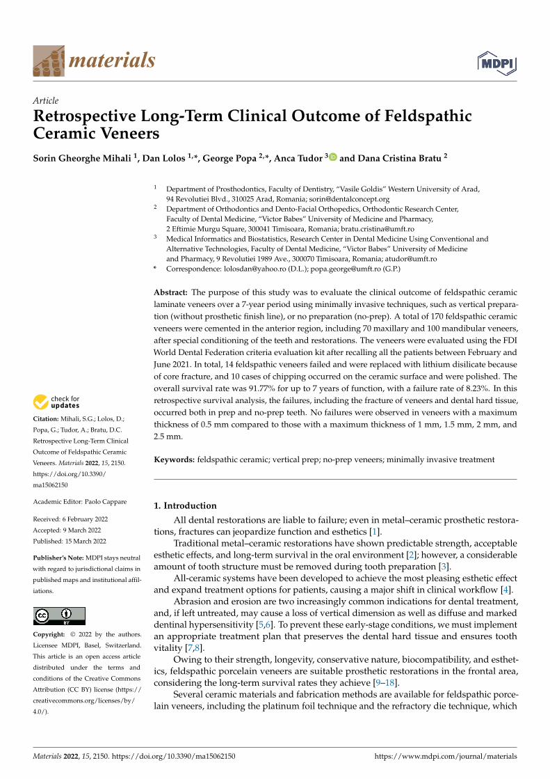
Figure 5. Color matching with: (a) VITA classical shade guide and (b) polar_eyes cross-polarization filter (Bio-Emulation™, Freiburg im Breisgau, Germany).
In the group in which we applied minimally invasive techniques, the objective was to obtain space for the future restoration, removing the maximum tooth convexity and any existing undercuts (Figure 6). The teeth were prepared using magnifying loupes (x4.3, Zeiss, Wetzlar, Germany) directly in the mock-up for reducing errors. As alternatives to full crowns, double veneers were used instead with a prepless technique (Figure 7).
Figure 3. Direct mock-up: (a) initial situation; (b) silicon index made by diagnostic wax-up;(c) intraoral fabricated with a temporary resign material and silicon index; (d) clinical aspect ofmock-up after finishing.
Materials 2022, 14, x FOR PEER REVIEW 5 of 16
(c) (d)
Figure 3. Direct mock-up: (a) initial situation; (b) silicon index made by diagnostic wax-up; (c) in-traoral fabricated with a temporary resign material and silicon index; (d) clinical aspect of mock-up after finishing.
(a) (b)
(c)
Figure 4. A microdontia of the lateral incisor: (a) initial situation; (b) additive direct mock-up, with enough space for final restoration; (c) no-prep technique applied by removing the composite from the natural teeth and preparing for impression.
(a)
(b)
Figure 5. Color matching with: (a) VITA classical shade guide and (b) polar_eyes cross-polarization filter (Bio-Emulation™, Freiburg im Breisgau, Germany).
In the group in which we applied minimally invasive techniques, the objective was to obtain space for the future restoration, removing the maximum tooth convexity and any existing undercuts (Figure 6). The teeth were prepared using magnifying loupes (x4.3, Zeiss, Wetzlar, Germany) directly in the mock-up for reducing errors. As alternatives to full crowns, double veneers were used instead with a prepless technique (Figure 7).
Figure 4. A microdontia of the lateral incisor: (a) initial situation; (b) additive direct mock-up, withenough space for final restoration; (c) no-prep technique applied by removing the composite fromthe natural teeth and preparing for impression.
Materials 2022, 15, 2150 5 of 16
Materials 2022, 14, x FOR PEER REVIEW 5 of 16
(c) (d)
Figure 3. Direct mock-up: (a) initial situation; (b) silicon index made by diagnostic wax-up; (c) in-traoral fabricated with a temporary resign material and silicon index; (d) clinical aspect of mock-up after finishing.
(a) (b)
(c)
Figure 4. A microdontia of the lateral incisor: (a) initial situation; (b) additive direct mock-up, with enough space for final restoration; (c) no-prep technique applied by removing the composite from the natural teeth and preparing for impression.
(a)
(b)
Figure 5. Color matching with: (a) VITA classical shade guide and (b) polar_eyes cross-polarization filter (Bio-Emulation™, Freiburg im Breisgau, Germany).
In the group in which we applied minimally invasive techniques, the objective was to obtain space for the future restoration, removing the maximum tooth convexity and any existing undercuts (Figure 6). The teeth were prepared using magnifying loupes (x4.3, Zeiss, Wetzlar, Germany) directly in the mock-up for reducing errors. As alternatives to full crowns, double veneers were used instead with a prepless technique (Figure 7).
Figure 5. Color matching with: (a) VITA classical shade guide and (b) polar_eyes cross-polarizationfilter (Bio-Emulation™, Freiburg im Breisgau, Germany).
In the group in which we applied minimally invasive techniques, the objective wasto obtain space for the future restoration, removing the maximum tooth convexity andany existing undercuts (Figure 6). The teeth were prepared using magnifying loupes (x4.3,Zeiss, Wetzlar, Germany) directly in the mock-up for reducing errors. As alternatives tofull crowns, double veneers were used instead with a prepless technique (Figure 7).
Materials 2022, 14, x FOR PEER REVIEW 6 of 16
Figure 6. Minimally invasive preparation (vertical preparation) in central incisors for closing a dia-stema.
(a) (b)
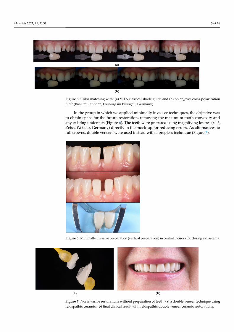
Figure 7. Noninvasive restorations without preparation of teeth: (a) a double veneer technique using feldspathic ceramic; (b) final clinical result with feldspathic double veneer ceramic restorations.
Provisional restorations were prepared to maintain tooth position, function, and es-thetics using a mock-up as described previously. Using a double-mixing technique, a full-arch, polyvinylsiloxane (ExpresXT, 3M ESPE, Seefeld, Germany) single impression was made for the opposing arch, and a type IV dental stone (GC Fujirock EP, Tokyo, Japan) was poured immediately.
All restorations were made by the same dental technician following the manufac-turer’s instructions. Each restoration was placed on the tooth after removing the provi-sional mock-up and assessed for proximal contacts, marginal adaptation, occlusal rela-tionships, and shade matching. Veneers were then etched for 60 s with 3% to <7% hydro-fluoric acid (IPS Ceramic Etching Gel, Ivoclar Vivadent, Schaan, Liechtenstein), washed and rinsed with water, and dried. Since etching with hydrofluoric acid leaves a significant amount of crystalline debris precipitate on the ceramic surface, the feldspathic restora-tions were also cleaned for 60 s using 36% orthophosphoric acid (Blue Etch, Cerkamed, Stalowa Wola, Poland) (Figure 8c) and ultrasonically cleaned in distilled water for 5 min. Thereafter, the etched surfaces were silanized (Monobond Plus, Ivoclar Vivadent, Schaan, Liechtenstein) for 60 s (Figure 8d) and dried to obtain a monolayer of silane (Figure 8e) [47]. A rubber dam was placed (Figure 9a), and the implicated teeth were sandblasted
Figure 6. Minimally invasive preparation (vertical preparation) in central incisors for closing a diastema.
Materials 2022, 14, x FOR PEER REVIEW 6 of 16
Figure 6. Minimally invasive preparation (vertical preparation) in central incisors for closing a dia-stema.
(a) (b)
Figure 7. Noninvasive restorations without preparation of teeth: (a) a double veneer technique using feldspathic ceramic; (b) final clinical result with feldspathic double veneer ceramic restorations.
Provisional restorations were prepared to maintain tooth position, function, and es-thetics using a mock-up as described previously. Using a double-mixing technique, a full-arch, polyvinylsiloxane (ExpresXT, 3M ESPE, Seefeld, Germany) single impression was made for the opposing arch, and a type IV dental stone (GC Fujirock EP, Tokyo, Japan) was poured immediately.
All restorations were made by the same dental technician following the manufac-turer’s instructions. Each restoration was placed on the tooth after removing the provi-sional mock-up and assessed for proximal contacts, marginal adaptation, occlusal rela-tionships, and shade matching. Veneers were then etched for 60 s with 3% to <7% hydro-fluoric acid (IPS Ceramic Etching Gel, Ivoclar Vivadent, Schaan, Liechtenstein), washed and rinsed with water, and dried. Since etching with hydrofluoric acid leaves a significant amount of crystalline debris precipitate on the ceramic surface, the feldspathic restora-tions were also cleaned for 60 s using 36% orthophosphoric acid (Blue Etch, Cerkamed, Stalowa Wola, Poland) (Figure 8c) and ultrasonically cleaned in distilled water for 5 min. Thereafter, the etched surfaces were silanized (Monobond Plus, Ivoclar Vivadent, Schaan, Liechtenstein) for 60 s (Figure 8d) and dried to obtain a monolayer of silane (Figure 8e) [47]. A rubber dam was placed (Figure 9a), and the implicated teeth were sandblasted
Figure 7. Noninvasive restorations without preparation of teeth: (a) a double veneer technique usingfeldspathic ceramic; (b) final clinical result with feldspathic double veneer ceramic restorations.
Materials 2022, 15, 2150 6 of 16
Provisional restorations were prepared to maintain tooth position, function, andesthetics using a mock-up as described previously. Using a double-mixing technique, afull-arch, polyvinylsiloxane (ExpresXT, 3M ESPE, Seefeld, Germany) single impression wasmade for the opposing arch, and a type IV dental stone (GC Fujirock EP, Tokyo, Japan) waspoured immediately.
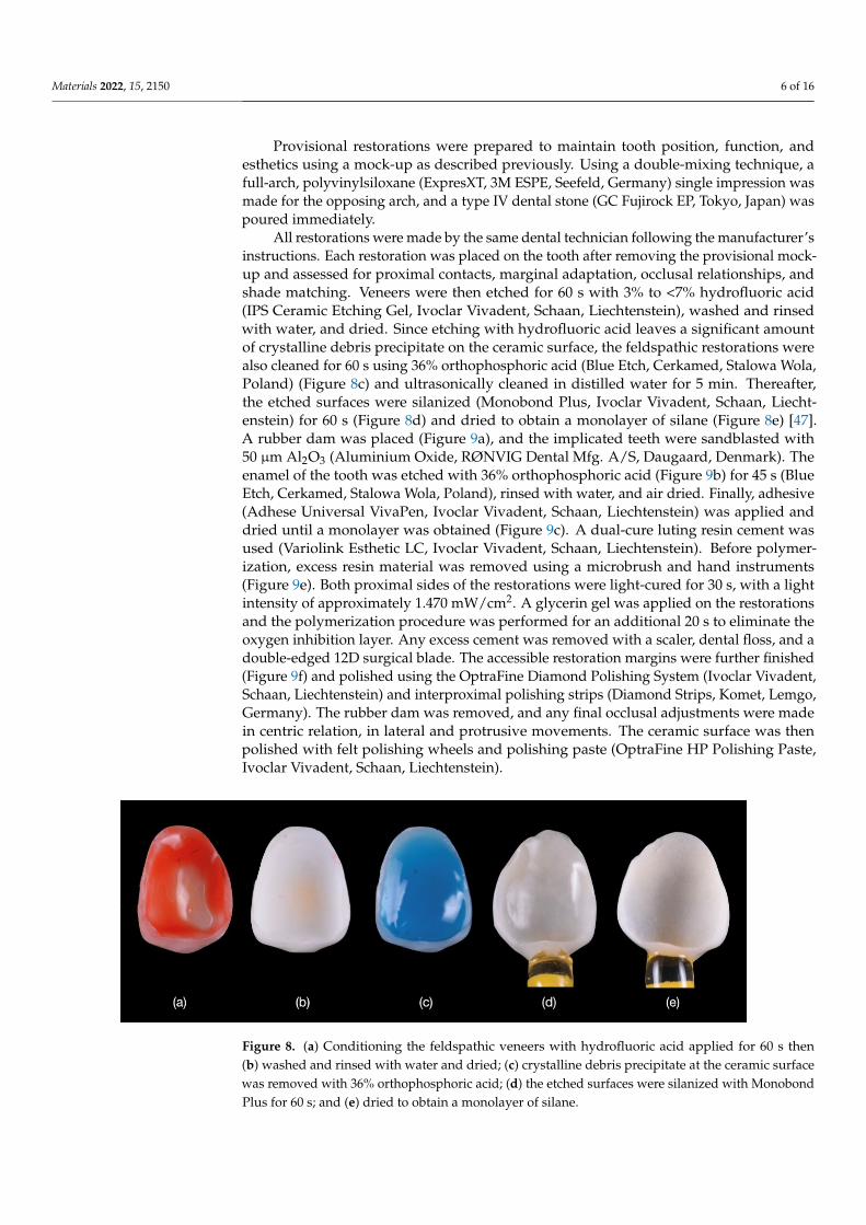
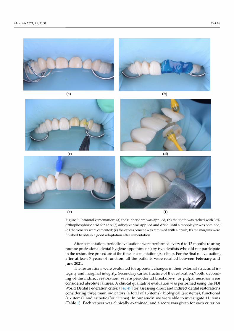
All restorations were made by the same dental technician following the manufacturer’sinstructions. Each restoration was placed on the tooth after removing the provisional mock-up and assessed for proximal contacts, marginal adaptation, occlusal relationships, andshade matching. Veneers were then etched for 60 s with 3% to <7% hydrofluoric acid(IPS Ceramic Etching Gel, Ivoclar Vivadent, Schaan, Liechtenstein), washed and rinsedwith water, and dried. Since etching with hydrofluoric acid leaves a significant amountof crystalline debris precipitate on the ceramic surface, the feldspathic restorations werealso cleaned for 60 s using 36% orthophosphoric acid (Blue Etch, Cerkamed, Stalowa Wola,Poland) (Figure 8c) and ultrasonically cleaned in distilled water for 5 min. Thereafter,the etched surfaces were silanized (Monobond Plus, Ivoclar Vivadent, Schaan, Liecht-enstein) for 60 s (Figure 8d) and dried to obtain a monolayer of silane (Figure 8e) [47].A rubber dam was placed (Figure 9a), and the implicated teeth were sandblasted with50 µm Al2O3 (Aluminium Oxide, RØNVIG Dental Mfg. A/S, Daugaard, Denmark). Theenamel of the tooth was etched with 36% orthophosphoric acid (Figure 9b) for 45 s (BlueEtch, Cerkamed, Stalowa Wola, Poland), rinsed with water, and air dried. Finally, adhesive(Adhese Universal VivaPen, Ivoclar Vivadent, Schaan, Liechtenstein) was applied anddried until a monolayer was obtained (Figure 9c). A dual-cure luting resin cement wasused (Variolink Esthetic LC, Ivoclar Vivadent, Schaan, Liechtenstein). Before polymer-ization, excess resin material was removed using a microbrush and hand instruments(Figure 9e). Both proximal sides of the restorations were light-cured for 30 s, with a lightintensity of approximately 1.470 mW/cm2. A glycerin gel was applied on the restorationsand the polymerization procedure was performed for an additional 20 s to eliminate theoxygen inhibition layer. Any excess cement was removed with a scaler, dental floss, and adouble-edged 12D surgical blade. The accessible restoration margins were further finished(Figure 9f) and polished using the OptraFine Diamond Polishing System (Ivoclar Vivadent,Schaan, Liechtenstein) and interproximal polishing strips (Diamond Strips, Komet, Lemgo,Germany). The rubber dam was removed, and any final occlusal adjustments were madein centric relation, in lateral and protrusive movements. The ceramic surface was thenpolished with felt polishing wheels and polishing paste (OptraFine HP Polishing Paste,Ivoclar Vivadent, Schaan, Liechtenstein).
Materials 2022, 14, x FOR PEER REVIEW 7 of 16
with 50 µm Al₂O₃ (Aluminium Oxide, RØNVIG Dental Mfg. A/S, Daugaard, Denmark). The enamel of the tooth was etched with 36% orthophosphoric acid (Figure 9b) for 45 s (Blue Etch, Cerkamed, Stalowa Wola, Poland), rinsed with water, and air dried. Finally, adhesive (Adhese Universal VivaPen, Ivoclar Vivadent, Schaan, Liechtenstein) was ap-plied and dried until a monolayer was obtained (Figure 9c). A dual-cure luting resin ce-ment was used (Variolink Esthetic LC, Ivoclar Vivadent, Schaan, Liechtenstein). Before polymerization, excess resin material was removed using a microbrush and hand instru-ments (Figure 9e). Both proximal sides of the restorations were light-cured for 30 s, with a light intensity of approximately 1.470 mW/cm2. A glycerin gel was applied on the resto-rations and the polymerization procedure was performed for an additional 20 s to elimi-nate the oxygen inhibition layer. Any excess cement was removed with a scaler, dental floss, and a double-edged 12D surgical blade. The accessible restoration margins were further finished (Figure 9f) and polished using the OptraFine Diamond Polishing System (Ivoclar Vivadent, Schaan, Liechtenstein) and interproximal polishing strips (Diamond Strips, Komet, Lemgo, Germany). The rubber dam was removed, and any final occlusal adjustments were made in centric relation, in lateral and protrusive movements. The ce-ramic surface was then polished with felt polishing wheels and polishing paste (OptraFine HP Polishing Paste, Ivoclar Vivadent, Schaan, Liechtenstein).
Figure 8. (a) Conditioning the feldspathic veneers with hydrofluoric acid applied for 60 s then (b) washed and rinsed with water and dried; (c) crystalline debris precipitate at the ceramic surface was removed with 36% orthophosphoric acid; (d) the etched surfaces were silanized with Monobond Plus for 60 s; and (e) dried to obtain a monolayer of silane.
After cementation, periodic evaluations were performed every 6 to 12 months (dur-ing routine professional dental hygiene appointments) by two dentists who did not par-ticipate in the restorative procedure at the time of cementation (baseline). For the final re-evaluation, after at least 7 years of function, all the patients were recalled between Febru-ary and June 2021.
(a) (b)
Figure 8. (a) Conditioning the feldspathic veneers with hydrofluoric acid applied for 60 s then(b) washed and rinsed with water and dried; (c) crystalline debris precipitate at the ceramic surfacewas removed with 36% orthophosphoric acid; (d) the etched surfaces were silanized with MonobondPlus for 60 s; and (e) dried to obtain a monolayer of silane.
Materials 2022, 15, 2150 7 of 16
Materials 2022, 14, x FOR PEER REVIEW 7 of 16
with 50 µm Al₂O₃ (Aluminium Oxide, RØNVIG Dental Mfg. A/S, Daugaard, Denmark). The enamel of the tooth was etched with 36% orthophosphoric acid (Figure 9b) for 45 s (Blue Etch, Cerkamed, Stalowa Wola, Poland), rinsed with water, and air dried. Finally, adhesive (Adhese Universal VivaPen, Ivoclar Vivadent, Schaan, Liechtenstein) was ap-plied and dried until a monolayer was obtained (Figure 9c). A dual-cure luting resin ce-ment was used (Variolink Esthetic LC, Ivoclar Vivadent, Schaan, Liechtenstein). Before polymerization, excess resin material was removed using a microbrush and hand instru-ments (Figure 9e). Both proximal sides of the restorations were light-cured for 30 s, with a light intensity of approximately 1.470 mW/cm2. A glycerin gel was applied on the resto-rations and the polymerization procedure was performed for an additional 20 s to elimi-nate the oxygen inhibition layer. Any excess cement was removed with a scaler, dental floss, and a double-edged 12D surgical blade. The accessible restoration margins were further finished (Figure 9f) and polished using the OptraFine Diamond Polishing System (Ivoclar Vivadent, Schaan, Liechtenstein) and interproximal polishing strips (Diamond Strips, Komet, Lemgo, Germany). The rubber dam was removed, and any final occlusal adjustments were made in centric relation, in lateral and protrusive movements. The ce-ramic surface was then polished with felt polishing wheels and polishing paste (OptraFine HP Polishing Paste, Ivoclar Vivadent, Schaan, Liechtenstein).
Figure 8. (a) Conditioning the feldspathic veneers with hydrofluoric acid applied for 60 s then (b) washed and rinsed with water and dried; (c) crystalline debris precipitate at the ceramic surface was removed with 36% orthophosphoric acid; (d) the etched surfaces were silanized with Monobond Plus for 60 s; and (e) dried to obtain a monolayer of silane.
After cementation, periodic evaluations were performed every 6 to 12 months (dur-ing routine professional dental hygiene appointments) by two dentists who did not par-ticipate in the restorative procedure at the time of cementation (baseline). For the final re-evaluation, after at least 7 years of function, all the patients were recalled between Febru-ary and June 2021.
(a) (b)
Materials 2022, 14, x FOR PEER REVIEW 8 of 16
(c) (d)
(e) (f)
Figure 9. Intraoral cementation: (a) the rubber dam was applied; (b) the tooth was etched with 36% orthophosphoric acid for 45 s; (c) adhesive was applied and dried until a monolayer was obtained; (d) the veneers were cemented; (e) the excess cement was removed with a brush; (f) the margins were finished to obtain a good adaptation after cementation.
The restorations were evaluated for apparent changes in their external structural in-tegrity and marginal integrity. Secondary caries, fracture of the restoration/tooth, debond-ing of the indirect restoration, severe periodontal breakdown, or pulpal necrosis were considered absolute failures. A clinical qualitative evaluation was performed using the FDI World Dental Federation criteria [48,49] for assessing direct and indirect dental res-torations considering three main indicators (a total of 16 items): biological (six items), functional (six items), and esthetic (four items). In our study, we were able to investigate 11 items (Table 1). Each veneer was clinically examined, and a score was given for each criterion (item) on a 5-point scale (e.g., 1, an excellent restoration; 5, a restoration that re-quired replacement). Each examinator used an intraoral mirror, sharp explorer, and peri-odontal probe specifically designed for evaluating the FDI World Dental Federation crite-ria. All the veneers received a baseline score of 1 (excellent restoration) at the moment of cementation.
Table 1. List of FDI World Dental Federation criteria used in our study to assess the esthetic, func-tional, and biological properties for the clinical evaluation of indirect restorations.
Esthetic properties C1 = Staining: (a) surface and (b) margin C2 = Color match and translucency C3 = Esthetic anatomical form
Functional properties C4 = Fracture of restorative material and retention C5 = Marginal adaptation C6 = Occlusal contour and wear
Biological properties C7 = Postoperative sensitivity and tooth vitality C8 = Recurrent caries C9 = Tooth integrity
Figure 9. Intraoral cementation: (a) the rubber dam was applied; (b) the tooth was etched with 36%orthophosphoric acid for 45 s; (c) adhesive was applied and dried until a monolayer was obtained;(d) the veneers were cemented; (e) the excess cement was removed with a brush; (f) the margins werefinished to obtain a good adaptation after cementation.
After cementation, periodic evaluations were performed every 6 to 12 months (duringroutine professional dental hygiene appointments) by two dentists who did not participatein the restorative procedure at the time of cementation (baseline). For the final re-evaluation,after at least 7 years of function, all the patients were recalled between February andJune 2021.
The restorations were evaluated for apparent changes in their external structural in-tegrity and marginal integrity. Secondary caries, fracture of the restoration/tooth, debond-ing of the indirect restoration, severe periodontal breakdown, or pulpal necrosis wereconsidered absolute failures. A clinical qualitative evaluation was performed using the FDIWorld Dental Federation criteria [48,49] for assessing direct and indirect dental restorationsconsidering three main indicators (a total of 16 items): biological (six items), functional(six items), and esthetic (four items). In our study, we were able to investigate 11 items(Table 1). Each veneer was clinically examined, and a score was given for each criterion
Materials 2022, 15, 2150 8 of 16
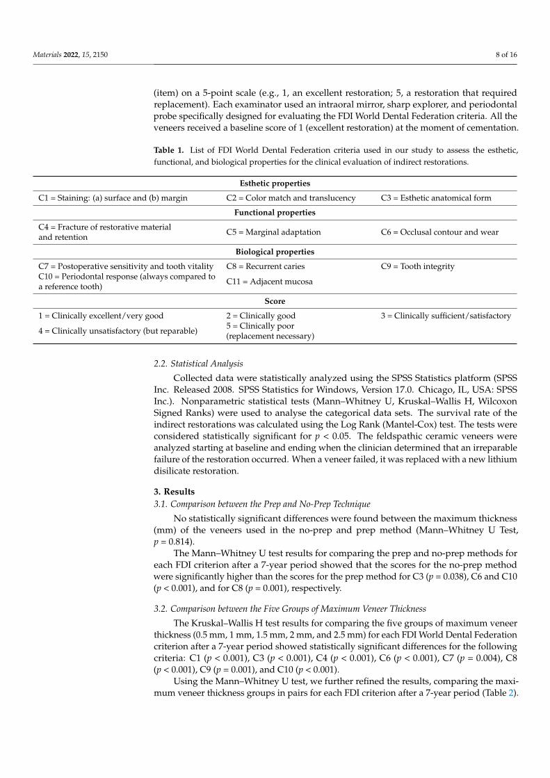
(item) on a 5-point scale (e.g., 1, an excellent restoration; 5, a restoration that requiredreplacement). Each examinator used an intraoral mirror, sharp explorer, and periodontalprobe specifically designed for evaluating the FDI World Dental Federation criteria. All theveneers received a baseline score of 1 (excellent restoration) at the moment of cementation.
Table 1. List of FDI World Dental Federation criteria used in our study to assess the esthetic,functional, and biological properties for the clinical evaluation of indirect restorations.
Esthetic properties
C1 = Staining: (a) surface and (b) margin C2 = Color match and translucency C3 = Esthetic anatomical form
Functional properties
C4 = Fracture of restorative materialand retention C5 = Marginal adaptation C6 = Occlusal contour and wear
Biological properties
C7 = Postoperative sensitivity and tooth vitality C8 = Recurrent caries C9 = Tooth integrityC10 = Periodontal response (always compared toa reference tooth) C11 = Adjacent mucosa
Score
1 = Clinically excellent/very good 2 = Clinically good 3 = Clinically sufficient/satisfactory
4 = Clinically unsatisfactory (but reparable) 5 = Clinically poor(replacement necessary)
2.2. Statistical Analysis
Collected data were statistically analyzed using the SPSS Statistics platform (SPSSInc. Released 2008. SPSS Statistics for Windows, Version 17.0. Chicago, IL, USA: SPSSInc.). Nonparametric statistical tests (Mann–Whitney U, Kruskal–Wallis H, WilcoxonSigned Ranks) were used to analyse the categorical data sets. The survival rate of theindirect restorations was calculated using the Log Rank (Mantel-Cox) test. The tests wereconsidered statistically significant for p < 0.05. The feldspathic ceramic veneers wereanalyzed starting at baseline and ending when the clinician determined that an irreparablefailure of the restoration occurred. When a veneer failed, it was replaced with a new lithiumdisilicate restoration.
3. Results3.1. Comparison between the Prep and No-Prep Technique
No statistically significant differences were found between the maximum thickness(mm) of the veneers used in the no-prep and prep method (Mann–Whitney U Test,p = 0.814).
The Mann–Whitney U test results for comparing the prep and no-prep methods foreach FDI criterion after a 7-year period showed that the scores for the no-prep methodwere significantly higher than the scores for the prep method for C3 (p = 0.038), C6 and C10(p < 0.001), and for C8 (p = 0.001), respectively.
3.2. Comparison between the Five Groups of Maximum Veneer Thickness
The Kruskal–Wallis H test results for comparing the five groups of maximum veneerthickness (0.5 mm, 1 mm, 1.5 mm, 2 mm, and 2.5 mm) for each FDI World Dental Federationcriterion after a 7-year period showed statistically significant differences for the followingcriteria: C1 (p < 0.001), C3 (p < 0.001), C4 (p < 0.001), C6 (p < 0.001), C7 (p = 0.004), C8(p < 0.001), C9 (p = 0.001), and C10 (p < 0.001).
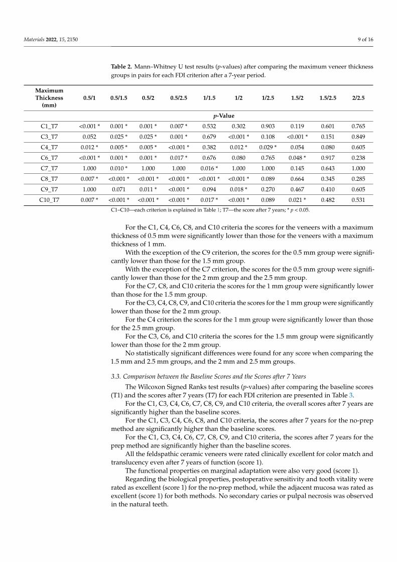
Using the Mann–Whitney U test, we further refined the results, comparing the maxi-mum veneer thickness groups in pairs for each FDI criterion after a 7-year period (Table 2).
Materials 2022, 15, 2150 9 of 16
Table 2. Mann–Whitney U test results (p-values) after comparing the maximum veneer thicknessgroups in pairs for each FDI criterion after a 7-year period.
MaximumThickness
(mm)0.5/1 0.5/1.5 0.5/2 0.5/2.5 1/1.5 1/2 1/2.5 1.5/2 1.5/2.5 2/2.5
p-Value
C1_T7 <0.001 * 0.001 * 0.001 * 0.007 * 0.532 0.302 0.903 0.119 0.601 0.765
C3_T7 0.052 0.025 * 0.025 * 0.001 * 0.679 <0.001 * 0.108 <0.001 * 0.151 0.849
C4_T7 0.012 * 0.005 * 0.005 * <0.001 * 0.382 0.012 * 0.029 * 0.054 0.080 0.605
C6_T7 <0.001 * 0.001 * 0.001 * 0.017 * 0.676 0.080 0.765 0.048 * 0.917 0.238
C7_T7 1.000 0.010 * 1.000 1.000 0.016 * 1.000 1.000 0.145 0.643 1.000
C8_T7 0.007 * <0.001 * <0.001 * <0.001 * <0.001 * <0.001 * 0.089 0.664 0.345 0.285
C9_T7 1.000 0.071 0.011 * <0.001 * 0.094 0.018 * 0.270 0.467 0.410 0.605
C10_T7 0.007 * <0.001 * <0.001 * <0.001 * 0.017 * <0.001 * 0.089 0.021 * 0.482 0.531
C1–C10—each criterion is explained in Table 1; T7—the score after 7 years; * p < 0.05.
For the C1, C4, C6, C8, and C10 criteria the scores for the veneers with a maximumthickness of 0.5 mm were significantly lower than those for the veneers with a maximumthickness of 1 mm.
With the exception of the C9 criterion, the scores for the 0.5 mm group were signifi-cantly lower than those for the 1.5 mm group.
With the exception of the C7 criterion, the scores for the 0.5 mm group were signifi-cantly lower than those for the 2 mm group and the 2.5 mm group.
For the C7, C8, and C10 criteria the scores for the 1 mm group were significantly lowerthan those for the 1.5 mm group.
For the C3, C4, C8, C9, and C10 criteria the scores for the 1 mm group were significantlylower than those for the 2 mm group.
For the C4 criterion the scores for the 1 mm group were significantly lower than thosefor the 2.5 mm group.
For the C3, C6, and C10 criteria the scores for the 1.5 mm group were significantlylower than those for the 2 mm group.
No statistically significant differences were found for any score when comparing the1.5 mm and 2.5 mm groups, and the 2 mm and 2.5 mm groups.
3.3. Comparison between the Baseline Scores and the Scores after 7 Years
The Wilcoxon Signed Ranks test results (p-values) after comparing the baseline scores(T1) and the scores after 7 years (T7) for each FDI criterion are presented in Table 3.
For the C1, C3, C4, C6, C7, C8, C9, and C10 criteria, the overall scores after 7 years aresignificantly higher than the baseline scores.
For the C1, C3, C4, C6, C8, and C10 criteria, the scores after 7 years for the no-prepmethod are significantly higher than the baseline scores.
For the C1, C3, C4, C6, C7, C8, C9, and C10 criteria, the scores after 7 years for theprep method are significantly higher than the baseline scores.
All the feldspathic ceramic veneers were rated clinically excellent for color match andtranslucency even after 7 years of function (score 1).
The functional properties on marginal adaptation were also very good (score 1).Regarding the biological properties, postoperative sensitivity and tooth vitality were
rated as excellent (score 1) for the no-prep method, while the adjacent mucosa was rated asexcellent (score 1) for both methods. No secondary caries or pulpal necrosis was observedin the natural teeth.
Materials 2022, 15, 2150 10 of 16
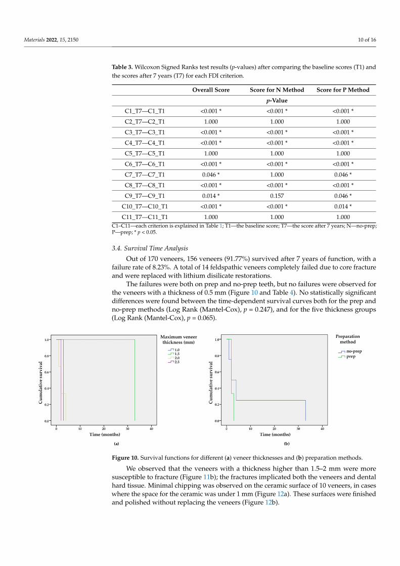
Table 3. Wilcoxon Signed Ranks test results (p-values) after comparing the baseline scores (T1) andthe scores after 7 years (T7) for each FDI criterion.
Overall Score Score for N Method Score for P Method
p-Value
C1_T7—C1_T1 <0.001 * <0.001 * <0.001 *
C2_T7—C2_T1 1.000 1.000 1.000
C3_T7—C3_T1 <0.001 * <0.001 * <0.001 *
C4_T7—C4_T1 <0.001 * <0.001 * <0.001 *
C5_T7—C5_T1 1.000 1.000 1.000
C6_T7—C6_T1 <0.001 * <0.001 * <0.001 *
C7_T7—C7_T1 0.046 * 1.000 0.046 *
C8_T7—C8_T1 <0.001 * <0.001 * <0.001 *
C9_T7—C9_T1 0.014 * 0.157 0.046 *
C10_T7—C10_T1 <0.001 * <0.001 * 0.014 *
C11_T7—C11_T1 1.000 1.000 1.000C1–C11—each criterion is explained in Table 1; T1—the baseline score; T7—the score after 7 years; N—no-prep;P—prep; * p < 0.05.
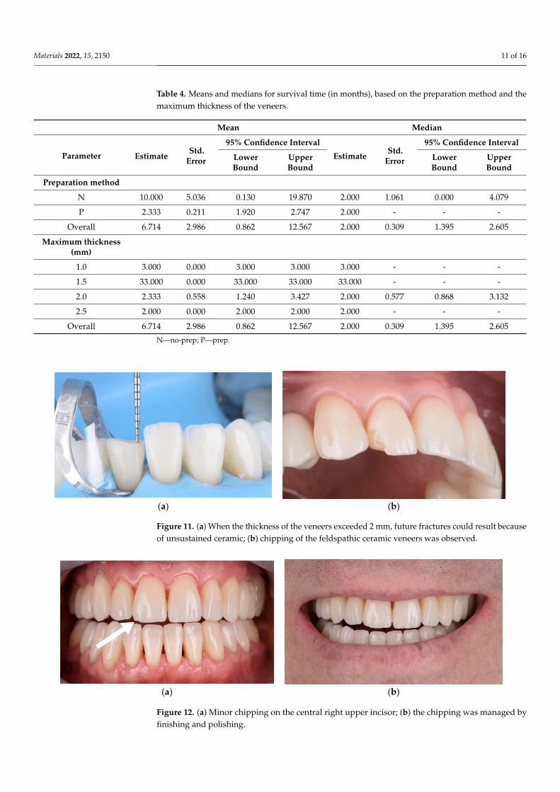
3.4. Survival Time Analysis
Out of 170 veneers, 156 veneers (91.77%) survived after 7 years of function, with afailure rate of 8.23%. A total of 14 feldspathic veneers completely failed due to core fractureand were replaced with lithium disilicate restorations.
The failures were both on prep and no-prep teeth, but no failures were observed forthe veneers with a thickness of 0.5 mm (Figure 10 and Table 4). No statistically significantdifferences were found between the time-dependent survival curves both for the prep andno-prep methods (Log Rank (Mantel-Cox), p = 0.247), and for the five thickness groups(Log Rank (Mantel-Cox), p = 0.065).
1
Figure 10. Survival functions for different (a) veneer thicknesses and (b) preparation methods.
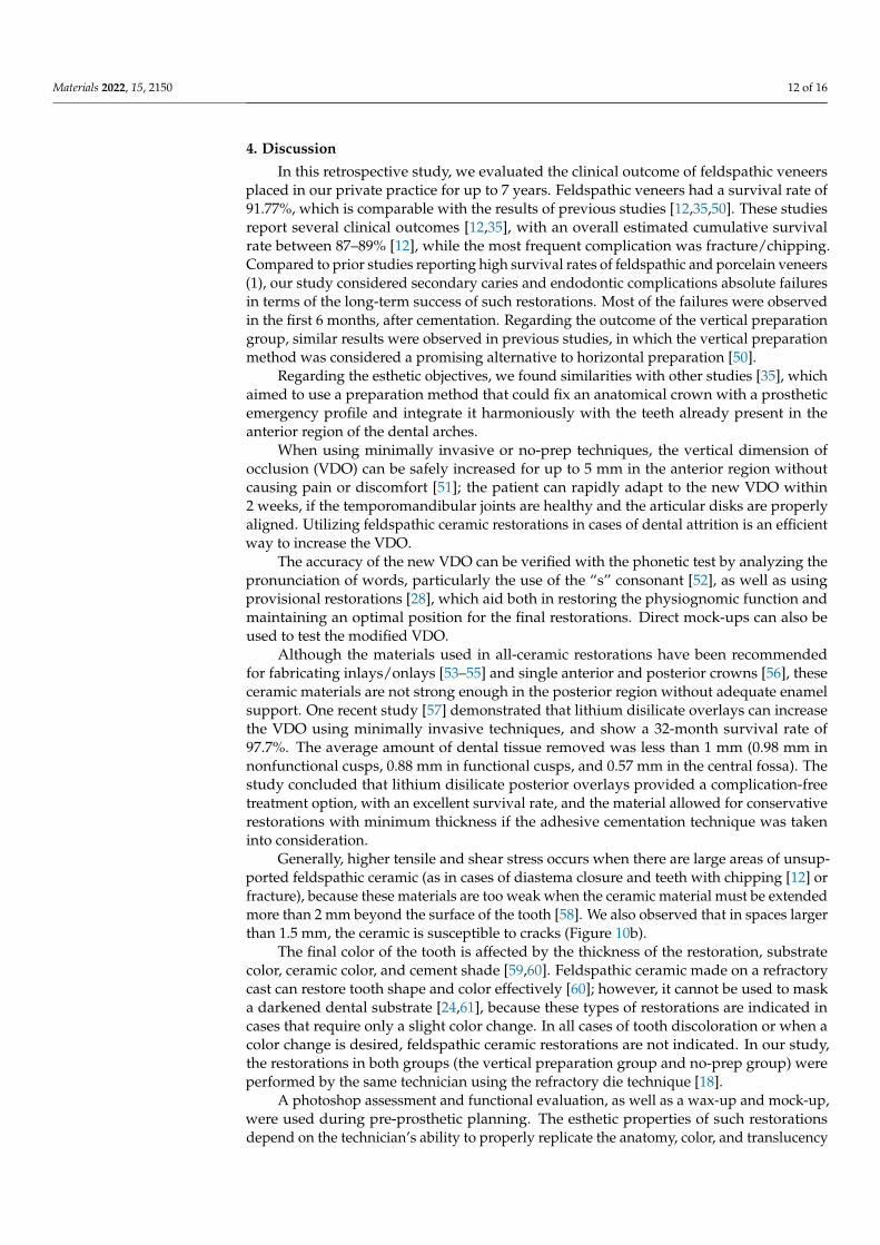
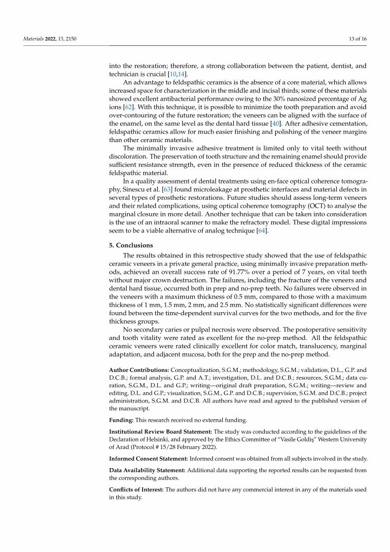
We observed that the veneers with a thickness higher than 1.5–2 mm were moresusceptible to fracture (Figure 11b); the fractures implicated both the veneers and dentalhard tissue. Minimal chipping was observed on the ceramic surface of 10 veneers, in caseswhere the space for the ceramic was under 1 mm (Figure 12a). These surfaces were finishedand polished without replacing the veneers (Figure 12b).
Materials 2022, 15, 2150 11 of 16
Table 4. Means and medians for survival time (in months), based on the preparation method and themaximum thickness of the veneers.
Mean Median
Parameter EstimateStd.
Error
95% Confidence Interval
EstimateStd.
Error
95% Confidence Interval
LowerBound
UpperBound
LowerBound
UpperBound
Preparation method
N 10.000 5.036 0.130 19.870 2.000 1.061 0.000 4.079
P 2.333 0.211 1.920 2.747 2.000 - - -
Overall 6.714 2.986 0.862 12.567 2.000 0.309 1.395 2.605
Maximum thickness(mm)
1.0 3.000 0.000 3.000 3.000 3.000 - - -
1.5 33.000 0.000 33.000 33.000 33.000 - - -
2.0 2.333 0.558 1.240 3.427 2.000 0.577 0.868 3.132
2.5 2.000 0.000 2.000 2.000 2.000 - - -
Overall 6.714 2.986 0.862 12.567 2.000 0.309 1.395 2.605
N—no-prep; P—prep.
Materials 2022, 14, x FOR PEER REVIEW 12 of 16
We observed that the veneers with a thickness higher than 1.5–2 mm were more sus-ceptible to fracture (Figure 11b); the fractures implicated both the veneers and dental hard tissue. Minimal chipping was observed on the ceramic surface of 10 veneers, in cases where the space for the ceramic was under 1 mm (Figure 12a). These surfaces were fin-ished and polished without replacing the veneers (Figure 12b).
(a) (b)
Figure 11. (a) When the thickness of the veneers exceeded 2 mm, future fractures could result be-cause of unsustained ceramic; (b) chipping of the feldspathic ceramic veneers was observed.
(a) (b)
Figure 12. (a) Minor chipping on the central right upper incisor; (b) the chipping was managed by finishing and polishing.
4. Discussion In this retrospective study, we evaluated the clinical outcome of feldspathic veneers
placed in our private practice for up to 7 years. Feldspathic veneers had a survival rate of 91.77%, which is comparable with the results of previous studies [12,35,50]. These studies report several clinical outcomes [12,35], with an overall estimated cumulative survival rate between 87–89% [12], while the most frequent complication was fracture/chipping. Compared to prior studies reporting high survival rates of feldspathic and porcelain ve-neers (1), our study considered secondary caries and endodontic complications absolute failures in terms of the long-term success of such restorations. Most of the failures were observed in the first 6 months, after cementation. Regarding the outcome of the vertical preparation group, similar results were observed in previous studies, in which the vertical preparation method was considered a promising alternative to horizontal preparation [50].
Regarding the esthetic objectives, we found similarities with other studies [35], which aimed to use a preparation method that could fix an anatomical crown with a prosthetic emergency profile and integrate it harmoniously with the teeth already present in the an-terior region of the dental arches.
Figure 11. (a) When the thickness of the veneers exceeded 2 mm, future fractures could result becauseof unsustained ceramic; (b) chipping of the feldspathic ceramic veneers was observed.
Materials 2022, 14, x FOR PEER REVIEW 12 of 16
We observed that the veneers with a thickness higher than 1.5–2 mm were more sus-ceptible to fracture (Figure 11b); the fractures implicated both the veneers and dental hard tissue. Minimal chipping was observed on the ceramic surface of 10 veneers, in cases where the space for the ceramic was under 1 mm (Figure 12a). These surfaces were fin-ished and polished without replacing the veneers (Figure 12b).
(a) (b)
Figure 11. (a) When the thickness of the veneers exceeded 2 mm, future fractures could result be-cause of unsustained ceramic; (b) chipping of the feldspathic ceramic veneers was observed.
(a) (b)
Figure 12. (a) Minor chipping on the central right upper incisor; (b) the chipping was managed by finishing and polishing.
4. Discussion In this retrospective study, we evaluated the clinical outcome of feldspathic veneers
placed in our private practice for up to 7 years. Feldspathic veneers had a survival rate of 91.77%, which is comparable with the results of previous studies [12,35,50]. These studies report several clinical outcomes [12,35], with an overall estimated cumulative survival rate between 87–89% [12], while the most frequent complication was fracture/chipping. Compared to prior studies reporting high survival rates of feldspathic and porcelain ve-neers (1), our study considered secondary caries and endodontic complications absolute failures in terms of the long-term success of such restorations. Most of the failures were observed in the first 6 months, after cementation. Regarding the outcome of the vertical preparation group, similar results were observed in previous studies, in which the vertical preparation method was considered a promising alternative to horizontal preparation [50].
Regarding the esthetic objectives, we found similarities with other studies [35], which aimed to use a preparation method that could fix an anatomical crown with a prosthetic emergency profile and integrate it harmoniously with the teeth already present in the an-terior region of the dental arches.
Figure 12. (a) Minor chipping on the central right upper incisor; (b) the chipping was managed byfinishing and polishing.
Materials 2022, 15, 2150 12 of 16
4. Discussion
In this retrospective study, we evaluated the clinical outcome of feldspathic veneersplaced in our private practice for up to 7 years. Feldspathic veneers had a survival rate of91.77%, which is comparable with the results of previous studies [12,35,50]. These studiesreport several clinical outcomes [12,35], with an overall estimated cumulative survivalrate between 87–89% [12], while the most frequent complication was fracture/chipping.Compared to prior studies reporting high survival rates of feldspathic and porcelain veneers(1), our study considered secondary caries and endodontic complications absolute failuresin terms of the long-term success of such restorations. Most of the failures were observedin the first 6 months, after cementation. Regarding the outcome of the vertical preparationgroup, similar results were observed in previous studies, in which the vertical preparationmethod was considered a promising alternative to horizontal preparation [50].
Regarding the esthetic objectives, we found similarities with other studies [35], whichaimed to use a preparation method that could fix an anatomical crown with a prostheticemergency profile and integrate it harmoniously with the teeth already present in theanterior region of the dental arches.
When using minimally invasive or no-prep techniques, the vertical dimension ofocclusion (VDO) can be safely increased for up to 5 mm in the anterior region withoutcausing pain or discomfort [51]; the patient can rapidly adapt to the new VDO within2 weeks, if the temporomandibular joints are healthy and the articular disks are properlyaligned. Utilizing feldspathic ceramic restorations in cases of dental attrition is an efficientway to increase the VDO.
The accuracy of the new VDO can be verified with the phonetic test by analyzing thepronunciation of words, particularly the use of the “s” consonant [52], as well as usingprovisional restorations [28], which aid both in restoring the physiognomic function andmaintaining an optimal position for the final restorations. Direct mock-ups can also beused to test the modified VDO.
Although the materials used in all-ceramic restorations have been recommendedfor fabricating inlays/onlays [53–55] and single anterior and posterior crowns [56], theseceramic materials are not strong enough in the posterior region without adequate enamelsupport. One recent study [57] demonstrated that lithium disilicate overlays can increasethe VDO using minimally invasive techniques, and show a 32-month survival rate of97.7%. The average amount of dental tissue removed was less than 1 mm (0.98 mm innonfunctional cusps, 0.88 mm in functional cusps, and 0.57 mm in the central fossa). Thestudy concluded that lithium disilicate posterior overlays provided a complication-freetreatment option, with an excellent survival rate, and the material allowed for conservativerestorations with minimum thickness if the adhesive cementation technique was takeninto consideration.
Generally, higher tensile and shear stress occurs when there are large areas of unsup-ported feldspathic ceramic (as in cases of diastema closure and teeth with chipping [12] orfracture), because these materials are too weak when the ceramic material must be extendedmore than 2 mm beyond the surface of the tooth [58]. We also observed that in spaces largerthan 1.5 mm, the ceramic is susceptible to cracks (Figure 10b).
The final color of the tooth is affected by the thickness of the restoration, substratecolor, ceramic color, and cement shade [59,60]. Feldspathic ceramic made on a refractorycast can restore tooth shape and color effectively [60]; however, it cannot be used to maska darkened dental substrate [24,61], because these types of restorations are indicated incases that require only a slight color change. In all cases of tooth discoloration or when acolor change is desired, feldspathic ceramic restorations are not indicated. In our study,the restorations in both groups (the vertical preparation group and no-prep group) wereperformed by the same technician using the refractory die technique [18].
A photoshop assessment and functional evaluation, as well as a wax-up and mock-up,were used during pre-prosthetic planning. The esthetic properties of such restorationsdepend on the technician’s ability to properly replicate the anatomy, color, and translucency
Materials 2022, 15, 2150 13 of 16
into the restoration; therefore, a strong collaboration between the patient, dentist, andtechnician is crucial [10,14].
An advantage to feldspathic ceramics is the absence of a core material, which allowsincreased space for characterization in the middle and incisal thirds; some of these materialsshowed excellent antibacterial performance owing to the 30% nanosized percentage of Agions [62]. With this technique, it is possible to minimize the tooth preparation and avoidover-contouring of the future restoration; the veneers can be aligned with the surface ofthe enamel, on the same level as the dental hard tissue [40]. After adhesive cementation,feldspathic ceramics allow for much easier finishing and polishing of the veneer marginsthan other ceramic materials.
The minimally invasive adhesive treatment is limited only to vital teeth withoutdiscoloration. The preservation of tooth structure and the remaining enamel should providesufficient resistance strength, even in the presence of reduced thickness of the ceramicfeldspathic material.
In a quality assessment of dental treatments using en-face optical coherence tomogra-phy, Sinescu et al. [63] found microleakage at prosthetic interfaces and material defects inseveral types of prosthetic restorations. Future studies should assess long-term veneersand their related complications, using optical coherence tomography (OCT) to analyse themarginal closure in more detail. Another technique that can be taken into considerationis the use of an intraoral scanner to make the refractory model. These digital impressionsseem to be a viable alternative of analog technique [64].
5. Conclusions
The results obtained in this retrospective study showed that the use of feldspathicceramic veneers in a private general practice, using minimally invasive preparation meth-ods, achieved an overall success rate of 91.77% over a period of 7 years, on vital teethwithout major crown destruction. The failures, including the fracture of the veneers anddental hard tissue, occurred both in prep and no-prep teeth. No failures were observed inthe veneers with a maximum thickness of 0.5 mm, compared to those with a maximumthickness of 1 mm, 1.5 mm, 2 mm, and 2.5 mm. No statistically significant differences werefound between the time-dependent survival curves for the two methods, and for the fivethickness groups.
No secondary caries or pulpal necrosis were observed. The postoperative sensitivityand tooth vitality were rated as excellent for the no-prep method. All the feldspathicceramic veneers were rated clinically excellent for color match, translucency, marginaladaptation, and adjacent mucosa, both for the prep and the no-prep method.
Author Contributions: Conceptualization, S.G.M.; methodology, S.G.M.; validation, D.L., G.P. andD.C.B.; formal analysis, G.P. and A.T.; investigation, D.L. and D.C.B.; resources, S.G.M.; data cu-ration, S.G.M., D.L. and G.P.; writing—original draft preparation, S.G.M.; writing—review andediting, D.L. and G.P.; visualization, S.G.M., G.P. and D.C.B.; supervision, S.G.M. and D.C.B.; projectadministration, S.G.M. and D.C.B. All authors have read and agreed to the published version ofthe manuscript.
Funding: This research received no external funding.
Institutional Review Board Statement: The study was conducted according to the guidelines of theDeclaration of Helsinki, and approved by the Ethics Committee of “Vasile Goldis” Western Universityof Arad (Protocol # 15/28 February 2022).
Informed Consent Statement: Informed consent was obtained from all subjects involved in the study.
Data Availability Statement: Additional data supporting the reported results can be requested fromthe corresponding authors.
Conflicts of Interest: The authors did not have any commercial interest in any of the materials usedin this study.
Materials 2022, 15, 2150 14 of 16
References1. Aslam, A.; Khan, D.A.; Hassan, S.H.; Ahmed, B. Ceramic Fracture in Metal-Ceramic Restorations: The Aetiology. Dent. Update
2017, 44, 448–456. [CrossRef] [PubMed]2. Heffernan, M.J.; Aquilino, S.A.; Diaz-Arnold, A.M.; Haselton, D.R.; Stanford, C.M.; Vargas, M.A. Relative translucency of six
all-ceramic systems. Part I: Core materials. J. Prosthet. Dent. 2002, 88, 4–9. [CrossRef] [PubMed]3. Edelhoff, D.; Sorensen, J.A. Tooth structure removal associated with various preparation designs for anterior teeth. J. Prosthet.
Dent. 2002, 87, 503–509. [CrossRef] [PubMed]4. Silva, L.H.D.; Lima, E.; Miranda, R.B.P.; Favero, S.S.; Lohbauer, U.; Cesar, P.F. Dental ceramics: A review of new materials and
processing methods. Braz. Oral Res. 2017, 31, e58. [CrossRef] [PubMed]5. Bosch, G.; Ender, A.; Mehl, A. Non- and minimally invasive full-mouth rehabilitation of patients with loss of vertical dimension
of occlusion using CAD/CAM: An innovative concept demonstrated with a case report. Int. J. Comput. Dent. 2015, 18, 273–286.[PubMed]
6. Savi, A.; Turillazzi, O.; Crescini, A.; Manfredi, M. Esthetic treatment of a diffuse amelogenesis imperfecta using pressed lithiumdisilicate and feldspathic ceramic restorations: 5-year follow up. J. Esthet. Restor. Dent. 2014, 26, 363–373. [CrossRef] [PubMed]
7. Tyas, M.J.; Anusavice, K.J.; Frencken, J.E.; Mount, G.J. Minimal intervention dentistry—A review. FDI Commission Project 1-97.Int. Dent. J. 2000, 50, 1–12. [CrossRef]
8. Vailati, F.; Gruetter, L.; Belser, U.C. Adhesively restored anterior maxillary dentitions affected by severe erosion: Up to 6-yearresults of a prospective clinical study. Eur. J. Esthet. Dent. 2013, 8, 506–530.
9. McLaren, E.A.; Whiteman, Y.Y. Ceramics: Rationale for material selection. Compend. Contin. Educ. Dent. 2010, 31, 666–668.[PubMed]
10. McLaren, E.A.; LeSage, B. Feldspathic veneers: What are their indications? Compend. Contin. Educ. Dent. 2011, 32, 44–49.11. Layton, D.M.; Clarke, M.; Walton, T.R. A systematic review and meta-analysis of the survival of feldspathic porcelain veneers
over 5 and 10 years. Int. J. Prosthodont. 2012, 25, 590–603. [PubMed]12. Morimoto, S.; Albanesi, R.B.; Sesma, N.; Agra, C.M.; Braga, M.M. Main Clinical Outcomes of Feldspathic Porcelain and Glass-
Ceramic Laminate Veneers: A Systematic Review and Meta-Analysis of Survival and Complication Rates. Int. J. Prosthodont. 2016,29, 38–49. [CrossRef] [PubMed]
13. Layton, D.M.; Walton, T.R. The up to 21-year clinical outcome and survival of feldspathic porcelain veneers: Accounting forclustering. Int. J. Prosthodont. 2007, 25, 604–612.
14. Peumans, M.; De Munck, J.; Fieuws, S.; Lambrechts, P.; Vanherle, G.; Van Meerbeek, B. A prospective ten-year clinical trial ofporcelain veneers. J. Adhes. Dent. 2004, 6, 65–76.
15. Pini, N.P.; Aguiar, F.H.; Lima, D.A.; Lovadino, J.R.; Terada, R.S.; Pascotto, R.C. Advances in dental veneers: Materials, applications,and techniques. Clin. Cosmet. Investig. Dent. 2012, 10, 9–16.
16. Strassler, H.E. Minimally invasive porcelain veneers: Indications for a conservative esthetic dentistry treatment modality.Gen. Dent. 2007, 55, 686–694, quiz 695–696, 712.
17. Smales, R.J.; Etemadi, S. Long-term survival of porcelain laminate veneers using two preparation designs: A retrospective study.Int. J. Prosthodont. 2004, 17, 323–326. [CrossRef]
18. Wildgoose, D.G.; Winstanley, R.B.; van Noort, R. The laboratory construction and teaching of ceramic veneers: A survey. J. Dent.1997, 25, 119–123. [CrossRef]
19. Venturini, A.B.; Prochnow, C.; May, L.G.; Bottino, M.C.; Felipe Valandro, L. Influence of hydrofluoric acid concentration on theflexural strength of a feldspathic ceramic. J. Mech. Behav. Biomed. Mater. 2015, 48, 241–248. [CrossRef]
20. Giordano, R.A., 2nd; Pelletier, L.; Campbell, S.; Pober, R. Flexural strength of an infused ceramic, glass ceramic, and feldspathicporcelain. J. Prosthet. Dent. 1995, 73, 411–418. [CrossRef]
21. Gresnigt, M.; Ozcan, M. Esthetic rehabilitation of anterior teeth with porcelain laminates and sectional veneers. J. Can. Dent.Assoc. 2011, 77, b143. [PubMed]
22. Toreskog, S. The minimally invasive and aesthetic bonded porcelain technique. Int. Dent. J. 2002, 52, 353–363. [CrossRef][PubMed]
23. Touati, B. Innovative dental ceramics: Expanding the material alternatives. Pract. Proced. Aesthet. Dent. 2005, 17, 357–358.[PubMed]
24. Federizzi, L.; Gomes, É.A.; Báratro, S.S.; Baratto-Filho, F.; Bacchi, A.; Spazzin, A.O. Use of Feldspathic Porcelain Veneers toImprove Smile Harmony: A 3-Year Follow-up Report. Braz. Dent. J. 2016, 27, 767–774. [CrossRef]
25. Giordano, R. Materials for chairside CAD/CAM-produced restorations. J. Am. Dent. Assoc. 2006, 137, 14S–21S. [CrossRef][PubMed]
26. Javaheri, D. Considerations for planning esthetic treatment with veneers involving no or minimal preparation. J. Am. Dent. Assoc.2007, 138, 331–337. [CrossRef]
27. Radz, G.M. Minimum thickness anterior porcelain restorations. Dent. Clin. N. Am. 2011, 55, 353–370, ix. [CrossRef]28. Magne, P.; Belser, U.C. Novel porcelain laminate preparation approach driven by a diagnostic mock-up. J. Esthet. Restor. Dent.
2004, 16, 7–16, discussion 17–18. [CrossRef]29. Mozayek, R.S.; Alkhalil, M.A.; Allaf, M.; Dayoub, S. Evaluation of the fracture strength of porcelain sectional veneers made from
different sintered feldspathic porcelains: An in vitro study. Dent. Med. Probl. 2019, 56, 273–278. [CrossRef]
Materials 2022, 15, 2150 15 of 16
30. Farias-Neto, A.; Gomes, E.M.; Sánchez-Ayala, A.; Sánchez-Ayala, A.; Vilanova, L.S. Esthetic Rehabilitation of the Smile withNo-Prep Porcelain Laminates and Partial Veneers. Case Rep. Dent. 2015, 2015, 452765. [CrossRef]
31. Guess Gierthmuehlen, P.C.; Steger, E. CAD/CAM Solutions for Minimally Invasive All-Ceramic Rehabilitation of ExtendedErosive Lesions. Compend. Contin. Educ. Dent. 2016, 37, 340–345. [PubMed]
32. da Cunha, L.F.; Gonzaga, C.C.; Saab, R.; Mushashe, A.M.; Correr, G.M. Rehabilitation of the dominance of maxillary centralincisors with refractory porcelain veneers requiring minimal tooth preparation. Quintessence Int. 2015, 46, 837–841. [PubMed]
33. Agustín-Panadero, R.; Solá-Ruíz, M.F.; Chust, C.; Ferreiroa, A. Fixed dental prostheses with vertical tooth preparations withoutfinish lines: A report of two patients. J. Prosthet. Dent. 2016, 115, 520–526. [CrossRef]
34. Imburgia, M.; Canale, A.; Cortellini, D.; Maneschi, M.; Martucci, C.; Valenti, M. Minimally invasive vertical preparation designfor ceramic veneers. Int. J. Esthet. Dent. 2016, 11, 460–471. [PubMed]
35. García-Gil, I.; Perez de la Calle, C.; Lopez-Suarez, C.; Pontevedra, P.; Suarez, M.J. Comparative analysis of trueness betweenconventional and digital impression in dental-supported fixed dental prosthesis with vertical preparation. J. Clin. Exp. Dent.2020, 12, e896–e901. [CrossRef] [PubMed]
36. Vailati, F.; Belser, U.C. Full-mouth adhesive rehabilitation of a severely eroded dentition: The three-step technique. Part 1. Eur. J.Esthet. Dent. 2008, 3, 30–44. [PubMed]
37. Vailati, F.; Belser, U.C. Full-mouth adhesive rehabilitation of a severely eroded dentition: The three-step technique. Part 2. Eur. J.Esthet. Dent. 2008, 3, 128–146.
38. Vailati, F.; Belser, U.C. Full-mouth adhesive rehabilitation of a severely eroded dentition: The three-step technique. Part 3. Eur. J.Esthet. Dent. 2008, 3, 236–257. [PubMed]
39. Grütter, L.; Vailati, F. Full-mouth adhesive rehabilitation in case of severe dental erosion, a minimally invasive approach followingthe 3-step technique. Eur. J. Esthet. Dent. 2013, 8, 358–375.
40. D’Arcangelo, C.; Vadini, M.; D’Amario, M.; Chiavaroli, Z.; De Angelis, F. Protocol for a new concept of no-prep ultrathin ceramicveneers. J. Esthet. Restor. Dent. 2018, 30, 173–179. [CrossRef]
41. Signore, A.; Kaitsas, V.; Tonoli, A.; Angiero, F.; Silvestrini-Biavati, A.; Benedicenti, S. Sectional porcelain veneers for a maxillarymidline diastema closure: A case report. Quintessence Int. 2013, 44, 201–206. [PubMed]
42. Vadini, M.; D’Amario, M.; De Angelis, F.; Falco, A.; D’Arcangelo, C. No-Prep Rehabilitation of Fractured Maxillary Incisors withPartial Veneers. J. Esthet. Restor. Dent. 2016, 28, 351–358. [CrossRef]
43. Piwowarczyk, A.; Blum, J.; Abendroth, H. Non-prep restoration of an ankylosed incisor: A case report. Quintessence Int. 2015, 46,281–285. [PubMed]
44. Furuse, A.Y.; Soares, J.V.; Cunali, R.S.; Gonzaga, C.C. Minimum intervention in restorative dentistry with V-shaped facial andpalatal ceramic veneers: A clinical report. J. Prosthet. Dent. 2016, 115, 527–530. [CrossRef]
45. Ostermann, F.; Meyer, G.; Kern, M. Survey of clinically used adhesive ceramic bonding methods—Follow up after 12 years.Dent. Mater. 2021, 37, e195–e200. [CrossRef] [PubMed]
46. Sateia, M.J. International classification of sleep disorders-third edition: Highlights and modifications. Chest 2014, 146, 1387–1394.[CrossRef] [PubMed]
47. Gresnigt, M.M.M.; Cune, M.S.; Schuitemaker, J.; van der Made, S.A.M.; Meisberger, E.W.; Magne, P.; Özcan, M. Performance ofceramic laminate veneers with immediate dentine sealing: An 11 year prospective clinical trial. Dent. Mater. 2019, 35, 1042–1052.[CrossRef]
48. Hickel, R.; Peschke, A.; Tyas, M.; Mjör, I.; Bayne, S.; Peters, M.; Hiller, K.A.; Randall, R.; Vanherle, G.; Heintze, S.D. FDI WorldDental Federation: Clinical criteria for the evaluation of direct and indirect restorations-update and clinical examples. Clin. OralInvestig. 2010, 14, 349–366. [CrossRef]
49. Cvar, J.F.; Ryge, G. Reprint of criteria for the clinical evaluation of dental restorative materials. 1971. Clin. Oral Investig. 2005, 9,215–232.
50. Kasem, A.T.; Sakrana, A.A.; Ellayeh, M.; Özcan, M. Evaluation of zirconia and zirconia-reinforced glass ceramic systems fabricatedfor minimal invasive preparations using a novel standardization method. J. Esthet. Restor. Dent. 2020, 32, 560–568. [CrossRef]
51. Abduo, J. Safety of increasing vertical dimension of occlusion: A systematic review. Quintessence Int. 2012, 43, 369–380. [PubMed]52. Silverman, M.M. Accurate measurement of vertical dimension by phonetics and the speaking centric space. Part I. Dent. Dig.
1951, 57, 261–265. [PubMed]53. Otto, T.; De Nisco, S. Computer-aided direct ceramic restorations: A 10-year prospective clinical study of Cerec CAD/CAM
inlays and onlays. Int. J. Prosthodont. 2002, 15, 122–128. [PubMed]54. Sjögren, G.; Molin, M.; van Dijken, J.W. A 10-year prospective evaluation of CAD/CAM-manufactured (Cerec) ceramic inlays
cemented with a chemically cured or dual-cured resin composite. Int. J. Prosthodont. 2004, 17, 241–246.55. Zimmer, S.; Göhlich, O.; Rüttermann, S.; Lang, H.; Raab, W.H.; Barthel, C.R. Long-term survival of Cerec restorations: A 10-year
study. Oper. Dent. 2008, 33, 484–487. [CrossRef]56. Bindl, A.; Mörmann, W.H. Survival rate of mono-ceramic and ceramic-core CAD/CAM-generated anterior crowns over 2-5 years.
Eur. J. Oral Sci. 2004, 112, 197–204. [CrossRef]57. Luciano, M.; Francesca, Z.; Michela, S.; Tommaso, M.; Massimo, A. Lithium disilicate posterior overlays: Clinical and biomechani-
cal features. Clin. Oral Investig. 2020, 24, 841–848. [CrossRef]58. Lee, B.; Gadow, R.; Mitic, V. Proceedings of the IV Advanced Ceramics and Applications Conference; Springer: Belgrade, Serbia, 2017.

Materials 2022, 15, 2150 16 of 16
59. Kürklü, D.; Azer, S.S.; Yilmaz, B.; Johnston, W.M. Porcelain thickness and cement shade effects on the color and translucency ofporcelain veneering materials. J. Dent. 2013, 41, 1043–1050. [CrossRef]
60. Igiel, C.; Weyhrauch, M.; Mayer, B.; Scheller, H.; Lehmann, K.M. Effects of ceramic layer thickness, cement color, and abutmenttooth color on color reproduction of feldspathic veneers. Int. J. Esthet. Dent. 2018, 13, 110–119.
61. Sari, T.; Ural, C.; Yüzbasioglu, E.; Duran, I.; Cengiz, S.; Kavut, I. Color match of a feldspathic ceramic CAD-CAM material forultrathin laminate veneers as a function of substrate shade, restoration color, and thickness. J. Prosthet. Dent. 2018, 119, 455–460.[CrossRef]
62. Kim, J.H.; Park, S.W.; Lim, H.P.; Park, C.; Yun, K.D. Biocompatibility Evaluation of Feldspathic Porcelain with Nano-Sized SilverIon Particles. J. Nanosci. Nanotechnol. 2018, 18, 1237–1240. [CrossRef] [PubMed]
63. Sinescu, C.; Negrutiu, M.L.; Todea, C.; Balabuc, C.; Filip, L.; Rominu, R.; Bradu, A.; Hughes, M.; Podoleanu, A.G. Qualityassessment of dental treatments using en-face optical coherence tomography. J. Biomed. Opt. 2008, 13, 54–65. [CrossRef] [PubMed]
64. Ferrini, F.; Sannino, G.; Chiola, C.; Capparé, P.; Gastaldi, G.; Gherlone, E.F. Influence of Intra-Oral Scanner (I.O.S.) on The MarginalAccuracy of CAD/CAM Single Crowns. Int. J. Environ. Res. Public Health 2019, 16, 544. [CrossRef] [PubMed]
Related Documents