Retrocyclins Kill Bacilli and Germinating Spores of Bacillus anthracis and Inactivate Anthrax Lethal Toxin * Received for publication, April 14, 2006, and in revised form, May 30, 2006 Published, JBC Papers in Press, June 21, 2006, DOI 10.1074/jbc.M603614200 Wei Wang ‡ , Chandrika Mulakala §1 , Sabrina C. Ward ¶ , Grace Jung ‡ , Hai Luong ‡ , Duy Pham ‡ , Alan J. Waring ‡ , Yiannis Kaznessis §1 , Wuyuan Lu , Kenneth A. Bradley § , and Robert I. Lehrer ‡2 From the Departments of ‡ Medicine and ¶ Microbiology, Immunology, and Molecular Genetics, David Geffen School of Medicine, UCLA, Los Angeles, California 90095, the § Department of Chemical Engineering and Materials Science, University of Minnesota, Minneapolis, Minnesota 55455, and the Institute for Human Virology, Biotechnology Institute, University of Maryland, Baltimore, Maryland 21201 -defensins are cyclic octadecapeptides encoded by the mod- ified -defensin genes of certain nonhuman primates. The recent demonstration that human -defensins could prevent deleterious effects of anthrax lethal toxin in vitro and in vivo led us to examine the effects of -defensins on Bacillus anthracis (Sterne). We tested rhesus -defensins 1–3, retrocyclins 1–3, and several analogues of RC-1. Low concentrations of -de- fensins not only killed vegetative cells of B. anthracis (Sterne) and rendered their germinating spores nonviable, they also inactivated the enzymatic activity of anthrax lethal factor and protected murine RAW-264.7 cells from lethal toxin, a mixture of lethal factor and protective antigen. Structure-function stud- ies indicated that the cyclic backbone, intramolecular tri-disul- fide ladder, and arginine residues of -defensins contributed substantially to these protective effects. Surface plasmon reso- nance studies showed that retrocyclins bound the lethal factor rapidly and with high affinity. Retrocyclin-mediated inhibition of the enzymatic activity of lethal factor increased substantially if the enzyme and peptide were preincubated before substrate was added. The temporal discrepancy between the rapidity of binding and the slowly progressive extent of lethal factor inhi- bition suggest that post-binding events, perhaps in situ oli- gomerization, contribute to the antitoxic properties of retrocy- clins. Overall, these findings suggest that -defensins provide molecular templates that could be used to create novel agents effective against B. anthracis and its toxins. Under normal circumstances Bacillus anthracis causes human infections only in individuals exposed to infected farm animals or their spore-contaminated products. The virulence of B. anthracis primarily derives from the hardiness of its spores, an anti-phagocytic capsule that surrounds its vegetative cells (1), and two secreted binary toxins: lethal toxin (LeTx) 3 and edema toxin (EdTx). Both toxins contain protective anti- gen (PA, 83 kDa). LeTx also contains lethal factor (LF, 90 kDa), and EdTx contains edema factor (EF, 89 kDa). The genes for all three toxin components, PA, LF, and EF, reside on the pXO1 plasmid (2), and those responsible for capsule synthesis exist on the pXO2 plasmid (3). Both of these plasmids are required for in vivo virulence (3). EF is an adenylate cyclase (4) and LF is a zinc-dependent metalloprotease that selectively attacks certain MAPK kinases (5, 6). PA is required to allow both of the other toxin compo- nents to enter host cells (7). When PA binds a cellular receptor (8), it is cleaved into PA63 (63 kDa) and PA20 (20 kDa). The PA20 diffuses away, and the residual receptor-bound PA63 molecules self-associate into ring-shaped heptamers (9) that bind EF or LF with high affinity (10 –12). Oligomerization of PA63 leads to endocytosis, which transports the complexes to an acidic compartment (13–15). Here, the heptameric pre-pore changes into an integral-membrane pore (16, 17) that translo- cates EF or LF into the cytosol (18). Immunization against PA is protective (19). Defensins are small, -sheet peptides that collectively pos- sess broad antibacterial, antifungal, and antiviral properties (20 –23). They are believed to be especially important as “first responders” to microbial and viral incursions and to play criti- cal roles in defending the mucosal surfaces that line the respi- ratory, gastrointestinal, and genitourinary tracts. Humans express 6 different -defensins and 30 or more -defensins (24, 25). Human -defensins (HNPs) are potent noncompetitive inhibitors of the metalloprotease activity of anthrax LF. They can protect murine macrophages from B. anthracis LeTx in vitro and provide protection to mice when co-injected with a lethal dose of LeTx (26). -Defensins are cyclic octadecapeptides that are encoded by mutated -defensin genes (27). They have been purified, as peptides, only from the leukocytes and bone marrow of rhesus * The work was also supported in part by National Institutes of Health (NIH) Grants AI056921 (to R. I. L.), AI057870 (to K. B.), and AI061482 (to W. L.). The costs of publication of this article were defrayed in part by the payment of page charges. This article must therefore be hereby marked “advertise- ment” in accordance with 18 U.S.C. Section 1734 solely to indicate this fact. 1 Supported by NIH Grant GM070989 and by the National Computational Science Alliance (under Grant TG-MCA04T033). 2 To whom correspondence should be addressed: Dept. of Medicine, David Geffen School of Medicine, UCLA, 10833 LeConte Ave., Los Angeles, CA 90095. Tel.: 310-825-0133; Fax: 310-206-8766; E-mail: rlehrer@mednet. ucla.edu. 3 The abbreviations used are: LeTx, lethal toxin; EdTx, edema toxin; EF, edema factor; HNP-1, human neutrophil peptide-1 (an -defensin); LF, lethal fac- tor; LGA, Lamarckian genetic algorithm; MAPKK-2, mitogen-activated pro- tein kinase kinase-2; MEC, minimum effective concentration; PA, protec- tive antigen; RC100 IAA , reduced and alkylated RC-1; RC-1XX, various retrocyclin analogues; RC-1, retrocyclin-1; RTD, rhesus -defensin; SPR, sur- face plasmon resonance; RU, resonance unit(s); BSA, bovine serum albu- min; r.m.s.d., root mean square deviation. THE JOURNAL OF BIOLOGICAL CHEMISTRY VOL. 281, NO. 43, pp. 32755–32764, October 27, 2006 © 2006 by The American Society for Biochemistry and Molecular Biology, Inc. Printed in the U.S.A. OCTOBER 27, 2006 • VOLUME 281 • NUMBER 43 JOURNAL OF BIOLOGICAL CHEMISTRY 32755

Welcome message from author
This document is posted to help you gain knowledge. Please leave a comment to let me know what you think about it! Share it to your friends and learn new things together.
Transcript

Retrocyclins Kill Bacilli and Germinating Spores of Bacillusanthracis and Inactivate Anthrax Lethal Toxin*
Received for publication, April 14, 2006, and in revised form, May 30, 2006 Published, JBC Papers in Press, June 21, 2006, DOI 10.1074/jbc.M603614200
Wei Wang‡, Chandrika Mulakala§1, Sabrina C. Ward¶, Grace Jung‡, Hai Luong‡, Duy Pham‡, Alan J. Waring‡,Yiannis Kaznessis§1, Wuyuan Lu�, Kenneth A. Bradley§, and Robert I. Lehrer‡2
From the Departments of ‡Medicine and ¶Microbiology, Immunology, and Molecular Genetics, David Geffen School of Medicine,UCLA, Los Angeles, California 90095, the §Department of Chemical Engineering and Materials Science, University of Minnesota,Minneapolis, Minnesota 55455, and the �Institute for Human Virology, Biotechnology Institute, University of Maryland,Baltimore, Maryland 21201
�-defensins are cyclic octadecapeptides encoded by the mod-ified �-defensin genes of certain nonhuman primates. Therecent demonstration that human �-defensins could preventdeleterious effects of anthrax lethal toxin in vitro and in vivo ledus to examine the effects of �-defensins on Bacillus anthracis(Sterne). We tested rhesus �-defensins 1–3, retrocyclins 1–3,and several analogues of RC-1. Low concentrations of �-de-fensins not only killed vegetative cells of B. anthracis (Sterne)and rendered their germinating spores nonviable, they alsoinactivated the enzymatic activity of anthrax lethal factor andprotected murine RAW-264.7 cells from lethal toxin, a mixtureof lethal factor and protective antigen. Structure-function stud-ies indicated that the cyclic backbone, intramolecular tri-disul-fide ladder, and arginine residues of �-defensins contributedsubstantially to these protective effects. Surface plasmon reso-nance studies showed that retrocyclins bound the lethal factorrapidly and with high affinity. Retrocyclin-mediated inhibitionof the enzymatic activity of lethal factor increased substantiallyif the enzyme and peptide were preincubated before substratewas added. The temporal discrepancy between the rapidity ofbinding and the slowly progressive extent of lethal factor inhi-bition suggest that post-binding events, perhaps in situ oli-gomerization, contribute to the antitoxic properties of retrocy-clins. Overall, these findings suggest that �-defensins providemolecular templates that could be used to create novel agentseffective against B. anthracis and its toxins.
Under normal circumstances Bacillus anthracis causeshuman infections only in individuals exposed to infected farmanimals or their spore-contaminated products. The virulenceof B. anthracis primarily derives from the hardiness of itsspores, an anti-phagocytic capsule that surrounds its vegetative
cells (1), and two secreted binary toxins: lethal toxin (LeTx)3and edema toxin (EdTx). Both toxins contain protective anti-gen (PA, 83 kDa). LeTx also contains lethal factor (LF, 90 kDa),and EdTx contains edema factor (EF, 89 kDa). The genes for allthree toxin components, PA, LF, and EF, reside on the pXO1plasmid (2), and those responsible for capsule synthesis exist onthe pXO2 plasmid (3). Both of these plasmids are required forin vivo virulence (3).EF is an adenylate cyclase (4) and LF is a zinc-dependent
metalloprotease that selectively attacks certain MAPK kinases(5, 6). PA is required to allow both of the other toxin compo-nents to enter host cells (7). When PA binds a cellular receptor(8), it is cleaved into PA63 (63 kDa) and PA20 (20 kDa). ThePA20 diffuses away, and the residual receptor-bound PA63molecules self-associate into ring-shaped heptamers (9) thatbind EF or LF with high affinity (10–12). Oligomerization ofPA63 leads to endocytosis, which transports the complexes toan acidic compartment (13–15). Here, the heptameric pre-porechanges into an integral-membrane pore (16, 17) that translo-cates EF or LF into the cytosol (18). Immunization against PA isprotective (19).Defensins are small, �-sheet peptides that collectively pos-
sess broad antibacterial, antifungal, and antiviral properties(20–23). They are believed to be especially important as “firstresponders” to microbial and viral incursions and to play criti-cal roles in defending the mucosal surfaces that line the respi-ratory, gastrointestinal, and genitourinary tracts. Humansexpress 6 different �-defensins and 30 ormore �-defensins (24,25). Human �-defensins (HNPs) are potent noncompetitiveinhibitors of the metalloprotease activity of anthrax LF. Theycan protect murine macrophages from B. anthracis LeTx invitro and provide protection to mice when co-injected with alethal dose of LeTx (26).
�-Defensins are cyclic octadecapeptides that are encoded bymutated �-defensin genes (27). They have been purified, aspeptides, only from the leukocytes and bone marrow of rhesus
* The work was also supported in part by National Institutes of Health (NIH)Grants AI056921 (to R. I. L.), AI057870 (to K. B.), and AI061482 (to W. L.). Thecosts of publication of this article were defrayed in part by the payment ofpage charges. This article must therefore be hereby marked “advertise-ment” in accordance with 18 U.S.C. Section 1734 solely to indicate this fact.
1 Supported by NIH Grant GM070989 and by the National ComputationalScience Alliance (under Grant TG-MCA04T033).
2 To whom correspondence should be addressed: Dept. of Medicine, DavidGeffen School of Medicine, UCLA, 10833 LeConte Ave., Los Angeles, CA90095. Tel.: 310-825-0133; Fax: 310-206-8766; E-mail: [email protected].
3 The abbreviations used are: LeTx, lethal toxin; EdTx, edema toxin; EF, edemafactor; HNP-1, human neutrophil peptide-1 (an �-defensin); LF, lethal fac-tor; LGA, Lamarckian genetic algorithm; MAPKK-2, mitogen-activated pro-tein kinase kinase-2; MEC, minimum effective concentration; PA, protec-tive antigen; RC100IAA, reduced and alkylated RC-1; RC-1XX, variousretrocyclin analogues; RC-1, retrocyclin-1; RTD, rhesus �-defensin; SPR, sur-face plasmon resonance; RU, resonance unit(s); BSA, bovine serum albu-min; r.m.s.d., root mean square deviation.
THE JOURNAL OF BIOLOGICAL CHEMISTRY VOL. 281, NO. 43, pp. 32755–32764, October 27, 2006© 2006 by The American Society for Biochemistry and Molecular Biology, Inc. Printed in the U.S.A.
OCTOBER 27, 2006 • VOLUME 281 • NUMBER 43 JOURNAL OF BIOLOGICAL CHEMISTRY 32755

macaques (28–30), whose three �-defensins are named rhesus�-defensins (RTDs) 1–3. Humans have multiple �-defensingenes, including some that are transcribed. However, humangenes and their transcripts contain a premature stop codon thataborts successful translation (27, 31). Retrocyclins 1–3 are syn-thetic �-defensin peptides, whose structures are based onhuman multiple �-defensin pseudogenes. Conceptually, theyrepresent peptides last produced by apes whose eventual prog-eny included gorillas, chimps, and humans. The purpose of thisstudy was to examine the effects of �-defensins on vegetativecells and spores of B. anthracis and on the enzymatic and cyto-toxic properties of anthrax LF.
EXPERIMENTAL PROCEDURES
Retrocyclin and Other Peptides
Retrocyclins (31) and HNP-1, -2, and -3 (32) were preparedby solid-phase peptide synthesis as described previously. Pep-tide concentrations were established by quantitative aminoacid analysis (for �-defensins) or by A280 measurement (forHNPs). Table 1 contains the sequence of every peptide used inthis study.
Spore Preparation
B. anthracis (Sterne strain 7702) spores were prepared asdescribed elsewhere (33). Briefly, B. anthracis was grown inTrypticase soy brothmedium (T8907, Sigma) at 30 °Cwith con-stant shaking at 250 rpm for 5–7 days until sporulation. Theculture was centrifuged at 6000 � g for 20 min at 4 °C. Thepelletwas resuspended in sterilewater, and cultured at 30 °C fortwo more days with constant shaking to promote further spor-
ulation and bacterial lysis. Complete spore formation was con-firmed by light microscopy. Spores were centrifuged at 6000 �g for 20 min at 4 °C and washed five times with sterile water.Before use, the spores were heated at 65 °C for 30min to kill anygerminated or germinating spores. No intact bacilli were pres-ent at this stage. Serial dilutions of the spore preparation wereplated on Trypticase soy agar plates to determine the concen-tration of colony forming units.
Radial Diffusion Assay
Two-stage radial diffusion assays were used to test the anti-microbial activity of peptides against B. anthracis spores andvegetative cells (34).Stage 1—1–4� 106 colony forming units were dispersed in a
thin 1% agarose underlay gel containing 10 mM phosphatebuffer (pH 7.4), 100 mM NaCl, and 1% (v/v) Trypticase soybroth. A 6 � 6 array of wells, each with a 3-mm diameter and9-�l capacity, was punched. Serially diluted peptide solutionscontaining 250, 79, 25, 7.9, 2.5, and 0.79 �g/ml peptides (8 �leach) were added to each set of 6 wells. The plate was incubatedat 37 °C for 3 h to allow the peptides to diffuse into the underlaygel.Stage 2—An overlay gel containing 60 mg/ml Trypticase soy
broth powdered medium plus 1% agarose was poured over theunderlay gel, and the plate was incubated overnight to allowsurviving bacteria to form micro-colonies. The clear zonesaround each well were measured 18–24 h later. To determinethe minimal effective concentration (MEC), a linear regressionfunction relating the adjusted diameter (zone diameter minusthe well diameter) to the log10 peptide concentration was cal-culated. The X-intercept defined by this function gives theMEC. Typically, the correlation coefficient (r2) was �0.98.
Enzymatic Assay
The enzymatic activity of anthrax lethal factor (LF), a zincmetalloprotease, was measured by monitoring cleavage of aspecific substrate by fluorescence resonance energy transfer.The substrate, purchased from Calbiochem, was an internallyquenched, N-acetylated, C-7-amino-4-methylcoumarin deriv-ative of a 14-mer mitogen-activated protein kinase/extracellu-lar signal-regulated-kinase-2 (MEK-2) peptide. Its cleavage byrecombinant LF (Calbiochem) resulted in increased fluores-cence that wasmonitored kinetically with an fmax fluorescencemicroplate reader (Molecular Devices, Sunnyvale, CA), withexcitation set at 360 nm and emission at 460 nm. Unless other-wise noted, 100 nM LF was incubated for 30 min at room tem-perature with the specified amount of �-defensin peptidebefore 50 �M substrate (final concentration) was added.
Murine Macrophage Intoxication
Murine macrophage-like RAW 264.7 cells were seeded in384-well plates at 4000 cells per well and incubated over-night at 37 °C. The medium was replaced by 20 �l of freshDulbecco’s modified Eagle’s medium containing 25 mMHEPES, 2 mM glutamine, 100 �g (each) penicillin and strep-tomycin, and 1% fetal bovine serum. The �- and �-defensinpeptides were serially diluted into the same medium, and 20�l was added to appropriate wells. Peptide concentrations
TABLE 1Sequences of the peptides used in this studyNoncyclic RC-100, the synthetic octadecapeptide precursor of RC-1 (RC-100), con-tains the three disulfide bonds found in RC-1. Protegrin PG-1, a potently antimi-crobial octadecapeptide originally isolated from porcine leukocytes (66), containstwo disulfide bonds.
Name/laboratoryID Sequence
HNP-1 ACYCRIPACIAGERRYGTCIYQGRLWAFCC
RC-100 cyclic�GICRCICGRGICRCICGR�
Chirality and orderRC-110 cyclic�RGCICRCIGRGCICRCIG� (all-D)RC-111 cyclic�RGCICRCIGRGCICRCIG�RC-112 cyclic�GICRCICGRGICRCICGR� (all-D)
Arginine substitutionsRC-100 cyclic�GICRCICGRGICRCICGR�RC-107G cyclic�GICRCICGGGICRCICGR�RC-107GG cyclic�GICRCICGGGICRCICGG�RC-107G2H2 cyclic�GICHCICGGGICHCICGG�RC-107G2Ha cyclic�GICRCICGGGICHCICGG�RC-107G2Hb cyclic�GICHCICGGGICRCICGG�RC-101 cyclic�GICRCICGKGICRCICGR�
Retrocyclins versus RTDsRC-100 cyclic�GICRCICGRGICRCICGR�RC-100b cyclic�GICRCICGRRICRCICGR�RC-100c cyclic�RICRCICGRRICRCICGR�RTD-1 cyclic�GFCRCLCRRGVCRCICTR�RTD-2 cyclic�GVCRCLCRRGVCRCICRR�RTD-3 cyclic�GFCRCICTRGFCRCICTR�
Cyclic backbone and SS bondsNoncyclic RC-100 GICRCICGRGICRCICGRReduced andalkylated RC-100
cyclic�GICRCICGRGICRCICGR�
Protegrin PG-1 RGGRLCYCRRRFCVCVGR-amide
Retrocyclins and the Anthrax bacillus
32756 JOURNAL OF BIOLOGICAL CHEMISTRY VOLUME 281 • NUMBER 43 • OCTOBER 27, 2006
ranged from 0 to 60 �g/ml. LeTx (20 �l) diluted in freshmedia was added to each well giving final concentrations of100 ng/ml PA and 100 ng/ml LF. Cells were incubated over-night at 37 °C. Cell viability was assayed with the CellTiter-Glo� luminescent cell viability kit per the manufacturer’s(Promega, Madison, WI) instructions. IC50 values wereobtained with GraphPad Prism Software.
Ultracentrifugation
Sedimentation equilibrium runs were performed at 25 °C in12-mm path length double sector cells on a Beckman OptimaXL-A analytical ultracentrifuge. Absorption was monitored at228 nm for 0.1 mg/ml samples and 260 nm for 1.0 mg/ml sam-ples. Peptide samples were in 100mMNaCl, 10mMTris, pH 7.4,and sedimentation equilibrium profiles were measured at40,000 and 50,000 rpm. The data were initially fitted with anonlinear least-squares exponential fit for a single ideal speciesusing the Beckman Origin-based software (version 3.01). Pre-liminary analysis of the association behavior used the globalanalysis software (the “multifit” option of the abovementionedsoftware) to analyze four scans simultaneously, correspondingto protein at 0.1mg/ml at 40,000 and 50,000 rpm and protein at1.0 mg/ml at 40,000 and 50,000 rpm. The partial specific vol-ume (0.711 of RC-1 was calculated from its amino acid compo-sition (35).
Surface Plasmon Resonance Studies
SPR experiments were performed on a BIAcore 3000 system(BIAcore AB, Uppsala, Sweden). Proteins were immobilized ona BIAcore CM5 sensor chip using the BIAcore amine-couplingprotocol. Analytes were introduced into the flow cells in a run-ning buffer containing 10 mM HEPES, pH 7.4, 150 mM NaCl, 3mM EDTA. The running buffer also contained 0.005% polysor-bate-20 to reduce nonspecific binding. Values were correctedfor background binding to the CM5 chip. Data were analyzedwith BIAevaluation 3.1 software, and curve fitting was donewith an assumption of 1:1 binding.SPR results are expressed in resonance units (RUs). To cali-
brate the instrument, we synthesized [14C]RC-2, which con-tained [14C]glycine (Sigma) and purchased 125I-bovine serumalbumin (BSA) from MP Biomedicals (Irvine, CA). The[14C]RC-2 had a specific activity of 21.6 �Ci/mg, and the 125I-BSA had a specific activity of 987 �Ci/mg. To calibrate thesystem for BSA, we immobilized amouse anti-BSAmonoclonalantibody (U.S. Biological, Swampscott, MA) on a CM5 biosen-sor chip. After measuring the binding of 125I-BSA (1 �g/ml) tothe biosensor chip, the bound analyte was recovered using theBIAcore Analyte Recovery Wizard� program, and its radioac-tivity wasmeasured in a Beckman liquid scintillation spectrom-eter. From seven suchmeasurements, we determined that 1 RUof BSA was equivalent to 13.35 � 0.55 pg (mean � S.E.). Weused this value to estimate the amount of immobilized LF onthe biosensor chip in experiments with retrocyclins. To obtaina conversion factor for RC-2, we used a biosensor that con-tained immobilized recombinant gp120 (BioDesign Interna-tional, Saco,ME). From these experiments, we determined that1 RU corresponded to 4.41� 0.15 pg of RC-2 (mean� S.E., n�
3). We used these values to estimate the stoichiometry withwhich retrocyclins bound to LF.
Computational Methods
Docking was accomplished with the AutoDock 3.06 suite ofprograms (36) which assumes that the macromolecule is rigid,while the ligand is allowed torsional flexibility. A Lamarckiangenetic algorithm (LGA) searches the conformational space ofthe ligand in the vicinity of the macromolecule and ranks thedocked molecules on the basis of its binding energy. Also avail-able are two local search methods based on the method of Solisand Wets (37) and an empirical free energy function that esti-mates the binding free energy (36). In the present work all pro-tein and ligand hydrogen atoms were explicitly modeled, withpolar and nonpolar atoms being assigned Lennard-Jones 12-10and 12-6 parameters, respectively. They were added to thenative and ligand complexed forms of anthrax lethal factor(Protein Data Bank designations 1J7N (38), 1PWW (39), and1YQY) (40) and the ligand (L2) of 1PWW using the WHAT IFweb interface (41).The NMR structures of RTD-1 and retrocyclin (RC)-2
(1HVZ and 2ATG) already had hydrogen atoms. RC-1was gen-erated by mutating RC-2 in silico with Pymol.4 All water mole-cules were removed during docking. Partial charges wereassigned to the protein atoms using all-atom charges of theAMBER force field (43). Atomic solvation parameters andatomic fragmental volumes were added with the AddSol pro-gram of AutoDock 3.06. The Lennard-Jones parameters usedfor Zn2�, taken form the work of Stote and Karplus (44), suc-cessfully reproduced the crystal structures of L1 and L2 (Table4). Hydrogen atoms for the hydroxamate ligand of 1YQY (L1)were added with BABEL,5 and partial charges were generatedwith GAMESS (46). Ligand rotatable bonds for all dockedligands were defined using the AutoTors module of AutoDock.Van der Waals and electrostatic energy grid maps were pre-
pared using AutoGrid (36). These grid maps define the cubicspace in the vicinity of receptor in which the search for theoptimally binding ligand conformer is focused. The grid pointswere spaced 0.375 Å apart and based on the centers of L1 andL2, with the grid sized to allow a 5-Å clearance on either side ofthe ligands in the x, y, and z dimensions. For RC-2 and RTD-1two grid sizes were used. The first grid was based on the NterminusMAPPK-2 peptide in complexwith anthrax lethal fac-tor (1JKY) (38), which spans the entire active site. This grid wastherefore centered on the MAPKK-2 peptide, also with a 5-Åclearance on either side in the x, y, and z dimensions and a gridspacing of 0.375Å. Later, when it was found that the RTD-1 andRC-2 docked only in a cavity close to the C-terminal end of theMAPKK-2 peptide in active site, the grid size was reduced tocover only that volume of space to improve the search effi-ciency. RC-1 was therefore docked using the smaller grid alone.The force-field parameters of AutoDock 2.4 were used to eval-uate nonbonded interaction energies instead of using theparameters of AutoDock 3.0, which estimates free energies.This was because the presence of a large number of conforma-
4 W. L. Delano (2002) The Pymol Molecular Graphics System, www.pymol.org.5 P. Walters and M. Stahl (1992) BABEL, smog.com/chem/babel/.
Retrocyclins and the Anthrax bacillus
OCTOBER 27, 2006 • VOLUME 281 • NUMBER 43 JOURNAL OF BIOLOGICAL CHEMISTRY 32757

tional degrees of freedom in the docked ligands presented chal-lenges with the estimation of the torsional free energy term(36). The reported binding energies are therefore representa-tive of binding enthalpies and not binding free energies. Elec-trostatic interactions were evaluated using a distance-depend-ent dielectric constant to model solvent effects.For the global search using the LGA, the size of the initial
random population was 200 individuals for the large grids and50 individuals otherwise, the maximal number of energy eval-uations was 2 � 107, the maximal number of generations was500, the number of top individuals that survived into the nextgeneration, the elitism, was 1, the probability that a gene wouldundergo a random change was 0.02, the crossover probabilitywas 0.80, and the average of the worst energy was calculatedover a window of ten generations.For a pure local search, the pseudo-Solis and Wets method
was used, whereas the Solis andWets method was used for theLGA part of the local search. The parameters used for localsearch in both cases were a maximum of 1000 iterations perlocal search, the probability of performing a local search on anindividual was 1.0, the maximal number of consecutive suc-cesses or failures before doubling or halving the step size of thelocal search was 4, and the lower bound on the step size, 0.01,was the termination criteria for the local search. For RTD-1 andRC-2, a total of 50 dockings was performed using the large gridbefore switching to the smaller grid. For the root-mean squareddeviation (r.m.s.d.) calculation of docked of L1 and L2, theircrystal coordinates were used as reference.
RESULTS
Activity against Spores and Bacilli—We used the capsule-deficient Sterne strain of B. anthracis to examine the antimi-crobial activity of retrocyclins. Radial diffusion and colonycounting assays were performed in the presence of physiologi-
cal NaCl concentrations. Table 2 summarizes the results of ourradial diffusion assays, which showed that RC-1 and HNP-1killed vegetative B. anthracis cells with an MEC � 1 �g/ml.Retrocyclin-1 also manifested this exceptional potency againstB. anthracis spores, but HNP-1 did not (MEC 24.9 � 0.49�g/ml). Because RC-112, an all D-amino acid version of RC-1,and RC-110, a retroenantio version of RC-1 were as effective asthe regular peptide, chiral interactions were apparently notrequired for antimicrobial activity. RC-111, the retro analog ofRC-1, showed reduced activity against vegetative cells (MEC13.6 � 2.78 �g/ml) but had excellent activity against spores(MEC 0.32 � 0.04 �g/ml).Table 2 also contains structure-function data. �-Defensins
possess a cyclic peptide backbone and three intramoleculardisulfide bonds. RC-100ox, the immediate synthetic precursorof RC-1, contains the three disulfide bonds but has free aminoand C termini. This peptide was3-fold less potent than cyclicRC-1 against vegetativeB. anthracis bacteria, and36-fold lesseffective against B. anthracis spores. RC-100IAA was derivedfrom RC-1 by reducing its disulfide bonds with dithiothreitoland alkylating the liberated cysteine residues with iodoacet-amide. RC-100IAA thereby retained the net charge (�4) andcyclic peptide backbone of RC-1 but lacked its internal tri-di-sulfide scaffold. RC100IAA lacked activity (MEC � 250 �g/ml)against vegetative cells and spores of B. anthracis (Sterne).Thus, both the cyclic backbone and the tri-disulfide ladder con-tributed to activity against B. anthracis.
In addition to its six cysteine residues, Retrocyclin-1 containsfour residues each of arginine, isoleucine, and glycine. To assessthe contribution of the arginines to its activity against B.anthracis (Sterne), we synthesized analogs in which 1, 2, 3, or 4of these arginines were replaced by a glycine and/or a histidineresidue. Replacing one (RC-107G) or both (RC-107GG) argi-
TABLE 2MEC against vegetative cells and spores of B. anthracis (Sterne)Values represent the mean � S.E. The number of determinations (n) appears in parentheses.
Peptide name Short ID Vegetative cells Spores�g/ml
Human neutrophil peptide-1 HNP-1 0.85 � 0.04 (3) 24.9 � 0.49 (3)RC-1 RC-100 0.89 � 0.03 (5) 0.69 � 0.05 (8)Chirality and residue orderRetroenantio-RC-1 RC-110 1.15 � 0.06 (4) 0.79 � 0.00 (3)Retro-RC-1 RC-111 13.6 � 2.78 (3) 0.32 � 0.04 (3)Enantio-RC-1 RC-112 0.99 � 0.05 (4) 0.51 � 0.02 (3)
Arginine substitutions(R9G)-RC-1 RC107G 1.08 � 0.04 (3) 1.61 � 0.084 (4)(R9,18G)-RC-1 RC107GG 2.80 � 0.06 (3) 3.31 � 0.525 (4)(R9,18G, R13H)-RC-1 RC107G2Ha 7.91 � 1.02 (3) �250 (3)(R9,18G, R4H)-RC-1 RC107G2Hb 24.8 � 0.17 (3) �250 (3)(R9,18G, R4,13H)-RC-1 RC107G2H2 �250 (3) �250 (3)
Retrocyclins versus RTDsRC-2 RC-100b 0.47 � 0.05 (4) 0.28 � 0.025 (4)RC-3 RC-100c 0.88 � 0.03 (3) 7.70 � 0.64 (3)Rhesus �-defensin-1 RTD-1 0.98 � 0.05 (3) 0.39 � 0.018 (3)Rhesus �-defensin-2 RTD-2 0.72 � 0.05 (3) 0.25 � 0.02 (3)Rhesus �-defensin-3 RTD-3 1.62 � 0.24 (3) 0.68 � 0.03 (3)R9K-RC-1 RC-101 0.32 � 0.03 (4) 0.58 � 0.01 (4)
Cyclic backbone and SS bondsNoncyclic RC-1 RC100ox 2.31 � 0.07 (3) 24.9 � 0.12 (3)Reduced and alkylated RC-1 RC100IAA �250 (3) �250 (3)Protegrin PG-1 PG-1 1.34 � 0.01 (3) 1.06 � 0.15 (3)
Retrocyclins and the Anthrax bacillus
32758 JOURNAL OF BIOLOGICAL CHEMISTRY VOLUME 281 • NUMBER 43 • OCTOBER 27, 2006

nine residues in the�-turn region(s) of RC-1 had relatively littleeffect on activity against vegetative cells or spores (Table 2).However, replacing all four arginines (RC-107G2H2) abolishedactivity (MEC� 250�g/ml).Whenwe replaced three arginines(RC-107G2Ha and RC-107G2Hb), this abolished activityagainst B. anthracis spores (MEC � 250 �g/ml), but activityagainst vegetative cells persisted (MEC 7.91 � 1.02 and 24.8 �0.17 �g/ml). Analogs of RC-2 containing a lysine instead of anarginine at position 4, 9, 10, 13, or 18 of RC-2 killed vegetativeB.anthracis cells with an MEC � 1 �g/ml, as did an analog inwhich bothArg-9 andArg-10were replaced by lysines (data notshown).We also examined activity against vegetative cells of B.
anthracis (Sterne) in colony count assays. Fig. 1 shows thatmid-logarithmic and stationary phase organisms showed simi-lar susceptibility to RC-2 and that its bactericidal activity wastime- and concentration-dependent. In contrast, whenwe usedcolony count assays to test activity against B. anthracis (Sterne)spores, we saw no decrease in colony forming units/ml after 3 hof incubation with 25 �g/ml RC-2 (data not shown). This sug-gests that the potent activity of RC-2 againstB. anthracis sporesseen in our radial diffusion assays (Table 2)was exerted after thespores had commenced to germinate, rather than against qui-escent spores.Effect on Enzymatic Activity—Anthrax LF is a highly sub-
strate-specific Zn2�-metalloprotease. Table 3 summarizes theability of 16 different �-defensins to inhibit the enzymatic activ-ity of LF. The tested peptides included RC-1 (RC-100) and itsretro (RC-111), enantio (RC-112), and retroenantio(RC-110) analogs. Although these peptides were similar incomposition, net charge, and sequence, the retro (RC-111)and enantio (RC-112) analogs were only half as potent asRC-1, and the retroenantio analog (RC-110) was 25% aseffective. Thus, chirality and polarity relative to the peptidebackbone also contributed to the ability to inactivate LF. Thecyclic backbone was extremely important, because the IC50of RC-100ox (the �-hairpin, noncyclic synthetic precursor ofRC100) was increased 5-fold relative to RC-1.
Replacing one�-turn arginine residewith glycine (RC-107G)had slight effect on potency, but replacing two of them (RC-107GG) reduced potency 2-fold. Further replacement of asingle �-sheet arginine with histidine (RC-107G2Ha andRC-107G2Hb) caused little further impairment, but replacingboth of these arginines with histidines (RC-107G2H2), abol-ished it completely. These findings suggest that the argininesmay operate in a pairwise fashion with respect to inhibiting theenzymatic activity of LF. Placing a lysine instead of an arginineat position 4, 9, 10, 13, or 18 of RC-2 neither enhanced nordiminished inhibitory activity against LF (data not shown).Retrocyclins 1–3 and RTDs 1–3 had fairly similar potency
(Table 3). Although RC-2 appeared somewhat more effectivethan RC-1, the difference was relatively small, and not statisti-cally significant. Retrocyclin-3was less effective thanRC-1 (p�0.05), despite having two additional arginine residues that gaveit a net charge of �6 instead of �4. The generally similar activ-ity of the retrocyclins 1–3 and RTD 1–3 suggests that theirability to inhibit LF likely resides in features they all share,namely a cyclic backbone, a conserved tri-disulfide ladder, andcertain arginine residues.Effect of Preincubation—Fig. 2a shows that by preincubating
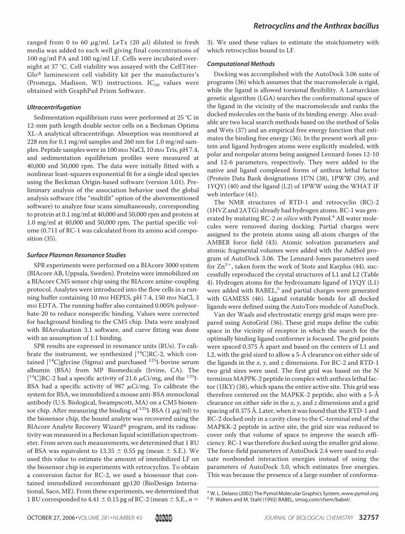
LF with retrocyclin, inhibition of the enzymatic activity of thetoxin was considerably enhanced. In the absence of preincuba-tion, 2.5 �M RC-1 inhibited enzymatic activity by 20%. Inhi-bition increased to 40% after a 5-min preincubation, to 60%after 15-min preincubation, and to 80% after a 30-min preincu-bation. In Table 3, inhibition was measured after a 30-min pre-incubation between recombinant LF and the indicated �-defen-sin. Fig. 2b shows that increasing the substrate concentrationdid not reverse the inhibition of LF by RC-2, clearly indicating
FIGURE 1. Colony count assays. Mid-logarithmic (a) and stationary phase (b)B. anthracis, Sterne strain bacteria were killed with similar kinetics by RC-2(RC-100b).
TABLE 3Inhibition of the enzymatic activity of anthrax LFData, shown as, aremean� S.E. values from at least three independent experimentswith each peptide. IC50 values for each peptide were compared to those obtained forRC-1 by unpaired t test.
Peptide Laboratory name Mean IC50 � S.E.�g/ml
RC-1 RC-100 4.23 � 0.82Retroenantio-RC-1 RC-110 19.9 � 2.01a
Retro-RC-1 RC-111 7.73 � 0.29b
Enantio-RC-1 RC-112 9.37 � 1.21b
Retrocyclins versus RTDsRC-1 RC-100 4.98 � 0.88RC-2 RC-100b 3.60 � 0.23RC-3 RC-100c 11.1 � 2.15bRhesus �-defensin-1 RTD-1 5.13 � 0.48Rhesus �-defensin-2 RTD-2 7.93 � 1.41Rhesus �-defensin-3 RTD-3 4.70 � 0.60
Arginine substitutionsRC-1 RC-100 4.23 � 0.82(R9G)-RC-1 RC107G 5.63 � 0.49(R9,18G)-RC-1 RC107GG 9.25 � 0.95b(R9,18G, R13H)-RC-1 RC107G2Ha 9.43 � 0.43a(R9,18G, R4H)-RC-1 RC107G2Hb 12.9 � 2.23b(R9,18G, R4,13H)-RC-1 RC107G2H2 �50a
Cyclic backbone and SS bondsNoncyclic RC-1 RC100ox 22.7 � 5.79bReduced and alkylated RC-1 RC100IAA �50aProtegrin PG-1 PG-1 14.8 � 2.41bHuman neutrophil peptide-1 HNP-1 6.58 � 1.48
a p � 0.01.b p � 0.05.
Retrocyclins and the Anthrax bacillus
OCTOBER 27, 2006 • VOLUME 281 • NUMBER 43 JOURNAL OF BIOLOGICAL CHEMISTRY 32759
that the process was not competitive. The Vmax of RC-treatedLF was considerably reduced, consistent with noncompetitiveinhibition, and the Km was also reduced, from 10 �M to 5�M, consistent with uncompetitive inhibition.Kinetics of Binding—We used SPR to examine the binding of
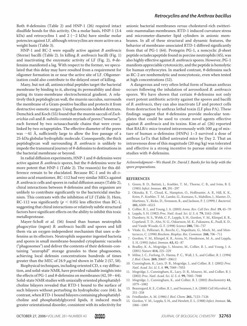
RC-1 to immobilized anthrax LF (Fig. 3). Binding isotherms areshown for two concentrations of RC-1: 4 �g/ml (2 �M) and 1�g/ml (0.5 �M). Binding was rapid, with maximal bindingreached after 5 min. From these isotherms and others
obtained at lower retrocyclin concentrations, the followingconstants were calculated for the binding of RC-1 to LF: kon,1.99 � 104; koff 1.51 � 103; Kd, 79.1 nM.
SPR experiments provide a readout in resonance units. Toconvert RUs to mass-based quantities, we performed experi-ments with [14C]RC-2 and 125I-BSA. From the former, wedetermined that 1 RU of RC-2 corresponded to 4.42 � 0.25 pg(mean � S.E., n � 3). For 125I-BSA, we determined that 1 RUwas equivalent to 13.35 � 0.55 pg of protein (mean � S.E., n �7). If we apply the same RU/pg conversion factors to RC-1 andLF, respectively, the following estimates result. The 4422RUs ofLF affixed to the biosensor chip represents 59.0 ng (or 0.66pmol) of this toxin.When the biosensorwas exposed to 4�g/mlRC-1, 4000 RU of the defensin was bound (Fig. 3). This cor-responds to 17.7 ng or9.23 pmol of retrocyclin. Thus, each LFmolecule bound 14 molecules of retrocyclin. At 1 �g/ml,when 1500 RU of retrocyclin was bound, the molar ratio was5.25 retrocyclins/LF.The different kinetics of binding (rapid) and protease inhibi-
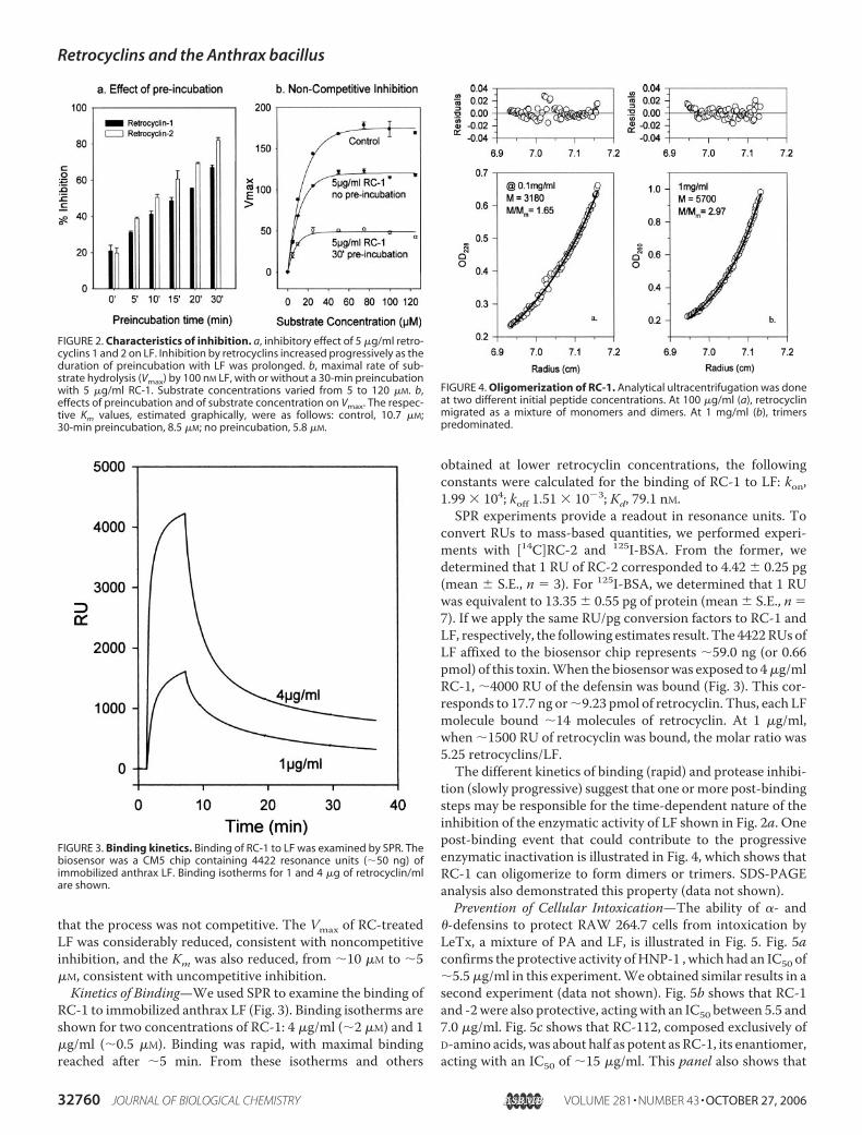
tion (slowly progressive) suggest that one ormore post-bindingsteps may be responsible for the time-dependent nature of theinhibition of the enzymatic activity of LF shown in Fig. 2a. Onepost-binding event that could contribute to the progressiveenzymatic inactivation is illustrated in Fig. 4, which shows thatRC-1 can oligomerize to form dimers or trimers. SDS-PAGEanalysis also demonstrated this property (data not shown).Prevention of Cellular Intoxication—The ability of �- and
�-defensins to protect RAW 264.7 cells from intoxication byLeTx, a mixture of PA and LF, is illustrated in Fig. 5. Fig. 5aconfirms the protective activity ofHNP-1 , which had an IC50 of5.5�g/ml in this experiment.We obtained similar results in asecond experiment (data not shown). Fig. 5b shows that RC-1and -2were also protective, actingwith an IC50 between 5.5 and7.0 �g/ml. Fig. 5c shows that RC-112, composed exclusively ofD-amino acids, was about half as potent as RC-1, its enantiomer,acting with an IC50 of 15 �g/ml. This panel also shows that
FIGURE 2. Characteristics of inhibition. a, inhibitory effect of 5 �g/ml retro-cyclins 1 and 2 on LF. Inhibition by retrocyclins increased progressively as theduration of preincubation with LF was prolonged. b, maximal rate of sub-strate hydrolysis (Vmax) by 100 nM LF, with or without a 30-min preincubationwith 5 �g/ml RC-1. Substrate concentrations varied from 5 to 120 �M. b,effects of preincubation and of substrate concentration on Vmax. The respec-tive Km values, estimated graphically, were as follows: control, 10.7 �M;30-min preincubation, 8.5 �M; no preincubation, 5.8 �M.
FIGURE 3. Binding kinetics. Binding of RC-1 to LF was examined by SPR. Thebiosensor was a CM5 chip containing 4422 resonance units (50 ng) ofimmobilized anthrax LF. Binding isotherms for 1 and 4 �g of retrocyclin/mlare shown.
FIGURE 4. Oligomerization of RC-1. Analytical ultracentrifugation was doneat two different initial peptide concentrations. At 100 �g/ml (a), retrocyclinmigrated as a mixture of monomers and dimers. At 1 mg/ml (b), trimerspredominated.
Retrocyclins and the Anthrax bacillus
32760 JOURNAL OF BIOLOGICAL CHEMISTRY VOLUME 281 • NUMBER 43 • OCTOBER 27, 2006

RC110, the retroenantio analog of RC-1, had very little protec-tive activity even though its net charge and composition mir-rored that of RC-1. The results shown in Fig. 5 (b and c) arerepresentative of four individual experiments, each done withtriplicate samples.Docking Studies—Docking was performed on three crystal
forms of LF: 1J7N, native LF (38); 1YQY, LF complexed with apotent hydroxamate inhibitor, L1, with an IC50 value of 60 nM(39); and 1PWW, LF with a nanopeptide fragment, L2, in itsactive site (40). The docking results are summarized in Table 4with the final docked energy listed for the best docked ligands.In AutoDock, the docked energy is the sum of nonbondedligand-receptor intermolecular energy and the nonbondedinternal energy of the ligand (Table 4). L1 and L2 were dockedas controls, and successfully reproduce the crystal structures inlocal searches with energies of118.8 kcal/mol and274.32kcal/mol and r.m.s.d. values of 0.75 and 0.57 Å from theircrystal coordinates, respectively, thus verifying the parame-ters used for Zn2�. A global search for L1 turned up a con-formation with even lower energy than the local search witha docked energy of 123.67 kcal/mol and an r.m.s.d. of 0.87Å, showing the effectiveness of the LGA of AutoDock. Aglobal search for L2, however, performs suboptimally, withan energy of 254.71 kcal/mol whose r.m.s.d. is 5.97 Å fromits crystal form. This is due to the large number of confor-mational degrees of freedom in L2.
Because AutoDock assumes that the receptor is rigid andtorsional flexibility is allowed in the ligand, the degree of flexi-bility of the ligand determines the computational complexity ofthe problem. Typical ligands docked with AutoDock usuallyhave less than ten torsional degrees of freedom, whereas L2,even though just a small peptide, has 58 torsional degrees offreedom. Our goal in this modeling work was to dock RTD-1,RC-1, andRC-2, which are octadecapeptideswith 44, 51, and 51torsional degrees of freedom, respectively (after excludingbackbone torsional degrees of freedom, because AutoDockcannot model torsional changes in loops). The computationalcomplexity is therefore similar to the computational complex-ity of L2. However, despite the difficulties in docking outlinedwith L2, we believe that docking circular peptides withAutoDock may still be feasible, because torsional changes inlinear peptides such as L2 would sample much greater confor-mational space than only side chain perturbations, as wouldlargely be the case with the RTDs and the RCs. Also, to reducethe search space, we focused docking primarily on the active-site cavity, even though the stoichiometry of LF/RC-1 bindingas determined by SPR indicates that RC-1 binds to multiplesites on the surface of LF. Our goal was, therefore, to search forRC or RTD binding that would directly interfere with the sub-strate binding.Several patterns emerge from our results. When docked in
the larger grid (see “Computational Methods”), all RC-2s andRTD-1s docked toward the C-ter-minal end of MAPKK-2 peptide inthe active-site cavity (Fig. 6). Itappears that the active site is toonarrow toward the N-terminalside to make entry possible fromthat direction. Access to the activesite is, therefore, most likely fromthe C-terminal side, and for latersearches, the grid size was reducedto cover only the C-terminal cavityto make search more efficient.RC-1 and -2 and RTD-1 dock
with energies of 303.03, 303.78,and 341.90 kcal/mol, respectively,which are significantly lower thanthat of the L2 peptide fragment withthe local search, thus implyingstrong binding may be occurring in
FIGURE 5. Protection from anthrax LeTx. RAW264.7 cells were incubated with LeTx and various concentra-tions of retrocyclin-1 (RC-1), retrocyclin-2 (RC-2), enantio-RC-1 (RC-112), and retroenantio-RC-1 (RC-110) in thepresence (solid symbols) or absence (open symbols) of anthrax LeTx, as described in the text. On the followingday, target cell survival was estimated by measuring their ATP content with a luciferase assay. These resultshave been normalized relative to the ATP content of control cells incubated without toxins or defensinpeptides.
TABLE 4Results of the computational docking study
Docked molecule Source ligand PDB receptor Search typea No. of atoms TorsionsDocked energy
r.m.s.d. No. of docksInternal Intermolecular Overall
kcal/mol Ao
L1 1YQY 1YQY L 42 8 17.65 101.15 118.80 0.75 200L1 1YQY 1YQY G 42 8 19.44 104.23 123.67 0.86 100L2 1PWW 1PWW L 193 58 72.14 202.18 274.32 0.57 200L2 1PWW 1PWW G 193 58 109.47 145.24 254.71 5.97 200RC-1 2ATGb 1J7N G 260 44c 154.29 148.74 303.03 131RC-2 2ATG 1J7N G 277 51c 142.36 161.42 303.78 217RTD-1 1HVZ 1J7N G 282 51c 173.99 167.91 341.90 171
aL refers to a local search, and G refers to an LGA-based global search.bRC-1 was generated by an in silicomutation on the RC-2 NMR structure.c Includes only side-chain torsional degrees of freedom. The circular peptide backbone was kept fixed during docking.
Retrocyclins and the Anthrax bacillus
OCTOBER 27, 2006 • VOLUME 281 • NUMBER 43 JOURNAL OF BIOLOGICAL CHEMISTRY 32761

the active site. Because the docked energies are representativeof binding enthalpies and not binding free energies, the trendsin their values should be interpreted with care. For instance,even though RTD-1 binds better than RC-2, their NMR struc-tures imply that RTD-1 is more flexible than RC-2. The loss ofconformational entropy of the former upon binding, therefore,would be larger when compared with the latter. This needs tobe quantified to make an accurate judgment of their relativeactivities.In the AutoDock output, the nonbonded interaction of
each ligand atom with the receptor is listed. It is thereforepossible to determine the contribution of each ligand residueto the intermolecular interaction energy. In the best docksfor RTD-1, RC-1, and RC-2 60%, 60, and 62% of the totalinteraction energy is contributed by the arginines. This is notnecessarily surprising because arginines hydrogen bond,besides interacting through electrostatic interactions, andhydrogen bonding energies an order of magnitude largerthan van der Waals interactions. In particular, because wehave the RC-107 series of mutants available, it is instructiveto see the energetic contribution of each of the arginines inRC-1. The energetic contributions in kilocalories/mol of thefour arginines are as follows: 22.59/R4, 0.22/R9, 56.75/R13, and 9.83/R18. Keeping in mind the inherent symme-try of RC-1 structure, these energies correlate well with theobserved inhibition of RC-1, RC-107G, and RC-107GGtoward LF. For example, R9G hardly affects the IC50, butR9,18G almost doubles it (Table 3).
The stoichiometry of RC-1 binding suggests that the bindingis far from specific and that there are several binding sites on theenzyme surface. DockedRTD-1, RC-1, andRC-2, each ofwhichprobably represent one of several possible bindingmodes, showsignificant overlap with the N-terminal MAPPK-2 peptide
binding site (Fig. 6) implying thatthey all compete for the same bind-ing site, which contradicts the non-competitive binding curves ob-served (Fig. 2b). However, the koffindicates that the half-life of thebinding is 7.6 min (0.69302/koff),implying the RC-1 binding can bepractically considered irreversiblecompared with substrate binding, ifthe latter is assumed to be diffusion-limited. Such irreversible bindingwithin the active site should alsogive noncompetitive binding curvesof the sort observed (Fig. 2b), be-cause the irreversibly bound inhibi-tor would reduce the effective con-centration of the active enzyme.Also, because the RCs self-aggre-gate, the slow onset of inhibitionobserved (Fig. 2a) can be explainedas follows: once the enzyme surfaceis covered with RCs, these boundRCs may form nucleation sites foraggregation and compete with the
binding to the active site itself, thus resulting in the slow onsetof inhibition.
DISCUSSION
In nonhuman primates, such as rhesus macaques, �-de-fensins are encoded by genes that encode a C-terminal “defen-sin” domain containing 12 residues, including 3 cysteines. Rhe-sus leukocytes trim and splice two such precursors into a cyclic,18-residue, �-defensin peptide. The human �-defensin genescontain a premature stop codon, and humans lack �-defensinpeptides. Although the sequences of retrocyclins 1, 2, and 3 arebased on human genome sequences, these peptides and theiranalogs were prepared by solid-phase chemical synthesis forthis study.Surprisingly few studies of �-defensin peptides have
appeared since their initial description by Selsted et al. in 1999(29), despite their novel characteristics and potential useful-ness. Whereas many cyclic peptides are produced by plants,bacteria, or fungi, �-defensins are the only known cyclic pep-tides of animal origin (47–49). The ability of �-defensins tobind carbohydrates (50) also makes them the smallest knownlectins. Their broad antiviral spectrum encompasses humanimmunodeficiency virus type 1, influenza A, and herpes sim-plex viruses (31, 51, 52), and its mechanisms have been thefocus of many of our recent studies.
�-Defensins were reported to kill some bacteria, but not oth-ers (29, 30, 53, 54). The present studies show thatB. anthracis ishighly susceptible to their antibiotic effects. Our studies werestimulated by a recent report of Kim et al. (26), showing thatHNP-, a human �-defensin, inhibited the enzymatic activity ofanthrax LF in a noncompetitive manner. We confirmed thisobservation for HNP-1 and extended it by showing that �-de-fensins also inhibit LF in a noncompetitive manner (Fig. 2).
FIGURE 6. Docking of �-defensins to the active site of anthrax lethal toxin. Docked RTD-1 (red), RC-1 (green),and RC-2 (blue) are shown with reference to the N-terminal MAPKK-2 peptide (yellow) in the active-site cavity ofanthrax lethal factor.
Retrocyclins and the Anthrax bacillus
32762 JOURNAL OF BIOLOGICAL CHEMISTRY VOLUME 281 • NUMBER 43 • OCTOBER 27, 2006
Both �-defensins (Table 2) and HNP-1 (26) required intactdisulfide bonds for this activity. On a molar basis, HNP-1 (3.4kDa) and retrocyclins 1 and 2 (2 kDa) have similar molarpotencies against LF, although retrocyclins aremore active on aweight basis (Table 3).HNP-1 and RC-2 were equally active against B anthracis
(Sterne) bacilli (Table 2). In killing B. anthracis bacilli (Fig. 1)and inactivating the enzymatic activity of LF (Fig. 2), �-de-fensins manifested a lag. With respect to the former, we specu-lated that this delay may have resulted from a requirement foroligomer formation in or near the active site of LF. Oligomer-ization could also contribute to the delayed onset of killing.Many, but not all, antimicrobial peptides target the bacterial
membrane by binding to it, altering its permeability and dissi-pating its trans-membrane electrochemical gradient. A rela-tively thick peptidoglycan wall, the murein sacculus, surroundsthe membrane of a Gram-positive bacillus and protects it fromdestructive osmotic surges. Using fluorescein-labeled dextrans,Demchick and Koch (55) found that themurein sacculi of Esch-erichia coli and B. subtilis containmyriads of pores (“tesserae”),each formed by two octasaccharide chains that were cross-linked by two octapeptides. The effective diameter of the poreswas 41 Å, sufficiently large to allow the free passage of a25-kDa globular hydrophilic molecule. Consequently, the thickpeptidoglycan wall surrounding B. anthracis is unlikely toimpede the transmural journey of �-defensins to destinations inthe bacterial membrane or beyond.In radial diffusion experiments, HNP-1 and �-defensins were
active against B. anthracis spores, but the �-defensins were farmore potent that HNP-1 (Table 2). The reason(s) for this dif-ference remain to be elucidated. Because RC-1 and its all-D-amino acid enantiomer, RC-112 had very similarMECs againstB. anthracis cells and spores in radial diffusion assays (Table 2),chiral interactions between �-defensins and this organism areunlikely to contribute significantly to the bactericidal mecha-nism. This contrasts with the inhibition of LF (Table 3). Here,RC-112 was significantly (p � 0.05) less effective than RC-1,suggesting that chiral interactions or relatively subtle structuralfactors have significant effects on the ability to inhibit this toxicmetalloprotease.Mayer-Scholl et al. (56) found than human neutrophils
phagocytize (ingest) B. anthracis bacilli and spores and killthem via an oxygen-independent mechanism that uses �-de-fensins as its effectors. Neutrophils sequester ingested bacteriaand spores in small membrane-bounded cytoplasmic vacuoles(“phagosomes”) and deliver the contents of their defensin-con-taining “azurophil” storage granules to these phagosomes,achieving local defensin concentrations hundreds of timesgreater than the MEC of 24.9 �g/ml shown in Table 2 (57, 58).
Biophysical techniques, including orientedCD, x-ray diffrac-tion, and solid-state NMR, have provided valuable insights intothe effects of PG-1 and �-defensins onmembranes (42, 59–64).Solid-stateNMR studies with uniaxially oriented phosphatidyl-choline bilayers revealed that RTD-1 bound to the surface ofsuch bilayers without perturbing its hydrophobic core (64). Incontrast, whenRTD-1boundbilayers containing phosphatidyl-choline and phosphatidylglycerol lipids, it induced muchgreater orientational disorder, consistent with its selectivity for
anionic bacterial membranes versus cholesterol-rich zwitteri-onic mammalian membranes. RTD-1 induced curvature stressand micrometer-diameter lipid cylinders in anionic mem-branes. In all of these structural and dynamic features, thebehavior of membrane-associated RTD-1 differed significantlyfrom that of PG-1 (64). Protegrin PG-1, a noncyclic �-sheetpeptide octadecapeptide found in porcine neutrophils (45), wasalso highly effective againstB. anthracis spores. However, PG-1manifests appreciable cytotoxicity, and the peptide is hemolyticfor human erythrocytes. In marked contrast, �-defensins suchas RC-2 are nonhemolytic and noncytotoxic, even when testedat high concentrations (52).A dangerous and very often lethal form of human anthrax
occurs following the inhalation of aerosolized B. anthracisspores. We have shown that certain �-defensins not onlyexert potent antibiotic activity against the spores and bacilliof B. anthracis, they can also inactivate LF and protect cellsfrom destruction by anthrax lethal toxin (LF plus PA). Thesefindings suggest that �-defensins provide molecular tem-plates that could be used to create novel agents effectiveagainst B. anthracis and its toxins. Kim et al. (26) reportedthat BALB/c mice treated intravenously with 500 �g of mix-ture of human �-defensins (HNPs) 1–3 survived a dose ofanthrax LeTx that killed untreated mice by Day 2. That anintravenous dose of this magnitude (20 mg/kg) was toleratedand effective is a strong incentive to pursue similar in vivostudies with �-defensins.
Acknowledgment—We thank Dr. David J. Banks for his help with thespore preparations.
REFERENCES1. Green, B. D., Battisti, L., Koehler, T. M., Thorne, C. B., and Ivins, B. E.
(1985) Infect. Immun. 49, 291–2972. Okinaka, R. T., Cloud, K., Hampton, O., Hoffmaster, A. R., Hill, K. K.,
Keim, P., Koehler, T. M., Lamke, G., Kumano, S., Mahillon, J., Manter, D.,Martinez, Y., Ricke, D., Svensson, R., and Jackson, P. J. (1999) J. Bacteriol.181, 6509–6515
3. Collier, R. J., and Young, J. A. (2003) Annu. Rev. Cell Dev. Biol. 19, 45–704. Leppla, S. H. (1982) Proc. Natl. Acad. Sci. U. S. A. 79, 3162–31665. Duesbery, N. S., Webb, C. P., Leppla, S. H., Gordon, V. M., Klimpel, K. R.,
Copeland, T. D., Ahn, N. G., Oskarsson, M. K., Fukasawa, K., Paull, K. D.,and Vande Woude, G. F. (1998) Science 280, 734–737
6. Vitale, G., Pellizzari, R., Recchi, C., Napolitani, G., Mock, M., and Mon-tecucco, C. (1998) Biochem. Biophys. Res. Commun. 248, 706–711
7. Gordon, V. M., Klimpel, K. R., Arora, N., Henderson, M. A., and Leppla,S. H. (1995) Infect. Immun. 63, 82–87
8. Bradley, K. A., Mogridge, J., Mourez, M., Collier, R. J., and Young, J. A.(2001) Nature 414, 225–229
9. Milne, J. C., Furlong, D., Hanna, P. C., Wall, J. S., and Collier, R. J. (1994)J. Biol. Chem. 269, 20607–20612
10. Cunningham, K., Lacy, D. B., Mogridge, J., and Collier, R. J. (2002) Proc.Natl. Acad. Sci. U. S. A. 99, 7049–7053
11. Mogridge, J., Cunningham, K., Lacy, D. B., Mourez, M., and Collier, R. J.(2002) Proc. Natl. Acad. Sci. U. S. A. 99, 7045–7048
12. Mogridge, J., Cunningham, K., and Collier, R. J. (2002) Biochemistry 41,1079–1082
13. Beauregard, K. E., Collier, R. J., and Swanson, J. A. (2000)CellMicrobiol. 2,251–258
14. Friedlander, A. M. (1986) J. Biol. Chem. 261, 7123–712615. Gordon, V. M., Leppla, S. H., and Hewlett, E. L. (1988) Infect. Immun. 56,
1066–1069
Retrocyclins and the Anthrax bacillus
OCTOBER 27, 2006 • VOLUME 281 • NUMBER 43 JOURNAL OF BIOLOGICAL CHEMISTRY 32763
16. Blaustein, R. O., Koehler, T. M., Collier, R. J., and Finkelstein, A. (1989)Proc. Natl. Acad. Sci. U. S. A. 86, 2209–2213
17. Milne, J. C., and Collier, R. J. (1993)Mol. Microbiol. 10, 647–65318. Wesche, J., Elliott, J. L., Falnes, P. O., Olsnes, S., and Collier, R. J. (1998)
Biochemistry 37, 15737–1574619. Friedlander, A. M., Welkos, S. L., and Ivins, B. E. (2002) Curr. Top. Micro-
biol. Immunol. 271, 33–6020. Ganz, T. (2004) C. R. Biol. 327, 539–54921. Lehrer, R. I. (2004) Nat. Rev. Microbiol. 2, 727–73822. Selsted, M. E., and Ouellette, A. J. (2005) Nat. Immunol. 6, 551–55723. Lehrer, R. I., Lichtenstein, A. K., andGanz, T. (1993)Annu. Rev. Immunol.
11, 105–12824. Scheetz, T., Bartlett, J. A.,Walters, J. D., Schutte, B. C., Casavant, T. L., and
McCray, P. B., Jr. (2002) Immunol. Rev. 190, 137–14525. Schutte, B. C., Mitros, J. P., Bartlett, J. A., Walters, J. D., Jia, H. P., Welsh,
M. J., Casavant, T. L., and McCray, P. B., Jr. (2002) Proc. Natl. Acad. Sci.U. S. A. 99, 2129–2133
26. Kim, C., Gajendran, N., Mittrucker, H. W., Weiwad, M., Song, Y. H.,Hurwitz, R., Wilmanns, M., Fischer, G., and Kaufmann, S. H. (2005) Proc.Natl. Acad. Sci. U. S. A. 102, 4830–4835
27. Nguyen, T. X., Cole, A.M., and Lehrer, R. I. (2003)Peptides 24, 1647–165428. Selsted, M. E. (2004) Curr. Protein Pept. Sci 5, 365–37129. Tang, Y. Q., Yuan, J., Osapay, G., Osapay, K., Tran, D., Miller, C. J., Ouel-
lette, A. J., and Selsted, M. E. (1999) Science 286, 498–50230. Leonova, L., Kokryakov, V. N., Aleshina, G., Hong, T., Nguyen, T., Zhao,
C., Waring, A. J., and Lehrer, R. I. (2001) J. Leukoc. Biol. 70, 461–46431. Cole, A. M., Hong, T., Boo, L. M., Nguyen, T., Zhao, C., Bristol, G., Zack,
J. A., Waring, A. J., Yang, O. O., and Lehrer, R. I. (2002) Proc. Natl. Acad.Sci. U. S. A. 99, 1813–1818
32. Wu, Z., Powell, R., and Lu, W. (2003) J. Am. Chem. Soc. 125, 2402–240333. Banks, D. J., Barnajian, M., Maldonado-Arocho, F. J., Sanchez, A. M., and
Bradley, K. A. (2005) Cell Microbiol. 7, 1173–118534. Lehrer, R. I., Rosenman, M., Harwig, S. S., Jackson, R., and Eisenhauer, P.
(1991) J. Immunol. Methods 137, 167–17335. Cohn, E. J., and Edsall, J.T. (1943) in Proteins, Amino Acids and Peptides as
Ions and Dipolar Ions (Cohn, E. J., and Edsall, J. T., eds) pp. 374–377,Reinhold Publishing Corporation, New York
36. Morris, G.M., Goodsell, D. S., Halliday, R. S., Huey, R., Hart,W. E., Belew,R.K., and Olson, A. J. (1998) J. Comput. Chem. 19, 1639–1662
37. Solis, F. J., and Wets, R. J. B. (1981)Math. Operation Res. 6, 19–3038. Pannifer, A. D.,Wong, T. Y., Schwarzenbacher, R., Renatus,M., Petosa, C.,
Bienkowska, J., Lacy, D. B., Collier, R. J., Park, S., Leppla, S. H., Hanna, P.,and Liddington, R. C. (2001) Nature 414, 229–233
39. Turk, B. E., Wong, T. Y., Schwarzenbacher, R., Jarrell, E. T., Leppla, S. H.,Collier, R. J., Liddington, R. C., and Cantley, L. C. (2004) Nat. Struct. Mol.Biol. 11, 60–66
40. Shoop, W. L., Xiong, Y., Wiltsie, J., Woods, A., Guo, J., Pivnichny, J. V.,Felcetto, T., Michael, B. F., Bansal, A., Cummings, R. T., Cunningham,B. R., Friedlander, A. M., Douglas, C. M., Patel, S. B., Wisniewski, D.,Scapin, G., Salowe, S. P., Zaller, D. M., Chapman, K. T., Scolnick, E. M.,
Schmatz, D. M., Bartizal, K., MacCoss, M., and Hermes, J. D. (2005) Proc.Natl. Acad. Sci. U. S. A. 102, 7958–7963
41. Vriend, G. (1990) J. Mol. Graph. 8, 52–5642. Mani, R., Buffy, J. J., Waring, A. J., Lehrer, R. I., and Hong, M. (2004)
Biochemistry 43, 13839–1384843. Cornell, W. D., Bayli, C. I., Gould, I. R., Merz, K. M. J., Ferguson, D. M.,
Spellmeyer, D. C., Fox, T., Caldwell, J.W., and Kollman, P. A. (1995) J. Am.Chem. Soc. 117, 5179–5197
44. Stote, R. H., and Karplus, M. (1995) Proteins 23, 12–3145. Kokryakov, V. N., Harwig, S. S., Panyutich, E. A., Shevchenko, A. A.,
Aleshina, G. M., Shamova, O. V., Korneva, H. A., and Lehrer, R. I. (1993)FEBS Lett. 327, 231–236
46. Schmidt, M. W., Baldridge, K. K., Boatz, J. A., Elbert, S. T., Gordon, M. S.,Jensen, J. H., Koseki, S., Matsunaga, N., Nguyen, K. A., Su, S. J., Windus,T. L., Dupuis, M., and Montgomery, J. A. (1993) J. Comput. Chem. 14,1347–1363
47. Trabi, M., and Craik, D. J. (2002) Trends Biochem. Sci. 27, 132–13848. Mulvenna, J. P., Wang, C., and Craik, D. J. (2006) Nucleic Acids Res. 34,
D192–D19449. Craik, D. J. (2006) Science 311, 1563–156450. Wang, W., Cole, A. M., Hong, T., Waring, A. J., and Lehrer, R. I. (2003)
J. Immunol. 170, 4708–471651. Leikina, E., Delanoe-Ayari, H., Melikov, K., Cho, M. S., Chen, A., Waring,
A. J., Wang, W., Xie, Y., Loo, J. A., Lehrer, R. I., and Chernomordik, L. V.(2005) Nat. Immunol. 6, 995–1001
52. Yasin, B., Wang, W., Pang, M., Cheshenko, N., Hong, T., Waring, A. J.,Herold, B. C.,Wagar, E. A., and Lehrer, R. I. (2004) J. Virol. 78, 5147–5156
53. Chong-Cerrillo, C., Selsted, M. E., Peterson, E. M., and de la Maza, L. M.(2003) J. Pept. Res. 61, 237–242
54. Tran, D., Tran, P. A., Tang, Y. Q., Yuan, J., Cole, T., and Selsted, M. E.(2002) J. Biol. Chem. 277, 3079–3084
55. Demchick, P., and Koch, A. L. (1996) J. Bacteriol. 178, 768–77356. Mayer-Scholl, A., Hurwitz, R., Brinkmann, V., Schmid, M., Jungblut, P.,
Weinrauch, Y., and Zychlinsky, A. (2005) PLoS. Pathog. 1, e2357. Lehrer, R. I., and Ganz, T. (1992) Ciba Found. Symp. 171, 276–29058. Rice, W. G., Ganz, T., Kinkade, J. M., Jr., Selsted, M. E., Lehrer, R. I., and
Parmley, R. T. (1987) Blood 70, 757–76559. Gidalevitz, D., Ishitsuka, Y., Muresan, A. S., Konovalov, O., Waring, A. J.,
Lehrer, R. I., and Lee, K. Y. (2003) Proc. Natl. Acad. Sci. U. S. A. 100,6302–6307
60. Heller, W. T., Waring, A. J., Lehrer, R. I., Harroun, T. A., Weiss, T. M.,Yang, L., and Huang, H. W. (2000) Biochemistry 39, 139–145
61. Weiss, T. M., Yang, L., Ding, L., Waring, A. J., Lehrer, R. I., and Huang,H. W. (2002) Biochemistry 41, 10070–10076
62. Buffy, J. J., Hong, T., Yamaguchi, S., Waring, A. J., Lehrer, R. I., and Hong,M. (2003) Biophys. J. 85, 2363–2373
63. Buffy, J. J., Waring, A. J., Lehrer, R. I., and Hong, M. (2003) Biochemistry42, 13725–13734
64. Buffy, J. J., McCormick, M. J., Wi, S., Waring, A., Lehrer, R. I., and Hong,M. (2004) Biochemistry 43, 9800–9812
Retrocyclins and the Anthrax bacillus
32764 JOURNAL OF BIOLOGICAL CHEMISTRY VOLUME 281 • NUMBER 43 • OCTOBER 27, 2006
Related Documents


![Bacillus anthracis - As Biological WeaponsBacillus anthracis - as biological weapons :JOLN (Bacillus anthracis) ± MDNREUR ELRORJLF]QD miotr Daniszewski Department of Invertebrate](https://static.cupdf.com/doc/110x72/613e1f0259df642846165479/bacillus-anthracis-as-biological-weapons-bacillus-anthracis-as-biological-weapons.jpg)