595 Retinal Vascular Geometry in Asian Persons with Diabetes and Retinopathy Carol Yim-lui Cheung, Ph.D., 1,2 Ecosse Lamoureux, Ph.D., 1,3 M. Kamran Ikram, M.D., Ph.D., 2,4 M. Bayu Sasongko, M.D., M.Epi., 3,5 Jie Ding, Ph.D., 1 Yingfeng Zheng, M.D., 1 Paul Mitchell, M.D., Ph.D., 6 Jie Jin Wang, M.Med., Ph.D., 3,6 and Tien Yin Wong, M.D., Ph.D. 1,2, Author Affiliations: 1 Singapore Eye Research Institute, Singapore National Eye Centre, Singapore; 2 Department of Ophthalmology, Yong Loo Lin School of Medicine, National University of Singapore, Singapore; 3 Centre for Eye Research Australia, University of Melbourne, Royal Victorian Eye and Ear Hospital, Victoria, Australia; 4 Department of Ophthalmology, Erasmus Medical Center, Rotterdam, the Netherlands; 5 Department of Ophthalmology, Faculty of Medicine, Gadjah Mada University, Yogyakarta, Indonesia; and 6 Centre for Vision Research, University of Sydney, Sydney, Australia Abbreviations: (BMI) body mass index, (CI) confidence interval, (DR) diabetic retinopathy, (HbA1c) glycated hemoglobin, (SIVA) Singapore I Vessel Assessment Keywords: diabetes, epidemiology, imaging, retinopathy Corresponding Author: Carol Yim-lui Cheung, Ph.D., Singapore Eye Research Institute, 11 Third Hospital Avenue, Singapore 168751; email address [email protected] Journal of Diabetes Science and Technology Volume 6, Issue 3, May 2012 © Diabetes Technology Society Abstract Purpose: Our purpose was to examine the relationship of retinal vascular parameters with diabetes and retinopathy in an older Asian population. Methods: Retinal photographs from participants of a population-based survey of Asian Malay persons aged 40–80 years were analyzed. Specific retinal vascular parameters (tortuosity, branching angle, fractal dimension, and caliber) were measured using a semiautomated computer-based program. Diabetes was defined as random plasma glucose ≥ 11.1 mmol/liter, the use of diabetes medication, or physician-diagnosed diabetes. Retinopathy signs were graded from photographs using the modified Airlie House classification system. Results: A total of 2735 persons were included in the study. Persons with diabetes ( n = 594) were more likely to have straighter (less tortuous) arterioles and wider arteriolar and venular caliber than those without diabetes ( n = 2141). Among subjects with diabetes, those with retinopathy had wider venular caliber than those without retinopathy (211.3 versus 204.9 µm, p = .001). Among nondiabetic subjects, however, those with retinopathy had more tortuous venules than those without retinopathy [5.19(×10 4 ) versus 4.27(×10 4 ), p < .001]. Conclusions: Retinal vascular parameters varied by diabetes and retinopathy status in this older Asian cohort. Our findings suggest that subtle alterations in retinal vascular architecture are influenced by diabetes. J Diabetes Sci Technol 2012;6(3):595-605 ORIGINAL ARTICLE

Welcome message from author
This document is posted to help you gain knowledge. Please leave a comment to let me know what you think about it! Share it to your friends and learn new things together.
Transcript

595
Retinal Vascular Geometry in Asian Persons with Diabetes and Retinopathy
Carol Yim-lui Cheung, Ph.D.,1,2 Ecosse Lamoureux, Ph.D.,1,3 M. Kamran Ikram, M.D., Ph.D.,2,4 M. Bayu Sasongko, M.D., M.Epi.,3,5 Jie Ding, Ph.D.,1 Yingfeng Zheng, M.D.,1
Paul Mitchell, M.D., Ph.D.,6 Jie Jin Wang, M.Med., Ph.D.,3,6 and Tien Yin Wong, M.D., Ph.D.1,2,
Author Affiliations: 1Singapore Eye Research Institute, Singapore National Eye Centre, Singapore; 2Department of Ophthalmology, Yong Loo Lin School of Medicine, National University of Singapore, Singapore; 3Centre for Eye Research Australia, University of Melbourne, Royal Victorian Eye and Ear Hospital, Victoria, Australia; 4Department of Ophthalmology, Erasmus Medical Center, Rotterdam, the Netherlands; 5Department of Ophthalmology, Faculty of Medicine, Gadjah Mada University, Yogyakarta, Indonesia; and 6Centre for Vision Research, University of Sydney, Sydney, Australia
Abbreviations: (BMI) body mass index, (CI) confidence interval, (DR) diabetic retinopathy, (HbA1c) glycated hemoglobin, (SIVA) Singapore I Vessel Assessment
Keywords: diabetes, epidemiology, imaging, retinopathy
Corresponding Author: Carol Yim-lui Cheung, Ph.D., Singapore Eye Research Institute, 11 Third Hospital Avenue, Singapore 168751; email address [email protected]
Journal of Diabetes Science and Technology Volume 6, Issue 3, May 2012 © Diabetes Technology Society
Abstract
Purpose:Our purpose was to examine the relationship of retinal vascular parameters with diabetes and retinopathy in an older Asian population.
Methods:Retinal photographs from participants of a population-based survey of Asian Malay persons aged 40–80 years were analyzed. Specific retinal vascular parameters (tortuosity, branching angle, fractal dimension, and caliber) were measured using a semiautomated computer-based program. Diabetes was defined as random plasma glucose ≥ 11.1 mmol/liter, the use of diabetes medication, or physician-diagnosed diabetes. Retinopathy signs were graded from photographs using the modified Airlie House classification system.
Results:A total of 2735 persons were included in the study. Persons with diabetes (n = 594) were more likely to have straighter (less tortuous) arterioles and wider arteriolar and venular caliber than those without diabetes (n = 2141). Among subjects with diabetes, those with retinopathy had wider venular caliber than those without retinopathy (211.3 versus 204.9 µm, p = .001). Among nondiabetic subjects, however, those with retinopathy had more tortuous venules than those without retinopathy [5.19(×104) versus 4.27(×104), p < .001].
Conclusions:Retinal vascular parameters varied by diabetes and retinopathy status in this older Asian cohort. Our findings suggest that subtle alterations in retinal vascular architecture are influenced by diabetes.
J Diabetes Sci Technol 2012;6(3):595-605
ORIGINAL ARTICLE
596
Retinal Vascular Geometry in Asian Persons with Diabetes and Retinopathy Cheung
www.journalofdst.orgJ Diabetes Sci Technol Vol 6, Issue 3, May 2012
Introduction
Diabetes mellitus is projected to affect 438.7 million people worldwide by 2030.1 An important microvascular complication is diabetic retinopathy (DR), which affects 1 in 3 diabetes patients, with as many as 1 in 10 having sight-threatening DR.2,3
Advances in digital retinal photography have provided an opportunity to assess the human microvasculature noninvasively and to measure subtle retinal vascular changes quantitatively, prior to development of clinical retinopathy signs. Previous studies, largely in white populations, have shown that one particular retinal vascular change—retinal vascular caliber (diameter)—is a marker of early microvascular damage related to diabetes and associated complications (e.g., DR, nephropathy, and cardiovascular disease).4–13
The human circulatory system is believed to conform to an optimum design principle,14 which allows sufficient blood distribution with the least amount of energy. Deviation from this optimal architecture therefore results in suboptimal and less efficient peripheral circulation.14–16 Studies have shown that a number of newer retinal vascular parameters capture the “optimality” state of the retinal circulation. These newer parameters, including tortuosity, branching angle, fractal dimension, and length-to-diameter ratio, have been linked to diabetes,17–20 hypertension,21–24 stroke,25,26 or chronic kidney disease,27 providing additional insights into early microvascular abnormalities prior to clinical disease.20,28 However, previous studies have been limited to relatively small and highly selected clinical samples of persons with diabetes.17–20 In particular, there are no population-based studies of these interrelationships in Asians.
In this study, we examined the association of quantitative retinal vascular parameters with diabetes and retinopathy signs in a large Asian population-based cohort.
Methods
Study PopulationData for this analysis were derived from the Singapore Malay Eye Study, a population-based cross-sectional study of eye diseases in urban Malay adults ranging in age between 40 and 80 years, residing in Southwestern Singapore. In brief, subjects were selected, using an age-stratified (by 10-year age group) random sampling method,
from a computer-generated list provided by the Singapore Ministry of Home Affairs. Of 4168 eligible participants, 3280 (78.7%) participated in the study, conducted from August 2004 through June 2006. The methodology and objectives of the study population are reported in detail elsewhere.29
Of 3280 participants, 3266 subjects (99.6%) had fundus photographs taken of either eye and 3100 had photographs from the right eyes available for retinal vascular analysis. We excluded eyes with poor image quality (n = 177), with evidence of previous laser treatment (n = 29), without at least six gradable arterioles or venules for vessel diameter measurement (n = 106), without an arteriolar or venular bifurcation within the measured area (n = 28), without an adequate measured area (n = 24), or with missing retinopathy grading data (n = 1). This left 2735 subjects for the final analysis (83.4% of 3280 participants).
Written, informed consent was obtained from each participant, and the study conducted adhered to the Declaration of Helsinki. Ethical approval was obtained by the Singapore Eye Research Institute Institutional Review Board. Participants underwent a standardized interview, systemic and ocular examination, and laboratory investigations.
Retinal PhotographyDigital fundus photography was taken using a 45º digital retinal camera (Canon CR-DGi with a 10D SLR digital camera back; Canon, Tokyo, Japan) after pupil dilation using tropicamide 1% and phenylephrine hydrochloride 2.5%. Two retinal images of each eye were obtained, one centered at the optic disc and another centered at the fovea.
Quantitative Measurements of Retinal Microvasculature We used a semiautomated computer-assisted program (Singapore I Vessel Assessment [SIVA], version 1.0) to measure the following retinal vascular parameters from digital photographs: tortuosity, branching angle, fractal dimension, and caliber. Trained graders, masked to participant characteristics, used the SIVA program to measure these parameters according to a standardized protocol. The measured area of retinal vascular parameters was standardized and defined within the region between 0.5 and 2.0 disc diameters away from the disc margin
597
Retinal Vascular Geometry in Asian Persons with Diabetes and Retinopathy Cheung
www.journalofdst.orgJ Diabetes Sci Technol Vol 6, Issue 3, May 2012
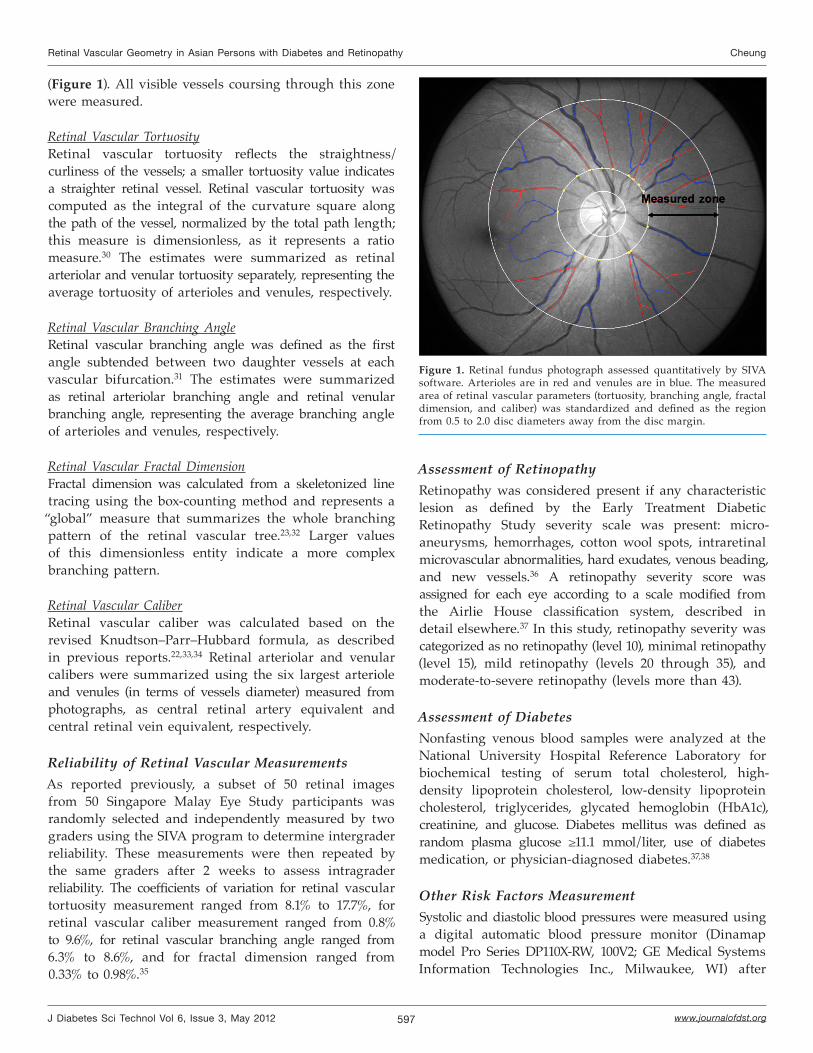
(Figure 1). All visible vessels coursing through this zone were measured.
Retinal Vascular TortuosityRetinal vascular tortuosity reflects the straightness/curliness of the vessels; a smaller tortuosity value indicates a straighter retinal vessel. Retinal vascular tortuosity was computed as the integral of the curvature square along the path of the vessel, normalized by the total path length; this measure is dimensionless, as it represents a ratio measure.30 The estimates were summarized as retinal arteriolar and venular tortuosity separately, representing the average tortuosity of arterioles and venules, respectively.
Retinal Vascular Branching AngleRetinal vascular branching angle was defined as the first angle subtended between two daughter vessels at each vascular bifurcation.31 The estimates were summarized as retinal arteriolar branching angle and retinal venular branching angle, representing the average branching angle of arterioles and venules, respectively.
Retinal Vascular Fractal DimensionFractal dimension was calculated from a skeletonized line tracing using the box-counting method and represents a
“global” measure that summarizes the whole branching pattern of the retinal vascular tree.23,32 Larger values of this dimensionless entity indicate a more complex branching pattern.
Retinal Vascular CaliberRetinal vascular caliber was calculated based on the revised Knudtson–Parr–Hubbard formula, as described in previous reports.22,33,34 Retinal arteriolar and venular calibers were summarized using the six largest arteriole and venules (in terms of vessels diameter) measured from photographs, as central retinal artery equivalent and central retinal vein equivalent, respectively.
Reliability of Retinal Vascular MeasurementsAs reported previously, a subset of 50 retinal images from 50 Singapore Malay Eye Study participants was randomly selected and independently measured by two graders using the SIVA program to determine intergrader reliability. These measurements were then repeated by the same graders after 2 weeks to assess intragrader reliability. The coefficients of variation for retinal vascular tortuosity measurement ranged from 8.1% to 17.7%, for retinal vascular caliber measurement ranged from 0.8% to 9.6%, for retinal vascular branching angle ranged from 6.3% to 8.6%, and for fractal dimension ranged from 0.33% to 0.98%.35
Figure 1. Retinal fundus photograph assessed quantitatively by SIVA software. Arterioles are in red and venules are in blue. The measured area of retinal vascular parameters (tortuosity, branching angle, fractal dimension, and caliber) was standardized and defined as the region from 0.5 to 2.0 disc diameters away from the disc margin.
Assessment of RetinopathyRetinopathy was considered present if any characteristic lesion as defined by the Early Treatment Diabetic Retinopathy Study severity scale was present: micro-aneurysms, hemorrhages, cotton wool spots, intraretinal microvascular abnormalities, hard exudates, venous beading, and new vessels.36 A retinopathy severity score was assigned for each eye according to a scale modified from the Airlie House classification system, described in detail elsewhere.37 In this study, retinopathy severity was categorized as no retinopathy (level 10), minimal retinopathy (level 15), mild retinopathy (levels 20 through 35), and moderate-to-severe retinopathy (levels more than 43).
Assessment of DiabetesNonfasting venous blood samples were analyzed at the National University Hospital Reference Laboratory for biochemical testing of serum total cholesterol, high-density lipoprotein cholesterol, low-density lipoprotein cholesterol, triglycerides, glycated hemoglobin (HbA1c), creatinine, and glucose. Diabetes mellitus was defined as random plasma glucose ≥11.1 mmol/liter, use of diabetes medication, or physician-diagnosed diabetes.37,38
Other Risk Factors MeasurementSystolic and diastolic blood pressures were measured using a digital automatic blood pressure monitor (Dinamap model Pro Series DP110X-RW, 100V2; GE Medical Systems Information Technologies Inc., Milwaukee, WI) after

598
Retinal Vascular Geometry in Asian Persons with Diabetes and Retinopathy Cheung
www.journalofdst.orgJ Diabetes Sci Technol Vol 6, Issue 3, May 2012
subjects were seated for at least 5 min. Blood pressure was measured twice, 5 min apart. A third measurement was made if the systolic blood pressure differed by more than 10 mm Hg or the diastolic by more than 5 mm Hg. The mean between the two closest readings was then taken as the blood pressure of that individual. Hypertension was defined as systolic blood pressure of 140 mm Hg or more or diastolic blood pressure of 90 mm Hg or more at examination, a history of physician-diagnosed hypertension, use of antihypertensive medication, or both. Current smokers were defined as those currently smoking any cigarettes (i.e., current versus past/never). Body mass index (BMI) was calculated as body weight (in kilograms) divided by body height (in meters) squared.
Statistical AnalysisAll statistical analyses were performed using SPSS statistics version 17.0. Retinal vascular measures (tortuosity, branching angle, fractal dimension, and caliber) were analyzed as continuous variables. Retinal vascular tortuosity was log-transformed because of its skewed distribution.
We first used analyses of covariance to estimate mean [95% confidence interval (CI)] retinal vascular measures by diabetes status to examine associations of diabetes (independent variables) with retinal vascular parameters (dependent variables). For each retinal vascular parameter, two multivariable models were constructed: model 1 adjusted for age and gender and model 2 additionally adjusted mean arterial blood pressure, BMI, total cholesterol and smoking status. Second, we used analyses of covariance to estimate mean (95% CI) retinal vascular measures with retinopathy status in persons with and without diabetes. Test of trend was determined by treating categorical retinopathy severity level as a continuous ordinal variable. For each retinal vascular parameter, two multivariable models were constructed: model 1 adjusted for age and gender and model 2 additionally adjusted for systolic blood pressure, BMI, total cholesterol, HbA1c, and smoking status (and diabetes duration for those with diabetes).
ResultsA total of 2735 subjects were included in the analysis (83.4% of 3280 participants). Compared with the excluded persons, persons included in the current study were younger (57.2 versus 66.2 years, p < .001), had lower mean arterial blood pressure (101.5 versus 105.6 mm Hg, p < .001), had a lower serum glucose level (6.71 versus 7.23 mmol/liter, p < .001), had a lower total cholesterol level (5.61 versus 5.72 mmol/liter, p = .046), had a
higher BMI (26.5 versus 25.8 kg/m2, p = .008), and were less likely to be current smokers (21.2% versus 15.3%, p = .002).
Table 1 shows the characteristics of the study population. Among participants with and without diabetes, those with retinopathy were more likely to have higher systolic blood pressure, serum glucose levels, and HbA1c levels compared with subjects without retinopathy.
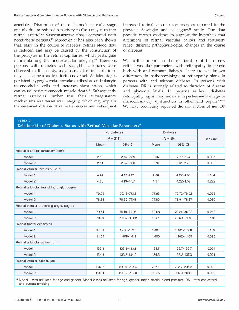
Table 2 shows that, after controlling for age, gender, mean arterial blood pressure, BMI, total cholesterol level, and smoking status, persons with diabetes were more likely to have straighter (less tortuous) arterioles, wider arteriolar caliber, and wider venular caliber than persons without diabetes. In contrast, venular tortuosity, arteriolar and venular branching angle, and fractal dimension were not significantly associated with diabetes.
Table 3 shows the relationship of retinal vascular para-meters with DR in persons with diabetes. Among persons with diabetes, subjects with DR had wider venular caliber than those without retinopathy (211.3 versus 204.9 µm, p = .001); wider venular caliber was significantly related to increased DR severity level (p trend < 0.001).
Table 4 shows the relationship of retinal vascular para-meters with retinopathy in nondiabetic persons. Among persons without diabetes, those with retinopathy had more tortuous venules than those without retinopathy (5.19[x104] versus 4.27[x104], p < .001); higher venular tortuosity value was significantly associated with increased retinopathy severity level (p-trend = 0.016).
DiscussionOur population-based study of older Asian cohort confirms previous findings that retinal vascular alterations, measured quantitatively from digital retinal images, are associated with diabetes and retinopathy. Independent of age, blood pressure, and vascular risk factors, persons with diabetes were more likely to have straighter retinal arterioles and wider arterioles and venules than persons without diabetes. Among persons with diabetes, those with retinopathy were more likely to have wider retinal venules than those without retinopathy; in contrast, among persons without diabetes, those with retinopathy signs were more likely to have more tortuous venules than those without retinopathy.
This is the first study to examine the relationship of a wide range of quantitative retinal vascular parameters

599
Retinal Vascular Geometry in Asian Persons with Diabetes and Retinopathy Cheung
www.journalofdst.orgJ Diabetes Sci Technol Vol 6, Issue 3, May 2012
Table 1.Characteristics of Study Populationa
All participants(n = 2735)
Participants without diabetes(n = 2141)
Participants with diabetes(n = 594)
No diabetes(n = 2141)
Diabetes(n = 594) pb No retinopathy
(n = 2073)
Any retinopathy
(n = 68)pb
No retinopathy(n = 437)
Any retinopathy (n = 157)
pb
Male gender, % 49.3 42.9 0.007 48.8 63.2 0.019 45.5 35.7 0.032
Hypertension, % 61.3 82.8 <0.001 61.1 69.1 0.179 81.9 85.4 0.329
Current smoking, % 23.0 14.9 <0.001 22.9 25.0 0.688 15.4 13.5 0.566
Mean (SD) Mean (SD) Mean (SD) Mean (SD) Mean (SD) Mean (SD)
Age, years 56.0 (10.6) 61.5 (9.3) <0.001 56.0 (10.6) 56.5 (11.2) 0.733 61.5 (9.5) 61.4 (8.5) 0.894
Systolic BP, mm Hg 143.4 (22.9) 152.5 (22.7) <0.001 143.1 (22.5) 153.9 (31.5) <0.001 151.0 (22.2) 156.8 (23.6) 0.005
Diastolic BP, mm Hg 79.7 (11.2) 79.0 (10.7) 0.130 79.6 (11.1) 82.6 (14.7) 0.031 79.0 (10.4) 78.8 (11.5) 0.843
BMI, kg/m2 26.1 (5.2) 27.7 (4.8) <0.001 26.1 (5.1) 26.7 (5.9) 0.365 27.8 (5.0) 27.3 (4.4) 0.310
Serum glucose, mmol/liter 5.49 (1.30) 11.11 (5.35) <0.001 5.48 (1.29) 5.94 (1.61) 0.005 10.77 (5.14) 12.10 (5.80) 0.009
HbA1c, % 5.85 (0.59) 8.46 (2.03) <0.001 5.84 (0.58) 6.06 (0.64) 0.003 8.27 (2.02) 9.00 (1.96) <0.001
Total cholesterol, mmol/liter 5.65 (1.11) 5.45 (1.24) <0.001 5.65 (1.11) 5.79 (1.29) 0.319 5.50 (1.21) 5.33 (1.34) 0.139
HDL cholesterol, mmol/liter 1.37 (0.33) 1.28 (0.29) <0.001 1.38 (0.33) 1.31 (0.36) 0.163 1.28 (0.29) 1.30 (0.29) 0.425
Duration of diabetes, years - 8.47 (8.44) - - - - 6.97 (7.40) 12.45 (9.70) <0.001a BP, blood pressure; HDL, high-density lipoprotein; SD, standard deviation.b p value is based on chi-square (categorical) or independent sample t-tests.
measured from retinal images of persons with diabetes and retinopathy signs in those with and without diabetes in a general unselected population cohort. Most previous studies have focused on one retinal vascular parameter (the caliber or diameter of the retinal vessels), were from highly selected clinic-based populations, and were largely on white persons.4,9–11,38 While some of our findings in this Asian cohort are consistent with these previous studies, others were not. For example, our finding that persons with diabetes were more likely to have wider arterioles and venules than nondiabetic persons is compatible with many others with similar findings.10,11,38
We also showed that people with diabetes had less tortuous arterioles than those without diabetes. We previously demonstrated that decreased retinal arteriolar tortuosity were associated with elevated blood pressure in our cohort,21 and Witt and associates39 found that decreased arteriolar tortuosity was associated with risk of ischemic heart disease mortality. Analysis of clinical trial participants from the Anglo-Scandinavian Cardiac Outcomes Trial, with a smaller sample size (159 with and 552 without diabetes), reported that individuals with diabetes also tended to have straighter retinal
arterioles than individuals without diabetes; however, the differences were not statistically significant.40
These findings, however, contrast with a clinic-based study from Sasongko and colleagues41 showing that persons with diabetes had more tortuous retinal arterioles and venules (224 with and 103 without diabetes). The reason for the discrepancies may be related to differences in race (Asian Malays in our study versus Caucasian white persons in Sasongko and colleagues’ study), age of participants (57.2 versus 53.8 years, p < .001), duration of diabetes (8.5 versus 16.2 years, p < .001), glycemic control (HbA1c level 6.4% versus 7.8, p < .001) and otherunmeasured characteristics. It is well recognized that the pathologic microvascular alterations in the diabetic retina are complex and vary at different stages of diabetes following the degree of hyperglycemia.42,43 Thus our finding that diabetes patients had less tortuous arterioles is plausible. Early hyperglycemia in diabetes may disrupt the function of ion channels [voltage-gated K+ channels, large conductance Ca2+-activated K+ channels (BK channels), Ca2+-activated CI- channels, and L-type Ca2+], which control the vascular smooth muscle tone as a center of retinal blood flow regulator in the retinal
600
Retinal Vascular Geometry in Asian Persons with Diabetes and Retinopathy Cheung
www.journalofdst.orgJ Diabetes Sci Technol Vol 6, Issue 3, May 2012
Table 2.Relationship of Diabetes Status with Retinal Vascular Parametersa
No diabetes Diabetes
p valueN = 2141 N = 594
Mean 95% CI Mean 95% CI
Retinal arteriolar tortuosity (×104)
Model 1 2.80 2.75–2.85 2.66 2.57–2.74 0.005
Model 2 2.81 2.75–2.86 2.70 2.61–2.79 0.036
Retinal venular tortuosity (×104)
Model 1 4.24 4.17–4.31 4.36 4.22–4.50 0.134
Model 2 4.28 4.19–4.37 4.37 4.22–4.52 0.272
Retinal arteriolar branching angle, degree
Model 1 76.65 76.18–77.12 77.62 76.72–78.52 0.063
Model 2 76.88 76.30–77.45 77.89 76.91–78.87 0.059
Retinal venular branching angle, degree
Model 1 79.54 79.10–79.98 80.08 79.24–80.93 0.268
Model 2 79.79 79.25–80.32 80.51 79.59–81.43 0.146
Retinal fractal dimension
Model 1 1.408 1.406–1.410 1.404 1.401–1.408 0.109
Model 2 1.409 1.407–1.411 1.406 1.402–1.409 0.095
Retinal arteriolar caliber, µm
Model 1 133.3 132.8–133.9 134.7 133.7–135.7 0.024
Model 2 134.3 133.7–134.9 136.3 135.2–137.3 0.001
Retinal venular caliber, µm
Model 1 202.7 202.0–203.4 205.1 203.7–206.5 0.002
Model 2 204.4 203.5–205.3 206.5 205.0–208.0 0.009a Model 1 was adjusted for age and gender. Model 2 was adjusted for age, gender, mean arterial blood pressure, BMI, total cholesterol
and current smoking
arterioles. Disruption of these channels at early stage (mainly due to reduced sensitivity to Ca2+) may turn into retinal arteriolar vasoconstrictive phase compared with nondiabetic persons.43 Moreover, it has also been shown that, early in the course of diabetes, retinal blood flow is reduced and may be caused by the constriction of the pericytes in the retinal capillaries, which participate in maintaining the microvascular integrity.44 Therefore, persons with diabetes with straighter arterioles were observed in this study, as constricted retinal arterioles may also appear as less tortuous vessel. At later stages, persistent hyperglycemia provokes adhesion of leukocyte to endothelial cells and increases shear stress, which can cause pericyte/smooth muscle death.43 Subsequently,retinal arterioles further lose their autoregulatory mechanisms and vessel wall integrity, which may explain the sustained dilation of retinal arterioles and subsequent
increased retinal vascular tortuosity as reported in the previous Sasongko and colleagues41 study. Our data provide further evidence to support the hypotheis that alterations in retinal vascular caliber and tortuosity reflect different pathophysiological changes in the course of diabetes.
We further report on the relationship of these new retinal vascular parameters with retinopathy in people both with and without diabetes. There are well-known differences in pathophysiology of retinopathy signs in persons with and without diabetes. In persons with diabetes, DR is strongly related to duration of disease and glycemia levels. In persons without diabetes, retinopathy signs may indicate hypertensive damage or microcirculatory dysfunction in other end organs.45–48 We have previously reported the risk factors of non-DR

601
Retinal Vascular Geometry in Asian Persons with Diabetes and Retinopathy Cheung
www.journalofdst.orgJ Diabetes Sci Technol Vol 6, Issue 3, May 2012
Table 3.Relationship of Diabetic Retinopathy with Retinal Vascular Parameters in Persons with Diabetesa
No DR DR
p
No DR (level 10)
Minimal DR (level 15)
Mild DR(level 20 to 35)
Moderate-to-severe DR (level ≥ 43)
p for trendN = 437 N = 157 N = 437 N = 37 N = 54 N = 66
Mean (95% CI)
Mean (95% CI)
Mean (95% CI)
Mean (95% CI)
Mean (95% CI)
Mean (95% CI)
Arteriolar tortuosity (×104)
Model 1 2.56 2.57 0.958 2.56 2.49 2.81 2.42 0.790
2.46–2.66 2.40–2.74 (2.46–2.66) (2.18–2.85) (2.52–3.14) (2.19–2.68)
Model 2 2.54 2.64 0.408 2.59 2.58 2.85 2.48 0.879
2.40–2.69 2.43–2.86 (2.46–2.73) (2.24 – 2.96) (2.54–3.19) (2.22–2.77)
Venular tortuosity (×104)
Model 1 4.18 4.5 0.059 4.18 4.6 4.71 4.27 0.638
4.02–4.35 4.21–4.80 (4.02–4.35) (4.03–5.25) (4.22–5.26) (3.86–4.73)
Model 2 4.20 4.53 0.098 4.21 4.67 4.80 4.27 0.742
3.96–4.46 4.17–4.92 (4.00–4.44) 4.06–5.39 (4.28–5.39) (3.81–4.78)
Arteriolar branching angle, degree
Model 1 76.77 77.36 0.582 76.76 76.8 79.07 76.26 0.884
75.67–77.86 75.52–79.20 (75.67–77.86) (73.05–80.55) (75.96–82.18) (73.42–79.11)
Model 2 77.75 77.78 0.983 77.54 77.79 79.74 77.18 0.878
76.07–79.44 75.41–80.15 (76.06–79.03) (73.83–81.75) (76.51–82.96) (74.01–80.34)
Venular branching angle, degree
Model 1 79.63 79.78 0.888 79.64 79.16 80.75 79.33 0.895
78.61–80.65 78.08–81.48 (78.63–80.65) (75.66–82.67) (77.88–83.62) (76.70–81.95)
Model 2 79.16 80.62 0.234 79.63 79.02 81.26 79.62 0.669
77.55–80.77 78.36–82.88 (78.25–81.01) (75.31–82.74) (78.27–84.24) (76.68–82.55)
Fractal dimension
Model 1 1.398 1.399 0.882 1.398 1.408 1.41 1.385 0.057
1.394–1.402 1.392–1.406 (1.394–1.402) (1.394–1.422) (1.399–1.422) (1.374–1.395)
Model 2 1.400 1.406 0.227 1.400 1.411 1.413 1.388 0.092
1.394–1.407 1.397–1.415 (1.395–1.406) (1.396–1.426) (1.400–1.425) (1.376–1.400)
Arteriolar caliber, µm
Model 1 134.9 135.3 0.719 134.9 133.1 135.3 136.7 0.190
133.7–136.1 133.3–137.3 (133.7–136.1) (129.0–137.1) (131.9–138.7) (133.6–139.8)
Model 2 135.5 136.7 0.391 136.2 134.7 136.7 139.3 0.051
133.8–137.3 134.2–139.1 (134.7–137.8) (130.6–138.8) (133.4–140.1) (136.0–142.4)
Venular caliber, µm
Model 1 203.0 209.2 <0.001 203.0 202.7 205.0 216.5 <0.001
201.3–204.6 206.4–212.0 (201.4–204.6) (197.1–208.3) (200.4–209.7) (212.2–220.8)
Model 2 204.9 211.3 0.001 205.9 205.4 207.8 219.8 <0.001
202.3–207.4 207.6–214.9 (203.7–208.1) (199.6–211.2) (203.1–212.5) (215.1–224.4)a Model 1 was adjusted for age and gender. Model 2 was adjusted for age, gender, mean arterial blood pressure, BMI, total cholesterol,
HbA1c, current smoking, and duration of diabetes.

602
Retinal Vascular Geometry in Asian Persons with Diabetes and Retinopathy Cheung
www.journalofdst.orgJ Diabetes Sci Technol Vol 6, Issue 3, May 2012
Table 4.Relationship of Retinopathy with Retinal Vascular Parameters in Persons without Diabetesa
No retinopathy Retinopathy
pNo retinopathy
(level = 10)Minimal retinopathy
(level = 15)
Mild-to-severe retinopathy(level ≥ 20)
p for trendN = 2073 N = 68 N = 2073 N = 51 N = 17
Mean (95% CI)
Mean (95% CI) Mean
(95% CI)Mean
(95% CI)Mean
(95% CI)
Arteriolar tortuosity (×104)
Model 1 2.83 2.68 0.257 2.83 2.69 2.68 0.543
2.78–2.88 2.45–2.94 (2.79–2.88) (2.42–2.98) (2.24–3.20)
Model 2 2.84 2.77 0.587 2.84 2.76 2.81 0.889
2.79–2.90 2.52 – 3.04 (2.79–2.90) (2.47–3.07) (2.33–3.38)
Venular tortuosity (×104)
Model 1 4.23 5.28 <0.001 4.23 5.26 5.38 0.009
4.18–4.30 4.83–5.77 (4.17–4.30) (4.74–5.82) (4.50–6.43)
Model 2 4.27 5.19 <0.001 4.27 5.13 5.35 0.016
4.18–4.36 4.72–5.69 (4.18–4.36) (4.60–5.72) (4.46–6.43)
Arteriolar branching angle, degree
Model 1 76.85 77.74 0.503 76.85 76.9 80.24 0.195
76.38–77.31 75.18–80.29 (76.39–77.31) (73.95–79.85) (75.13–85.34)
Model 2 77.01 77.87 0.534 77.00 76.67 81.37 0.107
76.43–77.59 75.18–80.56 (76.41–77.58) (73.55–79.78) (76.08–86.66)
Venular branching angle, degree
Model 1 79.67 79.35 0.799 79.67 78.55 81.75 0.399
79.23–80.11 76.95–81.76 (79.23–80.11) (75.78–81.33) (76.94–86.56)
Model 2 79.99 79.46 0.684 79.98 79.01 80.78 0.752
79.44–80.54 76.94 – 81.98 (79.44–80.53) (76.08–81.93) (75.82–85.75)
Fractal dimension
Model 1 1.409 1.415 0.239 1.409 1.422 1.394 0.145
1.407–1.411 1.405–1.425 (1.407–1.411) (1.411–1.434) (1.374–1.414)
Model 2 1.410 1.417 0.173 1.410 1.425 1.396 0.197
1.408–1.412 1.407–1.428 (1.408–1.412) (1.413–1.437) (1.376–1.417)
Arteriolar caliber, µm
Model 1 133.3 132.6 0.676 133.3 132.5 133.0 0.925
132.7–133.8 129.7–135.5 (132.7–133.8) (129.2–135.9) (127.2–138.8)
Model 2 134.4 135.0 0.667 134.4 135.0 134.90.846
133.7 – 135.0 132.1 – 137.9 (133.7 – 135.0) (131.7–138.3) (129.3–140.6)
Venular caliber, µm
Model 1 202.7 205.5 0.174 202.7 204.2 209.4 0.101
202.0- 203.5 201.6–209.5 (202.0–203.5) (199.6–208.8) (201.5–217.4)
Model 2 204.3 206.9 0.205 204.2 205.9 209.8 0.178
203.4–205.1 202.8 – 211.0 (203.4–205.1) (201.2- 210.7) (201.7–217.9)a Model 1 was adjusted for age and gender. Model 2 was adjusted for age, gender, mean arterial blood pressure, BMI, total cholesterol,
HbA1c, and current smoking.

603
Retinal Vascular Geometry in Asian Persons with Diabetes and Retinopathy Cheung
www.journalofdst.orgJ Diabetes Sci Technol Vol 6, Issue 3, May 2012
signs include higher systolic blood pressure and BMI and previous cardiovascular disease.45 In the current study, we extend these findings by showing that the pattern of relationship of retinal vasculature features with retinopathy differs between persons with and without diabetes. Among persons with diabetes, wider retinal venules were strongly associated with retinopathy; whereas, among persons without diabetes, more tortuous retinal venules were strongly associated with retinopathy. This may reflect differences in the underlying risk factors and etiology of retinopathy in persons with diabetes (largely driven by hyperglycemia) and in persons without diabetes (related to a wider range of vascular risk factors, including blood pressure).2,36,37,45–49
Fractal analysis has been used to assess the overall geometry of the retinal vascular network, but the few studies that have examined this have not provided clear pattern of associations with diabetes and retinopathy.17,19,50–53 For example, Cheung and coworkers19 showed that higher retinal fractal dimension was related to early DR, but Grauslund and associates17 showed that lower retinal fractal dimension was related to proliferative DR. Parsons-Wingerter and colleagues51 further demonstrated that retinal vessel density varies with severity of DR, but Yau and coworkers53 did not find an association between retinal fractal dimension and DR severity. In the only prospective study to date, Lim and associates50 found no association between retinal fractal dimension with incident early DR in young patients with type 1 diabetes. In our population-based study in older persons of mostly type 2 diabetes, we did not observe any statistically significant associations between retinal fractal dimension with DR in individuals with diabetes. Furthermore, we did not find any associations between branching angle with diabetes and retinopathy. It is likely that both retinal fractal dimension and branching angles are more strongly linked with hypertension and aging than diabetes in older persons.23,35,54
There are potential areas for further research. First, replication of our results in other populations would verify and strengthen our findings that different retinal vascular alterations are associated with diabetes, DR, and hyperglycemia. Second, prospective analyses are needed to establish the temporal sequence of the retinal vascular characteristics associated with prediabetes (e.g., impaired glucose tolerance), as well as with the development of clinical diabetes, early retinopathy signs, and more severe retinopathy. Third, an assessment of whether this novel retinal vascular imaging adds information to the prediction and risk stratification of either diabetes or
diabetic complications is needed for its translation to clinical practice. Finally, experimental animal work investigating these features in the retinal vasculature and the microvascular complications of diabetes will be important to understand the specific underlying pathophysiological mechanisms.
The strengths of this study include its large unselected-population-based sample, standardized assessment of retinal images, and quantitative, objective measures of the retinal vasculature using a computer-assisted program. Our study has a number of limitations. First, due to the cross-sectional nature of our data, the causal and temporal relationships between these retinal vascular signs, diabetes, and retinopathy cannot be examined. Second, despite the standardized protocols used, the retinal vasculature grading includes measurement errors related to subjective grader input (both intragrader and intergrader), potential variability in image quality55 (e.g., image contrast or brightness), and other unknown issues (e.g., pulse cycle56), which could lead to mis-classification or less precision of the measurement. Third, retinal fundus photography has lower resolution to image smaller vessels compared with fluorescein angiography. Nevertheless, fundus photography is clinically more accessible and less invasive than fluorescein angiography. Forth, there might be a difference in the early patho-genesis of DR in between type 1 and type 2 diabetes subjects; however, we are not able to discern between type 1 and type 2 diabetes in the current study cohort, as we did not collect data on the classification of diabetes. Finally, the measurement of serum glucose and lipids were not from fasting venous samples. The results may be different if fasting glucose was used to define diabetes.
Conclusions
We report on differential effects of diabetes on a range of retinal vascular features that reflect the pattern and architecture of the retinal vasculature. We demonstrated that, in older persons, these retinal vascular parameters, particularly tortuosity and caliber, are correlated with diabetes and retinopathy signs, although the specific pattern of associations and the underlying mechanisms for these changes are different. Our findings highlight the usefulness of studying the retinal vasculature in gaining insights and clues into early and later pathways in diabetes and its microvascular manifestations.

604
Retinal Vascular Geometry in Asian Persons with Diabetes and Retinopathy Cheung
www.journalofdst.orgJ Diabetes Sci Technol Vol 6, Issue 3, May 2012
Funding:
This work was supported by the National Medical Research Council (0796/2003) and STaR/0003/2008 Singapore Bio Imaging Consortium (grant C-011/2006).
References:
1. Shaw JE, Sicree RA, Zimmet PZ. Global estimates of the prevalence of diabetes for 2010 and 2030. Diabetes Res Clin Pract. 2010;87(1):4–14.
2. Cheung N, Mitchell P, Wong TY. Diabetic retinopathy. Lancet. 2010;376(9735):124–36.
3. Zhang X, Saaddine JB, Chou CF, Cotch MF, Cheng YJ, Geiss LS, Gregg EW, Albright AL, Klein BE, Klein R. Prevalence of diabetic retinopathy in the United States, 2005-2008. JAMA. 2010;304(6):649–56.
4. Wong TY, Klein R, Sharrett AR, Schmidt MI, Pankow JS, Couper DJ, Klein BE, Hubbard LD, Duncan BB; ARIC Investigators. Retinal arteriolar narrowing and risk of diabetes mellitus in middle-aged persons. JAMA. 2002;287(19):2528–33.
5. Wong TY, Shankar A, Klein R, Klein BE, Hubbard LD. Retinal arteriolar narrowing, hypertension, and subsequent risk of diabetes mellitus. Arch Intern Med. 2005;165(9):1060–5.
6. Nguyen TT, Wang JJ, Islam FM, Mitchell P, Tapp RJ, Zimmet PZ, Simpson R, Shaw J, Wong TY. Retinal arteriolar narrowing predicts incidence of diabetes: the Australian Diabetes, Obesity and Lifestyle (AusDiab) Study. Diabetes. 2008;57(3):536–9.
7. Nguyen TT, Wong TY. Retinal vascular changes and diabetic retinopathy. Curr Diab Rep. 2009;9(4):277–83.
8. Wong TY, Shankar A, Klein R, Klein BE. Retinal vessel diameters and the incidence of gross proteinuria and renal insufficiency in people with type 1 diabetes. Diabetes. 2004;53(1):179–84.
9. Klein R, Klein BE, Moss SE, Wong TY. Retinal vessel caliber and microvascular and macrovascular disease in type 2 diabetes: XXI: the Wisconsin Epidemiologic Study of Diabetic Retinopathy. Ophthalmology. 2007;114(10):1884–92.
10. Kifley A, Wang JJ, Cugati S, Wong TY, Mitchell P. Retinal vascular caliber, diabetes, and retinopathy. Am J Ophthalmol. 2007;143(6):1024–6.
11. Nguyen TT, Wang JJ, Sharrett AR, Islam FM, Klein R, Klein BE, Cotch MF, Wong TY. Relationship of retinal vascular caliber with diabetes and retinopathy: the Multi-Ethnic Study of Atherosclerosis (MESA). Diabetes Care. 2008;31(3):544–9.
12. Wong TY, Coresh J, Klein R, Muntner P, Couper DJ, Sharrett AR, Klein BE, Heiss G, Hubbard LD, Duncan BB. Retinal microvascular abnormalities and renal dysfunction: the atherosclerosis risk in communities study. J Am Soc Nephrol. 2004;15(9):2469–76.
13. Klein R, Klein BE, Moss SE, Wong TY, Hubbard L, Cruickshanks KJ, Palta M. The relation of retinal vessel caliber to the incidence and progression of diabetic retinopathy: XIX: the Wisconsin Epidemiologic Study of Diabetic Retinopathy. Arch Ophthalmol. 2004;122(1):76–83.
14. Murray CD. The physiological principle of minimum work: I. the vascular system and the cost of blood volume. Proc Natl Acad Sci U S A. 1926;12(3):207–14.
15. Changizi MA, Cherniak C. Modeling the large-scale geometry of human coronary arteries. Can J Physiol Pharmacol. 2000;78(8):603–11.
16. Rossitti S, Löfgren J. Optimality principles and flow orderliness at the branching points of cerebral arteries. Stroke. 1993;24(7):1029–32.
17. Grauslund J, Green A, Kawasaki R, Hodgson L, Sjolie AK, Wong TY. Retinal vascular fractals and microvascular and macrovascular complications in type 1 diabetes. Ophthalmology. 2010;117(7):1400–5.
18. Sasongko MB, Wang JJ, Donaghue KC, Cheung N, Benitez-Aguirre P, Jenkins A, Hsu W, Lee ML, Wong TY. Alterations in retinal microvascular geometry in young type 1 diabetes. Diabetes Care. 2010;33(6):1331–6.
19. Cheung N, Donaghue KC, Liew G, Rogers SL, Wang JJ, Lim SW, Jenkins AJ, Hsu W, Li Lee M, Wong TY. Quantitative assessment of early diabetic retinopathy using fractal analysis. Diabetes Care. 2009;32(1):106–10.
20. Benitez-Aguirre P, Craig ME, Sasongko MB, Jenkins AJ, Wong TY, Wang JJ, Cheung N, Donaghue KC. Retinal Vascular Geometry Predicts Incident Retinopathy in Young People With Type 1 Diabetes: A prospective cohort study from adolescence. Diabetes Care. 2011;34(7):1622–7.
21. Cheung CY, Zheng Y, Hsu W, Lee ML, Lau QP, Mitchell P, Wang JJ, Klein R, Wong TY. Retinal vascular tortuosity, blood pressure, and cardiovascular risk factors. Ophthalmology. 2011;118(5):812–8.
22. Cheung CY, Hsu W, Lee ML, Wang JJ, Mitchell P, Lau QP, Hamzah H, Ho M, Wong TY. A new method to measure peripheral retinal vascular caliber over an extended area. Microcirculation. 2010;17(7):495–503.
23. Liew G, Wang JJ, Cheung N, Zhang YP, Hsu W, Lee ML, Mitchell P, Tikellis G, Taylor B, Wong TY. The retinal vasculature as a fractal: methodology, reliability, and relationship to blood pressure. Ophthalmology. 2008;115(11):1951–6.
24. Hughes AD, Martinez-Perez E, Jabbar AS, Hassan A, Witt NW, Mistry PD, Chapman N, Stanton AV, Beevers G, Pedrinelli R, Parker KH, Thom SA. Quantification of topological changes in retinal vascular architecture in essential and malignant hypertension. J Hypertens. 2006;24(5):889–94.
25. Cheung N, Liew G, Lindley RI, Liu EY, Wang JJ, Hand P, Baker M, Mitchell P, Wong TY; Multi-Center Retina & Stroke Study Collaborative Group. Retinal fractals and acute lacunar stroke. Ann Neurol. 2010;68(1):107–11.
26. Doubal FN, MacGillivray TJ, Patton N, Dhillon B, Dennis MS, Wardlaw JM. Fractal analysis of retinal vessels suggests that a distinct vasculopathy causes lacunar stroke. Neurology. 2010;74(14):1102–7.
27. Sng CC, Sabanayagam C, Lamoureux EL, Liu E, Lim SC, Hamzah H, Lee J, Tai ES, Wong TY. Fractal analysis of the retinal vasculature and chronic kidney disease. Nephrol Dial Transplant. 2010;25(7):2252–8.
28. Sasongko MB, Wang JJ, Donaghue KC, Cheung N, Benitez-Aguirre P, Jenkins A, Hsu W, Lee ML, Wong TY. Alterations in retinal microvascular geometry in young type 1 diabetes. Diabetes Care. 2010;33(6):1331–6.
29. Foong AW, Saw SM, Loo JL, Shen S, Loon SC, Rosman M, Aung T, Tan DT, Tai ES, Wong TY. Rationale and methodology for a population-based study of eye diseases in Malay people: The Singapore Malay eye study (SiMES). Ophthalmic Epidemiol. 2007;14(1):25–35.
30. Hart WE, Goldbaum M, Cote B, Kube P, Nelson MR. Measurement and classification of retinal vascular tortuosity. Int J Med Inform. 1999;53(2-3):239–52.
31. Zamir M, Medeiros JA, Cunningham TK. Arterial bifurcations in the human retina. J Gen Physiol. 1979;74(4):537–48.
32. Mainster MA. The fractal properties of retinal vessels: embryological and clinical implications. Eye (Lond). 1990;4(Pt 1):235–41.
33. Hubbard LD, Brothers RJ, King WN, Clegg LX, Klein R, Cooper LS, Sharrett AR, Davis MD, Cai J. Methods for evaluation of retinal microvascular abnormalities associated with hypertension/sclerosis in the Atherosclerosis Risk in Communities Study. Ophthalmology. 1999;106(12):2269–80.
34. Knudtson MD, Lee KE, Hubbard LD, Wong TY, Klein R, Klein BE. Revised formulas for summarizing retinal vessel diameters. Curr Eye Res. 2003;27(3):143–9.
35. Cheung CY, Tay WT, Mitchell P, Wang JJ, Hsu W, Lee ML, Lau QP, Zhu AL, Klein R, Saw SM, Wong TY. Quantitative and qualitative retinal microvascular characteristics and blood pressure. J Hypertens. 2011;29(7):1380–91.
605
Retinal Vascular Geometry in Asian Persons with Diabetes and Retinopathy Cheung
www.journalofdst.orgJ Diabetes Sci Technol Vol 6, Issue 3, May 2012
36. Wong TY, Klein R, Islam FM, Cotch MF, Folsom AR, Klein BE, Sharrett AR, Shea S. Diabetic retinopathy in a multi-ethnic cohort in the United States. Am J Ophthalmol. 2006;141(3):446–455.
37. Wong TY, Cheung N, Tay WT, Wang JJ, Aung T, Saw SM, Lim SC, Tai ES, Mitchell P. Prevalence and risk factors for diabetic retinopathy: the Singapore Malay Eye Study. Ophthalmology. 2008;115(11):1869–75.
38. Islam FM, Nguyen TT, Wang JJ, Tai ES, Shankar A, Saw SM, Aung T, Lim SC, Mitchell P, Wong TY. Quantitative retinal vascular calibre changes in diabetes and retinopathy: the Singapore Malay eye study. Eye (Lond). 2009;23(8):1719–24.
39. Witt N, Wong TY, Hughes AD, Chaturvedi N, Klein BE, Evans R, McNamara M, Thom SA, Klein R. Abnormalities of retinal microvascular structure and risk of mortality from ischemic heart disease and stroke. Hypertension. 2006;47(5):975–81.
40. Stettler C, Witt N, Tapp RJ, Thom S, Allemann S, Tillin T, Stanton A, O’Brien E, Poulter N, Gallimore JR, Hughes AD, Chaturvedi N. Serum amyloid A, C-reactive protein, and retinal microvascular changes in hypertensive diabetic and nondiabetic individuals: an Anglo-Scandinavian Cardiac Outcomes Trial (ASCOT) substudy. Diabetes Care. 2009;32(6):1098–100.
41. Sasongko MB, Wong TY, Nguyen TT, Cheung CY, Shaw JE, Wang JJ. Retinal vascular tortuosity in persons with diabetes and diabetic retinopathy. Diabetologia. 2011;54(9):2409–16.
42. Konno S, Feke GT, Yoshida A, Fujio N, Goger DG, Buzney SM. Retinal blood flow changes in type I diabetes. A long-term follow-up study. Invest Ophthalmol Vis Sci. 1996;37(6):1140–8.
43. Curtis TM, Gardiner TA, Stitt AW. Microvascular lesions of diabetic retinopathy: clues towards understanding pathogenesis? Eye (Lond). 2009;23(7):1496–508.
44. Nagaoka T, Sato E, Takahashi A, Yokota H, Sogawa K, Yoshida A. Impaired retinal circulation in patients with type 2 diabetes mellitus: retinal laser Doppler velocimetry study. Invest Ophthalmol Vis Sci. 2010;51(12):6729–34.
45. Jeganathan VS, Cheung N, Tay WT, Wang JJ, Mitchell P, Wong TY. Prevalence and risk factors of retinopathy in an Asian population without diabetes: the Singapore Malay Eye Study. Arch Ophthalmol. 2010;128(1):40–5.
46. Yu T, Mitchell P, Berry G, Li W, Wang JJ. Retinopathy in older persons without diabetes and its relationship to hypertension. Arch Ophthalmol. 1998;116(1):83–9.
47. Klein R. Retinopathy in a population-based study. Trans Am Ophthalmol Soc. 1992;90:561–94.
48. Peng XY, Wang FH, Liang YB, Wang JJ, Sun LP, Peng Y, Friedman DS, Liew G, Wang NL, Wong TY. Retinopathy in persons without diabetes: the Handan Eye Study. Ophthalmology. 2010;117(3):531–7.
49. Nguyen TT, Wang JJ, Wong TY. Retinal vascular changes in pre-diabetes and prehypertension: new findings and their research and clinical implications. Diabetes Care. 2007;30(10):2708–15.
50. Lim SW, Cheung N, Wang JJ, Donaghue KC, Liew G, Islam FM, Jenkins AJ, Wong TY. Retinal vascular fractal dimension and risk of early diabetic retinopathy: a prospective study of children and adolescents with type 1 diabetes. Diabetes Care. 2009;32(11):2081–3.
51. Parsons-Wingerter P, Radhakrishnan K, Vickerman MB, Kaiser PK. Oscillation of angiogenesis with vascular dropout in diabetic retinopathy by VESsel GENeration analysis (VESGEN). Invest Ophthalmol Vis Sci. 2010;51(1):498–507.
52. Avakian A, Kalina RE, Sage EH, Rambhia AH, Elliott KE, Chuang EL, Clark JI, Hwang JN, Parsons-Wingerter P. Fractal analysis of region-based vascular change in the normal and non-proliferative diabetic retina. Curr Eye Res. 2002;24(4):274–80.
53. Yau JW, Kawasaki R, Islam FM, Shaw J, Zimmet P, Wang JJ, Wong TY. Retinal fractal dimension is increased in persons with diabetes but not impaired glucose metabolism: the Australian Diabetes, Obesity and Lifestyle (AusDiab) study. Diabetologia. 2010;53(9):2042–5.
54. Stanton AV, Wasan B, Cerutti A, Ford S, Marsh R, Sever PP, Thom SA, Hughes AD. Vascular network changes in the retina with age and hypertension. J Hypertens. 1995;13(12 Pt 2):1724–8.
55. Cosatto VF, Liew G, Rochtchina E, Wainwright A, Zhang Y, Hsu W, Lee ML, Lau QP, Hamzah HH, Mitchell P, Wong TY, Wang JJ. Retinal vascular fractal dimension measurement and its influence from imaging variation: results of two segmentation methods. Curr Eye Res. 2010;35(9):850–6.
56. Knudtson MD, Klein BE, Klein R, Wong TY, Hubbard LD, Lee KE, Meuer SM, Bulla CP. Variation associated with measurement of retinal vessel diameters at different points in the pulse cycle. Br J Ophthalmol. 2004;88(1):57–61.
Related Documents