©2012 American Academy of Ophthalmology. All rights reserved. No portion may be reproduced without express written consent of the American Academy of Ophthalmology. Retina 2012 The Winds of Change Program Directors Joan W Miller MD and Tarek S Hassan MD In conjunction with the American Society of Retina Specialists, the Macula Society, the Retina Society, and Club Jules Gonin McCormick Place Chicago, Illinois Friday – Saturday, November 9 – 10, 2012 Presented by The American Academy of Ophthalmology Sponsored by an unrestricted educational grant by Genentech and Regeneron Retina 2012 Planning Group Joan W Miller MD Program Director Tarek S Hassan MD Program Director Pravin U Dugel MD Peter K Kaiser MD Former Program Directors 2011 Allen C Ho MD Joan W Miller MD 2010 Daniel F Martin MD Allen C Ho MD 2009 Antonio Capone Jr MD Daniel F Martin MD 2008 M Gilbert Grand MD Antonio Capone Jr MD 2007 John T Thompson MD M Gilbert Grand MD 2006 Emily Y Chew MD John T Thompson MD 2005 Michael T Trese MD Emily Y Chew MD 2004 William F Mieler MD Michael T Trese MD 2003 Kirk H Packo MD William F Mieler MD 2002 Mark S Blumenkranz MD Kirk H Packo MD 2001 George A Williams MD Mark S Blumenkranz MD 2000 Julia A Haller MD George A Williams MD 1999 Stanley Chang MD Julia A Haller MD 1998 Harry W Flynn Jr MD Stanley Chang MD 1997 H MacKenzie Freeman MD Harry W Flynn Jr MD 1996 H MacKenzie Freeman MD 1995 Thomas M Aaberg Sr MD Paul Sternberg Jr MD Subspecialty Day Advisory Committee William F Mieler MD Associate Secretary Donald L Budenz MD MPH Daniel S Durrie MD Robert S Feder MD Leah Levi MBBS R Michael Siatkowski MD Jonathan B Rubenstein MD Secretary for Annual Meeting Staff Melanie R Rafaty CMP, Director, Scientific Meetings Ann L’Estrange, Scientific Meetings Specialist Brandi Garrigus, Presenter Coordinator Debra Rosencrance CMP CAE, Vice President, Meetings & Exhibits Patricia Heinicke Jr, Editor Mark Ong, Designer Gina Comaduran, Cover Design

Welcome message from author
This document is posted to help you gain knowledge. Please leave a comment to let me know what you think about it! Share it to your friends and learn new things together.
Transcript
©2012 American Academy of Ophthalmology. All rights reserved. No portion may be reproduced without express written consent of the American Academy of Ophthalmology.
Retina 2012The Winds of Change Program DirectorsJoan W Miller MD and Tarek S Hassan MD
In conjunction with the American Society of Retina Specialists, the Macula Society, the Retina Society, and Club Jules Gonin
McCormick PlaceChicago, IllinoisFriday – Saturday, November 9 – 10, 2012
Presented byThe American Academy of Ophthalmology
Sponsored by an unrestricted educational grant by Genentech and Regeneron
Retina 2012 Planning GroupJoan W Miller MDProgram Director Tarek S Hassan MDProgram DirectorPravin U Dugel MD Peter K Kaiser MD
Former Program Directors2011 Allen C Ho MD Joan W Miller MD2010 Daniel F Martin MD Allen C Ho MD2009 Antonio Capone Jr MD Daniel F Martin MD2008 M Gilbert Grand MD Antonio Capone Jr MD2007 John T Thompson MD M Gilbert Grand MD2006 Emily Y Chew MD John T Thompson MD
2005 Michael T Trese MD Emily Y Chew MD2004 William F Mieler MD Michael T Trese MD2003 Kirk H Packo MD William F Mieler MD2002 Mark S Blumenkranz MD Kirk H Packo MD2001 George A Williams MD Mark S Blumenkranz MD2000 Julia A Haller MD George A Williams MD1999 Stanley Chang MD Julia A Haller MD1998 Harry W Flynn Jr MD Stanley Chang MD1997 H MacKenzie Freeman MD Harry W Flynn Jr MD1996 H MacKenzie Freeman MD1995 Thomas M Aaberg Sr MD Paul Sternberg Jr MD
Subspecialty Day Advisory CommitteeWilliam F Mieler MDAssociate SecretaryDonald L Budenz MD MPHDaniel S Durrie MDRobert S Feder MD Leah Levi MBBSR Michael Siatkowski MDJonathan B Rubenstein MD Secretary for Annual Meeting
StaffMelanie R Rafaty CMP, Director, Scientific
Meetings Ann L’Estrange, Scientific Meetings SpecialistBrandi Garrigus, Presenter CoordinatorDebra Rosencrance CMP CAE, Vice
President, Meetings & ExhibitsPatricia Heinicke Jr, EditorMark Ong, DesignerGina Comaduran, Cover Design
ii 2012 Subspecialty Day | Retina
Dear Colleague:
On behalf of the American Academy of Ophthalmology and the American Society of Retina Spe-cialists, the Macula Society, the Retina Society, and Club Jules Gonin, it is our pleasure to welcome you to Chicago and to Retina 2012: The Winds of Change.
The standard components of Retina Subspecialty Day are the lectures and panel discussions pre-sented by leading experts from around the world. We have created opportunities for lively and spirited discussions of controversial issues, including a rapid-fire presentation of “My Coolest Surgical Video” by innovative surgical leaders, with commentary by an expert panel and followed by an audience vote, debate teams presenting arguments on contested topics in the management of diabetic retinopathy with audience pre- and post-debate voting, and expert panels on surgi-cal complications. We continue the tradition of holding discussion panels on the topics of AMD management, retinal vein occlusion, pediatric retinal surgery, and tumor management. We include “best of” approaches to create a core program that addresses what’s new in clinical practice, as well as practical issues that retina specialists face daily—from the status of new treatments for diabetic retinopathy to what health care reform means for the field of retina. Two sessions are reserved for presentation of late-breaking developments. The Schepens Lecture—delivered this year by Alan Bird MD on “Potential Therapeutic Approaches to AMD”—is certain to be a highlight. Finally, we include the popular Break With the Experts program on Friday from 3:12 to 3:54, which allows participants to move freely from topic to topic at their leisure and to interact with our faculty on a much more personal level. Our goal is that attendees will find Retina 2012: The Winds of Change to be an informative, interactive, and entertaining experience as we present new and useful informa-tion to benefit their professional lives. We thank the dedicated Academy Subspecialty Day staff and the Program Committee for their tireless work. Above all, we thank the outstanding faculty for their enthusiastic efforts in preparing their presentations and course materials to provide the most up-to-date and comprehensive review on the diagnosis and management of vitreoretinal diseases.
We strive for continual improvement of the Subspecialty Day Meetings and request that you assist us by completing the evaluation. We carefully review all comments to better understand your needs so please take a few moments to indicate the strengths and shortcomings of this program and sug-gest new ways to meet the needs of our international audience.
Again, we welcome you to Retina 2012: The Winds of Change. We hope you find it intellectually stimulating, educational and enjoyable.
Sincerely,
Joan W Miller MD Tarek S Hassan MD Program Director Program Director
2012 Subspecialty Day | Retina iii
Retina 2012 Contents
Program Directors’ Welcome Letter ii
CME iv
The Charles L Schepens MD Lecture v
Faculty Listing vi
Program Schedule xxiii
Section I: Vitreoretinal Surgery, Part I 1
Cool Surgical Video Panel 13
The Charles L Schepens MD Lecture 14
Section II: Non-neovascular AMD 15
Section III: Late Breaking Developments, Part I 34
Section IV: Pediatric Retina 35
Section V: Inherited Retinal Diseases 43
Section VI: Retinal Vein Occlusion 51
Section VII: Business of Retina 52
Section VIII: Neovascular AMD 63
Section IX: Imaging 86
Section X: Oncology 105
Section XI: Late Breaking Developments, Part II 118
Section XII: Diabetes 119
Section XIII: Vitreoretinal Surgery, Part II 129
Faculty Financial Disclosure 131
Presenter Index 139
Electronic version of Syllabi available at www.aao.org / 2012syllabi
iv 2012 Subspecialty Day | Retina
CME Credit
Academy’s CME Mission Statement
The purpose of the American Academy of Ophthalmology’s Continuing Medical Education (CME) program is to pres-ent ophthalmologists with the highest quality lifelong learning opportunities that promote improvement and change in physi-cian practices, performance or competence, thus enabling such physicians to maintain or improve the competence and profes-sional performance needed to provide the best possible eye care for their patients.
2012 Retina Subspecialty Day Meeting Learning Objectives
Upon completion of this activity, participants should be able to:
-ondary to retinal occlusive disease and diabetic retinopa-thy
and exudative AMD and other causes of CNV
-mentation
-tions, pediatric retinal diseases, and ocular oncology
2012 Retina Subspecialty Day Meeting Target Audience
The intended target audience for this program is vitreoretinal specialists, members in fellowship training and general ophthal-mologists who are engaged in the diagnosis and treatment of vitreoretinal diseases.
2012 Retina Subspecialty Day CME Credit
The American Academy of Ophthalmology is accredited by the Accreditation Council for Continuing Medical Education to pro-vide continuing medical education for physicians.
The American Academy of Ophthalmology designates this live activity for a maximum of 14 AMA PRA Category 1 Cred-its™. Physicians should claim only the credit commensurate with the extent of their participation in the activity.
Scientific Integrity and Disclosure of Financial Interest
The American Academy of Ophthalmology is committed to ensuring that all continuing medical education (CME) informa-tion is based on the application of research findings and the implementation of evidence-based medicine. It seeks to promote balance, objectivity and absence of commercial bias in its con-tent. All persons in a position to control the content of this activ-ity must disclose any and all financial interests. The Academy has mechanisms in place to resolve all conflicts of interest prior to an educational activity being delivered to the learners.
Attendance Verification for CME Reporting
Before processing your requests for CME credit, the Academy must verify your attendance at Subspecialty Day and/or the Joint Meeting. In order to be verified for CME or auditing purposes, you must either:
in the Final Program and/or Subspecialty Day Syllabus exchange voucher(s) onsite;
-rials did not arrive before you traveled to the meeting;
CME Credit Reporting
Grand Concourse Level 2.5; Academy Resource Center, Hall A - Booth 508Attendees whose attendance has been verified (see above) at the 2012 Joint Meeting can claim their CME credit online during the meeting. Registrants will receive an e-mail during the meeting with the link and instructions on how to claim credit.
Onsite, you may report credits earned during Subspecialty Day and/or the Joint Meeting at the CME Credit Reporting booth.
Academy Members: The CME credit reporting receipt is not a CME transcript. CME transcripts that include 2012 Joint Meet-ing credits entered onsite will be available to Academy members on the Academy’s website beginning Dec. 3, 2012.
NOTE: CME credits must be reported by Jan. 16, 2013. After the 2012 Joint Meeting, credits can be claimed at www.aao.org/cme.
The Academy transcript cannot list individual course atten-dance. It will list only the overall credits spent in educational activities at Subspecialty Day and/or the Joint Meeting.
Nonmembers: The Academy will provide nonmembers with verification of credits earned and reported for a single Academy-sponsored CME activity, but it does not provide CME credit transcripts. To obtain a printed record of your credits, you must report your CME credits onsite at the CME Credit Reporting booths.
Proof of Attendance
The following types of attendance verification will be available during the Joint Meeting and Subspecialty Day for those who need it for reimbursement or hospital privileges, or for nonmem-bers who need it to report CME credit:
Visit the Academy’s website for detailed CME reporting infor-mation.
2012 Subspecialty Day | Retina v
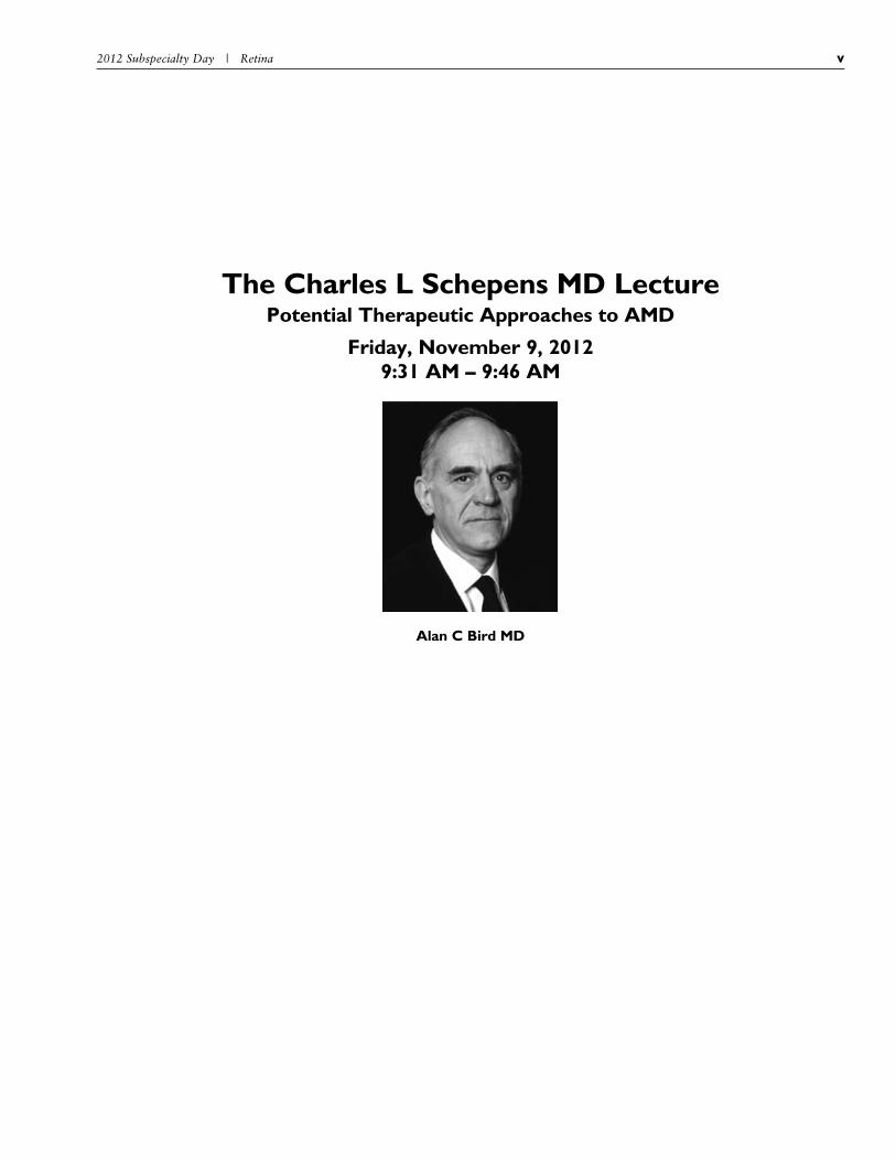
The Charles L Schepens MD LecturePotential Therapeutic Approaches to AMD
Friday, November 9, 20129:31 AM – 9:46 AM
Alan C Bird MD
vi 2012 Subspecialty Day | Retina
Thomas M Aaberg Jr MDAda, MI Founder and PresidentRetina Specialists of MichiganAssistant Clinical Professor of
OphthalmologyMichigan State University
Gary W Abrams MDDetroit, MI Professor of OphthalmologyKresge Eye InstituteWayne State University
David H Abramson MD FACSNew York, NY Chief, Ophthalmic Oncology ServiceMemorial Sloan-Kettering Cancer CenterProfessor of OphthalmologyWeill Cornell University
Lloyd P Aiello MD PhDBoston, MA Professor of OphthalmologyHarvard Medical SchoolHead, Section of Eye Research, andDirector, Beetham Eye InstituteJoslin Diabetes Center
Arthur W Allen Jr MDSan Francisco, CA Vice Chairman Department of OphthalmologyCalifornia Pacific Medical CenterPresidentPacific Eye Associates
J Fernando Arevalo MD FACSRiyadh, Saudi ArabiaProfessor of OphthalmologyWilmer Eye Institute and Johns Hopkins
University School of MedicineChief of Vitreoretina DivisionThe King Khaled Eye Specialist HospitalRiyadh, Kingdom of Saudi Arabia
Jorge G Arroyo MDBrookline, MA Associate Professor of OphthalmologyHarvard Medical SchoolDirector of Retina ServiceBeth Israel Deaconess Medical Center
Marcos P Avila MDGoiania, BrazilFull Professor of Ophthalmology andHead of the Ophthalmology DepartmentUniversidade Federal de Goiás
Carl C Awh MDNashville, TN PresidentTennessee Retina, PC
Faculty

106 Section X: Oncology 2012 Subspecialty Day | Retina
Practical Approaches to Needle Biopsy and Genetic Diagnosis for Ocular MelanomaThomas M Aaberg JR MD
I. Brachytherapy for Uveal Melanoma
A. Performed first by R Foster Moore on February 15, 1929
B. Local control of uveal melanoma now approaches 100% success.
C. 80 years after first brachytherapy case, patient sur-vival rates remain unchanged.
II. Micrometastatic disease occurs early in specific cases.
A. How do we identify these cases?
1. Improve our detection methods, or
2. Identify characteristics of tumors at greatest risk of metastasizing early.
B. What do we do once identified?
1. More aggressive metastatic disease surveillance, and/or
2. Initiate prophylactic interventions in the hopes of eradicating or at least controlling micrometa-static disease.
III. Identifying High-risk Patients
A. Clinical finding
1. Tumor size
2. Location
3. Cell type (epithelioid vs. spindle)
4. These characteristics do not sufficiently differen-tiate those at greatest risk from those at lowest risk.
B. Molecular diagnostics
1. Improved our understanding of melanoma
2. Improved our characterization of the tumor’s DNA/RNA makeup
3. Improving the identification of those patients at high risk for developing metastatic disease
4. Currently, the two most accepted methods
a. Measuring chromosomal gains and losses (chromosomal analysis)
b. Measuring the expression of specific genes by neoplastic cells (gene expression profiling)
C. Chromosome analysis
1. The most common statistically independent prognostic chromosomal expression associated with metastatic disease is monosomy Chromo-some 3, and less frequently gain of 8q.
2. Chromosomal analysis can be performed at most hospital laboratories via a number of methods:
a. SNP assay across the chromosome to assess for loss of heterozygosity
b. Fluorescence in situ hybridization
c. Array-based comparative genomic hybridiza-tion
3. Technical yield ranges from 50% to 91%. Greater tissue requirements compared to GEP
D. Gene expression profiling (GEP)
1. Proprietary PCR testing on a microfluidic plat-form
2. Castle laboratories.
3. Differentiates patients into 3 catagories: class 1a, class 1b, and class 2
4. Technical yield is 97%; utilizes preamplification and microfluidics
IV. Tissue procurement is necessary for cytogenetic and GEP testing.
A. Enucleated eyes: An “eye cap” (a transverse section through the eye to include a portion of the tumor) will provide ample tissue for chromosomal and GEP analysis.
B. Nonenucleated eyes
1. Require minimally invasive technics, such as fine needle aspiration biopsy
2. Excellent cytopathologist
V. Fine Needle Aspiration Biopsy (FNAB) of Uveal Melanoma
A. Techniques include:
1. Transscleral full thickness
2. Transscleral via a scleral flap
3. Transvitreal
B. Transscleral biopsy
1. Typically performed at the time of radiation plaque placement or placement of titanium but-tons for tumor localization and subsequent pro-ton beam irradiation
2. Equipment
a. Basic surgical tray (ie, Westcott scissors, tooth forceps, needle driver, etc.)
b. 27- or 25-gauge 5/8th-inch needle
c. 10 cc syringe

2012 Subspecialty Day | Retina Section X: Oncology 107
d. Length of tubing with appropriate male and female connectors
e. Optional: tissue glue, absolute alcohol
3. Technique
a. Create conjunctival flap.
b. Isolate 2 or more extraocular muscles on bridle sutures to provide stabilization of the globe.
c. Trans-illuminate the globe and mark out the trans-illumination defect.
d. Connect the needle to the syringe directly or via a length of tubing.
e. Make sure the biopsy field is completely dry.
f. It is necessary to know the thickness of the tumor and the thickest point of the tumor in order to minimize the risk of perforating the overlying retina.
i. Consider marking the needle with a mark-ing pen or small steri-strip to provide a “depth gauge.”
ii. In thin tumors, angling the needle may be wise as this improves the self-sealing nature of the needle pass, and decreases the risk of passing full thickness through the tumor.
iii. When angling the needle, the needle is introduced into the eye with the bevel out. Once in the eye beyond the bevel, rotate the needle 180 degrees, exposing the ostomy of the needle to the choroid and away from the sclera.
g. A sawing motion under active suction has improved my cell yield.
h. Release suction before exiting the eye.
i. Pass off the syringe
j. Immediately cover the site with a plaque, tis-sue glue, or absolute alcohol on a cotton tip applicator.
k. Process the specimen promptly.
i. Follow local laboratory protocol for tissue processing.
ii. Utilize cell prep solution provided by Castle.
(a) Detach needle and/or tubing from syringe.
(b) Draw air into syringe.
(c) Reattach needle and/or tubing to syringe.
(d) Forcefully express the air into the pro-vided tube.
(e) Aspirate the entire cell solution pro-vided. I “wash” the needle with the solution gently.
(f) Inject the wash into the designated tube.
C. Transvitreal biopsy
1. Indirect ophthalmoscopy
a. A 25-gauge 1.5-inch needle is connected to a 10 cc syringe via an intervening length of tub-ing.
b. Pass the needle through the pars plana.
c. Cross the vitreous cavity.
d. Enter the tumor at the apex.
e. Ten cc’s of suction is applied. If possible, a sawing motion is performed; however, when performing a transvitreal biopsy one must be exceedingly careful to have the ostomy of the needle well within the tumor and avoid aspi-rating the retina.
f. If retina is aspirated, release vacuum and push the needle back into the tumor, attempting to pull the retina out of the needle ostomy. Extract the needle slowly.
i. If the retina remains, place the needle back into the tumor and have the assistant pro-vide a very minute amount of reflux.
ii. Do not reflux within the vitreous cavity, as you may seed the vitreous.
g. Apply a digit or cotton tip applicator to the biopsy entry site to control intraocular hem-orrhage.
2. Via the operating room microscope using a chan-delier system and infusion.
a. Place a chandelier infusion system.
i. Visualization
ii. IOP control for hemostasis.
b. Place a second trocar through which the biopsy needle or other necessary instrumenta-tion will pass.
i. Diathermy
ii. Soft tip cannula (enables the delivery of thrombin)
D. Complications
1. Rhegmatogenous detachments: Surprisingly low incidence
a. Fibrin from blood likely closes the break.
b. The tumor acts as a tamponade (ie, internal buckle).
2. Subretinal hemorrhage: Often limited and resolves within 4 months

108 Section X: Oncology 2012 Subspecialty Day | Retina
3. Vitreous hemorrhage: Often limited and resolves within 4 months
4. Misdiagnosis caused by sampling errors
a. Tumors have heterogeneous distribution of monosomy 3 abnormality
b. GEP may be less affected by heterogeneous cell distribution since this is an assay of the tumor’s microenvironment.
5. Tumor spread or seeding along needle track
a. Needle biopsy of breast masses is one of the most widely accepted uses of FNAB.
b. Several studies have looked at the risk of seed-ing tumor cells following breast biopsy.
i. Tumor seeding incidence is approximately 1%.
ii. However, as the interval between biopsy and surgery lengthens, the incidence of seeding declines, suggesting that displaced tumor cells are not necessarily viable.
c. Data for FNAB in ophthalmology are less plentiful.
i. Foos (1988) and colleagues reported on 22 FNABs performed on enucleated eyes.
(a) Cells in 67% of all transscleral tracks
(b) Cells in 53% of transvitreal tracks
(c) However, the number of tumor cells was less than that associated with tumor growth in experimental models.
ii. Shields (2007) reported on 140 patients who underwent FNAB immediately prior to I125 brachytherapy. No cases of tumor recurrence along the biopsy track occurred.
iii. McCannel (2012) reported on 170 con-secutive patients with choroidal melanoma managed with FNAB and I125 brachy-therapy and 1-6 years of follow-up (mean 2.7 years).
(a) No cases of orbital dissemination occurred.
(b) Metastatic rate was 13% (compa-rable to the 13% rate reported by the COMS), suggesting FNAB did not worsen the prognosis for metastatic disease.
E. Billing/reimbursement
CPT code: 67415 = fine needle aspiration of orbital contents
F. Contraindications to FNAB
1. Children in which retinoblastoma is in the differ-ential diagnosis
2. No cytopathologist available
3. Inadequate visualization of the tumor
VI. Survey of Ocular Oncologists
109 ocular oncologists worldwide were asked to par-ticipate in the following survey questions. There were 50 respondents (46% response rate).
A. Do you perform some type of analysis that requires a FNAB? (see Figure 1)
B. How many uveal melanoma patients are not eligible for biopsy due to safety concerns?
1. Mean: 20.92%
2. Median: 10%
3. Range: 0%-100%
C. For eligible patients do you perform analysis for:
1. Enucleated eyes only: 13.5%
2. All cases: 86.5%
D. For what percent of uveal melanoma cases do you offer cytogenic testing? (see Figure 2)
E. For what percent of uveal melanoma cases do you offer molecular testing? (see Figure 3)
F. How many biopsies for uveal melanoma do you perform annually?
1. Mean: 27.41 biopsies
2. Median: 15 biopsies
3. Range: 0-250 biopsies
VII. Survey: Technique
A. Vitrector for biopsy (see Figure 4)
B. Size of needle gauge for biopsy (see Figure 5)
C. Flexible tubing to stabilize the needle position (see Figure 6)
D. Size of syringe (see Figure 7)
E. Scleral window or full-thickness sclera (see Figure 8)
F. What percent of biopsies are performed transscler-ally?
1. Mean: 70.31%
2. Range: 0%-100%
G. What percent of biopsies are performed transvit-really?
1. Mean: 29.69%
2. Range: 0%-100%)
H. If you perform both transscleral and transvitreal biopsies, what factors contribute to your decision process?
1. Tumor location
a. Anterior tumors = transscleral
b. Posterior tumors = transvitreal

2012 Subspecialty Day | Retina Section X: Oncology 109
Figure 2.
Figure 1.
Figure 3.
Figure 4.

110 Section X: Oncology 2012 Subspecialty Day | Retina
Figure 6.
Figure 5.
Figure 7.
Figure 8.

2012 Subspecialty Day | Retina Section X: Oncology 111
2. Tumor thickness
a. Tumors less than 1.5-2.0 mm = transvitreal
b. Tumors greater than 1.5-2.0 mm = trans-scleral
Table 1. Of those who do not do both transscleral and transvitreal:
Response Percentage Frequency
Only do transcleral 25.7% 9
Only do transvitreal 2.9% 1
I. After the biopsy is performed do you “seal” the biopsy site?
1. Yes = 54%
2. No = 46%
J. If so, with what?
1. Suture
2. Cryo
3. Glue: Isodent, Dermabond, Cyanoacrylate
K. How is the biopsy performed? (see Figure 9)
L. How many tumor sites? (see Figure 10)
M. Which fixative? (see Figure 12)
Figure 10.
Figure 9.
Figure 11.
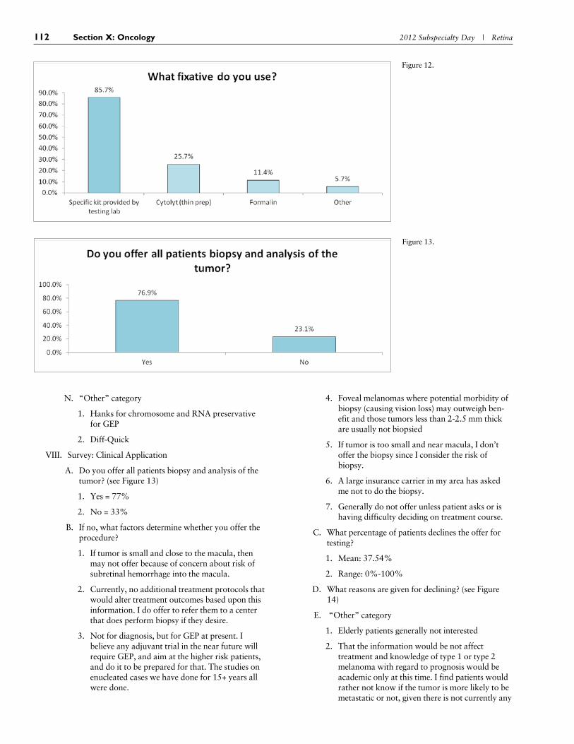
112 Section X: Oncology 2012 Subspecialty Day | Retina
N. “Other” category
1. Hanks for chromosome and RNA preservative for GEP
2. Diff-Quick
VIII. Survey: Clinical Application
A. Do you offer all patients biopsy and analysis of the tumor? (see Figure 13)
1. Yes = 77%
2. No = 33%
B. If no, what factors determine whether you offer the procedure?
1. If tumor is small and close to the macula, then may not offer because of concern about risk of subretinal hemorrhage into the macula.
2. Currently, no additional treatment protocols that would alter treatment outcomes based upon this information. I do offer to refer them to a center that does perform biopsy if they desire.
3. Not for diagnosis, but for GEP at present. I believe any adjuvant trial in the near future will require GEP, and aim at the higher risk patients, and do it to be prepared for that. The studies on enucleated cases we have done for 15+ years all were done.
4. Foveal melanomas where potential morbidity of biopsy (causing vision loss) may outweigh ben-efit and those tumors less than 2-2.5 mm thick are usually not biopsied
5. If tumor is too small and near macula, I don’t offer the biopsy since I consider the risk of biopsy.
6. A large insurance carrier in my area has asked me not to do the biopsy.
7. Generally do not offer unless patient asks or is having difficulty deciding on treatment course.
C. What percentage of patients declines the offer for testing?
1. Mean: 37.54%
2. Range: 0%-100%
D. What reasons are given for declining? (see Figure 14)
E. “Other” category
1. Elderly patients generally not interested
2. That the information would be not affect treatment and knowledge of type 1 or type 2 melanoma with regard to prognosis would be academic only at this time. I find patients would rather not know if the tumor is more likely to be metastatic or not, given there is not currently any
Figure 13.
Figure 12.

2012 Subspecialty Day | Retina Section X: Oncology 113
additional intervention that could help them if this were found to be the case.
3. Some decline to know, or have the information documented.
4. Irrational concern that biopsy will spread tumor cells and thereby worsen prognosis
5. No effective treatment
6. Age and health of the patient
7. They are made aware of the potential risks and possible benefits. They typically choose to avoid the risks.
8. Potential risks of hemorrhage, RD, or ESE
9. The knowledge will not prevent metastasis
10. Once they learn that there is no proven advan-tage in terms of longevity with intensive moni-toring, most are not compelled to seek genetic information.
F. How do you use the information clinically? (see Fig-ure 15)
G. Write-in responses
1. I generally have the patient see a medical oncolo-gist who specializes in melanoma; they generally increase metastatic surveillance, and have access to clinical trials. I specifically as the ophthalmol-ogist do not change my management.
2. I refer high-risk class II patients to an oncologist with experience with metastatic choroidal mela-
noma for their ongoing metastatic surveillance. They use a more frequent surveillance protocol, with more frequent imaging (liver ultrasound).
3. This is difficult at present! There are not great trials, metastatic surveillance is not shown to have survival benefit, and we are building this bridge as we walk on it. To my knowledge there isn’t a good prophylaxis yet either. But gathering the data to analyze, and stratify as soon as avail-able is essential.
4. At present, there is (in my opinion) no legitimate clinical trial of any adjuvant therapy for uveal melanoma. At present, there is also no compel-ling clinical evidence of any regimen or frquency of surveillance testing for uveal melanoma metastasis improves patient survival. In spite of this, I inform patients that they appear (based on evidence provided by the testing of their FNAB aspirates) to have low (GEP class 1A), relatively low (class 1B), or relatively high risk of develop-ing clinically apparent distant metatases in the future. I advise patients in all three groups about the current lack of compelling evidence about survival benefits of any method of treatment for uveal melanoma metastasis and the concurrent lack of evidence that any regimen or frequency of surveillance testing for metastasis changes their prognosis. At the same time, I inform them that an adjuvant therapy clinical trial based on legiti-mate preliminary evidence (not hype) may induce me to contact them as candidates for enrollment.
Figure 14.
Figure 15.

114 Section X: Oncology 2012 Subspecialty Day | Retina
5. More likely to utilize imaging as part of surveil-lance in class 2 patients vs. those with a class 1 profile.
6. The chromosome 3 work is iffy and the more recent RNA work has not been validated by another center. The results are not yet conclusive enough to change clinical management, in my opinion.
Selected Readings 1. Moore RF. Choroidal sarcoma treated by the intraocular insertion
of radon seeds. Br J Ophthalmol. 1930; 14(4):145-152.
2. Henkes HE, Manschott WA. The danger of diagnostic biopsy in eyes suspected of an intraocular tumour. Ophthalmologica 1963; 145:467-469.
3. Reese AB. Tumors of the Eye. 3 ed. Hagerstown, MD: Harper & Row; 1976.
4. Augsburger JJ, Shields JA. Fine-needle aspiration biopsy of solid intraocular tumors; indications, instrumentation and techniques. Ophthalmic Surg. 1984; 15:34-40.
5. Glasgo BJ, Brown HH, Zargoza AM, Foos RY. Quantitation of tumor seeding from fine needle aspiration of ocular melanomas. Am J Ophthalmol. 1988; 105:538-546.
6. Shields CL, Ganguly A, Materin MA, et al. Chromosome 3 analysis of uveal melanoma using fine-needle aspiration biopsy at the time of plaque radiotherapy in 140 consecutive cases: the Deborah Iver-son, MD, Lectureship. Arch Ophthalmol. 2007; 125(8):1017-1024.
7. Maat W, Jordanova ES, van Zelderen-Bhola SL, et al. The hetero-geneous distribution of monosomy 3 in uveal melanomas: implica-tions for prognostication based on fine needle aspiration biopsies. Arch Pathol Lab Med. 2007; 131:91-96.
8. Young TA, Burgess BL, Rao NP, Glasgow BJ, Straatsma BR. Trans-scleral fine-needle aspiration biopsy of macular choroidal mela-noma. Am J Ophthalmol. 2008; 145(2):297-302.
9. Schoenfield L, Pettay J, Tubbs RR, Singh AD. Variation of mono-somy 3 status within uveal melanoma. Arch Pathol Lab Med. 2009; 133:1219-1222.
10. Shields CL, Ganguly A, Bianciotto CG, Turaka K, Tavallali A, Shields JA. Prognosis of uveal melanoma in 500 cases using genetic testing of fine-needle aspiration biopsy specimens. Ophthalmology 2011; 118(2):396-401.
11. Loughran CF, Keeling CR. Seeding of tumour cells following breast biopsy: a literature review. Br J Radiol. 2011; 84(1006):869-874.
12. Robertson EG, Baxter G. Tumour seeding following percutaneous needle biopsy: the real story! Clin Radiol. 2011; 66(11):1007-1014.
13. Brenner RJ, Gordon LM. Malignant seeding following percutane-ous breast biopsy: documentation with comprehensive imaging and clinical implications. Breast J. 2011; 17(6):651-656.
Related Documents