Author's personal copy Taxonomy/Taxinomie Resurrection of New Caledonian maskray Neotrygon trigonoides (Myliobatoidei: Dasyatidae) from synonymy with N. kuhlii, based on cytochrome-oxidase I gene sequences and spotting patterns Re ´surrection de la raie pastenague de Nouvelle-Cale ´donie Neotrygon trigonoides (Myliobatoidei: Dasyatidae) de la synonymie avec N. kuhlii, sur la base des se ´quences du ge `ne de la cytochrome-oxydase I et des patterns de taches Philippe Borsa a, *, Irma S. Arlyza b , Wei-Jen Chen c , Jean-Dominique Durand d , Mark G. Meekan e , Kang-Ning Shen f,g a Institut de recherche pour le de ´veloppement (IRD), UR 227 « Biocomplexite ´ des e ´cosyste `mes re ´cifaux », 911, avenue Agropolis, 34394 Montpellier, France b Lembaga Ilmu Pengetahuan Indonesia (LIPI), Pusat Penelitihan Oseanografi (P2O), Ancol, Jakarta, Indonesia c Marine Phylogenomics Laboratory, Oceanography Institute, National Taiwan University, Taipei, Taiwan d IRD, UMR 5119 Ecosym, Universite ´ Montpellier 2, Montpellier, France e Australian Institute of Marine Science, Perth, Australia f Department of Environmental Biology and Fisheries Science, National Taiwan Ocean University, Keelung, Taiwan g Center of Excellence for Marine Bioenvironment and Biotechnology, National Taiwan Ocean University, Keelung, Taiwan C. R. Biologies 336 (2013) 221–232 A R T I C L E I N F O Article history: Received 21 December 2012 Accepted after revision 11 May 2013 Keywords: Indo-West Pacific Coral Triangle Coral Sea New Caledonia Molecular systematics A B S T R A C T The maskray from New Caledonia, Neotrygon trigonoides Castelnau, 1873, has been recently synonymized with the blue-spotted maskray, N. kuhlii (Mu ¨ ller and Henle, 1841), a species with wide Indo-West Pacific distribution, but the reasons for this are unclear. Blue- spotted maskray specimens were collected from the Indian Ocean (Tanzania, Sumatra) and the Coral Triangle (Indonesia, Taiwan, and West Papua), and N. trigonoides specimens were collected from New Caledonia (Coral-Sea). Their partial COI gene sequences were generated to expand the available DNA-barcode database on this species, which currently comprises homologous sequences from Ningaloo Reef, the Coral Triangle and the Great Barrier Reef (Coral-Sea). Spotting patterns were also compared across regions. Haplotypes from the Coral-Sea formed a haplogroup phylogenetically distinct from all other haplotypes sampled in the Indo-West Pacific. No clear-cut geographic composition relative to DNA-barcodes or spotting patterns was apparent in N. kuhlii samples across the Indian Ocean and the Coral Triangle. The New Caledonian maskray had spotting patterns markedly different from all the other samples. This, added to a substantial level of net * Corresponding author. IR UR 227 c/o Indonesian Biodiversity Research Center, Universitas Udayana, Jl Sesetan Gang Markisa 6, Denpasar, Indonesia. E-mail address: [email protected] (P. Borsa). Contents lists available at SciVerse ScienceDirect Comptes Rendus Biologies ww w.s cien c edir ec t.c om 1631-0691/$ – see front matter ß 2013 Published by Elsevier Masson SAS on behalf of Acade ´ mie des sciences. http://dx.doi.org/10.1016/j.crvi.2013.05.005

Welcome message from author
This document is posted to help you gain knowledge. Please leave a comment to let me know what you think about it! Share it to your friends and learn new things together.
Transcript
Author's personal copy
Taxonomy/Taxinomie
Resurrection of New Caledonian maskray Neotrygontrigonoides (Myliobatoidei: Dasyatidae) from synonymy withN. kuhlii, based on cytochrome-oxidase I gene sequences andspotting patterns
Resurrection de la raie pastenague de Nouvelle-Caledonie Neotrygon
trigonoides (Myliobatoidei: Dasyatidae) de la synonymie avec N. kuhlii,
sur la base des sequences du gene de la cytochrome-oxydase I et des
patterns de taches
Philippe Borsa a,*, Irma S. Arlyza b, Wei-Jen Chen c, Jean-Dominique Durand d,Mark G. Meekan e, Kang-Ning Shen f,g
a Institut de recherche pour le developpement (IRD), UR 227 « Biocomplexite des ecosystemes recifaux », 911, avenue Agropolis, 34394
Montpellier, Franceb Lembaga Ilmu Pengetahuan Indonesia (LIPI), Pusat Penelitihan Oseanografi (P2O), Ancol, Jakarta, Indonesiac Marine Phylogenomics Laboratory, Oceanography Institute, National Taiwan University, Taipei, Taiwand IRD, UMR 5119 Ecosym, Universite Montpellier 2, Montpellier, Francee Australian Institute of Marine Science, Perth, Australiaf Department of Environmental Biology and Fisheries Science, National Taiwan Ocean University, Keelung, Taiwang Center of Excellence for Marine Bioenvironment and Biotechnology, National Taiwan Ocean University, Keelung, Taiwan
C. R. Biologies 336 (2013) 221–232
A R T I C L E I N F O
Article history:
Received 21 December 2012
Accepted after revision 11 May 2013
Keywords:
Indo-West Pacific
Coral Triangle
Coral Sea
New Caledonia
Molecular systematics
A B S T R A C T
The maskray from New Caledonia, Neotrygon trigonoides Castelnau, 1873, has been
recently synonymized with the blue-spotted maskray, N. kuhlii (Muller and Henle, 1841), a
species with wide Indo-West Pacific distribution, but the reasons for this are unclear. Blue-
spotted maskray specimens were collected from the Indian Ocean (Tanzania, Sumatra) and
the Coral Triangle (Indonesia, Taiwan, and West Papua), and N. trigonoides specimens were
collected from New Caledonia (Coral-Sea). Their partial COI gene sequences were
generated to expand the available DNA-barcode database on this species, which currently
comprises homologous sequences from Ningaloo Reef, the Coral Triangle and the Great
Barrier Reef (Coral-Sea). Spotting patterns were also compared across regions. Haplotypes
from the Coral-Sea formed a haplogroup phylogenetically distinct from all other
haplotypes sampled in the Indo-West Pacific. No clear-cut geographic composition
relative to DNA-barcodes or spotting patterns was apparent in N. kuhlii samples across the
Indian Ocean and the Coral Triangle. The New Caledonian maskray had spotting patterns
markedly different from all the other samples. This, added to a substantial level of net
* Corresponding author. IR UR 227 c/o Indonesian Biodiversity Research Center, Universitas Udayana, Jl Sesetan Gang Markisa 6, Denpasar, Indonesia.
E-mail address: [email protected] (P. Borsa).
Contents lists available at SciVerse ScienceDirect
Comptes Rendus Biologies
ww w.s c ien c edi r ec t . c om
1631-0691/$ – see front matter � 2013 Published by Elsevier Masson SAS on behalf of Academie des sciences.
http://dx.doi.org/10.1016/j.crvi.2013.05.005
Author's personal copy
1. Introduction
Recent advances in the molecular phylogenetics ofstingrays (Myliobatoidei: [1]) have challenged the pre-viously accepted taxonomy of species in a number ofgenera [2–6]. Both a cytochrome-oxidase I gene (COI)-basedbarcoding survey of Australasian chondrichthyans [7], arecent COI-barcoding survey of northwestern Australianmyliobatoids [8] and a recent systematic survey ofelasmobranchs based on the nicotinamide-adenine dinu-cleotide dehydrogenase subunit two gene marker [3] havepointed to problematic cases relative to the definition ofspecies boundaries, when divergent haplotypes areobserved within a given nominal species. One of thesecases is the blue-spotted maskray, Neotrygon kuhlii (Mullerand Henle, 1841) [9].
The blue-spotted maskray under its current taxonomicdefinition has a wide Indo-Pacific distribution, from theRed Sea to southern Africa and from the western IndianOcean to the western Pacific, reaching Japan in the North,Tonga in the South-East and New Caledonia in the South[10]. It is currently the most frequently landed stingrayspecies in the Coral Triangle [11], hence it is a species ofcommercial interest but also one of conservation concerngiven the general vulnerability of elasmobranchs to fishingpressure [12,13]. N. kuhlii shows unusually high within-species diversity, with average nucleotide distances amonghaplotypes reaching 2.8%–3.0% at the COI locus [7,8]. Anintron-marker based diversity study has confirmed theremarkably high degree of genetic differentiation amongpopulations, but has failed to detect multiple crypticspecies within Coral Triangle N. kuhlii [14]. One study [8]
reported 3.5% nucleotide divergence at the COI locusbetween a sample from the Great Barrier Reef and samplesfrom northwestern Australia and the Coral Triangle. Arecent phylogeographic survey of Neotrygon spp. [15] hasconfirmed this preliminary result. Blue-spotted maskrayfrom the Coral-Sea deserve particular scrutiny as argued inthe following.
The New Caledonian maskray, N. trigonoides Castelnau,1873 [16] is currently considered as a junior synonym ofN. kuhlii [15,17,18]. In his description, F. de Castelnau [16]noted a large number of small black spots on the dorsalsurface of the stingray whereas in their description ofN. kuhlii, J. Muller and J. Henle [9] only mentioned ‘‘einzene,
kleine, runde blaue Flecken (3-6) auf jeder Brustflosse’’ andprovided a figure of N. kuhlii that illustrates this description(Fig. 1A). The two species, N. kuhlii and N. trigonoides, weresynonymized by P.R. Last and W.T. White [17] whocompared the holotype of Castelnau’s N. trigonoides towhat they identified as N. kuhlii from eastern Australia: ‘‘Aclose examination of the holotype of Raya trigonoides
Castelnau, 1873 from New Caledonia. . . confirmed that it isconspecific with eastern Australian forms of N. kuhlii’’.
In the present work, we re-examine spotting patternsin Castelnau (1873)’s N. trigonoides [16] and comparethem to Muller and Henle (1841)’s N. kuhlii [9] and to blue-spotted maskray specimens from a range of locations inthe Indo-West Pacific. We examine COI gene sequences forsamples of blue-spotted maskray from two distant sites inthe Coral-Sea, and from a number of sites in the IndianOcean and in the Coral Triangle. Finally, we provide aredescription of N. trigonoides based on the COI genesequence.
nucleotide divergence (2.6%) with typical N. kuhlii justifies considering the New
Caledonian maskray as a separate species, for which we propose to resurrect the name
Neotrygon trigonoides.
� 2013 Published by Elsevier Masson SAS on behalf of Academie des sciences.
R E S U M E
La raie pastenague a taches bleues, Neotrygon kuhlii (Muller et Henle, 1841), possede une
large distribution indo-ouest pacifique. Une raie pastenague tachetee de Nouvelle-
Caledonie, Neotrygon trigonoides Castelnau, 1873, a ete recemment placee en synonymie
de N. kuhlii, mais les raisons en sont obscures. Des specimens de raie pastenague a taches
bleues ont ete collectes dans l’ocean Indien (Tanzanie, Sumatra) et le triangle de Corail
(Indonesie, Taiwan et Papouasie occidentale), tandis que des specimens de N. trigonoides
actuellement designes comme N. kuhlii ont ete collectes en Nouvelle-Caledonie (mer de
Corail). Leurs sequences partielles du gene COI ont ete produites pour augmenter la base
de donnees de barcodes disponible sur cette espece, laquelle comprend deja des
sequences homologues de Ningaloo Reef, du triangle de Corail et du recif de la Grande-
Barriere (mer de Corail). Les patterns de taches ont ete egalement analyses sur
l’ensemble des regions echantillonnees. Les haplotypes de la mer de Corail forment un
ensemble phylogenetiquement distinct du reste de l’Indo-Ouest pacifique. Nous n’avons
pas observe de differences geographiques nettes entre les populations de N. kuhlii de
l’ocean Indien et celles du triangle de Corail, que ce soit au niveau des barcodes ADN ou
des patterns de taches. La raie pastenague tachetee de Nouvelle-Caledonie s’avere
posseder des patterns de taches tres differents de ceux de tous les autres specimens. Cela,
ajoute a un niveau de divergence nucleotidique coherent (2,6 %) avec les N. kuhlii
typiques, justifie le statut d’espece a part entiere de la forme presente en Nouvelle-
Caledonie. Nous proposons de retablir le nom N. trigonoides pour cette espece, dont la
distribution semble concerner l’ensemble de la mer de Corail.
� 2013 Publie par Elsevier Masson SAS pour l’Academie des sciences.
Mots cles :
Indo-Ouest
Pacifique
Triangle de Corail
Mer de Corail
Nouvelle-Caledonie
Systematique moleculaire
P. Borsa et al. / C. R. Biologies 336 (2013) 221–232222
Author's personal copy
2. Materials and methods
2.1. Samples
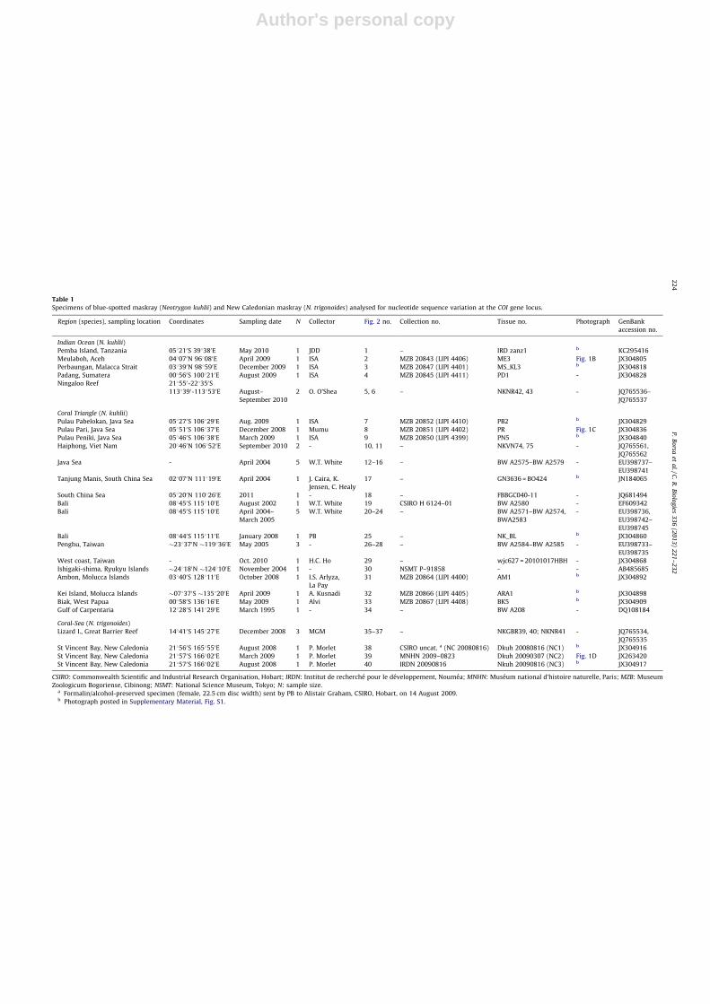
Table 1 lists the blue-spotted maskray individualssampled for DNA and barcoded for the present survey. Thislist includes 13 newly sampled specimens from the IndianOcean (nos. 1–4 in Table 1), the Coral Triangle (nos. 7–9;28; 30–32) and New Caledonia (nos. 37–39). All newlysampled specimens were deposited in zoological collec-tions (Table 1) except no. 1 (from Pemba Island, Tanzania),which was not retained. Table 1 also includes references tosequence data retrieved from GENBANK (http://www.ncbi.nlm.nih.gov/; accessed 25 November 2012) towhich the new sequences were compared.
2.2. Nucleotide sequences
A tissue fragment �0.05 cm3 to �1 cm3 was removed,using surgical scissors, from the pelvic or pectoral fins, orthe tail and was preserved in 95% ethanol at ambienttemperature. DNA extraction was done using either theViogene (Taiwan) tissue genomic DNA extraction protocol,or the DNEasy DNA extraction kit of Qiagen GmbH (Hilden,Germany). DNA was stored in 1X, pH 8.0 TE buffer(AppliChem, Darmstadt, Germany). Polymerase chainreaction (PCR) amplification of a fragment of the COI genewas done in a T-Gradient thermal cycler (Biometra,Gottingen, Germany) using 20 mL reaction mixture withthe following concentrations: 0.05 units/mL Taq DNApolymerase in 2� DFS-Taq Mastermix (Bioron GmbH,Ludwigshafen, Germany), 16 mM (NH4)2SO4, 65 mMTrisHCl, pH 8.8 at 25 8C, 0.01% Tween-20, 2.75 mM MgCl2,
0.8 mM dNTP mix, 0.4 mM of each primer (AITBiotech,Singapore) and 2 mL DNA template. The primers wereFishF1 (50- T C A A C C A A C C A C A A A G A C A T T G G C A C -30) and FishR1 (50- T A G A C T T C T G G G T G G C C A A A G AA T C A -30) [19]. PCR parameters were an initialdenaturation at 94 8C for 2 min, followed by 35 cycles ofheating (94 8C for 1 min), annealing (47 8C for 1 min) andextension (72 8C for 1 min) with a final extension step at72 8C for 10 min.
Individual MNHN 2009-0823 was also PCR-amplifiedfor the complete cytochrome b gene, using primers L14735
(50- A A A A A C C A C C G T T G T T A T T C A A C T A -30) andCB7 (50- C T C C A G T C T T C G G C T T A C A A G -30, slightlymodified from CB6ThrH-15930) [20,21]. The reactionvolume was 15 mL and the reaction mixture contained0.2 mM dNTPs, 1.5 mL 10 � PCR buffer (Bioman, Taipei),0.5 mM each of forward and reverse primer, 0.2 U Taq DNApolymerase (Bioman), and 1.0 mL template DNA. The PCRparameters were initial denaturation at 94 8C for 4 minfollowed by 35 cycles of denaturation (94 8C for 45 s),annealing (48 8C for 1 min), and extension (72 8C for 1 min),and a final extension step at 72 8C for 10 min.
PCR products were visualized on 1% agarose gels andtheir size was estimated as approximately 670 bp. Afterpurification (by isopropanol precipitation), 1 mL 1/8-diluted PCR product was subjected to sequencing reaction,in both forward and reverse directions, using the BigDyeTerminator v3.1 cycle-sequencing kit (Applied Biosystems,Foster City, CA, USA). Cycling conditions were according tothe manufacturer’s protocol. Sequencing reaction productswere cleaned by removing dye-terminator (CleanSEQ kit,Beckman Coulter, Beverly, MA, USA) and loaded onto anABI Prism 3100 DNA sequencer (Beckman Coulter).
2.3. Phylogenetic analysis
The phylogenetic analysis package MEGA5 [22] was usedto estimate nucleotide distance between major clades andnucleotide diversity within. Among the nucleotide sub-stitution models proposed to this effect by MEGA5, the mostlikely according to the Bayesian information criterion wasthe Tamura 3-parameter model [23] (T92) with non-uniform evolutionary rates among sites modelled bydiscrete gamma distribution (+G). Therefore, nucleotidedistance and diversity were estimated according to theT92+G model.
Phylogenetic analyses were done using the Neighbour-joining (NJ) algorithm on T92+G-modelled genetic dis-tances (MEGA5) and partitioned Maximum-likelihood(ML) as implemented in the RAxML-HPC [24] with itsgraphical interface raxmlGUI 0.93 [25]. For the partitionedML search with the mixed model of nucleotide substitu-tion, a GTR+G model (with four discrete rate categories)was used for each partition (respective to codon position)as RAxML only provides GTR [26] -related models of rateheterogeneity for nucleotide data [24]. ML tree search wasdone with 100 separate runs using the default algorithm ofthe program from a random starting tree for each run. Thefinal tree was selected among suboptimal trees in each runby comparing likelihood scores under the GTR+G model.Nodal support was assessed by bootstrapping the original
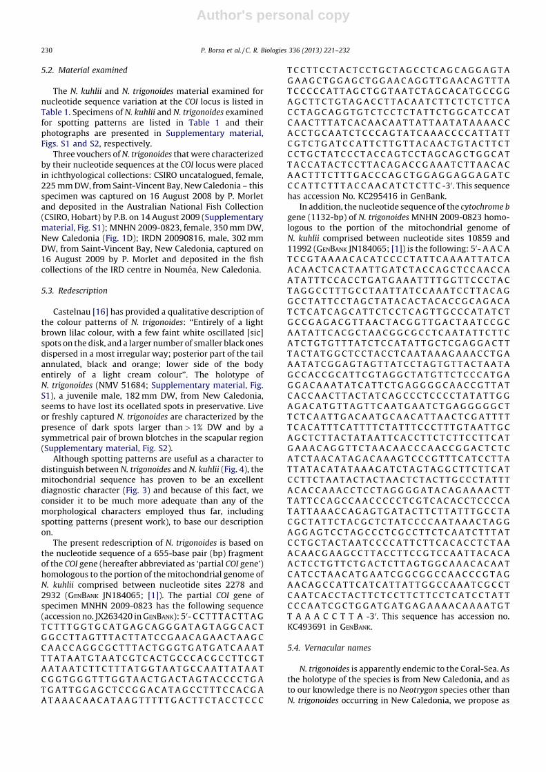
Fig. 1. Specimens of blue-spotted maskrays from various locations
showing variation in pigmentation patterns. A. Original drawing of a
specimen (presumably type material) of Neotrygon kuhlii (pl. 51 in [9]). B.
Specimen MZB 20843 (male N. kuhlii; no. 2 in Table 1) from Meulaboh,
Aceh (048070N 968080E; 28 April 2009; I.S.A.). C. Specimen MZB 20851
(male N. kuhlii, 260 mm DW; no. 8 in Table 1) from Pulau Pari, Java Sea
(058510S 1068370E; 14 December 2008). D. Specimen MNHN 2009-0823
(female N. trigonoides, 350 mm DW; no. 38 in Table 1) from Saint-Vincent
Bay, New Caledonia (218570S 1668020E; 07 March 2009; P.B.).
P. Borsa et al. / C. R. Biologies 336 (2013) 221–232 223
Author's personal copy
Table 1
Specimens of blue-spotted maskray (Neotrygon kuhlii) and New Caledonian maskray (N. trigonoides) analysed for nucleotide sequence variation at the COI gene locus.
Region (species), sampling location Coordinates Sampling date N Collector Fig. 2 no. Collection no. Tissue no. Photograph GenBank
accession no.
Indian Ocean (N. kuhlii)
Pemba Island, Tanzania 058210S 398380E May 2010 1 JDD 1 – IRD zanz1 b KC295416
Meulaboh, Aceh 048070N 968080E April 2009 1 ISA 2 MZB 20843 (LIPI 4406) ME3 Fig. 1B JX304805
Perbaungan, Malacca Strait 038390N 988590E December 2009 1 ISA 3 MZB 20847 (LIPI 4401) MS_KL3 b JX304818
Padang, Sumatera 008560S 1008210E August 2009 1 ISA 4 MZB 20845 (LIPI 4411) PD1 - JX304828
Ningaloo Reef 218550-228350S
1138390-1138530E August–
September 2010
2 O. O’Shea 5, 6 – NKNR42, 43 - JQ765536–
JQ765537
Coral Triangle (N. kuhlii)
Pulau Pabelokan, Java Sea 058270S 1068290E Aug. 2009 1 ISA 7 MZB 20852 (LIPI 4410) PB2 b JX304829
Pulau Pari, Java Sea 058510S 1068370E December 2008 1 Mumu 8 MZB 20851 (LIPI 4402) PR Fig. 1C JX304836
Pulau Peniki, Java Sea 058460S 1068380E March 2009 1 ISA 9 MZB 20850 (LIPI 4399) PN5 b JX304840
Haiphong, Viet Nam 208460N 1068520E September 2010 2 - 10, 11 – NKVN74, 75 - JQ765561,
JQ765562
Java Sea - April 2004 5 W.T. White 12–16 – BW A2575–BW A2579 - EU398737–
EU398741
Tanjung Manis, South China Sea 028070N 1118190E April 2004 1 J. Caira, K.
Jensen, C. Healy
17 – GN3636 = BO424 b JN184065
South China Sea 058200N 1108260E 2011 1 - 18 – FBBGC040-11 - JQ681494
Bali 088450S 1158100E August 2002 1 W.T. White 19 CSIRO H 6124–01 BW A2580 - EF609342
Bali 088450S 1158100E April 2004–
March 2005
5 W.T. White 20–24 – BW A2571–BW A2574,
BWA2583
- EU398736,
EU398742–
EU398745
Bali 088440S 1158110E January 2008 1 PB 25 – NK_BL b JX304860
Penghu, Taiwan �238370N �1198360E May 2005 3 - 26–28 – BW A2584–BW A2585 - EU398733–
EU398735
West coast, Taiwan - Oct. 2010 1 H.C. Ho 29 – wjc627 = 20101017HBH - JX304868
Ishigaki-shima, Ryukyu Islands �248180N �1248100E November 2004 1 - 30 NSMT P–91858 - - AB485685
Ambon, Molucca Islands 038400S 1288110E October 2008 1 I.S. Arlyza,
La Pay
31 MZB 20864 (LIPI 4400) AM1 b JX304892
Kei Island, Molucca Islands �078370S �1358200E April 2009 1 A. Kusnadi 32 MZB 20866 (LIPI 4405) ARA1 b JX304898
Biak, West Papua 008580S 1368160E May 2009 1 Alvi 33 MZB 20867 (LIPI 4408) BK5 b JX304909
Gulf of Carpentaria 128280S 1418290E March 1995 1 - 34 – BW A208 - DQ108184
Coral-Sea (N. trigonoides)
Lizard I., Great Barrier Reef 148410S 1458270E December 2008 3 MGM 35–37 – NKGBR39, 40; NKNR41 - JQ765534,
JQ765535
St Vincent Bay, New Caledonia 218560S 1658550E August 2008 1 P. Morlet 38 CSIRO uncat. a (NC 20080816) Dkuh 20080816 (NC1) b JX304916
St Vincent Bay, New Caledonia 218570S 1668020E March 2009 1 P. Morlet 39 MNHN 2009–0823 Dkuh 20090307 (NC2) Fig. 1D JX263420
St Vincent Bay, New Caledonia 218570S 1668020E August 2008 1 P. Morlet 40 IRDN 20090816 Nkuh 20090816 (NC3) b JX304917
CSIRO: Commonwealth Scientific and Industrial Research Organisation, Hobart; IRDN: Institut de recherche pour le developpement, Noumea; MNHN: Museum national d’histoire naturelle, Paris; MZB: Museum
Zoologicum Bogoriense, Cibinong; NSMT: National Science Museum, Tokyo; N: sample size.a Formalin/alcohol-preserved specimen (female, 22.5 cm disc width) sent by PB to Alistair Graham, CSIRO, Hobart, on 14 August 2009.b Photograph posted in Supplementary Material, Fig. S1.
P.
Bo
rsa
et
al.
/
C.
R.
Bio
log
ies
33
6
(20
13
)
22
1–
23
22
24
Author's personal copy
matrix of sequences [27] with the NJ algorithm and MLcriterion, based on 1000 pseudo-replicates.
2.4. Spotting patterns
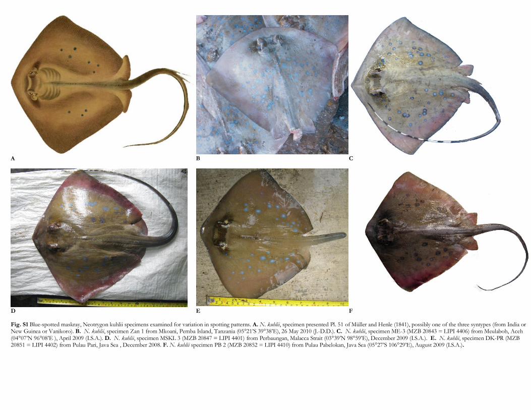
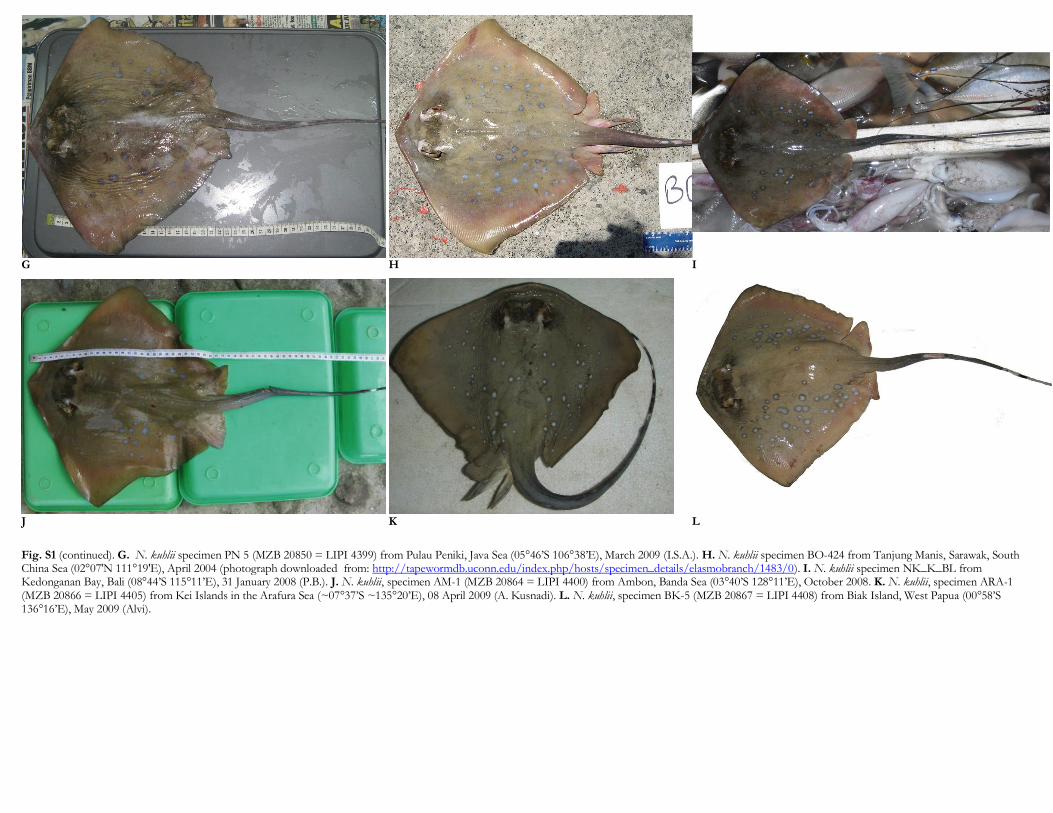
The sample of individuals chosen for studying spottingpatterns included the holotype of N. trigonoides (NMMV51684), the specimen represented Plate 51 of [9](presumably one of the three syntypes of N. kuhlii), andthose specimens analysed at the COI locus of which wepossessed a photograph of quality suitable to counting andsizing all spots present on the dorsal surface of the disk(Fig. 1 and Supplementary material, Figs. S1 and S2).
The number of ocellated spots on the dorsal side of anindividual was counted and their diameter was measuredrelative to disk width (DW). The maximal diameter of anocellated spot was then assigned to one of three classeschosen arbitrarily: small, when diameter � 2% DW; med-
ium, when diameter was comprised between 2% and 4%DW; and large, when diameter > 4% DW. Dark-brown orblack spots (> 1% DW) and speckles (� 1% DW) on the dorsalside of disk were also counted. Dark spots and speckleslocated in the mask area were not counted, because thequality of some of the pictures was not good enough todistinguish them from the brown background in this partof the head (Supplementary material, Figs. S1 and S2). Thesymmetrical pair of brown blotches in the scapular regionwas encoded 0 (when absent), 1 (when visible) or 2 (when
conspicuous). Spotting patterns were compared amongindividuals through correspondence analysis (CA) [28]. CAwas run using the FACTOMINER package [29] under R [30].Only dark spots and speckles are clearly visible onspecimen NMV 51684. The ocellated white spots men-tioned by Castelnau [16] have apparently faded inpreservative, as noted previously [17]. Because its spottingpatterns were characterized by three descriptor variablesonly (number of black speckles, number of black dots andpresence/absence of brown scapular blotch), specimenNMV 51684 was included as active element in a second CArun based on these three descriptor variables.
3. Results
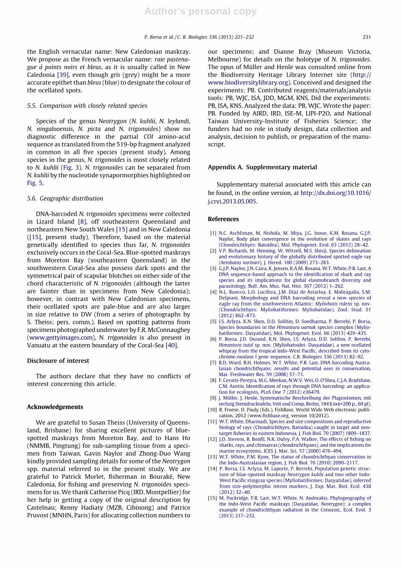
The NJ tree of COI haplotypes (Fig. 3) showed a maindichotomy separating maskrays from the Coral-Sea (north-ern Great Barrier Reef, New Caledonia) from all the otherblue-spotted maskray sampled in the Indo-West Pacific.The net nucleotide divergence between the two clades was0.026 whereas the nucleotide diversity within clades was0.003 and 0.027, respectively. By comparison, the netnucleotide distance between N. leylandi and N. picta was0.031. The ML tree constructed on the basis of the GTR+Gmodel yielded a slightly different topology (not shown),where the Coral-Sea maskray haplotypes formed ahaplogroup external to N. kuhlii, but whose monophylywas not supported enough statistically. In all other aspects,
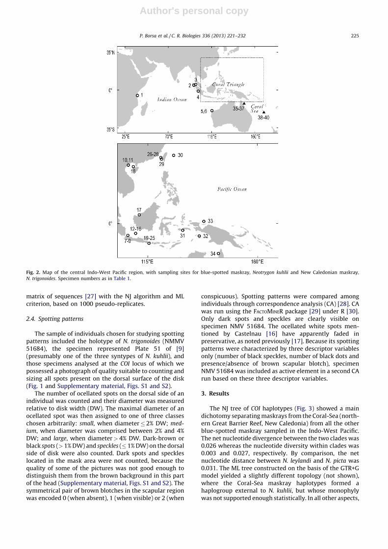
Fig. 2. Map of the central Indo-West Pacific region, with sampling sites for blue-spotted maskray, Neotrygon kuhlii and New Caledonian maskray,
N. trigonoides. Specimen numbers as in Table 1.
P. Borsa et al. / C. R. Biologies 336 (2013) 221–232 225
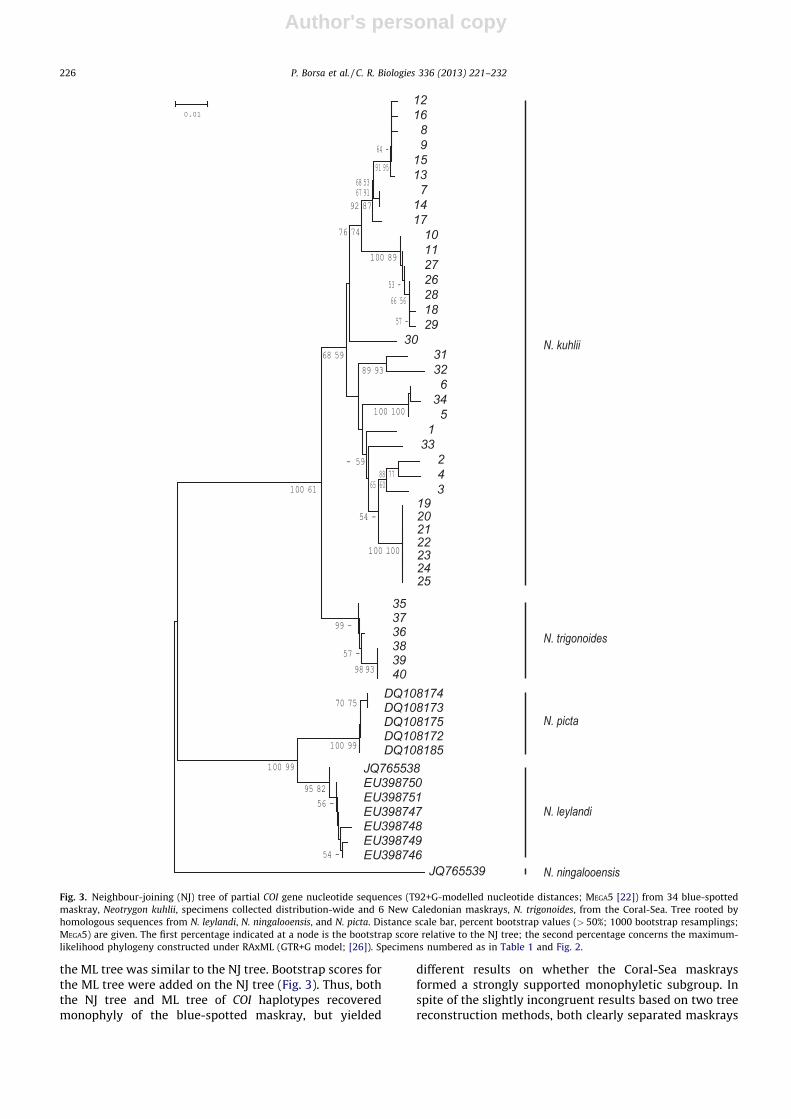
Author's personal copy
the ML tree was similar to the NJ tree. Bootstrap scores forthe ML tree were added on the NJ tree (Fig. 3). Thus, boththe NJ tree and ML tree of COI haplotypes recoveredmonophyly of the blue-spotted maskray, but yielded
different results on whether the Coral-Sea maskraysformed a strongly supported monophyletic subgroup. Inspite of the slightly incongruent results based on two treereconstruction methods, both clearly separated maskrays
30
12168
915137
1417
10112726281829
12645
13
243
19 20
2345
353736 383940
DQ108174DQ108173DQ108175DQ108172DQ108185
JQ765538EU398750EU398751EU398747EU398748EU398749EU398746
JQ765539
21
N. ningalooensis
N. leylandi
N. picta
N. trigonoides
N. kuhlii
Fig. 3. Neighbour-joining (NJ) tree of partial COI gene nucleotide sequences (T92+G-modelled nucleotide distances; MEGA5 [22]) from 34 blue-spotted
maskray, Neotrygon kuhlii, specimens collected distribution-wide and 6 New Caledonian maskrays, N. trigonoides, from the Coral-Sea. Tree rooted by
homologous sequences from N. leylandi, N. ningalooensis, and N. picta. Distance scale bar, percent bootstrap values (> 50%; 1000 bootstrap resamplings;
MEGA5) are given. The first percentage indicated at a node is the bootstrap score relative to the NJ tree; the second percentage concerns the maximum-
likelihood phylogeny constructed under RAxML (GTR+G model; [26]). Specimens numbered as in Table 1 and Fig. 2.
P. Borsa et al. / C. R. Biologies 336 (2013) 221–232226
Author's personal copy
from the Coral-Sea from all the other blue-spottedmaskrays sampled in the Indo-West Pacific.
Spotting patterns of New Caledonian maskray speci-mens were markedly different from all other N. kuhlii
specimens examined (Table 2). Diagnostic differencesincluded the absence of medium or large ocellated spots inNew Caledonian maskray, whereas all other N. kuhlii
specimens possessed a substantial proportion of these;the presence of a few dark spots in all specimens from NewCaledonia, vs. absence in all other N. kuhlii specimensexcept one; and the presence of a pair of brown scapularblotches, exclusively in specimens from New Caledonia.Moreover, ocellated spots were rather pale grey in NewCaledonian maskray specimens vs. pale-blue to blue in allother N. kuhlii specimens (Supplementary material, Figs.S1 and S2). Pictures of blue-spotted maskray takenunderwater at Lizard Island (Supplementary material,Fig. S2), which could not be utilized for the presentmorphological analysis, revealed features similar to thosecharacterizing New Caledonian specimens, i.e. numerousdark speckles, a few dark spots, and the scapular brownblotch.
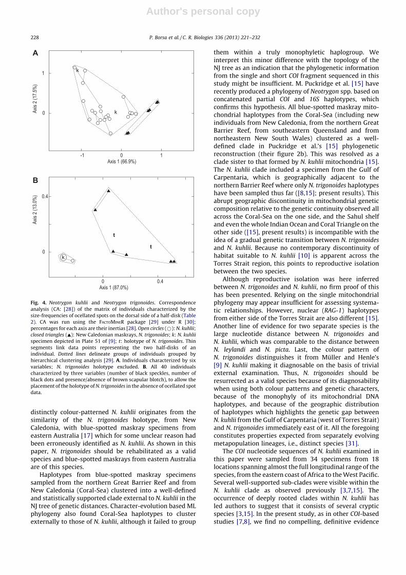
Correspondence analysis of the matrix presented inTable 2 determined two main clusters (Fig. 4A, B). Onecluster exclusively comprised the specimens from NewCaledonia; the analysis placed the holotype ofN. trigonoides within this cluster (Fig. 4B). The othercluster comprised all N. kuhlii specimens from the IndianOcean and the Coral Triangle and also comprised theN. kuhlii specimen referred to by Muller and Henle [9]along their description of the species (Fig. 4A, B).
4. Discussion
Contemporary species concepts are diverse but allrelate to the idea that species more or less can be defined assegments of lineages of the global genealogical networkpertaining to the metapopulation level of organization[31]. Properties of species may include reproductiveisolation from other species, the possession of fixedcharacter state differences, and monophyly [31–34].
Neotrygon trigonoides was originally defined as adistinct species from its unique spotting patterns [16].The reason for declaring N. trigonoides a synonym of the
Table 2
Neotrygon kuhlii and N. trigonoides. Matrix of individuals characterized by the numbers of ocellated spots [sorted into three size-classes: small: � 2% disk
width (DW); medium: > 2% DW and � 4% DW; large: > 4% DW], the number of dark speckles (� 1% DW), the number of dark spots (> 1% DW), and the
absence or presence of a scapular brown blotch (0: absent; 1: visible; 2: conspicuous) on the dorsal surface of left or right half-disk.
Species, individual no. Sampling region Side
of disk
N ocellated spots N dark
speckles
N dark
spots
Scapular
blotch
Fig. 2 no. Specimen no. Small Medium Large
Neotrygon kuhlii
- Pl. 51 of [9] Unknown Left
Right
2
1
1
5
0
0
2
1
0
0
0
0
1 zanz 1 Tanzania Left
Right
44
51
29
21
2
2
10
15
0
0
0
0
2 MZB 20843 Aceh Left
Right
28
29
13
11
0
0
16
16
0
0
0
0
3 MZB 20847 Malacca Strait Left
Right
2
4
14
10
0
1
5
3
0
0
0
0
7 MZB 20852 Java Sea Left
Right
5
9
8
3
1
0
21
17
0
0
0
0
8 MZB 20851 Java Sea Left
Right
22
18
9
6
2
2
12
8
0
0
0
0
9 MZB 20850 Java Sea Left
Right
12
22
12
11
2
0
2
1
0
0
0
0
17 BO424 S. China Sea Left
Right
35
41
22
6
0
0
16
13
0
0
0
0
25 NK BL Bali Left
Right
21
24
6
5
0
0
43
54
0
0
0
0
31 MZB 20864 Ambon Left
Right
9
2
7
12
0
0
5
2
0
0
0
0
32 MZB 20866 Kei Islands Left
Right
36
30
9
9
0
0
53
53
0
1
0
0
33 MZB 20867 Biak Left
Right
41
37
25
13
0
2
10
7
0
0
0
0
Neotrygon trigonoides
- NMV 51684 New Caledonia Left
Right
ND
ND
ND
ND
ND
ND
24
26
4
2
1
1
38 CSIRO uncat. New Caledonia Left
Right
54
38
0
0
0
0
36
25
1
1
2
2
39 MNHN 2009-0823 New Caledonia Left
Right
33
28
0
0
0
0
85
91
10
8
2
2
40 IRDN 20090816 New Caledonia Left
Right
15
16
0
0
0
0
55
57
13
13
2
2
N: number of spots or speckles; ND: no data.
P. Borsa et al. / C. R. Biologies 336 (2013) 221–232 227
Author's personal copy
distinctly colour-patterned N. kuhlii originates from thesimilarity of the N. trigonoides holotype, from NewCaledonia, with blue-spotted maskray specimens fromeastern Australia [17] which for some unclear reason hadbeen erroneously identified as N. kuhlii. As shown in thispaper, N. trigonoides should be rehabilitated as a validspecies and blue-spotted maskrays from eastern Australiaare of this species.
Haplotypes from blue-spotted maskray specimenssampled from the northern Great Barrier Reef and fromNew Caledonia (Coral-Sea) clustered into a well-definedand statistically supported clade external to N. kuhlii in theNJ tree of genetic distances. Character-evolution based MLphylogeny also found Coral-Sea haplotypes to clusterexternally to those of N. kuhlii, although it failed to group
them within a truly monophyletic haplogroup. Weinterpret this minor difference with the topology of theNJ tree as an indication that the phylogenetic informationfrom the single and short COI fragment sequenced in thisstudy might be insufficient. M. Puckridge et al. [15] haverecently produced a phylogeny of Neotrygon spp. based onconcatenated partial COI and 16S haplotypes, whichconfirms this hypothesis. All blue-spotted maskray mito-chondrial haplotypes from the Coral-Sea (including newindividuals from New Caledonia, from the northern GreatBarrier Reef, from southeastern Queensland and fromnortheastern New South Wales) clustered as a well-defined clade in Puckridge et al.’s [15] phylogeneticreconstruction (their figure 2b). This was resolved as aclade sister to that formed by N. kuhlii mitochondria [15].The N. kuhlii clade included a specimen from the Gulf ofCarpentaria, which is geographically adjacent to thenorthern Barrier Reef where only N. trigonoides haplotypeshave been sampled thus far ([8,15]; present results). Thisabrupt geographic discontinuity in mitochondrial geneticcomposition relative to the genetic continuity observed allacross the Coral-Sea on the one side, and the Sahul shelfand even the whole Indian Ocean and Coral Triangle on theother side ([15], present results) is incompatible with theidea of a gradual genetic transition between N. trigonoides
and N. kuhlii. Because no contemporary discontinuity ofhabitat suitable to N. kuhlii [10] is apparent across theTorres Strait region, this points to reproductive isolationbetween the two species.
Although reproductive isolation was here inferredbetween N. trigonoides and N. kuhlii, no firm proof of thishas been presented. Relying on the single mitochondrialphylogeny may appear insufficient for assessing systema-tic relationships. However, nuclear (RAG-1) haplotypesfrom either side of the Torres Strait are also different [15].Another line of evidence for two separate species is thelarge nucleotide distance between N. trigonoides andN. kuhlii, which was comparable to the distance betweenN. leylandi and N. picta. Last, the colour pattern ofN. trigonoides distinguishes it from Muller and Henle’s[9] N. kuhlii making it diagnosable on the basis of trivialexternal examination. Thus, N. trigonoides should beresurrected as a valid species because of its diagnosabilitywhen using both colour patterns and genetic characters,because of the monophyly of its mitochondrial DNAhaplotypes, and because of the geographic distributionof haplotypes which highlights the genetic gap betweenN. kuhlii from the Gulf of Carpentaria (west of Torres Strait)and N. trigonoides immediately east of it. All the foregoingconstitutes properties expected from separately evolvingmetapopulation lineages, i.e., distinct species [31].
The COI nucleotide sequences of N. kuhlii examined inthis paper were sampled from 34 specimens from 18locations spanning almost the full longitudinal range of thespecies, from the eastern coast of Africa to the West Pacific.Several well-supported sub-clades were visible within theN. kuhlii clade as observed previously [3,7,15]. Theoccurrence of deeply rooted clades within N. kuhlii hasled authors to suggest that it consists of several crypticspecies [3,15]. In the present study, as in other COI-basedstudies [7,8], we find no compelling, definitive evidence
Axis 1 (87.0%)
t
t
Axis
2 (13
.0%)
0 0.4
0.4
B
A
0
1
0
k
k
k
Axis 1 (66.9%)
Axis
2 (17
.5%)
0 1-1
Fig. 4. Neotrygon kuhlii and Neotrygon trigonoides. Correspondence
analysis (CA: [28]) of the matrix of individuals characterized by the
size-frequencies of ocellated spots on the dorsal side of a half-disk (Table
2). CA was run using the FACTOMINER package [29] under R [30];
percentages for each axis are their inertias [28]. Open circles (*): N. kuhlii;
closed triangles ( ): New Caledonian maskrays, N. trigonoides; k: N. kuhlii
specimen depicted in Plate 51 of [9]; t: holotype of N. trigonoides. Thin
segments link data points representing the two half-disks of an
individual. Dotted lines delineate groups of individuals grouped by
hierarchical clustering analysis [29]. A. Individuals characterized by six
variables; N. trigonoides holotype excluded. B. All 40 individuals
characterized by three variables (number of black speckles, number of
black dots and presence/absence of brown scapular blotch), to allow the
placement of the holotype of N. trigonoides in the absence of ocellated spot
data.
P. Borsa et al. / C. R. Biologies 336 (2013) 221–232228
Author's personal copy
favouring this interpretation (Fig. 3). Likewise, spottingpatterns as measured in our study did not allow a clearpartition of N. kuhlii into distinct groups. However, moredetailed nuclear-based investigations are needed, to testwhether different mitochondrial clades within N. kuhlii
may belong to reproductively isolated populations. TheRAG-1 marker has proven to be too conservative to thiseffect [15]. Size-polymorphic introns [14] might be moreappropriate. The few intron data available thus far [14] arecompatible with a discontinuous distribution of geneti-cally differentiated, possibly isolated populations. How-ever, denser geographic sampling is necessary to furtherinvestigate potential geographic boundaries or transitionzones.
5. Resurrection of Neotrygon trigonoides
Family Dasyatidae Jordan, 1888 [35]; Dasyatidaebelong to the superfamily Myliobatoidei recentlyredefined on the basis of mitochondrial genomesand nuclear genes [1]. Genus Neotrygon Castelnau,1873 [16]. N. trigonoides Castelnau, 1873, resurrectedspecies.
5.1. Previous references
Raya trigonoides [16]; Dasyatis kuhlii (non Muller &Henle, 1841) [36,37]; N. kuhlii (non Muller & Henle, 1841)[8,17,38]; N. trigonoides [5,16].
No. Nucleotide sit e no. 2222222222222222222222222222222222222222222222222222222222222222222222222222222222222333333333334444444444444444555555555555555555666666666666667777777777888888888889999922345556689012345566677889911234455566677889991123355667889001356688901223456678000022799136034627351392584736250924362691573938247287077803212335420283927725298476803697
1 ACCCAGAACAACCAAGTATTCAAGAGTTACCGTACTACAAAGCGCATCTTGAGCTCCCTCACTTGACT TACCAT TCCATTGAAT C2 G.............G..G.C.GG.......TA......G.....................G........G.T.CC..........3 G...G............G.C..G.......TA...............T...........TG...A........C...........4 G........G....G..G.C..G.......TA.....TG.............A.......G.C..........C...........
5 --.T....T.TT.....G....G.......T.C...........T........T......G............C .--------- -6 --.T....T.TT.....G....G.......T.C...........T........T......G............C .--------- -7 G......C........CG....G......TTA..T.........T...............GT.CA.....T..C.....C.....8 G.T..........G..CG....G......TTA..T....G.A..T...............GT.CA.....T..C.....C.....9 G.T..........G..CG....G......TTA..T....G....T...............GT.CA.....T..C.....C.....10 --.........TT...CG....G......TTA..T.........T....C......T...GTC.......T..C .--------- -11 --.........TT...CG....G......TTA..T.........T....C......T...GTC.......T..C .--------- -12 G.T..........G..CG....G......TTA..T....G....T.........C.....GT.CA.....T..C.....C.... .13 G.T..........G..CG....G......TTA..T....G....T...............GT.CA.....T..C.....CA....14 G......C........CG....G......TTA..T.........T...............GT.CA.....T..C.....C.....15 G.T..........G..CG....G......TTA..T....G....T...............GT.CA.....T..C.....C.....16 G.T..........G..CG....G....C.TTA..T....G....T...............GT.CA.....T..C.....C.....17 G...............C...T.G......TTA..T.........T...............GT.CA.....T..C.....C.....18 --........GTT...CG....G......TTA..T.........T....C......T...GTC.......T..C.T...CTG.C .19 G................GC...G...C.G.TA.........................T..G.......C....C...........20 G................GC...G...C.G.TA.........................T..G.......C....C...........21 G................GC...G...C.G.TA.........................T..G.......C....C...........22 G................GC...G...C.G.TA.........................T..G.......C....C...........23 G................GC...G...C.G.TA.........................T..G.......C....C.......... .24 G................GC...G...C.G.TA.........................T..G.......C....C...........25 G................GC...G...C.G.TA.........................T..G.......C....C...........26 G..........TT...CG....G......TTA..T.........T....C......T...GTC.......T..C...T.CTG.C.27 G..........TT...CG....G......TTA..T.........T....C......T...GTC.......T..C...T.CTG...28 G..........TT...CG....G......TTA..T.........T....C......T...GTC.......T..C...T.CTG.C .29 G..........TT...CG....G......TTA..T.........T....C......T...GTC...T...T..C...T.CTG.C.30 G........G.....A.G....G..A...TTA..TC..G.....T......G........GT..........GC....CCA....31 GT..................T.GAG......A............T.C........T..........T......C......A....32 GT...CC..........G..T.G.G......A...........CTG....C....T.................C......A....33 G................G..T..A....G.T....C....G...................G............C......AG...34 G..T....T.TT.....G....G.......T.C...........T...C....T......G............C..T..CA.G..35 --............G.CG......GA...TTA .C.CC.... .T.T...............G..... .C.....C C--------- -36 --............G.CG......GA...TTA .C.CC.... .T.T...............G....G .C.....C C--------- -37 --............G.CG......GA...TTA .C.CC.... .T.T...............G..... .C.....C C--------- -38 GT............G.CG......GA....TA .C.CC.... .T.T.............C.G..... .C.....C CT..C.A.. .T39 GT............G.CG......GA....TA .C.CC.... .T.T.............C.G..... .C.....C CT..C.A.. .T40 GT............G.CG......GA....TA .C.CC.... .T.T.............C.G..... .C.....C CT..C.A.. .T
Fig. 5. Variable nucleotide sites in COI gene sequences of Neotrygon kuhlii and Neotrygon trigonoides. Numbers allocated to specimens as in Table 1 and Fig. 2.
Nucleotide sites numbered according to [1] (GenBank JN184065). Highlighted: synapomorphies characterizing the New Caledonian maskray, N. trigonoides.
P. Borsa et al. / C. R. Biologies 336 (2013) 221–232 229
Author's personal copy
5.2. Material examined
The N. kuhlii and N. trigonoides material examined fornucleotide sequence variation at the COI locus is listed inTable 1. Specimens of N. kuhlii and N. trigonoides examinedfor spotting patterns are listed in Table 1 and theirphotographs are presented in Supplementary material,Figs. S1 and S2, respectively.
Three vouchers of N. trigonoides that were characterizedby their nucleotide sequences at the COI locus were placedin ichthyological collections: CSIRO uncatalogued, female,225 mm DW, from Saint-Vincent Bay, New Caledonia – thisspecimen was captured on 16 August 2008 by P. Morletand deposited in the Australian National Fish Collection(CSIRO, Hobart) by P.B. on 14 August 2009 (Supplementarymaterial, Fig. S1); MNHN 2009-0823, female, 350 mm DW,New Caledonia (Fig. 1D); IRDN 20090816, male, 302 mmDW, from Saint-Vincent Bay, New Caledonia, captured on16 August 2009 by P. Morlet and deposited in the fishcollections of the IRD centre in Noumea, New Caledonia.
5.3. Redescription
Castelnau [16] has provided a qualitative description ofthe colour patterns of N. trigonoides: ‘‘Entirely of a lightbrown lilac colour, with a few faint white oscillated [sic]spots on the disk, and a larger number of smaller black onesdispersed in a most irregular way; posterior part of the tailannulated, black and orange; lower side of the bodyentirely of a light cream colour’’. The holotype ofN. trigonoides (NMV 51684; Supplementary material, Fig.S1), a juvenile male, 182 mm DW, from New Caledonia,seems to have lost its ocellated spots in preservative. Liveor freshly captured N. trigonoides are characterized by thepresence of dark spots larger than > 1% DW and by asymmetrical pair of brown blotches in the scapular region(Supplementary material, Fig. S2).
Although spotting patterns are useful as a character todistinguish between N. trigonoides and N. kuhlii (Fig. 4), themitochondrial sequence has proven to be an excellentdiagnostic character (Fig. 3) and because of this fact, weconsider it to be much more adequate than any of themorphological characters employed thus far, includingspotting patterns (present work), to base our descriptionon.
The present redescription of N. trigonoides is based onthe nucleotide sequence of a 655-base pair (bp) fragmentof the COI gene (hereafter abbreviated as ‘partial COI gene’)homologous to the portion of the mitochondrial genome ofN. kuhlii comprised between nucleotide sites 2278 and2932 (GENBANK JN184065; [1]). The partial COI gene ofspecimen MNHN 2009-0823 has the following sequence(accession no. JX263420 in GENBANK): 50- C C T T T A C T T A GT C T T T G G T G C A T G A G C A G G G A T A G T A G G C A C TG G C C T T A G T T T A C T T A T C C G A A C A G A A C T A A G CC A A C C A G G C G C T T T A C T G G G T G A T G A T C A A A TT T A T A A T G T A A T C G T C A C T G C C C A C G C C T T C G TA A T A A T C T T C T T T A T G G T A A T G C C A A T T A T A A TC G G T G G G T T T G G T A A C T G A C T A G T A C C C C T G AT G A T T G G A G C T C C G G A C A T A G C C T T T C C A C G AA T A A A C A A C A T A A G T T T T T G A C T T C T A C C T C C C
T C C T T C C T A C T C C T G C T A G C C T C A G C A G G A G T AG A A G C T G G A G C T G G A A C A G G T T G A A C A G T T T AT C C C C C A T T A G C T G G T A A T C T A G C A C A T G C C G GA G C T T C T G T A G A C C T T A C A A T C T T C T C T C T T C AC C T A G C A G G T G T C T C C T C T A T T C T G G C A T C C A TC A A C T T T A T C A C A A C A A T T A T T A A T A T A A A A C CA C C T G C A A T C T C C C A G T A T C A A A C C C C A T T A T TC G T C T G A T C C A T T C T T G T T A C A A C T G T A C T T C TC C T G C T A T C C C T A C C A G T C C T A G C A G C T G G C A TT A C C A T A C T C C T T A C A G A C C G A A A T C T T A A C A CA A C T T T C T T T G A C C C A G C T G G A G G A G G A G A T CC C A T T C T T T A C C A A C A T C T C T T C -30. This sequencehas accession No. KC295416 in GenBank.
In addition, the nucleotide sequence of the cytochrome b
gene (1132-bp) of N. trigonoides MNHN 2009-0823 homo-logous to the portion of the mitochondrial genome ofN. kuhlii comprised between nucleotide sites 10859 and11992 (GENBANK JN184065; [1]) is the following: 50- A A C AT C C G T A A A A C A C A T C C C C T A T T C A A A A T T A T C AA C A A C T C A C T A A T T G A T C T A C C A G C T C C A A C C AA T A T T T C C A C C T G A T G A A A T T T T G G T T C C C T A CT A G G C C T T T G C C T A A T T A T C C A A A T C C T T A C A GG C C T A T T C C T A G C T A T A C A C T A C A C C G C A G A C AT C T C A T C A G C A T T C T C C T C A G T T G C C C A T A T C TG C C G A G A C G T T A A C T A C G G T T G A C T A A T C C G CA A T A T T C A C G C T A A C G G C G C C T C A A T A T T C T T CA T C T G T G T T T A T C T C C A T A T T G C T C G A G G A C T TT A C T A T G G C T C C T A C C T C A A T A A A G A A A C C T G AA A T A T C G G A G T A G T T A T C C T A G T G T T A C T A A T AG C C A C C G C A T T C G T A G G C T A T G T T C T C C C A T G AG G A C A A A T A T C A T T C T G A G G G G C A A C C G T T A TC A C C A A C T T A C T A T C A G C C C T C C C C T A T A T T G GA G A C A T G T T A G T T C A A T G A A T C T G A G G G G G C TT C T C A A T T G A C A A T G C A A C A T T A A C T C G A T T T TT C A C A T T T C A T T T T C T A T T T C C C T T T G T A A T T G CA G C T C T T A C T A T A A T T C A C C T T C T C T T C C T T C A TG A A A C A G G T T C T A A C A A C C C A A C C G G A C T C T CA T C T A A C A T A G A C A A A G T C C C G T T T C A T C C T T AT T A T A C A T A T A A A G A T C T A G T A G G C T T C T T C A TC C T T C T A A T A C T A C T A A C T C T A C T T G C C C T A T T TA C A C C A A A C C T C C T A G G G G A T A C A G A A A A C T TT A T T C C A G C C A A C C C C C T C G T C A C A C C T C C C C AT A T T A A A C C A G A G T G A T A C T T C T T A T T T G C C T AC G C T A T T C T A C G C T C T A T C C C C A A T A A A C T A G GA G G A G T C C T A G C C C T C G C C T T C T C A A T C T T T A TC C T G C T A C T A A T C C C C A T T C T T C A C A C C T C T A AA C A A C G A A G C C T T A C C T T C C G T C C A A T T A C A C AA C T C C T G T T C T G A C T C T T A G T G G C A A A C A C A A TC A T C C T A A C A T G A A T C G G C G G C C A A C C C G T A GA A C A G C C A T T C A T C A T T A T T G G C C A A A T C G C C TC A A T C A C C T A C T T C T C C T T C T T C C T C A T C C T A T TC C C A A T C G C T G G A T G A T G A G A A A A C A A A A T G TT A A A C C T T A -30. This sequence has accession no.KC493691 in GENBANK.
5.4. Vernacular names
N. trigonoides is apparently endemic to the Coral-Sea. Asthe holotype of the species is from New Caledonia, and asto our knowledge there is no Neotrygon species other thanN. trigonoides occurring in New Caledonia, we propose as
P. Borsa et al. / C. R. Biologies 336 (2013) 221–232230
Author's personal copy
the English vernacular name: New Caledonian maskray.We propose as the French vernacular name: raie pastena-
gue a points noirs et bleus, as it is usually called in NewCaledonia [39], even though gris (grey) might be a moreaccurate epithet than bleus (blue) to designate the colour ofthe ocellated spots.
5.5. Comparison with closely related species
Species of the genus Neotrygon (N. kuhlii, N. leylandi,N. ningalooensis, N. picta and N. trigonoides) show nodiagnostic difference in the partial COI amino-acidsequence as translated from the 519-bp fragment analyzedin common in all five species (present study). Amongspecies in the genus, N. trigonoides is most closely relatedto N. kuhlii (Fig. 3). N. trigonoides can be separated fromN. kuhlii by the nucleotide synapormorphies highlighted onFig. 5.
5.6. Geographic distribution
DNA-barcoded N. trigonoides specimens were collectedin Lizard Island [8], off southeastern Queensland andnortheastern New South Wales [15] and in New Caledonia([15], present study). Therefore, based on the materialgenetically identified to species thus far, N. trigonoides
exclusively occurs in the Coral-Sea. Blue-spotted maskraysfrom Moreton Bay (southeastern Queensland) in thesouthwestern Coral-Sea also possess dark spots and thesymmetrical pair of scapular blotches on either side of thechord characteristic of N. trigonoides (although the latterare fainter than in specimens from New Caledonia);however, in contrast with New Caledonian specimens,their ocellated spots are pale-blue and are also largerin size relative to DW (from a series of photographs byS. Theiss; pers. comm.). Based on spotting patterns fromspecimens photographed underwater by F.R. McConnaughey(www.gettyimages.com), N. trigonoides is also present inVanuatu at the eastern boundary of the Coral-Sea [40].
Disclosure of interest
The authors declare that they have no conflicts ofinterest concerning this article.
Acknowledgements
We are grateful to Susan Theiss (University of Queens-land, Brisbane) for sharing excellent pictures of blue-spotted maskrays from Moreton Bay, and to Hans Ho(NMMB, Pingtung) for sub-sampling tissue from a speci-men from Taiwan. Gavin Naylor and Zhong-Duo Wangkindly provided sampling details for some of the Neotrygon
spp. material referred to in the present study. We aregrateful to Patrick Morlet, fisherman in Bourake, NewCaledonia, for fishing and preserving N. trigonoides speci-mens for us. We thank Catherine Picq (IRD, Montpellier) forher help in getting a copy of the original description byCastelnau; Renny Hadiaty (MZB, Cibinong) and PatricePruvost (MNHN, Paris) for allocating collection numbers to
our specimens; and Dianne Bray (Museum Victoria,Melbourne) for details on the holotype of N. trigonoides.The opus of Muller and Henle was consulted online fromthe Biodiversity Heritage Library Internet site (http://www.biodiversitylibrary.org). Conceived and designed theexperiments: PB. Contributed reagents/materials/analysistools: PB, WJC, ISA, JDD, MGM, KNS. Did the experiments:PB, ISA, KNS. Analyzed the data: PB, WJC. Wrote the paper:PB. Funded by AIRD, IRD, ISE-M, LIPI-P2O, and NationalTaiwan University-Institute of Fisheries Science; thefunders had no role in study design, data collection andanalysis, decision to publish, or preparation of the manu-script.
Appendix A. Supplementary material
Supplementary material associated with this article can
be found, in the online version, at http://dx.doi.org/10.1016/
j.crvi.2013.05.005.
References
[1] N.C. Aschliman, M. Nishida, M. Miya, J.G. Inoue, K.M. Rosana, G.J.P.Naylor, Body plan convergence in the evolution of skates and rays(Chondrichthyes: Batoidea), Mol. Phylogenet. Evol. 63 (2012) 28–42.
[2] V.P. Richards, M. Henning, W. Witzell, M.S. Shivji, Species delineationand evolutionary history of the globally distributed spotted eagle ray(Aetobatus narinari), J. Hered. 100 (2009) 273–283.
[3] G.J.P. Naylor, J.N. Caira, K. Jensen, K.A.M. Rosana, W.T. White, P.R. Last, ADNA sequence-based approach to the identification of shark and rayspecies and its implications for global elasmobranch diversity andparasitology, Bull. Am. Mus. Nat. Hist. 367 (2012) 1–262.
[4] N.L. Ruocco, L.O. Lucifora, J.M. Dıaz de Astarloa, E. Mabragana, S.M.Delpiani, Morphology and DNA barcoding reveal a new species ofeagle ray from the southwestern Atlantic: Myliobatis ridens sp. nov.(Chondrichthyes: Myliobatiformes: Myliobatidae), Zool. Stud. 51(2012) 862–873.
[5] I.S. Arlyza, K.N. Shen, D.D. Solihin, D. Soedharma, P. Berrebi, P. Borsa,Species boundaries in the Himantura uarnak species complex (Mylio-batiformes: Dasyatidae), Mol. Phylogenet. Evol. 66 (2013) 429–435.
[6] P. Borsa, J.D. Durand, K.N. Shen, I.S. Arlyza, D.D. Solihin, P. Berrebi,Himantura tutul sp. nov. (Myliobatoidei: Dasyatidae), a new ocellatedwhipray from the tropical Indo-West Pacific, described from its cyto-chrome-oxidase I gene sequence, C.R. Biologies 336 (2013) 82–92.
[7] R.D. Ward, B.H. Holmes, W.T. White, P.R. Last, DNA barcoding Austra-lasian chondrichthyans: results and potential uses in conservation,Mar. Freshwater Res. 59 (2008) 57–71.
[8] F. Cerutti-Pereyra, M.G. Meekan, N.W.V. Wei, O. O’Shea, C.J.A. Bradshaw,C.M. Austin, Identification of rays through DNA barcoding: an applica-tion for ecologists, PLoS One 7 (2012) e36479.
[9] J. Muller, J. Henle, Systematische Beschreibung der Plagiostomen, mitsechzig Steindrucktafeln, Veit und Comp, Berlin, 1841(xxii+200 p., 60 pl).
[10] R. Froese, D. Pauly (Eds.), FishBase, World Wide Web electronic publi-cation, 2012 (www.fishbase.org, version 10/2012).
[11] W.T. White, Dharmadi, Species and size compositions and reproductivebiology of rays (Chondrichthyes, Batoidea) caught in target and non-target fisheries in eastern Indonesia, J. Fish Biol. 70 (2007) 1809–1837.
[12] J.D. Stevens, R. Bonfil, N.K. Dulvy, P.A. Walker, The effects of fishing onsharks, rays, and chimaeras (chondrichthyans), and the implications formarine ecosystems, ICES J. Mar. Sci. 57 (2000) 476–494.
[13] W.T. White, P.M. Kyne, The status of chondrichthyan conservation inthe Indo-Australasian region, J. Fish Biol. 76 (2010) 2090–2117.
[14] P. Borsa, I.S. Arlyza, M. Laporte, P. Berrebi, Population genetic struc-ture of blue-spotted maskray Neotrygon kuhlii and two other Indo-West Pacific stingray species (Myliobatiformes: Dasyatidae), inferredfrom size-polymorphic intron markers, J. Exp. Mar. Biol. Ecol. 438(2012) 32–40.
[15] M. Puckridge, P.R. Last, W.T. White, N. Andreakis, Phylogeography ofthe Indo-West Pacific maskrays (Dasyatidae, Neotrygon): a complexexample of chondrichthyan radiation in the Cenozoic, Ecol. Evol. 3(2013) 217–232.
P. Borsa et al. / C. R. Biologies 336 (2013) 221–232 231
Author's personal copy
[16] F. de Castelnau, Contribution to the ichthyology of Australia, Proc. Zool.Acclim. Soc. Victoria 2 (1873) 37–158.
[17] P.R. Last, W.T. White, Resurrection of the genus Neotrygon Castelnau(Myliobatoidei: Dasyatidae) with the description of Neotrygon picta sp.nov., a new species from northern Australia, In: P.R. Last, W.T. White,J.J. Pogonoski (eds.), Descriptions of new Australian chondrichthyans,CSIRO Mar. Atm. Res. Pap. 22, CSIRO, Hobart, 2008, p. 315–325.
[18] W.N. Eschmeyer (Ed.), Catalog of Fishes, California Academy ofSciences, San Francisco, 2012 (http://research.calacademy.org/re-search/ichthyology/catalog/fishcatmain.asp; electronic version accessed16 December 2012).
[19] R.D. Ward, T.S. Zemlak, B.H. Innes, P.R. Last, P.D.N. Hebert, DNA barcod-ing Australia’s fish species, Phil. Trans. Roy. Soc. Lond. B 360 (2005)1847–1857.
[20] S.R. Palumbi, A.P. Martin, S. Romano, W.O. McMillan, L. Stice, G. Gra-bowski, The Simple Fool’s Guide to PCR, Version 2. 0, Department ofZoology and Kewalo Marine Laboratory, University of Hawaii, Hono-lulu, 1991 (46 p).
[21] T. Kitamura, A. Takemura, S. Watabe, T. Taniuchi, M. Shimizu, Molecularphylogeny of the sharks and rays of Superorder Squalea based onmitochondrial cytochrome b gene, Fisheries Sci. 62 (1996) 340–343.
[22] K. Tamura, D. Peterson, N. Peterson, G. Stecher, M. Nei, S. Kumar, MEGA5: molecular evolutionary genetics analysis using maximum likeli-hood, evolutionary distance, and maximum parsimony methods,Mol. Biol. Evol. 28 (2011) 2731–2739.
[23] K. Tamura, Estimation of the number of nucleotide substitutions whenthere are strong transition-transversion and G+C content biases, Mol.Biol. Evol. 9 (1992) 678–687.
[24] A. Stamatakis, RAxML-VI-HPC: maximum likelihood-based phyloge-netic analyses with thousands of taxa and mixed models, Bioinformat-ics 22 (2006) 2688–2690.
[25] D. Silvestro, I. Michalak, raxmlGUI: a graphical front-end for RAxML,Org. Div. Evol. 12 (2012) 335–337.
[26] Z. Yang, Maximum likelihood phylogenetic estimation from DNAsequences with variable rates over sites: approximate methods, J.Mol. Evol. 39 (1994) 306–314.
[27] J. Felsenstein, Confidence limits on phylogenies: an approach using thebootstrap, Evolution 39 (1985) 783–791.
[28] J.P. Benzecri, L’analyse des donnees, 2, in : L’analyse des correspon-dances, Dunod, Paris, 1982.
[29] S. Le, J. Josse, F. Husson, FACTOMINER: an R package for multivariateanalysis, J. Stat. Software 25 (2008) 1.
[30] R. Ihaka, R. Gentleman, R: a language for data analysis and graphics, J.Comput. Graph. Stat. 5 (1996) 299–314.
[31] K. de Queiroz, Species concepts and species delimitation, Syst. Biol. 56(2007) 879–886.
[32] E. Mayr, Systematics and the Origin of Species, Columbia UniversityPress, New York, 1942 (334 p).
[33] J. Cracraft, Species concepts and speciation analysis, Curr. Ornithol. 1(1983) 159–187.
[34] M.J. Donoghue, A critique of the biological species concept andrecommendations for a phylogenetic alternative, Bryologist 88(1985) 172–181.
[35] D.S. Jordan, A Manual of the Vertebrate Animals of the Northern UnitedStates Including the District North and East of the Ozark Mountains,South of the Laurentian Hills, North of the Southern Boundary ofVirginia and East of the Missouri River Inclusive of Marine Species,5th ed., A.C. McClurg and Company, Chicago, 1890 (iii+375 p, entirelyrewritten and much enlarged).
[36] P. Laboute, R. Grandperrin, Poissons de Nouvelle-Caledonie, CatherineLedru, Noumea, 2000 (520 p).
[37] S.M. Theiss, T.J. Lisney, S.P. Collin, N.S. Hart, Colour vision and visualecology of the blue-spotted maskray, Dasyatis kuhlii Muller & Henle,1814, J. Comp. Physiol. A 193 (2007) 67–79.
[38] S.J. Pierce, S.A. Pardo, M.B. Bennett, Reproduction of the blue-spottedmaskray Neotrygon kuhlii (Myliobatoidei: Dasyatidae) in south-eastQueensland, Australia, J. Fish Biol. 74 (2009) 1291–1308.
[39] R. Fricke, M. Kulbicki, L. Wantiez, Checklist of the fishes of NewCaledonia, and their distribution in the Southwest Pacific Ocean (Pi-sces), Stuttg. Beitr. Naturk. A Neue Ser. 4 (2011) 341–463.
[40] International Hydrographic Organization, Limits of oceans and seas,third ed., Int. Hydrogr. Org. Spec. Publ. 23 (1953) 1–38.
P. Borsa et al. / C. R. Biologies 336 (2013) 221–232232
Supplementary material to:
“Resurrection of New Caledonian maskray Neotrygon trigonoides from synonymy with Neotrygon kuhlii (Myliobatoidei:
Dasyatidae), based on cytochrome-oxidase I gene sequences and spotting patterns”
by Philippe Borsa, Wei-Jen Chen, Irma S. Arlyza, Jean-Dominique Durand, Mark G. Meekan and Kang-Ning Shen
Fig. S1 here appended
Fig. S2 here appended
References
Castelnau F. de (1873) Contribution to the ichthyology of Australia, Proc. Zool. Acclim. Soc. Victoria 2, 37-158.
Last P.R., White W.T. (2008) Resurrection of the genus Neotrygon Castelnau (Myliobatoidei: Dasyatidae) with the description of
Neotrygon picta sp. nov., a new species from northern Australia. In Last P.R., White W.T., Pogonoski J.J. (eds.), Descriptions of
new Australian Chondrichthyans. CSIRO Mar. Atm. Res. Pap. 22, CSIRO, Hobart, pp. 315-325.
Müller J, Henle J. 1841. Systematische Beschreibung der Plagiostomen, mit sechzig Steindrucktafeln. Veit und Comp., Berlin, xxii+200
p., 60 pl.
A B
C
D
E F
Fig. S1 Blue-spotted maskray, Neotrygon kuhlii specimens examined for variation in spotting patterns. A. N. kuhlii, specimen presented Pl. 51 of Müller and Henle (1841), possibly one of the three syntypes (from India or New Guinea or Vanikoro). B. N. kuhlii, specimen Zan 1 from Mkoani, Pemba Island, Tanzania (05°21’S 39°38’E), 26 May 2010 (J.-D.D.). C. N. kuhlii, specimen ME-3 (MZB 20843 = LIPI 4406) from Meulaboh, Aceh (04°07’N 96°08’E ), April 2009 (I.S.A.). D. N. kuhlii, specimen MSKL 3 (MZB 20847 = LIPI 4401) from Perbaungan, Malacca Strait (03°39’N 98°59’E), December 2009 (I.S.A.). E. N. kuhlii, specimen DK-PR (MZB 20851 = LIPI 4402) from Pulau Pari, Java Sea , December 2008. F. N. kuhlii specimen PB 2 (MZB 20852 = LIPI 4410) from Pulau Pabelokan, Java Sea (05°27’S 106°29’E), August 2009 (I.S.A.).
G H I
J K L
Fig. S1 (continued). G. N. kuhlii specimen PN 5 (MZB 20850 = LIPI 4399) from Pulau Peniki, Java Sea (05°46’S 106°38’E), March 2009 (I.S.A.). H. N. kuhlii specimen BO-424 from Tanjung Manis, Sarawak, South China Sea (02°07'N 111°19'E), April 2004 (photograph downloaded from: http://tapewormdb.uconn.edu/index.php/hosts/specimen_details/elasmobranch/1483/0). I. N. kuhlii specimen NK_K_BL from Kedonganan Bay, Bali (08°44’S 115°11’E), 31 January 2008 (P.B.). J. N. kuhlii, specimen AM-1 (MZB 20864 = LIPI 4400) from Ambon, Banda Sea (03°40’S 128°11’E), October 2008. K. N. kuhlii, specimen ARA-1 (MZB 20866 = LIPI 4405) from Kei Islands in the Arafura Sea (~07°37’S ~135°20’E), 08 April 2009 (A. Kusnadi). L. N. kuhlii, specimen BK-5 (MZB 20867 = LIPI 4408) from Biak Island, West Papua (00°58’S 136°16’E), May 2009 (Alvi).
A B C
D E F
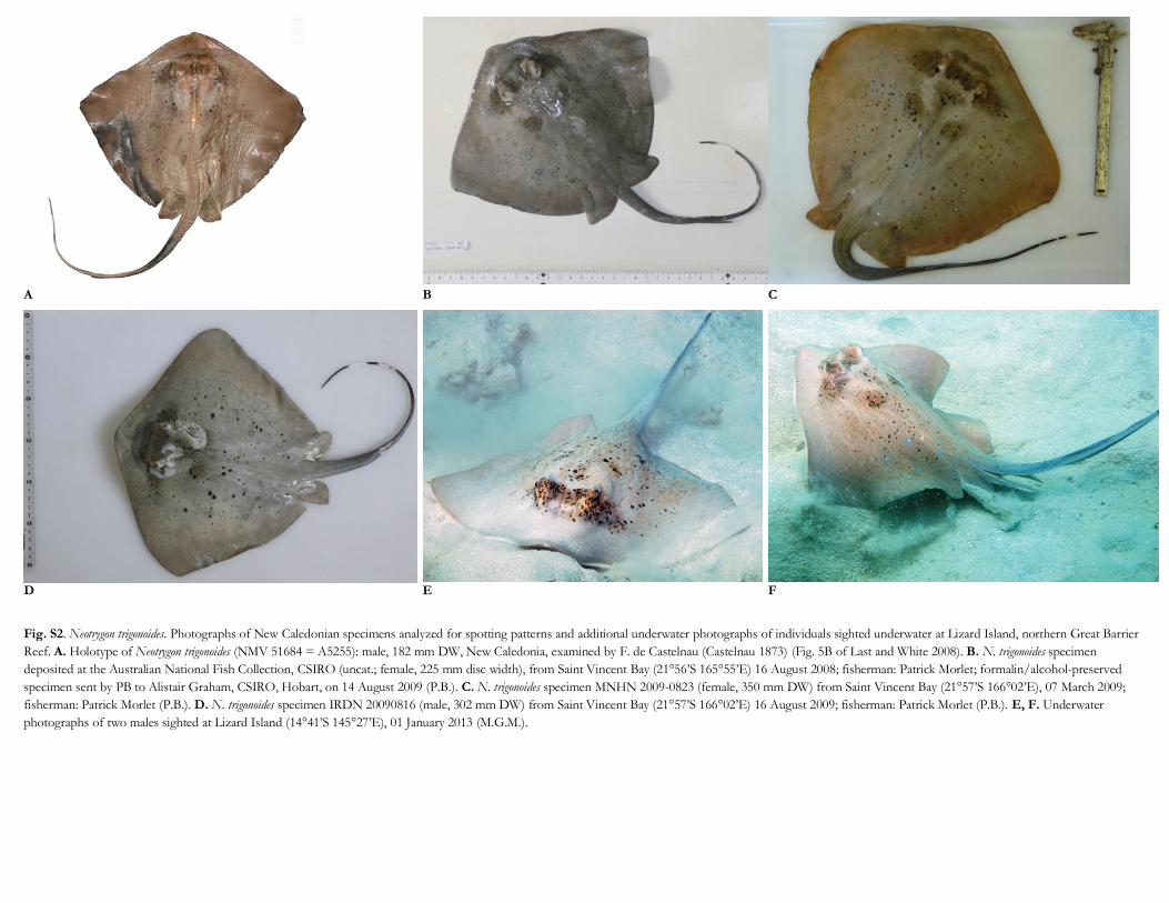
Fig. S2. Neotrygon trigonoides. Photographs of New Caledonian specimens analyzed for spotting patterns and additional underwater photographs of individuals sighted underwater at Lizard Island, northern Great Barrier
Reef. A. Holotype of Neotrygon trigonoides (NMV 51684 = A5255): male, 182 mm DW, New Caledonia, examined by F. de Castelnau (Castelnau 1873) (Fig. 5B of Last and White 2008). B. N. trigonoides specimen
deposited at the Australian National Fish Collection, CSIRO (uncat.; female, 225 mm disc width), from Saint Vincent Bay (21°56’S 165°55’E) 16 August 2008; fisherman: Patrick Morlet; formalin/alcohol-preserved
specimen sent by PB to Alistair Graham, CSIRO, Hobart, on 14 August 2009 (P.B.). C. N. trigonoides specimen MNHN 2009-0823 (female, 350 mm DW) from Saint Vincent Bay (21°57’S 166°02’E), 07 March 2009;
fisherman: Patrick Morlet (P.B.). D. N. trigonoides specimen IRDN 20090816 (male, 302 mm DW) from Saint Vincent Bay (21°57’S 166°02’E) 16 August 2009; fisherman: Patrick Morlet (P.B.). E, F. Underwater
photographs of two males sighted at Lizard Island (14°41’S 145°27’E), 01 January 2013 (M.G.M.).
Related Documents