Responses to Cytokines and Interferons that Depend upon JAKs and STATs George R. Stark, HyeonJoo Cheon, and Yuxin Wang Department of Cancer Biology, Lerner ResearchInstitute of the Cleveland Clinic, Cleveland, Ohio 44195 Correspondence: [email protected] Many cytokines and all interferons activate members of a small family of kinases (the Janus kinases [JAKs]) and a slightly larger family of transcription factors (the signal transducers and activators of transcription [STATs]), which are essential components of pathways that induce the expression of specific sets of genes in susceptible cells. JAK-STAT pathways are required for many innate and acquired immune responses, andthe activities of these pathways must be finely regulated to avoid major immune dysfunctions. Regulation is achieved through mechanisms that include the activation or induction of potent negative regulatory proteins, posttranslational modification of the STATs, and other modulatoryeffects that are cell-type specific. Mutations of JAKs and STATs can result in gains or losses of function and can predispose affected individuals to autoimmune disease, susceptibility to a variety of infec- tions, or cancer. Here we review recent developments in the biochemistry, genetics, and biologyof JAKs and STATs. B ecause the basic biochemistry of Janus ki- nase –signal transducers and activators of transcription (JAK-STAT) signaling pathways has been frequently and extensively reviewed (see, for example, Stark and Darnell 2012; Cai et al. 2015; O’Shea et al. 2015; Villarino et al. 2015), we present here only a brief summary. After a cytokine or interferon binds to its specific receptor, the receptor forms homodimers, het- erodimers, or trimers, depending on the cyto- kine, thus activating the tightly bound JAKs to cross-phosphorylate each other. The activated JAKs then phosphorylate specific tyrosine resi- dues in the cytoplasmic domains of the recep- tors, providing binding sites for the STATs through their highly conserved SH2 domains. The receptor-bound STATs are phosphorylated, each on a highly conserved tyrosine residue, af- ter which they leave the receptor as homo- or heterodimers whose association is strengthened by SH2-phosphotyrosine interactions. The phosphorylated STAT dimers are then trans- ported to the nucleus, where they bind to and activate specific promoters. The basic outline of JAK-STATsignaling (Fig. 1) shows that interfer- on (IFN)-g (type II IFN) and all of the cytokines primarily drive the formation of specific STAT homodimers, which then bind to DNA directly. However, in some cases, heterodimers involving STAT1 and STAT3 or STAT5A and STAT5B can also form. In contrast, IFN-b and the subtypes of IFN-a (collectively type I IFNs) and the sub- types of IFN-l (collectively type III IFNs) drive the formation of STAT1-STAT2 heterodimers, which then associate with the DNA-binding in- terferon regulatory protein 9 (IRF9) to form in- Editors: Warren J. Leonard and Robert D. Schreiber Additional Perspectives on Cytokines available at www.cshperspectives.org Copyright # 2018 Cold Spring Harbor Laboratory Press; all rights reserved; doi: 10.1101/cshperspect.a028555 Cite this article as Cold Spring Harb Perspect Biol 2018;10:a028555 1 on February 20, 2022 - Published by Cold Spring Harbor Laboratory Press http://cshperspectives.cshlp.org/ Downloaded from

Welcome message from author
This document is posted to help you gain knowledge. Please leave a comment to let me know what you think about it! Share it to your friends and learn new things together.
Transcript

Responses to Cytokines and Interferons thatDepend upon JAKs and STATs
George R. Stark, HyeonJoo Cheon, and Yuxin Wang
Department of Cancer Biology, Lerner Research Institute of the Cleveland Clinic, Cleveland, Ohio 44195
Correspondence: [email protected]
Many cytokines and all interferons activate members of a small family of kinases (the Januskinases [JAKs]) and a slightly larger family of transcription factors (the signal transducers andactivators of transcription [STATs]), which are essential components of pathways that inducethe expression of specific sets of genes in susceptible cells. JAK-STAT pathways are requiredfor many innate and acquired immune responses, and the activities of these pathways mustbe finely regulated to avoid major immune dysfunctions. Regulation is achieved throughmechanisms that include the activation or induction of potent negative regulatory proteins,posttranslational modification of the STATs, and other modulatory effects that are cell-typespecific. Mutations of JAKs and STATs can result in gains or losses of function and canpredispose affected individuals to autoimmune disease, susceptibility to a variety of infec-tions, or cancer. Here we review recent developments in the biochemistry, genetics, andbiology of JAKs and STATs.
Because the basic biochemistry of Janus ki-nase–signal transducers and activators of
transcription (JAK-STAT) signaling pathwayshas been frequently and extensively reviewed(see, for example, Stark and Darnell 2012; Caiet al. 2015; O’Shea et al. 2015; Villarino et al.2015), we present here only a brief summary.After a cytokine or interferon binds to its specificreceptor, the receptor forms homodimers, het-erodimers, or trimers, depending on the cyto-kine, thus activating the tightly bound JAKs tocross-phosphorylate each other. The activatedJAKs then phosphorylate specific tyrosine resi-dues in the cytoplasmic domains of the recep-tors, providing binding sites for the STATsthrough their highly conserved SH2 domains.The receptor-bound STATs are phosphorylated,each on a highly conserved tyrosine residue, af-
ter which they leave the receptor as homo- orheterodimers whose association is strengthenedby SH2-phosphotyrosine interactions. Thephosphorylated STAT dimers are then trans-ported to the nucleus, where they bind to andactivate specific promoters. The basic outline ofJAK-STAT signaling (Fig. 1) shows that interfer-on (IFN)-g (type II IFN) and all of the cytokinesprimarily drive the formation of specific STAThomodimers, which then bind to DNA directly.However, in some cases, heterodimers involvingSTAT1 and STAT3 or STAT5A and STAT5B canalso form. In contrast, IFN-b and the subtypesof IFN-a (collectively type I IFNs) and the sub-types of IFN-l (collectively type III IFNs) drivethe formation of STAT1-STAT2 heterodimers,which then associate with the DNA-binding in-terferon regulatory protein 9 (IRF9) to form in-
Editors: Warren J. Leonard and Robert D. Schreiber
Additional Perspectives on Cytokines available at www.cshperspectives.org
Copyright # 2018 Cold Spring Harbor Laboratory Press; all rights reserved; doi: 10.1101/cshperspect.a028555
Cite this article as Cold Spring Harb Perspect Biol 2018;10:a028555
1
on February 20, 2022 - Published by Cold Spring Harbor Laboratory Press http://cshperspectives.cshlp.org/Downloaded from
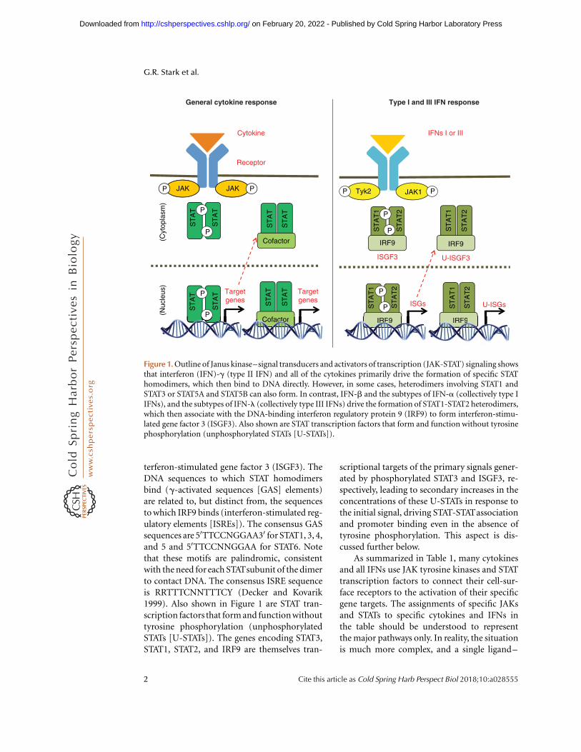
terferon-stimulated gene factor 3 (ISGF3). TheDNA sequences to which STAT homodimersbind (g-activated sequences [GAS] elements)are related to, but distinct from, the sequencesto which IRF9 binds (interferon-stimulated reg-ulatory elements [ISREs]). The consensus GASsequences are 50TTCCNGGAA30 for STAT1, 3, 4,and 5 and 50TTCCNNGGAA for STAT6. Notethat these motifs are palindromic, consistentwith the need for each STATsubunit of the dimerto contact DNA. The consensus ISRE sequenceis RRTTTCNNTTTCY (Decker and Kovarik1999). Also shown in Figure 1 are STAT tran-scription factors that form and function withouttyrosine phosphorylation (unphosphorylatedSTATs [U-STATs]). The genes encoding STAT3,STAT1, STAT2, and IRF9 are themselves tran-
scriptional targets of the primary signals gener-ated by phosphorylated STAT3 and ISGF3, re-spectively, leading to secondary increases in theconcentrations of these U-STATs in response tothe initial signal, driving STAT-STATassociationand promoter binding even in the absence oftyrosine phosphorylation. This aspect is dis-cussed further below.
As summarized in Table 1, many cytokinesand all IFNs use JAK tyrosine kinases and STATtranscription factors to connect their cell-sur-face receptors to the activation of their specificgene targets. The assignments of specific JAKsand STATs to specific cytokines and IFNs inthe table should be understood to representthe major pathways only. In reality, the situationis much more complex, and a single ligand–
Cytokine
JAKP P
Receptor
P
P
Targetgenes
Targetgenes
Tyk2 JAK1 PP
P
P
ISGs
IFNs I or III
U-ISGs
U-ISGF3ISGF3
P
P
General cytokine response Type I and III IFN response
JAK
ST
AT
ST
AT
ST
AT
(Cyt
opla
sm)
(Nuc
leus
)
P
PCofactor
Cofactor IRF9
IRF9 IRF9
IRF9
ST
AT
ST
AT
ST
AT
ST
AT
ST
AT
1S
TA
T1
ST
AT
1
ST
AT
2
ST
AT
2
ST
AT
1
ST
AT
2
ST
AT
2
ST
AT
PCofCCCCCCCC actccctctttcctctctccttcccccc oroooooooo IRF99999999999IRFRFRFRFRFRFFRRFRFRRFRRFFFFRRRFRRRFRFRFRFFFFRRFRFFFRRRRRFRR 9
Figure 1. Outline of Janus kinase–signal transducers and activators of transcription (JAK-STAT) signaling showsthat interferon (IFN)-g (type II IFN) and all of the cytokines primarily drive the formation of specific STAThomodimers, which then bind to DNA directly. However, in some cases, heterodimers involving STAT1 andSTAT3 or STAT5A and STAT5B can also form. In contrast, IFN-b and the subtypes of IFN-a (collectively type IIFNs), and the subtypes of IFN-l (collectively type III IFNs) drive the formation of STAT1-STAT2 heterodimers,which then associate with the DNA-binding interferon regulatory protein 9 (IRF9) to form interferon-stimu-lated gene factor 3 (ISGF3). Also shown are STAT transcription factors that form and function without tyrosinephosphorylation (unphosphorylated STATs [U-STATs]).
G.R. Stark et al.
2 Cite this article as Cold Spring Harb Perspect Biol 2018;10:a028555
on February 20, 2022 - Published by Cold Spring Harbor Laboratory Press http://cshperspectives.cshlp.org/Downloaded from

receptor pair may activate more than one STAT.The ratio of activated STATs can depend on theirrelative intracellular concentrations (specificexamples for interleukin (IL)-6, IL-21, and IL-27 are cited below), on the specific cell type, onwhether or not the cell has received prior signals(“priming”), and probably on other variablesas well. As one example of cell-type-specificresponses, van Boxel-Dezaire et al. (2010) foundthat different primary human leukocyte subsetsrespond quite differently to IFN-b. In Bcells and CD4þ T cells, IFN-b activates STAT3and STAT5 primarily, with biological effectsopposite from those driven by the “canonical”activation of STAT1 and STAT2 in the otherleukocyte subtypes that were studied.
FUNCTIONALLY IMPORTANT CHEMICALMODIFICATIONS OF THE STATs
The STATs are substrates for phosphorylation,methylation, and other posttranslational modi-fications that facilitate both positive and negativefine-tuning of the transcriptional responses.
Carboxy-Terminal Serine Phosphorylations
All the STATs except STAT2 share a functionallyimportant serine phosphorylation site withina P(M)SP motif located near their carboxyltermini. Carboxy-terminal serine phosphoryla-tion is stimulated by many different cytokinesand growth factors, and is mediated by manydifferent kinases, including extracellular signal-regulated kinase (ERK), p38, c-Jun amino-
terminal kinase (JNK), mechanistic target ofrapamycin (mTOR), nemo-like kinase (NLK),calcium/calmodulin-dependent protein kinaseII (CaMKII), IkB kinase 1 (IKK1), and proteinkinase C d (PKC-d) (Schindler et al. 2007).Phosphorylation increases the transactivationpotential of these proteins. The most intensivelyinvestigated phosphorylated serine residues areS727 in both STAT1 and STAT3. Phosphoryla-tion of S727 of STAT1 and STAT3 is necessaryfor full activation of transcription in responseto IFNs and IL-6 family cytokines, but thephosphorylation of S727 in either STAT1 orSTAT3 is not associated with increased tyrosinephosphorylation (Wen et al.1995). The serine toalanine mutation of S727 in STAT1 or STAT3leads to reduction of the cytokine-induced tran-scription of specific genes, the extent of whichis likely to vary in different cellular contexts. Inaddition to altering the transcriptional activa-tion of STATs, serine phosphorylation of S727 inSTAT1 and STAT3 has been correlated with en-hanced DNA-binding ability (Eilers et al. 1995;Zhang et al. 1995; Ng and Cantrell 1997). Vis-conti et al. (2000) showed that mutating S721of STAT4 decreased its transcriptional activi-ty in IL-12-treated cells. Phosphorylation ofboth S725 and S779 of STAT5A and of S730 ofSTAT5B negatively regulate transactivation in re-sponse to stimulation of mammary glands withprolactin (Yamashita et al. 1998; Benitah et al.2003). Wang et al. (2004) showed that IL-4 andIL-13 promote STAT6 phosphorylation on S756in human T cells. However, the contribution ofthis phosphorylation to function is not yet clear.
Table 1. Summary of cytokines and IFNs that use JAK tyrosine kinases and STAT transcription factors to connecttheir cell-surface receptors to the activation of specific gene targets
STATs JAKs Major cytokines
STAT1 JAK1, JAK2, TYK2 Type I, II, and III IFNsSTAT2 JAK1, TYK2 Type I and III IFNsSTAT3 JAK1, JAK2, JAK3, TYK2 IL-6 family cytokines, IL-10, IL-27, IL-21STAT4 JAK2, TYK2 IL-12, IL-23STAT5A/B JAK1, JAK2, JAK3 IL-2, IL-7, IL-9, IL-15, EPO, TPO, GM-CSF, GH, PRLSTAT6 JAK1, JAK2, JAK3, TYK2 IL-4, IL-13
STATs, Signal transducers and activators of transcription; JAKs, Janus kinases; IFN, interferon; IL, interleukin; EPO,
erythropoietin; TPO, thrombopoietin; GM-CSF, granulocyte macrophage colony-stimulating factor; GH, growth hormone;
PRL, prolactin.
JAKs and STATs
Cite this article as Cold Spring Harb Perspect Biol 2018;10:a028555 3
on February 20, 2022 - Published by Cold Spring Harbor Laboratory Press http://cshperspectives.cshlp.org/Downloaded from

Additional Serine and ThreoninePhosphorylations
Recent advances in mass spectrometry haverevolutionized the analysis of protein phos-phorylation by allowing rapid identificationof the sites modified with precision and sensi-tivity. Several additional serine and threoninemodifications sites on STATs have been found.
STAT1
Phosphorylation of S708 by TRIM6-activatedIKK1 regulates STAT1 homodimerization butnot ISGF3 formation in response to IFN (Te-noever et al. 2007; Rajsbaum et al. 2014).Ultimately, the phosphorylation of S708 facil-itates the induction of a subset of ISGs whoseprotein products are essential for the antiviralresponse in vitro and in vivo. Phosphorylationof S744/S747 has also been reported, butthe binding of ISGF3 to ISREs was unaffectedby a carboxy-terminal deletion of STAT1 thatremoved both of these residues (Tenoever et al.2007).
STAT2
The first serine phosphorylation of STAT2(S287) was revealed by the work of Steen et al.(2013). Phosphorylation-defective mutants ofS287 of STAT2 enhanced the ability of ISGF3to bind to DNA, revealing that this phosphory-lation is a negative regulatory event. We haveshown that the phosphorylation of T387 regu-lates the ability of ISGF3 to bind to DNA (Wanget al. 2017). This phosphorylation negativelyregulates the expression of most genes inducedby type I IFN, inhibiting the ability of IFNto protect cells against virus infection and toinhibit cell growth. In most untreated cell types,the great majority of STAT2 is phosphorylatedon T387 constitutively. T387 lies in a cyclin-dependent kinase (CDK) consensus sequence,and CDK inhibitors decrease T387 phosphory-lation markedly. Using CDK inhibitors toreverse the constitutive inhibitory phosphory-lation of T387 of STAT2 might enhance theefficacy of type I IFNs.
STAT3
Waitkus et al. (2014) have provided evidencethat GSK3 a/b directly phosphorylates STAT3,simultaneously on T714 and S727, and thatthese modifications are required for STAT3-dependent gene expression in response to simul-taneous activation of epidermal growth factorreceptor (EGFR) and protease-activated recep-tor 1 (PAR-1) in endothelial cells. Levels of bothT714 and S727 phosphorylation of STAT3 aresignificantly elevated in renal tumor tissues, sug-gesting that the GSK3-activated STAT3 signalingmay be important in this disease.
STAT5A
S127/S128 phosphorylation of STAT5A is re-quired for ERBB4-induced Y694 phosphoryla-tion and has a substantial impact on ERBB4-dependent regulation of STAT5A activity (Clarket al. 2005). The expression of a STAT5 mutantin which the S725 and S779 phosphorylationsites were altered prohibited transformationand induced apoptosis in bone marrow cells(Pircher et al. 1999; Xue et al. 2002; Friedbichleret al. 2010; Berger et al. 2014). S779 is phos-phorylated by p21-activated kinase (PAK) inhuman myeloid malignancies.
STAT5B
STAT5B constitutively phosphorylated on S193has been found in hematopoietic cancers (Mitraet al. 2012). This phosphorylation is dependenton the mTOR signaling pathway and positivelyregulates STAT5B DNA binding and transcrip-tional activity.
STAT6
Phosphorylation of STAT6 S707 is triggered bythe virus infection–responsive protein STING,which is located in the endoplasmic reticulum.Homodimers of STAT6 phosphorylated on Y641and S407 then activate specific target genes inthe nucleus that mediate immune cell homing(Chen et al. 2011). S707 is phosphorylated byJNK, which can be activated in response to cel-
G.R. Stark et al.
4 Cite this article as Cold Spring Harb Perspect Biol 2018;10:a028555
on February 20, 2022 - Published by Cold Spring Harbor Laboratory Press http://cshperspectives.cshlp.org/Downloaded from

lular stress or IL-1b. Phosphorylation of S707 isa negative regulatory event that decreases theDNA-binding ability of STAT6 following its ac-tivation by IL-4 (Shirakawa et al. 2011).
Lysine and Arginine Modifications
The lysine residues of proteins can be modifiedby acetylation or by the addition of one, two, orthree methyl groups, and the arginine residuescan be modified by methylation. Furthermore,these reactions are reversible, providing rich op-portunities to modify function. Many enzymescarry out the reversible acetylation and methyl-ation of histones, providing the chemical basisof the modification of chromatin structure andfunction known as the histone code (Allis andJenuwein 2016). In many cases, the specificallymodified lysine or arginine residues providedocking sites for the binding of accessory pro-teins that modulate function. As summarized inTable 2, several different lysine and arginine res-idues of STAT1 and STAT3 are methylated oracetylated, but we are not aware of reports ofthese modifications for any other STAT. In everycase, the enzymes responsible for STAT1 orSTAT3 modification were previously known tomodify histones. One of the earliest papers re-ports the dimethylation of R31 of STAT1, whichfacilitates IFN-dependent gene expression by
inhibiting the association of STAT1 with thenegative regulator PIAS1 (Mowen et al. 2001).It is very interesting that the effects of gain-of-function mutations of STAT1 that cause dis-seminated yeast infections in patients can beameliorated by reducing the level of PIAS1 orby facilitating STAT1 arginine methylation(Sampaio et al. 2013).
We have described the reversible dimethyla-tion of K140 of STAT3, which regulates STAT3-dependent gene expression negatively (Yang etal. 2010). In this case, the reaction is catalyzed bythe histone lysine methyltransferase SET9 andoccurs only after STAT3 has been bound to apromoter. The docking site for SET9 is providedby the phosphorylated S727 residue of STAT3,because the S727A mutant of STAT3 fails to re-cruit SET9 to the promoter. We reviewed severaladditional examples of the lysine methylation ofpromoter-bound transcription factors (Starket al. 2011). Our working hypothesis, whichneeds to be tested further, is that the promot-er-bound factor provides a docking site for ahistone-modifying enzyme that then catalyzesfunctionally important modifications, notonly of the transcription factor but potentiallyalso of local histones and the transcriptional ma-chinery itself. Another important modificationof STAT3 is the dimethylation of K49, carried outby the lysine methyltransferase EZH2 (Dasgupta
Table 2. Summary of lysine and arginine modifications of STAT1 and STAT3
STATs Modifications Sites
STAT1 Methylation R31me1 (Zhu et al. 2002)R31me2 (Mowen et al. 2001)
Acetylation K410/413ac (Kramer et al. 2006; Antunes et al. 2011; Kotla and Rao, 2015),controversial
Sumoylation K703sm (Ungureanu et al. 2003; Ungureanu et al. 2005; Gronholm et al. 2012)STAT3 Methylation R31me1 (Iwasaki et al. 2010)
K49me2 (Dasgupta et al. 2015b)K140 (Yang et al. 2010)
Acetylation K49/87ac (Ray et al. 2005; Hou et al. 2008; Nie et al. 2009)K679ac (Nie et al. 2009)K685ac (Yuan et al. 2005; Lee et al. 2009, 2016; Dasgupta et al. 2014;
Kang et al. 2015)K707ac (Nie et al. 2009)
Sumoylation K451sm (Zhou et al. 2016b)
Listed posttranslational modifications (PTMs) can be found at www.phosphosite.org.
STATs, Signal transducers and activators of transcription.
JAKs and STATs
Cite this article as Cold Spring Harb Perspect Biol 2018;10:a028555 5
on February 20, 2022 - Published by Cold Spring Harbor Laboratory Press http://cshperspectives.cshlp.org/Downloaded from

et al. 2015b). Failure to carry out this reactioninhibits the ability of STAT3 to activate the ex-pression of a substantial fraction of its targetgenes by an as-yet-unknown mechanism.
SUMOylation, Glycosylation, and AdditionalTyrosine Phosphorylation
Glycosylation (Gewinner et al. 2004) and tyro-sine phosphorylation of STATs at additionalsites, including STAT2, human, Y631 (Scarzelloet al. 2007), STAT5A, mouse, Y682/683 (Schal-ler-Schonitz et al. 2014), and STAT5B, rat, Y679(Kabotyanski and Rosen 2003), have been re-ported, but the functional importance of thesemodifications is not yet well established. On theother hand, SUMOylation of STAT1 on K703 isa modification of great functional importance(references in Table 2). This modification, cat-alyzed by PIAS1 (Rogers et al. 2003; Ungureanuet al. 2003), leads to inhibition of STAT1 func-tion (Rogers et al. 2003; Ungureanu et al. 2005),and modulation of the response to IFN-g isfacilitated by the SUMOylation of STAT1 (Be-gitt et al. 2011; Maarifi et al. 2015).
NEGATIVE REGULATION
Failure to regulate cytokine-stimulated re-sponses leads to catastrophic hyperinflamma-tory responses, and therefore elaborate mecha-nisms exist to achieve the necessary negativeregulation. A major mechanism involves thesuppressor of cytokine signaling (SOCS) pro-teins (reviewed by Kazi et al. 2014). These po-tent negative regulators are typically induced inresponse to acute exposure to specific cytokines,and they typically function by inhibiting STATactivation at the receptors (see Babon et al. 2014for an example of the SOCS3 and the IL-6 fam-ily of cytokines). Defective SOCS3 function hasbeen reported to contribute to many diseases,including allergy, autoimmune diseases such asrheumatoid arthritis, vascular inflammatory dis-eases, insulin resistance, and cancer, as reviewedby Yin et al. (2015). Many additional mecha-nisms contribute to adequate regulation of theinduction of and responses to IFNs, includingthe PIAS proteins, which inhibit the function
of activated STATs in the nucleus and also cata-lyze the inhibitory SUMOylation of STAT1, andprotein tyrosine phosphatases, which inactivatethe STATs (Hertzog and Williams 2013; Porrittand Hertzog 2015). Although the STAT3-in-duced expression of SOCS3 is important fordampening acute responses to IL-6 and othergp130-linked cytokines, tumor cells use an IL-6-induced association between the IL-6 recep-tor:gp130 complex and the EGFR to nullify theinhibitory effect of SOCS3 and thus to sustainSTAT3 activation constitutively (Wang et al.2013). It seems likely that specific mechanismsto prevent negative regulation of STATactivationwill be used whenever sustained STAT activationis necessary in normal physiology.
UNPHOSPHORYLATED STATs
Many studies have shown that U-STATs, whichlack phosphorylation of their highly conservedtyrosine residues, are located in nuclei, bindto promoters, and activate gene expression(Chatterjee-Kishore et al. 2000; Yang et al.2005, 2007; Cui et al. 2007; Cheon and Stark2009; Cheon et al. 2013; Park et al. 2015).Whether the phosphorylation of other residuesaffects the function of U-STATs is not clear, butK685 of U-STAT3 is acetylated, and mutation ofthis residue results in loss of expression of manyU-STAT3-induced genes (Dasgupta et al. 2014).U-STATs 1, 2, 3, 5, and 6 regulate gene expres-sion through their interactions with partnercofactors. The expression of U-STATs 1, 2, and3 is increased in response to cytokines that in-duce their tyrosine phosphorylation, but howthe expression of the other U-STATs is regulatedis not yet known. The roles of U-STATs inregulating gene expression were originally con-troversial, but it is now generally accepted thatU-STATs are critical transcription factors thatare involved in many biological events in bothnormal and pathological situations.
U-STATs as Positive Regulators of GeneExpression
The levels of the U-STAT1, U-STAT2, and U-STAT3 proteins are induced in response to sig-
G.R. Stark et al.
6 Cite this article as Cold Spring Harb Perspect Biol 2018;10:a028555
on February 20, 2022 - Published by Cold Spring Harbor Laboratory Press http://cshperspectives.cshlp.org/Downloaded from

nals that lead to the tyrosine phosphorylation ofeach STAT, because each phosphorylated STATbinds to the promoter of its own gene to induceexpression (Yang et al. 2005; Cheon et al. 2013).The U-STAT proteins then accumulate, translo-cate into nuclei, bind to target gene promoterstogether with their cofactors, and induce theexpression of these genes. U-STATs contributeto the steady-state constitutive expression ofspecific genes, while tyrosine-phosphorylatedSTATs induce rapid and transient responses inresponse to cytokine stimulation. Many targetgenes of U-STATs 1, 2, 3, and 6 encode proteinsthat promote cell survival and resistance tocell death, suggesting that the U-STAT systemhelps to sustain the survival of cells in stressfulenvironments.
U-STAT1 induces its target genes asU-ISGF3, a tripartite complex with U-STAT2and IRF9, and the expression of all threeU-ISGF3 components is increased in responseto either type I or type III IFN (Cheon et al.2013; Sung et al. 2015). U-STAT1 does not ac-tivate gene expression as a homodimer, becausehigh levels of U-STAT1 do not induce targetgene expression if U-STAT2 and IRF9 are notpresent in sufficient quantities (Cheon andStark 2009; Cheon et al. 2013). U-ISGF3 in-duces a subset of ISGs that are also induced inthe initial response to IFNs, resulting in theirprolonged expression. The phenotypes of cellsthat express high levels of only the U-ISGF3-induced genes are different from those of cellstreated with high levels of IFNs, which expressall of the ISGs in response to phosphorylatedISGF3. Cancer cells that express high levels of U-ISGF3 are more resistant to DNA damage, whileIFNs inhibit cancer cell proliferation andincrease their apoptosis (Borden et al. 2007).Similarly, hepatocytes expressing high levels ofU-ISGF3 are more resistant to IFN-a therapy,although U-ISGF3 itself suppresses viral repli-cation by inducing antiviral genes (Sung et al.2015). Phosphorylated STAT3 mediates the in-duction of U-STAT3 in response to IL-6 andother cytokines that activate gp130-linked re-ceptors (Yang et al. 2005). However, in strongcontrast to the situation for U-STAT1, U-STAT3induces the expression of a set of genes that is
completely different from the set induced byphosphorylated STAT3 (Yang et al. 2005).Some of the genes specifically induced byU-STAT3 are important oncogenes, includingSRC, MET, and MRAS. The mechanism bywhich U-STAT3 induces expression of targetgenes is only partially understood. However,for the expression of a subset of the inducedgenes, including RANTES, IL6, and IL8,U-STAT3 forms a complex with nuclear factorkB (NF-kB), which then binds to kB elementsin the promoters of a small fraction of NF-kBtarget genes (Yang et al. 2007). Cui et al. (2007)show that U-STAT6 constitutively activates theexpression of the COX2 gene. Similarly to theother U-STATs, U-STAT6 also forms a complexwith a cofactor protein, p300, facilitating bind-ing to the COX2 promoter.
U-STATs as Negative Regulators of GeneExpression
In contrast to their ability to regulate gene ex-pression positively, some U-STATs repress geneexpression instead. ChIP-seq data showing ge-nome-wide distribution reveals that U-STAT5and phosphorylated STAT5 bind to differentcis-acting elements in the genome, and differ-ently regulate gene expression (Park et al. 2015).In this study, phosphorylated STAT5 wasshown to bind to GAS elements in canonicalSTAT5-induced promoters in response tothrombopoietin (TPO) in mouse hematopoiet-ic stem cells. In the absence of TPO, however, alarge portion of U-STAT5 occupies binding sitesfor early growth response (EGR), an activatorthat promotes the expression of megakaryocyticgenes, thus repressing EGR-induced gene ex-pression. Only a small portion of U-STAT5binds to promoters that are occupied by phos-phorylated STAT5 in response to TPO, but therole of U-STAT5 is not known in that situation.Differently from U-STAT5, U-ISGF3 and phos-phorylated ISGF3 bind to similar ISRE elementsin the promoters of ISGs that are induced byboth transcription factors (Cheon et al. 2013).Using a ChIP-on-chip analysis, Testoni et al.(2011) showed that U-STAT2, possibly as a com-ponent of U-ISGF3, binds to more than half of
JAKs and STATs
Cite this article as Cold Spring Harb Perspect Biol 2018;10:a028555 7
on February 20, 2022 - Published by Cold Spring Harbor Laboratory Press http://cshperspectives.cshlp.org/Downloaded from

the ISG promoters investigated before IFN-atreatment, but the effect on the expression ofISGs is not clear. Although U-ISGF3 itself in-creases the expression of some ISGs in the ab-sence of IFN treatment, we do not yet knowwhether or not U-ISGF3 inhibits the ex-pression of other ISGs in response to IFN.
Nongenomic Activity of U-STAT3 inMitochondria
U-STAT3 plays important roles not only in thenucleus but also in mitochondria (Garama et al.2016). The mitochondrial activity of STAT3 isnot dependent on Y705 phosphorylation, butphosphorylation of S727 is necessary (Goughet al. 2009, 2014). Mitochondrial U-STAT3regulates the activity of the electron transportchain, which is required for adenosine triphos-phate (ATP) production, and the opening ofthe mitochondrial permeability transitionpore (Gough et al. 2009; Wegrzyn et al. 2009).In RAS-transformed cells, the MEK–ERKpathway drives the phosphorylation of STAT3on S727, and RAS-transformed cells carryinga mutation of S727 are partially resistant toinhibitors of the ERK pathway (Gough et al.2013).
STRUCTURES AND INTRACELLULARLOCATIONS OF STATS
Phosphorylated STATs are translocated into thenucleus after cytokine stimulation, but U-STATsshuttle constitutively between cytoplasm andnucleus (Meyer and Vinkemeier 2004; Pranadaet al. 2004; Liu et al. 2005; Iyer and Reich 2007;Vogt et al. 2011). U-STATs 1, 3, and 5 formantiparallel dimers, whereas phosphorylatedSTAT dimers are in a parallel conformationthat is stabilized by phosphotyrosine–SH2 do-main interactions, allowing both DNA-bindingdomains to contact the GAS sequences simulta-neously, resulting in strong binding (Mao et al.2005; Neculai et al. 2005; Wenta et al. 2008;Timofeeva et al. 2012; Nkansah et al. 2013).In addition, phosphorylated homodimers ofSTAT1 bind to each other to form tetramers,facilitating gene expression in response to type
II (but not type I) IFNs. Consistently, the F77Amutation of murine STAT1, which disruptsdimer–dimer interactions, blunts signalingin response to type II IFNs in mice (Begittet al. 2014). Interestingly, Droescher et al.(2011) found that all activated STATs can formparacrystalline arrays in the nuclei of cytokine-stimulated cells, with important biologicalconsequences. However, for STAT1 only, thisphenomenon can be prevented by modificationof the protein by SUMO (Begitt et al. 2011;Droescher et al. 2011). STAT2 forms a stableantiparallel heterodimer with either the un-phosphorylated or tyrosine-phosphorylatedforms of STAT1; these heterodimers are nottransported to the nucleus and have no tran-scriptional activity, so STAT2 is a pervasive neg-ative regulator of STAT1-dependent functions(Ho et al. 2016). However, when IRF9 is presentin sufficient quantity, antiparallel STAT1-STAT2heterodimers will be converted to U-ISGF3(Cheon et al. 2013) or to hemiphosphorylatedISGF3 (Morrow et al. 2011), in which the rela-tive orientation of the two STATs becomes par-allel. They are then transported to the nucleusand bind to DNA to activate transcription.Thus, the availability of free STAT1 to signal inresponse, for example, to IFN-g or IL-27 (Hoet al. 2016), will depend on the relative concen-trations of both STAT2 and IRF9.
CHROMATIN REMODELING BY STATs
There are a few intriguing reports that STATsfunction to affect chromatin structure indepen-dently of their ability to activate transcription.U-STAT92E (the only Drosophila STAT) stabi-lizes heterochromatin in association with het-erochromatin protein 1 (HP1) (Shi et al. 2008).Phosphorylation of U-STAT92E causes hetero-chromatin instability and promotes gene ex-pression. Human U-STAT5A binds to HP1a,repressing the expression of multiple oncogenes(Hu et al. 2013). The overexpression of STAT5AY704F, which cannot be phosphorylated at thissite, has effects on global gene expression simi-lar to the effects of over expressing HP1a. Thephosphorylation of STAT1 remodels chromatinto generate a local environment appropriate for
G.R. Stark et al.
8 Cite this article as Cold Spring Harb Perspect Biol 2018;10:a028555
on February 20, 2022 - Published by Cold Spring Harbor Laboratory Press http://cshperspectives.cshlp.org/Downloaded from

the activation of target gene expression. Whenhuman histocompatibility (MHC) genes areactivated by IFN-g, phosphorylated STAT1,which binds to specific elements of the targetgene promoter and recruits the chromatin re-modeling enzyme BRG1, causes the release ofentire MHC locus loops from compacted chro-matin (Christova et al. 2007).
JAK AND STAT MUTATIONS
Naturally Occurring STAT Mutations
Because the STATs have essential roles in infec-tious disease, immunity, and cancer, manynaturally occurring mutations have been dis-covered that affect human health, providinga rich source of structural and functional infor-mation. All seven STATs form homo- andheterodimers following phosphorylation oftheir tyrosine residues, and thus there is greatpotential for dominant effects in which onlyone partner is mutated. These mutations canaffect many different protein–protein interac-tions, leading to dominant phenotypes thatreflect either loss of function (LOF) or gain offunction (GOF), as summarized recently forSTAT3 (Chandrasekaran et al. 2016). However,STAT mutations will be manifest in human dis-ease only if they lead to a discernable biologicalphenotype and, although many dominant mu-tations have been described for STAT1 andSTAT3, far fewer have emerged for the otherSTATs. Because the structures of the STAT di-mers are similar, it seems likely that germlinedominant mutations will occur with similar fre-quencies for all the STATs, but with far less fre-quent phenotypic consequences for STATs 2, 4,5, and 6. In their summary of all human prima-ry immunodeficiencies, Boisson et al. (2015)point out that the known autosomal-recessivedeficiencies are all caused by alleles with LOF,and that 44 out of 61 autosomal-dominantdefects are caused by LOF mutations. Negativeregulation of cytokine-dependent signaling isvital to prevent overstimulation of immuneresponses, so that, within the 17 examplesof GOF-dominant mutations, those affectingSTAT1 lead to infection, autoimmunity, and
malignancy, whereas GOF mutations of STAT3affect autoimmunity, allergy, and autoinflam-mation. Y705, S727, and K49 mutations ofSTAT3 have not yet been reported, presum-ably because they would not be consistentwith survival.
Gain-of-Function Somatic Mutations inCancer
The constitutive activation of JAK-STAT signal-ing pathways in cancer cells, through somaticmutation and other mechanisms, drives prolif-eration and resistance to stresses, and helps toovercome barriers to perpetual cell growth.Because this topic has been well reviewed re-cently (O’Shea et al. 2015; Pilati and Zucman-Rossi 2015; Thomas et al. 2015), we present onlya few illustrative examples here. As recent exam-ples for the STATs, activating somatic mutationsof STAT5B lead to leukemia (Rajala et al. 2013)and activating mutations of STAT6 lead tofollicular lymphoma (Yildiz et al. 2015). ForJAK2, the activating V617F mutation causespolycythemia vera and other myeloproliferativediseases (Spivak 2010).
The Gain-of-Function STAT1 Paradox inInfectious Disease
Resolution of infection depends heavily on IFNresponses, and specific STAT1 and JAK muta-tions that lead to increased susceptibility toinfection have been extensively reviewed (Casa-nova et al. 2012; Boisson et al. 2015). BecauseSTAT1 is required for all known responses to allthree IFN subtypes, dominant STAT1 mutationslead to increased susceptibility to a wide rangeof infectious agents. For example, some domi-nant LOF mutations of STAT1 underlie chronicinfections, such as candidiasis (van de Veerdonket al. 2011) and disseminated mycobacterialdisease (Sampaio et al. 2012). It is surprisingthat germline GOF mutations that lead toconstitutive STAT1 activity predispose affectedindividuals to diseases resulting from infectionwith a variety of mycobacteria and fungi (Sam-paio et al. 2013; Uzel et al. 2013; Kumar et al.2014). As summarized by Zerbe et al. (2016),
JAKs and STATs
Cite this article as Cold Spring Harb Perspect Biol 2018;10:a028555 9
on February 20, 2022 - Published by Cold Spring Harbor Laboratory Press http://cshperspectives.cshlp.org/Downloaded from

“STAT1 LOF mutations are associated with viral,mycobacterial . . . and bacterial infections, whileGOF mutations are associated with mucocuta-neous and invasive fungal infections and viralinfections.” STAT1 GOF mutations also causefailure to control persistent JC virus infectionsin the central nervous system, leading to pro-gressive multifocal leukoencelphalopathy inhumans (Zerbe et al. 2016). Furthermore, per-sistent activation of IFN signaling, which de-pends on STAT1 activation, facilitates persistentlymphocytic choriomeningitis virus (LCMV)infection in mice (Teijaro et al. 2013; Oldstone2015). Why are we betrayed by STAT1-depen-dent systems whose primary roles should be toprotect us from infection? As summarized byMichael Oldstone (2015) for the example ofpersistent LCMV infection in mice, productionof type I IFN leads to the expression of IL-10and PD-1/PD-L1, which in turn cause loss ofantiviral T-cell function, so that inhibiting theIFN response helps to cure the persistent infec-tion! The general problem is that the up-regu-lation of strong inhibitory responses to acute orpersistent infections must be balanced by theneed to limit these responses, to avoid killinguninfected cells, and to avoid autoimmunity,resulting in a complex system that is poised ona veritable knife-edge and thus susceptible tomisregulation.
There are a few examples of human germlinemutations in STATs other than STAT1 or STAT3that affect infection or inflammation. Patientswith an abnormally low level of STAT2 expres-sion are susceptible to virus infections (Shahniet al. 2015) but, very surprisingly, the completeloss of STAT2 expression does not seem to elim-inate completely the host defense against manyviruses, but does sensitize affected individualsto measles (Hambleton et al. 2013). For STAT4,some polymorphisms influence the risk ofdeveloping juvenile arthritis (Fan et al. 2015).
STATs AND IFNs
Mechanisms of Misregulation of IFN Signaling
The complexity of how negative regulators fine-tune IFN responses and the consequences of
failure to regulate these responses effectivelyhave been well reviewed recently (Hertzog andWilliams 2013; Porritt and Hertzog 2015). Inhepatitis C virus (HCV) infections, the viruspersists even though many IFN-induced pro-teins are expressed at a high level in the liversof chronically infected patients. However, thelevels of tyrosine-phosphorylated STAT1 andSTAT2 are low, and the patients respond poorlyto exogenous IFN (Shin et al. 2016). The under-lying mechanism is complex, revealing someimportant general principles (Sung et al.2015). The levels of U-ISGF3 are high in theselivers because of chronic exposure to IFN-l and,as a result, the downstream gene ISG15 is con-stitutively activated. Increased concentrationsof the ISG15 protein stabilize USP18, a negativeregulator of the response to type I IFNs (Zhanget al. 2015). Negative regulators play an essentialrole in modulating IFN responses that, if notwell controlled, are extremely deleterious. Theexpected phenotype of a cell with a low level ofIFN-dependent signaling, as a result of eitherchronic exposure to a low level of IFN or to aGOF mutation of STAT1, is failure to respondeffectively to the high level of IFN that would beproduced in response to an infection. As exam-ples, we note GOF STAT1 mutations that lead toincreased constitutive STAT1 tyrosine phos-phorylation and STAT1-dependent gene expres-sion, but decreased ability of the affected cellsto respond to restimulation by IFN (Sampaio etal. 2013; Uzel et al. 2013). We anticipate thatincreased expression of prominent negativeregulators, such as the SOCS and PIAS proteinsin response to GOF STATmutations, will help toexplain why cells bearing these mutations failto respond well to a high level of the relevantcytokine, especially IFN. We also suspect thatU-ISGF3, which activates some promoters(Cheon et al. 2013), also binds to other promot-ers without activating them, thus competingwith phosphorylated ISGF3 to inhibit theIFN-induced responses of these promoters.
It is fascinating to observe that not all mam-mals use the IFN system in the same way. Zhouet al. (2016a) found that at least one speciesof bats expresses type I IFNs constitutively inthe absence of exogenous stimulation, leading
G.R. Stark et al.
10 Cite this article as Cold Spring Harb Perspect Biol 2018;10:a028555
on February 20, 2022 - Published by Cold Spring Harbor Laboratory Press http://cshperspectives.cshlp.org/Downloaded from
to the constitutive expression of U-ISGF3 andproviding resistance to viruses that are patho-genic in other mammals. Some bats are espe-cially sensitive to fungal infections (Verant et al.2014), and it seems possible that desensitizationto increased production of IFN on infectionmay contribute to this situation. It is also rele-vant that constitutive IFN signaling at very lowlevels in normal individuals modulates pro-found biological effects (Gough et al. 2012),including the regulation of basal STAT1 expres-sion, as shown by the failure of Y701F STAT1 tosustain STAT1 gene expression in transgenicmice (Majoros et al. 2016). Another possibilityfor the deleterious effects of GOF mutations inSTAT1 and STAT3 is the competition of thesetwo proteins for activation by specific receptors,including the receptors for IL-21 (Wan et al.2015), IL-6 (Costa-Pereira et al. 2002), andIFN-g (Qing and Stark 2004). In the case ofIL-21, STAT1 and STAT3 have opposing rolesin regulating the function of CD4þ T cells(Wan et al. 2015). The relative concentrationsof STAT1 and STAT3 are determined not only byendogenous factors but also by the actions ofcytokines, because activated STAT3 drives theexpression of the STAT3 gene (Yang et al.2007) and ISGF3 drives the expression of notonly the STAT1 gene but also the STAT2 andIRF9 genes (Cheon et al. 2013). Therefore,GOF mutations in STAT1 or STAT3 are likelyto alter the steady-state levels of these two pro-teins and thus affect the biological responses tocytokines such as IL-21.
Good and Bad IFNs in Cancer
Cancers are constitutively exposed to IFNsthat are produced by immune cells in thetumor microenvironment, especially macro-phages and dendritic cells. In addition, cancercells make type I IFNs themselves in responseto endogenous or induced DNA damage andin response to the enhanced expression ofdouble-stranded RNAs that are encoded byendogenous retrovirus-like DNA sequences,following reduction in the extent of the DNAmethylation that normally suppresses their ex-pression (reviewed by Cheon et al. 2014 and
Borden 2017). Acute exposure to endogenousor exogenous (therapeutic) IFN often leads tothe arrest or death of cancer cells (Borden 2017).In particular, the IFN that is induced in re-sponse to DNA damage facilitates the arrest orkilling of cancer cells (Widau et al. 2014; Yu et al.2015). On the other hand, chronic exposure to alow level of IFN leads to the expression of genescomprising the IFN-related DNA damage-resis-tance signature (IRDS) (Weichselbaum et al.2008), which is virtually identical to the patternof gene expression observed in response toU-ISGF3 (see above). The IRDS phenotype incancer is characterized by resistance to DNAdamage, very likely because of the action ofone or more IFN-induced protein. Cancer cellshave to survive the toxic effects of endogenousIFNs that arise from constitutive DNA damageand the formation of endogenous double-stranded RNA (Leonova et al. 2013) and exog-enous IFNs produced by immune cells in thetumor microenvironment. They do this bydesensitizing the full response to IFNs, whichotherwise would induce the expression of manycytotoxic or cytostatic proteins while retainingthe partial response to IFN that is driven byU-ISGF3, which induces the expression ofproteins that provide protection against DNAdamage. How the cancer cells manage to achievesuch a selective response to IFNs remains to beelucidated. Important factors are likely to be theamounts and types of IFN that are present andthe modulatory effects of the many othersignaling pathways that are activated by othercytokines in the tumor microenvironment.
SUMMARY AND CONCLUSIONS
Many complex mechanisms are required forappropriate control of how cells respond tocytokines and IFNs, including multiple post-translational modifications of the STATs andinhibition by many constitutive and inducednegative regulators. Furthermore, all the re-sponses are time-dependent, with kinetics thatare regulated in many ways as well, includingchanging levels of STAT expression and modu-lation of the effects of the negative regulators.How a cell responds to a specific cytokine or IFN
JAKs and STATs
Cite this article as Cold Spring Harb Perspect Biol 2018;10:a028555 11
on February 20, 2022 - Published by Cold Spring Harbor Laboratory Press http://cshperspectives.cshlp.org/Downloaded from
is determined not only by the cell type but alsoby the experience of that cell, in which all thesignals that the cell is receiving from the envi-ronment are integrated into a specific pattern ofbehavior. Defects in control become evident inpatients with rare germline defects in the STATsand in the abnormal responses of cancer cells toextracellular signals. We can anticipate manymore years of important new discoveries as themany layers of this amazing system are exposedto our view by ongoing research.
REFERENCES
Allis CD, Jenuwein T. 2016. The molecular hallmarks ofepigenetic control. Nat Rev Genet 17: 487–500.
Antunes F, Marg A, Vinkemeier U. 2011. STAT1 signaling isnot regulated by a phosphorylation-acetylation switch.Mol Cell Biol 31: 3029–3037.
Babon JJ, Varghese LN, Nicola NA. 2014. Inhibition of IL-6family cytokines by SOCS3. Semin Immunol 26: 13–19.
Begitt A, Droescher M, Knobeloch KP, Vinkemeier U. 2011.SUMO conjugation of STAT1 protects cells from hyper-responsiveness to IFNg. Blood 118: 1002–1007.
Begitt A, Droescher M, Meyer T, Schmid CD, Baker M,Antunes F, Knobeloch KP, Owen MR, Naumann R, Deck-er T, et al. 2014. STAT1-cooperative DNA binding distin-guishes type 1 from type 2 interferon signaling. Nat Im-munol 15: 168–176.
Benitah SA, Valeron PF, Rui H, Lacal JC. 2003. STAT5a acti-vation mediates the epithelial to mesenchymal transitioninduced by oncogenic RhoA. Mol Biol Cell 14: 40–53.
Berger A, Hoelbl-Kovacic A, Bourgeais J, Hoefling L, WarschW, Grundschober E, Uras IZ, Menzl I, Putz EM, Hoer-mann G, et al. 2014. PAK-dependent STAT5 serine phos-phorylation is required for BCR-ABL-induced leukemo-genesis. Leukemia 28: 629–641.
Boisson B, Quartier P, Casanova JL. 2015. Immunologicalloss-of-function due to genetic gain-of-function inhumans: Autosomal dominance of the third kind. CurrOpin Immunol 32: 90–105.
Borden EC. 2017. Interferon signaling in cancer therapy:New opportunities. Nat Rev Drug Discov (in press).
Borden EC, Sen GC, Uze G, Silverman RH, Ransohoff RM,Foster GR, Stark GR. 2007. Interferons at age 50: Past,current and future impact on biomedicine. Nat Rev DrugDiscov 6: 975–990.
Cai B, Cai JP, Luo YL, Chen C, Zhang S. 2015. The specificroles of JAK/STAT signaling pathway in sepsis. Inflam-mation 38: 1599–1608.
Casanova JL, Holland SM, Notarangelo LD. 2012. Inbornerrors of human JAKs and STATs. Immunity 36: 515–528.
Chandrasekaran P, Zimmerman O, Paulson M, Sampaio EP,Freeman AF, Sowerwine KJ, Hurt D, Alcantara-MontielJC, Hsu AP, Holland SM. 2016. Distinct mutations at thesame positions of STAT3 cause either loss or gain of func-tion. J Allergy Clin Immunol 138: 1222–1224.
Chatterjee-Kishore M, Wright KL, Ting JPY, Stark GR. 2000.How Stat1 mediates constitutive gene expression: A com-plex of unphosphorylated Stat1 and IRF1 supports tran-scription of the LMP2 gene. EMBO J 19: 4111–4122.
Chen H, Sun H, You F, Sun W, Zhou X, Chen L, Yang J, WangY, Tang H, Guan Y, et al. 2011. Activation of STAT6 bySTING is critical for antiviral innate immunity. Cell 147:436–446.
Cheon H, Stark GR. 2009. Unphosphorylated STAT1 pro-longs the expression of interferon-induced immune reg-ulatory genes. Proc Natl Acad Sci 106: 9373–9378.
Cheon H, Holvey-Bates EG, Schoggins JW, Forster S, Hert-zog P, Imanaka N, Rice CM, Jackson MW, Junk DJ, StarkGR. 2013. IFNb-dependent increases in STAT1, STAT2,and IRF9 mediate resistance to viruses and DNA damage.EMBO J 32: 2751–2763.
Cheon H, Borden EC, Stark GR. 2014. Interferons and theirstimulated genes in the tumor microenvironment. SeminOncol 41: 156–173.
Christova R, Jones T, Wu PJ, Bolzer A, Costa-Pereira AP,Watling D, Kerr IM, Sheer D. 2007. P-STAT1 mediateshigher-order chromatin remodelling of the humanMHC in response to IFN. J Cell Sci 120: 3262–3270.
Clark DE, Williams CC, Duplessis TT, Moring KL, NotwickAR, Long W, Lane WS, Beuvink I, Hynes NE, Jones FE.2005. ERBB4/HER4 potentiates STAT5A transcriptionalactivity by regulating novel STAT5A serine phosphoryla-tion events. J Biol Chem 280: 24175–24180.
Costa-Pereira AP, Tininini S, Strobl B, Alonzi T, Schlaak JF,Is’harc H, Gesualdo I, Newman SJ, Kerr IM, Poli V. 2002.Mutational switch of an IL-6 response to an interferon-g-like response. Proc Natl Acad Sci 99: 8043–8047.
Cui X, Zhang L, Luo J, Rajasekaran A, Hazra S, Cacalano N,Dubinett SM. 2007. Unphosphorylated STAT6 contrib-utes to constitutive cyclooxygenase-2 expression inhuman non-small cell lung cancer. Oncogene 26: 4253–4260.
Dasgupta M, Unal H, Willard B, Yang J, Karnik SS, Stark GR.2014. Critical role for lysine 685 in gene expression me-diated by transcription factor unphosphorylated STAT3.J Biol Chem 289: 30763–30771.
Dasgupta A, Chen KH, Tian L, Archer SL. 2015a. Gonefission: An asymptomatic STAT2 mutation elongatesmitochondria and causes human disease following viralinfection. Brain 138: 2802–2806.
Dasgupta M, Dermawan JK, Willard B, Stark GR. 2015b.STAT3-driven transcription depends upon the dimethy-lation of K49 by EZH2. Proc Natl Acad Sci 112: 3985–3990.
Decker T. 2016. Emancipation from transcriptional latency:Unphosphorylated STAT5 as guardian of hematopoieticdifferentiation. EMBO J 35: 555–557.
Decker T, Kovarik P. 1999. Transcription factor activity ofSTAT proteins: Structural requirements and regulation byphosphorylation and interacting proteins. Cell Mol LifeSci 55: 1535–1546.
Droescher M, Begitt A, Marg A, Zacharias M, Vinkemeier U.2011. Cytokine-induced paracrystals prolong the activityof signal transducers and activators of transcription(STAT) and provide a model for the regulation of proteinsolubility by small ubiquitin-like modifier (SUMO).J Biol Chem 286: 18731–18746.
G.R. Stark et al.
12 Cite this article as Cold Spring Harb Perspect Biol 2018;10:a028555
on February 20, 2022 - Published by Cold Spring Harbor Laboratory Press http://cshperspectives.cshlp.org/Downloaded from

Eilers A, Georgellis D, Klose B, Schindler C, Ziemiecki A,Harpur AG, Wilks AF, Decker T. 1995. Differentiation-regulated serine phosphorylation of STAT1 promotesGAF activation in macrophages. Mol Cell Biol 15:3579–3586.
Fan ZD, Wang FF, Huang H, Huang N, Ma HH, Guo YH,Zhang YY, Qian XQ, Yu HG. 2015. STAT4 rs7574865 G/Tand PTPN22 rs2488457 G/C polymorphisms influencethe risk of developing juvenile idiopathic arthritis in HanChinese patients. PLoS ONE 10: e0117389.
Friedbichler K, Kerenyi MA, Kovacic B, Li G, Hoelbl A,Yahiaoui S, Sexl V, Mullner EW, Fajmann S, Cerny-Rei-terer S, et al. 2010. Stat5a serine 725 and 779 phosphor-ylation is a prerequisite for hematopoietic transforma-tion. Blood 116: 1548–1558.
Garama DJ, White CL, Balic JJ, Gough DJ. 2016. Mitochon-drial STAT3: Powering up a potent factor. Cytokine 87:20–25.
Gewinner C, Hart G, Zachara N, Cole R, Beisenherz-Huss C,Groner B. 2004. The coactivator of transcription CREB-binding protein interacts preferentially with the glycosy-lated form of Stat5. J Biol Chem 279: 3563–3572.
Gough DJ, Corlett A, Schlessinger K, Wegrzyn J, Larner AC,Levy DE. 2009. Mitochondrial STAT3 supports Ras-de-pendent oncogenic transformation. Science 324: 1713–1716.
Gough DJ, Messina NL, Clarke CJ, Johnstone RW, Levy DE.2012. Constitutive type I interferon modulates homeo-static balance through tonic signaling. Immunity 36:166–174.
Gough DJ, Koetz L, Levy DE. 2013. The MEK–ERK pathwayis necessary for serine phosphorylation of mitochondrialSTAT3 and Ras-mediated transformation. PLoS ONE 8:e83395.
Gough DJ, Marie IJ, Lobry C, Aifantis I, Levy DE. 2014.STAT3 supports experimental K-RasG12D-induced mu-rine myeloproliferative neoplasms dependent on serinephosphorylation. Blood 124: 2252–2261.
Gronholm J, Vanhatupa S, Ungureanu D, Valiaho J, LaitinenT, Valjakka J, Silvennoinen O. 2012. Structure-functionanalysis indicates that sumoylation modulates DNA-binding activity of STAT1. BMC Biochem 13: 20.
Hambleton S, Goodbourn S, Young DF, Dickinson P, Mo-hamad SM, Valappil M, McGovern N, Cant AJ, HackettSJ, Ghazal P, et al. 2013. STAT2 deficiency and suscepti-bility to viral illness in humans. Proc Natl Acad Sci 110:3053–3058.
Hertzog PJ, Williams BR. 2013. Fine tuning type I interferonresponses. Cytokine Growth Factor Rev 24: 217–225.
Ho J, Pelzel C, Begitt A, Mee M, Elsheikha HM, Scott DJ,Vinkemeier U. 2016. STAT2 is a pervasive cytokine regu-lator due to its inhibition of STAT1 in multiple signalingpathways. PLoS Biol 14: e2000117.
Hou T, Ray S, Lee C, Brasier AR. 2008. The STAT3 NH2-terminal domain stabilizes enhanceosome assembly byinteracting with the p300 bromodomain. J Biol Chem283: 30725–30734.
Hu X, Dutta P, Tsurumi A, Li J, Wang J, Land H, Li WX.2013. Unphosphorylated STAT5A stabilizes heterochro-matin and suppresses tumor growth. Proc Natl Acad Sci110: 10213–10218.
Iwasaki H, Kovacic JC, Olive M, Beers JK, Yoshimoto T,Crook MF, Tonelli LH, Nabel EG. 2010. Disruption ofprotein arginine N-methyltransferase 2 regulates leptinsignaling and produces leanness in vivo through loss ofSTAT3 methylation. Circ Res 107: 992–1001.
Iyer J, Reich NC. 2007. Constitutive nuclear import of latentand activated STAT5a by its coiled coil domain. FASEB J22: 391–400.
Kabotyanski EB, Rosen JM. 2003. Signal transduction path-ways regulated by prolactin and Src result in differentconformations of activated Stat5b. J Biol Chem 278:17218–17227.
Kang HJ, Yi YW, Hou SJ, Kim HJ, Kong Y, Bae I, Brown ML.2015. Disruption of STAT3-DNMT1 interaction by SH-I-14 induces re-expression of tumor suppressor genes andinhibits growth of triple-negative breast tumor. Oncotar-get doi: 10.18632/oncotarget.4054.
Kazi JU, Kabir NN, Flores-Morales A, Ronnstrand L. 2014.SOCS proteins in regulation of receptor tyrosine kinasesignaling. Cell Mol Life Sci 71: 3297–3310.
Kotla S, Rao GN. 2015. Reactive oxygen species (ROS) me-diate p300–dependent STAT1 protein interaction withperoxisome proliferator-activated receptor (PPAR)-g inCD36 protein expression and foam cell formation. J BiolChem 290: 30306–30320.
Kramer OH, Baus D, Knauer SK, Stein S, Jager E, StauberRH, Grez M, Pfitzner E, Heinzel T. 2006. Acetylation ofStat1 modulates NF-kB activity. Genes Dev 20: 473–485.
Kumar N, Hanks ME, Chandrasekaran P, Davis BC, Hsu AP,Van Wagoner NJ, Merlin JS, Spalding C, La Hoz RM,Holland SM, et al. 2014. Gain-of-function signal trans-ducer and activator of transcription 1 (STAT1) mutation-related primary immunodeficiency is associated with dis-seminated mucormycosis. J Allergy Clin Immunol 134:236–239.
Lee JL, Wang MJ, Chen JY. 2009. Acetylation and activationof STAT3 mediated by nuclear translocation of CD44.J Cell Biol 185: 949–957.
Lee SC, Min HY, Jung HJ, Park KH, Hyun SY, Cho J, Woo JK,Kwon SJ, Lee HJ, Johnson FM, et al. 2016. Essential role ofinsulin-like growth factor 2 in resistance to histone de-acetylase inhibitors. Oncogene 35: 5515–5526.
Leonova KI, Brodsky L, Lipchick B, Pal M, Novototskaya L,Chenchik AA, Sen GC, Komarova EA, Gudkov AV. 2013.p53 cooperates with DNA methylation and a suicidalinterferon response to maintain epigenetic silencing ofrepeats and noncoding RNAs. Proc Natl Acad Sci 110:E89–E98.
Levine RL, Gilliland DG. 2008. Myeloproliferative disorders.Blood 112: 2190–2198.
Liu L, McBride KM, Reich NC. 2005. STAT3 nuclear importis independent of tyrosine phosphorylation and mediat-ed by importin-3. Proc Natl Acad Sci 102: 8150–8155.
Maarifi G, Maroui MA, Dutrieux J, Dianoux L, Nisole S,Chelbi-Alix MK. 2015. Small ubiquitin-like modifieralters IFN response. J Immunol 195: 2312–2324.
Majoros A, Platanitis E, Szappanos D, Cheon H, Vogl C,Shukla P, Stark GR, Sexl V, Schreiber R, Schindler C,et al. 2016. Response to interferons and antibacterialinnate immunity in the absence of tyrosine-phosphor-ylated STAT1. EMBO Rep 17: 367 – 382.
JAKs and STATs
Cite this article as Cold Spring Harb Perspect Biol 2018;10:a028555 13
on February 20, 2022 - Published by Cold Spring Harbor Laboratory Press http://cshperspectives.cshlp.org/Downloaded from

Mao X, Ren Z, Parker GN, Sondermann H, Pastorello MA,Wang W, McMurray JS, Demeler B, Darnell JE, Chen X.2005. Structural bases of unphosphorylated STAT1 asso-ciation and receptor binding. Mol Cell 17: 761–771.
Meyer T, Vinkemeier U. 2004. Nucleocytoplasmic shuttlingof STAT transcription factors. Eur J Biochem 271: 4606–4612.
Meyer T, Begitt A, Lodige I, van Rossum M, Vinkemeier U.2002. Constitutive and IFN-g-induced nuclear import ofSTAT1 proceed through independent pathways. EMBO J21: 344–354.
Mitra A, Ross JA, Rodriguez G, Nagy ZS, Wilson HL, KirkenRA. 2012. Signal transducer and activator of transcrip-tion 5b (Stat5b) serine 193 is a novel cytokine-inducedphospho-regulatory site that is constitutively activated inprimary hematopoietic malignancies. J Biol Chem 287:16596–16608.
Morrow AN, Schmeisser H, Tsuno T, Zoon KC. 2011. Anovel role for IFN-stimulated gene factor 3II in IFN-gsignaling and induction of antiviral activity in humancells. J Immunol 186: 1685–1693.
Mowen KA, Tang J, Zhu W, Schurter BT, Shuai K, Hersch-man HR, David M. 2001. Arginine methylation of STAT1modulates IFNa/b-induced transcription. Cell 104:731–741.
Neculai D, Neculai AM, Verrier S, Straub K, Klumpp K,Pfitzner E, Becker S. 2005. Structure of the unphosphory-lated STAT5a dimer. J Biol Chem 280: 40782–40787.
Ng J, Cantrell D. 1997. STAT3 is a serine kinase target in Tlymphocytes. Interleukin 2 and T cell antigen receptorsignals converge upon serine 727. J Biol Chem 272:24542–24549.
Nie Y, Erion DM, Yuan Z, Dietrich M, Shulman GI, HorvathTL, Gao Q. 2009. STAT3 inhibition of gluconeogenesis isdownregulated by SirT1. Nat Cell Biol 11: 492–500.
Nkansah E, Shah R, Collie GW, Parkinson GN, Palmer J,Rahman KM, Bui TT, Drake AF, Husby J, Neidle S, etal. 2013. Observation of unphosphorylated STAT3 coreprotein binding to target dsDNA by PEMSA and X-raycrystallography. FEBS Lett 587: 833–839.
Oldstone MB. 2015. A Jekyll and Hyde profile: Type 1 inter-feron signaling plays a prominent role in the initiationand maintenance of a persistent virus infection. J InfectDis 212: S31–S36.
O’Shea JJ, Schwartz DM, Villarino AV, Gadina M, McInnesIB, Laurence A. 2015. The JAK-STAT pathway: Impact onhuman disease and therapeutic intervention. Annu RevMed 66: 311–328.
Park HJ, Li J, Hannah R, Biddie S, Leal-Cervantes AI,Kirschner K, Flores Santa Cruz D, Sexl V, Gottgens B,Green AR. 2015. Cytokine-induced megakaryocytic dif-ferentiation is regulated by genome-wide loss of a uSTATtranscriptional program. EMBO J 35: 580–594.
Pilati C, Zucman-Rossi J. 2015. Mutations leading to con-stitutive active gp130/JAK1/STAT3 pathway. CytokineGrowth Factor Rev 26: 499–506.
Pircher TJ, Petersen H, Gustafsson JA, Haldosen LA. 1999.Extracellular signal-regulated kinase (ERK) interactswith signal transducer and activator of transcription(STAT) 5a. Mol Endocrinol 13: 555–565.
Porritt RA, Hertzog PJ. 2015. Dynamic control of type I IFNsignalling by an integrated network of negative regula-tors. Trends Immunol 36: 150–160.
Pranada AL, Metz S, Herrmann A, Heinrich PC, Muller-Newen G. 2003. Real time analysis of STAT3 nucleocyto-plasmic shuttling. J Biol Chem 279: 15114–15123.
Qing Y, Stark GR. 2004. Alternative activation of STAT1 andSTAT3 in response to interferon-g. J Biol Chem 279:41679–41685.
Rajala HL, Eldfors S, Kuusanmaki H, van Adrichem AJ,Olson T, Lagstrom S, Andersson EI, Jerez A, ClementeMJ, Yan Y, et al. 2013. Discovery of somatic STAT5b mu-tations in large granular lymphocytic leukemia. Blood121: 4541–4550.
Rajsbaum R, Versteeg GA, Schmid S, Maestre AM, Belicha-Villanueva A, Martinez-Romero C, Patel JR, Morrison J,Pisanelli G, Miorin L, et al. 2014. Unanchored K48-linkedpolyubiquitin synthesized by the E3-ubiquitin ligaseTRIM6 stimulates the interferon-IKK1 kinase-mediatedantiviral response. Immunity 40: 880–895.
Ray S, Boldogh I, Brasier AR. 2005. STAT3 NH2-terminalacetylation is activated by the hepatic acute-phase re-sponse and required for IL-6 induction of angiotensino-gen. Gastroenterology 129: 1616–1632.
Rogers RS, Horvath CM, Matunis MJ. 2003. SUMO modi-fication of STAT1 and its role in PIAS-mediated inhibi-tion of gene activation. J Biol Chem 278: 30091–30097.
Sampaio EP, Bax HI, Hsu AP, Kristosturyan E, Pechacek J,Chandrasekaran P, Paulson ML, Dias DL, Spalding C,Uzel G, et al. 2012. A novel STAT1 mutation associatedwith disseminated mycobacterial disease. J Clin Immunol32: 681–689.
Sampaio EP, Hsu AP, Pechacek J, Bax HI, Dias DL, PaulsonML, Chandrasekaran P, Rosen LB, Carvalho DS, Ding L,et al. 2013. Signal transducer and activator of transcrip-tion 1 (STAT1) gain-of-function mutations and dissem-inated coccidioidomycosis and histoplasmosis. J AllergyClin Immunol 131: 1624–1634.
Scarzello AJ, Romero-Weaver AL, Maher SG, Veenstra TD,Zhou M, Qin A, Donnelly RP, Sheikh F, Gamero AM.2007. A mutation in the SH2 domain of STAT2 prolongstyrosine phosphorylation of STAT1 and promotes type IIFN-induced apoptosis. Mol Biol Cell 18: 2455–2462.
Schaller-Schonitz M, Barzan D, Williamson AJ, Griffiths JR,Dallmann I, Battmer K, Ganser A, Whetton AD, ScherrM, Eder M. 2014. BCR-ABL affects STAT5A and STAT5Bdifferentially. PLoS ONE 9: e97243.
Schindler C, Levy DE, Decker T. 2007. JAK-STAT signaling:From interferons to cytokines. J Biol Chem 282: 20059–20063.
Shahni R, Cale CM, Anderson G, Osellame LD, HambletonS, Jacques TS, Wedatilake Y, Taanman JW, Chan E, QasimWV. et al. 2015. Signal transducer and activator of tran-scription 2 deficiency is a novel disorder of mitochondri-al fission. Brain 138: 2834–2846.
Shi S, Larson K, Guo D, Lim SJ, Dutta P, Yan SJ, Li WX. 2008.Drosophila STAT is required for directly maintaining HP1localization and heterochromatin stability. Nat Cell Biol10: 489–496.
Shin EC, Sung PS, Park SH. 2016. Immune responses andimmunopathology in acute and chronic viral hepatitis.Nat Rev Immunol 16: 509–523.
G.R. Stark et al.
14 Cite this article as Cold Spring Harb Perspect Biol 2018;10:a028555
on February 20, 2022 - Published by Cold Spring Harbor Laboratory Press http://cshperspectives.cshlp.org/Downloaded from

Shirakawa T, Kawazoe Y, Tsujikawa T, Jung D, Sato S, UesugiM. 2011. Deactivation of STAT6 through serine 707 phos-phorylation by JNK. J Biol Chem 286: 4003–4010.
Spivak JL. 2010. Narrative review: Thrombocytosis, polycy-themia vera, and JAK2 mutations: The phenotypic mim-icry of chronic myeloproliferation. Ann Intern Med 152:300–306.
Stark GR, Darnell JE Jr. 2012. The JAK-STAT pathway attwenty. Immunity 36: 503–514.
Stark GR, Wang Y, Lu T. 2011. Lysine methylation of pro-moter-bound transcription factors and relevance to can-cer. Cell Res 21: 375–380.
Steen HC, Nogusa S, Thapa RJ, Basagoudanavar SH, GillAL, Merali S, Barrero CA, Balachandran S, Gamero AM.2013. Identification of STAT2 serine 287 as a novel regu-latory phosphorylation site in type I interferon-inducedcellular responses. J Biol Chem 288: 747–758.
Sung PS, Cheon H, Cho CH, Hong SH, Park DY, Seo HI,Park SH, Yoon SK, Stark GR, Shin EC. 2015. Roles ofunphosphorylated ISGF3 in HCV infection and interfer-on responsiveness. Proc Natl Acad Sci 112: 10443–10448.
Teijaro JR, Ng C, Lee AM, Sullivan BM, Sheehan KC, WelchM, Schreiber RD, de la Torre JC, Oldstone MB. 2013.Persistent LCMV infection is controlled by blockade oftype I interferon signaling. Science 340: 207–211.
Tenoever BR, Ng SL, Chua MA, McWhirter SM, Garcia-Sastre A, Maniatis T. 2007. Multiple functions of theIKK-related kinase IKK1 in interferon-mediated antiviralimmunity. Science 315: 1274–1278.
Testoni B, Vollenkle C, Guerrieri F, Gerbal-Chaloin S, Blan-dino G, Levrero M. 2011. Chromatin dynamics of geneactivation and repression in response to interferon (IFN )reveal new roles for phosphorylated and unphosphory-lated forms of the transcription factor STAT2. J Biol Chem286: 20217–20227.
Thomas SJ, Snowden JA, Zeidler MP, Danson SJ. 2015. Therole of JAK/STAT signalling in the pathogenesis, progno-sis and treatment of solid tumours. Br J Cancer 113: 365–371.
Timofeeva OA, Chasovskikh S, Lonskaya I, Tarasova NI,Khavrutskii L, Tarasov SG, Zhang X, KorostyshevskiyVR, Cheema A, Zhang L, et al. 2012. Mechanisms ofunphosphorylated STAT3 transcription factor bindingto DNA. J Biol Chem 287: 14192–14200.
Ungureanu D, Vanhatupa S, Kotaja N, Yang J, Aittomaki S,Janne OA, Palvimo JJ, Silvennoinen O. 2003. PIAS pro-teins promote SUMO-1 conjugation to STAT1. Blood102: 3311–3313.
Ungureanu D, Vanhatupa S, Gronholm J, Palvimo JJ, Silven-noinen O. 2005. SUMO-1 conjugation selectively mod-ulates STAT1-mediated gene responses. Blood 106: 224–226.
Uzel G, Sampaio EP, Lawrence MG, Hsu AP, Hackett M,Dorsey MJ, Noel RJ, Verbsky JW, Freeman AF, Janssen E,et al. 2013. Dominant gain-of-function STAT1 mutationsin FOXP3 wild-type immune dysregulation-polyendoc-rinopathy-enteropathy-X-linked-like syndrome. J AllergyClin Immunol 131: 1611–1623.
van Boxel-Dezaire AH, Zula JA, Xu Y, Ransohoff RM, Ja-cobberger JW, Stark GR. 2010. Major differences in theresponses of primary human leukocyte subsets to IFN-b.J immunol 185: 5888–5899.
van de Veerdonk FL, Plantinga TS, Hoischen A, SmeekensSP, Joosten LA, Gilissen C, Arts P, Rosentul DC, Carmi-chael AJ, Smits-van der Graaf CA, et al. 2011. STAT1mutations in autosomal dominant chronic mucocutane-ous candidiasis. N Engl J Med 365: 54–61.
Verant ML, Meteyer CU, Speakman JR, Cryan PM, LorchJM, Blehert DS. 2014. White-nose syndrome initiates acascade of physiologic disturbances in the hibernatingbat host. BMC Physiol 14: 10.
Villarino AV, Kanno Y, Ferdinand JR, O’Shea JJ. 2015. Mech-anisms of Jak/STAT signaling in immunity and disease. JImmunol 194: 21–27.
Visconti R, Gadina M, Chiariello M, Chen EH, Stancato LF,Gutkind JS, O’Shea JJ. 2000. Importance of the MKK6/p38 pathway for interleukin-12-induced STAT4 serinephosphorylation and transcriptional activity. Blood 96:1844–1852.
Vogt M, Domoszlai T, Kleshchanok D, Lehmann S, SchmittA, Poli V, Richtering W, Muller-Newen G. 2011. The roleof the N-terminal domain in dimerization and nucleo-cytoplasmic shuttling of latent STAT3. J Cell Sci 124:900–909.
Waitkus MS, Chandrasekharan UM, Willard B, Tee TL,Hsieh JK, Przybycin CG, Rini BI, Dicorleto PE. 2014.Signal integration and gene induction by a functionallydistinct STAT3 phosphoform. Mol Cell Biol 34: 1800–1811.
Wan CK, Andraski AB, Spolski R, Li P, Kazemian M, Oh J,Samsel L, Swanson PA2nd, McGavern DB, Sampaio EP,et al. 2015. Opposing roles of STAT1 and STAT3 in IL-21function in CD4þT cells. Proc Natl Acad Sci 112: 9394 –9399.
Wang Y, Malabarba MG, Nagy ZS, Kirken RA. 2004. Inter-leukin 4 regulates phosphorylation of serine 756 in thetransactivation domain of Stat6. Roles for multiple phos-phorylation sites and Stat6 function. J Biol Chem 279:25196–25203.
Wang Y, van Boxel-Dezaire AH, Cheon H, Yang J, Stark GR.2013. STAT3 activation in response to IL-6 is prolongedby the binding of IL-6 receptor to EGF receptor. Proc NatlAcad Sci 110: 16975–16980.
Wang Y, Nan J, Willard B, Wang X, Yang J, Stark GR. 2017.Negative regulation of type I IFN signaling by phosphor-ylation of STAT2 on T387. EMBO J 36: 202–212.
Wegrzyn J, Potla R, Chwae YJ, Sepuri NBV, Zhang Q, KoeckT, Derecka M, Szczepanek K, Szelag M, Gornicka A, et al.2009. Function of mitochondrial Stat3 in cellular respi-ration. Science 323: 793–797.
Weichselbaum RR, Ishwaran H, Yoon T, Nuyten DS, BakerSW, Khodarev N, Su AW, Shaikh AY, Roach P, Kreike B, etal. 2008. An interferon-related gene signature for DNAdamage resistance is a predictive marker for chemother-apy and radiation for breast cancer. Proc Natl Acad Sci105: 18490–18495.
Wen Z, Zhong Z, Darnell JE Jr. 1995. Maximal activation oftranscription by Stat1 and Stat3 requires both tyrosineand serine phosphorylation. Cell 82: 241–250.
Wenta N, Strauss H, Meyer S, Vinkemeier U. 2008. Tyrosinephosphorylation regulates the partitioning of STAT1 be-tween different dimer conformations. Proc Natl Acad Sci105: 9238–9243.
JAKs and STATs
Cite this article as Cold Spring Harb Perspect Biol 2018;10:a028555 15
on February 20, 2022 - Published by Cold Spring Harbor Laboratory Press http://cshperspectives.cshlp.org/Downloaded from

Widau RC, Parekh AD, Ranck MC, Golden DW, Kumar KA,Sood RF, Pitroda SP, Liao Z, Huang X, Darga TE, et al.2014. RIG-I-like receptor LGP2 protects tumor cells fromionizing radiation. Proc Natl Acad Sci 111: E484–E491.
Xue HH, Fink DW Jr, Zhang X, Qin J, Turck CW, LeonardWJ. 2002. Serine phosphorylation of Stat5 proteins inlymphocytes stimulated with IL-2. Int Immunol 14:1263–1271.
Yamashita H, Xu J, Erwin RA, Farrar WL, Kirken RA, Rui H.1998. Differential control of the phosphorylation state ofproline-juxtaposed serine residues Ser725 of Stat5a andSer730 of Stat5b in prolactin-sensitive cells. J Biol Chem273: 30218–30224.
Yang J, Chatterjee-Kishore M, Staugaitis SM, Nguyen H,Schlessinger K, Levy DE, Stark GR. 2005. Novel roles ofunphosphorylated STAT3 in oncogenesis and transcrip-tional regulation. Cancer Res 65: 939–947.
Yang J, Liao X, Agarwal MK, Barnes L, Auron PE, Stark GR.2007. Unphosphorylated STAT3 accumulates in responseto IL-6 and activates transcription by binding to NFkB.Genes Dev 21: 1396–1408.
Yang J, Huang J, Dasgupta M, Sears N, Miyagi M, Wang B,Chance MR, Chen X, Du Y, Wang Y, et al. 2010. Reversiblemethylation of promoter-bound STAT3 by histone-mod-ifying enzymes. Proc Natl Acad Sci 107: 21499–21504.
Yildiz M, Li H, Bernard D, Amin NA, Ouillette P, Jones S,Saiya-Cork K, Parkin B, Jacobi K, Shedden K, et al. 2015.Activating STAT6 mutations in follicular lymphoma.Blood 125: 668–679.
Yin Y, Liu W, Dai Y. 2015. SOCS3 and its role in associateddiseases. Hum Immunol 76: 775–780.
Yu Q, Katlinskaya YV, Carbone CJ, Zhao B, Katlinski KV,Zheng H, Guha M, Li N, Chen Q, Yang T, et al. 2015.
DNA-damage-induced type I interferon promotes sen-escence and inhibits stem cell function. Cell Rep 11: 785–797.
Yuan ZL, Guan YJ, Chatterjee D, Chin YE. 2005. Stat3 di-merization regulated by reversible acetylation of a singlelysine residue. Science 307: 269–273.
Zerbe CS, Marciano BE, Katial RK, Santos CB, Adamo N,Hsu AP, Hanks ME, Darnell DN, Quezado MM, FreinC, et al. 2016. Progressive multifocal leukoencephalop-athy in primary immune deficiencies: Stat1 gain offunction and review of the literature. Clin Infect Dis62: 986 – 994.
Zhang X, Blenis J, Li HC, Schindler C, Chen-Kiang S. 1995.Requirement of serine phosphorylation for formation ofSTAT-promoter complexes. Science 267: 1990–1994.
Zhang X, Bogunovic D, Payelle-Brogard B, Francois-New-ton V, Speer SD, Yuan C, Volpi S, Li Z, Sanal O, MansouriD, et al. 2015. Human intracellular ISG15 prevents inter-feron-a/b over-amplification and auto-inflammation.Nature 517: 89–93.
Zhou P, Tachedjian M, Wynne JW, Boyd V, Cui J, Smith I,Cowled C, Ng JH, Mok L, Michalski WP, et al. 2016a.Contraction of the type I IFN locus and unusual consti-tutive expression of IFN-a in bats. Proc Natl Acad Sci 113:2696–2701.
Zhou Z, Wang M, Li J, Xiao M, Chin YE, Cheng J, Yeh ET,Yang J, Yi J. 2016b. SUMOylation and SENP3 regulateSTAT3 activation in head and neck cancer. Oncogene 35:5826–5838.
Zhu W, Mustelin T, David M. 2002. Arginine methylation ofSTAT1 regulates its dephosphorylation by T cell proteintyrosine phosphatase. J Biol Chem 277: 35787–35790.
G.R. Stark et al.
16 Cite this article as Cold Spring Harb Perspect Biol 2018;10:a028555
on February 20, 2022 - Published by Cold Spring Harbor Laboratory Press http://cshperspectives.cshlp.org/Downloaded from

15, 20172018; doi: 10.1101/cshperspect.a028555 originally published online JuneCold Spring Harb Perspect Biol
George R. Stark, HyeonJoo Cheon and Yuxin Wang STATsResponses to Cytokines and Interferons that Depend upon JAKs and
Subject Collection Cytokines
HomeostasisRegulators of Inflammation and Tissue−−
Interleukin (IL)-33 and the IL-1 Family of Cytokines
Ajithkumar Vasanthakumar and Axel KalliesCancer Immunityand Inhibiting Spontaneous and Therapeutic
and Its Important Roles in PromotingγInterferon
SchreiberElise Alspach, Danielle M. Lussier and Robert D.
Treatment of Human DiseasesTargeting IL-10 Family Cytokines for the
Xiaoting Wang, Kit Wong, Wenjun Ouyang, et al. DiseaseCrossroads of Health and Autoinflammatory Inflammasome-Dependent Cytokines at the
Mohamed LamkanfiHanne Van Gorp, Nina Van Opdenbosch and
InfectionResponses During Acute and Chronic Viral Cytokine-Mediated Regulation of CD8 T-Cell
Masao Hashimoto, Se Jin Im, Koichi Araki, et al.
Regulating Immunity and Tissue HomeostasisInnate Lymphoid Cells (ILCs): Cytokine Hubs
RosMaho Nagasawa, Hergen Spits and Xavier Romero
Cytokines in Cancer ImmunotherapyThomas A. Waldmann Plasticity
T Helper Cell Differentiation, Heterogeneity, and
Jinfang Zhu
Conventions and Private IdiosyncrasiesThe Tumor Necrosis Factor Family: Family
David WallachCells in Mouse and HumanDevelopment, Diversity, and Function of Dendritic
Carlos G. BriseñoDavid A. Anderson III, Kenneth M. Murphy and
FamilyIFN Regulatory Factor (IRF) Transcription Factor The Interferon (IFN) Class of Cytokines and the
YanaiHideo Negishi, Tadatsugu Taniguchi and Hideyuki
Cytokines and Long Noncoding RNAsSusan Carpenter and Katherine A. Fitzgerald
http://cshperspectives.cshlp.org/cgi/collection/ For additional articles in this collection, see
Copyright © 2018 Cold Spring Harbor Laboratory Press; all rights reserved
on February 20, 2022 - Published by Cold Spring Harbor Laboratory Press http://cshperspectives.cshlp.org/Downloaded from

Health and Diseasec) Family of Cytokines inβ Common (βRole of the
Broughton, et al.Timothy R. Hercus, Winnie L. T. Kan, Sophie E.
ImmunityNegative Regulation of Cytokine Signaling in
et al.Akihiko Yoshimura, Minako Ito, Shunsuke Chikuma,
Roles in CancerInterleukin (IL)-12 and IL-23 and Their Conflicting
Juming Yan, Mark J. Smyth and Michele W.L. Teng
Cancer Inflammation and Cytokines
MantovaniMaria Rosaria Galdiero, Gianni Marone and Alberto
http://cshperspectives.cshlp.org/cgi/collection/ For additional articles in this collection, see
Copyright © 2018 Cold Spring Harbor Laboratory Press; all rights reserved
on February 20, 2022 - Published by Cold Spring Harbor Laboratory Press http://cshperspectives.cshlp.org/Downloaded from
Related Documents