Response control networks are selectively modulated by attention to rare events and memory load regardless of the need for inhibition Sobanawartiny Wijeakumar a, ⁎, Vincent A. Magnotta a,b , Aaron T. Buss c , Joseph P. Ambrose a , Timothy A. Wifall a , Eliot Hazeltine a , John P. Spencer d a University of Iowa, Department of Psychology and Delta Center, Iowa City 52242, IA, USA b University of Iowa, Department of Radiology and Delta Center, Iowa City 52242, IA, USA c University of Tennessee, Department of Psychology, Knoxville, TN 37996, USA d University of East Anglia, School of Psychology, Norwich NR4 7TJ, UK abstract article info Article history: Received 2 March 2015 Accepted 3 July 2015 Available online 17 July 2015 Keywords: Response inhibition Response selection Go/Nogo Simon fMRI Insula Recent evidence has sparked debate about the neural bases of response selection and inhibition. In the current study, we employed two reactive inhibition tasks, the Go/Nogo (GnG) and Simon tasks, to examine questions central to these debates. First, we investigated whether a fronto-cortical-striatal system was sensitive to the need for inhibition per se or the presentation of infrequent stimuli, by manipulating the proportion of trials that do not require inhibition (Go/Compatible trials) relative to trials that require inhibition (Nogo/Incompatible trials). A cortico-subcortical network composed of insula, putamen, and thalamus showed greater activation on salient and infrequent events, regardless of the need for inhibition. Thus, consistent with recent findings, key parts of the fronto-cortical-striatal system are engaged by salient events and do not appear to play a selective role in response inhibition. Second, we examined how the fronto-cortical-striatal system is modulated by work- ing memory demands by varying the number of stimulus-response (SR) mappings. Right inferior parietal lobule showed decreasing activation as the number of SR mappings increased, suggesting that a form of associative memory – rather than working memory – might underlie performance in these tasks. A broad motor planning and control network showed similar trends that were also modulated by the number of motor responses re- quired in each task. Finally, bilateral lingual gyri were more robustly engaged in the Simon task, consistent with the role of this area in shifts of visuo-spatial attention. The current study sheds light on how the fronto- cortical-striatal network is selectively engaged in reactive control tasks and how control is modulated by manipulations of attention and memory load. © 2015 Elsevier Inc. All rights reserved. Introduction Inhibitory control is a pervasive cognitive process. It is needed in the context of immediate threats such as stopping entry into the street in the face of an on-coming car, as well as to suppress urges so that we actively choose a more desirable response option over an alternative prepotent response. Not surprisingly, inhibitory control changes dramatically over development with robust individual differences in adulthood, and has been implicated in multiple forms of psychopathol- ogy including attention deficit hyperactivity disorder (Aron, 2011; Bhaijiwala et al., 2014) and obsessive-compulsive disorder (Tolin et al., 2014). A central challenge to studying inhibitory control is that it comes in many flavors. A recent review by Aron provides a useful taxonomy, classifying inhibitory control along two key dimensions (Aron, 2011). The first dimension contrasts global control and selective control. In global inhibitory control tasks (Aron and Verbruggen, 2008), global inhibition of the motor system is required whenever a specific stimulus is presented, while in selective control tasks, the specifics of the stimulus determine the control needed to slow down the system to give enough time for one particular set of response tendencies to win out over another when conflict is detected (for detailed review, see Aron (2011). The second dimension in Aron’s taxonomy contrasts reactive and proactive control (Aron, 2011). In the former case, participants must inhibit a behavior in reaction to a specific stimulus after a response has been prepared. This type of control is often studied in a stop- signal paradigm where participants are instructed to stop a previously prepared response when a stop-signal is presented. Proactive control, by contrast, occurs where there is some advance control process that modulates behavior before the presentation of a response cue. Proactive control often implicates attentional or working memory processes that NeuroImage 120 (2015) 331–344 ⁎ Corresponding author at: E11 Seashore Hall, Department of Psychology, University of Iowa, Iowa City, IA 52245, USA. E-mail address: [email protected] (S. Wijeakumar). http://dx.doi.org/10.1016/j.neuroimage.2015.07.026 1053-8119/© 2015 Elsevier Inc. All rights reserved. Contents lists available at ScienceDirect NeuroImage journal homepage: www.elsevier.com/locate/ynimg

Welcome message from author
This document is posted to help you gain knowledge. Please leave a comment to let me know what you think about it! Share it to your friends and learn new things together.
Transcript

NeuroImage 120 (2015) 331–344
Contents lists available at ScienceDirect
NeuroImage
j ourna l homepage: www.e lsev ie r .com/ locate /yn img
Response control networks are selectivelymodulated by attention to rareevents and memory load regardless of the need for inhibition
SobanawartinyWijeakumar a,⁎, Vincent A.Magnotta a,b, Aaron T. Buss c, Joseph P. Ambrose a, TimothyA.Wifall a,Eliot Hazeltine a, John P. Spencer d
a University of Iowa, Department of Psychology and Delta Center, Iowa City 52242, IA, USAb University of Iowa, Department of Radiology and Delta Center, Iowa City 52242, IA, USAc University of Tennessee, Department of Psychology, Knoxville, TN 37996, USAd University of East Anglia, School of Psychology, Norwich NR4 7TJ, UK
⁎ Corresponding author at: E11 Seashore Hall, DepartmIowa, Iowa City, IA 52245, USA.
E-mail address: [email protected]
http://dx.doi.org/10.1016/j.neuroimage.2015.07.0261053-8119/© 2015 Elsevier Inc. All rights reserved.
a b s t r a c t
a r t i c l e i n f oArticle history:Received 2 March 2015Accepted 3 July 2015Available online 17 July 2015
Keywords:Response inhibitionResponse selectionGo/NogoSimonfMRIInsula
Recent evidence has sparked debate about the neural bases of response selection and inhibition. In the currentstudy, we employed two reactive inhibition tasks, the Go/Nogo (GnG) and Simon tasks, to examine questionscentral to these debates. First, we investigated whether a fronto-cortical-striatal system was sensitive to theneed for inhibition per se or the presentation of infrequent stimuli, by manipulating the proportion of trialsthat do not require inhibition (Go/Compatible trials) relative to trials that require inhibition (Nogo/Incompatibletrials). A cortico-subcortical network composed of insula, putamen, and thalamus showed greater activation onsalient and infrequent events, regardless of the need for inhibition. Thus, consistent with recent findings, keyparts of the fronto-cortical-striatal system are engaged by salient events and do not appear to play a selectiverole in response inhibition. Second, we examined how the fronto-cortical-striatal system is modulated by work-ing memory demands by varying the number of stimulus-response (SR) mappings. Right inferior parietal lobuleshowed decreasing activation as the number of SR mappings increased, suggesting that a form of associativememory – rather than working memory – might underlie performance in these tasks. A broad motor planningand control network showed similar trends that were also modulated by the number of motor responses re-quired in each task. Finally, bilateral lingual gyri were more robustly engaged in the Simon task, consistentwith the role of this area in shifts of visuo-spatial attention. The current study sheds light on how the fronto-cortical-striatal network is selectively engaged in reactive control tasks and how control is modulated bymanipulations of attention and memory load.
© 2015 Elsevier Inc. All rights reserved.
Introduction
Inhibitory control is a pervasive cognitive process. It is needed in thecontext of immediate threats such as stopping entry into the street inthe face of an on-coming car, as well as to suppress urges so that weactively choose a more desirable response option over an alternativeprepotent response. Not surprisingly, inhibitory control changesdramatically over development with robust individual differences inadulthood, and has been implicated in multiple forms of psychopathol-ogy including attention deficit hyperactivity disorder (Aron, 2011;Bhaijiwala et al., 2014) and obsessive-compulsive disorder (Tolinet al., 2014).
A central challenge to studying inhibitory control is that it comes inmany flavors. A recent review by Aron provides a useful taxonomy,
ent of Psychology, University of
u (S. Wijeakumar).
classifying inhibitory control along two key dimensions (Aron, 2011).The first dimension contrasts global control and selective control. Inglobal inhibitory control tasks (Aron and Verbruggen, 2008), globalinhibition of themotor system is required whenever a specific stimulusis presented, while in selective control tasks, the specifics of thestimulus determine the control needed to slow down the system togive enough time for one particular set of response tendencies to winout over another when conflict is detected (for detailed review, seeAron (2011).
The second dimension in Aron’s taxonomy contrasts reactive andproactive control (Aron, 2011). In the former case, participants mustinhibit a behavior in reaction to a specific stimulus after a responsehas been prepared. This type of control is often studied in a stop-signal paradigm where participants are instructed to stop a previouslyprepared response when a stop-signal is presented. Proactive control,by contrast, occurs where there is some advance control process thatmodulates behavior before the presentation of a response cue. Proactivecontrol often implicates attentional or working memory processes that

332 S. Wijeakumar et al. / NeuroImage 120 (2015) 331–344
modulate control in task-appropriate ways. For instance, activelymaintaining information inworkingmemory (WM) can have inhibitoryconsequences, suppressing the influence of potentially distractinginformation.
Given the challenges of teasing apart different aspects of inhibitorycontrol at the behavioral level, many studies have examined inhibitorycontrol at the neural level. Data from both neurophysiology and fMRIhave revealed a fronto-cortical-basal ganglia network critically involvedin reactive control. This network includes the inferior frontal cortex(IFC), the pre-supplementary motor area (preSMA), the basal ganglia,and aspects of the motor system including thalamus and motor cortex(Aron et al., 2014a,b; Braver et al., 2001; Garavan et al., 2002, 2003;McNab et al., 2008; Menon et al., 2001; Mostofsky et al., 2003; Raeet al., 2015; Rubia et al., 2003; Simmonds et al., 2008). This samenetwork may play a key role in ‘braking’ in proactive control tasks(Aron, 2011), but proactive control likely also involves other WMsystems including the dorso-lateral prefrontal cortex (DLPFC) (Barberet al., 2013; Hester et al., 2004; McNab et al., 2008).
In the present report,we focus on a recent controversy regarding theneural systems that underlie reactive inhibitory control. A large body ofevidence suggests that a fronto-cortical-striatal network is actively in-volved in inhibitory control, with a specific part of this network – rIFCand preSMA (Rae et al., 2015) – playing a braking function in reactivetasks. But a recent paper suggests that this fronto-striatal network isalso engaged in attentionally-demanding conditions that do not haveobvious inhibitory requirements (Erika-Florence et al., 2014). For in-stance, these researchers found increased activation in the rIFC networkin response to infrequent cues across four task variants, even in taskswith no inhibitory demands. These data are consistent with prior stud-ies that also suggested an attentional/WM role for the fronto-striatalnetwork (Erika-Florence et al., 2014; Hampshire, 2015; Hampshireet al., 2010; McNab et al., 2008). More recently, Swick and Chathamhave pointed out that tasks need to be designed such that they containconditions matched for saliency and attentional demands amongstother elements (Swick and Chatham, 2014). Thus, at the heart of thiscontroversy is whether there is a right-lateralized network for inhibito-ry control or a network involved in a broader class of control operations,including attention to rare events and the modulation of processing viatask goals in working memory.
Here, we examine this controversy using two different reactive con-trol tasks – one task that requires global reactive control – the GnG task– and one that requires selective reactive control – the Simon task. Bystudying tasks along the global-to-selective control dimension,1 wehope to tap a range of tasks relevant to daily life that may have broadimplications for populations with deficits in inhibitory control.
We examined two central questions about how the role of fronto-cortical-striatal system may differ during selective versus globalreactive control. First, is the fronto-cortical-striatal system sensitive tothe need for inhibition per se or the need for control on rare,attentionally-demanding trials? To address this question, we variedthe response frequency of trials that do not require motoric inhibition(Go trials). In a frequent condition, participants completed a block ofGnG trials with many Go trials and few Nogo trials. We contrastedperformance in this condition with a block of trials with frequentNogo trials and fewGo trials. If fronto-cortical-striatal networks are sen-sitive to the inhibitory demands of the task, we expected to see greateractivation on trials that require inhibition than during trials that do notrequire inhibition. By contrast, if fronto-cortical-striatal networks aresensitive to the need for control during rare, attentionally-demandingevents, we expected to see greater activation during infrequent trials,regardless of whether these trials occurred during a frequent Go block
1 Note that although the GnG and Simon tasks differ along this key dimension, thesetasks can be conceptualized in other ways as well. For instance, the Simon task is oftendiscussed as a ‘resistance to interference’ task. Critically, these different conceptualizationsare not mutually exclusive.
or a frequent Nogo block. An important question is whether such effectsgeneralize across tasks. Thus, the same participants completed a Simontask where the frequencies of Compatible and Incompatible trials weremanipulated across blocks in an analogous fashion.
The second question we examined was whether activation of thefronto-cortical-striatal system is modulated by the need for inhibitionper se or by theWMdemands of the task. To examine this issue,we var-ied the memory load, while holding attentional demands constant(i.e., equal numbers of Go/Compatible and Nogo/Incompatible trials).In particular, we changed the number of stimulus-response (SR)mappings that participants had to maintain in both the GnG andSimon tasks. Previous studies have demonstrated that WM mainte-nance has a particular neural signature – activation increases as theWM load increases (Pessoa et al., 2002; Pessoa and Ungerleider, 2004;Todd and Marois, 2004). Thus, if WM is critically involved in thesetasks, we would expect to see an increase in activation as the load in-creases within WM-specific regions of the fronto-cortical-striatal net-work. Data from several studies are consistent with this hypothesis.For instance, an increase in activation was observed within middlefrontal gyrus, left middle temporal gyrus, thalamus, and rostral anddorsal ACC/preSMA as the memory load was increased in a GnG task(Hester et al., 2004).
Materials and methods
Participants
Twenty right-handed native English-speaking participants (agerange 25±4 years; 9 women) took part in the experiment. All of themwere students at the University of Iowa. All participants had normal orcorrected vision. All participants signed an informed consent form ap-proved by the Ethics Committee at the University of Iowa.
Procedure
The experimental paradigmswere created using E-prime version 2.0and were run on an HP computer (Windows 7). Participants wereinstructed that they would be given a set of response mappings thatwould be indicated before the start of each block. There were nopractice trials, but participants were shown the sequence of events fora couple of trials to make sure they knew what they were going to doin the scanner.
In the GnG task, observers were asked to press a button when theysaw a Go stimulus and withhold their response when they saw a Nogostimulus (see Fig. 1B). In the Simon task, participants were asked topress the left button for one set of colors and the right button for a sec-ond set of colors (see Fig. 1C). On half the trials, stimuli were presentedin the compatible hemifield (i.e., the color associated with a left buttonpress was presented in the left hemifield), while on the other half oftrials, stimuli were presented in the incompatible hemifield (i.e., thecolor associated with a left button press was presented in the righthemifield).
Stimuli were all the same shape and varied in color. The colors wereequally distributed in CIELAB 1976 color space, a perceptually uniformcolor space and color-appearance model developed by the CommissionInternationale de l’E´clairage. The shape was chosen from Drucker andAguirre (Drucker and Aguirre, 2009). Colors used for the GnG taskwere separated by 30 degrees in color space from those colors used inthe Simon task (see Fig. 1A). Within a task, the colors associated withspecific responses (i.e., Go color and Nogo color) were chosen bygoing around the colorwheel in a clockwise direction. The chosen colorswere separated by 60 degrees in color space such that directly adjacentcolors were associated with different response types. This prevents par-ticipants from adopting any sort of color category response strategy.Participants indicated the response for each trial using left and right
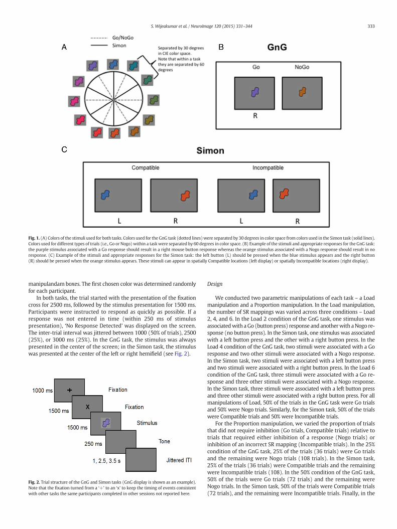
Fig. 1. (A) Colors of the stimuli used for both tasks. Colors used for the GnG task (dotted lines)were separated by 30 degrees in color space from colors used in the Simon task (solid lines).Colors used for different types of trials (i.e., Go or Nogo) within a taskwere separated by 60 degrees in color space. (B) Example of the stimuli and appropriate responses for the GnG task:the purple stimulus associated with a Go response should result in a right mouse button response whereas the orange stimulus associated with a Nogo response should result in noresponse. (C) Example of the stimuli and appropriate responses for the Simon task: the left button (L) should be pressed when the blue stimulus appears and the right button(R) should be pressed when the orange stimulus appears. These stimuli can appear in spatially Compatible locations (left display) or spatially Incompatible locations (right display).
333S. Wijeakumar et al. / NeuroImage 120 (2015) 331–344
manipulandam boxes. The first chosen color was determined randomlyfor each participant.
In both tasks, the trial started with the presentation of the fixationcross for 2500 ms, followed by the stimulus presentation for 1500 ms.Participants were instructed to respond as quickly as possible. If aresponse was not entered in time (within 250 ms of stimuluspresentation), ‘No Response Detected’ was displayed on the screen.The inter-trial interval was jittered between 1000 (50% of trials), 2500(25%), or 3000 ms (25%). In the GnG task, the stimulus was alwayspresented in the center of the screen; in the Simon task, the stimuluswas presented at the center of the left or right hemifield (see Fig. 2).
Fig. 2. Trial structure of the GnG and Simon tasks (GnG display is shown as an example).Note that the fixation turned from a ‘+’ to an ‘x’ to keep the timing of events consistentwith other tasks the same participants completed in other sessions not reported here.
Design
We conducted two parametric manipulations of each task – a Loadmanipulation and a Proportion manipulation. In the Load manipulation,the number of SR mappings was varied across three conditions – Load2, 4, and 6. In the Load 2 condition of the GnG task, one stimulus wasassociatedwith aGo (button press) response and anotherwith aNogo re-sponse (no button press). In the Simon task, one stimulus was associatedwith a left button press and the other with a right button press. In theLoad 4 condition of the GnG task, two stimuli were associated with a Goresponse and two other stimuli were associated with a Nogo response.In the Simon task, two stimuli were associated with a left button pressand two stimuli were associated with a right button press. In the Load 6condition of the GnG task, three stimuli were associated with a Go re-sponse and three other stimuli were associated with a Nogo response.In the Simon task, three stimuli were associated with a left button pressand three other stimuli were associated with a right button press. For allmanipulations of Load, 50% of the trials in the GnG task were Go trialsand 50% were Nogo trials. Similarly, for the Simon task, 50% of the trialswere Compatible trials and 50% were Incompatible trials.
For the Proportion manipulation, we varied the proportion of trialsthat did not require inhibition (Go trials, Compatible trials) relative totrials that required either inhibition of a response (Nogo trials) orinhibition of an incorrect SR mapping (Incompatible trials). In the 25%condition of the GnG task, 25% of the trials (36 trials) were Go trialsand the remaining were Nogo trials (108 trials). In the Simon task,25% of the trials (36 trials) were Compatible trials and the remainingwere Incompatible trials (108). In the 50% condition of the GnG task,50% of the trials were Go trials (72 trials) and the remaining wereNogo trials. In the Simon task, 50% of the trials were Compatible trials(72 trials), and the remaining were Incompatible trials. Finally, in the

Table 1Percentage (%) of correct trials for Go, Compatible and Incompatible trials. Overall accuracyof 86.5 % and above was achieved.
Percentage of correct trials(%)
Go NoGo Compatible Incompatible
Load 2 96.5 99.7 91.5 94.2Load 4 96.7 97.6 92.3 92.0Load 6 93.1 94.1 86.5 86.8Prop 25 93.5 97.1 91.8 93.9Prop 75 94.6 97.9 93.1 92.1
334 S. Wijeakumar et al. / NeuroImage 120 (2015) 331–344
75% condition of the GnG task, 75% of the trials were Go trials (108trials) and the remaining were Nogo trials. In the Simon task, 75% ofthe trials were Compatible trials (108 trials) and the remainingwere In-compatible trials. The Load was maintained at 4 stimulus-responsemappings across all manipulations of Proportion.
Participants completed five total runs on the GnG scanning day:Load 2 (50% Go, 50% Nogo), Load 4 (50% Go), Load 6 (50% Go), 25% con-dition (25% Go trials at Load 4), and the 75% condition (75% Go trials atLoad 4). Participants completed five runs on the Simon scanning day:Load 2 (50% Compatible), Load 4 (50% Compatible), Load 6 (50%Compatible), 25% condition (25% Compatible trials at Load 4), and the75% condition (75% Compatible trials at Load 4). There were eight pos-sible orders, randomly selected for each day: 6-4-2-25-75, 6-4-2-75-25, 2-4-6-25-75, 2-4-6-75-25, 25-75-2-4-6, 75-25-2-4-6, 25-75-6-4-2or 75-25-6-4-2. The order of the scanning days (GnG first versusSimon first) was counterbalanced across participants.
Image acquisition and processing
A 3T Siemens TIM Trio magnetic resonance imaging system with a12-channel head coil located at theMagnetic Resonance Research Facil-ity at the University of Iowawas used. Anatomical T1weighted volumeswere collected using an MP-RAGE sequence. Functional BOLD imagingwas acquired using an axial 2D echo-planar gradient echo sequencewith the following parameters: TE=30 ms, TR=2000 ms, flip angle=70°, FOV=240×240 mm, matrix=64×64, slice thickness/gap=4.0/1.0 mm, and bandwidth=1920 Hz/pixel. Each run was approximately15 minutes and collected 454 volumes.
Head movement was restricted using foam padding inserted be-tween the observer’s head and the head coil. Both tasks were presentedusing E-prime software and a high-resolution projection system. Thestimuli were subtended at a visual angle of 3.2–4.2 degrees. Responseswere recorded by a manipulandum strapped to the participants’hands. The timing of the presented stimuli was synchronized to thetrigger pulse from the MRI scanner.
Data were analyzed using Analysis of Functional NeuroImages(AFNI) software. Standard preprocessing was used that included slicetiming correction, outlier removal, motion correction, and spatialsmoothing (Gaussian FWHM=8mm). First-level analyses consisted ofconstructing a general linear model using afni_proc with regressors formotion, drifts in baseline, and ten regressors of interest for each task.In the GnG task, the regressors of interest were onset of the stimuluspresentations for Go and Nogo trials at Loads 2, 4 and 6 and at Propor-tions 25% and 75%. Similarly, in the Simon task, the regressors of interestwere onset of the stimulus presentations for Compatible and Incompat-ible trials at Loads 2, 4 and 6 and at Proportions 25% and 75%.
To analyze the group-level data, we used the percent signal changemaps for each regressor in two three-factor ANOVAs designed to identi-fy group-level effects associated with the Load and Proportion manipu-lations. The factors for the Load ANOVA were Task (GnG, Simon), Type(Go/Compatible, Nogo/Incompatible), and Load (2, 4 and 6). The factorsfor the Proportion ANOVA were Task (GnG, Simon), Type (Go/Compat-ible, Nogo/Incompatible), and Proportion (25%, 50%, 75%). Note that theType factor grouped trials that do not require any form of inhibition(Go/Compatible) and trials that require some form of inhibition (inhibi-tion of amotor response onNogo trials, and inhibition of an incorrect SRmapping on Incompatible trials). Although in the latter case we effec-tively group two different senses of inhibition, it is important to notethat differences between them could emerge in regions that show a sig-nificant Task × Type interaction.
Group level analyses were done using AFNI’s 3dMVM function.Resultant functional images of main effects and interactions werecorrected for family-wise errors using 3dClustSim (corrected at alphab0.05, corresponding to a cluster size threshold of and a cluster thresh-old of N27 voxels i.e 1158 ml). Centers of mass for the resulting signifi-cant clusters are reported in later tables. If effects shows clusters that
were large and could be broken into more regions, they wereintersected with an atlas and then re-labeled. 3dROIStats was thenused to compute the average percent signal change for each subjectacross all conditions for each cluster identified as significant withinthe main effect or interaction. For example, if a cluster in the right infe-rior parietal lobule showed amain effect of Load, an average percent sig-nal change was calculated for each subject across all voxels in thatcluster for Loads 2, 4, and 6. Paired two-tailed t-tests were performedon these average percent signal change values. The threshold for theset-tests were maintained at pb0.05. Note that no further correction formultiple comparisons was performed given that a correction wasalready carried out at the level of the omnibus ANOVAs. Moreover, thegoal of these t-tests was primarily descriptive in nature – to help de-scribe the pattern of data underlying the ANOVA effect in question.
ROI-based analyses were also carried out within 10 mm spherical re-gions defined using coordinates from previous reports. The goal was toinvestigate whether the need for Inhibitory control as the Proportion ofeach trial type was manipulated elicited activation in targeted ROIsincluding right inferior frontal cortex (IFG) – (Hampshire, 2015), pre-supplementary area (PreSMA) – (Hampshire et al., 2010) and rightsuperior temporal gyrus (STG) and right inferior parietal lobule (IPL) –collectively referred to as the right temporo-parietal junction (Corbettaet al., 2000). A 3-factor (Task, Type, Proportion) ANOVA was computedwithin this mask using a voxel-wise correction with pb0.01. Voxels thatshowed a main effect or interaction were clustered, and we computedan average percent signal change for the relevant effect. Paired two-tailed t-tests were performed on these average percent signal changevalues with pb0.05. Note that average percent signal change withineach ROI was also calculated for each subject. A three-factorial ANOVAondata fromeachROI revealed no significantmain effects or interactions.
Results
Behavioral results
Only correct trials were analyzed as accuracy was over 86% for bothtasks and all conditions (see Table 1).
For the GnG task, two (Load, Proportion) one-factor repeated mea-sures ANOVAswere conducted on reaction times (RTs) for Go trials. Re-sults from the ANOVA examining the effect of Proportion (25%, 50%,75%) on RTs revealed a main effect of Proportion, F(2,38) = 6.546,pb0.005. Post-hoc comparisons using t-tests with revealed that RTswere significantly slower when 25% of the trials were Go trials as com-pared towhen 75% of the trials were Go trials, pb0.005 (Fig. 3A). Resultsfrom the ANOVA examining the effect of Load (2, 4, 6) revealed a maineffect of Load, F(2,38) = 48.892, pb0.001. Post-hoc comparisons re-vealed that RTs increased with increasing Load for all pairwise compar-isons, pb0.001 (Fig. 3B).
For the Simon task, results from a two-way ANOVA with Proportion(25%, 50%, 75%) and Compatibility (Compatible, Incompatible) as fac-tors revealed a main effect of Compatibility on RTs, F(1,19) = 14.251,pb0.005, with greater RTs for Incompatible trials than for Compatibletrials (Fig. 3C). A second two-way ANOVA with Load (2, 4, 6) and Com-patibility as factors revealed a main effect of Load on RTs, F(2,38) =

Fig. 3. (Top) Effect of Proportion on RTs for (A) Go trials. RTs at 25%were significantly higher than at 75%. (B.) Compatible and Incompatible trials. RTs to Incompatible trials were greaterthan for Compatible trials. (Bottom) Effect of Load on RTs for (C.) Go trials. RTs increasedwith increase in number of stimulus-response (SR) mappings. (D.) Compatible and Incompatibletrials. RTs increasedwith increase in number of SRmappings across Compatible and Incompatible trials. Across all Loads, RTs to Incompatible trials were greater than to Compatible trials.
Table 2Regions activated for Proportion × Type interaction (pb0.05, corrected).
Cluster Region Hemi BA Talaraich (RAI) (mm) Size (ml) F(4,76)
x y z
Insula L 13 29.1 −9.5 7 4630 8.03R 13/47 −33.9 −14 3.6 3473 9.32
Thalamus R - −2.6 18.7 5 3773 8.28Putamen R - −26.8 10.7 −3 1372 7.60
335S. Wijeakumar et al. / NeuroImage 120 (2015) 331–344
46.785, pb .001, with increasing RTs as load increased. Post-hoccomparisons revealed that all pairwise Load comparisons differedsignificantly, pb0.001 (Fig. 3D). There was also a main effect ofCompatibility, F(1,19) = 10.384, pb0.005 with greater RTs for Incom-patible trials than for Compatible trials. Note that the interactionbetween Load and Compatibility was not significant.
Overview of fMRI analysis approach
We analyzed the fMRI data using two omnibus ANOVAs. The firstthree-way ANOVA focused on the Proportion manipulation to examinewhether Inhibitory control areas are sensitive to the need for control oninfrequent trials. This ANOVA included Proportion (25%, 50%, 75%), Type(Go/Compatible, Nogo/Incompatible), and Task (GnG, Simon) aswithin-subject factors. Our central question is addressed by examiningmain effects of Proportion and Proportion-related interactions. Thesecond ANOVA focused on the Load manipulation to examine whetherparticular brain regions show an increase in activation as the numberof SR mappings increases consistent with the neural signature ofWM. This ANOVA included Load (2, 4, 6), Type (Go/Compatible, Nogo/Incompatible), and Task (GnG, Simon) as within-subject factors. Here,our central question is addressed by examining main effects of Loadand any Load-related interactions.
Critically, the omnibus ANOVAs have common factors of Type andTask even though they involve largely independent sets of data. This al-lows us to partition all effects that do not involve the Proportion or Loadfactors into two key categories: Task-general effects (e.g., Type main ef-fects) and Task-specific effects (e.g., Task main effects and Task-relatedinteractions). The latter effects reveal the brain systems critically in-volved in global reactive control versus selective reactive control. Notethat, Nogo trials do not require a response whereas Incompatible trialsdo require a response. Thus, Type × Task interactions may also reflectdifferences in the amount of motor output.
How is brain activity modulated as the proportion of each Type is varied?The Proportion × Type × Task ANOVA yielded significant Proportion
× Type and Proportion × Task interactions, but no other significantProportion-related effects after family-wise correction.
Proportion × Type interactions. When Go/Compatible trials wereinfrequent (25% condition), there was robust activation on these trialsrelative to Nogo/Incompatible trials in bilateral insula, right putamen,and right thalamus (see Table 2 and Fig. 4). By contrast, as Nogo/Incompatible trials became less frequent (75% condition), therewas a robust increase in activation on these trials relative to Go/Compatible trials in the same regions. Indeed, in right putamen, thepattern completely reversed across the Proportionmanipulation, show-ing robust activation on infrequent trials, regardless of whether they re-quired inhibition. Thus, activation in these regions reflects the need forcontrol on infrequent types rather than the need for inhibition per se.
Proportion × Task interactions. There was a significant Proportion × Taskinteraction in the thalamus (see Table 3 and Fig. 5), indicating that therewere different demands placed on the need for control in the two tasks.In particular, the need for control was greatest in the GnG task in the25% condition where the infrequent trials required an active response(i.e., the Go trials). Thus, the thalamus is responsive to the need for con-trol on infrequent trialswhen a response is needed (vs. Nogo trials). TheSimon task requires an active response on both Compatible and Incom-patible Types. Here we see the greatest modulation of thalamic activityin the 75% condition when Incompatible trials were infrequent, that is,when an infrequent response must be activated and this responserequires inhibition of a strongly prepotent response.
ROI-based analyses. Whole-brain analyses did not yield significant dif-ferences in activation for regions in right IFG and pre-SMA as the needfor control was varied. It is possible effects in these canonical

Fig. 4. Bar plots showing average percent signal change± S.E.M. across Go/Compatible trials and Nogo/Incompatible trials at Proportions of 25 %, 50 % and 75 %. Significance at p b 0.05 isdenoted by ‘*’.
336 S. Wijeakumar et al. / NeuroImage 120 (2015) 331–344
inhibitory-control regions were weak and might not have survivedwhole-brain correction for multiple comparisons. Thus, we conducteda ROI-based analysis, examining activity in right IFG, preSMA, andright TPJ. Results from a three-factorial ANOVA revealed a significantType × Proportion interaction in the right STG and a significant Task ×Proportion × Task interaction in the right middle temporal gyrus(MTG). Fig. 6a shows the Type × Proportion effect for right STG. Thisarea followed the same pattern reported in the whole brain analyses:there was greater activation on Go/Compatible trials in the 25% condi-tion and there was an increase in activation on Nogo/Incompatiable tri-als as these trials became less frequent in the 50% and 75% conditions.This trend was also observed in right MTG, but only for the GnG task(see Fig. 6b). Note that there were no significant effects in the rightIFC and preSMA ROIs.
How is brain activity modulated as the memory load increases?The Load × Type × Task ANOVA revealed main effects of Load and
Load × Type interactions. No other Load-related effects reached signifi-cance after family-wise correction.
Table 3Region activated for an interaction between Proportion and Task (pb0.05, corrected).
Cluster Region Hemi BA Talaraich (RAI)(mm)
Size (ml) F(4,76)
x y z
Thalamus R - −10 2.3 12 1501 7.53
Main effects of load. Activation in the right inferior parietal lobule waslargest for a Load of two items and decreased with an increase in Load(Table 4 and Fig. 7). This pattern is the opposite of what is typically ob-served in studies of WM. This suggests that the SR mappings in thesetasks are not actively maintained in WM. It is possible that anotherform of memory such as associative memory is responsible for remem-bering the task rules.We return to this possibility in greater detail below.
Load × Type interactions. A broad network of regions showed significantLoad × Type interactions (see Table 5 and Figs. 8 and 9). This networkincluded the bilateral cerebellum, bilateral superior temporal gyrus,right transverse temporal gyrus, right parahippocampal gyrus, right lin-gual gyrus, right fusiform gyrus, right insula, and right cuneus/posteriorcingulate cortex.
In the right transverse temporal gyrus, left superior temporal gyrus,right insula, and bilateral cerebellum, therewas a significant decrease inactivation as the Load increased, consistentwith the general pattern ob-served in the right inferior parietal lobule for the main effect of Load.Here, however, the decrease over Load only occurred for the Go/Com-patible trials – there was no significant change in activation as theLoad increased on Nogo/Incompatible trials. We also directly comparedactivation on the Go/Compatible and Nogo/Incompatible trials at eachLoad. Here, all regions showed significantly greater activation on Go/Compatible trials at Load 2 relative to Nogo/Incompatible trials. Greateractivation on Go/Compatible trials versus Nogo/Incompatible trials wasalso evident at Loads 4 and 6 in the right transverse temporal gyrus andright fusiform gyrus.

Fig. 5. Bar plots showing average percent signal change± S.E.M. for GnG and Simon tasks at Proportions of 25%, 50% and 75%. Note, Go and Nogo trials were pooled together for the GnGtask and Compatible and Incompatible trials were pooled together for the Simon task. Significance at p b 0.05 is denoted by ‘*’.
337S. Wijeakumar et al. / NeuroImage 120 (2015) 331–344
Once again, these results are not consistent with the hypothesis thatthe SR mappings are actively maintained in WM. Further, the differen-tial activation on Go/Compatible trials suggests that these mappingsare represented differently than mappings during Nogo/Incompatibletrials. It is possible, for instance, that the mappings during the Go trialsare stored in an associative memory that varies systematically byLoad, while the mappings during Nogo trials are not remembered. Wereturn to this possibility in the Discussion.
Do the demands of trial Type generalize across tasks?Across the two omnibus ANOVAs, there was a broad network of
brain regions that showed a common Typemain effect in both analyseswith greater activation on Go/Compatible versus Nogo/Incompatibletrials. This network included bilateral transverse temporal gyrus,
Fig. 6. Bar plots showing average percent signal change ± S.E.M. ROIs for (a) Right superior te
bilateral insula, bilateral superior temporal gyrus, left cingulate gyrus,left inferior parietal lobule, left precentral gyrus, bilateral medial frontalgyrus, right cerebellum, left putamen and bilateral thalamus (seeTable 6). An example pattern of activation is shown in Fig. 10B for theleft transverse temporal gyrus. This region showed greater activationon Go/Compatible trials when there were no inhibitory demands, sug-gesting that this region is actively involved in response selection and/orgeneration.
Note that the spatial pattern of results showed good overlap acrossthe two omnibus ANOVAs. An example is shown in Fig. 10A. Yellowcolors show voxels with a significant Type main effect in both ANOVAs,while red colors show voxels with an effect that was unique to oneANOVA or the other. Given the robust overlap, we only show significantfindings in Table 6 for clusters that intersect across both ANOVAs.
mporal gyrus (b) Right middle temporal gyrus. Significance at p b 0.05 is denoted by ‘*’.

Table 4Region activated for the main effect of Load (pb0.05, corrected).
Cluster Region Hemi BA Talaraich (RAI)(mm)
Size (ml) F(2,38)
x y z
Inferior Parietal Lobule R 13/41 −44.2 32 22.3 1415 11.49
Table 5Regions activated for Load × Type interaction (pb0.05, corrected).
Cluster Region Hemi BA Talaraich (RAI)(mm)
Size(ml)
F(4,76)
x y z
Cerebellum L - −6 45 −13 5317 8.82Transverse TemporalGyrus
R 41/13 −44.2 25.9 12 2144 8.44
Cuneus / PosteriorCingulate Cortex
R 30 −10.9 67.8 13.3 1758 7.68
Cerebellum R - −35.3 52.5 −23 1715 7.49Insula R - −33 −4.3 5 1629 8.33Superior Temporal Gyrus L 41 41.5 34 7.1 1501 8.21
R 13 −45.3 47.3 18.2 1286 9.61Parahippocampal Gyrus R 30 −24 −19 46 1029 8.18
338 S. Wijeakumar et al. / NeuroImage 120 (2015) 331–344
What patterns of brain activity are specific to each task?The two omnibus ANOVAs revealed two sets of task-specific effects:
significant Task main effects and significant Task × Type interactions.Given that the latter results relate to the Type main effects discussedin the previous section, we discuss these effects first and then concludewith the Task main effect.
Lingual Gyrus R 19 16 −49 −1 300 10.10Fusiform Gyrus R 37 −37 25 −7.5 129 7.00
Task × Type interactions. A broad network of regions showed clusterswith significant Task × Type interactions. An example for the left insulais shown in Fig. 11B. Go trials elicited greater activation thanNogo trials,with comparable activation on Compatible and Incompatible trials. Aswith the Type main effect described above, there was robust spatialoverlap in results from the two omnibus ANOVAs. Fig. 11A shows an ex-ample of the overlap between these regions.
Table 7 shows the full set of results for clusters that intersectedacross both ANOVAs. The areas showing common activation acrossboth ANOVAs were left transverse temporal gyrus, left insula, left supe-rior temporal gyrus, left inferior parietal lobule, left postcentral gyrus,bilateral medial frontal gyrus, right cerebellum, left putamen and bilat-eral thalamus. In all of these regions, therewas significantly stronger ac-tivation on Go trials than on Nogo trials. There was stronger activationfor Go trials than Compatible trials in left insula, left postcentral gyrus,right cerebellum, left putamen and left thalamus. Incompatible trialselicited greater activation than Nogo trials in the left transverse tempo-ral gyrus, left insula, left IPL, bilateralmedial frontal gyrus, right cerebel-lum, left putamen and bilateral thalamus. Note that, these results wereconsistent across both ANOVAs.
What is driving the Task × Type interactions? Recall that activationdecreased as the number of Go/Compatible S-R mappings increased(Load main effect), and this effect was particularly robust on Go/Com-patible trials (Load × Type interaction). It is possible we are seeing a re-lated effect here. In GnG, there were 1, 2, or 3 stimuli that required anactive response. In Simon, there were 2, 4, or 6 stimuli that required aresponse. The Task × Type interaction might reflect this differenceacross tasks, with greatest activation on the Go trials given that thereare few SR mappings where a response is required, weaker activationin Simon given that there aremany SRmappingswhere a response is re-quired, and weakest activation on Nogo trials because no response was
Fig. 7. Bar plots showing average percent signal change ± S.E.M. for Loads 2, 4 and 6 (pb0.05, unificance at p b 0.001 is denoted by ‘*’.
required. Thus, there appears to be weaker engagement of motor-related areas as the number of stimuli that require a manual responseacross tasks is increased.
Task main effect. The Simon task elicited positive activation in bilaterallingual gyri whereas activation was negative for the GnG task. This re-sult was consistent across both ANOVAs (see Table 8 and Fig. 12). In-creased lingual gyri activation has been previously linked to shifts ofvisuo-spatial attention to the contralateral hemifield of stimulation.The Simon task places a heavy burden on control of spatial attention, be-cause on Compatible trials, spatial attention can facilitate respondingwhile on Incompatible trials, spatial attention can delay correctresponding. Increased lingual gyri activation may reflect heightenedsensitivity to spatial information in the Simon task. By contrast, stimuliin the GnG task were always presented in the center of the display andGo responses were mapped to a single response button. Both of thesefactors could have resulted in attenuated activation in the lingual gyriin the GnG task.
Discussion
Previous studies investigating inhibitory control have shown that adistributed fronto-cortical-striatal system is involved in response inhi-bition and response selection including IFG/IFJ, DLPFC, pre-SMA, SMA,insula, IPL, motor and pre-motor cortices, and sub-cortical regionssuch as the caudate, putamen, and thalamus. The present study usedtwo reactive control tasks – GnG and Simon – to investigate whetherthis broad network is sensitive to the inhibitory demands of the taskor the need for control as attentional and WM demands are varied.
ncorrected). Note, that beta values were computed by pooling across Type and Task. Sig-

Fig. 8. Regions activated for an interaction between Load and Type. Significance at p b 0.05 is denoted by ‘*’.
339S. Wijeakumar et al. / NeuroImage 120 (2015) 331–344
Below, we discuss findings that emerged from our multi-factorialANOVAs and then discuss how these regions work together to carryout cognitive control.
A cortical-subcortical circuit is activated by the need for control on rareevents
Previous fMRI studies have shown robust IFC (Aron, 2011; Liddleet al., 2001; Munakata et al., 2011; Simmonds et al., 2008; Wageret al., 2005), insula (Dodds et al., 2011; Wager et al., 2005), and sub-cortical activation (Kelly et al., 2004; Liddle et al., 2001; Wager et al.,2005) in reactive inhibitory control tasks such as the GnG task. Afterfindings from recent work, Aron and colleagues updated their claimsto report that rIFG was activated by a stop signal or an unexpectedevent and engaged a brake by slowing down, pausing or completelystopping an action (Aron et al., 2014a,b). Most studies, however, haveprobed response control by employing a smaller number of trials that
Fig. 9. Bar plots showing average percent signal change ± S.E.M. for the inter
require inhibition than trials that do not (Kelly et al., 2004; Mostofskyet al., 2003; Wager et al., 2005). This confounds inhibitory controlwith modulation of response selection processes on rare,attentionally-demanding events. Thus, in the present study, we variedthe proportion of trials to probewhether cortical and subcortical activa-tion was specific to the need for inhibition on Nogo and Incompatibletrials, or whether these networks were robustly active onattentionally-demanding rare events regardless of the need for inhibi-tion. Results show that bilateral insula, right putamen, and right thala-mus were active on infrequent trials, regardless of whether these trialsrequire or do not require inhibition and regardless of whether the taskrequired ‘global’ or ‘selective’ control.
Results showing insula activation on rare, attentionally demandingtrials are consistent with previous studies that have identified thisarea as part of a salience network (Menon and Uddin, 2010; Sridharanet al., 2008; Uddin, 2015). The insular cortex facilitates detection of tar-get stimuli amongst distractors in oddball paradigms, suggesting this
action between Load and Type. Significance at p b 0.05 is denoted by ‘*’.

Table 6Regions activated for amain effect of Type for the intersection between the Proportion andLoad ANOVAs (pb0.05, corrected). Only regions with clusters N 2 voxels (86 ml) arepresented and discussed.
Cluster Region Hemi BA Size(ml)
Talaraich (RAI) (mm) LoadF(X,Y)
ProportionF(X,Y)
x y z
Transversetemporalgyrus
L 41 472 42.6 25 10 28.25 27.11R 41 129 −40.2 25.5 10 18.44 25.70
Insula L 13 7546 38.1 13.5 13.9 40.57 34.44R 13 472 −43 28.4 17.4 22.91 23.83L 13 43 33.2 1 6.5 34.67 18.37
Superiortemporalgyrus
L 41 858 44.1 29.8 15.6 38.76 30.75R 41 257 −46.2 27.6 15.4 27.92 28.49
Cingulate Gyrus L 24 300 6.7 2 2.3 35 32.52 29.91Inferior parietallobule
L 13 129 43.8 32.4 24 45.26 24.56
Precentral Gyrus L 13 129 44.6 −0.5 6.5 39.51 31.87Medial FrontalGyrus
L 6 600 3.3 6.9 49.9 35.16 27.67R 6 300 −2.6 5.7 48.9 29.92 24.79
Cerebellum R 19 1115 −20.6 57.3 −15.1 46.26 22.73R 37 5274 −23.1 45.8 −18.2 51.11 28.54
Putamen L - 557 26.5 9.6 2 33.66 20.46Thalamus L - 2958 11.7 18.8 10.3 27.34 31.66
R - 515 11.7 18.8 10.3 25.41 24.82
Fig. 11. (A) Regions activated by an interaction between Type and Task for the Proportionand Load ANOVAs. Regions common across both the Proportion and Load ANOVAs areshown in yellow. Regions unique to the Proportion or Load ANOVAs are shown in red.(B) Canonical bar plot of left insula but also representative of all activated regions reportedin Table 7, showing average beta coefficient values representative of all Go and Compatible(Comp) trials and Nogo and Incompatible (Incomp) trials for GnG and Simon tasks.Significance at p b 0.05 is denoted by ‘*’.
340 S. Wijeakumar et al. / NeuroImage 120 (2015) 331–344
area plays a role in salience detection (Crottaz-Herbette and Menon,2006; Downar et al., 2000). The insular cortex jointly with the IFC hasalso been implicated in switching between the default mode networkand the central executive network in saliency-detection tasks thatspanned the visual and auditory modalities (Sridharan et al., 2008).This is consistent with a review of insular cortex function by Menonand colleagueswho suggested that the function of the insula is to detectsalient events and to switch between large-scale networkswhen salientevents are detected via strong coupling between the insula, the anteriorcingulate cortex, and the motor system (Menon and Uddin, 2010).
Fig. 10. (A) Regions activated for a main effect of Type for the Proportion and LoadANOVAs. Regions common across both the Proportion and Load ANOVAs are shown inyellow. Regions unique to the Proportion or Load ANOVAs are shown in red.(B) Canonical bar plot of left transverse temporal gyrus but also representative of allactivated regions reported in Table 6. Significance at p b 0.05 is denoted by ‘*’.
A recent study tried to dissociate the functional roles of the right in-ferior frontal cortex and anterior insula, which have both been implicat-ed in inhibitory control (Cai et al., 2014). Their meta-analysis of 70published studies employing the stop-signal task and GnG suggestedthat the right anterior insula is important for saliency detection butrIFC is important for inhibitory control. By contrast, the right insulaand IFC were reported to not be uniquely engaged in inhibitory control;rather, that these areas belonged to a distributed network engaged inattentional and working memory processes (Erika-Florence et al.,2014). Consistent with this, insular cortex was robustly activated in
Table 7Regions activated for an interaction between Task and Type for the intersection betweenthe Proportion and Load ANOVAs (pb0.05, corrected). Only regions with clusters N 2voxels (86 ml) are presented and discussed.
Cluster Region Hemi BA Size(ml)
Talaraich (RAI) (mm) LoadF(X,Y)
ProportionF(X,Y)
x y z
TransverseTemporalGyrus
L 41 557 39.9 26.6 10 24.99 20.26
Insula L 13 7203 37 15.3 14.9 32.79 26.07L 13 86 33.2 −0.6 6.5 23.78 20.40
SuperiorTemporalGyrus
L 41 1029 43.1 30.2 15.3 28.53 22.82
Inferior ParietalLobule
L 13 129 43.8 32.2 24 33.15 26.15
PostcentralGyrus
L 40 129 51.4 25.5 15.5 33.57 18.62
Medial FrontalGyrus
L 6 643 3.1 5.3 49.9 26.83 23.84R - 343 −1.8 6.2 49.8 23.37 18.52R - 2015 −20.2 59.2 −15.1 19.70 26.23
Cerebellum R - 5917 −19.7 47.4 −16.5 25.98 30.48R - 214 −7.7 46.5 −13.6 23.79 25.63
Putamen L - 1372 24.8 4.7 4.3 20.25 17.37Thalamus L - 3173 12.9 19 10.5 21.42 25.36
R - 986 −11 12.8 13.4 19.21 27.09
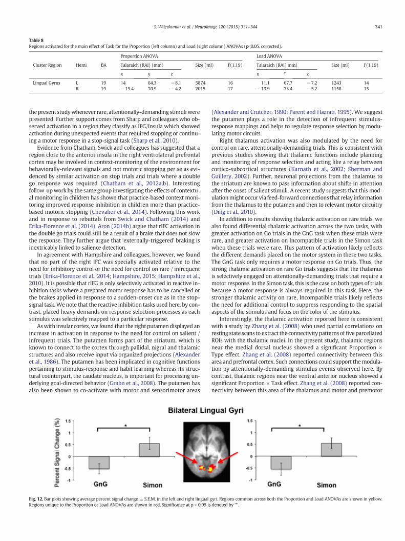
Table 8Regions activated for the main effect of Task for the Proportion (left column) and Load (right column) ANOVAs (pb0.05, corrected).
Proportion ANOVA Load ANOVA
Cluster Region Hemi BA Talaraich (RAI) (mm) Size (ml) F(1,19) Talaraich (RAI) mm) Size (ml) F(1,19)
x y z x y z
Lingual Gyrus L 19 14 64.3 −8.1 5874 16 11.1 67.7 −7.2 1243 14R 19 −15.4 70.9 −4.2 2015 17 −13.9 73.4 −5.2 1158 15
341S. Wijeakumar et al. / NeuroImage 120 (2015) 331–344
thepresent studywhenever rare, attentionally-demanding stimuliwerepresented. Further support comes from Sharp and colleagues who ob-served activation in a region they classify as IFG/Insula which showedactivation during unexpected events that required stopping or continu-ing a motor response in a stop-signal task (Sharp et al., 2010).
Evidence from Chatham, Swick and colleagues has suggested that aregion close to the anterior insula in the right ventrolateral prefrontalcortex may be involved in context-monitoring of the environment forbehaviorally-relevant signals and not motoric stopping per se as evi-denced by similar activation on stop trials and trials where a doublego response was required (Chatham et al., 2012a,b). Interestingfollow-upwork by the same group investigating the effects of contextu-al monitoring in children has shown that practice-based context moni-toring improved response inhibition in children more than practice-based motoric stopping (Chevalier et al., 2014). Following this workand in response to rebuttals from Swick and Chatham (2014) andErika-Florence et al. (2014), Aron (2014b) argue that rIFC activation inthe double go trials could still be a result of a brake that does not slowthe response. They further argue that ‘externally-triggered’ braking isinextricably linked to salience detection.
In agreement with Hampshire and colleagues, however, we foundthat no part of the right IFC was specially activated relative to theneed for inhibitory control or the need for control on rare / infrequenttrials (Erika-Florence et al., 2014; Hampshire, 2015; Hampshire et al.,2010). It is possible that rIFG is only selectively activated in reactive in-hibition tasks where a prepared motor response has to be cancelled orthe brakes applied in response to a sudden-onset cue as in the stop-signal task.We note that the reactive inhibition tasks used here, by con-trast, placed heavy demands on response selection processes as eachstimulus was selectively mapped to a particular response.
Aswith insular cortex,we found that the right putamen displayed anincrease in activation in response to the need for control on salient /infrequent trials. The putamen forms part of the striatum, which isknown to connect to the cortex through pallidal, nigral and thalamicstructures and also receive input via organized projections (Alexanderet al., 1986). The putamen has been implicated in cognitive functionspertaining to stimulus-response and habit learning whereas its struc-tural counterpart, the caudate nucleus, is important for processing un-derlying goal-directed behavior (Grahn et al., 2008). The putamen hasalso been shown to co-activate with motor and sensorimotor areas
Fig. 12. Bar plots showing average percent signal change ± S.E.M. in the left and right lingual gRegions unique to the Proportion or Load ANOVAs are shown in red. Significance at p b 0.05 is
(Alexander and Crutcher, 1990; Parent and Hazrati, 1995). We suggestthe putamen plays a role in the detection of infrequent stimulus-response mappings and helps to regulate response selection by modu-lating motor circuits.
Right thalamus activation was also modulated by the need forcontrol on rare, attentionally-demanding trials. This is consistent withprevious studies showing that thalamic functions include planningand monitoring of response selection and acting like a relay betweencortico-subcortical structures (Karnath et al., 2002; Sherman andGuillery, 2002). Further, neuronal projections from the thalamus tothe striatum are known to pass information about shifts in attentionafter the onset of salient stimuli. A recent study suggests that this mod-ulationmight occur via feed-forward connections that relay informationfrom the thalamus to the putamen and then to relevant motor circuitry(Ding et al., 2010).
In addition to results showing thalamic activation on rare trials, wealso found differential thalamic activation across the two tasks, withgreater activation on Go trials in the GnG task when these trials wererare, and greater activation on Incompatible trials in the Simon taskwhen these trials were rare. This pattern of activation likely reflectsthe different demands placed on the motor system in these two tasks.The GnG task only requires a motor response on Go trials. Thus, thestrong thalamic activation on rare Go trials suggests that the thalamusis selectively engaged on attentionally-demanding trials that require amotor response. In the Simon task, this is the case on both types of trialsbecause a motor response is always required in this task. Here, thestronger thalamic activity on rare, Incompatible trials likely reflectsthe need for additional control to suppress responding to the spatialaspects of the stimulus and focus on the color of the stimulus.
Interestingly, the thalamic activation reported here is consistentwith a study by Zhang et al. (2008) who used partial correlations onresting state scans to extract the connectivity patterns offive parcellatedROIs with the thalamic nuclei. In the present study, thalamic regionsnear the medial dorsal nucleus showed a significant Proportion ×Type effect. Zhang et al. (2008) reported connectivity between thisarea and prefrontal cortex. Such connections could support themodula-tion by attentionally-demanding stimulus events observed here. Bycontrast, thalamic regions near the ventral anterior nucleus showed asignificant Proportion × Task effect. Zhang et al. (2008) reported con-nectivity between this area of the thalamus and motor and premotor
yri. Regions common across both the Proportion and Load ANOVAs are shown in yellow.denoted by ‘*’.

342 S. Wijeakumar et al. / NeuroImage 120 (2015) 331–344
cortex. Such connections are consistent with our findings showingmodulation of ventral anterior thalamus by both attentional demandsand demands on motor output.
An interesting result that emerged from our ROI analyses is the sig-nificant interaction between Proportion and Type in the right STG. Thisarea showed effects similar to those observed in whole-brain analysesfrom the insular-putamen-thalamic network with greater activationon infrequent trials. A long of line of research from Corbetta and col-leagues have proposed that a ventro-parietal network with a specificfocus on the right TPJ acts as a ‘circuit-breaker’ to direct attention to arelevant stimulus that is outside of the current focus (Corbetta et al.,2000; Corbetta and Shulman, 2002). Further research has demonstratedthat similar effects occur when using visual and auditory stimulationand not just with changes in spatial information (Braver et al., 2001;Downar et al., 2000, 2001). In line with proposals by Corbetta and col-leagues,we suggest that the insular network is responsible for detectinginfrequent / salient events, and then lateralized TPJ serves as an ‘alertingsystem’, directing attention away from the current set of task rules(i.e., away from the frequent events). It is interesting that MTG onlyshowed a modulation of activity in the GnG task. Thus, parts of TPJ ap-pear to be selectively engaged in tasks that activate or inactivatemotor responses. It is important to note that these results did notemerge in thewhole-brain analysis; thus, additional work will be need-ed to tease apart interactions between the insular network and TPJ.
In summary, ourmanipulation of the proportion of trials that requireinhibition revealed a pattern activation in the insula, putamen, thala-mus, and TPJ that indicated these regions are sensitive to rare and infre-quent stimuli, rather than to the need for inhibitory control. A centralquestion moving forward is precisely how these regions modulate re-sponse selection system in the brain. It is possible that the activationpatterns observed here reflect active global inhibition or down-regulation of the motor system in the face of the need for control onrare events (Erika-Florence et al., 2014). That is, insula, putamen, andthalamus might be slowing down response processes in the face ofrare events to facilitate accurate response selection, with TPJ workingto actively shift attention to competing alternatives. It is also possible,however, that insula, putamen, and thalamus are enhancing processingor up-regulating response selection areas in the face of response conflicton rare, attentionally-demanding events (Erika-Florence et al., 2014).That is, these brain areas might boost activation in response selectionregions on rare events when conflict is detected. Note that existingmodels of response inhibition and response selection such as themodel by Weicki and Frank have only feed-forward connections be-tween cortical areas and subcortical regions of the basal ganglia(Wiecki and Frank, 2013). It will be important in future theoreticalwork to understand how reciprocal connectivity might enable down-regulation vs. up-regulation and to tease apart differential predictionsfor these two alternatives.
Activation in the response selection network decreases as the memory loadincreases
Previous studies have suggested that response selection and inhibi-tion arises from distributed cortical and subcortical networks involvedin attention and working memory. For instance, several studies havedemonstrated that frontal and parietal cortices are robustly activatedin both inhibitory control and working memory tasks (Barber et al.,2013; Bunge et al., 2001; Hester et al., 2004; Kelly et al., 2006; McNabet al., 2008; Mostofsky et al., 2003; Perlstein et al., 2003). In particular,McNab and colleagues used conjunction analysis and showed right infe-rior frontal gyrus activation across two working memory and two inhi-bition tasks (McNab et al., 2008). Similarly, Barber et al. (2013) showedthat the dorsolateral prefrontal cortex was recruited in two GnG taskswhen working memory was required to inhibit a response.
To address how working memory demands affect fronto-cortical-striatal control systems, we modulated the need for control in two
reactive inhibition tasks by varying the memory load, while holdingthe attentional demands constant (i.e., Go/Compatible and Nogo/Incompatible trials were equally frequent). If the fronto-cortical-striatal network is sensitive to the attentional and WM demandsof the task, we expected to see the same areas activated by the atten-tional manipulation also active as the memory load was varied. TheLoadmanipulation also speaks to the nature of thememory representa-tion underlying response selection in these tasks. Previous studies havedemonstrated thatWMmaintenance has a particular neural signature –activation increases as theWM load increases (Pessoa et al., 2002; ToddandMarois, 2004). Thus, ifWMwas critically involved in these tasks, wewould expect to see an increase in activation as the load increasedwith-in WM-specific regions of the fronto-cortical-striatal network. For in-stance, Hester et al. (2004) found an increase in activation as thememory load was increased in a GnG task within middle frontalgyrus, left middle temporal gyrus, thalamus, and rostral and dorsalACC/preSMA (for related results, see Bunge et al. (2001).
Interestingly, results from IPL in the current study showed the oppo-site pattern – activation in the right IPL decreased as the memory loadincreased. Previous studies suggest that IPL is responsible for processingsensory andmotor information (Andersen, 2011; Andersen et al., 1985;Fogassi et al., 2005; Fogassi and Luppino, 2005; Hyvarinen, 1981). Func-tional sub-divisions of the monkey IPL have distinguished areas 7a and7b in the monkey cortex responsible for visual and visuomotor func-tions respectively. One hypothesis, the vasomotor integration hypothe-sis suggests that IPL combines information from sensory and motornodes - for detailed review, see Andersen (2011). This is consistentwith our findings showing modulation of IPL activation as the numberof stimulus-response mappings was varied.
Anatomical tracing experiments have identified many connectionsfrom the IPL to other cortical regions, including the superior temporalregion, parahippocampal area, cingulate cortex, and the insula(Andersen, 2011). Interestingly, in the current study, these regionsalong with the transverse temporal gyrus, cerebellum, and lingualgyrus showed the same pattern of activation as rIPL but only for trialsthat did not require inhibition. Specifically, these regions showedsignificantly greater activation on Go/Compatible trials than Nogo/Incompatible trials at Load 2, and as the load increased, activation onGo/Compatible trials decreased. It is possible that the absence of aload effect on Nogo/Incompatible trials in these regions reflects weakmemory traces on these trials. Recent behavioral work suggests that re-sponse inhibition reduces attention to stimuli, yieldingweakermemorytraces at encoding and retrieval (Chiu and Egner, 2015).
The current study showed decreasing activation with increasingnumber of SR mappings in the right IPL. On the other hand, Hesteret al. (2004) showed the opposite pattern of activation. Why do theseresults differ? There are several differences across studies thatmight ex-plain our findings. Hester et al. (2004) used a verbal working memorytask rather than a visual working memory task. Thus, it is possible thatthere are important differences in how these twoworkingmemory sys-tems are engaged in response selection and inhibition tasks. In addition,Hester et al. (2004) only examined effects of memory load in a GnG taskwhere the Nogo trials were infrequent. It is possible that their findingsreflect an interaction between attentional and working memory pro-cesses not observed here given that we held the attentional demandsconstant across load conditions. Finally, we note that results fromHester et al. (2004) were mixed in that several fronto-cortical-striatalareas showed either no change in activation as the load increased or adecrease in activation as the load increased as reported here (e.g., inthe cerebellum).
Clearly, our findings are not consistentwith the typical neural signa-ture observed in studies ofworkingmemory that activation increases asthe load increases. An alternative possibility is that our results reflectthe engagement of an associativememory system rather than aworkingmemory system. For instance, in Wiecki and Frank’s model of responseselection and inhibition, SR mappings are stored in an associative

343S. Wijeakumar et al. / NeuroImage 120 (2015) 331–344
memory (Wiecki and Frank, 2013).More SRmappingsmake it harder toresolve the competition among different associative weights, slowingdown decision-making and enabling conflict-monitoring networks toexert control. It is possible that such effects yield a decrease in theBOLD response as thememory load is increased because growth of acti-vation is limited by enhanced competition. Future work will be neededto examine the possibility.
Note that our region of interest in the right IPL lies in the vicinity ofright STG andMTG, whichwere activated when Proportion wasmanip-ulated. Corbetta et al. (2000) demonstrated two regions of activation inthe vicinity of right TPJ (right STG, right IPL) while reorienting attentiontowards novel stimuli. In the current study, we did not explore the ef-fects of manipulating Load whilst manipulating Proportion; instead,we maintained Proportion at 50% in all Load conditions. It is possiblewe might see interactions between right IPL and STG/MTG when bothfactors are manipulated together.
A distributed cortico-subcortical system is engaged by motor outputdemands
Go/Compatible trials elicited greater activation than Nogo/Incom-patible trials within a host of cortical and subcortical regions. Thebroad network of regions showing this pattern is known to be involvedin managing sensory-motor mappings as well as motor planning andcontrol. For instance, the insula, putamen, and thalamus are involvedin detecting salient stimuli and activating appropriate motor circuitryin response to stimulation (Cai et al., 2014; Menon and Uddin, 2010;Uddin, 2015). Similarly, IPL has been associated with visuo-spatialattention, managing SR mappings, and processing sensory and motorinformation (Andersen et al., 1985, 1990; Mattingley et al., 1998). Fur-ther, medial frontal gyrus (Rushworth et al., 2004; Talati and Hirsch,2005) and cerebellum have been implicated in associative learning aswell as motor selection, planning, and coordination (Stoodley andSchmahmann, 2010; Timmann et al., 2010).
Interestingly, activation in these same areas was also modulated bythe Task. In particular, there was greater activation on Go trials relativeto Nogo trials in the GnG task. There was also greater activation on Gotrials relative to compatible trials in the Simon task. Further, Incompat-ible trials also elicited greater activation than Nogo trials. One primarydifference between the tasks is that there is a motor response only onGo trials in the GnG task, but a motor response is required on everytrial in the Simon task. Consequently, there would be fewer competingmotor options in the GnG task, and more competing motor options inthe Simon task. If motor planning / motor control areas are particularlysensitive to the amount of competition between motor responses, thenthis would explain the difference across tasks because there are moreresponse options in the Simon task where a weaker hemodynamic re-sponse was observed.
Lingual gyrus shows differential activation across tasks reflecting shifts ofspatial attention
Bilateral lingual gyrus was activated for the Simon task but showedsuppressed activation for the GnG task. The lingual gyrus is involvedin basic visual processing and has been implicated in inhibitory control(Braet et al., 2009; Menon et al., 2001; Odlaug et al., 2014; Schulz et al.,2004) but its role has not been well explored. Some studies have sug-gested that the lingual gyrus might be involved in visuo-spatialattention (Hopfinger et al., 2000; Mangun et al., 1998). For instance, ac-tivation was observed in the lingual gyrus in a task where participantshad to pay attention to a stimulus presented in the contralateralhemifield (Mangun et al., 1998) . Similarly, lingual gyrus was also acti-vated when a stimulus was presented in the contralateral hemisphere,an effect that was strengthened when tactile stimulation was adminis-tered to the same side as visual stimulation (Macaluso et al., 2000).Using effective connectivity, they reported back-projections between
right IPL and lingual gyrus and suggested that tactile informationcould be passed on from postcentral gyrus to the lingual gyrus via IPL.
One of the primary differences between the GnG and Simon tasks isthat shifts of spatial attention are required in the Simon task, whereasstimuli are always presented centrally in the GnG task. Moreover, theSimon task requires modulation of spatial attention because on incom-patible trials, one must suppress the irrelevant spatial information. Wesuspect the differential engagement of lingual gyrus reflects this differ-ential engagement of spatial attention across the two tasks. In light ofevidence fromMacaluso et al. (2000), futurework should explore causalconnectivity between the lingual gyrus and IPL/post-central gyrus be-cause all of these areas were robustly active in the present study.
To summarize and conclude, this study addresses issues that are cur-rently a source of debate in thefield of response selection and inhibition.Our findings clearly show modulation of insular cortex, putamen, andthalamus in response to rare, salient events rather than as a functionof inhibitory demands. We suggest that the insular cortex is involvedin the detection of salient or infrequent events. In concert with the pu-tamen and thalamus, detection then modulates motoric circuits withthe thalamus acting as a relay to pass information to other cortico-subcortical structures. In addition, coupling with TPJ helps shift thefocus of attention toward the infrequent task rules. Perhaps in concertwith such effects, lingual gyrus plays a role in shifting visuo-spatial at-tention to task-relevant features when this is required by the task. Ourfindings further suggest that a form of associativememory underlies re-sponse selection across a broad motor planning network that includesinsula, parahippocampal gyrus, STG, and cingulate cortex. The right IPLappears to play a central role in this selection process or serves as acentral relay among these regions, which reportedly all have anatomicconnections to this critical response selection area.
Acknowledgments
JPS acknowledges support from NSF BCS-1029082.We would also like to thank Nicholas T. Fox for his help with
collecting data.
Conflict of interest
The authors have no conflict of interest.
References
Alexander, G.E., Crutcher, M.D., 1990. Functional architecture of basal ganglia circuits:neural substrates of parallel processing. Trends Neurosci. 13, 266–271.
Alexander, G.E., DeLong, M.R., Strick, P.L., 1986. Parallel organization of functionally segre-gated circuits linking basal ganglia and cortex. Annu. Rev. Neurosci. 9, 357–381.
Andersen, R.A., 2011. Inferior parietal lobule function in spatial perception andvisuomotor integration. Supplement 5: Handbook of physiology, the nervous system,higher functions of the brain: ICompr Physiolpp. 483–518.
Andersen, R.A., Essick, G.K., Siegel, R.M., 1985. Encoding of spatial location by posterior pa-rietal neurons. Science 230, 456–458.
Andersen, R.A., Asanuma, C., Essick, G., Siegel, R.M., 1990. Corticocortical connections ofanatomically and physiologically defined subdivisions within the inferior parietallobule. J. Comp. Neurol. 296, 65–113.
Aron, A.R., 2011. From reactive to proactive and selective control: developing a richermodel for stopping inappropriate responses. Biol. Psychiatry 69, e55–e68.
Aron, A.R., Verbruggen, F., 2008. Stop the presses: dissociating a selective from a globalmechanism for stopping. Psychol. Sci. 19, 1146–1153.
Aron, A.R., Robbins, T.W., Poldrack, R.A., 2014a. Inhibition and the right inferior frontalcortex: one decade on. Trends Cogn. Sci. 18, 177–185.
Aron, A.R., R., T. W., Poldrack, R.A., 2014b. Right inferior frontal cortex: addressing the re-buttals. Front. Hum. Neurosci. 8.
Barber, A.D., Caffo, B.S., Pekar, J.J., Mostofsky, S.H., 2013. Effects of working memory de-mand on neural mechanisms of motor response selection and control. J. Cogn.Neurosci. 25, 1235–1248.
Bhaijiwala, M., Chevrier, A., Schachar, R., 2014. Withholding and canceling a response inADHD adolescents. Brain Behav. 4, 602–614.
Braet, W., Johnson, K.A., Tobin, C.T., Acheson, R., Bellgrove, M.A., Robertson, I.H., Garavan,H., 2009. Functional developmental changes underlying response inhibition anderror-detection processes. Neuropsychologia 47, 3143–3151.
344 S. Wijeakumar et al. / NeuroImage 120 (2015) 331–344
Braver, T.S., Barch, D.M., Gray, J.R., Molfese, D.L., Snyder, A., 2001. Anterior cingulate cortexand response conflict: effects of frequency, inhibition and errors. Cereb. Cortex 11,825–836.
Bunge, S.A., Ochsner, K.N., Desmond, J.E., Glover, G.H., Gabrieli, J.D., 2001. Prefrontal re-gions involved in keeping information in and out of mind. Brain 124, 2074–2086.
Cai,W., Ryali, S., Chen, T., Li, C.S., Menon, V., 2014. Dissociable roles of right inferior frontalcortex and anterior insula in inhibitory control: evidence from intrinsic and task-related functional parcellation, connectivity, and response profile analyses acrossmultiple datasets. J. Neurosci. 34, 14652–14667.
Chatham, C.H., Claus, E.D., Kim, A., Curran, T., Banich, M.T., Munakata, Y., 2012a. Cognitivecontrol reflects context monitoring, not motoric stopping, in response inhibition.PLoS One 7, e31546.
Chatham, C.H., Claus, E.D., Kim, A., Curran, T., Banich, M.T., Munakata, Y., 2012b. Cognitivecontrol reflects context monitoring, not motoric stopping, in response inhibition.PLoS One 7, e31546.
Chevalier, N., Chatham, C.H., Munakata, Y., 2014. The practice of going helps children tostop: the importance of context monitoring in inhibitory control. J. Exp. Psychol.Gen. 143, 959–965.
Chiu, Y.C., Egner, T., 2015. Inhibition-induced forgetting: when more control leads to lessmemory. Psychol. Sci. 26, 27–38.
Corbetta, M., Shulman, G.L., 2002. Control of goal-directed and stimulus-driven attentionin the brain. Nat. Rev. Neurosci. 3, 201–215.
Corbetta, M., Kincade, J.M., Ollinger, J.M., McAvoy, M.P., Shulman, G.L., 2000. Voluntaryorienting is dissociated from target detection in human posterior parietal cortex.Nat. Neurosci. 3, 292–297.
Crottaz-Herbette, S., Menon, V., 2006. Where and when the anterior cingulate cortexmodulates attentional response: combined fMRI and ERP evidence. J. Cogn. Neurosci.18, 766–780.
Ding, J.B., Guzman, J.N., Peterson, J.D., Goldberg, J.A., Surmeier, D.J., 2010. Thalamic gatingof corticostriatal signaling by cholinergic interneurons. Neuron 67, 294–307.
Dodds, C.M., Morein-Zamir, S., Robbins, T.W., 2011. Dissociating inhibition, attention, andresponse control in the frontoparietal network using functional magnetic resonanceimaging. Cereb. Cortex 21, 1155–1165.
Downar, J., Crawley, A.P., Mikulis, D.J., Davis, K.D., 2000. A multimodal cortical network forthe detection of changes in the sensory environment. Nat. Neurosci. 3, 277–283.
Downar, J., Crawley, A.P., Mikulis, D.J., Davis, K.D., 2001. The effect of task relevance on thecortical response to changes in visual and auditory stimuli: an event-related fMRIstudy. Neuroimage 14, 1256–1267.
Drucker, D.M., Aguirre, G.K., 2009. Different spatial scales of shape similarity representa-tion in lateral and ventral LOC. Cereb. Cortex 19, 2269–2280.
Erika-Florence,M., Leech, R., Hampshire, A., 2014. A functional network perspective on re-sponse inhibition and attentional control. Nat. Commun. 5, 4073.
Fogassi, L., Luppino, G., 2005. Motor functions of the parietal lobe. Curr. Opin. Neurobiol.15, 626–631.
Fogassi, L., Ferrari, P.F., Gesierich, B., Rozzi, S., Chersi, F., Rizzolatti, G., 2005. Parietal lobe:from action organization to intention understanding. Science 308, 662–667.
Garavan, H., Ross, T.J., Murphy, K., Roche, R.A., Stein, E.A., 2002. Dissociable executivefunctions in the dynamic control of behavior: inhibition, error detection, and correc-tion. Neuroimage 17, 1820–1829.
Garavan, H., Ross, T.J., Kaufman, J., Stein, E.A., 2003. A midline dissociation between error-processing and response-conflict monitoring. Neuroimage 20, 1132–1139.
Grahn, J.A., Parkinson, J.A., Owen, A.M., 2008. The cognitive functions of the caudate nu-cleus. Prog. Neurobiol. 86, 141–155.
Hampshire, A., 2015. Putting the brakes on inhibitory models of frontal lobe function.Neuroimage 113, 340–355.
Hampshire, A., Chamberlain, S.R., Monti, M.M., Duncan, J., Owen, A.M., 2010. The role ofthe right inferior frontal gyrus: inhibition and attentional control. Neuroimage 50,1313–1319.
Hester, R., Murphy, K., Garavan, H., 2004. Beyond common resources: the cortical basis forresolving task interference. Neuroimage 23, 202–212.
Hopfinger, J.B., Buonocore, M.H., Mangun, G.R., 2000. The neural mechanisms of top-down attentional control. Nat. Neurosci. 3, 284–291.
Hyvarinen, J., 1981. Regional distribution of functions in parietal association area 7 of themonkey. Brain Res. 206, 287–303.
Karnath, H.O., Himmelbach, M., Rorden, C., 2002. The subcortical anatomy of humanspatial neglect: putamen, caudate nucleus and pulvinar. Brain 125, 350–360.
Kelly, A.M., Hester, R., Murphy, K., Javitt, D.C., Foxe, J.J., Garavan, H., 2004. Prefrontal-subcortical dissociations underlying inhibitory control revealed by event-relatedfMRI. Eur. J. Neurosci. 19, 3105–3112.
Kelly, A.M., Hester, R., Foxe, J.J., Shpaner, M., Garavan, H., 2006. Flexible cognitive control:effects of individual differences and brief practice on a complex cognitive task.Neuroimage 31, 866–886.
Liddle, P.F., Kiehl, K.A., Smith, A.M., 2001. Event-related fMRI study of response inhibition.Hum. Brain Mapp. 12, 100–109.
Macaluso, E., Frith, C.D., Driver, J., 2000. Modulation of human visual cortex by crossmodalspatial attention. Science 289, 1206–1208.
Mangun, G.R., Buonocore, M.H., Girelli, M., Jha, A.P., 1998. ERP and fMRImeasures of visualspatial selective attention. Hum. Brain Mapp. 6, 383–389.
Mattingley, J.B., Husain, M., Rorden, C., Kennard, C., Driver, J., 1998. Motor role of humaninferior parietal lobe revealed in unilateral neglect patients. Nature 392, 179–182.
McNab, F., Leroux, G., Strand, F., Thorell, L., Bergman, S., Klingberg, T., 2008. Common andunique components of inhibition and working memory: an fMRI, within-subjects in-vestigation. Neuropsychologia 46, 2668–2682.
Menon, V., Uddin, L.Q., 2010. Saliency, switching, attention and control: a network modelof insula function. Brain Struct. Funct. 214, 655–667.
Menon, V., Adleman, N.E., White, C.D., Glover, G.H., Reiss, A.L., 2001. Error-related brainactivation during a Go/NoGo response inhibition task. Hum. Brain Mapp. 12,131–143.
Mostofsky, S.H., Schafer, J.G., Abrams, M.T., Goldberg, M.C., Flower, A.A., Boyce, A.,Courtney, S.M., Calhoun, V.D., Kraut, M.A., Denckla, M.B., Pekar, J.J., 2003. fMRI evi-dence that the neural basis of response inhibition is task-dependent. Brain Res.Cogn. Brain Res. 17, 419–430.
Munakata, Y., Herd, S.A., Chatham, C.H., Depue, B.E., Banich, M.T., O'Reilly, R.C., 2011. Aunified framework for inhibitory control. Trends Cogn. Sci. 15, 453–459.
Odlaug, B.L., Chamberlain, S.R., Derbyshire, K.L., Leppink, E.W., Grant, J.E., 2014. Impairedresponse inhibition and excess cortical thickness as candidate endophenotypes fortrichotillomania. J. Psychiatr. Res. 59, 167–173.
Parent, A., Hazrati, L.N., 1995. Functional anatomy of the basal ganglia. II. The place ofsubthalamic nucleus and external pallidum in basal ganglia circuitry. Brain Res.Brain Res. Rev. 20, 128–154.
Perlstein, W.M., Dixit, N.K., Carter, C.S., Noll, D.C., Cohen, J.D., 2003. Prefrontal cortexdysfunction mediates deficits in working memory and prepotent responding inschizophrenia. Biol. Psychiatry 53, 25–38.
Pessoa, L., Ungerleider, L.G., 2004. Neural correlates of change detection and changeblindness in a working memory task. Cereb. Cortex 14, 511–520.
Pessoa, L., Gutierrez, E., Bandettini, P., Ungerleider, L., 2002. Neural correlates of visualworking memory: fMRI amplitude predicts task performance. Neuron 35, 975–987.
Rae, C.L., Hughes, L.E., Anderson, M.C., Rowe, J.B., 2015. The prefrontal cortex achieves in-hibitory control by facilitating subcortical motor pathway connectivity. J. Neurosci.35, 786–794.
Rubia, K., Smith, A.B., Brammer, M.J., Taylor, E., 2003. Right inferior prefrontal cortexmediates response inhibition while mesial prefrontal cortex is responsible for errordetection. Neuroimage 20, 351–358.
Rushworth, M.F., Walton, M.E., Kennerley, S.W., Bannerman, D.M., 2004. Action sets anddecisions in the medial frontal cortex. Trends Cogn. Sci. 8, 410–417.
Schulz, K.P., Fan, J., Tang, C.Y., Newcorn, J.H., Buchsbaum, M.S., Cheung, A.M., Halperin,J.M., 2004. Response inhibition in adolescents diagnosedwith attention deficit hyper-activity disorder during childhood: an event-related FMRI study. Am. J. Psychiatry161, 1650–1657.
Sharp, D.J., Bonnelle, V., De Boissezon, X., Beckmann, C.F., James, S.G., Patel, M.C., Mehta,M.A., 2010. Distinct frontal systems for response inhibition, attentional capture, anderror processing. Proc. Natl. Acad. Sci. U. S. A. 107, 6106–6111.
Sherman, S.M., Guillery, R.W., 2002. The role of the thalamus in the flow of information tothe cortex. Philos. Trans. R. Soc. Lond. B Biol. Sci. 357, 1695–1708.
Simmonds, D.J., Pekar, J.J., Mostofsky, S.H., 2008. Meta-analysis of Go/No-go tasks demon-strating that fMRI activation associated with response inhibition is task-dependent.Neuropsychologia 46, 224–232.
Sridharan, D., Levitin, D.J., Menon, V., 2008. A critical role for the right fronto-insular cor-tex in switching between central-executive and default-mode networks. Proc. Natl.Acad. Sci. U. S. A. 105, 12569–12574.
Stoodley, C.J., Schmahmann, J.D., 2010. Evidence for topographic organization in the cer-ebellum of motor control versus cognitive and affective processing. Cortex 46,831–844.
Swick, D., Chatham, C.H., 2014. Ten years of inhibition revisited. Front. Hum. Neurosci. 8,329.
Talati, A., Hirsch, J., 2005. Functional specializationwithin themedial frontal gyrus for per-ceptual go/no-go decisions based on "what," "when," and "where" related informa-tion: an fMRI study. J. Cogn. Neurosci. 17, 981–993.
Timmann, D., Drepper, J., Frings, M., Maschke, M., Richter, S., Gerwig, M., Kolb, F.P., 2010.The human cerebellum contributes to motor, emotional and cognitive associativelearning. A review. Cortex 46, 845–857.
Todd, J.J., Marois, R., 2004. Capacity limit of visual short-termmemory in human posteriorparietal cortex. Nature 428, 751–754.
Tolin, D.F., Witt, S.T., Stevens, M.C., 2014. Hoarding disorder and obsessive-compulsivedisorder show different patterns of neural activity during response inhibition. Psychi-atry Res. 221, 142–148.
Uddin, L.Q., 2015. Salience processing and insular cortical function and dysfunction. Nat.Rev. Neurosci. 16, 55–61.
Wager, T.D., Sylvester, C.Y., Lacey, S.C., Nee, D.E., Franklin, M., Jonides, J., 2005. Commonand unique components of response inhibition revealed by fMRI. Neuroimage 27,323–340.
Wiecki, T.V., Frank, M.J., 2013. A computational model of inhibitory control in frontal cor-tex and basal ganglia. Psychol. Rev. 120, 329–355.
Zhang, D., Snyder, A.Z., Fox, M.D., Sansbury, M.W., Shimony, J.S., Raichle, M.E., 2008.Intrinsic functional relations between human cerebral cortex and thalamus.J. Neurophysiol. 100, 1740–1748.
Related Documents