Respiratory Models & Histology Lab Book Page 339

Respiratory Models & Histology Lab Book Page 339.
Dec 26, 2015
Welcome message from author
This document is posted to help you gain knowledge. Please leave a comment to let me know what you think about it! Share it to your friends and learn new things together.
Transcript
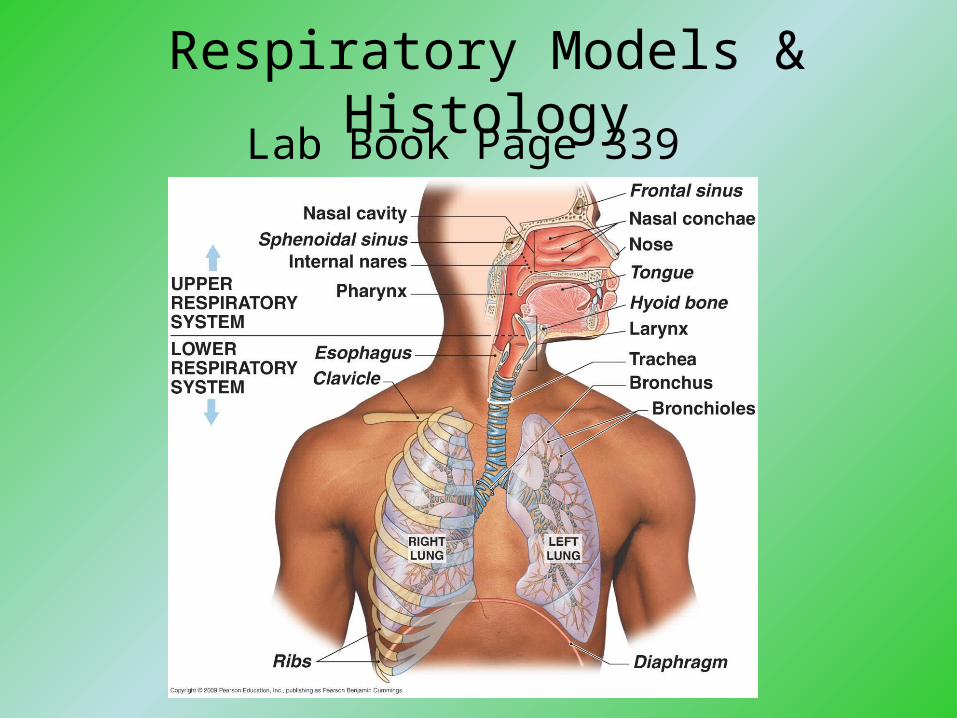
Respiratory Models & Histology
Lab Book Page 339

Fig. not in your lab book
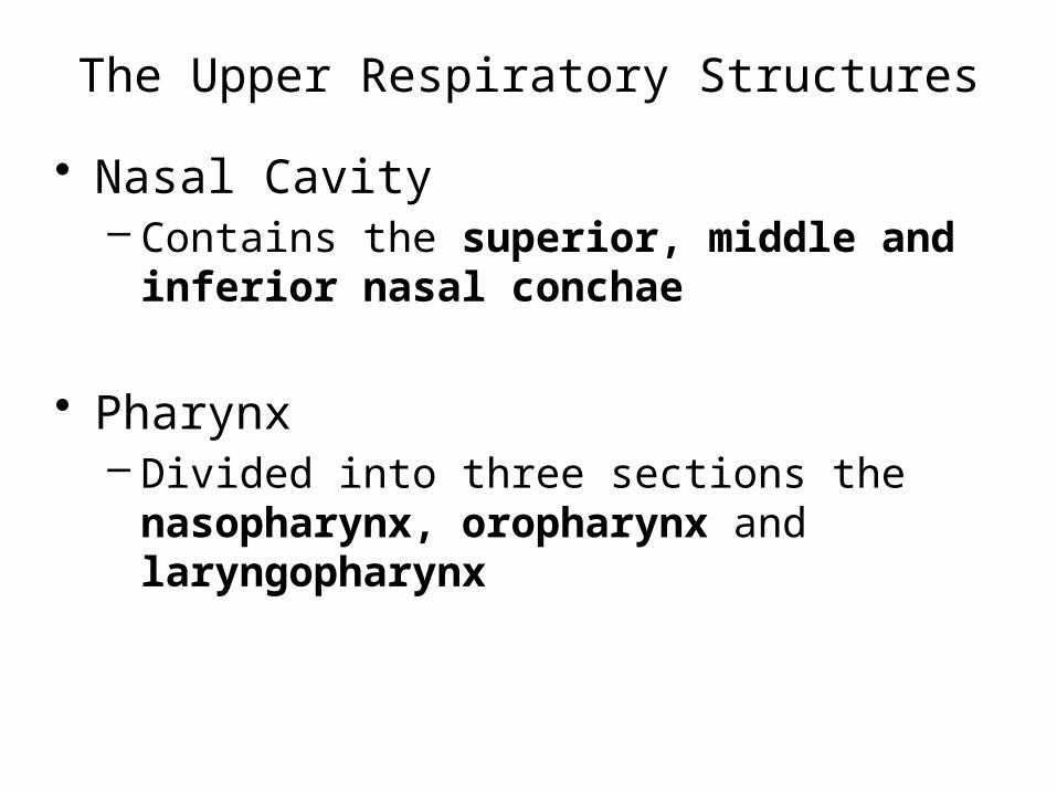
The Upper Respiratory Structures
• Nasal Cavity– Contains the superior, middle and inferior
nasal conchae
• Pharynx– Divided into three sections the nasopharynx,
oropharynx and laryngopharynx

Lab Book page 340
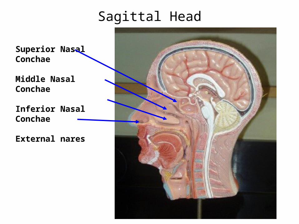
Sagittal Head
Superior Nasal Conchae
Middle Nasal Conchae
Inferior Nasal Conchae
External nares

Sagittal Head
Frontal Sinus
Sphenoid (Sphenoidal)Sinus
Nasopharynx
Oropharynx
Laryngopharynx

Sagittal Head
Larynx – Epiglottis
Larynx – Thyroid Cartilage
Esophagus

The Lower Respiratory Structures
• Larynx– Made of cartilage and ligaments– 3 Major cartilages of the larynx:
• Thyroid cartilage• Cricoid cartilage• Epiglottis
– Contains vocal cords, which surround an opening called the glottis
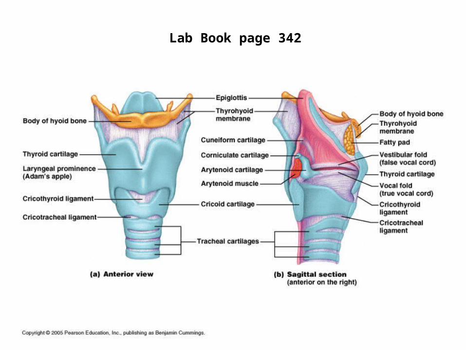
Lab Book page 342
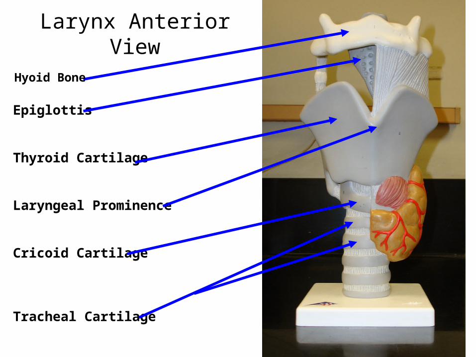
Larynx Anterior View
Epiglottis
Thyroid Cartilage
Laryngeal Prominence
Cricoid Cartilage
Tracheal Cartilage
Hyoid Bone
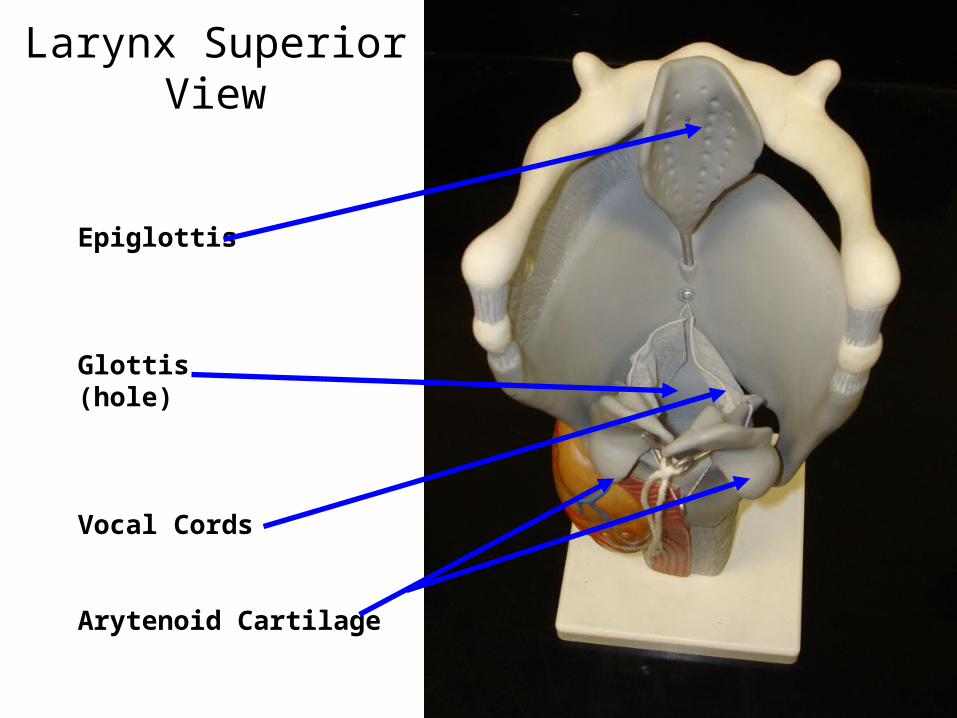
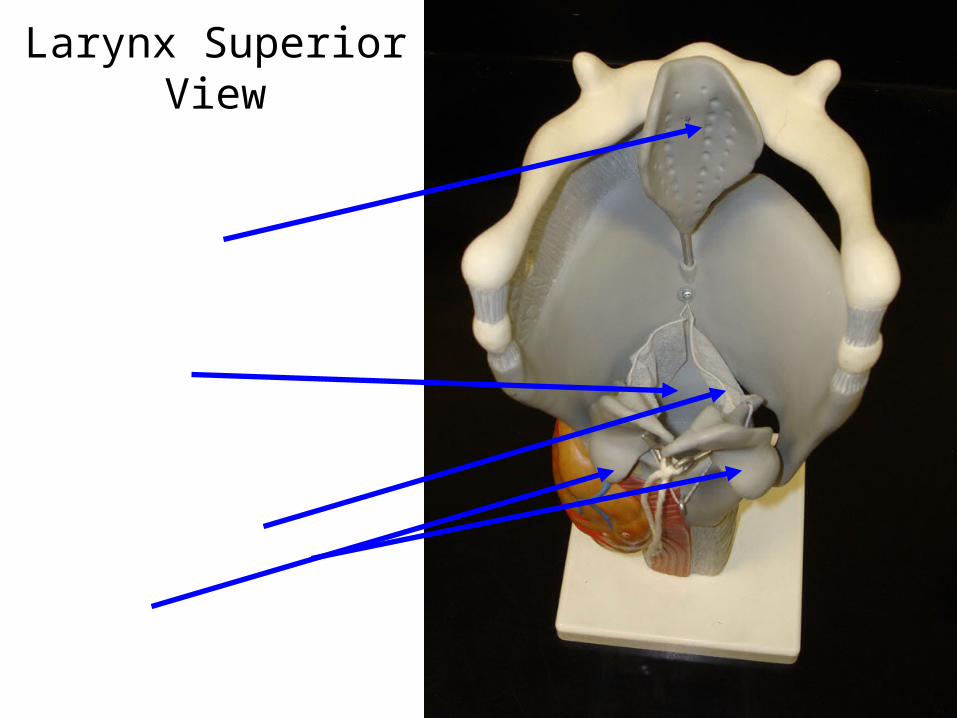
Larynx Superior View
Epiglottis
Glottis(hole)
Vocal Cords
Arytenoid Cartilage
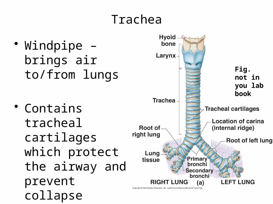
Trachea
• Windpipe – brings air to/from lungs
• Contains tracheal cartilages which protect the airway and prevent collapse
Fig. not in you lab book

Bronchial Tree
Fig. not in you lab book

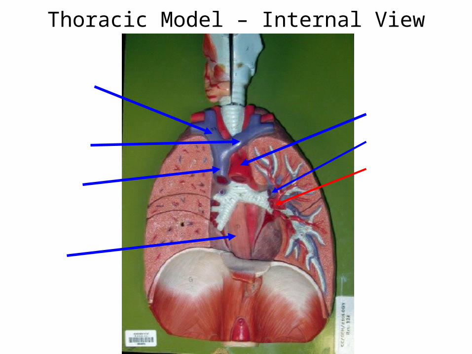
Thoracic Model – Internal View
Right Brachiocephalic vein
Left Brachiocephalic vein
Superior Vena Cava
Esophagus
Aortic Arch
Pulmonary Arteries
Pulmonary Veins

Thoracic Model – Internal View
Trachea
Primary bronchus
Secondary bronchus
Tertiary Bronchus

Thoracic Model – External View
Right LungSuperior Lobe
Right LungMiddle Lobe
Right LungInferior Lobe
Diaphragm
Left LungSuperior Lobe
Cardiac notch of left lung
Left LungInferior Lobe

2 Respiratory Slides
• 1. Slide of the TRACHEA
• 2. Slide of a BRONCHIOLE
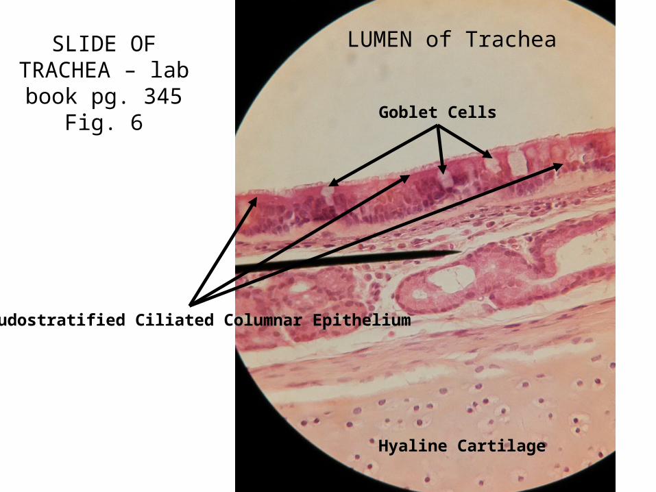
SLIDE OF TRACHEA – lab
book pg. 345 Fig. 6
LUMEN of Trachea
Pseudostratified Ciliated Columnar Epithelium
Hyaline Cartilage
Goblet Cells

Close-up of Trachea showing a Goblet Cell
Goblet Cell

Bronchiole & Alveoli
Fig. not in you lab book
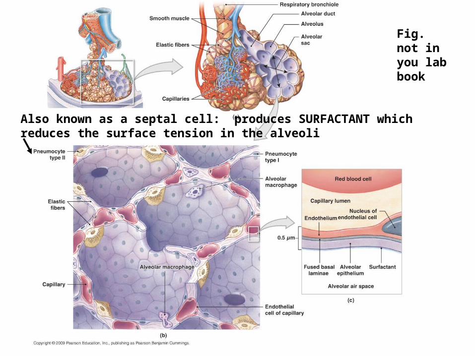
Also known as a septal cell: produces SURFACTANT which reduces the surface tension in the alveoli
Fig. not in you lab book
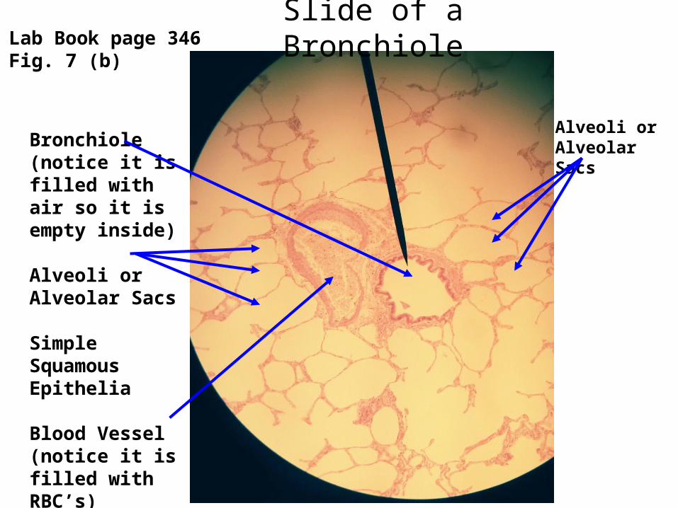
Slide of a Bronchiole
Bronchiole(notice it is filled with air so it is empty inside)
Alveoli or Alveolar Sacs
Simple Squamous Epithelia
Blood Vessel(notice it is filled with RBC’s)
Lab Book page 346Fig. 7 (b)
Alveoli or Alveolar Sacs
Practice Slides!!!
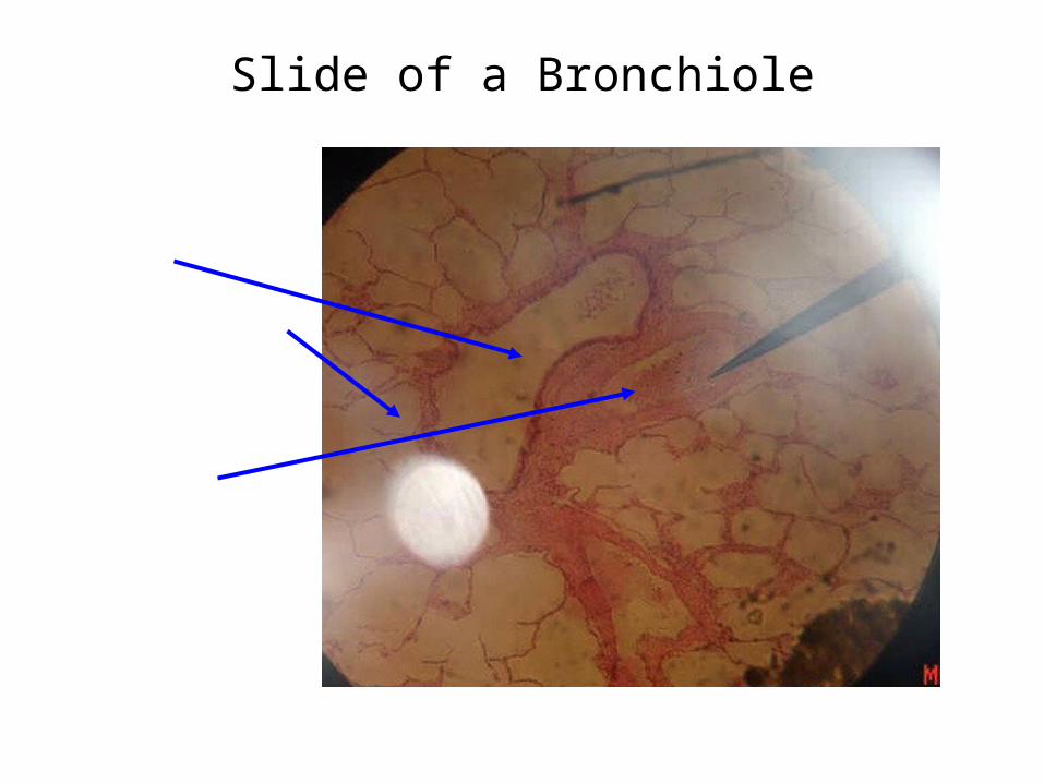
Slide of a Bronchiole

Thoracic Model – External View
Thoracic Model – Internal View

Thoracic Model – Internal View

Larynx Anterior View
Larynx Superior View

Sagittal Head

Sagittal Head

Sagittal Head
Related Documents