respirator Bohr Effect Alterations in hemoglobin’s structure Shift to the right in the oxyhemoglobin dissociation curve Loading of O 2 is not affected – the flat upper portion is not altered Unloading of O 2 is enhanced

Respiratory Bohr Effect Alterations in hemoglobin’s structure Alterations in hemoglobin’s structure Shift to the right in the oxyhemoglobin dissociation.
Dec 16, 2015
Welcome message from author
This document is posted to help you gain knowledge. Please leave a comment to let me know what you think about it! Share it to your friends and learn new things together.
Transcript
respiratory
Bohr Effect
Alterations in hemoglobin’s structure Shift to the right in the oxyhemoglobin
dissociation curve Loading of O2 is not affected
– the flat upper portion is not altered Unloading of O2 is enhanced
– along steep lower portion, more O2 is unloaded at a given PO2 with the shift

respiratory
respiratory
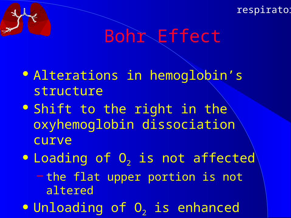
Myoglobin and Muscle Oxygen Storage
Skeletal & cardiac muscle contain compound myoglobin.
Each myoglobin contains only one heme in contrast to 4 in hemoglobin (Hb).
Myoglobin binds and retains O2 at low pressures.
Facilitates oxygen transfer to mitochondria at start of exercise and intense exercise when cellular PO2 ↓ greatly.

respiratory
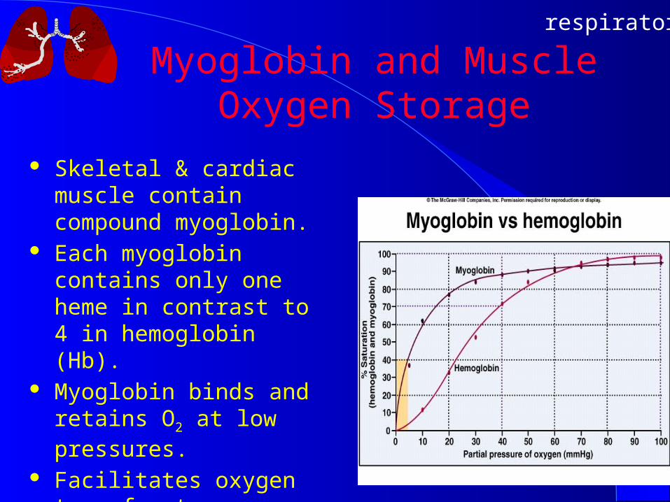
Carbon Dioxide Transportin the Blood
Dissolved in plasma – CO2 is 20 times more soluble than O2
– 7% to 10% of CO2 is dissolved
Combined with amino compounds– hemoglobin is most common– Haldane effect: Hb’s de-oxygenation enables bind CO2
– about 20% of CO2 is carried as carbamino compounds
Bicarbonate – about 70% carried as bicarbonate
respiratory
Carbon Dioxide Transportin the Blood

respiratory
Formation of Bicarbonate at Tissue Level
CO2 diffuses into RBC Enzyme, carbonic anhydrase, absent in plasma
but present in RBC drives reaction of CO2 + H2O => H2CO3
H2CO3 dissociates a proton => HCO3- + H+
CO2 + H2O => H2CO3 => HCO3- + H+
HCO3- moves into plasma via HCO3
- / Cl- anion exchanger to prevent electrical imbalance
Hb acts as buffer and accepts the H+


respiratory
Bicarbonate in the Lungs
In lungs, carbon dioxide diffuses from plasma into alveoli; lowers plasma PCO2.
HCO3- + H+ recombine to form carbonic acid.
H2CO3 dissociates to H2O and CO2, allowing carbon dioxide to exit through the lungs.
CO2 + H2O <= H2CO3 <= HCO3- + H+

respiratory
Ventilatory Regulation
Two factors regulate pulmonary ventilation: Neural input from higher brain centers
provides primary drive to ventilate Gaseous and chemical state of blood: humoral
factors

respiratory
Pulmonary Ventilation Control
Clusters neurons in medulla oblongata referred to as respiratory center.
Inspiratory center activates diaphragm & intercostals.
Expiratory center inhibits inspiratory neurons.
Stretch receptors assist regulation of breathing
Pneumotaxic & apneustic centers contribute (depth).

respiratory
Humoral Factors Chemoreceptors are specialized neurons. Chemoreceptors monitor blood conditions, provide
feedback– Peripheral located in aortic arch and bifurcation of
common carotid respond to CO2, “temperature”-no, H+
– Central located in medulla affected by PCO2 & H+
Specialized receptors in lungs sensitive to stretch and irritants act to provide feedback
Interaction among factors controls ventilation– CO2 production is closely associated with
ventilation rate
respiratory
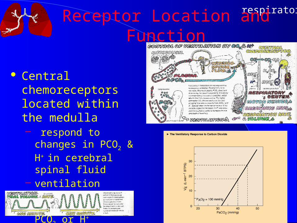
Receptor Location and Function
Central chemoreceptors located within the medulla– respond to changes in
PCO2 & H+ in cerebral spinal fluid
– ventilation increases with elevations of PCO2 or H+
respiratory
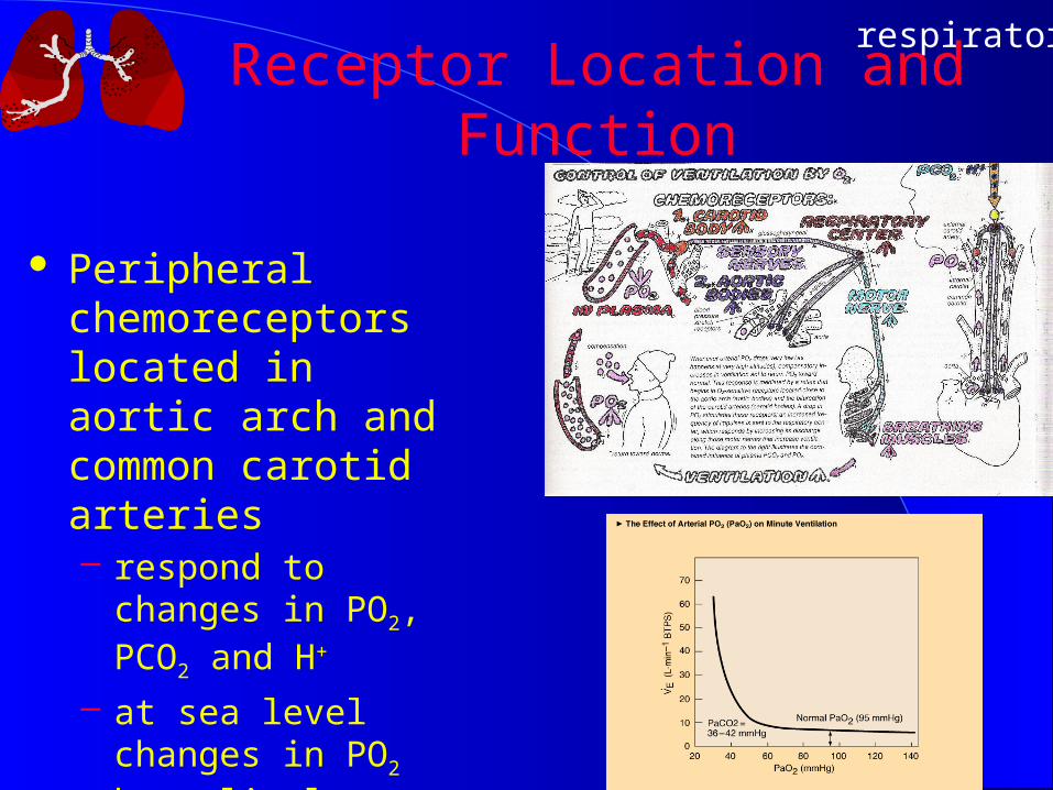
Receptor Location and Function
Peripheral chemoreceptors located in aortic arch and common carotid arteries– respond to changes in
PO2, PCO2 and H+
– at sea level changes in PO2 have little effect on VE
respiratory
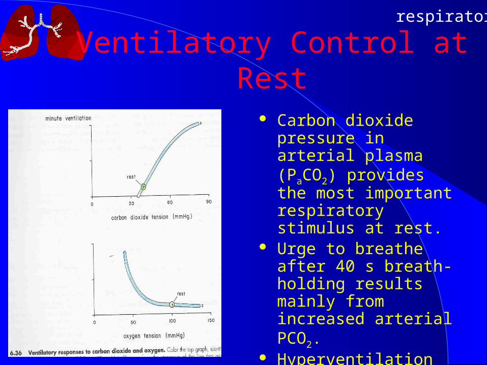
Ventilatory Control at Rest
Carbon dioxide pressure in arterial plasma (PaCO2) provides the most important respiratory stimulus at rest.
Urge to breathe after 40 s breath-holding results mainly from increased arterial PCO2.
Hyperventilation decreases Alveolar PCO2 to 15 mm Hg, which decreases PaCO2 below normal, allows longer breath holding.

respiratory
Ventilatory Control in Exercise
Very rapid increase at start of exercise
Chemical stimuli cannot explain initial hyperpnea during exercise.
Nonchemical factors mediate the rapid response– Cortical: motor cortex– Peripheral:
mechanoreceptors in joints, tendons and muscles
respiratory
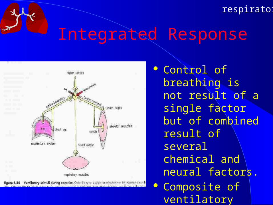
Integrated Response
Control of breathing is not result of a single factor but of combined result of several chemical and neural factors.
Composite of ventilatory response to exercise.

respiratory
References
Axen and Axen. 2001. Illustrated Principles of Exercise Physiology. Prentice Hall.
Kapit, Macey, Meisami. 1987. Physiology Coloring Book. Harper & Row.
McArdle, Katch, Katch. 2006. Image Collection Essentials of Exercise Physiology, 3rd ed. Lippincott William & Wilkens.
Related Documents