applied sciences Article Respiration Monitoring for Premature Neonates in NICU Yue Sun 1, *, Wenjin Wang 2 , Xi Long 2 , Mohammed Meftah 2 , Tao Tan 1 , Caifeng Shan 2 , Ronald M. Aarts 1 and Peter H. N. de With 1 1 Department of Electrical Engineering, Eindhoven University of Technology, 5612 WH Eindhoven, The Netherlands 2 Philips Research, High Tech Campus 34, 5656 AE Eindhoven, The Netherlands * Correspondence: [email protected] Received: 18 October 2019; Accepted: 28 November 2019; Published: 2 December 2019 Abstract: In this paper, we investigate an automated pipeline to estimate respiration signals from videos for premature infants in neonatal intensive care units (NICUs). Two flow estimation methods, namely the conventional optical flow- and deep learning-based flow estimation methods, were employed and compared to estimate pixel motion vectors between adjacent video frames. The respiratory signal is further extracted via motion factorization. The proposed methods were evaluated by comparing our automated extracted respiration signals to that extracted from chest impedance on videos of five premature infants. The overall average cross-correlation coefficients are 0.70 for the optical flow-based method and 0.74 for the deep flow-based method. The average root mean-squared errors are 6.10 and 4.55 for the optical flow- and the deep flow-based methods, respectively. The experimental results are promising for further investigation and clinical application of the video-based respiration monitoring method for infants in NICUs. Keywords: respiration; respiration rate; video processing; remote sensing; biomedical monitoring 1. Introduction Vital signs, such as heart rate, blood pressure, respiratory rate, and body temperature, are physical parameters that can be measured and used to assess physiological state and functioning. Monitoring of vital parameters is a crucial topic in neonatal daily care. Premature infants have an immature respiratory control that predisposes them to apnea/periodic breathing, haemoglobin oxygen desaturation, and bradycardia [1,2]. Apnea is defined as the status of cessation of respiratory airflow, whereas periodic breathing is characterized by groups of respiratory movements interrupted by small intervals of apnea [3]. Continuous monitoring of respiration for premature infants is critical to detect abnormalities in breathing and help develop early treatments to prevent significant hypoxia and central depression from apnea. A long-term continuous monitoring approach of respiration may also be used to assess sleep stage, which varies with different clinical conditions [4,5]. The existing methods for monitoring respiration include nasal thermocouples, spirometers, transthoracic inductance, respiratory effort belt transducer, piezoelectric transducer, optical sensor (pulse oximetry), strain gauge, impedance plethysmography, and electrocardiogram (ECG). Currently, the respiration of premature infants in a neonatal intensive care unit (NICU) is monitored by bedside monitors. ECG is considered as the standard reference measurement for respiration, since ECG can provide stable and robust monitoring for a NICU. However, the pressure of the contact sensor may also change the local skin perfusion, which yields not a true measurement as compared to the non-contact sensor. The electrodes may exert pressure to the skin, yielding to tissue compression and vascular insufficiency. As a consequence, applying the ECG electrodes on infant skin for a long time Appl. Sci. 2019, 9, 5246; doi:10.3390/app9235246 www.mdpi.com/journal/applsci

Welcome message from author
This document is posted to help you gain knowledge. Please leave a comment to let me know what you think about it! Share it to your friends and learn new things together.
Transcript

applied sciences
Article
Respiration Monitoring for Premature Neonatesin NICU
Yue Sun 1,*, Wenjin Wang 2, Xi Long 2 , Mohammed Meftah 2, Tao Tan 1, Caifeng Shan 2,Ronald M. Aarts 1 and Peter H. N. de With 1
1 Department of Electrical Engineering, Eindhoven University of Technology, 5612 WH Eindhoven,The Netherlands
2 Philips Research, High Tech Campus 34, 5656 AE Eindhoven, The Netherlands* Correspondence: [email protected]
Received: 18 October 2019; Accepted: 28 November 2019; Published: 2 December 2019�����������������
Abstract: In this paper, we investigate an automated pipeline to estimate respiration signalsfrom videos for premature infants in neonatal intensive care units (NICUs). Two flow estimationmethods, namely the conventional optical flow- and deep learning-based flow estimation methods,were employed and compared to estimate pixel motion vectors between adjacent video frames.The respiratory signal is further extracted via motion factorization. The proposed methods wereevaluated by comparing our automated extracted respiration signals to that extracted from chestimpedance on videos of five premature infants. The overall average cross-correlation coefficientsare 0.70 for the optical flow-based method and 0.74 for the deep flow-based method. The averageroot mean-squared errors are 6.10 and 4.55 for the optical flow- and the deep flow-based methods,respectively. The experimental results are promising for further investigation and clinical applicationof the video-based respiration monitoring method for infants in NICUs.
Keywords: respiration; respiration rate; video processing; remote sensing; biomedical monitoring
1. Introduction
Vital signs, such as heart rate, blood pressure, respiratory rate, and body temperature,are physical parameters that can be measured and used to assess physiological state and functioning.Monitoring of vital parameters is a crucial topic in neonatal daily care. Premature infants have animmature respiratory control that predisposes them to apnea/periodic breathing, haemoglobin oxygendesaturation, and bradycardia [1,2]. Apnea is defined as the status of cessation of respiratory airflow,whereas periodic breathing is characterized by groups of respiratory movements interrupted by smallintervals of apnea [3]. Continuous monitoring of respiration for premature infants is critical to detectabnormalities in breathing and help develop early treatments to prevent significant hypoxia andcentral depression from apnea. A long-term continuous monitoring approach of respiration may alsobe used to assess sleep stage, which varies with different clinical conditions [4,5].
The existing methods for monitoring respiration include nasal thermocouples, spirometers,transthoracic inductance, respiratory effort belt transducer, piezoelectric transducer, optical sensor(pulse oximetry), strain gauge, impedance plethysmography, and electrocardiogram (ECG). Currently,the respiration of premature infants in a neonatal intensive care unit (NICU) is monitored by bedsidemonitors. ECG is considered as the standard reference measurement for respiration, since ECGcan provide stable and robust monitoring for a NICU. However, the pressure of the contact sensormay also change the local skin perfusion, which yields not a true measurement as compared to thenon-contact sensor. The electrodes may exert pressure to the skin, yielding to tissue compression andvascular insufficiency. As a consequence, applying the ECG electrodes on infant skin for a long time
Appl. Sci. 2019, 9, 5246; doi:10.3390/app9235246 www.mdpi.com/journal/applsci

Appl. Sci. 2019, 9, 5246 2 of 11
increases the risk of trauma and infections. Removing the adhesives from the immature skin, as part ofregular care, can damage the immature skin of preterm infants, as well as cause stress and pain [6,7].This technique is impractical for home care, since the sensors have to be placed by skilled caregivers,and wearing the sensors also causes inconvenience for everyday life [8,9].
A non-contact monitoring system is a good alternative to improve infant comfort and safety andit also has the potential for monitoring at home. Few works to date have investigated video-basedcontactless methods for monitoring respiration. In this study, we developed and evaluated a contactlessrespiration measurement method based on video monitoring. The contribution of our method is:(1) we propose an approach for non-contact respiration monitoring; and (2) we validated the methodon clinical data acquired in NICU.
2. Related Work
In the applications of adults, Deng et al. [10] presented a design and implementation of a novelsleep monitoring system that simultaneously analyzed respiration, head posture, and body posturefor adults. For respiration, the region of breathing movement was automatically determined and theintensity estimated, yielding a waveform indicating respiratory rhythms with an accuracy of 96% inrecognizing abnormal breathing. However, the accuracy of the respiration rate was not provided.Chazal et al. [11] applied a contactless biomotion sensor of Doppler radar for respiration monitoring.Respiratory frequency and magnitude were used for the classification of determining sleep/wake states,which achieved the accuracy of 69% for the wake and 88% for sleep state. Gupta et al. [12] estimatedheart rate (HR) accurately, using face videos acquired from a low-cost camera for adults. The facevideo consisting of frontal, profile, or multiple faces, was divided into multiple overlapping fragmentsto determine HR estimates. The HR estimates were fused using quality-based fusion, which aimed tominimize illumination and face deformations. Prathosh et al. [13] proposed a general framework forestimating a periodic signal, which was applied to derive a computationally inexpensive method forestimating respiratory pattern using two-dimensional cameras that did not critically depend on theregion of interest. Specifically, the patterns were estimated by imaging changes in the reflected lightcaused by respiration-induced motion. Estimation of the pattern was cast as a blind deconvolutionproblem and was solved through a method comprising subspace projection and statistical aggregation.
In terms of the applications for infants, Werth et al. reviewed the unobtrusive measurements forindicating sleep state in preterm infants [14]. Abbas et al. [15] attempted to detect respiration rate ofneonates on a real-time basis using infrared thermography. They analyzed the anterior naris (nostrils)temperature profile associated with the inspiration and expiration phases. However, the region ofinterest (ROI) was assumed to be fixed after initialization. Moreover, the method is not practicalfor NICU infants, since the faces of premature infants in a NICU are often occluded by feedingtubes and/or breathing masks. In practice, the temperature inside an incubator is continuouslymonitored and adjusted by caregivers in order to help infants maintain their body temperature in anormal range. However, the controlled environmental temperature might affect the accuracy of thethermography-based respiration monitoring method. Koolen et al. [16] extracted the respiration ratefrom video data included in polysomnography. They used Eulerian video magnification (EVM) toamplify the respiration movements, which was followed by optical flow to estimate the respirationmotion and therefore, obtained a respiration signal. Independent component analysis and principalcomponent analysis were applied to improve signal quality. Finally, the results showed a detectionaccuracy of 94.12% for sleeping-stage patients. Antognoli et al. [17] applied a digital webcam (WeC)and an EVM algorithm to measure HR and respiration rate (RR). The accumulated RGB values of amanually selected ROI were calculated as a single signal, from which the power spectral density wasfurther estimated and used for peak extraction. The evaluation based on data of seven patients yieldeda root mean-squared error (RMSE) of 12.2 for the HR and 7.6 for the RR. However, the limitation ofa pulse-based respiration extraction is that it extracts the respiratory modulation in blood volumechanges, which is both subject dependent (different respiratory efforts) and measurement-location

Appl. Sci. 2019, 9, 5246 3 of 11
dependent. The modulation effect varies in different body parts. Moreover, the peak selection fromspectrograms was based on common knowledge of normal HR and RR frequency ranges, which is notsuitable for infants under clinical conditions of different diseases.
Methods on remote sensing of respiration or other contactless physiological measures have beendeveloped for adults and infants while our applications focus on premature infants in NICU.
3. Methods
3.1. Material
Our study was conducted with videos recorded at the Máxima Medical Center in Veldhoven,The Netherlands, by a fixed-position high-definition camera (Camera model IDS uEye monochrome)filming the infant’s entire body in the direction of the foot to the head. Figure 1 shows an example of acaptured video frame. We decided on the recording position of the camera by considering: (1) littleor no interruption to daily routine care; and (2) a good viewpoint for observing vertical movementof the infant chest, where the movement is assumed to be with maximum respiratory motion energyin the vertical direction. For all infant recordings, written consent was obtained from the parents.The resolution of each video frame is 736 × 480 pixels, while the frame rate is 8 fps. The videoswere recorded under uncontrolled, regular hospital lighting conditions. Five infants with an averagegestational age of 29.6 ± 2.8 weeks (range 27+0–33+6 weeks), an average postnatal age of 1.2 ± 0.6weeks (range 0+4–2+1 weeks), and an average weight of 1555 ± 682.4 g (range 755–2410 g) were filmed.Parallel with the video capturing, standard chest impedance (CI) signals were recorded simultaneouslyas reference standards.
Figure 1. Example of an acquired video frame.
3.2. Motion Matrix Calculation
The flowchart of the proposed system is shown in Figure 2, where motion matrix estimation is anessential step in the pipeline. We estimate dthe pattern of apparent motion including respiration ofinfants in videos. Motion matrix estimation can be defined as the distribution of apparent velocities ofmovement in successive images.
Figure 2. Flowchart of the proposed video-based respiration monitoring system.
Two flow estimation methods were employed to estimate pixel motion vectors between adjacentvideo frames. First, the conventional optical flow method [18] was utilized and evaluated. However,

Appl. Sci. 2019, 9, 5246 4 of 11
the optimal flow is more sensitive to high gradient texture, whereas in our case the infant chest iseither bare or covered by a blanket. Thus, in both cases, the chest area, which mostly indicates therespiratory motion, lacks gradient texture. Deep flow [19] is sensitive to the entire part of movingobjects with less regard to texture information. Therefore, better performance is expected by using aflow estimation method based on deep learning.
For both methods, the derived motion vectors contain respiration information, induced by themotion of the abdominal wall and chest wall. We only considered the vertical motion vector, sincethe respiration related motion is mainly in the vertical direction in the captured videos. For infantsunder one year of age, it is also recommended by the American Academy of Pediatrics (AAP) thatthey should be placed to sleep on their backs every time being laid down to sleep. This lowers the riskof sudden infant death syndrome (SIDS).
3.2.1. Conventional Optical Flow
The optical flow should satisfy Equation (1):
∂I∂x
Vx +∂I∂y
Vy +∂I∂t
= 0, (1)
where I is the intensity matrix of a frame in a video and Vx are Vy are the actual optical flow parameters.We deployed the classic dense optical flow algorithm proposed by Barron et al. [18], where second-orderdifferential equations based on the Hessian matrix are used to constrain two-dimensional (2D) velocity.The Barron method creates flow fields with 100% density. However, the conventional optical flowmethod is limited at estimating motion in poorly textured areas, which lack gradient variation.
3.2.2. Deep Flow
The above conventional optical flow approach only computes image matching at a single scale.However, for complicated situations with complex motions, the traditional approach may not beable to effectively capture the interaction or dependency relationships. Brox and Malik [20] proposedto add the addition of a descriptor matching term in the variational approach, which allows betterhandling large displacements. The matching provides a guide using correspondences from sparsedescriptor matching.
In our study, we applied the method from Weinzaepfel et al. [19]. A descriptor matching algorithmwas incorporated, which is based on deep convolutions with six layers, interleaving convolutions andmax-pooling. In the proposed framework, dense sampling is applied to efficiently retrieve quasi-densecorrespondences, while incorporating a smoothing effect on the descriptors matches. Figure 3 showsthe framework for deep flow estimation.
3.3. Respiratory Description
The results of flow calculation were captured in the rows of the derived motion matrix M (of sizeN × W, where N denotes the number of pixels in a video frame and W is the total number of framesin a video), which contain the motion derivatives that represent the velocity magnitudes of the pixeltrajectories in the vertical direction.
The spatial statistics of the flow matrix was further analyzed by applying robust principalcomponent analysis (PCA) [21]. PCA includes the eigendecomposition of a data covariance matrixor singular value decomposition of a data matrix. The decomposition task projects the original dataonto an orthogonal subspace, where each direction is mutually de-correlated and the most informativedata information becomes available in the first several principal components. In our study, instead ofconsidering the originally obtained matrix, the first eigenvalue of the covariance matrix of flows wasanalyzed, since the first eigenvalue represents the major motion component. Besides, it is beneficial toreduce residual motion noise that has lower energy than the respiratory motion in each video.

Appl. Sci. 2019, 9, 5246 5 of 11
Figure 3. Framework of deep flow.
The motion matrix M is masked by sub-spatiotemporal regions, mi, where each mi is a local maskthat stores W consecutive squared blocks. For each mi, we can generate the eigenvectors that satisfythe following general condition, specified by
mi · Di = λ · Di s.t. Det(mi − λi · I) = 0, (2)
where Det(·) denotes the matrix determinant, I represents the identity matrix, and Di and λi areeigenvectors and eigenvalues, respectively.
3.4. Evaluation
In this study, we employed a sliding window of 120 s with a step size of 1 s to estimate therespiration rate. The respiration rate was calculated by first averaging the time intervals betweenbreathing peaks, followed by converting those numbers to a frequency value, expressed in the unit ofbreaths per minute (bpm).
It is well known that the respiration signal can be estimated from the CI signals. To evaluatethe performance of respiration estimation using our video-based algorithms, we computed thecross-correlation coefficients and the RMSE as measures to compare the respiration rates of thereference breathing signal from the CI and our extracted respiration signals. The cross-correlationcoefficient calculation is initially based on a zero-mean process. Thus, this calculation is only forcomparing the respiration rate variance of our estimation and the reference standard. In addition,correlation plots and Bland–Altman plots [22,23] were also created.
4. Experimental Results and Discussion
Figure 4 shows a visual comparison of the reference breathing signal from the CI and two extractedrespiration signals by the proposed optical flow- and deep flow-based methods. The three re-scaled1D signals from the CI, optical flow- and deep flow-based motion estimations are depicted in Figure 5.

Appl. Sci. 2019, 9, 5246 6 of 11
Figure 4. Monitored respiration from the CI, and corresponding extracted respiration signals from asegment, based on optical flow and deep flow.
(a)
(b)
Figure 5. Data patterns for chest impedance and video-based observation: (a) re-scaled 1D signals;and (b) zoom in of each interval indicated in (a).

Appl. Sci. 2019, 9, 5246 7 of 11
Table 1 summarizes the estimation of respiration rate using two different optical flow methods.Table 2 shows the root mean-squared errors (RMSE) and cross-correlation (CC) coefficients of thereference breathing signal from the CI, compared to our optical flow- and deep flow-based results.The results obtained from deep flow are more accurate than those of the conventional optical flowapproach. The average cross-correlation coefficients of all videos is 0.74 and the average RMSE is 4.55,using the proposed deep learning-based method. Despite the limited number of measurements, thesepreliminary results provide a promising result for further investigation of the neonatal video-basedrespiration monitoring method. The overall RMSE for our deep learning-based method (4.55) showsthe feasibility to apply our automated respiration for clinical use.
Table 1. Duration of each video, and measured mean and standard deviations of reference and ouroptical flow- and deep flow-based methods.
Patient ID Duration (h:m:s)Mean ± std
Reference (CI) Optical Flow Deep Flow
1 00:50:13 51.12 ± 6.00 47.13 ± 4.96 48.67 ± 4.69
2 00:22:00 55.41 ± 6.73 51.18 ± 7.35 53.23 ± 7.07
3 00:26:54 48.78 ± 3.58 44.12 ± 2.91 46.75 ± 2.80
4 00:09:22 52.36 ± 5.25 48.75 ± 3.17 50.05 ± 3.72
5 00:06:15 61.61 ± 3.80 51.56 ± 4.29 55.58 ± 3.28
Table 2. Root mean-squared errors (RMSE) and cross-correlation (CC) coefficients of reference breathingsignal from the CI compared to our optical flow- and deep flow-based results.
Patient ID RMSE CC Coefficients
Optical Flow Deep Flow Optical Flow Deep Flow
1 5.09 4.32 0.85 0.82
2 4.94 3.10 0.94 0.95
3 3.86 3.51 0.73 0.75
4 5.75 5.11 0.47 0.52
5 10.85 6.71 0.49 0.66
Average 6.10 4.55 0.70 0.74
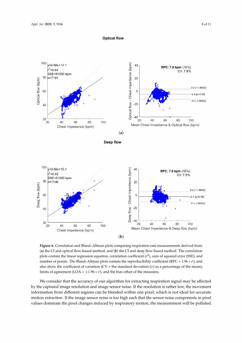
From both the correlation and Bland–Altman analyses in Figure 6a,b, the deep flow based-methodproduces a lower error than the optical flow-based method, especially when breathing rates are lessthan 50 bpm. The absolute values for the overall mean errors reduce from 4.8 to 2.7 bpm in the opticalflow and deep flow cases, respectively.
There is a residual error made by our automated processing pipeline, where the automatedmethod underestimates the respiration rate. This occurs because our band filter fails to remove motioncaused by interruptions in the video, for example movement resulting from nurse care-handling. Inthe future, we will investigate a supervised approach to replace unsupervised filters to extract thenoise from respiration signals.
We compared the effect of two different flow estimation methods on the final respirationcalculation. The results show that the deep flow approach is both sensitive to homogeneous regionsand the boundary area, whereas the conventional approach is more sensitive to the boundary area.This advantage of deep flow-based approach improves the whole processing pipeline by increasingboth the accuracy and robustness.
Appl. Sci. 2019, 9, 5246 8 of 11
(a)
(b)
Figure 6. Correlation and Bland–Altman plots comparing respiration rate measurements derived from:(a) the CI and optical flow-based method; and (b) the CI and deep flow-based method. The correlationplots contain the linear regression equation, correlation coefficient (r2), sum of squared error (SSE), andnumber of points. The Bland–Altman plots contain the reproducibility coefficient (RPC = 1.96 ∗ σ), andalso show the coefficient of variation (CV = the standard deviation (σ) as a percentage of the mean),limits of agreement (LOA = ±1.96 ∗ σ), and the bias offset of the measures.
We consider that the accuracy of our algorithm for extracting respiration signal may be affectedby the captured image resolution and image sensor noise. If the resolution is rather low, the movementinformation from different regions can be blended within one pixel, which is not ideal for accuratemotion extraction. If the image sensor noise is too high such that the sensor noise components in pixelvalues dominate the pixel changes induced by respiratory motion, the measurement will be polluted.

Appl. Sci. 2019, 9, 5246 9 of 11
The quantitative analysis regarding this perspective is a complicated matter and will be performed asfuture work.
Currently, the work is carried out as a feasibility study. The focus of this study is the installation,adaptation, and validation of camera-based monitoring technology in the NICU setting. We have notinvestigated the performance on preterm infants related to specific diseases. In the future, we willfurther validate our algorithm on infants having different health situations.
Our recordings were taken from real clinical practice without interfering with the clinical workflow.Our algorithm works when the respiratory motion can be observed by the camera, even as subtlemovement, i.e., infants can be either naked or covered by a blanket (as long as the infant body hascontact with the blanket such that the movement information from the thorax-abdomen can stillbe derived).
Our algorithm relies on the intensity of video frames for motion extraction (i.e., no color orchromaticity information is used). Therefore, for the nighttime condition, it is possible to measure therespiration signal by using the same software algorithms that we have proposed and just altering to aninfrared camera with an infrared lighting source. During low-light conditions, the performance of oursystem may be affected by the noise induced by the camera sensor.
The highlight of using a video-based method to monitor respiration is its contact-free operation.Both CI and polysomnography need electrodes attached to the patient’s skin, which increase the riskof skin irritation. Therefore, our method can improve the comfort level and convenience. A furtherbenefit of using a camera is that it enables more measurements than contact-based bio-sensors,including physiological signals (e.g., breathing rate, heart rate, and blood oxygen saturation) [24,25]and contexture signals (e.g., body motion, activities, and facial expressions) [26–29]. This will enrichthe functionality of a health monitoring system. Our system can be constructed with a generic webcamand an embedded computing platform, which forms a cost-effective solution. In principle, one cameracan monitor multiple subjects/infants simultaneously, as long as they are captured by the camera view,while each contact-based bio-sensor can only monitor one single subject/infant.
5. Conclusions
In this study, we applied an automated pipeline to estimate respiration signals from videosfor premature infants in NICUs. We compared our automated extracted respiration signals to thatextracted from the CI. The preliminary results are promising for further investigation of the video-basedrespiration monitoring method and for applying our automated respiration extraction for infants inNICUs. Experiments showed that the deep learning-based method outperforms the optical flow-basedmethod in accuracy (low RMSE) and robustness. In the future, we will investigate the possibility ofdirectly applying a deep learning framework to estimate respiration rate. For example, an LSTM-basedsystem can effectively incorporate temporal information for a regression task and is expected to furtherenhance the obtained results.
Author Contributions: Conceptualization, Y.S., W.W., X.L., and P.H.N.d.W.; methodology, Y.S., W.W., and T.T.;software, Y.S., W.W., X.L., and T.T.; validation, Y.S., and X.L.; formal analysis, Y.S.; investigation, Y.S.; resources,M.M.; data curation, X.L.; writing—original draft preparation, Y.S.; writing—review and editing, Y.S., W.W., X.L.,C.S., R.M.A., and P.H.N.d.W.; visualization, Y.S.; and supervision, P.H.N.d.W.
Funding: This research received no external funding.
Conflicts of Interest: The authors declare no conflict of interest.
References
1. Sale, S.M. Neonatal apnoea. Best Pract. Res. Clin. Anaesthesiol. 2010, 24, 323–336. [CrossRef] [PubMed]2. Chernick, V.; Heldrich, F.; Avery, M.E. Periodic breathing of premature infants. J. Pediatr. 1964, 64, 330–340.
[CrossRef]3. Poets, C.F.; Stebbens, V.A.; Samuels, M.P.; Southall, D.P. The relationship between bradycardia, apnea, and
hypoxemia in preterm infants. Pediatr. Res. 1993, 34, 144. [CrossRef] [PubMed]

Appl. Sci. 2019, 9, 5246 10 of 11
4. Prechtl, H.; Theorell, K.; Blair, A. Behavioural state cycles in abnormal infants. Dev. Med. Child Neurol. 1973,15, 606–615. [CrossRef] [PubMed]
5. Prechtl, H.F. The behavioural states of the newborn infant (a review). Brain Res. 1974, 76, 185–212. [CrossRef]6. Lund, C.H.; Nonato, L.B.; Kuller, J.M.; Franck, L.S.; Cullander, C.; Durand, D.K. Disruption of barrier
function in neonatal skin associated with adhesive removal. J. Pediatr. 1997, 131, 367–372. [CrossRef]7. Afsar, F. Skin care for preterm and term neonates. Clin. Exp. Dermatol. Clin. Dermatol. 2009, 34, 855–858.
[CrossRef] [PubMed]8. Baker, G.; Norman, M.; Karunanithi, M.; Sullivan, C. Contactless monitoring for sleep disordered-breathing,
respiratory and cardiac co-morbidity in an elderly independent living cohort. Eur. Respir. J. 2015, 46, PA3379.9. Matthews, G.; Sudduth, B.; Burrow, M. A non-contact vital signs monitor. Crit. Rev. Biomed. Eng. 2000, 28,
173–178. [CrossRef] [PubMed]10. Deng, F.; Dong, J.; Wang, X.; Fang, Y.; Liu, Y.; Yu, Z.; Liu, J.; Chen, F. Design and Implementation of a
Noncontact Sleep Monitoring System Using Infrared Cameras and Motion Sensor. IEEE Trans. Instrum. Meas.2018, 67, 1555–1563. [CrossRef]
11. De Chazal, P.; O’Hare, E.; Fox, N.; Heneghan, C. Assessment of sleep/wake patterns using a non-contactbiomotion sensor. In Proceedings of the 2008 30th Annual International Conference of the IEEE Engineeringin Medicine and Biology Society, Vancouver, BC, Canada, 20–25 August 2008; pp. 514–517.
12. Gupta, P.; Bhowmick, B.; Pal, A. Accurate heart-rate estimation from face videos using quality-basedfusion. In Proceedings of the IEEE International Conference on Image Processing (ICIP), Beijing, China,17–20 September 2017; pp. 4132–4136. [CrossRef]
13. Prathosh, A.P.; Praveena, P.; Mestha, L.K.; Bharadwaj, S. Estimation of Respiratory Pattern From VideoUsing Selective Ensemble Aggregation. IEEE Trans. Signal Process. 2017, 65, 2902–2916. [CrossRef]
14. Werth, J.; Atallah, L.; Andriessen, P.; Long, X.; Zwartkruis-Pelgrim, E.; Aarts, R.M. Unobtrusive sleep statemeasurements in preterm infants—A review. Sleep Med. Rev. 2017, 32, 109–122. [CrossRef] [PubMed]
15. Abbas, A.K.; Heimann, K.; Jergus, K.; Orlikowsky, T.; Leonhardt, S. Neonatal non-contact respiratorymonitoring based on real-time infrared thermography. Biomed. Eng. Online 2011, 10, 93. [CrossRef][PubMed]
16. Koolen, N.; Decroupet, O.; Dereymaeker, A.; Jansen, K.; Vervisch, J.; Matic, V.; Vanrumste, B.; Naulaers, G.;Van Huffel, S.; De Vos, M. Automated Respiration Detection from Neonatal Video Data. In Proceedings ofthe International Conference on Pattern Recognition Applications and Methods ICPRAM, Lisbon, Portugal,10–12 January 2015; pp. 164–169.
17. Antognoli, L.; Marchionni, P.; Nobile, S.; Carnielli, V.; Scalise, L. Assessment of cardio-respiratory rates bynon-invasive measurement methods in hospitalized preterm neonates. In Proceedings of the 2018 IEEEInternational Symposium on Medical Measurements and Applications (MeMeA), Rome, Italy, 11–13 June2018; pp. 1–5.
18. Barron, J.L.; Fleet, D.J.; Beauchemin, S.S.; Burkitt, T. Performance of optical flow techniques. In Proceedingsof the 1992 IEEE Computer Society Conference on Computer Vision and Pattern Recognition, Champaign,IL, USA, 15–18 June 1992; pp. 236–242.
19. Weinzaepfel, P.; Revaud, J.; Harchaoui, Z.; Schmid, C. DeepFlow: Large Displacement Optical Flow withDeep Matching. In Proceedings of the IEEE International Conference on Computer Vision, Sydney, Australia,1–8 December 2013; pp. 1385–1392. [CrossRef]
20. Brox, T.; Malik, J. Large Displacement Optical Flow: Descriptor Matching in Variational Motion Estimation.IEEE Trans. Pattern Anal. Mach. Intell. 2011, 33, 500–513. [CrossRef] [PubMed]
21. Pearson, K. LIII. On lines and planes of closest fit to systems of points in space. Lond. Edinb. Dublin Philos.Mag. J. Sci. 1901, 2, 559–572. [CrossRef]
22. Bland, J.M.; Altman, D. Statistical methods for assessing agreement between two methods of clinicalmeasurement. Lancet 1986, 327, 307–310. [CrossRef]
23. Klein, R. Bland-Altman and Correlation Plot. Mathworks File Exch. 2014. Available online: http://www.mathworks.com/matlabcentral/fileexchange/45049-bland-altman-and-correlation-plot (accessed on11 September 2015).
24. Van Luijtelaar, R.; Wang, W.; Stuijk, S.; de Haan, G. Automatic roi detection for camera-based pulse-ratemeasurement. In Proceedings of the Asian Conference on Computer Vision, Singapore, 1–2 November 2014;pp. 360–374.

Appl. Sci. 2019, 9, 5246 11 of 11
25. Wang, W.; den Brinker, A.C.; Stuijk, S.; de Haan, G. Robust heart rate from fitness videos. Physiol. Meas.2017, 38, 1023. [CrossRef] [PubMed]
26. Sun, Y.; Kommers, D.; Wang, W.; Joshi, R.; Shan, C.; Tan, T.; Aarts, R.M.; van Pul, C.; Andriessen, P.;de With, P.H. Automatic and continuous discomfort detection for premature infants in a NICU usingvideo-based motion analysis. In Proceedings of the 2019 41st Annual International Conference of the IEEEEngineering in Medicine and Biology Society (EMBC), Berlin, Germany, 23–27 July 2019; pp. 5995–5999.
27. Sun, Y.; Shan, C.; Tan, T.; Long, X.; Pourtaherian, A.; Zinger, S.; de With, P.H. Video-based discomfortdetection for infants. Mach. Vis. Appl. 2019, 30, 933–944. [CrossRef]
28. Sun, Y.; Shan, C.; Tan, T.; Tong, T.; Wang, W.; Pourtaherian, A.; de With, P.H.N. Detecting discomfort ininfants through facial expressions. Physiol. Meas. 2019. [CrossRef] [PubMed]
29. Wu, Y.; Huang, T.S. Vision-based gesture recognition: A review. In Proceedings of the International GestureWorkshop, Gif-sur-Yvette, France, 17–19 March 1999; pp. 103–115.
© 2019 by the authors. Licensee MDPI, Basel, Switzerland. This article is an open accessarticle distributed under the terms and conditions of the Creative Commons Attribution(CC BY) license (http://creativecommons.org/licenses/by/4.0/).
Related Documents