ARTICLE Resolving the structure of phage–bacteria interactions in the context of natural diversity Kathryn M. Kauffman 1,5,8 , William K. Chang 2,8 , Julia M. Brown 2,6 , Fatima A. Hussain 1,7 , Joy Yang 1 , Martin F. Polz 1,3 ✉ & Libusha Kelly 2,4 ✉ Microbial communities are shaped by viral predators. Yet, resolving which viruses (phages) and bacteria are interacting is a major challenge in the context of natural levels of microbial diversity. Thus, fundamental features of how phage-bacteria interactions are structured and evolve in the wild remain poorly resolved. Here we use large-scale isolation of environmental marine Vibrio bacteria and their phages to obtain estimates of strain-level phage predator loads, and use all-by-all host range assays to discover how phage and host genomic diversity shape interactions. We show that lytic interactions in environmental interaction networks (as observed in agar overlay) are sparse—with phage predator loads being low for most bacterial strains, and phages being host-strain-specific. Paradoxically, we also find that although overlap in killing is generally rare between tailed phages, recombination is common. Toge- ther, these results suggest that recombination during cryptic co-infections is an important mode of phage evolution in microbial communities. In the development of phages for bioengineering and therapeutics it is important to consider that nucleic acids of introduced phages may spread into local phage populations through recombination, and that the like- lihood of transfer is not predictable based on lytic host range. https://doi.org/10.1038/s41467-021-27583-z OPEN 1 Department of Civil and Environmental Engineering, Massachusetts Institute of Technology, Cambridge, MA 02139, USA. 2 Department of Systems and Computational Biology, Albert Einstein College of Medicine, Bronx, NY 10461, USA. 3 Division of Microbial Ecology, Department of Microbiology and Ecosystem Science, Centre for Microbiology and Environmental Systems Science, University of Vienna, Vienna, Austria. 4 Department of Microbiology and Immunology, Albert Einstein College of Medicine, Bronx, NY 10461, USA. 5 Present address: Department of Oral Biology, The University at Buffalo, Buffalo, NY 14214, USA. 6 Present address: Bigelow Laboratory for Ocean Sciences, East Boothbay, ME 04544, USA. 7 Present address: Ragon Institute of MGH, MIT, and Harvard, Cambridge, MA 02139, USA. 8 These authors contributed equally: Kathryn M. Kauffman and William K. Chang. ✉ email: [email protected]; [email protected] NATURE COMMUNICATIONS | (2022)13:372 | https://doi.org/10.1038/s41467-021-27583-z | www.nature.com/naturecommunications 1 1234567890():,;

Welcome message from author
This document is posted to help you gain knowledge. Please leave a comment to let me know what you think about it! Share it to your friends and learn new things together.
Transcript
ARTICLE
Resolving the structure of phage–bacteriainteractions in the context of natural diversityKathryn M. Kauffman 1,5,8, William K. Chang2,8, Julia M. Brown2,6, Fatima A. Hussain 1,7, Joy Yang1,
Martin F. Polz 1,3✉ & Libusha Kelly 2,4✉
Microbial communities are shaped by viral predators. Yet, resolving which viruses (phages)
and bacteria are interacting is a major challenge in the context of natural levels of microbial
diversity. Thus, fundamental features of how phage-bacteria interactions are structured and
evolve in the wild remain poorly resolved. Here we use large-scale isolation of environmental
marine Vibrio bacteria and their phages to obtain estimates of strain-level phage predator
loads, and use all-by-all host range assays to discover how phage and host genomic diversity
shape interactions. We show that lytic interactions in environmental interaction networks (as
observed in agar overlay) are sparse—with phage predator loads being low for most bacterial
strains, and phages being host-strain-specific. Paradoxically, we also find that although
overlap in killing is generally rare between tailed phages, recombination is common. Toge-
ther, these results suggest that recombination during cryptic co-infections is an important
mode of phage evolution in microbial communities. In the development of phages for
bioengineering and therapeutics it is important to consider that nucleic acids of introduced
phages may spread into local phage populations through recombination, and that the like-
lihood of transfer is not predictable based on lytic host range.
https://doi.org/10.1038/s41467-021-27583-z OPEN
1 Department of Civil and Environmental Engineering, Massachusetts Institute of Technology, Cambridge, MA 02139, USA. 2Department of Systems andComputational Biology, Albert Einstein College of Medicine, Bronx, NY 10461, USA. 3 Division of Microbial Ecology, Department of Microbiology andEcosystem Science, Centre for Microbiology and Environmental Systems Science, University of Vienna, Vienna, Austria. 4 Department of Microbiology andImmunology, Albert Einstein College of Medicine, Bronx, NY 10461, USA. 5Present address: Department of Oral Biology, The University at Buffalo, Buffalo, NY14214, USA. 6Present address: Bigelow Laboratory for Ocean Sciences, East Boothbay, ME 04544, USA. 7Present address: Ragon Institute of MGH, MIT, andHarvard, Cambridge, MA 02139, USA. 8These authors contributed equally: Kathryn M. Kauffman and William K. Chang. ✉email: [email protected];[email protected]
NATURE COMMUNICATIONS | (2022) 13:372 | https://doi.org/10.1038/s41467-021-27583-z | www.nature.com/naturecommunications 1
1234
5678
90():,;
Phages are important predators of bacteria—they shape thestructure, function, and evolution of natural microbialcommunities, and they are potential tools to manipulate
microbial communities for industrial, bioengineering, and ther-apeutic applications1. Key to understanding the roles of phages innatural communities, and to their design and use as efficient androbust tools, is knowledge of their host ranges in the context ofthe systems in which they exist or will be used2. Yet, how phagehost ranges are structured in complex microbial communitiesremains challenging to address3 because the local genomicdiversity of phage and bacterial strains is high, and phage-bacteriainteractions are specific. Thus, resolving the structure of inter-actions at the strain-level requires systematic assays of host rangesof phages against panels of potential host strains. The largest suchstudy in the context of natural microbial communities was per-formed by Moebus and Nattkemper in the 1970s4. Later re-analyses of the structure of the Moebus-Nattkemper matrix byFlores et al. in 20135 found this network to have a statisticallymodular structure and numerous singleton interactions. Thisconfirmed predictions made by Flores et al., in their prior largescale meta-analysis of 38 phage-bacteria interaction networks(PBINs)6, that whereas interactions in laboratory PBINs werelargely nested, larger environmental sampling would revealmodularity in interactions. However, as neither phages nor bac-teria of the Moebus-Nattkemper matrix were genome sequenced,the relation of the observed modules to bacterial and phagegenomic diversity could not be determined – and thus how phageand bacterial phylogenetic diversity shape the structure of PBINsin natural communities remains unclear.
In this work, we analyze a PBIN for which genomes of themajority of member phages and bacteria have been sequenced toaddress how environmental PBINs are structured in marinemicrobial communities. We show that the biological basis ofmodular structure in large-scale PBINs varies across modules andcan be defined by either phage or bacterial phylogenetic bound-aries; and we find that whereas overlaps in killing host ranges ofphages are rare, local pools of phage genomes are highlyrecombined. We propose two models that reconcile the con-trasting low overlap in killing among tailed phages with theprevalence of recombined genomes, and point to cryptic co-infections of bacteria by multiple phages as being important inthe ecology and evolution of phage-bacteria interactions inmicrobial communities.
ResultsCo-occurring lytic phage predator loads appear low. To eval-uate phage predation on closely related bacteria in the environ-ment, we focused on the well-characterized7–10 coastal marineheterotrophic Vibrionaceae bacteria as a model system. We iso-lated 1440 strains, predominantly in the genus Vibrio, over threedays (ordinal day 222, 261, and 286) during the course of the3-month 2010 Nahant Collection Time Series11 and sequencedthe housekeeping gene hsp60 to initially resolve their phylogeneticrelationships. Using these isolates as bait we quantified con-centrations of lytic phages present for each strain in seawatercollected on the same days. By using direct plating agar overlaymethods12,13 with virus concentrates14, rather than enrichments,we were able to obtain estimates of concentrations of co-occurring plaque-forming phages for each bacterial strain. Inprevious work14 we showed that use of oxalate solution in thisviral concentration procedure allows initial recovery of 49–55% ofinfective viruses (see Methods), as well as stable storage - thus,direct and doubled counts provide approximate lower and upperbounds of plaque forming units (PFUs) per ml of seawater con-centrate in these assays. Of the 1287 total bacterial strains which
both grew for the bait assay and for which we were able to obtainhsp60 gene sequences, 285 (22%) were plaque positive – revealingsensitivity to killing by co-occurring phages.
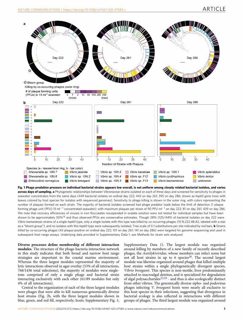
Our large-scale bait assay revealed that, at the strain level, lyticphage predation pressure on the majority of coastal ocean Vibrioappears low (<134 plaque forming phage L−1; limit of detectionbased on doubled counts assuming 50% recovery efficiency)compared to total virus-like-particle concentrations (1010 VLP L−1)common in coastal marine environments15 (Fig. 1, showingundoubled counts). As individual strains of the most abundantVibrio species in our study typically occur at concentrations of onaverage <1 cell ml−1 16, these findings indicate that encounterrates should be very low between these phages and their hosts.These observations are consistent with previous studies of lytic(plaque forming) phage predator loads on heterotrophic marinebacteria (largely in the family Vibrionaceae and genus Vibrio17, aswell as in the genus Pseudoalteromonas18) by Moebus, which alsoshowed the majority of bacterial strains were subject to 0–10 PFUml−1 in water samples collected in the same year. Theseobservations suggest that mechanisms that increase encounterrates between Vibrio phages and their hosts—such as hostblooms, spatial structure on small scales, and broad host range -should be important features of phage-bacteria interactions insystems where individual host strains are rare.
Lytic phage-bacteria interactions within the Vibrionaceae areoverall sparse and modular. To investigate the host ranges of thephages in this system we purified one phage from each plaque-positive host for further study, representing a final set of 248independent phage isolates (hosts: Supplementary Fig. 1, phages:Supplementary Data 8). In previous work we showed that thesephages represent phylogenetically diverse dsDNA viruses rangingin size from 10 kb – 349kb19, including non-tailed members ofthe recently proposed family Autolykiviridae20, as well as repre-sentatives of the three morphotypes of the Caudovirales (aspredicted by Virfam21). Host ranges of each of the phages wereassayed against a panel of 294 genome-sequenced bacterialstrains, including all plaque-positive hosts and 18 additionalVibrio strains (selected to represent additional populations ofVibrionaceae; for details on these additional strains see Supple-mentary Data 1 sheet A and filter for all bacterial strains withidentifiers without the prefix 10 N). Of these hosts, 279 were lysedby at least one of the 248 phages in the host range assay and wereincluded in subsequent analyses, with genomes of 259 memberbacteria and all phages sequenced (Supplementary Data 1).
In this large-scale study of the host ranges of 248 phages on279 hosts, we found that the majority of bacteria were resistant tothe majority of phages and that interactions were overall sparse –with only 1436 lytic interactions observed in agar overlay(hereafter “killing”) out of 69,192 possible interactions. Wefurther found that killing interactions were organized in anoverall modular fashion – with groups of phages and bacteriaclustering into 89 discrete interconnected sets (“modules”, Fig. 2a,subset shown is 248 phages and 259 hosts with sequencedgenomes, details in Supplementary Data 1) using the BiMatmodularity evaluation methods developed and employed byFlores et al.22 to investigate the Moebus-Nattkemper matrix4,5.These features of our matrix are strikingly similar to those of thesimilarly large matrix generated by Moebus and Nattkempermatrix in the 1980s (Table 1). However, unlike this previousmatrix, performed at a time when genome sequencing of allmembers was not possible, we could now also investigate thestructure of phage-bacteria interactions in light of genomic andphylogenetic diversity to understand the biological basis of themodular structure observed.
ARTICLE NATURE COMMUNICATIONS | https://doi.org/10.1038/s41467-021-27583-z
2 NATURE COMMUNICATIONS | (2022) 13:372 | https://doi.org/10.1038/s41467-021-27583-z | www.nature.com/naturecommunications
Diverse processes define membership of different interactionmodules. The structure of the phage-bacteria interaction networkin this study indicates that both broad and narrow host rangestrategies are important in the coastal marine environment.Whereas the three largest modules represented the majority oflytic interactions observed in agar overlay (53% of all interactions,768/1436 total infections), the majority of modules were single-tons comprised of only a single phage and bacterial straininteracting exclusively with each other (61/89 modules but only4% of all interactions).
Central to the organization of each of the three largest moduleswere phages that were able to kill numerous genomically diversehost strains (Fig. 2b, with the three largest modules shown inblue, green, and red fill, respectively; hosts: Supplementary Fig. 1;
Supplementary Data 1). The largest module was organizedaround killing by members of a new family of recently describedphages, the Autolykiviridae, whose members can infect some butnot all host strains in up to 6 species20. The second largestmodule was likewise organized around phages that killed multiplehost strains within a single phylogenetically divergent species,Vibrio breoganii. This species is non-motile, lives predominantlyattached to macroalgal detritus, and is specialized for degradationof algal polysaccharides23,24 - and thus is also ecologically distinctfrom other vibrios. The genomically diverse sipho- and podovirusphages infecting V. breoganii hosts were nearly all exclusive tothis host species in their infections, suggesting that divergence inbacterial ecology is also reflected in interactions with differentgroups of phages. The third largest module was organized around
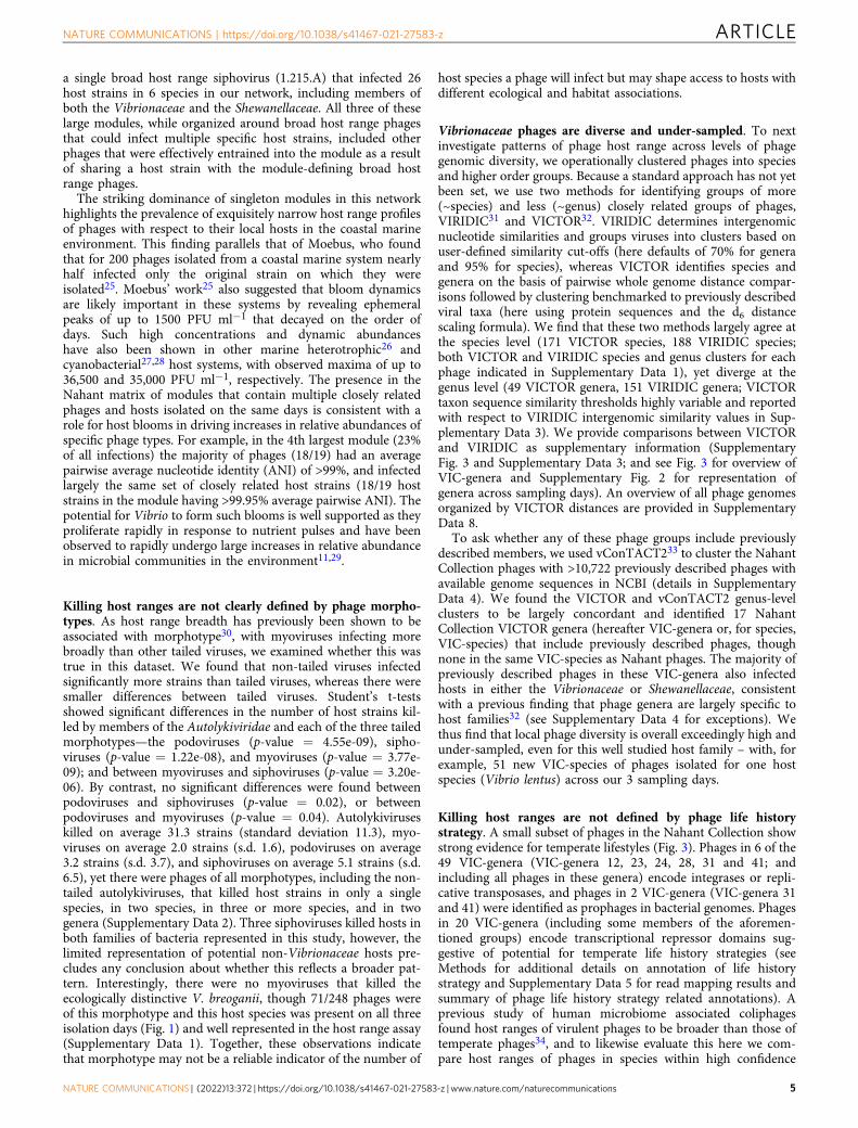
Fig. 1 Phage predation pressure on individual bacterial strains appears low overall, is not uniform among closely related bacterial isolates, and variesacross days of sampling. a Phylogenetic relationships between Vibrionaceae strains isolated on each of three days and screened for sensitivity to phages inseawater concentrates from the same days (449 bacterial isolates on ordinal day 222; 443 on day 261; 395 on day 286; shown as hsp60 gene trees withleaves colored by host species for isolates with sequenced genomes). Sensitivity to phage killing is shown in the outer ring, with colors representing thenumber of plaques formed on each strain. The majority of bacterial isolates screened had phage predator loads below the limit of detection (1 plaqueforming phage unit (PFU) 15 ml−1 concentrated seawater); with maximum plaques per strain of 90 PFU ml−1 on day 222; 81 on day 261; 439 on day 286.We note that recovery efficiencies of viruses in iron flocculates resuspended in oxalate solution were not tested for individual samples but have beenshown to be approximately 50%14 and thus observed PFUs are conservative estimates. Though 28% (125/449) of bacterial isolates on day 222 wereVibrio tasmaniensis strains of a single hsp60 type, only a single isolate with this type was killed by co-occurring phages (10 N.222.48.A2, labeled with a staras a “bloom group”), and no isolates with this hsp60 type were subsequently isolated. Tree scale of 0.1 substitutions per site indicated by red bars. b Strainskilled by co-occurring phages (43 plaque-positive on ordinal day 222; 101 on day 261; 141 on day 286) were targeted for genome sequencing and used insubsequent host range assays. Underlying data provided in Supplementary Data 1, see Methods for strain sets analyzed.
NATURE COMMUNICATIONS | https://doi.org/10.1038/s41467-021-27583-z ARTICLE
NATURE COMMUNICATIONS | (2022) 13:372 | https://doi.org/10.1038/s41467-021-27583-z | www.nature.com/naturecommunications 3
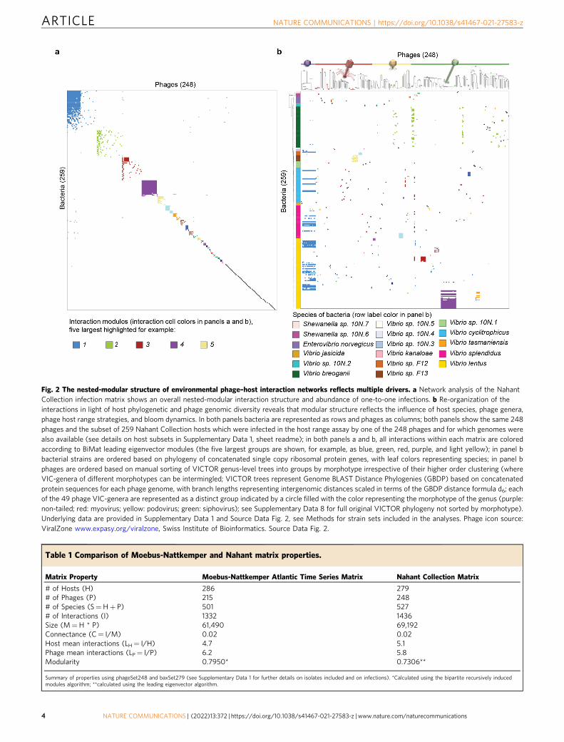
Fig. 2 The nested-modular structure of environmental phage–host interaction networks reflects multiple drivers. a Network analysis of the NahantCollection infection matrix shows an overall nested-modular interaction structure and abundance of one-to-one infections. b Re-organization of theinteractions in light of host phylogenetic and phage genomic diversity reveals that modular structure reflects the influence of host species, phage genera,phage host range strategies, and bloom dynamics. In both panels bacteria are represented as rows and phages as columns; both panels show the same 248phages and the subset of 259 Nahant Collection hosts which were infected in the host range assay by one of the 248 phages and for which genomes werealso available (see details on host subsets in Supplementary Data 1, sheet readme); in both panels a and b, all interactions within each matrix are coloredaccording to BiMat leading eigenvector modules (the five largest groups are shown, for example, as blue, green, red, purple, and light yellow); in panel bbacterial strains are ordered based on phylogeny of concatenated single copy ribosomal protein genes, with leaf colors representing species; in panel bphages are ordered based on manual sorting of VICTOR genus-level trees into groups by morphotype irrespective of their higher order clustering (whereVIC-genera of different morphotypes can be intermingled; VICTOR trees represent Genome BLAST Distance Phylogenies (GBDP) based on concatenatedprotein sequences for each phage genome, with branch lengths representing intergenomic distances scaled in terms of the GBDP distance formula d6; eachof the 49 phage VIC-genera are represented as a distinct group indicated by a circle filled with the color representing the morphotype of the genus (purple:non-tailed; red: myovirus; yellow: podovirus; green: siphovirus); see Supplementary Data 8 for full original VICTOR phylogeny not sorted by morphotype).Underlying data are provided in Supplementary Data 1 and Source Data Fig. 2, see Methods for strain sets included in the analyses. Phage icon source:ViralZone www.expasy.org/viralzone, Swiss Institute of Bioinformatics. Source Data Fig. 2.
Table 1 Comparison of Moebus-Nattkemper and Nahant matrix properties.
Matrix Property Moebus-Nattkemper Atlantic Time Series Matrix Nahant Collection Matrix
# of Hosts (H) 286 279# of Phages (P) 215 248# of Species (S=H+ P) 501 527# of Interactions (I) 1332 1436Size (M=H * P) 61,490 69,192Connectance (C= I/M) 0.02 0.02Host mean interactions (LH= I/H) 4.7 5.1Phage mean interactions (LP= I/P) 6.2 5.8Modularity 0.7950* 0.7306**
Summary of properties using phageSet248 and baxSet279 (see Supplementary Data 1 for further details on isolates included and on infections). *Calculated using the bipartite recursively inducedmodules algorithm; **calculated using the leading eigenvector algorithm.
ARTICLE NATURE COMMUNICATIONS | https://doi.org/10.1038/s41467-021-27583-z
4 NATURE COMMUNICATIONS | (2022) 13:372 | https://doi.org/10.1038/s41467-021-27583-z | www.nature.com/naturecommunications
a single broad host range siphovirus (1.215.A) that infected 26host strains in 6 species in our network, including members ofboth the Vibrionaceae and the Shewanellaceae. All three of theselarge modules, while organized around broad host range phagesthat could infect multiple specific host strains, included otherphages that were effectively entrained into the module as a resultof sharing a host strain with the module-defining broad hostrange phages.
The striking dominance of singleton modules in this networkhighlights the prevalence of exquisitely narrow host range profilesof phages with respect to their local hosts in the coastal marineenvironment. This finding parallels that of Moebus, who foundthat for 200 phages isolated from a coastal marine system nearlyhalf infected only the original strain on which they wereisolated25. Moebus’ work25 also suggested that bloom dynamicsare likely important in these systems by revealing ephemeralpeaks of up to 1500 PFU ml−1 that decayed on the order ofdays. Such high concentrations and dynamic abundanceshave also been shown in other marine heterotrophic26 andcyanobacterial27,28 host systems, with observed maxima of up to36,500 and 35,000 PFU ml−1, respectively. The presence in theNahant matrix of modules that contain multiple closely relatedphages and hosts isolated on the same days is consistent with arole for host blooms in driving increases in relative abundances ofspecific phage types. For example, in the 4th largest module (23%of all infections) the majority of phages (18/19) had an averagepairwise average nucleotide identity (ANI) of >99%, and infectedlargely the same set of closely related host strains (18/19 hoststrains in the module having >99.95% average pairwise ANI). Thepotential for Vibrio to form such blooms is well supported as theyproliferate rapidly in response to nutrient pulses and have beenobserved to rapidly undergo large increases in relative abundancein microbial communities in the environment11,29.
Killing host ranges are not clearly defined by phage morpho-types. As host range breadth has previously been shown to beassociated with morphotype30, with myoviruses infecting morebroadly than other tailed viruses, we examined whether this wastrue in this dataset. We found that non-tailed viruses infectedsignificantly more strains than tailed viruses, whereas there weresmaller differences between tailed viruses. Student’s t-testsshowed significant differences in the number of host strains kil-led by members of the Autolykiviridae and each of the three tailedmorphotypes—the podoviruses (p-value = 4.55e-09), sipho-viruses (p-value = 1.22e-08), and myoviruses (p-value = 3.77e-09); and between myoviruses and siphoviruses (p-value = 3.20e-06). By contrast, no significant differences were found betweenpodoviruses and siphoviruses (p-value = 0.02), or betweenpodoviruses and myoviruses (p-value = 0.04). Autolykiviruseskilled on average 31.3 strains (standard deviation 11.3), myo-viruses on average 2.0 strains (s.d. 1.6), podoviruses on average3.2 strains (s.d. 3.7), and siphoviruses on average 5.1 strains (s.d.6.5), yet there were phages of all morphotypes, including the non-tailed autolykiviruses, that killed host strains in only a singlespecies, in two species, in three or more species, and in twogenera (Supplementary Data 2). Three siphoviruses killed hosts inboth families of bacteria represented in this study, however, thelimited representation of potential non-Vibrionaceae hosts pre-cludes any conclusion about whether this reflects a broader pat-tern. Interestingly, there were no myoviruses that killed theecologically distinctive V. breoganii, though 71/248 phages wereof this morphotype and this host species was present on all threeisolation days (Fig. 1) and well represented in the host range assay(Supplementary Data 1). Together, these observations indicatethat morphotype may not be a reliable indicator of the number of
host species a phage will infect but may shape access to hosts withdifferent ecological and habitat associations.
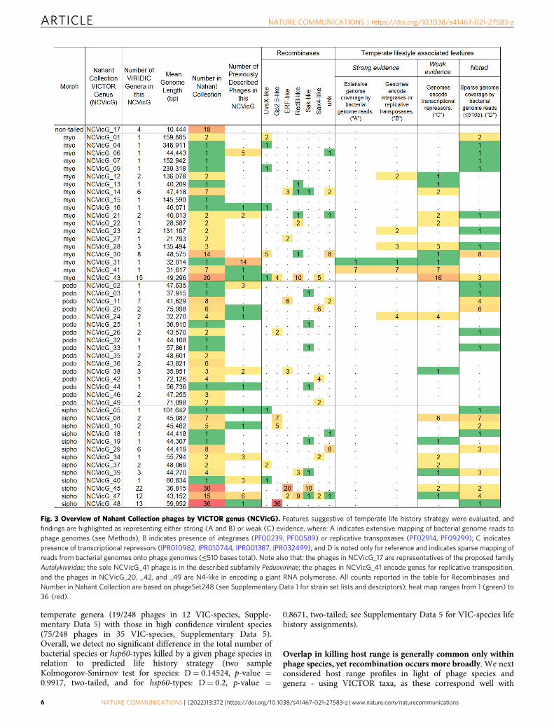
Vibrionaceae phages are diverse and under-sampled. To nextinvestigate patterns of phage host range across levels of phagegenomic diversity, we operationally clustered phages into speciesand higher order groups. Because a standard approach has not yetbeen set, we use two methods for identifying groups of more(~species) and less (~genus) closely related groups of phages,VIRIDIC31 and VICTOR32. VIRIDIC determines intergenomicnucleotide similarities and groups viruses into clusters based onuser-defined similarity cut-offs (here defaults of 70% for generaand 95% for species), whereas VICTOR identifies species andgenera on the basis of pairwise whole genome distance compar-isons followed by clustering benchmarked to previously describedviral taxa (here using protein sequences and the d6 distancescaling formula). We find that these two methods largely agree atthe species level (171 VICTOR species, 188 VIRIDIC species;both VICTOR and VIRIDIC species and genus clusters for eachphage indicated in Supplementary Data 1), yet diverge at thegenus level (49 VICTOR genera, 151 VIRIDIC genera; VICTORtaxon sequence similarity thresholds highly variable and reportedwith respect to VIRIDIC intergenomic similarity values in Sup-plementary Data 3). We provide comparisons between VICTORand VIRIDIC as supplementary information (SupplementaryFig. 3 and Supplementary Data 3; and see Fig. 3 for overview ofVIC-genera and Supplementary Fig. 2 for representation ofgenera across sampling days). An overview of all phage genomesorganized by VICTOR distances are provided in SupplementaryData 8.
To ask whether any of these phage groups include previouslydescribed members, we used vConTACT233 to cluster the NahantCollection phages with >10,722 previously described phages withavailable genome sequences in NCBI (details in SupplementaryData 4). We found the VICTOR and vConTACT2 genus-levelclusters to be largely concordant and identified 17 NahantCollection VICTOR genera (hereafter VIC-genera or, for species,VIC-species) that include previously described phages, thoughnone in the same VIC-species as Nahant phages. The majority ofpreviously described phages in these VIC-genera also infectedhosts in either the Vibrionaceae or Shewanellaceae, consistentwith a previous finding that phage genera are largely specific tohost families32 (see Supplementary Data 4 for exceptions). Wethus find that local phage diversity is overall exceedingly high andunder-sampled, even for this well studied host family – with, forexample, 51 new VIC-species of phages isolated for one hostspecies (Vibrio lentus) across our 3 sampling days.
Killing host ranges are not defined by phage life historystrategy. A small subset of phages in the Nahant Collection showstrong evidence for temperate lifestyles (Fig. 3). Phages in 6 of the49 VIC-genera (VIC-genera 12, 23, 24, 28, 31 and 41; andincluding all phages in these genera) encode integrases or repli-cative transposases, and phages in 2 VIC-genera (VIC-genera 31and 41) were identified as prophages in bacterial genomes. Phagesin 20 VIC-genera (including some members of the aforemen-tioned groups) encode transcriptional repressor domains sug-gestive of potential for temperate life history strategies (seeMethods for additional details on annotation of life historystrategy and Supplementary Data 5 for read mapping results andsummary of phage life history strategy related annotations). Aprevious study of human microbiome associated coliphagesfound host ranges of virulent phages to be broader than those oftemperate phages34, and to likewise evaluate this here we com-pare host ranges of phages in species within high confidence
NATURE COMMUNICATIONS | https://doi.org/10.1038/s41467-021-27583-z ARTICLE
NATURE COMMUNICATIONS | (2022) 13:372 | https://doi.org/10.1038/s41467-021-27583-z | www.nature.com/naturecommunications 5
temperate genera (19/248 phages in 12 VIC-species, Supple-mentary Data 5) with those in high confidence virulent species(75/248 phages in 35 VIC-species, Supplementary Data 5).Overall, we detect no significant difference in the total number ofbacterial species or hsp60-types killed by a given phage species inrelation to predicted life history strategy (two sampleKolmogorov-Smirnov test for species: D= 0.14524, p-value =0.9917, two-tailed, and for hsp60-types: D= 0.2, p-value =
0.8671, two-tailed; see Supplementary Data 5 for VIC-species lifehistory assignments).
Overlap in killing host range is generally common only withinphage species, yet recombination occurs more broadly. We nextconsidered host range profiles in light of phage species andgenera - using VICTOR taxa, as these correspond well with
Fig. 3 Overview of Nahant Collection phages by VICTOR genus (NCVicG). Features suggestive of temperate life history strategy were evaluated, andfindings are highlighted as representing either strong (A and B) or weak (C) evidence, where: A indicates extensive mapping of bacterial genome reads tophage genomes (see Methods); B indicates presence of integrases (PF00239, PF00589) or replicative transposases (PF02914, PF09299); C indicatespresence of transcriptional repressors (IPR010982, IPR010744, IPR001387, IPR032499); and D is noted only for reference and indicates sparse mapping ofreads from bacterial genomes onto phage genomes (≤510 bases total). Note also that: the phages in NCVicG_17 are representatives of the proposed familyAutolykiviridae; the sole NCVicG_41 phage is in the described subfamily Peduovirinae; the phages in NCVicG_41 encode genes for replicative transposition,and the phages in NCVicG_20, _42, and _49 are N4-like in encoding a giant RNA polymerase. All counts reported in the table for Recombinases andNumber in Nahant Collection are based on phageSet248 (see Supplementary Data 1 for strain set lists and descriptors); heat map ranges from 1 (green) to36 (red).
ARTICLE NATURE COMMUNICATIONS | https://doi.org/10.1038/s41467-021-27583-z
6 NATURE COMMUNICATIONS | (2022) 13:372 | https://doi.org/10.1038/s41467-021-27583-z | www.nature.com/naturecommunications
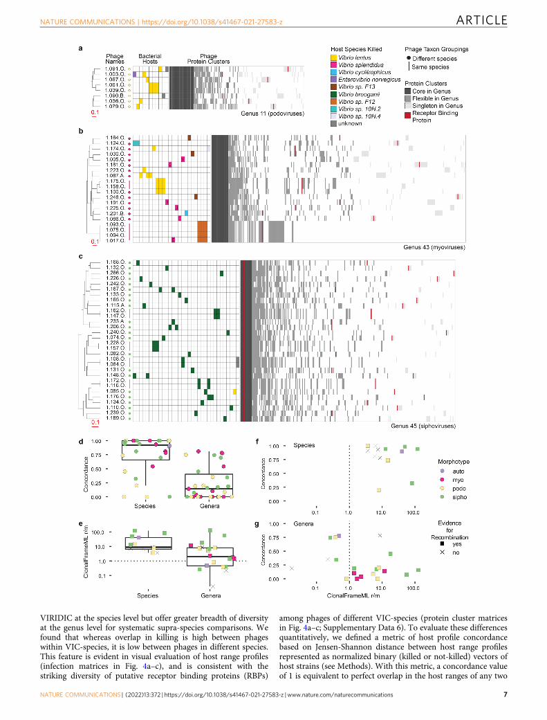
VIRIDIC at the species level but offer greater breadth of diversityat the genus level for systematic supra-species comparisons. Wefound that whereas overlap in killing is high between phageswithin VIC-species, it is low between phages in different species.This feature is evident in visual evaluation of host range profiles(infection matrices in Fig. 4a–c), and is consistent with thestriking diversity of putative receptor binding proteins (RBPs)
among phages of different VIC-species (protein cluster matricesin Fig. 4a–c; Supplementary Data 6). To evaluate these differencesquantitatively, we defined a metric of host profile concordancebased on Jensen-Shannon distance between host range profilesrepresented as normalized binary (killed or not-killed) vectors ofhost strains (see Methods). With this metric, a concordance valueof 1 is equivalent to perfect overlap in the host ranges of any two
NATURE COMMUNICATIONS | https://doi.org/10.1038/s41467-021-27583-z ARTICLE
NATURE COMMUNICATIONS | (2022) 13:372 | https://doi.org/10.1038/s41467-021-27583-z | www.nature.com/naturecommunications 7
phages, and a value of 0 represents no shared hosts. At the VIC-species level we found that concordance values were generallyhigh (Fig. 4d), with phages in 10 VIC-species showing perfectoverlap in their host range, including 3 VIC-species with memberphages isolated on different days (this analysis included 105phages, representing the 28 VIC-species with >1 member; seeSource Data Fig. 4 sheet A). By contrast, within VIC-genera,concordance in host range among members was generally low(Fig. 4d), even when calculated using a conservative approachthat yields higher estimates of concordance when VIC-species ina VIC-genus contain multiple members (see Methods, see SourceData Fig. 4 for concordances calculated using all members of agenus [sheet B] or with only a single representative of each species[sheet C], and see Supplementary Fig. 4 for scaling of con-cordance value with size of subsampled groups.).
The differences in host range concordance at the phage VIC-species and VIC-genus levels suggested that there should becorresponding differences in levels of recombination betweenphage genomes within these groups, yet we found recombinationto be occurring both within and between phage VIC-species. Weobserved this qualitatively in the distributions of flexible geneswithin VIC-genera, where in some cases nearest-neighbor phagesat the whole-genome level share flexible protein cluster genes notwith each other, but instead with phages of different VIC-specieswithin their VIC-genus (protein cluster matrices in Fig. 4–c;Supplementary Data 6). To quantitatively evaluate relativecontributions of homologous recombination and mutation(r/m) to diversification, we analyzed regions of phage genomesconserved at the VIC-species- and VIC-genus-levels. At the VIC-species level we found that homologous recombination was thegreater contributor (r/m > 1; one-sample Wilcoxon test, nullhypothesis mean = 1, p-value 0.0002) to diversification for themajority of VIC-species with sufficient members to test (Fig. 4e,Source Data Fig. 4 sheet D). This finding of r/m > 1 within phageVIC-species is consistent with similar findings in otherenvironments35–37 and suggests that, even in this seeminglyrare-encounter opportunity system, phage virion concentrationscan reach high enough local concentrations to result in co-infections between members of the same VIC-species in cells oftheir shared hosts.
Surprisingly we also found a signal for r/m > 1 (one-sampleWilcoxon test, null hypothesis mean = 1, p-value 0.0192) at theVIC-genus level (Fig. 4e), even when calculated using aconservative approach that could underestimate this value inVIC-genera containing VIC-species with multiple members (seeMethods, Source Data Fig. 4 sheet E). Considering the possibilitythat this metric could reflect high rates of recombination withinonly a single species within a genus, we also evaluate r/m withingenera using only a single representative of each species within a
genus and find that using this approach r/m decreases to <1 foronly a single VIC-genus [NCVicG_24] and increases to >1 for adifferent VIC-genus [NCVicG_17, the Autolykiviridae]. It isnotable that substituting tools with stricter relatedness thresholds(such as VIRIDIC) to define genera would result in a largernumber of quasi-genus groups and that therefore detecting r/m as>1 within VIC-genera is a conservative estimate of the potentialextent of homologous recombination occurring between phagesin supra-species level taxa.
The importance of recombination at both the VIC-species andVIC-genus levels indicates that overlap in killing between phagesis not predictive of their potential for recombination in thecontext of natural microbial communities. This is corroboratedwhen both quantitative metrics are considered together, whichshows a lack of a positive association between host rangeconcordance and r/m on both the VIC-genus (Fig. 4f, Spearmancorrelation −0.2695786) and VIC-species levels (Fig. 4g, Spear-man correlation −0.2417582).
To understand the potential maximal extent of recent geneflow among all phages in this collection we used a k-mer basedapproach and found evidence of sequence sharing betweenphages of different Caudovirales morphotypes with non-overlapping host killing. We first used the liberal metric ofoccurrence of sharing of any 100% identity 25-base pair (25-mer)length sequences between phages, such 25-mers are sufficientlyunique in bacterial genomes to be used to recapitulate strain andspecies level relatedness38, and provide a marker for potentialrecent horizontal gene transfer events39. Using this approach, wefound far greater potential connectivity of gene flow thansuggested by overlaps in host killing (Fig. 5a, b, Source DataFig. 5). Notably, however, despite extensive overlap in killing hostrange between autolykiviruses and tailed phages, there were nocases of shared 25-mers between phages in these two groups—afeature consistent with their lack of any shared protein clusters(Supplementary Data 4 and 6).
Considering next the more conservative metric of totalnumbers of 25-mers shared between any two phage genomeswe also found evidence for sequence sharing between divergenttailed phages with non-overlapping host ranges (Fig. 5c, Supple-mentary Fig. 3); with the maximum number of shared 25-mersbetween any pair of phages of different morphotypes being 6,169.In the collection, some pairs of phages of different morphotypesshow evidence of extensive sequence similarity in non-structuralgenome regions (e.g. Fig. 5d, similarity between NCVicG45 si-phoviruses with the singleton NCVicG25 podovirus), parallelingthe observation that the podovirus P22 shares sequence similarityto lambdoid siphoviruses in its non-structural genes40; other pairsshare only a single 25-mer, for example in a DNA polymerasegene (e.g. in protein cluster mmseq 2145 in podovirus 1.262.O
Fig. 4 Host range overlap is high within phage VIC-species and low within phage VIC-genera, but recombination occurs both within and between VIC-species. VIC-genus trees for each of a group of podoviruses (a), myoviruses (b), and siphoviruses (c) represent VICTOR Genome BLAST DistancePhylogenies (GBDP) based on concatenated protein sequences for each phage genome, with branch lengths representing intergenomic distances scaled interms of the GBDP distance formula d6 (complete tree with all phages shown in Supplementary Data 8, with underlying data in Newick format provided inSource Data Fig. 2. Filled in cells in the host range matrices aligned to the right of phage names (in rows) show host strains (in columns) killed by eachphage. Protein cluster matrices aligned to the right of the host range matrices show all the MMseqs2 protein sequence clusters present in each genus(columns), rank sorted based on the number of phages in the VIC-genus in which they occur. Quantified host range profiles for phages across thecollection show that: d overlap in killing profiles (concordance) is high within VIC-species (28 VIC-species with ≥2 phages, 105 phages total) but lowwithin VIC-genera (31 VIC-genera with≥2 phages, including cases of genera represented by only 1 species, 230 phages total; two-sided Welch’s t-test p-value = 1.45e-07); that e, recombination in conserved regions is commonly a greater contributor to genomic diversity in both species and genera (samephage counts as in panel d); and, f and g, that there is no relationship between concordance in killing and recombination for either VIC-species or VIC-genera, respectively. Underlying data and strain information available in Supplementary Data 1 and 6, and in Source Data Fig. 4, see Methods fordescription of differences in results when considering only single VIC-species representatives in VIC-genus-level analyses. Boxplot features: central line-median; box limits-1st and 3rd quartiles; upper whisker-largest value no larger than 1.5 * IQR (inter-quartile range); lower whisker-smallest value no smallerthan 1.5 * IQR. Source Data Fig. 4.
ARTICLE NATURE COMMUNICATIONS | https://doi.org/10.1038/s41467-021-27583-z
8 NATURE COMMUNICATIONS | (2022) 13:372 | https://doi.org/10.1038/s41467-021-27583-z | www.nature.com/naturecommunications
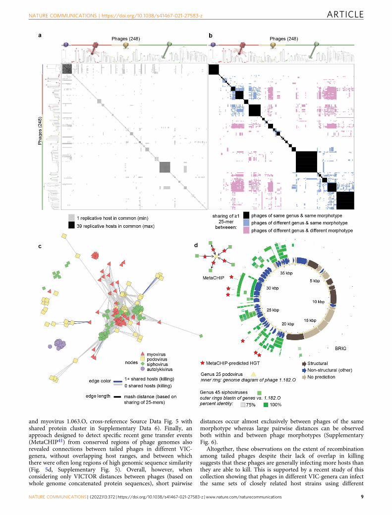
and myovirus 1.063.O, cross-reference Source Data Fig. 5 withshared protein cluster in Supplementary Data 6). Finally, anapproach designed to detect specific recent gene transfer events(MetaCHIP41) from conserved regions of phage genomes alsorevealed connections between tailed phages in different VIC-genera, without overlapping host ranges, and between whichthere were often long regions of high genomic sequence similarity(Fig. 5d, Supplementary Fig. 5). Overall, however, whenconsidering only VICTOR distances between phages (based onwhole genome concatenated protein sequences), short pairwise
distances occur almost exclusively between phages of the samemorphotype whereas large pairwise distances can be observedboth within and between phage morphotypes (SupplementaryFig. 6).
Altogether, these observations on the extent of recombinationamong tailed phages despite their lack of overlap in killingsuggests that these phages are generally infecting more hosts thanthey are able to kill. This is supported by a recent study of thiscollection showing that phages in different VIC-genera can infectthe same sets of closely related host strains using different
NATURE COMMUNICATIONS | https://doi.org/10.1038/s41467-021-27583-z ARTICLE
NATURE COMMUNICATIONS | (2022) 13:372 | https://doi.org/10.1038/s41467-021-27583-z | www.nature.com/naturecommunications 9
receptors. While these receptors are highly monomorphic at thebacterial species/population level, killing profiles are non-overlapping due to differential bacterial carriage of highly specificphage defense systems (Hussain and Dubert et al.42, where the“orange” phages in the reference are VIC-species 165 and 99 inVIC-genus 47, and the “purple” phages are VIC-species 144 inVIC-genus 48). Thus, co-infections of host cells by multipledifferent phages (necessary to allow for recombination betweenphage genomes) are likely far more common in naturalcommunities than predicted by killing host ranges.
Nucleotide sharing between phages can lead to cross-mappingof sequencing reads. Considering the potential for widespreadsequence sharing to influence mapping of viral reads to referencegenomes, we investigated cross-mapping using a recently devel-oped rapid k-mer based pseudo-alignment approach43. We foundthat cross-mapping of reads can occur between phages of dif-ferent VIC-species, VIC-genera, and morphotypes within thiscollection. False positive classifications of reference presence werereduced when using shorter simulated read lengths as a result ofoverall lower collateral (false positive) sequence coverage whenthe basis for the mapping was a single 31-mer match in thesequence (Supplementary Fig. 7, see Methods). This observedpotential for cross-mapping calls for a cautious approach in usingread-mapping to reference genomes in determining whetherspecific phages are present in metagenomic samples or predictingwhich hosts virus pools are interacting with.
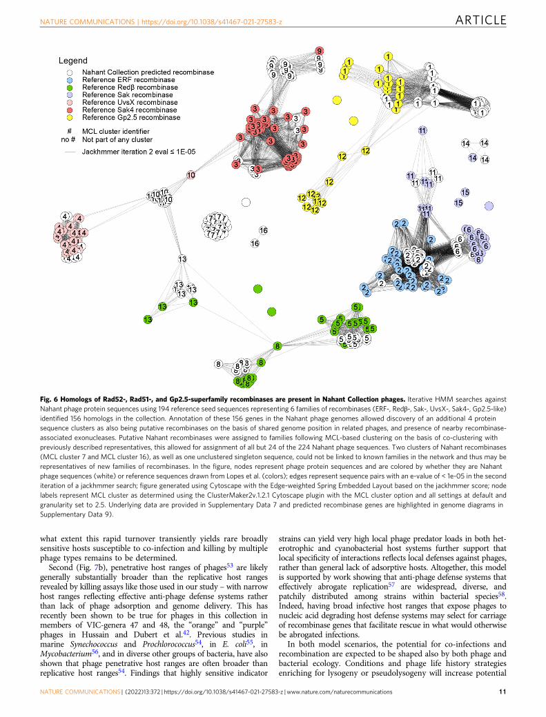
Recombinases are prevalent in Vibrionaceae phage genomes.The overall prevalence of sequence sharing observed betweenphage genomes suggests that they harbor homologous recombi-nation systems, and indeed we find that recombinase genes areencoded by the majority of tailed phages in this collection. Lowfidelity single strand annealing protein (SSAP) based recombinasesystems such as those in the Rad52-superfamily (e.g. Redβ/RecT-,ERF-, and Sak-families) are common in temperate phages44, andare thought to play an important role in their extensive genomemodularity and mosaicism45. Such recombinases have beenshown to be associated with large-scale recombination events ofup to 79% genome length between incoming temperate phagesand resident prophages46 and are useful tools for in vitro geneticengineering (recombineering)47–50 as they can facilitate recom-bination between sequence regions with as little as 23-bpsequence identity51. Noting a number of putative SSAP recom-binase genes in our initial annotations, we sought to more sys-tematically evaluate their representation in our diverse collectionof phages. Using representative sequences44 as seeds for iterative
searches, followed by gene neighborhood analysis, we identifiedputative recombinases in 196/230 (85%) tailed phages (in pha-geSet248, see Methods and Supplementary Data 8 showing gen-ome diagrams with recombinases highlighted), with 117 of theseresembling low-fidelity Rad52-superfamily and Sak4-like Rad51-superfamily recombinases commonly associated almost exclu-sively with temperate phages44 (Fig. 6, Supplementary Data 7).That no recombinases were identified among the autolykiviruses,though these viruses also showed evidence for high rates of intra-species recombination (when calculated using single representa-tives of each species rather than all members, Source Data Fig. 4,sheet F), indicates distinct pathways underlying observedrecombination in this group. These results suggest that just ashorizontal gene transfer in microbial communities may allowbacteria to evolve resistance to phages, recombination and geneticexchange between phages may likewise be important in over-coming this resistance.
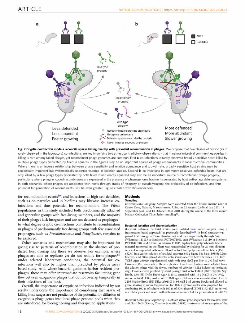
DiscussionThe findings of this work appear at first contradictory: thatrecombined phage genomes commonly co-exist in naturalmicrobial communities, yet overlap in phage killing in the contextof natural diversity is rare. Previous work has spoken to theimportance of recombination in generation of phage diversityglobally and locally52, and our observations imply that recombi-nants are generated in co-infections of shared hosts that are morefrequent than are revealed by killing assays. That these largelyunobserved implied co-infections are occurring in recent evolu-tionary and ecological time is indicated by the higher contribu-tion of homologous recombination than mutation to sequencedivergence in many phage groups in our dataset.
We propose two main complementary model scenarios tounify and reconcile these apparently contradictory observations –one addressing trade-offs between growth and phage defense inbacteria and the other pointing to recombination as an importantmechanism for phage survival in the face of selective pressure bybacterial anti-phage defenses.
First (Fig. 7a), recombinant phages may be disproportionatelygenerated during co-infections in broadly sensitive (killed) hosts.Previous work with the marine phototroph Synechoccocus byWaterbury and Valois27 showed that the rarest bacterial strainswere the fastest growing as well as the most sensitive to phages.As a result of their lower relative abundances in the environment,these ecologically important broadly phage sensitive hosts maythus be underrepresented in cultivated isolate collections such asours. Recent work42 has also demonstrated that rapid turnover inanti-phage defense systems42 is key to fine-scale differences inphage sensitivity among closely related bacterial strains, and to
Fig. 5 Overlap in killing of host strains is sparse among tailed phages and does not reflect sequence sharing between phages of different VIC-generaor morphotypes. Matrix representations of pairwise comparisons between phages, show that: a occurrence of shared replicative hosts, does not reflect boccurrence of sharing of ≥1 25-mer between phages of different VIC-genera and morphotypes; in both panels phages are ordered based on manual sortingof VICTOR genus-level trees into groups by morphotype irrespective of their higher order clustering (where VIC-genera of different morphotypes can beintermingled, see Methods for details and Supplementary Data 8 for full original VICTOR phylogeny not sorted by morphotype); each of the 49 VIC-generaare represented as a distinct group indicated by a circle filled with the color representing the morphotype of the VIC-genus (purple: non-tailed; red:myovirus; yellow: podovirus; green: siphovirus). VICTOR trees represent Genome BLAST Distance Phylogenies (GBDP) based on concatenated proteinsequences for each phage genome, with branch lengths representing intergenomic distances scaled in terms of the GBDP distance formula d6. Extent ofsequence sharing that can occur between phage morphotypes and genera that are largely non-overlapping in host killing is revealed by: c networkvisualization of phage genome connectivity by Mash distance, which is defined by sharing of 25-mers; and d occurrence of extensive sequence similaritybetween phages of different morphotypes, as visualized here using Blast Ring Image Generator (BRIG), for which specific directional horizontal genetransfer events can also be detected using MetaCHIP. Source data for panels a, b, c, and d are provided in Source Data Fig. 5, see Methods for strain setsincluded in the analyses; annotations for genes shown in panel d are provided in Supplementary Data 6, see Methods for information on methods fordefining “structural”, “non-structural” (defined as “other” in Supplementary Data 6), or “no prediction”. Phage icon source: ViralZone www.expasy.org/viralzone, Swiss Institute of Bioinformatics. Source Data Fig. 5.
ARTICLE NATURE COMMUNICATIONS | https://doi.org/10.1038/s41467-021-27583-z
10 NATURE COMMUNICATIONS | (2022) 13:372 | https://doi.org/10.1038/s41467-021-27583-z | www.nature.com/naturecommunications
what extent this rapid turnover transiently yields rare broadlysensitive hosts susceptible to co-infection and killing by multiplephage types remains to be determined.
Second (Fig. 7b), penetrative host ranges of phages53 are likelygenerally substantially broader than the replicative host rangesrevealed by killing assays like those used in our study – with narrowhost ranges reflecting effective anti-phage defense systems ratherthan lack of phage adsorption and genome delivery. This hasrecently been shown to be true for phages in this collection inmembers of VIC-genera 47 and 48, the “orange” and “purple”phages in Hussain and Dubert et al.42. Previous studies inmarine Synechococcus and Prochlorococcus54, in E. coli55, inMycobacterium56, and in diverse other groups of bacteria, have alsoshown that phage penetrative host ranges are often broader thanreplicative host ranges54. Findings that highly sensitive indicator
strains can yield very high local phage predator loads in both het-erotrophic and cyanobacterial host systems further support thatlocal specificity of interactions reflects local defenses against phages,rather than general lack of adsorptive hosts. Altogether, this modelis supported by work showing that anti-phage defense systems thateffectively abrogate replication57 are widespread, diverse, andpatchily distributed among strains within bacterial species58.Indeed, having broad infective host ranges that expose phages tonucleic acid degrading host defense systems may select for carriageof recombinase genes that facilitate rescue in what would otherwisebe abrogated infections.
In both model scenarios, the potential for co-infections andrecombination are expected to be shaped also by both phage andbacterial ecology. Conditions and phage life history strategiesenriching for lysogeny or pseudolysogeny will increase potential
Fig. 6 Homologs of Rad52-, Rad51-, and Gp2.5-superfamily recombinases are present in Nahant Collection phages. Iterative HMM searches againstNahant phage protein sequences using 194 reference seed sequences representing 6 families of recombinases (ERF-, Redβ-, Sak-, UvsX-, Sak4-, Gp2.5-like)identified 156 homologs in the collection. Annotation of these 156 genes in the Nahant phage genomes allowed discovery of an additional 4 proteinsequence clusters as also being putative recombinases on the basis of shared genome position in related phages, and presence of nearby recombinase-associated exonucleases. Putative Nahant recombinases were assigned to families following MCL-based clustering on the basis of co-clustering withpreviously described representatives, this allowed for assignment of all but 24 of the 224 Nahant phage sequences. Two clusters of Nahant recombinases(MCL cluster 7 and MCL cluster 16), as well as one unclustered singleton sequence, could not be linked to known families in the network and thus may berepresentatives of new families of recombinases. In the figure, nodes represent phage protein sequences and are colored by whether they are Nahantphage sequences (white) or reference sequences drawn from Lopes et al. (colors); edges represent sequence pairs with an e-value of < 1e-05 in the seconditeration of a jackhmmer search; figure generated using Cytoscape with the Edge-weighted Spring Embedded Layout based on the jackhmmer score; nodelabels represent MCL cluster as determined using the ClusterMaker2v.1.2.1 Cytoscape plugin with the MCL cluster option and all settings at default andgranularity set to 2.5. Underlying data are provided in Supplementary Data 7 and predicted recombinase genes are highlighted in genome diagrams inSupplementary Data 9).
NATURE COMMUNICATIONS | https://doi.org/10.1038/s41467-021-27583-z ARTICLE
NATURE COMMUNICATIONS | (2022) 13:372 | https://doi.org/10.1038/s41467-021-27583-z | www.nature.com/naturecommunications 11
for recombination events59, and infections at high cell densities,such as on particles and in biofilms may likewise increase co-infections and thus potential for recombination. The Vibriopopulations in this study included both predominantly attachedand generalist groups with free-living members, and the majorityof their phages lack integrases and are not detected as prophages -to what degree cryptic co-infections contribute to recombinationin phages of predominantly free-living groups with few associatedprophages, such as Prochlorococcus and Pelagibacter, remains tobe explored.
Other scenarios and mechanisms may also be important forgiving rise to patterns of recombination in the absence of pre-dicted host overlap like those we observe. For example, wherephages are able to replicate yet do not readily form plaques60
under selected laboratory conditions, the potential for co-infections will also be higher than predicted by plaque assaybased study. And, where bacterial genomes harbor resident pro-phages, these may offer intermediate reservoirs facilitating geneflow between exogenous phages that do not overlap temporally intheir infections of the host.
Overall, the importance of cryptic co-infections indicated by ourresults underscores the importance of considering that assays ofkilling host ranges are not predictive of the potential for diffusion ofexogenous phage genes into local phage genome pools when theyare introduced for bioengineering and therapeutic applications.
MethodsSamplingEnvironmental sampling. Samples were collected from the littoral marine zone atCanoe Cove, Nahant, Massachusetts, USA, on 22 August (ordinal day 222), 18September (261) and 13 October (286) 2010, during the course of the three monthNahant Collection Time Series sampling11.
Bacterial isolation and characterizationBacterial isolation. Bacterial strains were isolated from water samples using afractionation-based approach7 as previously described19,20. In brief, seawater waspassed first through a 63um plankton net and then sequentially through 5um(Whatman 111113 or Sterlitech PCT5047100), 1um (Whatman 111110 or SterlitechPCT1047100), and 0.2um (Whatman 111106) hydrophilic polycarbonate filters;material recovered on the filters was resuspended by shaking for 20min; dilutionseries of resuspended cells were filtered onto 0.2um polyethersulfone filters (Pall66234) in a carrier solution of artificial seawater (40 g Sigma Sea Salts, S9883; 0.2umfiltered), and filters placed directly onto Vibrio-selective MTCBS plates (BD DifcoTCBS Agar 265020, supplemented with with 10 g NaCl per liter to 2% final w/v).Colonies (96) from each of three replicates of each size fraction were selected fromthe dilution plates with the fewest numbers of colonies (1,152 isolates per isolationday). Colonies were purified by serial passage, first onto TSB-II (Difco Tryptic SoyBroth, 1.5% BD Difco Bacto Agar 214010, amended with 15 g NaCl to 2% w/v),second onto MTCBS, finally onto TSB-II again. Colonies were inoculated into 1ml of2216 Marine Broth (BD Difco 279110) in 96-well 2 ml culture blocks and allowed togrow, shaking at room temperature, for 48 h. Glycerol stocks were prepared bycombining 100 ul of culture with 100 ul of 50% glycerol (BDH 1172-4LP) in 96-wellmicrotiter plates and sealed with adhesive aluminum foil for preservation at −80 °C.
Bacterial hsp60 gene sequencing. To obtain hsp60 gene sequences for isolates, Lyseand Go (LNG) (Pierce, Thermo Scientific 78882) treatments of subsamples of the
Fig. 7 Cryptic-coinfection models reconcile sparse killing overlap with prevalent recombination in phages.We propose that two classes of cryptic (as inrarely observed in the laboratory) co-infections are key in unifying two at first contradictory observations - that in natural microbial communities overlap inkilling is rare among tailed phages, yet recombinant phage genomes are common. First a, co-infections in rarely observed broadly sensitive hosts killed bymultiple phage types (indicated by filled in squares in the figure) may be an important source of phage recombinants in local microbial communities.Where there is an inverse relationship between phage sensitivity and relative abundance and growth rate, broadly sensitive host strains may beecologically important but systematically underrepresented in isolation studies. Second b, co-infections in commonly observed defended hosts that areonly killed by a few phage types (indicated by both filled in and empty squares) may also be an important source of recombinant phage progeny,particularly where phage encoded recombinases are expressed in the presence of phage genome fragments generated by host anti-phage defense systems.In both scenarios, where phages are associated with hosts through states of lysogeny or pseudolysogeny, the probability of co-infections, and thuspotential for generation of recombinants, will be even greater. Figure created with BioRender.com.
ARTICLE NATURE COMMUNICATIONS | https://doi.org/10.1038/s41467-021-27583-z
12 NATURE COMMUNICATIONS | (2022) 13:372 | https://doi.org/10.1038/s41467-021-27583-z | www.nature.com/naturecommunications
same overnights cultures used in the bait assay (described below) were used directlyas template in PCR amplification reactions. PCR reactions were prepared in 30 ulvolumes, as follows: 1 ul LNG template, 3 ul 10x buffer, 3 ul 2 mM dNTPs, 3 ul2um hsp60-F primer, 3 ul 2um hsp60-R primer, 0.3 ul NEB Taq, 16.7 ul PCR-gradeHOH; with hsp60-F (H279) primer sequence: 5′-GAA TTC GAI III GCI GGI GAYGGI ACI ACI AC-3′, and hsp60-R (H280) primer sequence: 5′-CGC GGG ATCCYK IYK ITC ICC RAA ICC IGG IGC YTT-3′61 (Supplementary Table 1). PCRthermocycling conditions were as follows: initial denaturation at 94 °C for 2 min;35 cycles of 94 °C for 1 min, 37 C for 1 min, 72 °C for 1 min; final annealing at72 °C for 6 min; hold at 10 °C. PCR products were cleaned up by isopropyl alcohol(IPA) precipitation, as follows: addition of 100 ul 75% IPA to 30 ul PCR reactionproduct, gentle inversion mixing followed by 25 min incubation at RT, 30 mincentrifugation at 2800 rcf, addition of 50 ul 70% IPA with gentle inversion wash,centrifugation at 2000 rcf, inversion on paper towels to remove IPA, 10 min cen-trifugation at 700 rcf, air drying in PCR hood for 30 min, resuspension in 30 ulPCR HOH. PCR products were Sanger sequenced (Genewiz, Inc.) using hsp60-Rprimer, as follows: 5 ul of 5 um hsp60-R primer, 7 ul nuclease free water, 3 ul DNAtemplate. For a subset of strains hsp60 sequences were obtained from subsequentlydetermined whole-genome sequences. Hsp60 sequences were aligned to the hsp60sequence previously published for Vibrio 1S_84 and trimmed to 422 bases usingGeneious (https://www.geneious.com/). Accession numbers for these 1287 strainsare provided in Supplementary Data 1, where they are identified as baxSet1287.
Bacterial hsp60 phylogenies. A phylogenetic tree of relationships among bacterialisolates screened in the bait assay (described below) was produced based on a422 bp fragment of the hsp60 gene, derived either from Sanger or whole genomesequences; with E. coli K12 serving as the outgroup. Sequences from each of thethree days of isolation were aligned using muscle v.3.8.3162 with default settings(muscle -in $seqsALL -out $seqsALL.muscleAln), and a single tree including all1287 sequences from all the days was generated using FastTree v.2.1.863 (FastTree-gtr -gamma -nt -spr 4 -slow < $seqsALL.muscleAln > $seqsALL.muscleAln.fasttree).For presentation in Fig. 1 three sub-trees including only nodes from each day wereproduced using PareTree v.1.0.264 (java -jar PareTree1.0.2.jar -t O -del not-Day222.txt -f $seqsALL.$round.muscleAln.fasttree.DAY222). Trees were visualizedusing iTOL65 and painted with metadata for each of the strains, including: sensi-tivity to killing in agar overlay by co-occurring phage predators collected on thesame day and, for the subset of strains that were genome sequenced and alsoincluded in the host range matrix, the bacterial species, based on concatenatedribosomal protein analysis using RiboTree66 as described below. Isolation days foreach of the strains included in these analyses are provided in SupplementaryData 1, where these strains are identified as baxSet1287.
Bacterial genome sequencing and assignment to populations. To assign genome-sequenced bacterial isolates used in the host range assay to species, we use theRiboTree tool66 to produce a phylogeny based on concatenated single copy ribo-somal proteins as in23. We include strains of previously described Vibrionaceae inpreliminary analyses as reference strains and assign species names to new isolatesbased on clustering with named representatives, as well as provide placeholdernames for newly identified clades with no previously described representatives.Trees were visualized using iTOL65 and the representation including only thosestrains included in the host range assay is shown in Supplementary Fig. 1; popu-lation assignments and accession numbers for this set of 294 genomes, which alsoincludes a small number of previously isolated bacterial strains that were includedin the host range assay (described below), are provided in Supplementary Data 1,where they are identified as baxSet294.
Viral isolation and characterization. We have previously described features of theviruses of the Nahant Collection20, as well as approaches used for the standardi-zation of their genome assemblies19, additional details are provided below.
Viral sample collection. The iron chloride flocculation approach was used to gen-erate 1000-fold concentrated viral samples from 0.2 um-filtered seawater, as fol-lows. For each isolation day, triplicate 4 L seawater samples were filtered through0.2 um polyethersulfone cartridge filters (Millipore, Sterivex, SVGP0150) intocollection bottles, spiked with 400 uL of FeCl3 solution (10 gL−1 Fe; as 4.83 gFeCl3•6H2O (Mallinckrodt 5029) into 100 ml H2O), and allowed to incubate atroom temperature for at least 1 h. Virus-containing flocs were then recovered fromthe sample by filtration onto 90 mm 0.2 um polycarbonate filters (Millipore, Iso-pore, GTTP09030) under gentle vacuum in a 90 mm glass cup-frit system (e.gKontes funnel 953755-0090, fritted base 953752-0090, and clamp 953753-0090);once liquid was fully passed, the funnel was removed and, with the vacuum pumpleft on, the filters were folded into quarters, removed from the fritted base, andinserted into a 7 ml borosilicate glass vial. A volume of 4 ml of oxalate-EDTAsolution (prepared from stock solution as 10 ml 2 M Mg2EDTA (J.T. Baker,JTL701-5), 10 ml 2.5 M Tris-HCl (Promega PAH5123), 25 ml 1M oxalic acid(Mallinckrodt 2752); adjusted to pH 6 with 10M NaOH (J.T. Baker, 3722-01); finalvolume 100 ml; used within 7 days of preparation and maintained at room tem-perature in the dark) was added to the vial and the sample allowed to dissolve atroom temperature for at least 30 min before transfer to storage at 4 °C. A reagent
used in this original formulation (JT-Baker 7501 Mg2EDTA) is no longer availableand an updated recipe is provided elsewhere67.
Bait assay and associated viral plaque archival. In order to obtain estimates of co-occurring phage predator loads at bacterial strain level resolution, and generate pla-ques from which to isolate phages, we exposed 1440 purified bacterial isolates tophage concentrates from their same day of isolation (1334 yielded lawns sufficient toevaluate for plaques, and hsp60 sequences could be determined for 1287 of these).Bacterial strains screened included 480 isolates from each ordinal day, representing120 strains from each of 4 size-fractionation classes (0.2 um, 1.0 um, 5.0 um, 63 um)details of isolation origin are provided for each strain in Supplementary Data 1, anddescription of naming conventions is as previously described19. For the bait assay eachstrain was mixed in agar overlay with seawater concentrates containing viruses (15 ulconcentrate, equivalent to 15ml unconcentrated seawater assuming 100% recoveryefficiency; derived from pooling of three replicate virus concentrates from each day).We note that recoveries were not tested for individual samples and that previoustests14 of recovery efficiency have shown that resuspension of iron flocculates inoxalate solution yields initial recoveries of approximately 50% (49 ± 3% and 55 ± 11%for a marine sipho- and myo-virus respectively, at 24 h post re-suspension) and showslow decay rate over time (47 ± 5% and 73 ± 16% for a marine sipho- and myo-virusrespectively, at 38 days post re-suspension). All of our assays were performedapproximately 8–9 months post-sampling from oxalate concentrates stored at 4 °C.Agar overlays were performed based on the previously described Tube-free method13,as follows. Bacterial strains were prepared for agar overlay plating by streaking outfrom glycerol stocks onto 2216MB agar plates with 1.5% agar (Difco, BD Bacto,214010), and allowed to grow for 2 days at room temperature. Strains were theninoculated into 1ml 2216MB in a 96-well culture block and incubated 24 h at roomtemperature shaking at 275 rpm on a VWR DS500E orbital shaker. Immediately priorto use in direct plating the OD600 was measured in 96-well microtiter plates andsubsamples were taken for Lyse and Go (LNG) processing for DNA (10 ul culture, 10ul LNG). Phage concentrates were prepared for plating by pooling 1.2ml from each ofthe concentrate replicates into a 7ml borosilicate scintillation vial. Cultures weretransferred from overnight culture blocks to 96-well PCR plates in 100 ul volume and15 ul of pooled phage concentrate was added to cultures one row at a time, with eachrow plated in agar overlay before adding phage concentrate to the next row ofbacterial cultures. Mixed samples of 100 ul bacterial overnight culture and 15 ulpooled phage concentrate were transferred to the surface of bottom agar plates(2216MB, 1% agar, 5% glycerol, 125ml L−1 of chitin supplement [40 g L−1 coarselyground chitin, autoclaved, 0.2 um filtered]). A 2.5ml volume of 52 °C molten top agar(2216MB, 0.4% agar, 5% glycerol BDH 1172-4LP) was added to the surface of thebottom agar and swirled around to incorporate and evenly disperse the mixed bac-terial and phage sample into an agar overlay lawn. Agar overlay lawns were held atroom temperature for 14–16 days and observed for plaque formation. Glycerol wasincorporated into this assay to facilitate detection of plaques68. Chitin supplement wasincorporated into this assay to facilitate detection of phages interacting with receptorsupregulated in response to chitin degradation products. A variety of preliminary testsexploring potential optimizations to agar compositions for direct plating indicatedthat the addition of chitin did not negatively impact recovery of plaques with controlphage strains tested. After approximately 2 weeks, plaques on agar overlay lawns werecataloged and described with respect to plaque morphology and plaques were pickedfor storage based on the previously described Archiving Plaques method13, as follows.All plaques were archived from plates containing less than 25 plaques, on plates withlarger numbers of plaques a random subsample of plaques from each distinct mor-phology were archived. A polypropylene 96-well PCR plate was filled with 200 ulaliquots of 0.2 um filtered 2216MB, agar plugs were collected from plates using a 1mlbarrier pipette tip and ejected into the 2216MB, skipping one well between eachsample to minimize potential for cross-contamination, for a final count of 48 phageplugs per plate. Plaque plugs were soaked at 4 °C for several hours to allow elution ofphage particles into the media. After soaking, 96-well plates were centrifuged at 2,000rcf for 3 min before proceeding to the next step. Plug soaks were then processed fortwo independent storage treatments. For storage at 4 °C, plates were processed bytransferring 150 ul of eluate from each well to a 0.2 um filtration plate (Millipore,Multiscreen HTS GV 0.22um Filter Plate MSGVS22) and gently filtered undervacuum to remove bacteria, the cell-free filtrates containing eluted phage particlesfrom each plaque plug were stored at 4 °C. For storage at −20 °C, 50 ul of 50%glycerol was added to the residual ~50 ul of the plug elution, often still containing theagar plug. In this way all plaques were characterized and many plaques from eachstrain were archived in two independent sets of conditions. Total plaque counts for allstrains included in the bait assay are represented in Fig. 1, and provided in Supple-mentary Data 1, where they are identified as baxSet1287. Notes on limitations to theassay: Water temperatures on each of the three isolations days were 13.8 °C, 16.3 °C,and 14.2 °C, for days 222, 261, and 286; as bait assays were performed at roomtemperature (approximately 22 °C) some phages requiring lower temperatures maynot have yielded plaques. The majority of plates were evaluated for plaque formationtwice, on day 1 and day 13, thus any plaques appearing after day 1 and disappearingbefore day 13 – for example as a result of overgrowth of lysogens—are likely to havebeen missed in these assays.
Viral purification. A subset of plaques archived during the bait assay was selectedfor phage purification, genome sequencing, and host range characterization. This
NATURE COMMUNICATIONS | https://doi.org/10.1038/s41467-021-27583-z ARTICLE
NATURE COMMUNICATIONS | (2022) 13:372 | https://doi.org/10.1038/s41467-021-27583-z | www.nature.com/naturecommunications 13
subset included single randomly-selected representatives from each plaque-positivebacterial strain. Minor details of the purification and lysate preparation variedacross samples but were largely as follows. Phages were purified from inoculaderived primarily from −20 °C plaque archives, and secondarily from 4 °C archiveswhen primary attempts with −20 °C stocks failed to produce plaques. Three serialpassages were performed using Molten Streaking for Singles13 method. Agaroverlay lawns for passages were prepared by aliquoting 100 ul of host overnightculture (4 ml 2216MB, colony inoculum from streak on 2216MB with 1.5% BactoAgar, shaken overnight at RT at 250 rpm on VWR DS500E orbital shaker) onto astandard size bottom agar plate and adding 2.5 ml of molten 52 °C top agar as inthe bait assay, swirling to disperse the host into the top agar and form a lawn, andstreaking-in phage with a toothpick either from the plaque archive or directly fromwell-separated plaques in overlays from the previous step in serial purification.Following plaque formation on the third serial passage plate plaque plugs werepicked using barrier tip 1 ml pipettes and ejected into 250 ul of 2216MB to eluteovernight at 4 °C. Plaque eluates were spiked with 20 ul of host culture and grownwith shaking for several hours to generate a primary small-scale lysate. Small scaleprimary lysates were centrifuged to pellet cells and titered by drop spot assay toestimate optimal inoculum volume to achieve confluent lysis in a 150 mm agaroverlay plate lysate. Plate lysates were generated by mixing 250 ul of overnight hostculture with primary lysate and plating in 7.5 ml agar overlay. After development ofconfluent lysis of lawns as compared against negative control without phageaddition, the lysates were harvested by addition of 25 ml of 2216MB, shredding ofthe agar overlay with a dowel, and collection of the broth and top agar. Freshlyharvested lysates were stored at 4 °C overnight for elution of phage particles, thefollowing day lysates were centrifuged at 5,000 rcf for 20 min and the supernatantfiltered through a 0.2 um Sterivex filter into a 50 ml tube and stored at 4 °C.
Viral genome sequencing. Sequencing of Nahant Collection viruses was described inprevious work19, and was performed as follows. For DNA extraction approximately18ml of phage lysate was concentrated using a 30 kD centrifugal filtration device(Millipore, Amicon Ultra Centrifugal Filters, Ultracel 30 K, UFC903024) and washedwith 1:100 2216MB to reduce salt concentrations inhibitory to downstream nucleasetreatments. Concentrates were brought to approximately 500 ul using 1:100 diluted2216MB and then treated with DNase I and RNase A (Qiagen RNase A 100mg mL−1) for 65min at 37 °C to digest unencapsidated nucleic acids. Nuclease treatedconcentrates were extracted using an SDS, KOAc, phenol-chloroform extraction andresuspended in EB Buffer (Qiagen 19086) for storage at 20 °C. Phage genomic DNAwas sheared by sonication in preparation for genome library preparation. DNAconcentrations of extracts were determined using PicoGreen (Invitrogen, Quant-iTPicoGreen dsDNA Reagent and Kits P7589) in a 96-well format and samples broughtto 5 ug in 100 ul final volume of PCR-grade water diluent for sonication. Sampleswere sonicated in batches of 6 for 6 cycles of 5min each, at an interval of 30 s on/offon the Low Intensity setting of the Biogenode Bioruptor to enrich for a fragment sizeof ~300 bp. Illumina constructs were prepared from sheared DNA as follows: endrepair of sheared DNA (NEB, Quick Blunting Kit, E1201L), 0.72×/0.21× dSPRI(AMPure XP SPRI Beads) size selection to enrich for ~300 bp sized fragments,ligation (NEB, Quick Ligation Kit, M2200L) of Illumina adapters and unique pairs offorward and reverse barcodes for each sample, SPRI (AMPure XP SPRI Beads) cleanup, nick translation (NEB, Bst DNA polymerase, M0275L), and final SPRI (AMPureXP SPRI Beads) clean up (Rodrigue et al., 2010). Constructs were enriched by PCRusing PE primers following qPCR-based normalization of template concentrations.Enrichment PCRs were prepared in octuplicate 25 ul volumes, with the recipe: 1 ulIllumina construct template, 5 ul 5x Phusion polymerase buffer (NEB, 5X Phusion HFReaction Buffer, B0518S), 0.5 ul 10mM dNTPs (NEB, dNTP Mix (1 mM; 0.5ml),N1201AA), 0.25 ul 40 uM IGA-PCR-PE-F primer, 0.25 ul 40 uM IGA-PCR-PE-Rprimer, 0.25 ul Phusion polymerase (NEB, Phusion High Fidelity DNA Pol, M0530L),17.75 ul PCR-grade water. PCR thermocycling conditions were as follows: initialdenaturation at 98 °C for 20 sec; batch dependent number of cycles (range of 12–28)of 98 °C for 15 sec, 60 °C for 20 see, 72 °C for 20 sec; final annealing at 72 °C for 5 min;hold at 10 °C. For each sample 8 replicate enrichment PCR reactions were pooled andpurified by 0.8x SPRI beads (AMPure XP) clean up. Each sample was then checked byBioanalyzer (2100 expert High Sensitivity DNA Assay) to confirm the presence of aunimodal distribution of fragments with a peak between 350–500 bp. Sequencing ofphage genomes was distributed over 4 paired-end sequencing runs as follows: HiSeqlibrary of 18 samples pooled with 18 external samples, 3 MiSeq libraries each con-taining ~100 multiplexed phage genomes. Accession numbers for all sequenced phagegenomes are provided in Supplementary Data 1, where they are identified as pha-geSet283; the subset of phages used in the majority of analyses in this work areidentified as phageSet248 and exclude non-independent isolates derived from thesame plaque, as well as well as identical phages isolated from multiple independentplaques from the same host strain in the bait assay.
Viral protein clustering. To characterize and annotate groups of proteins inassembled viral genomes in the Nahant Collection19, proteins were clustered usingMMseqs2 v. 2.2339469 with default parameter settings, the 21,937 proteins reportedin the GenBank files associated with each of the 283 Nahant Collection phagegenomes were clustered into 5,929 clusters including 2,978 singletons. MMseqs2cluster assignments for each protein sequence are provided in SupplementaryData 6.
Viral protein cluster annotation. All proteins were annotated using InterProScan70
v.5.39–77.0; eggNOG-mapper71,72 v.2 using both automated and viral HMMselection options; Meta-iPVP73; and with best matches to 9518 Viral OrthologousGroups74 HMM profiles (obtained at http://dmk-brain.ecn.uiowa.edu/pVOGs/downloads.html); search was performed with hmmer, requiring a bitscore of 50 orgreater (highest e-value 5.80E-13), as follows: hmmsearch -o $out_dir/$hmm_group.$hmmfile.$prots_short_name.hmm.out -tblout $out_dir/$hmm_group.$hmmfile.$prots_short_name.hmm.tbl.out -noali -T 50 $hmmfile$prots_dir/$prots_file. Annotations for viral protein clusters are provided in Sup-plementary Data 6.
Receptor binding proteins (RBPs) were annotated as follows. RBPs weredefined here to include both globular and fibrous host interacting proteins andgeneral protein annotations were reviewed for similarity to known phagereceptor binding proteins and supplemented with Phyre275, HHpred, andliterature review76. Annotated RBPs were mapped onto phage genome diagramsand additional RBPs were annotated based on gene order conservation withphages in the same genus for which RBPs were already identified; annotatedRBPs were then used to iteratively search against all Nahant Collectionphage proteins using the jackhmmer search tool in the HMMER77 v.3.2.1package (jackhmmer -cpu 16 -N 3 -E 0.00001 -incE 0.01 -incdomE 0.01 -o$run.$1.vs.$2.jackhmmer.iters-$iters.out -tblout $run.$1.vs.$2.jackhmmer.iters-$iters.tbl.out -domtblout $run.$1.vs.$2.jackhmmer.iters-$iters.dom.tbl.out$queryFASTAS $subjectFASTAS) and new hits were manually reviewed. Allannotations were performed on a protein-cluster level and annotations ofproteins and protein clusters as “adsorption - RBP” are indicated inSupplementary Data 6.
Recombinases were annotated as follows: Homologs of single strand annealingprotein recombinases in the Rad52, Rad51 and Gp2.5 superfamilies in the NahantCollection phages were identified as described below. First, iterative HMM searcheswere performed against the Nahant Collection phage proteins using as seeds 194recombinases identified in Lopes et al.44 (excluding RecET fusion proteinYP_512292.1; http://biodev.extra.cea.fr/virfam/table.aspx), these represent 6 familiesof SSAP recombinases (UvsX, Sak4, Sak, RedB, ERF, and Gp2.5); searches wereperformed using the jackhmmer function of HMMER v.3.1.2 (jackhmmer -cpu 16 -N5 -E 0.00001 -incE 0.01 -incdomE 0.01 -o $run.$1.vs.$2.jackhmmer.out -tblout$run.$1.vs.$2.jackhmmer.tbl.out -domtblout $run.$1.vs.$2.jackhmmer.dom.tbl.out$queryFASTAS $subjectFASTAS) – this yielded 156 proteins. Second, all hits wereplotted onto genome diagrams for all phages in the collection and additionalcandidate recombinases identified based on gene neighborhood comparisons(Supplementary Data 9) – this step identified 4 additional protein clusters (mmseqs297, 149, 2211, and 600), totaling 224 proteins. Third, all proteins clusters werecurated by manual review of annotations made using InterProScan70, EggNOG-mapper71, and Phyre275 (annotations provided in Supplementary Data 6) to identifypotential false positives (none identified), and references to recombinases inannotations. Where these annotation methods did not provide additional support,sequences were evaluated for additional support using HHpred78 (hhsearch -cpu 8 -i../results/full.a3m -d /cluster/toolkit/production/databases/hh-suite/mmcif70/pdb70 -o../results/2058109.hhr -oa3m../results/2058109.a3m -p 20 -Z 250 -loc -z 1 -b 1 -B 250 -ssm2 -sc 1 -seq 1 -dbstrlen 10000 -norealign -maxres 32000 -contxt /cluster/toolkit/production/bioprogs/tools/hh-suite-build-new/data/context_data.crf) as implementedon the MPI Bioinformatics Toolkit webserver (mmseq 2896 and 5138 both gave >99%probability hits to DNA repair protein Rad52 with PDB ID 5JRB_G), or JackHMMER(-E 1 -domE 1 -incE 0.01 -incdomE 0.03 -mx BLOSUM62 -pextend 0.4 -popen 0.02-seqdb uniprotkb) as implemented on the EMBL-EBI webserver (mmseq2990 showed hits to diverse RedB family RecT-like sequences at e-value ≤1e-05).Following this third step, there were 3 protein clusters for which support was limited,these were included in the final dataset as putative SSAP recombinases but arehighlighted here. Protein cluster mmseq 297 (present in 21 phages in 6 genera): wasalways encoded by genes adjacent to genes in protein cluster mmseq 3923, which wasitself a recombinase associated exonuclease that was found either adjacent to mmseq297 or to the well-supported putative SSAP recombinase mmseq 3721 (sometimesseparated by one gene from mmseq 3721). Protein cluster mmseq 600 (present in 2phages in 2 genera): was encoded adjacent to a protein cluster annotated as arecombination associated exonuclease; iterative HHMER searches of a mmseq 600cluster representative (AUR82881.1) against Viruses in UniProtKB using jackhmmeryielded hits to proteins in mmseq 297 in iteration 3. Protein cluster mmseq 2990(present in 1 phage): was encoded adjacent to two small proteins encoding putativerecombination associated exonucleases and was in the same genomic position relativeto neighboring genes as putative recombinases in related phages in the genus. Finally,all putative SSAP recombinase genes were assigned to a recombinase family byclustering based on 2 iterations of all-by-all HMM jackhmmer sequence similaritysearches of all candidates and the reference seed set of Lopes44 (jackhmmer -cpu 16 -N2 -E 0.00001 -incE 0.01 -incdomE 0.01 -o $run.$1.vs.$2.jackhmmer.out -tblout$run.$1.vs.$2.jackhmmer.tbl.out -domtblout $run.$1.vs.$2.jackhmmer.dom.tbl.out$queryFASTAS $subjectFASTAS); similarities were were visualized using Cytoscapev.3.3.0 using the “Edge-weighted Spring Embedded Layout” based on jackhmmerscore, clusters were identified using the ClusterMaker2 v.1.2.1 Cytoscape plugin withthe MCL cluster option and all settings at default and Granularity=2.5. Proteins in 3mmseq clusters (149, 297, 600) did not fall into MCL clusters with recombinases fromthe annotated seed set and therefore are described as “unknown” rather than beingassigned to a family of recombinases. All final assignments of genes to a recombinase
ARTICLE NATURE COMMUNICATIONS | https://doi.org/10.1038/s41467-021-27583-z
14 NATURE COMMUNICATIONS | (2022) 13:372 | https://doi.org/10.1038/s41467-021-27583-z | www.nature.com/naturecommunications
superfamily and family, as well as all associated annotations, are provided inSupplementary Data 6 (sheet A.prots_overview column anno_Recombinase_manual).Additional details regarding seed sequences and MCL cluster assignments associatedwith recombinase analyses are provided in Supplementary Data 7 which contains amain descriptor sheet (00.readme), an overview of the 224 Nahant phages withrecombinases (sheet 01.NahantPhageRecombinases_224), a table of InterPro domainsassociated with each of the reference and Nahant recombinases, with specific mmseqsand MCL clusters (sheet 02.IPR_annos_Lopes+Nahant), a list of all references used(sheet 03.List1_LOPES_ALL.noETfusion), the output of the iterative jackhmmersearch with seeds against all Nahant Collection proteins (sheet04.List1_vs_NahantProts), the output of the all-by-all jackhmmer search for 194references and 224 putative Nahant recombinases (sheet 05.Lopes+Nahant224_v_self2iter), and information on the assignment of all Nahant andreference proteins to MCL clusters as shown in Fig. 6(06.Recombinase_assign_by_MCL).
All proteins were assigned to one of three broad categories - structural, other(non-structural), or no prediction - based on manual review of annotations derivedfrom: NCBI product ID, Virfam21, PhANNs79, pVOGs74, eggNOG-mapper72,Phyre275, the MPI Bioinformatics implementation of HHpred78, and targetedannotations of predicted receptor binding proteins and recombinases (seedescriptions for targeted annotations in Methods, above). Protein clusters (mmseqgroups) were reviewed for conflicting calls and ultimately all proteins within eachprotein cluster (mmseqsID) were assigned to a single category. All assignments,and annotations on which they were based, are provided in Supplementary Data 6.
The approach for assigning annotations to these broad categories was asfollows: Step 1) All genes identified as putative recombinases through targetedannotations were assigned as “other”. Step 2) All genes identified as putativereceptor binding proteins through targeted annotations were assigned as“structural”. Step 3) Genes not assigned to a category in steps 1 and 2, and whichwere identified by Virfam as “head-neck-tail” associated were assigned as follows:Genes annotated by Virfam as a terminase (TerL) were assigned as “other”; genesannotated by Virfam as a major capsid protein (MCP), portal (portal), adaptor(Ad1, Ad2, Ad3), head-closure (Hc1, Hc2, Hc3), tail completion (Tc1, Tc2), majortail protein (MTP), neck (Ne), or sheath (sheath) were assigned as “structural”.Step 4) Genes not assigned to a category in steps 1–3, were assigned as “structural”or “other” (non-structural) if identified as such by PhANNs with a confidenceof ≥95%. Cases where conflicting annotations were observed between PhANNs andother annotations were flagged for review in subsequent steps. Step 5) Genes withannotations of VOG0263 (DNA transfer protein); terminal protein, any referenceto internal virion protein, DNA circularization protein, and MuF-like proteins wereassigned as “other”; in the case of conflict the Step 5 annotation superseded theprior annotations. Step 6) Genes with annotation as a terminase (large subunit,small subunit, and unspecified) by any of the tools (requiring ≥ 90% confidence ifbased on Phyre2) were assigned as “other”. Step 7) All genes lacking support acrossannotations were assigned as “no prediction”, high confidence Phyre2 predictionsqualitatively judged as inappropriate were disregarded. Step 8) Genes flagged inStep 4 were reviewed and assigned as “structural” when containing any structuralrelated genes (i.e. those listed in Step 3 and any others identifiable as structuralbased on words in the annotations and consensus across tools, e.g. containing theword baseplate, capsid, coat, head, spike, tail, whisker, fibritin). Additional targetedannotation by HHpred was used to facilitate assignment to “structural” (knownstructural proteins as described for Step 3 and in the aforementioned list), “other”(non-structural), “no prediction” (e.g. no assignable function based on availableannotations and a PhANNs confidence of <95% for its category of “other”). Step 9)All protein clusters (genes with the same mmseqsID) were reviewed for consistencyof annotation among member genes, and additional targeted annotation byHHpred was used to facilitate assignment to “structural” (known structuralproteins as described for Step 3 and Step 8), “other” (non-structural), “noprediction” (e.g. no assignable function based on available annotations, a PhANNsconfidence of <95% for its category of “other”). In cases where existing assignmentsof genes within the protein cluster contained both “no prediction” and “other”calls, the “no prediction” call prevailed where these represented more than ~30% ofthe calls across all genes in the cluster.
Annotation of viral potential for temperate lifestyleOverview. We identified 6 genera of phages as likely representing temperate phages(indicated in Fig. 2).
Bacterial genome read mapping. In order to evaluate the possibility that phagesclosely related to the Nahant Collection phages reside in the bacterial hosts in thisstudy as prophages we used a read mapping approach. Briefly, reads from each of276 bacterial genomes isolated from Nahant were mapped against each of the 248phages and coverage in terms of total bases and genes was considered. Overall,results of this analysis are consistent with our other approaches for assessingpotential for lysogeny in these phages. Confirmed as prophages are the knownactive prophage (1 phage, NCVICG_31; this is the sole case of a prophage beingisolated from its own host of isolation and was the sole phage in NCVICG_31, amyovirus in the subfamily Peduovirinae) and the transposable phages (7 phages,NCVICG_41), both genera of which show extensive recruitment of bacterial reads,covering 100 and 93% of their genomes, respectively. In addition to these 8 phages,
58 phages recruited bacterial genome reads covering up to 510 bases (range:30–510 bases covered). Investigation of the genes to which these reads mappedshowed that in only one case was a gene covered at ≥ 70%, this was a single strandbinding protein in one phage in NVICG_43 (a group which includes 20 members).Where there was any mapping of reads the patterns were bimodal, with eitherextensive coverage (which we describe in Fig. 2 as strong evidence, in category A)or very limited coverage (which we note in Fig. 2 as weak evidence, in category B).
Results of the analyses are reported in the supplementary data as follows: Thetotal number of bases covered by bacterial genome reads for each individual phagegenome are reported in a summary form that indicates both the total bases coveredwhen all bacterial genome reads are considered together (aggregate) and when eachbacterial genome’s reads are considered alone (individual) (Supplementary Data 5);additional data on minimum, maximum, and average read depth for each phageand each analysis type (individual vs aggregate) are also reported (SupplementaryData 5); all genes covered at ≥70% by bacterial genome mappings are indicated inthe column entitled reads_mapping_from_bacterial_genome in sheet D ofSupplementary Data 5.
Read mapping analyses were performed as follows: Demultiplexed bacterial genomereads were quality trimmed using fastp80 v.0.20.1 with default settings (example: fastp -i$1\_1.fastq -I $1\_2.fastq -o $fastpDir/$1\_1.FASTP.fastq -O $fastpDir/$1\_2.FASTP.fastq -html $fastpDir/$1.FASTP.html -json $fastpDir/$1.FASTP.json).Phage genomes were indexed for read mapping using bwa81 v.0.7.17 with defaultsettings example: Forward and reverse reads bacterial genome reads were thenindependently mapped onto phage genomes and resulting bam files sorted usingsamtools82 v.1.8 (here the example for mapping of forward reads: while read -r line; docd $phageDir; (bwa mem -t 12 $line $reads/$1\_1.FASTP.fastq | samtools view -h -F 4 - |samtools sort -@12 -o $interim/$1.vs.$line.BWA_MEM_ALN.FORWARDv2.sam.sorted.bam -) 2» $interim/$1.vs.$line.BWA_MEM_ALN.FORWARDv2.sam.sorted.bam.log; done < $scriptsDir/phageGenomes.names). Forward and reverse read mappings were merged and sortedusing samtools and genome coverage evaluated using bedtools83 v.2.29.2; this was doneusing both an “individual” approach where reads were merged only for each bacterialgenome alone versus each phage (example: while read -r line; do cd $interim; (samtoolsmerge -@$SLURM_CPUS_PER_TASK -$1.vs.$line.BWA_MEM_ALN.FORWARDv2.sam.sorted.bam$1.vs.$line.BWA_MEM_ALN.REVERSEv2.sam.sorted.bam | samtools sort -@$SLURM_CPUS_PER_TASK - -o - | bedtools genomecov -ibam --d > $1.vs.$line.BWA_MEM_ALN.FandR.merged.sorted.bam.bed) 2»$1.vs.$line.merge2bed.log; done < $scriptsDir/phageGenomes.names #note that the .bedfile is in fact a genomecov output file format and not a bed file format) and using an“aggregate” approach where reads from all bacterial genomes were merged for mappingonto each phage genome (example: while read -r line; do cd $interimSelect276; (samtoolsmerge -@$SLURM_CPUS_PER_TASK - 10 N*.vs.$line.BWA_MEM_ALN.* | samtoolssort -@$SLURM_CPUS_PER_TASK - -o $outDir276/ALL276.vs.$line.merged.sorted.bam) 2» $outDir276/ALL276.vs.$line.merge2bam.log;done < $scriptsDir/phageGenomes.names followed by while read -r line; do cd$interimSelect276; (bedtools genomecov -ibam $outDir276/ALL276.vs.$line.merged.sorted.bam -d > $outDir276/ALL276.vs.$line.merged.sorted.bam.genomecov) 2» ALL276.vs.$line.bam2genomecov.log;done < $scriptsDir/phageGenomes.names). Summary mapping information wasobtained from genomcov files using awk84 (example: while read -r line; do cd $interim;(awk -v OFS= ‘\t’ ‘BEGIN {count = 0} {if ($3 > 1) count=count+1} END {printFILENAME,count,“coveredBases”}’$1.vs.$line.BWA_MEM_ALN.FandR.merged.sorted.bam.bed »$1.vs.$line.awkGenomeCovInfos.summary && awk -v OFS= ‘\t’ ‘END {printFILENAME,NR,“totalBases”}’$1.vs.$line.BWA_MEM_ALN.FandR.merged.sorted.bam.bed »$1.vs.$line.awkGenomeCovInfos.summary && awk -v OFS= ‘\t’ ‘{sum=sum+ $3} END{print FILENAME,sum/NR,“averageDepth”}’$1.vs.$line.BWA_MEM_ALN.FandR.merged.sorted.bam.bed »$1.vs.$line.awkGenomeCovInfos.summary && awk -v OFS= ‘\t’ ‘BEGIN {max=0} {if($3>max) max= $3} END {print FILENAME,max,“maxDepth”}’$1.vs.$line.BWA_MEM_ALN.FandR.merged.sorted.bam.bed » $1.vs.$line.awkGenomeCovInfos.summary && awk -vOFS= ‘\t’ ‘BEGIN {min=999999} {if ($3<min) min= $3} END {printFILENAME,min,“minDepth”}’$1.vs.$line.BWA_MEM_ALN.FandR.merged.sorted.bam.bed »$1.vs.$line.awkGenomeCovInfos.summary && awk -v OFS= ‘\t’ ‘BEGIN {count = 0} {if($3==0) count=count+1} END {print FILENAME,count,“uncoveredBases”}’$1.vs.$line.BWA_MEM_ALN.FandR.merged.sorted.bam.bed »$1.vs.$line.awkGenomeCovInfos.summary) 2» $1.vs.$line.awkGenomeCovInfos.log;done < $scriptsDir/phageGenomes.names). Phage genome gff3 files were converted tobed files using bedops85 v.2.4.39 (example: for file in *gff3; do cd $gffDir;gff2bed < $file > $file.bed; done) and any genes with 70% or more overlapped byrecruited read regions identified using bedops (example: while read line; do cd $gffDir;bedops -element-of 70% $line.gff3.bed.GENE.bed ALL276.vs.$line.merged.sorted.bam.bed» ALL276.vs.$line.genesCovered_70pct.bed; done < $scriptsDir/phageGenomes.names).All programs were installed using conda v.4.9.0.
Gene-based annotation. Integrases were identified in phages in 5 of the genera. Thephages in NCVICG_12 (2 phages), NCVICG_24 (4 phages), and NCVICG_31 (1
NATURE COMMUNICATIONS | https://doi.org/10.1038/s41467-021-27583-z ARTICLE
NATURE COMMUNICATIONS | (2022) 13:372 | https://doi.org/10.1038/s41467-021-27583-z | www.nature.com/naturecommunications 15
phage), were identified as encoding integrases based on annotations with iterativejackhmmer searches with PF00589 phage integrase seed alignment, and byEggNOG-mapper and InterProScan annotation. The phages in NCVICG_23 andNCVICG_28 were identified as encoding PF00239 family integrases based onInterProScan annotation. The phages in NCVICG_41 (7 phages) were identified asencoding a Mu-like transposase, PF02914, on the basis of InterProScan annota-tions. Iterative jackhmmer searches with N15 phage linear plasmid maintenancegene SopA (NP_046923.1) and E. coli ParA (AAA99230.1) yielded no hits to any ofthe Nahant Collection phages. Finally, phages in 19 genera in the collection encodegenes with transcriptional repressor domains represented by IPR010982,IPR010744, IPR001387, IPR032499.
Viral genome clustering. To understand how the diversity of viral genomes in theNahant Collection is organized, we use the VICTOR classifier32, which determinesgenome to genome distances between concatenated amino acid sequences of viralproteomes using the Genome-BLAST Distance Phylogeny method86 and clustersthese using OPTSIL87 and criteria optimized by benchmarking to reference ICTVprokaryotic virus taxonomic units available at the time of the development of thetool32, with the fraction of links required for cluster fusion of 0.588. Averagesupport values for the phylogenomic trees using the d0, d4, and d6VICTOR for-mulas were 49%, 31%, and 51%, respectively, and results presented here were thosederived from the d6 formula, for which 171 species-level and 49 genus-levelclusters. VICTOR taxonomy assignments for all phages included in the analysis areprovided in Supplementary Data 1 sheet B, where they are identified as phage-Set283. Genome diagrams of all phages ordered by VICTOR distance are shown inSupplementary Data 8 (with all genes colored by and labeled based on the proteincluster number to which they belong) and Supplementary Data 9 (with onlyrecombinases highlighted), these figures were generated in R v.3.6.1 with thepackages genoPlotR v.0.8.10, ape v.5.4-1, and readr v.1.3.1.
Viral genome relatedness to previously described phages. To assess therobustness of the predicted VICTOR-based genus level groupings and their relationto previously identified viruses with known hosts we clustered the Nahant Col-lection phages with previously described phage genomes. We use the curated list ofNCBI phage genomes generously provided as a public resource by the laboratory ofAndrew Millard (16,103 phage genomes: http://s3.climb.ac.uk/ADM_share/crap/website/26Aug2019_phages.gb.gz; methods now published89). We reduced the fulllist of 16,103 phage genomes to 10,663 genomes based on 95% identity clusteringusing the dedupe.sh tool in BBtools; we then added Nahant Collection phages(identified as phageSet283 in Supplementary Data 1 sheet B) not already includedin the list, yielding a total of 10,722 phage genomes (see Supplementary Data 4sheet A for a list of all phage genome accessions included). We next used vCon-TACT2 v.0.9.1029 to predict viral genome clusters (vcontact -raw-proteins $raw-prots -rel-mode ‘Diamond’ -proteins-fp $gene2genome -db‘ProkaryoticViralRefSeq94-Merged’ -pcs-mode MCL -vcs-mode ClusterONE -c1-bin/home/k6logc/miniconda3/bin/cluster_one-1.0.jar -output-dir $outdir; see Supple-mentary Data 4 sheet B for full clustering output for all members).
The vConTACT2 analysis allowed us to identify 47 previously described phagesthat belong to 17 Nahant Collection VICTOR genera; none of these were found tobe members of the same species as Nahant phages when examined together withVICTOR (Supplementary Data 4 for information about 47 previously describedphages). The majority of previously described phages in Nahant CollectionVICTOR genera also infected hosts in either the Vibrionaceae or Shewanellaceae (asecond family of hosts also represented in this study), consistent with a previousfinding that phage genera are specific to host families32. However, in 4 of the 17genera, previously described phages had hosts in Gammaproteobacterial ordersthat were non-Vibrionales, including the Enterobacterales, Aeromonadales,Pasteurellales, and Alteromonadales. The genus of phages for which previousisolates had the most diverse hosts (NCVICG_31) contains phages that, in the 2019taxonomy revision by the International Committee on the Taxonomy of Viruses,were assigned to multiple genera within the phage subfamily Peduovirinae; and wasrepresented in the Nahant Collection by only a single phage - the sole case in thisstudy of isolating a host-derived prophage (as the result of a prophage forming aplaque on its own host in the bait assay).
With respect to the Nahant phages alone, correspondence between VICTORgenera and vConTACT2 clusters was overall high (see Supplementary Data 4sheet C for cluster assignments for Nahant phages and Supplementary Data 4sheet D for correspondence between genera and vConTACT2 clusters).However, we found that a number of VICTOR genera (NCVICG) over-clusteredphages as compared with vConTACT2, as follows. The representatives of thenon-tailed family Autolykiviridae were identified as all being members of a singlegenus by VICTOR (NCVICG_17) but were split into 2 vConTACT2 clusters.Representatives of NVCG_36 were split across 2 vConTACT2 clusters. The twophages in NCVICG_36 were identified as separate outliers in vConTACT2. Thephages in NCVICG_47 were split across 3 vConTACT2 subclusters within asingle cluster. And, three VICTOR genera (NCVICG: 14, 21, and 46) thatotherwise exactly corresponded with vConTACT2 clusters, each also included amember classified as an outlier by vConTACT2. Finally, 7 phages (members ofNCVICG: 7, 9, 12, and 28) that were included as inputs to the vConTACT2
analysis were excluded from the summary outputs in multiple run attempts forreasons that are unclear.
Host rangeHost range assay. Host range of viruses was determined as follows, and as alsopreviously described13,20. Cell-free phage lysates were stamped onto host agar overlaylawns and observed for changes in lawn morphology proximal to each stamp. Phageapplication to host lawns was performed using a 96-well blotter (BelArt, Bel-blotter96-tip replicator 378760002) that was set into a microtiter plate containing arrayedphage lysate, transferred to the surface of the host lawn, and allowed to remain incontact for several minutes. Each 96-stamp contained 3 replicates of each phagelysate, distributed across three panels (columns 1–4, 5–8, 9–12) each with a uniquearray of the 32 samples (including one negative control). 96-well blotters weremicrowave steam sterilized (Tommee Tippee, Closer to Nature Microwave SteamSterilizer) in batches for continuing re-use during plating sessions. Bacterial strainswere prepared for the infection assay by inoculating 1ml volumes of 2216MB in 2ml96-well culture blocks directly from glycerol stocks and shaking them at RT forapproximately 48 h. Agar overlays were prepared by transferring 250 ul aliquots ofhost culture to bottom agar plates (2216MB, 1% Bacto Agar, 5% glycerol; in 150mmdiameter plates) and adding 7ml of molten 52 °C top agar (2216MB, 0.4% BactoAgar, 5% glycerol). Phages were prepared by distributing lysates into a 2ml 96-wellculture block in panels as described above, aliquots of <200 ul were then transferredinto shallow microtiter plates so that the blotter could phage lysate by capillary action.Host lawns were stamped with phage lysates within 5–6 h of plating lawns. Agaroverlays were assessed for changes in lawn morphology associated with phagetreatment and scored blind with respect to phage identity and arrangement ofreplicates. Plates were scored for the presence of interactions on days 1, 2, 3, 7, 14, 21,and 30, and the outer bands of the interaction zones were marked with a differentcolor for each time point. After 30 days the interactions for each strain were recordedand the approximate diameter for each interaction at each time point was recorded.During recording of the interactions for each plate an additional qualitative measureof confidence in the projected positive or negative call of the interaction was made.For example, where 2 of 3 replicates were positive for a phage on a lawn with no otherpositive interactions such an interaction would be called by the qualitative measure as“real”; alternatively, where 2 of 3 replicates were positive for a phage on a lawncontaining several other positive interactions the qualitative measure might call thesereplicates “contam” if they were high-titer interactions and occurred in close proxi-mity to other positive interactions. Evaluation of changes in clearing sizes showed thatthe majority of clearings (>90%) increased in size over the course of the observationperiod, consistent with these representing replicative infections rather than inhibitionor killing from without. If any of the clearings that do not increase in size representnon-replicative interactions this would reduce the total true killing interactions furtherand underscore our finding of the general sparsity of interactions. A list of all infectionpairs and information regarding plaque sizes and increases are provided in Supple-mentary Data 1 sheet C, and represent interactions between phages identified in theSupplement as phageSet248 and hosts identified as baxSet279.
Characterization of phage-host interaction featuresBiMat modularity analysis. To characterize large scale features of the infectionnetwork we use the BiMat MatLab package22 as described in5,6,90. Modularity wasquantified using the leading eigenvector method, with a Kernighan-Lin tuning stepperformed after module detection, and nestedness was quantified using overlap anddecreasing fill (NODF). Statistical significance of modularity and nestedness wastested against 1000 random matrices generated using the equiprobable method,preserving the overall matrix connectivity. Modularity values were as follows: Qbvalue 0.7306, mean 0.4362, std 0.0047, z-score 63.2774, t-score 2001.0077, percentile100; Qr value 0.9318, mean 0.1004, std 0.0219, z-score 37.9184, t-score 1199.0848,percentile 100. Nestedness values were as follows: Nestedness value: 0.0300, mean0.0230, std 0.0005, z-score 14.0305, t-score 443.6833, percentile 100. The 248 phagesincluded in the analysis (phageSet248 in Supplementary Data 1) are genomesequenced phages isolated during the Nahant study, excluding cases of duplicatephages purified from the same plaque and excluding duplicate phages purified fromdifferent plaques on the same host; the 279 bacteria included in the analyses(baxSet279 in Supplementary Data 1) include all bacterial strains screened in thehost range assay for which there was a positive interaction with a phage in pha-geSet248 (ie. host strains that were assayed but not killed by any phages were notincluded); these represent 1,436 infections out of a possible set of 69,192 and yield aconnectance or fill of 0.021. To facilitate visual comparisons between the matrix ofinteractions between phages and bacteria with known species assignments (Fig. 2b)and the BiMat results, the representation of the BiMat analysis as shown in the maintext (Fig.2a) includes only the subset of interactions between phages in phageSet248and the 259 bacterial strains (baxSet259) that is the intersection of the 279 bacterialstrains (baxSet279) included in the BiMat analysis and the 294 bacterial genomes(baxSet294) for which genomes were available. BiMat module assignments for allphages and hosts are provided in Supplementary Data 1 sheet C.
Average nucleotide identity. FastANI v.1.32 was used to determine averagenucleotide identity (ANI) for phages and hosts as follows. For phages, run para-meters were: kmer size of 16, fragment length of 100, minimum fraction of shortergenome coverage of 75% (fastANI -kmer 16 -fragLen 100 -minFraction 0.75 -matrix
ARTICLE NATURE COMMUNICATIONS | https://doi.org/10.1038/s41467-021-27583-z
16 NATURE COMMUNICATIONS | (2022) 13:372 | https://doi.org/10.1038/s41467-021-27583-z | www.nature.com/naturecommunications
-ql $genomesPathsList -rl $genomesPathsList -o phageSet248_k16fL100-Frax75.fastani.out). For bacteria, run parameters were: kmer size of 16, fragmentlength of 3000, minimum fraction of shorter genome coverage of 50% (fastANI-kmer 16 -fragLen 3000 -minFraction 0.5 -matrix -ql $genomesPathsList -rl $gen-omesPathsList -o baxSet294_k16fL3000Frax50.fastani.out). Results of ANI analysesare provided in Supplementary Data 1.
Host range divergence analyses within VIC-species and VIC-genera. To quantifyoverlap in host range profiles between phages we develop a metric of host rangedivergence (represented as concordance (1-divergence) in the main text and Fig. 4).We normalize the binary vector xi ¼ x1; x2; ¼ ; xm
� �representing the killing host
range of a phage i across all m hosts so that it sums to 1, and interpret the resultpi ¼ xi=∑
mj xj as a probability distribution of killing across all m hosts for a single
phage i. We then define the scaled host range divergence of a given genus consisting ofn phages to be the normalized generalized Jensen-Shannon divergence (gJSD) of theirinfection probability vectors, Dh ¼ gJSD p1; ¼ ; pn
� �=log2 nð Þ. This has the property
that 0 ≤Dh ≤ 1, where Dh ¼ 0 means the host ranges of all phages overlap exactly,and Dh ¼ 1 means none of the host ranges have any overlap with each other. Wenote that we took a conservative approach in determining genus-level concordancesas presented in Fig. 3 by including all phages within each VIC-genus in the calculationof concordance rather than collapsing intra-species blooms, which are largely com-prised of phages with overlapping host ranges. Because high overlap of host rangewithin VIC-species increases the value of the overall genus-level concordance metric(because of the higher number of pairwise comparisons with high values of con-cordance within species), this approach will be affected by the evenness of speciesabundances within a genus and including all members yields up to >24 times highervalues of the concordance metric than when only including single representatives ofeach species in this dataset. Calculations of concordance are provided in Source DataFig. 4 for VIC-genera using both the approach of using all members (Source DataFig. 4 sheet B, with phages identified as phageSet248) and that of using only singlerepresentatives from each VIC-species (Source Data Fig. 4 sheet C, with phagesidentified as phageSet171); values for VIC-species are provided in Source Data Fig. 4sheet A. A Welch’s t-test between the set of VIC-genus level concordances and the setof VIC-species level concordances yielded a p-value of 1.45e-07, suggesting thatconcordance differs significantly between the VIC-genus and VIC-species levels.Analyses and visualizations were performed in R 3.6.1 with the following packages:ggrepel 0.8.1, ape 5.3, combinat 0.0-8, Infotheo 1.2.0, philentropy 0.4.0.9000, igraph1.2.4.1, ggraph 2.0.0, cowplot 1.0.0, data.table 1.12.2, ggplot2 3.2.1, tidyverse 1.3.0,ggtree 2.0.4, and patchwork 1.1.1.
Characterization of sequence sharingHomologous recombination within species and genera. To assess the extent ofrecombination between closely related viruses we used viral species and genera asoperationally defined by VICTOR as the framework and estimated effective relativecontribution of recombination over mutation (r/m) as follows. HomBlocks v.1.091
was used with default parameter settings to identify, extract, and trim conservedregions within genera based on alignments with progressiveMauve (build February13, 2015)92 and trimAl v.1.293. Phylogenetic relationships between sequences werepredicted using IQ-TREE v.1.6.1294. ClonalFrameML v.1.1295 was then used toevaluate evidence for recombination within species and genera. Estimates of rela-tive effect of recombination to mutation (r/m) were based on the formula r/m = R/theta * delta * nu; where R/theta is the ratio of recombination to mutation rate,delta is mean import length, and nu is the nucleotide distance between importedsequences. We include in our analysis a control set of closely related siphovirusgenomes which in a previous study were found to have an r/m of 23.5035, accessionnumbers for these phages are provided in Source Data Fig. 4 sheet G; using themethods we describe here we find a similar r/m of 18.3. All analyses were per-formed only where there were ≥3 phages per species or genus. For genus levelcalculations of r/m we performed analyses using two different sets of genomes: first,as presented in Fig. 4e, g including all phages in each genus (Source Data Fig. 4sheet E, using phages identified as phageSet248); second, including only 1 repre-sentative from each species (Source Data Fig. 4 sheet F, using phages identified asphageSet171). As for estimates of host range divergence, estimates of r/m areaffected by evenness of species abundances within a genus and including allmembers can result in reduced estimates of r/m; for example, for NCVICG_17, theAutolykiviridae, estimates of genus-level r/m are >285 higher when consideringonly species representatives rather than all members of the genus.
Sharing of 25-mers. To assess nucleotide sequence sharing between phages in thecollection overall, we used methods that were not limited by requirement for largescale pairwise genome conservation. We use Mash v.2.2.296 to create a sketch filefor all phage genomes (mash sketch -o $out -k 400000 -s 25 -i $concatGenomesFile),using a k-mer size of 25 and a sufficiently large sketch size to capture all 25-mers inthe largest phage genome; we next generate the mash distance output (mash dist$out.msh $out.msh -i >> $out.msh.ALLbyALL.dist), which includes a count ofshared 25-mers between all pairs of phage genomes. To compare the infection andk-mer sharing networks, we created a binary vector reflecting whether each pair ofphages shares at least one host in the observed infection matrix. Similarly, wecreated a binary vector reflecting whether each pair of phages shares at least one
25-mer between their respective genomes, as reported by Mash. We then calculatedthe mutual information between these vectors using the R function mutinforma-tion() from package infotheo 1.2.0. Using mutual information analysis to askwhether phages that share ≥1 25-mer also share ≥1 hosts, we found that one matrixdoes not predict the other (0.018 bits out of a maximum value of 1 predicted). Hostsharing information and Mash outputs for all phage-phage pairs are provided inSource Data Fig. 5 and were based on analysis of phages identified in Supple-mentary Data 1 as phageSet248.
BRIG plotting. To visualize regions of genome conservation and divergence withinsets of phages as compared to a reference, as shown in Fig. 4d we used the BLASTRing Image Generator tool BRIG97.
Transfers with identifiable directionality between genera. To use a method that couldpredict directionality of transfer between phages in different genera, we usedMetaCHIP41, providing VICTOR genus as the grouping variable. This method is aconservative estimator as it requires that candidate regions be conserved within donorgroups but not in recipient groups. Modifications were performed to the source codeto accept the amino acid-based phylogenetic tree as output by VICTOR as thephylogeny used in the MetaCHIP analysis. MetaCHIP parameters were set to 90%identity cutoff, 200 bp alignment length cutoff, 75% coverage cutoff, 80% end matchidentity cutoff, and 10 Kbp flanking region length. MetaCHIP results are provided inSource Data Fig. 5.
Potential for cross-recruitment of reads between viral genomes. To assess thepotential for cross-recruitment of metagenomic reads to lead to false positive detec-tion of phage genomes in metagenomic sequencing datasets we simulated Illuminareads for each of the phage genomes using parameters representative of high qualityviral metagenome datasets and then asked whether they yielded a positive identifi-cation using a recently developed reference mapping tool designed for rapid char-acterization of large phage metagenomes. We included 248 phages of the NahantCollection, as well as 47 previously described phage genomes that in vConTACT2analyses (above) were found to be members of the same VIC-genera as the NahantCollection phages. Simulated Illumina paired end reads were generated usingDWGSIM v.1.1.1198, with settings of: outer distance 500, standard deviation of readlengths 50, number of reads 100000, read lengths for both reads of 250 bp, and errorrate 0.0010 (dwgsim -d 500 -s 50 -N 100000 -1 250 -2 250 -r 0.0010 -c 0 -S 2 $genome$genome.simreads). Cross recruitment of reads to each of the 295 phages was per-formed independently for simulated reads from each phage using default settings ofFastViromeExplorer43 for criteria required to define a genome as present in a readdataset: genome coverage [0.1]; ratio of coverage to expected coverage [0.3], whichconsiders evenness of read mapping; and minimum number of mapped reads [10].Notably, performing this analysis with 100bp-length read sets eliminated cross-genusmappings that were identified using 250 bp read sets. This is thought to occur becauseFastViromeExplorer uses a Kallisto99 based pseudoalignment approach, whichrequires only a single matching 31-mer to map a given read to a target genomes, andthus where a 100 bp read and a 250 bp read both share only a single 31-mer with areference genome the 250 bp read will result in overall greater coverage of the genome.All information about the phages included in the cross-recruitment analysis, and theassociated data, are provided in Source Data Supplementary Fig. 7.
Biological materials availability. Phage and bacterial isolates are available fromthe authors upon request.
Reporting summary. Further information on research design is available in the NatureResearch Reporting Summary linked to this article.
Data availabilityInformation about bacteria and phage isolates, infections, and BiMat module assignments areprovided in Supplementary Data 1. Summary data describing host taxa by phage taxa andmorphotypes are provided in Supplementary Data 2. Information on assignments of phagesto VIRIDIC groups are in Supplementary Data 3. Results of vConTACT analyses are availablein Supplementary Data 4, this includes a list of phages obtained from the group website ofAndrew Millard (http://s3.climb.ac.uk/ADM_share/crap/website/26Aug2019_phages.gb.gz).Summary data describing mapping of bacterial genome reads to phage genomes are providedin Supplementary Data 5. Annotations of all proteins and protein clusters of phages in thiswork are provided in Supplementary Data 6. Information regarding annotation phagerecombinases is provided in Supplementary Data 7, this includes a table describing seedsequences based on the original source information at http://biodev.extra.cea.fr/virfam/table.aspx. The full VICTOR tree for all phages, together with genome diagrams colored andlabeled by protein cluster identifiers, is shown in Supplementary Data 8. The full VICTORtree for all phages, with recombinases highlighted and colored by type, is shown inSupplementary Data 9. Source data are provided with this paper in the Source Data file forthe underlying matrices in Fig. 2, the underlying killing concordance and recombination plotsin Fig. 4, underlying host and nucleotide sharing in Fig. 5, and the cross mapping informationin Supplementary Fig. 7. New bacterial genomes and hsp60 sequence data deposited with thiswork are included under the Nahant Collection of NCBI BioProject with accession numberPRJNA328102. Source data are provided with this paper.
NATURE COMMUNICATIONS | https://doi.org/10.1038/s41467-021-27583-z ARTICLE
NATURE COMMUNICATIONS | (2022) 13:372 | https://doi.org/10.1038/s41467-021-27583-z | www.nature.com/naturecommunications 17
Code availabilityAll custom codes associated with this work are available from the authors upon request.
Received: 10 August 2020; Accepted: 12 November 2021;
References1. Sheth, R. U., Cabral, V., Chen, S. P. & Wang, H. H. Manipulating bacterial
communities by in situ microbiome engineering. Trends Genet. 32, 189–200(2016).
2. Koskella, B. & Meaden, S. Understanding bacteriophage specificity in naturalmicrobial communities. Viruses 5, 806–823 (2013).
3. Coenen, A. R. & Weitz, J. S. Limitations of correlation-based inference incomplex virus-microbe communities. mSystems 3, e00084–18 (2018).
4. Moebus, K. & Nattkemper, H. Bacteriophage sensitivity patterns amongbacteria isolated from marine waters. Helgoländer Meeresunters 34, 375–385(1981).
5. Flores, C. O., Valverde, S. & Weitz, J. S. Multi-scale structure and geographicdrivers of cross-infection within marine bacteria and phages. ISME J 7,520–532 (2013).
6. Flores, C. O., Meyer, J. R., Valverde, S., Farr, L. & Weitz, J. S. Statisticalstructure of host–phage interactions. Proc. Natl. Acad. Sci. USA 108,E288–E297 (2011).
7. Hunt, D. E. et al. Resource partitioning and sympatric differentiation amongclosely related bacterioplankton. Science 320, 1081–1085 (2008).
8. Preheim, S. P. et al. Metapopulation structure of Vibrionaceae among coastalmarine invertebrates. Env. Microbiol. 13, 265–275 (2011).
9. Szabo, G. et al. Reproducibility of Vibrionaceae population structure in coastalbacterioplankton. ISME J 7, 509–519 (2013).
10. Arevalo, P., VanInsberghe, D., Elsherbini, J., Gore, J. & Polz, M. F. A reverseecology approach based on a biological definition of microbial populations.Cell 178, 820–834.e14 (2019).
11. Martin-Platero, A. M. et al. High resolution time series reveals cohesive butshort-lived communities in coastal plankton. Nat. Commun. 9, 266 (2018).
12. Swanstrom, M. & Adams, M. H. Agar layer method for production of hightiter phage stocks. Proc. Soc. Exp. Biol. Med. 78, 372–375 (1951).
13. Kauffman, K. M. & Polz, M. F. Streamlining standard bacteriophage methodsfor higher throughput. MethodsX 5, 159–172 (2018).
14. John, S. G. et al. A simple and efficient method for concentration of oceanviruses by chemical flocculation. Environ. Microbiol. Rep. 3, 195–202 (2011).
15. Chibani-Chennoufi, S., Bruttin, A., Dillmann, M.-L. & Brüssow, H. Phage-host interaction: an ecological perspective. J. Bacteriol. 186, 3677–3686 (2004).
16. Thompson, J. R. et al. Genotypic diversity within a natural coastalbacterioplankton population. Science 307, 1311–1313 (2005).
17. Moebus, K. & Nattkemper, H. Taxonomic investigations of bacteriophagesensitive bacteria isolated from marine waters. Helgoländer Meeresunters 36,357–373 (1983).
18. Duhaime, M. B., Wichels, A., Waldmann, J., Teeling, H. & Glöckner, F. O.Ecogenomics and genome landscapes of marine Pseudoalteromonas phageH105/1. ISME J 5, 107–121 (2011).
19. Kauffman, K. M. et al. Viruses of the Nahant Collection, characterization of251 marine Vibrionaceae viruses. Sci. Data 5, 180114 (2018).
20. Kauffman, K. M. et al. A major lineage of non-tailed dsDNA viruses asunrecognized killers of marine bacteria. Nature. https://doi.org/10.1038/nature25474 (2018) .
21. Lopes, A., Tavares, P., Petit, M.-A., Guérois, R. & Zinn-Justin, S. Automatedclassification of tailed bacteriophages according to their neck organization.BMC Genomics 15, 1027 (2014).
22. Flores, C. O., Poisot, T., Valverde, S. & Weitz, J. S. BiMat: a MATLAB package tofacilitate the analysis of bipartite networks. Methods Ecol. Evol. 7, 127–132 (2016).
23. Hehemann, J.-H. et al. Adaptive radiation by waves of gene transfer leads tofine-scale resource partitioning in marine microbes. Nat. Commun. 7,ncomms12860 (2016).
24. Corzett, C. H. et al. Evolution of a vegetarian vibrio: metabolic specializationof V. breoganii to macroalgal substrates. J. Bacteriol. JB.00020-18. https://doi.org/10.1128/JB.00020-18 (2018).
25. Moebus, K. Further investigations on the concentration of marinebacteriophages in the water around Helgoland, with reference to the phage-host systems encountered. Helgoländer Meeresunters 46, 275–292 (1992).
26. Ahrens, R. Untersuchungen zur verbreitung von phagen der gattungagrobacterium in der ostsee. Kiel. Meeresforsch 27, 102–112 (1971).
27. Waterbury, J. B. & Valois, F. W. Resistance to co-occurring phages enablesmarine synechococcus communities to coexist with cyanophages abundant inseawater. Appl. Env. Microbiol. 59, 3393–3399 (1993).
28. Dekel‐Bird, N. P., Sabehi, G., Mosevitzky, B. & Lindell, D. Host-dependentdifferences in abundance, composition and host range of cyanophages fromthe Red Sea. Environ. Microbiol. 17, 1286–1299 (2015).
29. Gilbert, J. A. et al. Defining seasonal marine microbial community dynamics.ISME J 6, 298–308 (2012).
30. Sullivan, M. B., Waterbury, J. B. & Chisholm, S. W. Cyanophages infecting theoceanic cyanobacterium Prochlorococcus. Nature 424, 1047–1051 (2003).
31. Moraru, C., Varsani, A. & Kropinski, A. M. VIRIDIC—a novel tool tocalculate the intergenomic similarities of prokaryote-infecting viruses. Viruses12, 1268 (2020).
32. Meier-Kolthoff, J. P. & Göker, M. VICTOR: genome-based phylogeny andclassification of prokaryotic viruses. Bioinformatics 33, 3396–3404 (2017).
33. Bin Jang, H. et al. Taxonomic assignment of uncultivated prokaryotic virusgenomes is enabled by gene-sharing networks. Nat. Biotechnol. 37, 632–639(2019).
34. Mathieu, A. et al. Virulent coliphages in 1-year-old children fecal samples arefewer, but more infectious than temperate coliphages. Nat. Commun. 11, 378(2020).
35. Kupczok, A. et al. Rates of mutation and recombination in siphoviridae phagegenome evolution over three decades. Mol. Biol. Evol. 35, 1147–1159 (2018).
36. Babenko, V. V. et al. Phages associated with horses provide new insights intothe dominance of lateral gene transfer in virulent bacteriophages evolution innatural systems. 542787. https://doi.org/10.1101/542787.
37. Kupczok, A. & Dagan, T. Rates of molecular evolution in a marinesynechococcus phage lineage. Viruses 11, 720 (2019).
38. Greenfield, P. & Roehm, U. Answering biological questions by querying k-merdatabases. Concurr. Comput. Pract. Exp 25, 497–509 (2013).
39. Bernard, G., Greenfield, P., Ragan, M. A. & Chan, C. X. k-mer similarity,networks of microbial genomes, and taxonomic rank. mSystems 3, e00257–18(2018).
40. Ackermann, H.-W. The lambda - P22 problem. Bacteriophage 5, 1 (2015).41. Song, W., Wemheuer, B., Zhang, S., Steensen, K. & Thomas, T. MetaCHIP:
community-level horizontal gene transfer identification through thecombination of best-match and phylogenetic approaches. Microbiome 7, 36(2019).
42. Hussain, F. A. et al. Rapid evolutionary turnover of mobile genetic elementsdrives microbial resistance to viruses. https://doi.org/10.1101/2021.03.26.437281 (2021).
43. Tithi, S. S., Aylward, F. O., Jensen, R. V. & Zhang, L. FastViromeExplorer: apipeline for virus and phage identification and abundance profiling inmetagenomics data. PeerJ 6, e4227 (2018).
44. Lopes, A., Amarir-Bouhram, J., Faure, G., Petit, M.-A. & Guerois, R. Detectionof novel recombinases in bacteriophage genomes unveils Rad52, Rad51 andGp2.5 remote homologs. Nucleic Acids Res. 38, 3952–3962 (2010).
45. Martinsohn, J. T., Radman, M. & Petit, M.-A. The λ red proteins promoteefficient recombination between diverged sequences: implications forbacteriophage genome mosaicism. PLOS Genet. 4, e1000065 (2008).
46. Labrie, S. J. & Moineau, S. Abortive infection mechanisms and prophagesequences significantly influence the genetic makeup of emerging lyticlactococcal phages. J. Bacteriol. 189, 1482–1487 (2007).
47. Murphy, K. C. Use of bacteriophage λ recombination functions to promotegene replacement in Escherichia coli. J. Bacteriol. 180, 2063–2071 (1998).
48. Zhang, Y., Buchholz, F., Muyrers, J. P. P. & Stewart, A. F. A new logic forDNA engineering using recombination in Escherichia coli. Nat. Genet. 20,123–128 (1998).
49. Copeland, N. G., Jenkins, N. A. & Court, D. L. Recombineering: a powerfulnew tool for mouse functional genomics. Nat. Rev. Genet. 2, 769–779 (2001).
50. Filsinger, G. T. et al. Characterizing the portability of phage-encodedhomologous recombination proteins. Nat. Chem. Biol. 1–9 https://doi.org/10.1038/s41589-020-00710-5 (2021).
51. Sawitzke, J. A. et al. Probing cellular processes with oligo-mediatedrecombination and using the knowledge gained to optimize recombineering. J.Mol. Biol. 407, 45–59 (2011).
52. Bellas, C. M., Schroeder, D. C., Edwards, A., Barker, G. & Anesio, A. M.Flexible genes establish widespread bacteriophage pan-genomes in cryoconitehole ecosystems. Nat. Commun. 11, 4403 (2020).
53. Hyman, P. & Abedon, S. T. Chapter 7 - Bacteriophage Host Range andBacterial Resistance. (ed. Microbiology, B.-A. in A.) Vol. 70, 217–248(Academic Press, 2010).
54. Zborowsky, S. & Lindell, D. Resistance in marine cyanobacteria differs againstspecialist and generalist cyanophages. Proc. Natl. Acad. Sci. USA 116,16899–16908 (2019).
55. Maffei, E. et al. Systematic exploration of Escherichia coli phage-hostinteractions with the BASEL phage collection. PLoS Biol. https://doi.org/10.1101/2021.03.08.434280 (2021).
56. Mayer, O. et al. Fluorescent reporter DS6A mycobacteriophages reveal uniquevariations in infectibility and phage production in mycobacteria. J. Bacteriol.198, 3220–3232 (2016).
ARTICLE NATURE COMMUNICATIONS | https://doi.org/10.1038/s41467-021-27583-z
18 NATURE COMMUNICATIONS | (2022) 13:372 | https://doi.org/10.1038/s41467-021-27583-z | www.nature.com/naturecommunications
57. Doron, S. et al. Systematic discovery of antiphage defense systems in themicrobial pangenome. Science 359, eaar4120 (2018).
58. Bernheim, A. & Sorek, R. The pan-immune system of bacteria: antiviraldefence as a community resource. Nat. Rev. Microbiol. 18, 113–119 (2020).
59. Paepe, M. D. et al. Temperate phages acquire DNA from defective prophagesby relaxed homologous recombination: the role of Rad52-like recombinases.PLOS Genet. 10, e1004181 (2014).
60. Łoś, J. M., Golec, P., Węgrzyn, G., Węgrzyn, A. & Łoś, M. Simple method forplating Escherichia coli bacteriophages forming very small plaques or noplaques under standard conditions. Appl. Environ. Microbiol. 74, 5113–5120(2008).
61. Goh, S. H. et al. HSP60 gene sequences as universal targets for microbialspecies identification: studies with coagulase-negative staphylococci. J. Clin.Microbiol. 34, 818–823 (1996).
62. Edgar, R. C. MUSCLE: a multiple sequence alignment method with reducedtime and space complexity. BMC Bioinformatics 5, 113 (2004).
63. Price, M. N., Dehal, P. S. & Arkin, A. P. FastTree 2–approximately maximum-likelihood trees for large alignments. PLoS One 5, e9490 (2010).
64. Hodcroft, E. PareTree 1.0: Remove Sequences, Bootstraps, and Branch LengthsFrom Your Trees! http://emmahodcroft.com/PareTree.html
65. Letunic, I. & Bork, P. Interactive Tree Of Life (iTOL): an online tool forphylogenetic tree display and annotation. Bioinformatics 23, 127–128 (2007).
66. Arevalo, P. philarevalo/RiboTree (2017).67. Poulos, B. T., John, S. G. & Sullivan, M. B. Iron Chloride Flocculation of
Bacteriophages from Seawater. in Bacteriophages: Methods and Protocols, 3(eds Clokie, M. R. J., Kropinski, A. M. & Lavigne, R.) 49–57 (Springer, 2018).https://doi.org/10.1007/978-1-4939-7343-9_4.
68. Santos, S. B. et al. The use of antibiotics to improve phage detection andenumeration by the double-layer agar technique. BMC Microbiol 9, 148(2009).
69. Steinegger, M. & Söding, J. MMseqs2 enables sensitive protein sequencesearching for the analysis of massive data sets. Nat. Biotechnol. 35, 1026–1028(2017).
70. Jones, P. et al. InterProScan 5: genome-scale protein function classification.Bioinformatics 30, 1236–1240 (2014).
71. Huerta-Cepas, J. et al. Fast Genome-Wide Functional Annotation throughOrthology Assignment by eggNOG-Mapper. Mol. Biol. Evol. https://doi.org/10.1093/molbev/msx148 (2017).
72. Huerta-Cepas, J. et al. eggNOG 5.0: a hierarchical, functionally andphylogenetically annotated orthology resource based on 5090 organisms and2502 viruses. Nucleic Acids Res. 47, D309–D314 (2019).
73. Charoenkwan, P., Nantasenamat, C., Hasan, Md. M. & Shoombuatong, W.Meta-iPVP: a sequence-based meta-predictor for improving the prediction ofphage virion proteins using effective feature representation. J. Comput. AidedMol. Des. https://doi.org/10.1007/s10822-020-00323-z (2020).
74. Grazziotin, A. L., Koonin, E. V. & Kristensen, D. M. Prokaryotic VirusOrthologous Groups (pVOGs): a resource for comparative genomics andprotein family annotation. Nucleic Acids Res. 45, D491–D498 (2017).
75. Kelley, L. A., Mezulis, S., Yates, C. M., Wass, M. N. & Sternberg, M. J. E. ThePhyre2 web portal for protein modeling, prediction and analysis. Nat. Protoc.10, 845–858 (2015).
76. Davidson, A. R., Cardarelli, L., Pell, L. G., Radford, D. R. & Maxwell, K. L.Long Noncontractile Tail Machines of Bacteriophages. In Viral MolecularMachines. 115–142 (Springer, Boston, MA, 2012). https://doi.org/10.1007/978-1-4614-0980-9_6.
77. Eddy, S. R. Accelerated Profile HMM Searches. PLOS Comput. Biol. 7,e1002195 (2011).
78. Zimmermann, L. et al. A completely reimplemented MPI bioinformaticstoolkit with a new HHpred server at its core. J. Mol. Biol. https://doi.org/10.1016/j.jmb.2017.12.007 (2017).
79. Cantu, V. A. et al. PhANNs, a fast and accurate tool and web server to classifyphage structural proteins. PLOS Comput. Biol. 16, e1007845 (2020).
80. Chen, S., Zhou, Y., Chen, Y. & Gu, J. fastp: an ultra-fast all-in-one FASTQpreprocessor. Bioinformatics 34, i884–i890 (2018).
81. Li, H. Aligning sequence reads, clone sequences and assembly contigs withBWA-MEM. arXiv [q-bio.GN]. arXiv. http://arxiv.org/abs/1303.3997 (2013).
82. Danecek, P. et al. Twelve years of SAMtools and BCFtools. GigaScience 10,giab008 (2021).
83. Quinlan, A. R. BEDTools: the Swiss-army tool for genome feature analysis.Curr. Protoc. Bioinformatics 47, 11.12.1–11.12.34 (2014).
84. Aho, A. V., Kernighan, B. W. & Weinberger, P. J. Awk — a pattern scanningand processing language. Softw. Pract. Exp 9, 267–279 (1979).
85. Neph, S. et al. BEDOPS: high-performance genomic feature operations.Bioinformatics 28, 1919–1920 (2012).
86. Meier-Kolthoff, J. P., Auch, A. F., Klenk, H.-P. & Göker, M. Genomesequence-based species delimitation with confidence intervals and improveddistance functions. BMC Bioinformatics 14, 60 (2013).
87. Göker, M., García-Blázquez, G., Voglmayr, H., Tellería, M. T. & Martín, M. P.Molecular taxonomy of phytopathogenic fungi: a case study in Peronospora.PLoS ONE 4, e6319 (2009).
88. Meier-Kolthoff, J. P. et al. Complete genome sequence of DSM 30083 T,the type strain (U5/41 T) of Escherichia coli, and a proposal fordelineating subspecies in microbial taxonomy. Stand. Genomic Sci. 9, 2(2014).
89. Cook, R. et al. INfrastructure for a PHAge REference Database: Identificationof large-scale biases in the current collection of phage genomes. https://doi.org/10.1101/2021.05.01.442102 (2021).
90. Weitz, J. S. et al. Phage–bacteria infection networks. Trends Microbiol. 21,82–91 (2013).
91. Bi, G., Mao, Y., Xing, Q. & Cao, M. HomBlocks: a multiple-alignmentconstruction pipeline for organelle phylogenomics based on locally collinearblock searching. Genomics 110, 18–22 (2018).
92. Darling, A. E., Mau, B. & Perna, N. T. progressiveMauve: multiple genomealignment with gene gain, loss and rearrangement. PLoS ONE 5, e11147(2010).
93. Capella-Gutiérrez, S., Silla-Martínez, J. M. & Gabaldón, T. trimAl: a tool forautomated alignment trimming in large-scale phylogenetic analyses.Bioinformatics 25, 1972–1973 (2009).
94. Nguyen, L.-T., Schmidt, H. A., von Haeseler, A. & Minh, B. Q. IQ-TREE: a fastand effective stochastic algorithm for estimating maximum-likelihoodphylogenies. Mol. Biol. Evol. 32, 268–274 (2015).
95. Didelot, X. & Wilson, D. J. ClonalFrameML: efficient inference ofrecombination in whole bacterial genomes. PLOS Comput. Biol. 11, e1004041(2015).
96. Ondov, B. D. et al. Mash: fast genome and metagenome distance estimationusing MinHash. Genome Biol 17, 132 (2016).
97. Alikhan, N.-F., Petty, N. K., Ben Zakour, N. L. & Beatson, S. A. BLAST RingImage Generator (BRIG): simple prokaryote genome comparisons. BMCGenomics 12, 402 (2011).
98. Homer, N. nh13/DWGSIM. https://github.com/nh13/DWGSIM (2019).99. Bray, N. L., Pimentel, H., Melsted, P. & Pachter, L. Near-optimal probabilistic
RNA-seq quantification. Nat. Biotechnol. 34, 525–527 (2016).
AcknowledgementsWe are grateful to Jan Meier-Kolthoff and Markus Göker, for running their VICTORtool analysis on the Nahant Collection phage genomes on their cluster. We thankMichael Cutler, Philip Arevalo, and Joseph Elsherbini for support in sequencing,assembly, and annotation of bacterial genomes. We thank David VanInsberghe forsupport in early phage genome analyses. We thank all members of the 2010 Polz lab forsupport with field sampling and bacterial isolation, and in particular Michael Cutler,Alison Takemura, Hong Xue, Tara Soni, and Gitta Szabo. We thank the Edward Ruby labfor sharing Vibrio fischeri strains for inclusion in the host range assay. This work wassupported by grants from the National Science Foundation OCE 1435993 and 1435868 toM.P. and L.K., respectively, the Simons Foundation (LIFE ID 572792) to M.P., the NSFGRFP to F.H., and the WHOI Ocean Ventures Fund to K.K. J.Y. was supported by theDepartment of Energy Computational Science Graduate Fellowship Program of theOffice of Science and National Nuclear Security Administration in the Department ofEnergy under contract DE-FG02-97ER25308.
Author contributionsK.K. and W.C. designed the study, performed analyses, and wrote the manuscript withsubstantial contributions throughout from J.B., F.H., J.Y., M.P. and L.K.
Competing interestsThe authors declare no competing interests.
Additional informationSupplementary information The online version contains supplementary materialavailable at https://doi.org/10.1038/s41467-021-27583-z.
Correspondence and requests for materials should be addressed to Martin F. Polz orLibusha Kelly.
Peer review information Nature Communications thanks Nathan Ahlgren, KarinHolmfeldt and the other, anonymous, reviewer(s) for their contribution to the peerreview of this work.
Reprints and permission information is available at http://www.nature.com/reprints
Publisher’s note Springer Nature remains neutral with regard to jurisdictional claims inpublished maps and institutional affiliations.
NATURE COMMUNICATIONS | https://doi.org/10.1038/s41467-021-27583-z ARTICLE
NATURE COMMUNICATIONS | (2022) 13:372 | https://doi.org/10.1038/s41467-021-27583-z | www.nature.com/naturecommunications 19
Open Access This article is licensed under a Creative CommonsAttribution 4.0 International License, which permits use, sharing,
adaptation, distribution and reproduction in any medium or format, as long as you giveappropriate credit to the original author(s) and the source, provide a link to the CreativeCommons license, and indicate if changes were made. The images or other third partymaterial in this article are included in the article’s Creative Commons license, unlessindicated otherwise in a credit line to the material. If material is not included in thearticle’s Creative Commons license and your intended use is not permitted by statutoryregulation or exceeds the permitted use, you will need to obtain permission directly fromthe copyright holder. To view a copy of this license, visit http://creativecommons.org/licenses/by/4.0/.
© The Author(s) 2022
ARTICLE NATURE COMMUNICATIONS | https://doi.org/10.1038/s41467-021-27583-z
20 NATURE COMMUNICATIONS | (2022) 13:372 | https://doi.org/10.1038/s41467-021-27583-z | www.nature.com/naturecommunications
Related Documents