doi: 10.1152/japplphysiol.00087.2009 106:1720-1729, 2009. First published 19 March 2009; J Appl Physiol Kovanen and Antti A. Mero Juha J. Hulmi, Jörgen Tannerstedt, Harri Selänne, Heikki Kainulainen, Vuokko mTOR signaling pathway and myostatin in men Resistance exercise with whey protein ingestion affects You might find this additional info useful... 59 articles, 32 of which you can access for free at: This article cites http://jap.physiology.org/content/106/5/1720.full#ref-list-1 4 other HighWire-hosted articles: This article has been cited by http://jap.physiology.org/content/106/5/1720#cited-by including high resolution figures, can be found at: Updated information and services http://jap.physiology.org/content/106/5/1720.full can be found at: Journal of Applied Physiology about Additional material and information http://www.the-aps.org/publications/jappl This information is current as of May 13, 2013. http://www.the-aps.org/. © 2009 the American Physiological Society. ISSN: 8750-7587, ESSN: 1522-1601. Visit our website at year (monthly) by the American Physiological Society, 9650 Rockville Pike, Bethesda MD 20814-3991. Copyright physiology, especially those papers emphasizing adaptive and integrative mechanisms. It is published 12 times a publishes original papers that deal with diverse area of research in applied Journal of Applied Physiology by guest on May 13, 2013 http://jap.physiology.org/ Downloaded from

Welcome message from author
This document is posted to help you gain knowledge. Please leave a comment to let me know what you think about it! Share it to your friends and learn new things together.
Transcript

doi: 10.1152/japplphysiol.00087.2009106:1720-1729, 2009. First published 19 March 2009;J Appl Physiol
Kovanen and Antti A. MeroJuha J. Hulmi, Jörgen Tannerstedt, Harri Selänne, Heikki Kainulainen, VuokkomTOR signaling pathway and myostatin in menResistance exercise with whey protein ingestion affects
You might find this additional info useful...
59 articles, 32 of which you can access for free at: This article citeshttp://jap.physiology.org/content/106/5/1720.full#ref-list-1
4 other HighWire-hosted articles: This article has been cited by http://jap.physiology.org/content/106/5/1720#cited-by
including high resolution figures, can be found at: Updated information and serviceshttp://jap.physiology.org/content/106/5/1720.full
can be found at: Journal of Applied Physiology about Additional material and informationhttp://www.the-aps.org/publications/jappl
This information is current as of May 13, 2013.
http://www.the-aps.org/. © 2009 the American Physiological Society. ISSN: 8750-7587, ESSN: 1522-1601. Visit our website at year (monthly) by the American Physiological Society, 9650 Rockville Pike, Bethesda MD 20814-3991. Copyrightphysiology, especially those papers emphasizing adaptive and integrative mechanisms. It is published 12 times a
publishes original papers that deal with diverse area of research in appliedJournal of Applied Physiology
by guest on May 13, 2013
http://jap.physiology.org/D
ownloaded from
HIGHLIGHTED TOPIC Regulation of Protein Metabolism in Exercise and Recovery
Resistance exercise with whey protein ingestion affects mTOR signalingpathway and myostatin in men
Juha J. Hulmi,1 Jorgen Tannerstedt,2 Harri Selanne,3 Heikki Kainulainen,1 Vuokko Kovanen,4
and Antti A. Mero1
1Department of Biology of Physical Activity and Neuromuscular Research Center, University of Jyvaskyla, Jyvaskyla,Finland; 2Astrand Laboratory, Swedish School of Sport and Health Sciences and Department of Physiologyand Pharmacology, Karolinska Institutet, Stockholm, Sweden; 3LIKES Research Center, Jyvaskyla, Finland; and 4Departmentof Health Sciences, University of Jyvaskyla, Jyvaskyla, Finland
Submitted 28 January 2009; accepted in final form 17 March 2009
Hulmi JJ, Tannerstedt J, Selanne H, Kainulainen H, Kov-anen V, Mero AA. Resistance exercise with whey protein inges-tion affects mTOR signaling pathway and myostatin in men. J ApplPhysiol 106: 1720 –1729, 2009. First published March 19, 2009;doi:10.1152/japplphysiol.00087.2009.—Signaling pathways sense lo-cal and systemic signals and regulate muscle hypertrophy. The effectsof whey protein ingestion on acute and long-term signaling responsesof resistance exercise are not well known. Previously untrained youngmen were randomized into protein (n � 9), placebo (n � 9), andcontrol (n � 11) groups. Vastus lateralis (VL) muscle biopsies weretaken before and 1 h and 48 h after a leg press of 5 � 10 repetitions[resistance exercise (RE)] and after 21 wk (2 times per week) ofresistance training (RT). Protein (15 g of whey) or nonenergeticplacebo was ingested before and after a single RE bout and each REworkout throughout the RT. The protein group increased its bodymass and VL muscle thickness (measured by ultrasonography) al-ready at week 10.5 (P � 0.05). At week 21, the protein and placebogroups had similarly increased their myofiber size. No changes wereobserved in the nonexercised controls. However, the phosphorylationof p70S6K and ribosomal protein S6 (rpS6) were increased at 1 hpost-RE measured by Western blotting, the former being the greatestwith protein ingestion. Mammalian target of rapamycin (mTOR)phosphorylation was increased after the RE bout and RT only in theprotein group, whereas the protein ingestion prevented the post-REdecrease in phosphorylated eukaryotic initiation factor 4E bindingprotein 1 (p-4E-BP1). Akt phosphorylation decreased after RT,whereas no change was observed in phosphorylated eukaryotic elon-gation factor 2. A post-RE decrease in muscle myostatin proteinoccurred only in the placebo group. The results indicate that resistanceexercise rapidly increases mTOR signaling and may decrease myo-statin protein expression in muscle and that whey protein increasesand prolongs the mTOR signaling response.
hypertrophy; training; nutrition; S6K1; skeletal muscle
ADEQUATE MUSCLE MASS is crucial for human well-being. It is,therefore, important to identify the mechanisms that stimulatemuscle hypertrophy or prevent atrophy. The most efficient wayto increase the size of a skeletal muscle is by resistance training(RT) in combination with protein-containing nutrition. Musclehypertrophy due to RT and protein nutrition seems largely toresult from cumulative acute increases in muscle protein syn-
thesis. One resistance exercise (RE) bout can within 1 hincrease muscle protein synthesis (9), which can last up to 72 hafter exercise (39). Protein ingestion before or after a bout ofRE has been shown to significantly enhance this response (52)and be possibly more beneficial in terms of muscle hypertrophythan nutrient ingestion at other times of day (7, 11). It wouldthus be important to understand how protein ingestion affectspathways regulating intracellular hypertrophy of muscle in thecontext of a bout of RE and long-term RT.
Muscle protein synthesis and hypertrophy are stimulated bythe mammalian target of rapamycin (mTOR) pathway proteinkinase enzymes that are activated or inactivated by phosphor-ylation or dephosphorylation at different amino acid sites (13,28). In this pathway, phosphorylation of eukaryotic initiationfactor 4E (eIF4E) binding protein (4E-BP1) and p70S6K/S6K1(p70 ribosomal S6 kinase) by mTOR have been shown to beimportant in muscle protein synthesis and hypertrophy (2, 12,28, 33, 35, 40, 43, 51). The importance of 4E-BP1 is due tothe fact that its phosphorylation prevents the interaction andinhibition of 4E-BP1 with eIF4E and therefore increasestranslation of the protein synthesis (24, 42). On the otherhand, p70S6K affects muscle hypertrophy at least throughribosomal protein S6 (rpS6) as well as possibly throughsome other proteins such as eukaryotic elongation factor 2(eEF2) (44, 45).
Ingestion of protein with carbohydrate or only branched-chain amino acids in the context of a bout of RE has beenshown to increase the phosphorylation of mTOR (3), p70S6K
(26, 30), and rpS6 (26, 30) at 0–4 h post-RE in humans. Theeffect of an intact protein source alone, such as whey, on thispathway in humans, and especially in the longer term after about of RE, i.e., from 12 to 72 h or after months of resistancetraining, is unknown.
The mTOR pathway is opposed by myostatin signaling,which inhibits muscle growth (37, 46), partially possiblythrough inhibiting mTOR signaling (1). The only publishedstudies so far on the myostatin response in humans to a bout ofRE or long-term RT combined with protein ingestion are basedon studies carried out recently in our laboratory (21, 22). Thesestudies suggested that protein ingestion may acutely hinder theRE-induced decrease in myostatin mRNA expression in bothyoung and old men; however, it remains unknown whether thatwould also lead to a change in a protein level of myostatin.
Address for reprint requests and other correspondence: J. Hulmi, Dept. ofBiology of Physical Activity, Univ. of Jyvaskyla, P.O. Box 35, 40014 Jyvas-kyla, Finland (e-mail: [email protected]).
J Appl Physiol 106: 1720–1729, 2009.First published March 19, 2009; doi:10.1152/japplphysiol.00087.2009.
8750-7587/09 $8.00 Copyright © 2009 the American Physiological Society http://www. jap.org1720
by guest on May 13, 2013
http://jap.physiology.org/D
ownloaded from

The purpose of this randomized controlled and double-blinded trial was to examine acute and long-term/chronicresponses to resistance training in terms of protein signalingknown to be related to muscle hypertrophy. Specifically, ourmain focus was to investigate these pathways when a high-quality milk protein fraction, whey (15), is supplemented to anormal diet both immediately before and after a resistanceexercise workout. We hypothesized that ingestion of wheyproteins immediately before and after a resistance exercisebout would have fast acute but not long-lasting effects on thephosphorylation levels of the mTOR signaling pathway and onmyostatin protein expression.
MATERIALS AND METHODS
Subjects
The subjects were randomly assigned to either a whey proteingroup (n � 9, age 24.7 � 5.0 yr), placebo group (n � 9, 27.4 � 3.1yr), or control group (n � 11, 25.2 � 2.7 yr). Anthropometric detailsof the subjects are presented in Table 1. The subjects were recruitedfor the study by advertising in newspapers and through e-mail lists. Asubgroup from a previous study (22) was used in the present study.
All the subjects were examined by a physician, and none hadmedical problems that might confound the results of this investigation.None of the subjects had any regular RT experience, but they weremoderately active. Their normal habitual activities included walking,jogging, swimming, or ball games, and they were urged to continuethe normal activities and daily living exactly the same during theexperimental period. The subjects had a typical Finnish diet contain-ing a rather large amount of protein and moderate amounts of fat andcarbohydrates. The subjects were urged to continue their normal dietthroughout the project. Before the investigation, each subject wasinformed about the experimental design and possible associated risks
and discomforts. Each then signed an informed consent to participatein the study, which was approved by the local Ethics Committee of theUniversity of Jyvaskyla and was conducted in accordance with theDeclaration of Helsinki.
Design
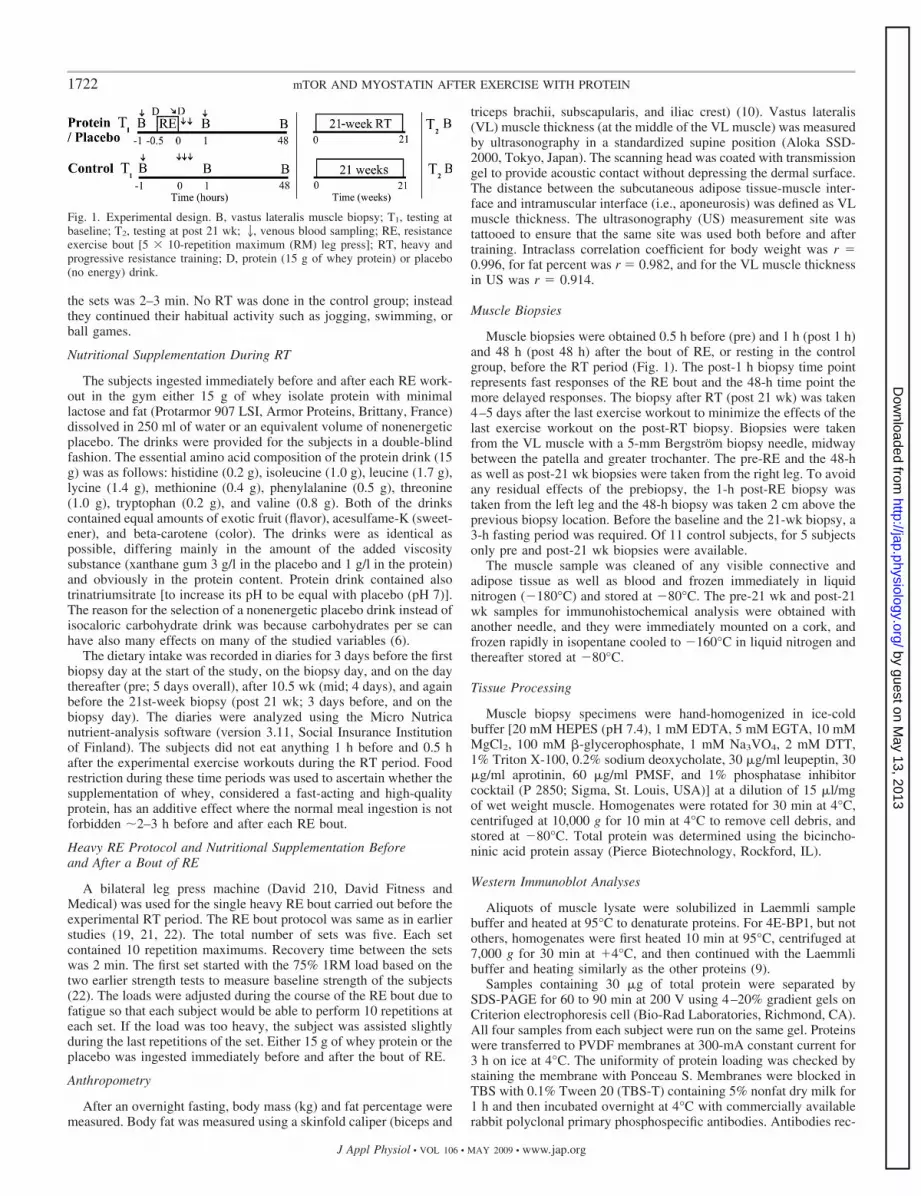
This investigation examined acute and long-term responses ofadding high-quality protein to a normal diet (with no other nutritionalsupplements). The experimental design involved both a single bout ofRE and 0- to 48-h postexercise responses to it, and following that, 21wk of RT consisting of 42 RE workouts (Fig. 1). A control group wasincluded, and all the measurements were always carried out at thesame time of day to exclude the effects of biopsy sampling or effectsof time of year or daily variations (47, 57). All the measurements werepreceded by at least 2 days of rest from physical activity.
Experimental RT
Whole body heavy RE workouts were carried out twice a week. Aminimum of 2 days of rest was required between workouts. Alltraining sessions were supervised by experienced trainers who en-sured that proper techniques and progression were used in eachexercise (32). The leg exercises included two exercises for the legextensor muscles, the bilateral leg press and bilateral knee extension,and one exercise for the leg flexors, bilateral knee flexion. The RTprogram also included exercises for the other main muscle groups:chest and shoulders (bench press), upper back, trunk extensors andflexors, upper arms, ankle extensors, and hip abductors and adductors.RT was performed with progressive training loads of 40–85% of thesubject’s one-repetition maximum (1RM) in a periodized trainingprogram. For each exercise in a workout the number of sets increased(from 2–3 to 3–5) and the number of repetitions in each set decreased(from 15–20 to 5–6) during the 21-wk RT period. The loads wereindividually determined throughout the RT period. Recovery between
Table 1. Anthropometry: height, body mass, fat percent, muscle fiber cross-sectional area, and muscle thicknessof the protein, placebo, and control groups
Variable/Group Baseline 10.5 wk 21 wk P Valuepre vs. 21 P Value�%group21
Height, cmProtein 181.8�6.9Placebo 181.0�6.2Control 181.9�4.7
Body mass, kgProtein 76.7�8.1 80.1�9.5*† 80.1�9.6*† 0.009 �0.001Placebo 75.9�8.0 77.6�8.5 78.2�8.9*† 0.03 0.02Control 74.5�7.8 75.6�8.4* 74.5�8.1 0.93
Fat, %Protein 16.8�4.0 17.7�4.3 17.5�4.6 0.35 0.50Placebo 17.3�3.9 17.2�4.5 17.1�3.9 �0.99 0.46Control 16.6�3.5 17.5�4.5 16.9�4.6 0.64
Fiber size: type I, �m2
Protein 4,650�178 6,582�511*† 0.009 0.03Placebo 4,198�185 5,910�288*† 0.003 0.04Control 4,940�411 5,099�391 0.21
Fiber size: type II, �m2
Protein 5,021�402 7,599�576*† 0.002 0.01Placebo 4,617�336 6,951�484*† 0.003 0.04Control 5,501�407 5,629�380 0.72
Muscle thickness, cmProtein 2.61�0.14 2.89�0.18*† 2.93�0.19*† 0.003 0.03Placebo 2.47�0.22 2.72�0.32 2.74�0.25* 0.046 0.12Control 2.68�0.15 2.65�0.17 2.78�0.13 0.10
Values are means � SD, except muscle size variables, which are means � SE. P valuepre vs.21 designates Holm-Bonferroni corrected P values compared withbaseline. P value�%group21 designates the difference between the training group and the control group in percent change between the baseline and post-21 wkvalues. *Significant (P � 0.05) P value vs. pre. †Significant value compared with percentage change vs. the control group. See text for further description ofgroups and time points.
1721mTOR AND MYOSTATIN AFTER EXERCISE WITH PROTEIN
J Appl Physiol • VOL 106 • MAY 2009 • www.jap.org
by guest on May 13, 2013
http://jap.physiology.org/D
ownloaded from
the sets was 2–3 min. No RT was done in the control group; insteadthey continued their habitual activity such as jogging, swimming, orball games.
Nutritional Supplementation During RT
The subjects ingested immediately before and after each RE work-out in the gym either 15 g of whey isolate protein with minimallactose and fat (Protarmor 907 LSI, Armor Proteins, Brittany, France)dissolved in 250 ml of water or an equivalent volume of nonenergeticplacebo. The drinks were provided for the subjects in a double-blindfashion. The essential amino acid composition of the protein drink (15g) was as follows: histidine (0.2 g), isoleucine (1.0 g), leucine (1.7 g),lycine (1.4 g), methionine (0.4 g), phenylalanine (0.5 g), threonine(1.0 g), tryptophan (0.2 g), and valine (0.8 g). Both of the drinkscontained equal amounts of exotic fruit (flavor), acesulfame-K (sweet-ener), and beta-carotene (color). The drinks were as identical aspossible, differing mainly in the amount of the added viscositysubstance (xanthane gum 3 g/l in the placebo and 1 g/l in the protein)and obviously in the protein content. Protein drink contained alsotrinatriumsitrate [to increase its pH to be equal with placebo (pH 7)].The reason for the selection of a nonenergetic placebo drink instead ofisocaloric carbohydrate drink was because carbohydrates per se canhave also many effects on many of the studied variables (6).
The dietary intake was recorded in diaries for 3 days before the firstbiopsy day at the start of the study, on the biopsy day, and on the daythereafter (pre; 5 days overall), after 10.5 wk (mid; 4 days), and againbefore the 21st-week biopsy (post 21 wk; 3 days before, and on thebiopsy day). The diaries were analyzed using the Micro Nutricanutrient-analysis software (version 3.11, Social Insurance Institutionof Finland). The subjects did not eat anything 1 h before and 0.5 hafter the experimental exercise workouts during the RT period. Foodrestriction during these time periods was used to ascertain whether thesupplementation of whey, considered a fast-acting and high-qualityprotein, has an additive effect where the normal meal ingestion is notforbidden �2–3 h before and after each RE bout.
Heavy RE Protocol and Nutritional Supplementation Beforeand After a Bout of RE
A bilateral leg press machine (David 210, David Fitness andMedical) was used for the single heavy RE bout carried out before theexperimental RT period. The RE bout protocol was same as in earlierstudies (19, 21, 22). The total number of sets was five. Each setcontained 10 repetition maximums. Recovery time between the setswas 2 min. The first set started with the 75% 1RM load based on thetwo earlier strength tests to measure baseline strength of the subjects(22). The loads were adjusted during the course of the RE bout due tofatigue so that each subject would be able to perform 10 repetitions ateach set. If the load was too heavy, the subject was assisted slightlyduring the last repetitions of the set. Either 15 g of whey protein or theplacebo was ingested immediately before and after the bout of RE.
Anthropometry
After an overnight fasting, body mass (kg) and fat percentage weremeasured. Body fat was measured using a skinfold caliper (biceps and
triceps brachii, subscapularis, and iliac crest) (10). Vastus lateralis(VL) muscle thickness (at the middle of the VL muscle) was measuredby ultrasonography in a standardized supine position (Aloka SSD-2000, Tokyo, Japan). The scanning head was coated with transmissiongel to provide acoustic contact without depressing the dermal surface.The distance between the subcutaneous adipose tissue-muscle inter-face and intramuscular interface (i.e., aponeurosis) was defined as VLmuscle thickness. The ultrasonography (US) measurement site wastattooed to ensure that the same site was used both before and aftertraining. Intraclass correlation coefficient for body weight was r �0.996, for fat percent was r � 0.982, and for the VL muscle thicknessin US was r � 0.914.
Muscle Biopsies
Muscle biopsies were obtained 0.5 h before (pre) and 1 h (post 1 h)and 48 h (post 48 h) after the bout of RE, or resting in the controlgroup, before the RT period (Fig. 1). The post-1 h biopsy time pointrepresents fast responses of the RE bout and the 48-h time point themore delayed responses. The biopsy after RT (post 21 wk) was taken4–5 days after the last exercise workout to minimize the effects of thelast exercise workout on the post-RT biopsy. Biopsies were takenfrom the VL muscle with a 5-mm Bergstrom biopsy needle, midwaybetween the patella and greater trochanter. The pre-RE and the 48-has well as post-21 wk biopsies were taken from the right leg. To avoidany residual effects of the prebiopsy, the 1-h post-RE biopsy wastaken from the left leg and the 48-h biopsy was taken 2 cm above theprevious biopsy location. Before the baseline and the 21-wk biopsy, a3-h fasting period was required. Of 11 control subjects, for 5 subjectsonly pre and post-21 wk biopsies were available.
The muscle sample was cleaned of any visible connective andadipose tissue as well as blood and frozen immediately in liquidnitrogen (180°C) and stored at 80°C. The pre-21 wk and post-21wk samples for immunohistochemical analysis were obtained withanother needle, and they were immediately mounted on a cork, andfrozen rapidly in isopentane cooled to 160°C in liquid nitrogen andthereafter stored at 80°C.
Tissue Processing
Muscle biopsy specimens were hand-homogenized in ice-coldbuffer [20 mM HEPES (pH 7.4), 1 mM EDTA, 5 mM EGTA, 10 mMMgCl2, 100 mM -glycerophosphate, 1 mM Na3VO4, 2 mM DTT,1% Triton X-100, 0.2% sodium deoxycholate, 30 �g/ml leupeptin, 30�g/ml aprotinin, 60 �g/ml PMSF, and 1% phosphatase inhibitorcocktail (P 2850; Sigma, St. Louis, USA)] at a dilution of 15 �l/mgof wet weight muscle. Homogenates were rotated for 30 min at 4°C,centrifuged at 10,000 g for 10 min at 4°C to remove cell debris, andstored at 80°C. Total protein was determined using the bicincho-ninic acid protein assay (Pierce Biotechnology, Rockford, IL).
Western Immunoblot Analyses
Aliquots of muscle lysate were solubilized in Laemmli samplebuffer and heated at 95°C to denaturate proteins. For 4E-BP1, but notothers, homogenates were first heated 10 min at 95°C, centrifuged at7,000 g for 30 min at �4°C, and then continued with the Laemmlibuffer and heating similarly as the other proteins (9).
Samples containing 30 �g of total protein were separated bySDS-PAGE for 60 to 90 min at 200 V using 4–20% gradient gels onCriterion electrophoresis cell (Bio-Rad Laboratories, Richmond, CA).All four samples from each subject were run on the same gel. Proteinswere transferred to PVDF membranes at 300-mA constant current for3 h on ice at 4°C. The uniformity of protein loading was checked bystaining the membrane with Ponceau S. Membranes were blocked inTBS with 0.1% Tween 20 (TBS-T) containing 5% nonfat dry milk for1 h and then incubated overnight at 4°C with commercially availablerabbit polyclonal primary phosphospecific antibodies. Antibodies rec-
Fig. 1. Experimental design. B, vastus lateralis muscle biopsy; T1, testing atbaseline; T2, testing at post 21 wk; 2, venous blood sampling; RE, resistanceexercise bout [5 � 10-repetition maximum (RM) leg press]; RT, heavy andprogressive resistance training; D, protein (15 g of whey protein) or placebo(no energy) drink.
1722 mTOR AND MYOSTATIN AFTER EXERCISE WITH PROTEIN
J Appl Physiol • VOL 106 • MAY 2009 • www.jap.org
by guest on May 13, 2013
http://jap.physiology.org/D
ownloaded from
ognized phosphorylated Akt on Ser473, mTOR on Ser2448, p70S6K onThr389, rpS6 on Ser235/236, 4E-BP1 on Thr37/46, and eEF2 on Thr56
(Cell Signaling Technology, Beverly, MA) and COOH-terminal myo-statin protein (Chemicon/Millipore AB 3239) (38). The rabbit poly-clonal antibody used was raised against a peptide residing in theCOOH terminus of myostatin corresponding to amino acids 349–364of human myostatin and, therefore, being a similar antibody to thoseused previously (14, 50).
All the antibodies were diluted 1:2,000 (except eEF2 on Thr56,which was 1:3,000) in TBS-T containing 2.5% nonfat dry milk.Membranes were then washed in TBS-T, incubated with secondaryantibody (horseradish peroxidase-conjugated anti-rabbit IgG; CellSignaling Technology) diluted 1:5,000 in TBS-T with 2.5% milk for1 h followed by washing in TBS-T. Phosphorylated proteins werevisualized by ECL according to the manufacturer’s protocol (SuperSignalWest femto maximum sensitivity substrate, Pierce Biotechnology)and quantified using a ChemiDoc XRS in combination with QuantityOne software (version 4.6.3. Bio-Rad Laboratories).
The membranes described above were incubated in Restore West-ern blot stripping buffer (Pierce Biotechnology) for 30 min andreprobed with appropriate antibodies for detection of the total expres-sion levels of Akt and rpS6 (rabbit monoclonal) (Cell SignalingTechnology) and p70S6K (Santa Cruz Biotechnology) by immunoblotanalysis as described above.
Blood analysis
The blood samples were drawn from the antecubital vein beforeand 0, 15, 30, and 60 min after the bout of RE using 21-gaugedisposable needles. Blood was centrifuged at 3,500 rpm in 4°C for 10min to separate serum and stored frozen at 80°C until assayed.Serum testosterone, sex hormone-binding globulin (SHBG), and in-sulin concentrations were analyzed by an immunometric chemilumi-nescence method with an Immulite 1000 (DPC, Los Angeles, CA).The sensitivity of the assay for testosterone and coefficient of varia-tion (CV) are 0.5 nmol/l and 5.7%, for SHBG 0.2 nmol/l and 2.4%,and for insulin 2 mIU/l and 3.4%, respectively. Free testosterone wascalculated from total testosterone and SHBG concentrations (56). Theresults are presented as uncorrected to plasma volume changes asthere were no differences between the protein and placebo group inthe decrease of the plasma volume during and after the bout of RE(data not shown).
Immunohistochemistry
Muscle fiber cross-sectional area. Serial 8-�m-thick transversesections were cut on a cryomicrotome (Leica CM 3000) at 24°C.Fiber type was classified by staining using myofibrillar ATPasemethod according to the earlier study (31). Fiber sarcolemma wasvisualized with an antibody against dystrophin (DYS2, NovocastraLaboratories) using avidin-biotin peroxidase kit (Vectastain PK-4002,Vector Laboratories) with diaminobenzidine (Abbott Laboratories) asa chromogen. The measurements of fiber cross-sectional area (CSA)comprised an average of 125 � 57 type I and 129 � 61 type II musclefibers. Stained cross sections were analyzed by Tema Image-AnalysisSystem (Scan Beam) using a microscope (Olympus BX 50) and colorvideo camera (Sanyo High Resolution CCD).
Immunohistochemical staining of rpS6 and mTOR. For immuno-histochemical staining of rpS6 and mTOR, 8-�m longitudinal andcross sections before the RT period from resting state muscle of thepresent subjects were fixed 15 min with 4% PFA-PBS, permeabilizedwith 0.2% Triton-X for 10 min, and blocked 30 min with 3%BSA-PBS and thereafter incubated with primary antibodies overnightat �4°C. Double immunolabeling was performed using a rabbitmonoclonal antibody against rpS6 or rabbit polyclonal antibodiesagainst phospho-rpS6 on Ser235/236 or phospho-mTOR on Ser2448
(Cell Signaling Technology; 1:40 in 1% BSA-PBS) with either mousemonoclonal antibody against human slow myosin heavy chain
(MyHC I) [Developmental Studies Hybridoma Bank (DSHB), IowaCity, IA: A4.951] diluted to 1:150 (18), or mouse monoclonal anti-body against caveolin-3 (1:100) (BD Transduction Laboratories) tovisualize muscle sarcolemma. Nuclei were stained by Hoechst 33258(Sigma, St. Louis, MO). Secondary antibodies used were goat anti-rabbit Alexa Fluor 488 or 546 and goat anti-mouse Alexa Fluor 546or 488 (Molecular Probes, Eugene, OR). Negative controls were doneby omitting the primary or secondary antibody. An Olympus BX-50Flight microscope (Olympus Optical, Tokyo, Japan) with Olympuscolor CCD camera (Colorview III, Olympus Optical) and Analysissoftware (version 5.0, Soft-Imaging System, Munster, Germany) wereused for the imaging and analysis. Two samples were also furtheranalyzed using Olympus IX81 confocal microscope with imagingsystem and software (Olympus Fluoview 1.6a) (29).
Statistical Analyses
All data are expressed as means � SD, except where designated.The data were analyzed by a repeated-measures general linear modelANOVA. Any violations of the assumptions of sphericity wereexplored and, if needed, corrected with a Greenhouse-Geisser orHuynh-Feldt estimator. The Shapiro-Wilk test revealed that Westernblot data were not normally distributed, and therefore for the statisticaltests, all those values were log-transformed. Holm-Bonferroni posthoc tests were performed to localize the effects. SPSS version 13.0 forWindows was used for statistical analyses (SPSS, Chicago, IL). Thelevel of significance was set at P � 0.05.
RESULTS
Daily Nutrient Intake
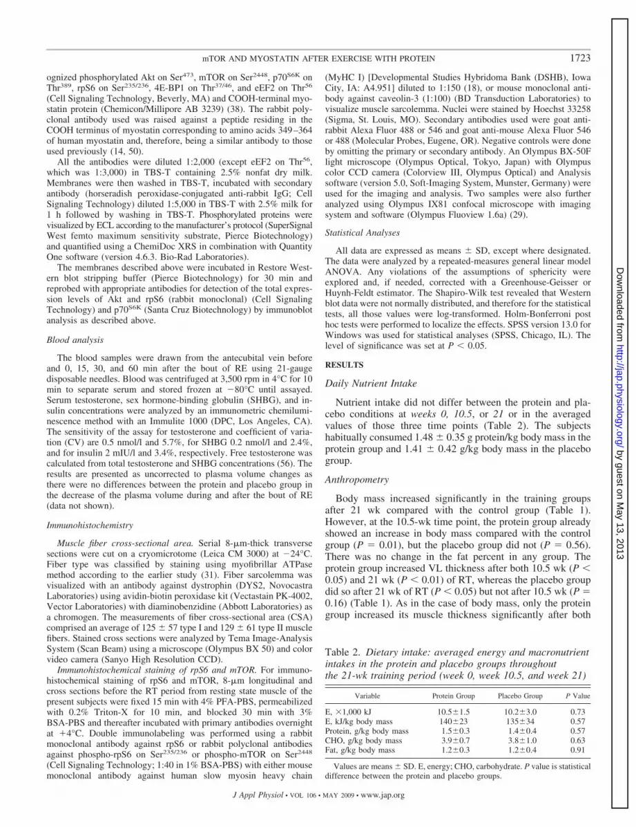
Nutrient intake did not differ between the protein and pla-cebo conditions at weeks 0, 10.5, or 21 or in the averagedvalues of those three time points (Table 2). The subjectshabitually consumed 1.48 � 0.35 g protein/kg body mass in theprotein group and 1.41 � 0.42 g/kg body mass in the placebogroup.
Anthropometry
Body mass increased significantly in the training groupsafter 21 wk compared with the control group (Table 1).However, at the 10.5-wk time point, the protein group alreadyshowed an increase in body mass compared with the controlgroup (P � 0.01), but the placebo group did not (P � 0.56).There was no change in the fat percent in any group. Theprotein group increased VL thickness after both 10.5 wk (P �0.05) and 21 wk (P � 0.01) of RT, whereas the placebo groupdid so after 21 wk of RT (P � 0.05) but not after 10.5 wk (P �0.16) (Table 1). As in the case of body mass, only the proteingroup increased its muscle thickness significantly after both
Table 2. Dietary intake: averaged energy and macronutrientintakes in the protein and placebo groups throughoutthe 21-wk training period (week 0, week 10.5, and week 21)
Variable Protein Group Placebo Group P Value
E, �1,000 kJ 10.5�1.5 10.2�3.0 0.73E, kJ/kg body mass 140�23 135�34 0.57Protein, g/kg body mass 1.5�0.3 1.4�0.4 0.57CHO, g/kg body mass 3.9�0.7 3.8�1.0 0.63Fat, g/kg body mass 1.2�0.3 1.2�0.4 0.91
Values are means � SD. E, energy; CHO, carbohydrate. P value is statisticaldifference between the protein and placebo groups.
1723mTOR AND MYOSTATIN AFTER EXERCISE WITH PROTEIN
J Appl Physiol • VOL 106 • MAY 2009 • www.jap.org
by guest on May 13, 2013
http://jap.physiology.org/D
ownloaded from
10.5 and 21 wk compared with the control group (P � 0.05),while the placebo group only approached a trend after 21 wk(P � 0.12).
Acute RE Bout at Week 0 and Training Volumes
The total volume of the work performed in the RE bout(loads � sets � repetitions) was similar in the placebo (88.7 �15.4 kg/kg body weight) and protein group (91.5 � 15.7 kg/kgbody wt) at week 0. Average training volumes (loads � sets �repetitions) for the leg extensor muscles (leg press and kneeextension) were calculated for weeks 1–7, 8–14, and 15–21.No significant difference was found between the protein andplacebo groups (data not shown).
Western Blotting Results
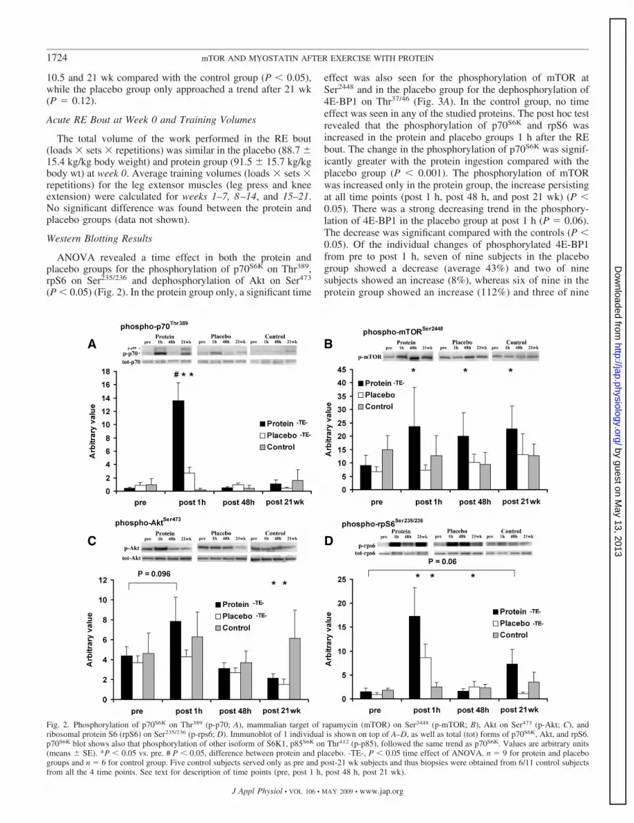
ANOVA revealed a time effect in both the protein andplacebo groups for the phosphorylation of p70S6K on Thr389,rpS6 on Ser235/236 and dephosphorylation of Akt on Ser473
(P � 0.05) (Fig. 2). In the protein group only, a significant time
effect was also seen for the phosphorylation of mTOR atSer2448 and in the placebo group for the dephosphorylation of4E-BP1 on Thr37/46 (Fig. 3A). In the control group, no timeeffect was seen in any of the studied proteins. The post hoc testrevealed that the phosphorylation of p70S6K and rpS6 wasincreased in the protein and placebo groups 1 h after the REbout. The change in the phosphorylation of p70S6K was signif-icantly greater with the protein ingestion compared with theplacebo group (P � 0.001). The phosphorylation of mTORwas increased only in the protein group, the increase persistingat all time points (post 1 h, post 48 h, and post 21 wk) (P �0.05). There was a strong decreasing trend in the phosphory-lation of 4E-BP1 in the placebo group at post 1 h (P � 0.06).The decrease was significant compared with the controls (P �0.05). Of the individual changes of phosphorylated 4E-BP1from pre to post 1 h, seven of nine subjects in the placebogroup showed a decrease (average 43%) and two of ninesubjects showed an increase (8%), whereas six of nine in theprotein group showed an increase (112%) and three of nine
Fig. 2. Phosphorylation of p70S6K on Thr389 (p-p70; A), mammalian target of rapamycin (mTOR) on Ser2448 (p-mTOR; B), Akt on Ser473 (p-Akt; C), andribosomal protein S6 (rpS6) on Ser235/236 (p-rps6; D). Immunoblot of 1 individual is shown on top of A–D, as well as total (tot) forms of p70S6K, Akt, and rpS6.p70S6K blot shows also that phosphorylation of other isoform of S6K1, p85S6K on Thr412 (p-p85), followed the same trend as p70S6K. Values are arbitrary units(means � SE). *P � 0.05 vs. pre. # P � 0.05, difference between protein and placebo. -TE-, P � 0.05 time effect of ANOVA. n � 9 for protein and placebogroups and n � 6 for control group. Five control subjects served only as pre and post-21 wk subjects and thus biopsies were obtained from 6/11 control subjectsfrom all the 4 time points. See text for description of time points (pre, post 1 h, post 48 h, post 21 wk).
1724 mTOR AND MYOSTATIN AFTER EXERCISE WITH PROTEIN
J Appl Physiol • VOL 106 • MAY 2009 • www.jap.org
by guest on May 13, 2013
http://jap.physiology.org/D
ownloaded from

a decrease (32%) (between-group difference in the change:P � 0.03). The phosphorylation of Akt decreased after 21wk of RT in both training groups (P � 0.05). There was,however, no change in the phospho-eEF2 (p-eEF2) or totalprotein expression of p70S6K, Akt, and rpS6. The myostatinCOOH-terminal protein was decreased at post 1 h in theplacebo group (P � 0.02) but not in the protein or controlgroups (Fig. 3C).
There were no significant correlations between the RE-induced change in the protein kinases or in the myostatinprotein with corresponding changes in VL fiber size ormuscle thickness (by ultrasonography) or VL CSA [by MRIpreviously (22)].
Immunohistochemistry
The CSA of type I and II fiber types increased significantlyand similarly after 21 wk of RT in both the protein and placebogroups (P � 0.01) and also significantly (P � 0.05) comparedwith the control group (Table 1).
Both phosphorylated mTOR at Ser2448 and rpS6 at Ser235/236
as well as total rpS6 were primarily localized close to thenuclei and sarcolemma, outside the area where contractileproteins are located (Fig. 4). The signal for these proteinsemanated in large part from inside the muscle fibers but also tosome extent from outside the sarcolemma. No clear cell-typedifference was seen.
Serum Testosterone and Insulin
Compared with the control group, serum total testosteroneconcentration elevated significantly during the bout of RE onlyin the placebo group (P � 0.04). No differences between thegroups were observed in free testosterone or in serum insulin(not shown).
DISCUSSION
The main findings of the present study were that ingestion ofwhey proteins before and after a bout of RE rapidly increasedthe phosphorylation of p70S6K and also prevented the decreasein the phosphorylation of 4E-BP1. Moreover, the RE boutacutely decreased the active form of myostatin protein, butonly when protein was not supplemented. The phosphoryla-tion of mTOR remained increased after the RE bout frompost 1 h to post 48 h and also after 21 wk of RT when theprotein was ingested. However, RT itself decreased Aktphosphorylation. The control group results ensured that theresults were not due to repeated biopsy, diurnal rhythm, ortime of year (47, 57).
Whey protein rapidly increases mTOR signaling. In thepresent study, whey protein ingestion rapidly increased thephosphorylation of p70S6K on Thr389 at 1 h post-RE, showingthe activation of the TORC1 complex, including mTOR and itsregulatory proteins (25, 54, 58). Probably also the activation ofp70S6K was increased especially in the protein group because
Fig. 3. Phosphorylation of eukaryotic initiation factor 4E binding protein 1 (4E-BP1) on Thr37/46 (p-4E-BP1; A), eukaryotic elongation factor 2 (eEF2) on Thr56
(p-eEF2; B), and myostatin 26 kDa (MSTN) COOH-terminal protein (C). Ponceau S (Pon S) staining shows equal protein loading. Immunoblot of 1 individualis shown at right. See text for further explanation. There was a between-group difference between the protein and placebo group and also between the placeboand control group in the change from pre to post 1 h (*P � 0.05).
1725mTOR AND MYOSTATIN AFTER EXERCISE WITH PROTEIN
J Appl Physiol • VOL 106 • MAY 2009 • www.jap.org
by guest on May 13, 2013
http://jap.physiology.org/D
ownloaded from

phosphorylation from this site is the chief event in the activa-tion of p70S6K (44) and since there was also a tendency forlarger phosphorylation of one of its downstream target rpS6(protein vs. placebo, P � 0.11) and the increase in mTORphosphorylation at Ser2448 was observed only in the proteingroup. This site of mTOR has been shown to be phosphory-lated by p70S6K (5). The present results agree with those of a
recent study showing that protein intake together with carbo-hydrate before, during, and 1 h after a RE bout increasedphosphorylation of p70S6K at post 0–4 h compared withcarbohydrate only (30). Interestingly, the phosphorylation ofthe second isoform of S6K1, p85S6K, clearly followed the samepattern in the present study as that of p70S6K (see Fig. 2,protein blot image).
Fig. 4. Confocal microscopy images of localization of total (A) and phosphorylated rpS6 (B) and phopshorylated mTOR (C) in muscle cross sections. Nucleiwere stained with Hoechst 33258, sarcolemma with antibody against caveolin-3, and myofibrillar area with an antibody against myosin heavy chain I (MyHC I). Allimages are representative of 2 subjects visualized with confocal microscope and of a total of 5 subjects with epifluorescence microscope. PhosphorylatedmTOR at Ser2448, rpS6 at Ser235/236, and total rpS6 were primarily localized close to the nuclei and sarcolemma (caveolin-3), outside the area wherecontractile proteins are located (MyHC). The images were taken with the settings in which the secondary antibody (not shown) only gave minimal signal.Scale bars are 50 �m.
1726 mTOR AND MYOSTATIN AFTER EXERCISE WITH PROTEIN
J Appl Physiol • VOL 106 • MAY 2009 • www.jap.org
by guest on May 13, 2013
http://jap.physiology.org/D
ownloaded from

The most important component responsible for the increasedphosphorylation of p70S6K with the whey protein ingestion isprobably its large content of branched-chain amino acids thatcan elicit a similar p70S6K response in the context of a bout ofRE as observed in the present study (3, 26). S6K1/p70S6K hasbeen shown in animal and cell models to be especially impor-tant in muscle hypertrophy (2, 40, 48). Moreover, in humans anacute increase in the phosphorylation of p70S6K after a bout ofRE has also correlated with a long-term loading-inducedincrease in fiber size as well as fat-free mass in trainedhumans (51), and with a RE-induced myofibrillar proteinsynthesis (33).
Whey protein intake alone prevented a RE-induced decreasein the phosphorylation of eukaryotic initiation factor 4E(eIF4E) binding protein (4E-BP1). This supports a recentfinding in humans with a slightly different time scale andnutrients (30). An RE bout per se has also previously decreasedthe phosphorylation of 4E-BP1 shortly after exercise whenprotein or amino acids are not supplemented (8, 9, 30, 36).Prevention of the dephosphorylation of 4E-BP1 after a bout ofRE by ingestion of whey proteins probably prevents associa-tion of the 4E-BP1 with eIF4E (43). This allows a largerincrease in protein synthesis (12, 28, 35, 43), a phenomenonpreviously observed after the ingestion of whey proteins (52,53). It can be speculated that whey protein alone can affectmTOR signaling TORC1 dependently and independently as thephosphorylation of 4E-BP1 from this site (Thr37/46) may occurindependently of TORC1 unlike the phosphorylation of p70S6K
(25, 54, 58). Our results suggest that these effects occurredindependently of blood insulin or the phosphorylation of Akt atSer473.
The phosphorylated mTOR is localized mainly close to thesarcolemmal membrane as has been shown in rodents (17, 41)while rpS6 and phospho-rpS6 (p-rpS6) were mostly locatedvery close to the nuclei as has been found earlier with p-rpS6(30). Magnification showed that rpS6 usually surrounded thenuclei, which is theoretically optimal for efficient proteinsynthesis.
Protein and training affects the phosphorylation of mTORand Akt, respectively. In contrast to the rapid changes, the REbout itself did not seem to have a consistent effect on thephosphorylation of the mTOR pathway proteins at 48 hpost-RE or after a longer term RT, supporting recent humanstudies investigating time points 48 h (36) and 24 h post-RE (8,36), and �4 days after a RT period (34, 59). The onlylong-lasting effect of protein ingestion was the increased phos-phorylation of mTOR, which remained increased in the proteingroup from 1 to 48 h after the RE bout and also after 21 wk ofRT. Surprisingly, the phosphorylation of Akt decreased �0.5-fold in both training groups after 21 wk of RT. In contrast, inprevious studies 8–10 wk of RT increased the phopshorylationof Akt at Ser473 (34, 59). This different response may, owing topossible complexity of the temporal pattern of the Akt phos-phorylation state, depend on the timing of the biopsies oradditionally on the length or type of the training period orpossibly the nutritional state.
The time point 4–5 days post-RE when the biopsy was takenrepresents roughly the time point when the next RE workoutwould have taken place. Therefore, the pre and post-21 wkbiopsies are comparable. The observed more “chronic” Aktand mTOR responses to the RT with or without protein
ingestion were probably due to 1) a very long-lasting 4- to5–day effect from the last RE workout, and/or 2) a morepermanent RT-induced change in the basal state of phosphor-ylation of these proteins. These changes may affect the level ofthe protein synthesis of the resting muscle (59) and/or theamount of the signal needed to activate the cascade throughthese pathways. It seems obvious that the change in the state ofphosphorylation of at least some of the mTOR pathway proteinkinases can be rather chronic and less transient due to, e.g.,aging or exercise (16, 34, 59) and, at least partially, also due toprotein ingestion.
Muscle Hypertrophy after RT, and Myostatin
The earlier MRI results obtained from the present studydesign showed a larger increase in VL muscle hypertrophywith whey protein ingestion (22). The present study alsoshowed a somewhat faster increase in VL muscle thickness andbody mass with protein ingestion. However, a larger protein-induced increase after the full 21-wk RT in fiber size was notobserved. Recently, in older men, there were no positiveeffects of 10 g of casein protein ingested also immediatelybefore and after a RE workout (55). It is possible that largereffects on muscle size would have been seen in the presentstudy using subjects with a higher level of RT background orwhose habitual ingestion of protein is smaller than �1.4–1.5g/body weight (49). Therefore, while the positive effects of theprotein or amino acid ingestion on muscle hypertrophy signal-ing can often be clear when studied acutely after each exercise,especially when the study was performed in a fasting state, thelong-term positive effects may not be as robust with normaldaily high protein consumption.
The present study is the first in humans showing thatmyostatin peptide concentration, thought to be the active formof myostatin, can follow the decreased mRNA transcript ofmyostatin after a RE bout. Interestingly, protein ingestionseemed to prevent the decrease in myostatin after the RE bout.This may have hindered larger hypertrophy in the proteingroup, which could have been predicted from the mTORpathway results because myostatin inhibits muscle growth (14,37, 46). The hindering effect of protein ingestion for down-regulating myostatin expression after the bout of RE supportsour earlier mRNA-level findings among younger (22) and oldermen (21), suggesting that the change in myostatin was tran-scriptionally regulated. Indeed, with the present subjects themyostatin mRNA and protein level changes from pre to post1 h also correlated positively (r � 0.66, P � 0.007). It isassumed that the detected �26-kDa myostatin is a glycosylatedtightly bound dimer of a 110-amino acid COOH-terminalpeptide of myostatin and/or that the monomer of myostatin isstrongly bound by some other protein (14, 50). Recently, amyostatin propeptide of size 28 kDa and myostatin proteincomplexes of size 50 kDa were not changed 24 h after a boutof RE in humans (27).
Whey protein also seemed to prevent the elevation in serumtotal testosterone seen in the placebo group after the bout ofRE, thereby supporting the previous results of protein ingestion(4, 20, 23). The testosterone response may be due to a decreasein the synthesis/secretion of testosterone and/or an increase inmetabolic clearance. As was the case with myostatin proteinconcentration, the effect of protein ingestion on testosterone
1727mTOR AND MYOSTATIN AFTER EXERCISE WITH PROTEIN
J Appl Physiol • VOL 106 • MAY 2009 • www.jap.org
by guest on May 13, 2013
http://jap.physiology.org/D
ownloaded from

was, however, small. Thus the physiological significance ofthese responses warrants future studies.
In conclusion, resistance exercise rapidly increases mTORsignaling, and whey protein increases and prolongs the mTORsignaling response to exercise and training. Active form ofmyostatin peptide rapidly decreases after a RE bout whenprotein nutrition is not supplemented.
ACKNOWLEDGMENTS
We thank Hanna Salmijarvi, Marja Katajavuori, Liisa Kiviluoto, MarkoHaverinen, Paavo Rahkila, Tuovi Nykanen, Risto Puurtinen, and Aila Olli-kainen for help in the data collection and analysis. We also thank the verydedicated group of subjects who made this project possible. The monoclonalantibody A4.951 for MyHC I, developed by Dr. H. M. Blau (18), was obtainedfrom the Developmental Studies Hybridoma Bank developed under the aus-pices of the National Institute of Child Health and Human Development andmaintained by the Univ. of Iowa, Dept. of Biological Science, Iowa City, IA52242.
GRANTS
The Finnish Ministry of Education and the Ellen and Artturi NyyssonenFoundation (Juha Hulmi personal grant) supported this research.
REFERENCES
1. Amirouche A, Durieux AC, Banzet S, Koulmann N, Bonnefoy R,Mouret C, Bigard X, Peinnequin A, Freyssenet D. Down-regulation ofAkt/mammalian target of rapamycin signaling pathway in response tomyostatin overexpression in skeletal muscle. Endocrinology 150: 286–294, 2009.
2. Baar K, Esser K. Phosphorylation of p70(S6k) correlates with increasedskeletal muscle mass following resistance exercise. Am J Physiol CellPhysiol 276: C120–C127, 1999.
3. Blomstrand E, Eliasson J, Karlsson HK, Kohnke R. Branched-chainamino acids activate key enzymes in protein synthesis after physicalexercise. J Nutr 136, Suppl: 269S–73S, 2006.
4. Chandler RM, Byrne HK, Patterson JG, Ivy JL. Dietary supplementsaffect the anabolic hormones after weight-training exercise. J Appl Physiol76: 839–845, 1994.
5. Chiang GG, Abraham RT. Phosphorylation of mammalian target ofrapamycin (mTOR) at Ser-2448 is mediated by p70S6 kinase. J Biol Chem280: 25485–25490, 2005.
6. Creer A, Gallagher P, Slivka D, Jemiolo B, Fink W, Trappe S.Influence of muscle glycogen availability on ERK1/2 and Akt signalingafter resistance exercise in human skeletal muscle. J Appl Physiol 99:950–956, 2005.
7. Cribb PJ, Hayes A. Effects of supplement timing and resistance exerciseon skeletal muscle hypertrophy. Med Sci Sports Exerc 38: 1918–1925,2006.
8. Deldicque L, Atherton P, Patel R, Theisen D, Nielens H, Rennie MJ,Francaux M. Decrease in Akt/PKB signalling in human skeletal muscleby resistance exercise. Eur J Appl Physiol 104: 57–65, 2008.
9. Dreyer HC, Fujita S, Cadenas JG, Chinkes DL, Volpi E, RasmussenBB. Resistance exercise increases AMPK activity and reduces 4E-BP1phosphorylation and protein synthesis in human skeletal muscle. J Physiol576: 613–624, 2006.
10. Durnin JV, Womersley J. Body fat assessed from total body density andits estimation from skinfold thickness: measurements on 481 men andwomen aged from 16 to 72 years. Br J Nutr 32: 77–97, 1974.
11. Esmarck B, Andersen JL, Olsen S, Richter EA, Mizuno M, Kjaer M.Timing of postexercise protein intake is important for muscle hypertrophywith resistance training in elderly humans. J Physiol 535: 301–311, 2001.
12. Gautsch TA, Anthony JC, Kimball SR, Paul GL, Layman DK, Jef-ferson LS. Availability of eIF4E regulates skeletal muscle protein syn-thesis during recovery from exercise. Am J Physiol Cell Physiol 274:C406–C414, 1998.
13. Glass DJ. Skeletal muscle hypertrophy and atrophy signaling pathways.Int J Biochem Cell Biol 37: 1974–1984, 2005.
14. Gonzalez-Cadavid NF, Taylor WE, Yarasheski K, Sinha-Hikim I, MaK, Ezzat S, Shen R, Lalani R, Asa S, Mamita M, Nair G, Arver S,Bhasin S. Organization of the human myostatin gene and expression in
healthy men and HIV-infected men with muscle wasting. Proc Natl AcadSci USA 95: 14938–14943, 1998.
15. Ha E, Zemel MB. Functional properties of whey, whey components, andessential amino acids: mechanisms underlying health benefits for activepeople. J Nutr Biochem 14: 251–258, 2003.
16. Haddad F, Adams GR. Aging-sensitive cellular and molecular mecha-nisms associated with skeletal muscle hypertrophy. J Appl Physiol 100:1188–1203, 2006.
17. Hornberger TA, Chu WK, Mak YW, Hsiung JW, Huang SA, Chien S.The role of phospholipase D and phosphatidic acid in the mechanicalactivation of mTOR signaling in skeletal muscle. Proc Natl Acad Sci USA103: 4741–4746, 2006.
18. Hughes SM, Cho M, Karsch-Mizrachi I, Travis M, Silberstein L,Leinwand LA, Blau HM. Three slow myosin heavy chains sequentiallyexpressed in developing mammalian skeletal muscle. Dev Biol 158:183–199, 1993.
19. Hulmi JJ, Ahtiainen JP, Kaasalainen T, Pollanen E, Hakkinen K,Alen M, Selanne H, Kovanen V, Mero AA. Postexercise myostatin andactivin IIb mRNA levels: effects of strength training. Med Sci SportsExerc 39: 289–297, 2007.
20. Hulmi JJ, Ahtiainen JP, Selanne H, Volek JS, Hakkinen K, KovanenV, Mero AA. Androgen receptors and testosterone in men—effects ofprotein ingestion, resistance exercise and fiber type. J Steroid BiochemMol Biol 110: 130–137, 2008.
21. Hulmi JJ, Kovanen V, Lisko I, Selanne H, Mero AA. The effects ofwhey protein on myostatin and cell cycle-related gene expression re-sponses to a single heavy resistance exercise bout in trained older men.Eur J Appl Physiol 102: 205–213, 2008.
22. Hulmi JJ, Kovanen V, Selanne H, Kraemer WJ, Hakkinen K, MeroAA. Acute and long-term effects of resistance exercise with or withoutprotein ingestion on muscle hypertrophy and gene expression. AminoAcids (July 27, 2008). doi:10.1007/s00726-008-0150-6.
23. Hulmi JJ, Volek JS, Selanne H, Mero AA. Protein ingestion prior tostrength exercise affects blood hormones and metabolism. Med Sci SportsExerc 37: 1990–1997, 2005.
24. Inoki K, Li Y, Zhu T, Wu J, Guan KL. TSC2 is phosphorylated andinhibited by Akt and suppresses mTOR signalling. Nat Cell Biol 4:648–657, 2002.
25. Jacinto E, Loewith R, Schmidt A, Lin S, Ruegg MA, Hall A, Hall MN.Mammalian TOR complex 2 controls the actin cytoskeleton and is rapa-mycin insensitive. Nat Cell Biol 6: 1122–1128, 2004.
26. Karlsson HK, Nilsson PA, Nilsson J, Chibalin AV, Zierath JR, Blom-strand E. Branched-chain amino acids increase p70S6k phosphorylationin human skeletal muscle after resistance exercise. Am J Physiol Endo-crinol Metab 287: E1–E7, 2004.
27. Kim JS, Petrella JK, Cross JM, Bamman MM. Load-mediated down-regulation of myostatin mRNA is not sufficient to promote myofiberhypertrophy in humans: a cluster analysis. J Appl Physiol 103: 1488–1495, 2007.
28. Kimball SR, Jefferson LS. New functions for amino acids: effects ongene transcription and translation. Am J Clin Nutr 83: 500S–507S, 2006.
29. Kivela R, Havas E, Vihko V. Localisation of lymphatic vessels andvascular endothelial growth factors-C and -D in human and mouse skeletalmuscle with immunohistochemistry. Histochem Cell Biol 127: 31–40,2007.
30. Koopman R, Pennings B, Zorenc AH, van Loon LJ. Protein ingestionfurther augments S6K1 phosphorylation in skeletal muscle followingresistance type exercise in males. J Nutr 137: 1880–1886, 2007.
31. Korhonen MT, Cristea A, Alen M, Hakkinen K, Sipila S, Mero A,Viitasalo JT, Larsson L, Suominen H. Aging, muscle fiber type, andcontractile function in sprint-trained athletes. J Appl Physiol 101: 906–917, 2006.
32. Kraemer WJ, Adams K, Cafarelli E, Dudley GA, Dooly C, Feigen-baum MS, Fleck SJ, Franklin B, Fry AC, Hoffman JR, Newton RU,Potteiger J, Stone MH, Ratamess NA, Triplett-McBride T; and Amer-ican College of Sports Medicine. American College of Sports Medicineposition stand Progression models in resistance training for healthy adults.Med Sci Sports Exerc 34: 364–380, 2002.
33. Kumar V, Selby A, Rankin D, Patel R, Atherton P, Hildebrandt W,Williams J, Smith K, Seynnes O, Hiscock N, Rennie MJ. Age-relateddifferences in the dose-response relationship of muscle protein synthesis toresistance exercise in young and old men. J Physiol 15: 211–217, 2009.
34. Leger B, Cartoni R, Praz M, Lamon S, Deriaz O, Crettenand A,Gobelet C, Rohmer P, Konzelmann M, Luthi F, Russell AP. Akt
1728 mTOR AND MYOSTATIN AFTER EXERCISE WITH PROTEIN
J Appl Physiol • VOL 106 • MAY 2009 • www.jap.org
by guest on May 13, 2013
http://jap.physiology.org/D
ownloaded from

signalling through GSK-3beta, mTOR and Foxo1 is involved in humanskeletal muscle hypertrophy and atrophy. J Physiol 576: 923–933, 2006.
35. Mader S, Lee H, Pause A, Sonenberg N. The translation initiation factoreIF-4E binds to a common motif shared by the translation factor eIF-4gamma and the translational repressors 4E-binding proteins. Mol Cell Biol15: 4990–4997, 1995.
36. Mascher H, Tannerstedt J, Brink-Elfegoun T, Ekblom B, GustafssonT, Blomstrand E. Repeated resistance exercise training induces differentchanges in mRNA expression of MAFbx and MuRF-1 in human skeletalmuscle. Am J Physiol Endocrinol Metab 294: E43–E51, 2008.
37. McPherron AC, Lawler AM, Lee SJ. Regulation of skeletal musclemass in mice by a new TGF-beta superfamily member. Nature 387: 83–90,1997.
38. Mendler L, Baka Z, Kovacs-Simon A, Dux L. Androgens negativelyregulate myostatin expression in an androgen-dependent skeletal muscle.Biochem Biophys Res Commun 361: 237–242, 2007.
39. Miller BF, Olesen JL, Hansen M, Dossing S, Crameri RM, WellingRJ, Langberg H, Flyvbjerg A, Kjaer M, Babraj JA, Smith K, RennieMJ. Coordinated collagen and muscle protein synthesis in human patellatendon and quadriceps muscle after exercise. J Physiol 567: 1021–1033,2005.
40. Ohanna M, Sobering AK, Lapointe T, Lorenzo L, Praud C, Petrou-lakis E, Sonenberg N, Kelly PA, Sotiropoulos A, Pende M. Atrophy ofS6K1(/) skeletal muscle cells reveals distinct mTOR effectors for cellcycle and size control. Nat Cell Biol 7: 286–294, 2005.
41. Parkington JD, Siebert AP, LeBrasseur NK, Fielding RA. Differentialactivation of mTOR signaling by contractile activity in skeletal muscle.Am J Physiol Regul Integr Comp Physiol 285: R1086–R1090, 2003.
42. Pause A, Belsham GJ, Gingras AC, Donze O, Lin TA, Lawrence JC,Jr, Sonenberg N. Insulin-dependent stimulation of protein synthesis byphosphorylation of a regulator of 5�-cap function. Nature 371: 762–767,1994.
43. Prod’homme M, Balage M, Debras E, Farges MC, Kimball S, Jeffer-son L, Grizard J. Differential effects of insulin and dietary amino acidson muscle protein synthesis in adult and old rats. J Physiol 563: 235–248,2005.
44. Pullen N, Thomas G. The modular phosphorylation and activation ofp70s6k. FEBS Lett 410: 78–82, 1997.
45. Ruvinsky I, Meyuhas O. Ribosomal protein S6 phosphorylation: fromprotein synthesis to cell size. Trends Biochem Sci 31: 342–348, 2006.
46. Schuelke M, Wagner KR, Stolz LE, Hubner C, Riebel T, Komen W,Braun T, Tobin JF, Lee SJ. Myostatin mutation associated with grossmuscle hypertrophy in a child. N Engl J Med 350: 2682–2688, 2004.
47. Sedliak M, Finni T, Cheng S, Kraemer WJ, Hakkinen K. Effect oftime-of-day-specific strength training on serum hormone concentrationsand isometric strength in men. Chronobiol Int 24: 1159–1177, 2007.
48. Shima H, Pende M, Chen Y, Fumagalli S, Thomas G, Kozma SC.Disruption of the p70(s6k)/p85(s6k) gene reveals a small mouse pheno-type and a new functional S6 kinase. EMBO J 17: 6649–6659, 1998.
49. Tarnopolsky MA, Atkinson SA, MacDougall JD, Chesley A, PhillipsS, Schwarcz HP. Evaluation of protein requirements for trained strengthathletes. J Appl Physiol 73: 1986–1995, 1992.
50. Taylor WE, Bhasin S, Artaza J, Byhower F, Azam M, Willard DH, JrKull FC, Jr, Gonzalez-Cadavid N. Myostatin inhibits cell proliferationand protein synthesis in C2C12 muscle cells. Am J Physiol EndocrinolMetab 280: E221–E228, 2001.
51. Terzis G, Georgiadis G, Stratakos G, Vogiatzis I, Kavouras S, MantaP, Mascher H, Blomstrand E. Resistance exercise-induced increase inmuscle mass correlates with p70S6 kinase phosphorylation in humansubjects. Eur J Appl Physiol 102: 145–152, 2008.
52. Tipton KD, Elliott TA, Cree MG, Aarsland AA, Sanford AP, WolfeRR. Stimulation of net muscle protein synthesis by whey protein ingestionbefore and after exercise. Am J Physiol Endocrinol Metab 292: E71–E76,2007.
53. Tipton KD, Elliott TA, Cree MG, Wolf SE, Sanford AP, Wolfe RR.Ingestion of casein and whey proteins result in muscle anabolism afterresistance exercise. Med Sci Sports Exerc 36: 2073–2081, 2004.
54. Vary TC, Anthony JC, Jefferson LS, Kimball SR, Lynch CJ. Rapa-mycin blunts nutrient stimulation of eIF4G, but not PKCepsilon phosphor-ylation, in skeletal muscle. Am J Physiol Endocrinol Metab 293: E188–E196, 2007.
55. Verdijk LB, Jonkers RA, Gleeson BG, Beelen M, Meijer K, SavelbergHH, Wodzig WK, Dendale P, van Loon LJ. Protein supplementationbefore and after exercise does not further augment skeletal muscle hyper-trophy after resistance training in elderly men. Am J Clin Nutr 89:608–616, 2009.
56. Vermeulen A, Verdonck L, Kaufman JM. A critical evaluation ofsimple methods for the estimation of free testosterone in serum. J ClinEndocrinol Metab 84: 3666–3672, 1999.
57. Vissing K, Andersen JL, Schjerling P. Are exercise-induced genesinduced by exercise? FASEB J 19: 94–96, 2005.
58. Wang X, Beugnet A, Murakami M, Yamanaka S, Proud CG. Distinctsignaling events downstream of mTOR cooperate to mediate the effects ofamino acids and insulin on initiation factor 4E-binding proteins. Mol CellBiol 25: 2558–2572, 2005.
59. Wilkinson SB, Phillips SM, Atherton PJ, Patel R, Yarasheski KE,Tarnopolsky MA, Rennie MJ. Differential effects of resistance andendurance exercise in the fed state on signalling molecule phosphor-ylation and protein synthesis in human muscle. J Physiol 586: 3701–3717, 2008.
1729mTOR AND MYOSTATIN AFTER EXERCISE WITH PROTEIN
J Appl Physiol • VOL 106 • MAY 2009 • www.jap.org
by guest on May 13, 2013
http://jap.physiology.org/D
ownloaded from
Related Documents