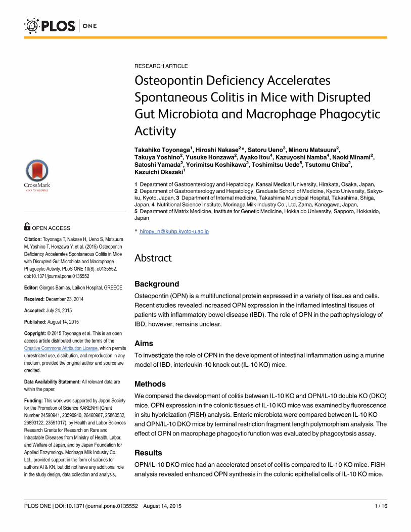
RESEARCH ARTICLE Osteopontin Deficiency Accelerates Spontaneous Colitis in Mice with Disrupted Gut Microbiota and Macrophage Phagocytic Activity Takahiko Toyonaga 1 , Hiroshi Nakase 2 *, Satoru Ueno 3 , Minoru Matsuura 2 , Takuya Yoshino 2 , Yusuke Honzawa 2 , Ayako Itou 4 , Kazuyoshi Namba 4 , Naoki Minami 2 , Satoshi Yamada 2 , Yorimitsu Koshikawa 2 , Toshimitsu Uede 5 , Tsutomu Chiba 2 , Kazuichi Okazaki 1 1 Department of Gastroenterology and Hepatology, Kansai Medical University, Hirakata, Osaka, Japan, 2 Department of Gastroenterology and Hepatology, Graduate School of Medicine, Kyoto University, Sakyo- ku, Kyoto, Japan, 3 Department of Internal medicine, Takashima Municipal Hospital, Takashima, Shiga, Japan, 4 Nutritional Science Institute, Morinaga Milk Industry Co., Ltd, Zama, Kanagawa, Japan, 5 Department of Matrix Medicine, Institute for Genetic Medicine, Hokkaido University, Sapporo, Hokkaido, Japan * [email protected] Abstract Background Osteopontin (OPN) is a multifunctional protein expressed in a variety of tissues and cells. Recent studies revealed increased OPN expression in the inflamed intestinal tissues of patients with inflammatory bowel disease (IBD). The role of OPN in the pathophysiology of IBD, however, remains unclear. Aims To investigate the role of OPN in the development of intestinal inflammation using a murine model of IBD, interleukin-10 knock out (IL-10 KO) mice. Methods We compared the development of colitis between IL-10 KO and OPN/IL-10 double KO (DKO) mice. OPN expression in the colonic tissues of IL-10 KO mice was examined by fluorescence in situ hybridization (FISH) analysis. Enteric microbiota were compared between IL-10 KO and OPN/IL-10 DKO mice by terminal restriction fragment length polymorphism analysis. The effect of OPN on macrophage phagocytic function was evaluated by phagocytosis assay. Results OPN/IL-10 DKO mice had an accelerated onset of colitis compared to IL-10 KO mice. FISH analysis revealed enhanced OPN synthesis in the colonic epithelial cells of IL-10 KO mice. PLOS ONE | DOI:10.1371/journal.pone.0135552 August 14, 2015 1 / 16 a11111 OPEN ACCESS Citation: Toyonaga T, Nakase H, Ueno S, Matsuura M, Yoshino T, Honzawa Y, et al. (2015) Osteopontin Deficiency Accelerates Spontaneous Colitis in Mice with Disrupted Gut Microbiota and Macrophage Phagocytic Activity. PLoS ONE 10(8): e0135552. doi:10.1371/journal.pone.0135552 Editor: Giorgos Bamias, Laikon Hospital, GREECE Received: December 23, 2014 Accepted: July 24, 2015 Published: August 14, 2015 Copyright: © 2015 Toyonaga et al. This is an open access article distributed under the terms of the Creative Commons Attribution License, which permits unrestricted use, distribution, and reproduction in any medium, provided the original author and source are credited. Data Availability Statement: All relevant data are within the paper. Funding: This work was supported by Japan Society for the Promotion of Science KAKENHI (Grant Number 24590941, 23590940, 26460967, 25860532, 26893122, 23591017), by Health and Labor Sciences Research Grants for Research on Rare and Intractable Diseases from Ministry of Health, Labor, and Welfare of Japan, and by Japan Foundation for Applied Enzymology. Morinaga Milk Industry Co., Ltd., provided support in the form of salaries for authors AI & KN, but did not have any additional role in the study design, data collection and analysis,

Welcome message from author
This document is posted to help you gain knowledge. Please leave a comment to let me know what you think about it! Share it to your friends and learn new things together.
Transcript
RESEARCH ARTICLE
Osteopontin Deficiency AcceleratesSpontaneous Colitis in Mice with DisruptedGut Microbiota and Macrophage PhagocyticActivityTakahiko Toyonaga1, Hiroshi Nakase2*, Satoru Ueno3, Minoru Matsuura2,Takuya Yoshino2, Yusuke Honzawa2, Ayako Itou4, Kazuyoshi Namba4, Naoki Minami2,Satoshi Yamada2, Yorimitsu Koshikawa2, Toshimitsu Uede5, Tsutomu Chiba2,Kazuichi Okazaki1
1 Department of Gastroenterology and Hepatology, Kansai Medical University, Hirakata, Osaka, Japan,2 Department of Gastroenterology and Hepatology, Graduate School of Medicine, Kyoto University, Sakyo-ku, Kyoto, Japan, 3 Department of Internal medicine, TakashimaMunicipal Hospital, Takashima, Shiga,Japan, 4 Nutritional Science Institute, Morinaga Milk Industry Co., Ltd, Zama, Kanagawa, Japan,5 Department of Matrix Medicine, Institute for Genetic Medicine, Hokkaido University, Sapporo, Hokkaido,Japan
Abstract
Background
Osteopontin (OPN) is a multifunctional protein expressed in a variety of tissues and cells.
Recent studies revealed increased OPN expression in the inflamed intestinal tissues of
patients with inflammatory bowel disease (IBD). The role of OPN in the pathophysiology of
IBD, however, remains unclear.
Aims
To investigate the role of OPN in the development of intestinal inflammation using a murine
model of IBD, interleukin-10 knock out (IL-10 KO) mice.
Methods
We compared the development of colitis between IL-10 KO and OPN/IL-10 double KO (DKO)
mice. OPN expression in the colonic tissues of IL-10 KOmice was examined by fluorescence
in situ hybridization (FISH) analysis. Enteric microbiota were compared between IL-10 KO
and OPN/IL-10 DKOmice by terminal restriction fragment length polymorphism analysis. The
effect of OPN onmacrophage phagocytic function was evaluated by phagocytosis assay.
Results
OPN/IL-10 DKOmice had an accelerated onset of colitis compared to IL-10 KO mice. FISH
analysis revealed enhanced OPN synthesis in the colonic epithelial cells of IL-10 KOmice.
PLOS ONE | DOI:10.1371/journal.pone.0135552 August 14, 2015 1 / 16
a11111
OPEN ACCESS
Citation: Toyonaga T, Nakase H, Ueno S, MatsuuraM, Yoshino T, Honzawa Y, et al. (2015) OsteopontinDeficiency Accelerates Spontaneous Colitis in Micewith Disrupted Gut Microbiota and MacrophagePhagocytic Activity. PLoS ONE 10(8): e0135552.doi:10.1371/journal.pone.0135552
Editor: Giorgos Bamias, Laikon Hospital, GREECE
Received: December 23, 2014
Accepted: July 24, 2015
Published: August 14, 2015
Copyright: © 2015 Toyonaga et al. This is an openaccess article distributed under the terms of theCreative Commons Attribution License, which permitsunrestricted use, distribution, and reproduction in anymedium, provided the original author and source arecredited.
Data Availability Statement: All relevant data arewithin the paper.
Funding: This work was supported by Japan Societyfor the Promotion of Science KAKENHI (GrantNumber 24590941, 23590940, 26460967, 25860532,26893122, 23591017), by Health and Labor SciencesResearch Grants for Research on Rare andIntractable Diseases from Ministry of Health, Labor,and Welfare of Japan, and by Japan Foundation forApplied Enzymology. Morinaga Milk Industry Co.,Ltd., provided support in the form of salaries forauthors AI & KN, but did not have any additional rolein the study design, data collection and analysis,

OPN/IL-10 DKOmice had a distinctly different enteric bacterial profile with a significantly
lower abundance of Clostridium subcluster XIVa and a greater abundance of Clostridiumcluster XVIII compared to IL-10 KOmice. Intracellular OPN deletion in macrophages
impaired phagocytosis of fluorescence particle-conjugated Escherichia coli in vitro. Exoge-nous OPN enhanced phagocytosis by OPN-deleted macrophages when administered at
doses of 1 to 100 ng/ml, but not 1000 ng/ml.
Conclusions
OPN deficiency accelerated the spontaneous development of colitis in mice with disrupted
gut microbiota and macrophage phagocytic activity.
IntroductionOsteopontin (OPN), a phosphorylated glycoprotein originally identified in bone, binds tohydroxyapatite and regulates ectopic calcium deposition [1]. OPN is also expressed in variousother tissues and cells, including intestinal epithelial cells and immune cells, such as macro-phages, dendritic cells, and T lymphocytes [2]. Two forms of OPN with distinct functions havebeen identified [3]. One is the classical form of OPN secreted from various cells, includingintestinal epithelial cells and activated T lymphocytes (secreted form of OPN [sOPN]), and theother is the intracellular form of OPN (iOPN), which was identified in macrophages [4] anddendritic cells [5]. Although macrophages constitutively express iOPN, they hardly secrete anyOPN, even after stimulation with lipopolysaccharides (LPS) [6].
OPN is implicated in macrophage migration and several macrophage functions [3]. Gener-ally, exogenous OPN enhances macrophage migration in vitro and in vivo [7]. Exogenous OPNworks as a pro-inflammatory cytokine by inducing interleukin-12 (IL-12) and suppressing IL-10 expression in macrophages through interactions with αvβ3 integrin and CD44 [8]. Previousstudies in OPN-deficient mice revealed that OPN is essential for efficient development of T-helper 1 immune responses, and thus plays an important role in protecting against microbialand viral infection [9, 10]. OPN deficiency in macrophages can also result in impaired phago-cytosis [11]. Thus, OPN has a crucial role in the development of both adaptive and innateimmune responses.
Inflammatory bowel disease (IBD) is a relapse-remitting disorder characterized by recurrentintestinal inflammation throughout the gastrointestinal tract. The precise pathophysiology ofIBD remains unknown, but a dysregulated immune response toward enteric bacteria is a strongcomponent [12]. Especially, intestinal macrophages underlying the subepithelial lamina pro-pria play a crucial role in not only intestinal homeostasis but also in the pathophysiology ofIBD by responding to commensal microbes. Indeed, biologic agents targeting tumor necrosisfactor-alpha (TNF-α), a representative cytokine secreted from activated macrophages, are well-known for their efficacy in the clinical treatment of IBD [13]. Increased levels of OPN in theintestinal mucosa and the serum of patients with active IBD suggest that OPN is involved inthe development of intestinal inflammation [14–16]. Experimental studies regarding the effectsof OPN on colonic inflammation, however, have produced conflicting results. In the acutephase of dextran sulfate sodium (DSS)-induced colitis, one study reported exacerbated colitisin OPN-deficient mice compared to wild-type mice with reduced nitric oxide synthase (NOS)expression and impaired phagocytic activity in intestinal macrophages, whereas another studyreported attenuated colitis with reduced macrophage migration in the inflamed colonic tissues
Osteopontin Deficiency Accelerates Spontaneous Colitis in Mice
PLOS ONE | DOI:10.1371/journal.pone.0135552 August 14, 2015 2 / 16
decision to publish, or preparation of the manuscript.The specific roles of these authors are articulated inthe ‘author contributions’ section.
Competing Interests: AI & KN are employees ofMorinaga Milk Industry Co., Ltd. There are nopatents, products in development or marketedproducts to declare. This does not alter the authors’adherence to all the PLOS ONE policies on sharingdata and materials.

of OPN-deficient mice [11, 17]. On the other hand, attenuated colitis was reported in thechronic phase of DSS- and trinitrobenzene sulfonic acid-induced colitis [11, 18]. Thus, whetherOPN ameliorates or exacerbates colonic inflammation is unclear, despite emerging evidencethat OPN is associated with the pathophysiology of human IBD [11, 17–19].
In the present study, we investigated the role of OPN in the development of colitis using amurine experimental colitis model: interleukin-10 knockout (IL-10 KO) mice that developspontaneous colitis in response to enteric bacteria [20]. Like human IBD, dysregulated activa-tion of intestinal macrophages plays a crucial role in the development of colitis in IL-10 KOmice, as demonstrated in mice lacking IL-10 receptors or signal transducers and activators oftranscription 3 in a macrophage-specific manner [21, 22]. Our findings revealed that OPNdeficiency accelerated the development of spontaneous colitis in mice with disrupted gutmicrobiota and macrophage phagocytic activity.
Materials and Methods
Ethics statementThis study was performed in strict accordance with the recommendations in the Guide for theCare and Use of Laboratory Animals of the National Institutes of Health. The protocol wasapproved by the Animal Protection Committee of Kyoto University (Approval Number Med-Kyo14291). All mice were euthanized by cervical dislocation under diethyl ether anesthesia,and all efforts were made to minimize suffering.
MiceThe C57BL/6NCrSlc wild-type (WT) mice were purchased from Japan SLC (Shizuoka, Japan).The B6.129P2-Il10tm1Cgn/J (IL-10 KO) mice were purchased from Charles River Japan (Kana-gawa, Japan). The OPN KOmice were a kind gift from Toshimitsu Uede (Department ofMatrix Medicine, Institute for Genetic Medicine, Hokkaido University). The OPN/IL-10 DKOmice were generated by intercrossing OPN KO and IL-10 KO mice. The mice were fed stan-dard laboratory chow and supplied drinking water ad libitum. All mice were confirmed nega-tive for Sendai virus, mouse hepatitis virus, ectomelia virus, LCM virus,Mycoplasma spp.,Clostridium piliforme, Corynebacterium kutscheri, and Salmonella spp. based on monitoringduring the experiments. We used 48 WT, 74 IL-10 KO, 24 OPN KO, and 74 OPN/IL-10 DKOfemale mice at 3 to 12 weeks of age for the experiments.
Clinical scoreClinical signs of IL-10 KO and OPN/IL-10 DKOmice were evaluated over time, as previouslydescribed [23]. Each of the following subscores was given a value of either 0 or 1: excreted peri-anal mucus, rectal prolapse, diarrhea, or weight loss greater than 5%. The total clinical scorewas the sum of the four subscores.
Microscopic assessment of colitisThe mice were euthanized, and the entire colon was removed. Rectums were dissected, fixed in10% formaldehyde, dehydrated in ethanol, and embedded in paraffin. Transverse sections wereprepared, stained with hematoxylin and eosin (HE), and histologically analyzed in a blindmanner. Histologic damage was quantified by the histologic scoring systems as described previ-ously [24].
Osteopontin Deficiency Accelerates Spontaneous Colitis in Mice
PLOS ONE | DOI:10.1371/journal.pone.0135552 August 14, 2015 3 / 16
Quantitative analysis of gene expressionCollected rectal tissues were quickly frozen in liquid nitrogen for later extraction of the mRNA.Total RNA was extracted using the guanidium isothiocyanate-phenol-chloroformmethod.Extracted RNA was reverse-transcribed with SuperScript II Reverse Transcriptase (Invitrogen,Carlsbad, CA, USA) and the resulting complementary DNAs were analyzed for the expression ofglyceraldehyde phosphate dehydrogenase (GAPDH), interleukin-6 (IL-6), interferon-γ (IFN-γ),interleukin-17A (IL-17A), and CD11bmRNA using the LightCycler 480 System II (Roche AppliedScience, Indianapolis, IN, USA). The primer sequences were as follows:GAPDH, 5’-CAA CTTTGT CAA GCT CAT TTC C-3’ (forward), 5’-GGT CCA GGG TTT CTT ACT CC-3’ (reverse);IL-6, 5’-AGT CCG GAG AGG AGA CTT CA-3’ (forward), 5’-ATT TCC ACG ATT TCC CAGAG-3’ (reverse); IFN-γ; 5’-AGC TCT TCC TCA TGG CTG TT-3’ (forward), 5’-ATC TGGCTC TGC AGG ATT TT-3’ (reverse); IL-17A, 5’-TCT CTG ATG CTG TTG CTG CT-3’ (for-ward), 5’-CGT GGA ACG GTT GAG GTA GT-3’ (reverse); and CD11b, 5’-GCT CCG GTA GCATCA ACA AC-3’ (forward), 5’-AGT GAA TCC GGA ACT CGT CCG-3’ (reverse). The resultinggene expression levels of target molecules were normalized based on the expression of GAPDH.
Fluorescence in situ hybridizationFor detection of the secreted phosphoprotein 1 gene coding OPN, we adapted Quantigene View-RNA (Affymetrix, Santa Clara, CA, USA) using a custom probe set targeting the secreted phos-phoprotein 1 gene (Affymetrix; GenBank Accession Number. NM_009263). Fluorescence in situhybridization (FISH) was performed according to the manufacturer’s instructions. Briefly, rectaltissues collected from 4-week-oldWT, IL-10 KO, and OPN/IL-10 DKOmice were quickly fro-zen in OCT and dissected to prepare 5-μm transverse sections. After fixation, washing, anddehydration in ethanol, the sections were digested in Protease GF (Affymetrix) and then hybrid-ized with custom-designed QuantiGene ViewRNA probes against target RNA sequences.Bound probes were preamplified and subsequently amplified according to the manufacturer’sinstructions. Probe oligonucleotide conjugated to alkaline phosphatase type 1 was added, fol-lowing by Fast Red substrate. Nuclei were visualized by Hoechst staining. In some experiments,the sections were further incubated with rabbit anti-mouse E-cadherin antibody (1:100 dilution;Abcam, Cambridge, MA, USA), and signals were visualized by Alexa Fluor 488-conjugatedanti-rabbit antibody (1:1000 dilution; Invitrogen). Images were acquired using a fluorescencemicroscope (BIOREVO BZ-9000; Keyence, Osaka, Japan) with the BZ-Analyzer version 2.1software and composed in Photoshop Elements 12 (Adobe Systems Inc., San Jose, CA, USA).
Preparation and stimulation of bone marrow-derived macrophagesMurine bone marrow cells were obtained from femur and tibia bone marrow. Cells were cul-tured in complete Roswell Park Memorial Institute (RPMI) 1640 medium (Gibco, Invitrogen,Grand Island, NY, USA) containing 10% heat-inactivated fetal bovine serum, 100 mg/ml ofstreptomycin (Sigma-Aldrich, St. Louis, MO, USA), 100 mg/ml of penicillin (Sigma-Aldrich),and 40 ng/ml of recombinant macrophage colony-stimulating factor (Peprotech, Rocky Hill,NJ, USA) for 5 d at 37°C. Bone marrow-derived macrophages (BMDMs) were harvested andseeded at 5×10⁵ cells/well in 6-well culture plates (Greiner Bio-One, Monroe, NC, USA). Afterovernight incubation with 10 ng/ml IFN-γ(R&D Systems, Minneapolis, MN, USA), the wellswere washed and challenged with stimulators as follows: Pam3CSK4 (Toll-like receptor [TLR]2 ligand) 50 to 500 ng/ml, LPS (TLR4 ligand) 100 ng/ml, and ODN1585 (TLR9 ligand) 500 nM(Pam3CSK4 and ODN1585 from InvivoGen [San Diego, CA, USA] and LPS from Sigma-Aldrich). The cells were stimulated for 24 h before the supernatants were collected and storedat -80°C until assayed.
Osteopontin Deficiency Accelerates Spontaneous Colitis in Mice
PLOS ONE | DOI:10.1371/journal.pone.0135552 August 14, 2015 4 / 16

Enzyme-linked immunosorbent assayThe TNF-α, IL-6, and interleukin-12p40 (IL-12p40) levels in the cell supernatants were quanti-fied using a mouse enzyme-linked immunosorbent assay (ELISA) kit (eBioscience, San Diego,CA, USA) according to the manufacturer’s instructions.
Isolation of lamina propria mononuclear cellsLamina propria mononuclear cells (LPMCs) were isolated from colonic tissues of IL-10 KOand OPN/IL-10 DKOmice as previously described [25]. Briefly, the entire colons wereremoved from euthanized mice and washed with phosphate-buffered saline (PBS). After dis-rupting epithelial cells in Hank’s Balanced Salt Solution containing 5 mM EDTA and 1 mMdithiothreitol, the colonic tissues were repeatedly digested in PBS containing 0.5 mg/ml colla-genase D (Roche Applied Science), 0.5 mg/ml DNase I (Sigma-Aldrich), and 3 mg/ml DispaseII (Roche Applied Science) at 37°C for 30 minutes under slow rotation. Collected supernatantswere spun down, and the obtained pellet was re-suspended in 40% Percoll solution and over-laid on 80% Percoll solution. LPMCs were obtained from the interphase and washed with PBS.Total RNA was extracted from LPMCs, and gene expression was analyzed as described above.The primer sequences were as follows: nitric oxide synthase 2 (NOS2), 5’-ATG CAA CAT GGGAGC CAC AGC-3’ (forward), 5’-CTG GGA TTT CAG CCT CAT GG-3’ (reverse); TNF-α,5’-CAT GCA CCA CCA TCA AGG AC-3’ (forward), 5’-GGC CTG AGA TCT TAT CCA GCC-3’ (reverse); IL-12p40, 5’-GGA AGC ACG GCA GCA GAA TA-3’ (forward), 5’-TGA CCTCCA CCT GTG AGT TC-3’ (reverse).
Terminal restriction fragment length polymorphism analysisTerminal restriction fragment length polymorphism (T-RFLP) analysis was performed asdescribed previously [26]. Briefly, murine cecal contents were suspended in a solution contain-ing 100 mM Tris-HCl (pH 9.0), 40 mM EDTA, 4 M guanidine thiocyanate, and 0.001% bro-mothymol blue. Fecal solids in the suspension were broken down using the FastPrep 24Instrument (MP Biomedicals, Irvine, CA, USA) with zirconia beads. Fecal DNA was extractedfrom the suspension by an automatic nucleic acid extractor (Precision System Science, Chiba,Japan) using MagDEA DNA 200 (GC) (Precision System Science) as the reagent.
The 16S rRNA gene was amplified from fecal DNA using the primers of 5’-FAM-labeled516f (5’-TGCCAGCAGCCGCGGTA-3’; Escherichia coli [E. coli] positions 516 to 532) and1510r (5’-GGTTACCTTGTTACGACTT-3’; E. coli positions 1510 to 1492). The resulting 16SrDNA amplicons were treated with 10 U of Bsl l (New England BioLabs, MA, USA) for 3hours, and the digestives were fractionated using an ABI PRISM 3130xl Genetic Analyzer(Applied Biosystems, Foster City, CA, USA) with the DNA analysis software Gene Mapper.The major terminal restriction fragments similar in size of 1–3 bp were summarized to opera-tional taxonomic units (OTUs), and the corresponding bacterial groups were estimated bycomputer simulation based on the human intestinal microbiota database [27]. The percentageof each bacterial group to total enteric bacteria was represented by the percentage of eachsummed OTU area to total OTU area [28]. Cluster analyses were performed using Gene Mathssoftware (Applied Maths, Kortrijk, Belgium) based on the obtained T-RFLP profiles, and thesimilarity among samples was evaluated by principal component analysis.
Preparation of thioglycollate-elicited peritoneal macrophagesMice were intraperitoneally injected with 3 ml of 4% thioglycollate (Eilken Chemical Co.,Tokyo, Japan) and peritoneal exudate cells were harvested on Day 4. After removing
Osteopontin Deficiency Accelerates Spontaneous Colitis in Mice
PLOS ONE | DOI:10.1371/journal.pone.0135552 August 14, 2015 5 / 16
erythrocytes, the cells were subjected to positive selection using anti-CD11b magnetic microbe-ads (Miltenyi Biotec, Auburn, CA, USA) to select CD11b-positive thioglycollate-elicited perito-neal macrophages (TEPMs).
Phagocytosis assayA phagocytosis assay was performed using pHrodo Green E. coli BioParticles Conjugate (Invi-trogen) according to the manufacturer’s instructions. Briefly, TEPMs were seeded at 2.5×10⁵
cells/well in 96-well cell culture plates (Greiner Bio-One) and incubated overnight in completeRPMI 1640 medium containing 40 ng/ml recombinant macrophage colony-stimulating factor.The wells were washed with PBS and challenged with fluorescent particles diluted in Live CellImaging Solution (Invitrogen). In several wells, recombinant OPN (rOPN, R&D Systems) wasadded at a concentration of 1, 10, 100, or 1000 ng/ml. The cells were incubated at 37°C andfluorescence intensity was measured at the indicated times using a fluorescence microplatereader (Infinite F200 PRO; TECAN, Kawasaki, Japan). Phagocytic activity was evaluated bysubtracting the average fluorescence intensity of the no-cell negative-control wells from that ofall experimental wells and indicated as the relative value.
Identification of bacterial species in the mesenteric lymph nodeBacterial species in the mesenteric lymph nodes (MLNs) of the mice were detected as previ-ously reported with some modifications [29]. MLNs were obtained from mice under sterileconditions and mechanically disrupted with zirconia beads. Bacterial DNA was extracted usinga bacterial DNA/RNA extraction kit (MORA-EXTRACT; Kyokuto Pharmaceuticals, Tokyo,Japan). After the first amplification of bacterial 16S rRNA with universal primers (27F; 5’-AGA GTT TGA TCC TGG CTC AG-3’, 1492R; 5’-GGT TAC CTT GTT ACG ACT T-3’),nested PCR was performed using internal primers (35F; 5’-CCT GGC TCA GGA TGA ACG-3’, 519R; 5’-ATT ACC GCG GCK GCT C-3’). The amplified product was electrophoresed ona 1% agarose gel, and purified from the agarose gel using the QIAEX II Gel Extraction Kit(QIAGEN, Düsseldorf, Germany). The purified product was ligated into the pCR4-TOPO vec-tor, and transformed into E. coli TOP10 cells using the TOPO-TA Cloning Kit (Invitrogen).
The 16S rRNA genes were amplified from cultures using the TempliPhi system (GE Health-care, NJ, US), and these were used as templates for DNA sequencing. The products were ana-lyzed using an ABI 3730 DNA Analyzer (Applied Biosystems). All nucleotide sequences werechecked by BLAST analysis against all entries in the DNA Data Bank of Japan database andaligned using DNASIS Pro (Hitachi Software Engineering, Yokohama, Japan).
Statistical analysisAll numerical data are expressed as means ± SD or SEM. Differences between groups were ana-lyzed using a non-parametric Mann-Whitney U test or one-way analysis of variance with Bon-ferroni’s correction for multiple comparisons. A P value of less than 0.05 was consideredsignificant.
Results
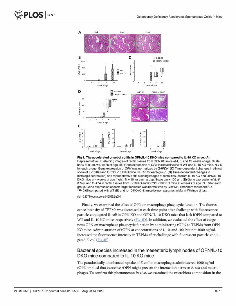
Disruption of OPN accelerated the onset of colitis in IL-10 KOmiceOPN KOmice did not develop spontaneous colitis (Fig 1A). Therefore, we first deleted OPN inIL-10 KO mice that had increased OPNmRNA expression in the colon from 4 weeks of age(Fig 1B), and compared the development of colitis in these mice with that in IL-10 KO mice.Clinical signs, such as perianal mucus excretion and diarrhea, appeared in OPN/IL-10 DKO
Osteopontin Deficiency Accelerates Spontaneous Colitis in Mice
PLOS ONE | DOI:10.1371/journal.pone.0135552 August 14, 2015 6 / 16

mice from 4 weeks of age, whereas IL-10 KO mice showed no sign at this time (Fig 1C). Histo-chemical examination revealed the initiation of colitis between 3 and 4 weeks of age in both IL-10 KO and OPN/IL-10 DKOmice. OPN/IL-10 DKOmice rapidly developed colitis within 1week, however, and had increased infiltrating immune cells and remarkable epithelial hyper-plasia in the rectum compared to IL-10 KO mice at 4 weeks of age (Fig 1D). The histologicscore was significantly higher in OPN/IL-10 DKOmice than in IL-10 KO mice at 4 weeks ofage, but there was no significant difference in the histologic score at 8 and 12 weeks of agebetween IL-10 KO and OPN/IL-10 DKOmice. Gene expression of several pro-inflammatorycytokines, such as IL-6, IFN-γ, and IL-17A was upregulated in the rectal tissues of 4-week-oldOPN/IL-10 DKOmice compared with IL-10 KO mice (Fig 1E).
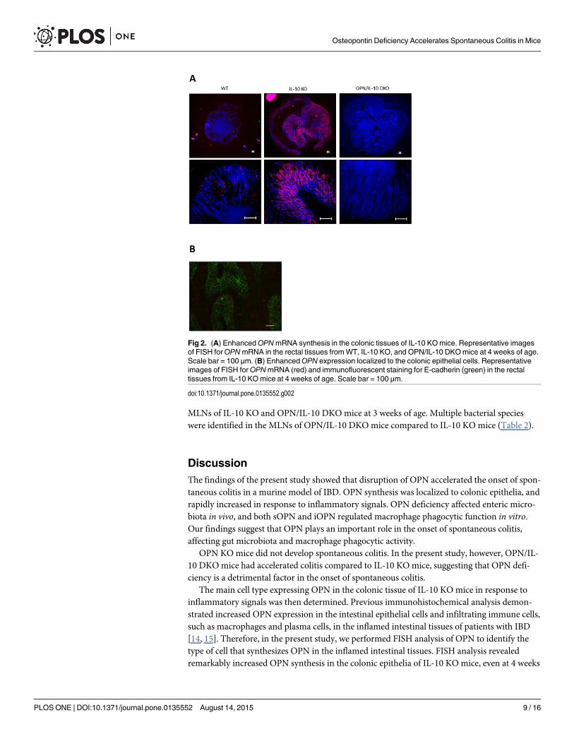
OPN synthesis was enhanced in the colonic epithelial cells of IL-10 KOmiceWe next performed FISH analysis of OPN to determine the main cell type that synthesizesOPN in the colonic tissues of IL-10 KO mice. The FISH analysis revealed remarkably enhancedOPNmRNA synthesis in the rectal tissues of 4-week-old IL-10 KO mice compared with WTmice (Fig 2A). OPN synthesis was observed in the E-cadherin expressing colonic epithelialcells of IL-10 KO mice (Fig 2B).
OPN deficiency affected enteric microbiota in IL-10 KOmiceTo elucidate the role of OPN secreted from colonic epithelia in the innate immune responses,we examined the impact of OPN deficiency on enteric bacteria in IL-10 KO mice. We com-pared the enteric microbiota between IL-10 KO and OPN/IL-10 DKOmice at 3 weeks of ageby T-RFLP analysis. The T-RFLP profiles showed significantly lower abundance of Clostridiumsubcluster XIVa and greater abundance of Clostridium cluster XVIII in OPN/IL-10 DKOmicecompared to IL-10 KO mice (Fig 3A, Table 1). Principal component analysis of the T-RFLPprofiles revealed a distinct difference in the enteric bacterial composition between IL-10 KOand OPN/IL-10 DKOmice (Fig 3B).
Both sOPN and iOPN affected the phagocytic function of macrophagesWe further investigated the role of OPN in the innate immune responses by examining theeffect of OPN on macrophage migration and functions, such as cytokine production andphagocytosis. First, we evaluated macrophage migration by quantifying CD11b gene expressionin the colon. Gene expression of CD11b in the rectal tissues was similar between OPN/IL-10DKOmice and IL-10 KO mice at 4 weeks of age, but it was significantly lower in IL-10 KOmice at 12 weeks of age (Fig 4A). Second, we generated BMDMs fromWT, OPN KO, IL-10KO, and OPN/IL-10 DKOmice, and stimulated them with TLR ligands. BMDMs from IL-10KO and OPN/IL-10 DKOmice secreted higher amounts of TNF-α, IL-12p40, and IL-6 thanBMDMs fromWT and OPN KOmice after stimulation with Pam3CSK4 and LPS. The secretedamounts of these pro-inflammatory cytokines, however, did not differ significantly betweenWT and OPN KOmice, and IL-10 KO and OPN/IL-10 DKOmice after stimulation with anyligand (Fig 4B). Third, we isolated LPMCs from colonic tissues of IL-10 KO and OPN/IL-10DKOmice, and examined their phenotype during colitis (Fig 4C). Gene expression of NOS2 inLPMCs from OPN/IL-10 DKOmice was significantly lower at 4 weeks of age, but significantlyhigher at 8 and 12 weeks of age than that in LPMCs from IL-10 KO mice. LPMCs from OPN/IL-10 DKOmice expressed higher levels of TNF-α and IL-12p40 than LPMCs from IL-10 KOmice at 4, 8, and 12 weeks of age.
Osteopontin Deficiency Accelerates Spontaneous Colitis in Mice
PLOS ONE | DOI:10.1371/journal.pone.0135552 August 14, 2015 7 / 16
Finally, we examined the effect of OPN on macrophage phagocytic function. The fluores-cence intensity of TEPMs was decreased at each time point after challenge with fluorescenceparticle-conjugated E. coli in OPN KO and OPN/IL-10 DKOmice that lack iOPN compared toWT and IL-10 KO mice, respectively (Fig 4D). In addition, we evaluated the effect of exoge-nous OPN on macrophage phagocytic function by administering rOPN to TEPMs from OPNKOmice. Administration of rOPN at concentrations of 1, 10, and 100, but not 1000 ng/ml,increased the fluorescence intensity in TEPMs after challenge with fluorescent particle-conju-gated E. coli (Fig 4E).
Bacterial species increased in the mesenteric lymph nodes of OPN/IL-10DKOmice compared to IL-10 KOmiceThe paradoxically unenhanced uptake of E. coli in macrophages administered 1000 ng/mlrOPN implied that excessive rOPN might prevent the interaction between E. coli and macro-phages. To confirm this phenomenon in vivo, we examined the microbiota composition in the
Fig 1. The accelerated onset of colitis in OPN/IL-10 DKOmice compared to IL-10 KOmice. (A)Representative HE-staining images of rectal tissues from OPN KOmice at 4, 8, and 12 weeks of age. Scalebar = 100 μm. wk, week of age. (B) Gene expression ofOPN in rectal tissues of WT and IL-10 KOmice. N = 6for each group. Gene expression ofOPN was normalized byGAPDH. (C) Time-dependent changes in clinicalscore of IL-10 KO and OPN/IL-10 DKOmice. N = 10 for each group. (D) Time-dependent changes inhistologic scores (left) and representative HE-staining images of rectal tissues from IL-10 KO and OPN/IL-10DKOmice at 4 weeks of age (right). N = 10 for each group. Scale bar = 100 μm. (E) Gene expression of IL-6,IFN-γ, and IL-17A in rectal tissues from IL-10 KO and OPN/IL-10 DKOmice at 4 weeks of age. N = 6 for eachgroup. Gene expression of each target molecule was normalized byGAPDH. Error bars represent SD.*P<0.05 compared with WT (B) and IL-10 KO (C-E) mice by non-parametric Mann-Whitney U test.
doi:10.1371/journal.pone.0135552.g001
Osteopontin Deficiency Accelerates Spontaneous Colitis in Mice
PLOS ONE | DOI:10.1371/journal.pone.0135552 August 14, 2015 8 / 16
MLNs of IL-10 KO and OPN/IL-10 DKOmice at 3 weeks of age. Multiple bacterial specieswere identified in the MLNs of OPN/IL-10 DKOmice compared to IL-10 KO mice (Table 2).
DiscussionThe findings of the present study showed that disruption of OPN accelerated the onset of spon-taneous colitis in a murine model of IBD. OPN synthesis was localized to colonic epithelia, andrapidly increased in response to inflammatory signals. OPN deficiency affected enteric micro-biota in vivo, and both sOPN and iOPN regulated macrophage phagocytic function in vitro.Our findings suggest that OPN plays an important role in the onset of spontaneous colitis,affecting gut microbiota and macrophage phagocytic activity.
OPN KOmice did not develop spontaneous colitis. In the present study, however, OPN/IL-10 DKOmice had accelerated colitis compared to IL-10 KO mice, suggesting that OPN defi-ciency is a detrimental factor in the onset of spontaneous colitis.
The main cell type expressing OPN in the colonic tissue of IL-10 KO mice in response toinflammatory signals was then determined. Previous immunohistochemical analysis demon-strated increased OPN expression in the intestinal epithelial cells and infiltrating immune cells,such as macrophages and plasma cells, in the inflamed intestinal tissues of patients with IBD[14, 15]. Therefore, in the present study, we performed FISH analysis of OPN to identify thetype of cell that synthesizes OPN in the inflamed intestinal tissues. FISH analysis revealedremarkably increased OPN synthesis in the colonic epithelia of IL-10 KO mice, even at 4 weeks
Fig 2. (A) EnhancedOPNmRNA synthesis in the colonic tissues of IL-10 KOmice. Representative imagesof FISH forOPNmRNA in the rectal tissues fromWT, IL-10 KO, and OPN/IL-10 DKOmice at 4 weeks of age.Scale bar = 100 μm. (B) EnhancedOPN expression localized to the colonic epithelial cells. Representativeimages of FISH forOPNmRNA (red) and immunofluorescent staining for E-cadherin (green) in the rectaltissues from IL-10 KOmice at 4 weeks of age. Scale bar = 100 μm.
doi:10.1371/journal.pone.0135552.g002
Osteopontin Deficiency Accelerates Spontaneous Colitis in Mice
PLOS ONE | DOI:10.1371/journal.pone.0135552 August 14, 2015 9 / 16
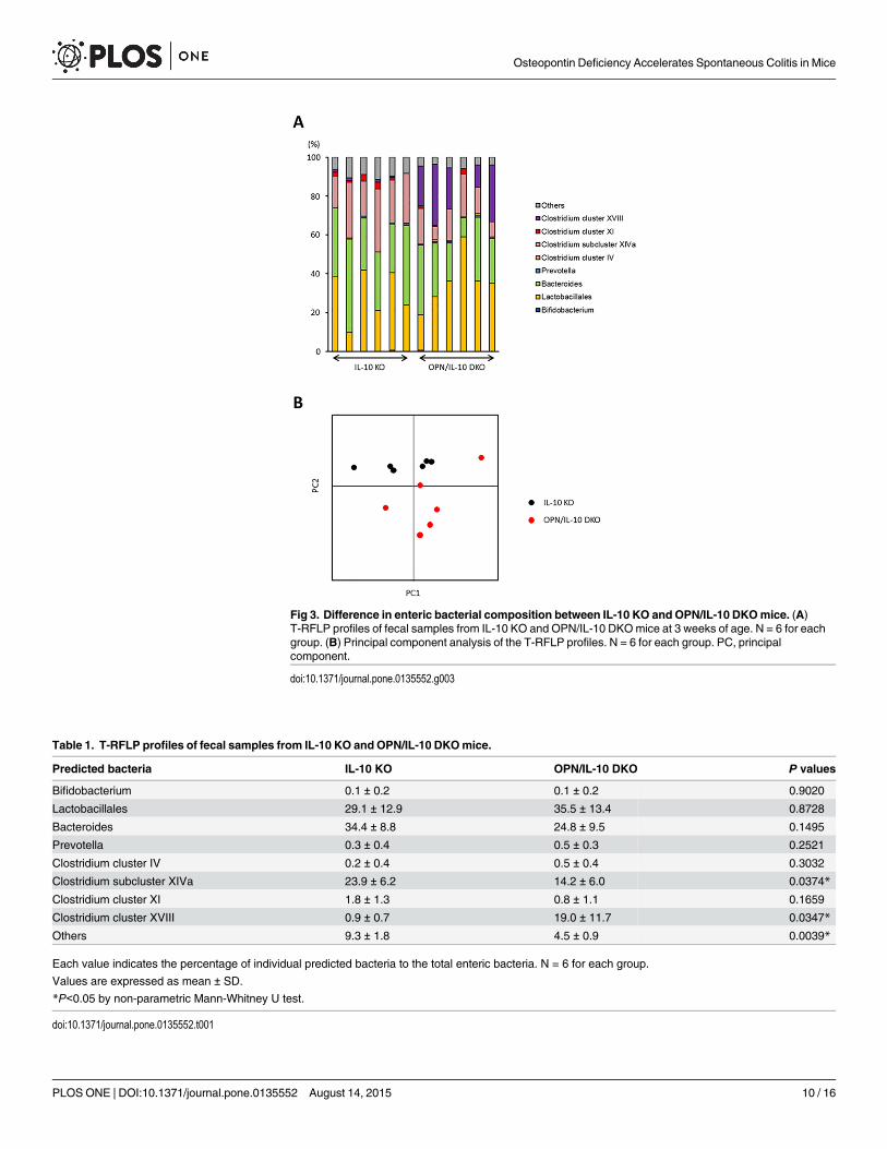
Fig 3. Difference in enteric bacterial composition between IL-10 KO and OPN/IL-10 DKOmice. (A)T-RFLP profiles of fecal samples from IL-10 KO and OPN/IL-10 DKOmice at 3 weeks of age. N = 6 for eachgroup. (B) Principal component analysis of the T-RFLP profiles. N = 6 for each group. PC, principalcomponent.
doi:10.1371/journal.pone.0135552.g003
Table 1. T-RFLP profiles of fecal samples from IL-10 KO and OPN/IL-10 DKOmice.
Predicted bacteria IL-10 KO OPN/IL-10 DKO P values
Bifidobacterium 0.1 ± 0.2 0.1 ± 0.2 0.9020
Lactobacillales 29.1 ± 12.9 35.5 ± 13.4 0.8728
Bacteroides 34.4 ± 8.8 24.8 ± 9.5 0.1495
Prevotella 0.3 ± 0.4 0.5 ± 0.3 0.2521
Clostridium cluster IV 0.2 ± 0.4 0.5 ± 0.4 0.3032
Clostridium subcluster XIVa 23.9 ± 6.2 14.2 ± 6.0 0.0374*
Clostridium cluster XI 1.8 ± 1.3 0.8 ± 1.1 0.1659
Clostridium cluster XVIII 0.9 ± 0.7 19.0 ± 11.7 0.0347*
Others 9.3 ± 1.8 4.5 ± 0.9 0.0039*
Each value indicates the percentage of individual predicted bacteria to the total enteric bacteria. N = 6 for each group.
Values are expressed as mean ± SD.
*P<0.05 by non-parametric Mann-Whitney U test.
doi:10.1371/journal.pone.0135552.t001
Osteopontin Deficiency Accelerates Spontaneous Colitis in Mice
PLOS ONE | DOI:10.1371/journal.pone.0135552 August 14, 2015 10 / 16

Fig 4. The impact of OPN on cytokine production and phagocytic function of macrophages. (A) Gene expression of CD11b in rectal tissues from IL-10KO and OPN/IL-10 DKOmice at 4, 8, and 12 weeks of age. N = 5 for each group. Gene expression ofCD11b was normalized byGAPDH. Error barsrepresent SD. *P<0.05 compared with IL-10 KOmice by non-parametric Mann-Whitney U test. (B) Cytokine production from BMDMs obtained fromWT,OPN KO, IL-10 KO, and OPN/IL-10 DKOmice after stimulation with Pam3CSK4 (50 to 500 ng/ml), LPS (100 ng/ml), or ODN1585 (500 nM). N = 6 for eachgroup. Error bars represent SEM. *P<0.05, one-way ANOVA with Bonferroni’s correction. (C) Gene expression of NOS2, TNF-α, and IL-12p40 in LPMCs
Osteopontin Deficiency Accelerates Spontaneous Colitis in Mice
PLOS ONE | DOI:10.1371/journal.pone.0135552 August 14, 2015 11 / 16

of age, compared to WT mice, suggesting that epithelial cells are the main cells that synthesizeOPN in the colonic tissues in response to inflammatory signals.
To elucidate the role of OPN secreted from colonic epithelia in mucosal immune responses,we examined the impact of OPN deficiency on enteric microbiota by comparing the entericbacterial composition between IL-10 KO and OPN/IL-10 DKOmice at 3 weeks of age, justbefore the initiation of colitis. Surprisingly, T-RFLP analysis revealed a significant difference inthe bacterial profiles with decreased abundance of Clostridium subcluster XIVa and increasedabundance of Clostridium cluster XVIII in OPN/IL-10 DKOmice compared to IL-10 KO mice.Although the role of Clostridium cluster XVIII in mucosal immunity remains uncertain, Clos-tridium subcluster XIVa has a protective effect on DSS- and oxazolone-induced murine colitisby inducing regulatory T lymphocytes in the colon [30]. Thus, an intriguing notion is thatOPN secreted from colonic epithelia prevents the development of colitis in IL-10 KO mice byaffecting enteric bacteria, including Clostridium subcluster XIVa. Further study is necessary toelucidate how OPN affects enteric bacteria.
Next, we investigated the effect of OPN on macrophages, which are deeply involved in theinitiation of colitis in IL-10 KO mice by sensing luminal bacterial components and producing avariety of pro-inflammatory cytokines [21]. We first examined CD11bmRNA expression levelsin the colonic tissues of IL-10 KO and OPN/IL-10 DKOmice, which probably reflect macro-phage numbers. There are several reports suggesting that OPN induces macrophage migrationin vitro [4, 7]. The role of OPN in macrophage recruitment in vivo, however, still lacks consen-sus [9, 31]. In this study, CD11bmRNA expression in the rectal tissues of OPN/IL-10 DKOmice was similar to that in IL-10 KO mice at 4 weeks of age, but lower than that in IL-10 KOmice at 8 and 12 weeks of age. This finding suggests that the effect of OPN on macrophagerecruitment depends on the phase of colitis in this model and might not contribute to the
isolated from IL-10 KO and OPN/IL-10 DKOmice at 4, 8, and 12 weeks of age. N = 4 for each group. Gene expression of each target molecule wasnormalized byGAPDH. Error bars represent SD. *P<0.05 compared with IL-10 KOmice by non-parametric Mann-Whitney U test. (D) Changes offluorescence intensity in TEPMs fromWT, OPN KO, IL-10 KO, and OPN/IL-10 DKOmice after challenged with fluorescent particle conjugated-E.coli.Fluorescence intensity was measured at the indicated times and is shown in arbitrary units (AU). N = 6 for each group. Error bars represent SEM. *P<0.05compared with TEPMs fromWTmice; ✝P<0.05 compared with IL-10 KOmice by one-way ANOVA with Bonferroni’s correction. (E) Changes of fluorescenceintensity in TEPMs from OPN KOmice after challenged with fluorescence particle conjugated-E.coli with or without recombinant OPN (rOPN) at 1, 10, 100,and 1000 ng/ml. Fluorescence intensity was measured at the indicated times and is shown in arbitrary units (AU). N = 6 for each group. Error bars representSEM. *P<0.05 compared to TEPMs from OPN KOmice without rOPN (0 ng/ml) by one-way ANOVAwith Bonferroni’s correction.
doi:10.1371/journal.pone.0135552.g004
Table 2. Identified bacterial species in the mesenteric lymph nodes of IL-10 KO and OPN/IL-10 DKOmice.
Mouse Identified bacteria
IL-10 KO Propionibacterium acnes isolate Asn14
OPN/IL-10 DKO Streptococcus sp. Smarlab 3301444
Staphylococcus sp. CHNDP23
Staphylococcus lentus
Corynebacterium imitans
Corynebacterium accolens
Corynebacterium sp. CIP102857
Staphylococcus sp. CNJ924 PL04
Bacterial species identified in the mesenteric lymph nodes of IL-10 KO and OPN/IL-10 DKO mice at 3
weeks of age.
N = 8 for each group.
doi:10.1371/journal.pone.0135552.t002
Osteopontin Deficiency Accelerates Spontaneous Colitis in Mice
PLOS ONE | DOI:10.1371/journal.pone.0135552 August 14, 2015 12 / 16
accelerated colitis in OPN/IL-10 DKOmice at 4 weeks of age. Then, we focused on the effect ofOPN on cytokine production from macrophages. In previous studies, exogenous OPNenhanced the expression of several pro-inflammatory cytokines, such as IL-1β, TNF-α, IL-12,and IL-6, whereas it suppressed the expression of IL-10 in murine macrophages and humanperipheral monocytes [8, 14, 32, 33]. Thus, theoretically, the lack of sOPN might lead to theamelioration of colitis in IL-10 KO mice by decreasing the production of pro-inflammatorycytokines from intestinal macrophages. In this regard, the rapidly deteriorating colitis in OPN/IL-10 DKOmice cannot be completely explained by the effect of sOPN deficiency on macro-phage cytokine production. Therefore, we focused on the effect of iOPN on macrophage cyto-kine production. We stimulated BMDMs fromWT, OPN KO, IL-10 KO, and OPN/IL-10DKOmice with the ligands of TLR2, TLR4, and TLR9, all of which recognize bacterial compo-nents on the surface or inside of macrophages [34]. Comparison of the amounts of secretedpro-inflammatory cytokines, however, revealed no effect of iOPN deficiency on macrophagecytokine production. Taken together, these findings indicate that the effect of OPN deficiencyon macrophage cytokine production does not contribute to the rapidly deteriorating colitis inOPN/IL-10 KO mice.
The macrophage phenotype during colitis was compared between IL-10 KO and OPN/IL-10 DKOmice to determine the effect of OPN on macrophage function in vivo. LPMCs isolatedfrom OPN/IL-10 DKOmice expressed higher levels of TNF-α and IL-12p40 than those fromIL-10 KO mice at 4 weeks of age, suggesting that intestinal macrophages are highly activated inOPN/IL-10 DKOmice at an early stage of colitis compared to IL-10 KO mice. On the otherhand, gene expression of NOS2, a representative marker of pro-inflammatory macrophages[35], was significantly lower in LPMCs from OPN/IL-10 DKOmice at 4 weeks of age com-pared to IL-10 KO mice, whereas it became significantly higher after 8 weeks of age (Fig 4C).Several reports showed that OPN suppressed NOS2mRNA expression in murine macrophagesin vitro, but the effect of OPN on NOS2mRNA expression and NO production in macrophagesremains uncertain in vivo [36, 37]. Remarkably attenuated NOS expression was reported in theintestinal macrophages of OPN KOmice compared to WT mice in the DSS-induced colitismodel [11]. Our findings suggest that the effect of OPN on NOS2 expression in macrophagesmight be context-specific and dependent on the phase of colitis.
We then examined the effect of OPN on macrophage phagocytic function. We first assessedthe effect of iOPN deficiency on macrophage phagocytic function. Shimizu et al. reported thatBMDMs from OPN KOmice have impaired ability to phagocytose fluorescent beads [38].Also, Heilmann et al. reported that macrophages isolated from the lamina propria of OPN KOmice with DSS-induced colitis have impaired ability to phagocytose fluorescein isothiocyanate-labeled E. coli [11]. Here, we showed impaired ability to phagocytose fluorescence-conjugatedE. coli in TEPMs from OPN KO and OPN/IL-10 DKOmice compared to TEPMs fromWTand IL-10 KO mice, respectively. Consistent with previous studies, our data clearly demon-strate that iOPN deficiency impaired the phagocytic function of macrophages, although themechanism is not yet clear. We next assessed the effect of sOPN on the phagocytic function ofmacrophages using TEPMs from OPN KOmice. Administration of 1, 10, and 100, but not1000 ng/ml rOPN enhanced the phagocytic function of TEPMs from OPN KOmice. Severalreports have demonstrated that OPN can function as an opsonin to facilitate macrophagephagocytosis [39, 40]. Schack et al. showed enhanced phagocytosis of OPN-coated Staphylococ-cus aureus through αxβ2 integrin expressed on the surface of phorbol myristate acetate-stimu-lated U937 cells [39]. Although we did not demonstrate the binding of rOPN to E. coli in thisstudy, our findings suggest that rOPN enhances bacterial uptake in macrophages by binding toseveral bacterial species, including E. coli. In contrast, 1000 ng/ml rOPN did not enhancephagocytosis in TEPMs from OPN KOmice. This finding suggests that excessive rOPN
Osteopontin Deficiency Accelerates Spontaneous Colitis in Mice
PLOS ONE | DOI:10.1371/journal.pone.0135552 August 14, 2015 13 / 16

between macrophages and bacteria might prevent their contact and thereby decrease bacterialuptake by macrophages. This phenomenon is consistent with the report by Heilmann et al.that a low dose (100 ng/ml), but not a high dose (500 ng/ml), of OPN enhances the ability ofhuman macrophages to phagocytose fluorescein isothiocyanate-labeled E. coli [11]. In thisregard, a large amount of OPN secreted from colonic epithelia in response to inflammatory sig-nals might enhance mucosal defense by limiting the interaction between enteric bacteria andunderlying mucosal immune cells, including macrophages, in vivo. The multiple bacterial spe-cies detected in the MLNs of OPN/IL-10 DKOmice compared to IL-10 KO mice at 3 weeks ofage supports the notion of impaired mucosal defense in OPN/IL-10 DKOmice.
The findings of the present study suggest an important role of OPN in the onset of sponta-neous colitis in mice. OPN deficiency affected the enteric microbiota, and both sOPN andiOPN regulated macrophage phagocytic function in vitro. Further studies regarding the directeffect of OPN on enteric bacteria, and the effect of OPN on macrophage phagocytic activity invivo during spontaneous colitis will provide new insights into the role of OPN in the develop-ment of colitis as well as in the pathophysiology of IBD.
Author ContributionsConceived and designed the experiments: HN TC KO. Performed the experiments: TT SUMM TY YH AI KN NM SY YK. Analyzed the data: TT SU HN. Contributed reagents/materi-als/analysis tools: TU. Wrote the paper: TT HN.
References1. Oldberg A, Franzén A, Heinegård D (1986) Cloning and sequence analysis of rat bone sialoprotein
(osteopontin) cDNA reveals an Arg-Gly-Asp cell-binding sequence. Proc Natl Acad Sci USA 83: 8819–8823. PMID: 3024151
2. Brown LF, Berse B, Van deWater L, Papadopoulos-Sergiou A, Perruzzi CA, Manseau EJ, et al. (1992)Expression and distribution of osteopontin in human tissues: widespread association with luminal epi-thelial surfaces. Mol Biol Cell 3: 1169–1180. PMID: 1421573
3. Rittling SR (2011) Osteopontin in macrophage function. Expert Rev Mol Med 26:13.
4. Zhu B, Suzuki K, Goldberg HA, Rittling SR, Denhardt DT, McCulloch CA, et al. (2004) Osteopontinmodulates CD44-dependent chemotaxis of peritoneal macrophages through G-protein-coupled recep-tors: evidence of a role for an intracellular form of osteopontin. J Cell Physiol 198: 155–167. PMID:14584055
5. Shinohara ML, Lu L, Bu J, Werneck MB, Kobayashi KS, Glimcher LH, et al. (2006) Osteopontin expres-sion is essential for interferon-alpha production by plasmacytoid dendritic cells. Nat Immunol 7: 498–506. PMID: 16604075
6. Shinohara ML, JanssonM, Hwang ES, Werneck MB, Glimcher LH, Cantor H (2005) T-bet-dependentexpression of osteopontin contributes to T cell polarization. Proc Natl Acad Sci USA 102: 17101–17106. PMID: 16286640
7. Weber GF, Zawaideh S, Hikita S, Kumar VA, Cantor H, Ashkar S. (2002) Phosphorylation-dependentinteraction of osteopontin with its receptors regulates macrophage migration and activation. J LeukocBiol 72: 752–761. PMID: 12377945
8. Ashkar S, Weber GF, Panoutsakopoulou V, Sanchirico ME, Jansson M, Zawaideh S, et al. (2000) Eta-1 (osteopontin): an early component of type-1 (cell-mediated) immunity. Science 287: 860–864. PMID:10657301
9. Nau GJ, Liaw L, Chupp GL, Berman JS, Hogan BL, Young RA (1999) Attenuated host resistanceagainst Mycobacterium bovis BCG infection in mice lacking osteopontin. Infect Immun 67: 4223–4230.PMID: 10417195
10. Rollo EE, Hempson SJ, Bansal A, Tsao E, Habib I, Rittling SR, et al. (2005) The cytokine osteopontinmodulates the severity of rotavirus diarrhea. J Virol 79: 3509–3516. PMID: 15731245
11. Heilmann K, Hoffmann U, Witte E, Loddenkemper C, Sina C, Schreiber S, et al. (2009) Osteopontin astwo-sided mediator of intestinal inflammation. J Cell Mol Med 13: 1162–1174. doi: 10.1111/j.1582-4934.2008.00428.x PMID: 18627421
Osteopontin Deficiency Accelerates Spontaneous Colitis in Mice
PLOS ONE | DOI:10.1371/journal.pone.0135552 August 14, 2015 14 / 16
12. Sartor RB (2006) Mechanisms of disease: pathogenesis of Crohn's disease and ulcerative colitis. NatClin Pract Gastroenterol Hepatol 3: 390–407. PMID: 16819502
13. Cader MZ, Kaser A. (2013) Recent advances in inflammatory bowel disease: mucosal immune cells inintestinal inflammation. Gut 62: 1653–1664. doi: 10.1136/gutjnl-2012-303955 PMID: 24104886
14. Sato T, Nakai T, Tamura N, Okamoto S, Matsuoka K, Sakuraba A, et al. (2005) Osteopontin/Eta-1 upre-gulated in Crohn's disease regulates the Th1 immune response. Gut 54: 1254–1262. PMID: 16099792
15. Masuda H, Takahashi Y, Asai S, Hemmi A, Takayama T (2005) Osteopontin expression in ulcerativecolitis is distinctly different from that in Crohn's disease and diverticulitis. J Gastroenterol 40: 409–413.PMID: 15868372
16. Mishima R, Takeshima F, Sawai T, Ohba K, Ohnita K, Isomoto H, et al. (2007) High plasma osteopontinlevels in patients with inflammatory bowel disease. J Clin Gastroenterol 41: 167–172. PMID: 17245215
17. Zhong J, Eckhardt ER, Oz HS, Bruemmer D, de Villiers WJ. (2006) Osteopontin deficiency protectsmice from Dextran sodium sulfate-induced colitis. InflammBowel Dis 12: 790–796. PMID: 16917234
18. Oz HS, Zhong J, de Villiers WJ (2012) Osteopontin ablation attenuates progression of colitis in TNBSmodel. Dig Dis Sci 57: 1554–1561. doi: 10.1007/s10620-011-2009-z PMID: 22173746
19. Da Silva AP, Pollett A, Rittling SR, Denhardt DT, Sodek J, Zohar R (2006) Exacerbated tissue destruc-tion in DSS-induced acute colitis of OPN-null mice is associated with downregulation of TNF-alphaexpression and non-programmed cell death. J Cell Physiol 208: 629–639. PMID: 16741956
20. Sellon RK, Tonkonogy S, Schultz M, Dieleman LA, Grenther W, Balish E, et al. (1998) Resident entericbacteria are necessary for development of spontaneous colitis and immune system activation in inter-leukin-10-deficient mice. Infect Immun 66: 5224–5231. PMID: 9784526
21. Zigmond E, Bernshtein B, Friedlander G, Walker CR, Yona S, Kim KW, et al. (2014) Macrophage-restricted interleukin-10 receptor deficiency, but not IL-10 deficiency, causes severe spontaneous coli-tis. Immunity 40: 720–733. PMID: 24792913
22. Takeda K, Clausen BE, Kaisho T, Tsujimura T, Terada N, Förster I, et al. (1999) Enhanced Th1 activityand development of chronic enterocolitis in mice devoid of Stat3 in macrophages and neutrophils.Immunity 10: 39–49. PMID: 10023769
23. Scheinin T, Butler DM, Salway F, Scallon B, Feldmann M (2003) Validation of the interleukin-10 knock-out mouse model of colitis: antitumour necrosis factor-antibodies suppress the progression of colitis.Clin Exp Immunol 133: 38–43. PMID: 12823276
24. Mikami S, Nakase H, Yamamoto S, Takeda Y, Yoshino T, Kasahara K, et al. (2008) Blockade ofCXCL12/CXCR4 axis ameliorates murine experimental colitis. J Pharmacol Exp Ther 327: 383–392.doi: 10.1124/jpet.108.141085 PMID: 18716065
25. Weigmann B, Tubbe I, Seidel D, Nicolaev A, Becker C, Neurath MF (2007) Isolation and subsequentanalysis of murine lamina propria mononuclear cells from colonic tissue. Nat Protoc 2: 2307–2311.PMID: 17947970
26. Nagashima K, Hisada T, Sato M, Mochizuki J (2003) Application of new primer-enzyme combinationsto terminal restriction fragment length polymorphism profiling of bacterial populations in human feces.Appl Environ Microbiol 69: 1251–1262. PMID: 12571054
27. Nagashima K, Mochizuki J, Hisada T, Suzuki S, Shimomura K (2006) Phylogenetic analysis of 16Sribosomal RNA gene sequences from Human fecal microbiota and improved utility of terminal restric-tion fragment length polymorphism profiling. Bioscience Microflora 25: 99–107.
28. Fujimoto T, Imaeda H, Takahashi K, Kasumi E, Bamba S, Fujiyama Y, et al. (2013) Decreased abun-dance of Faecalibacterium prausnitzii in the gut microbiota of Crohn's disease. J Gastroenterol Hepatol28: 613–619. doi: 10.1111/jgh.12073 PMID: 23216550
29. Ichimura S, Nagano M, Ito N, Shimojima M, Egashira T, Miyamoto C, et al. (2007) Evaluation of theinvader assay with the BACTECMGIT 960 system for prompt isolation and identification of mycobacte-rial species from clinical specimens. J Clin Microbiol 45: 3316–3322. PMID: 17687020
30. Atarashi K, Tanoue T, Oshima K, SudaW, Nagano Y, Nishikawa H, et al. (2013) Treg induction by arationally selected mixture of Clostridia strains from the humanmicrobiota. Nature 500: 232–236. doi:10.1038/nature12331 PMID: 23842501
31. Bruemmer D, Collins AR, Noh G, WangW, Territo M, Arias-Magallona S, et al. (2003) Angiotensin II-accelerated atherosclerosis and aneurysm formation is attenuated in osteopontin-deficient mice. J ClinInvest 112: 1318–1331. PMID: 14597759
32. Aziz MM, Ishihara S, Mishima Y, Oshima N, Moriyama I, Yuki T, et al. (2009) MFG-E8 attenuates intes-tinal inflammation in murine experimental colitis by modulating osteopontin-dependent alphavbeta3integrin signaling. J Immunol 182: 7222–7232. doi: 10.4049/jimmunol.0803711 PMID: 19454719
Osteopontin Deficiency Accelerates Spontaneous Colitis in Mice
PLOS ONE | DOI:10.1371/journal.pone.0135552 August 14, 2015 15 / 16

33. Naldini A, Leali D, Pucci A, Morena E, Carraro F, Nico B, et al. (2006) Cutting edge: IL-1beta mediatesthe proangiogenic activity of osteopontin-activated humanmonocytes. J Immunol 177: 4267–4270.PMID: 16982859
34. Smith PD, Smythies LE, Shen R, Greenwell-Wild T, Gliozzi M, Wahl SM (2011) Intestinal macrophagesand response to microbial encroachment. Mucosal Immunol. 4: 31–42. doi: 10.1038/mi.2010.66 PMID:20962772
35. Mosser DM, Edwards JP (2008) Exploring the full spectrum of macrophage activation. Nat Rev Immu-nol 8: 958–969. doi: 10.1038/nri2448 PMID: 19029990
36. Rollo EE, Laskin DL, Denhardt DT (1996) Osteopontin inhibits nitric oxide production and cytotoxicityby activated RAW264.7 macrophages. J Leukoc Biol 60: 397–404. PMID: 8830797
37. Gao C, Guo H, Mi Z, Grusby MJ, Kuo PC (2007) Osteopontin induces ubiquitin-dependent degradationof STAT1 in RAW264.7 murine macrophages. J Immunol 178: 1870–1881. PMID: 17237438
38. Shimizu S, Okuda N, Kato N, Rittling SR, Okawa A, Shinomiya K, et al. (2010) Osteopontin deficiencyimpairs wear debris-induced osteolysis via regulation of cytokine secretion frommurine macrophages.Arthritis Rheum 62: 1329–1337. doi: 10.1002/art.27400 PMID: 20155835
39. Schack L, Stapulionis R, Christensen B, Kofod-Olsen E, Skov Sørensen UB, Vorup-Jensen T, et al.(2009) Osteopontin enhances phagocytosis through a novel osteopontin receptor, the alphaXbeta2integrin. J Immunol 182: 6943–6950. doi: 10.4049/jimmunol.0900065 PMID: 19454691
40. Pedraza CE, Nikolcheva LG, Kaartinen MT, Barralet JE, McKee MD (2008) Osteopontin functions asan opsonin and facilitates phagocytosis by macrophages of hydroxyapatite-coated microspheres: impli-cations for bone wound healing. Bone 43: 708–716. doi: 10.1016/j.bone.2008.06.010 PMID: 18656563
Osteopontin Deficiency Accelerates Spontaneous Colitis in Mice
PLOS ONE | DOI:10.1371/journal.pone.0135552 August 14, 2015 16 / 16
Related Documents