RESEARCH ARTICLE Lipopolysaccharide-Induced Middle Ear Inflammation Disrupts the cochlear Intra- Strial Fluid–Blood Barrier through Down- Regulation of Tight Junction Proteins Jinhui Zhang 1,2 , Songlin Chen 2 , Zhiqiang Hou 2 , Jing Cai 2 , Mingmin Dong 1 , Xiaorui Shi 2 * 1 Department of Otolaryngology/Head and Neck Surgery, First Affiliated Hospital of Zhengzhou University, Zhengzhou, Henan, China, 2 Oregon Hearing Research Center, Department of Otolaryngology/Head and Neck Surgery, Oregon Health & Science University, Portland, Oregon, United States of America * [email protected] Abstract Middle ear infection (or inflammation) is the most common pathological condition that causes fluid to accumulate in the middle ear, disrupting cochlear homeostasis. Lipopolysac- charide, a product of bacteriolysis, activates macrophages and causes release of inflamma- tory cytokines. Many studies have shown that lipopolysaccharides cause functional and structural changes in the inner ear similar to that of inflammation. However, it is specifically not known how lipopolysaccharides affect the blood-labyrinth barrier in the stria vascularis (intra-strial fluid–blood barrier), nor what the underlying mechanisms are. In this study, we used a cell culture-based in vitro model and animal-based in vivo model, combined with immunohistochemistry and a vascular leakage assay, to investigate lipopolysaccharide effects on the integrity of the mouse intra-strial fluid–blood barrier. Our results show lipo- polysaccharide-induced local infection significantly affects intra-strial fluid–blood barrier component cells. Pericytes and perivascular-resident macrophage-like melanocytes are particularly affected, and the morphological and functional changes in these cells are ac- companied by substantial changes in barrier integrity. Significant vascular leakage is found in the lipopolysaccharide treated-animals. Consistent with the findings from the in vivo ani- mal model, the permeability of the endothelial cell monolayer to FITC-albumin was signifi- cantly higher in the lipopolysaccharide-treated monolayer than in an untreated endothelial cell monolayer. Further study has shown the lipopolysaccharide-induced inflammation to have a major effect on the expression of tight junctions in the blood barrier. Lipopolysaccha- ride was also shown to cause high frequency hearing loss, corroborated by previous reports from other laboratories. Our findings show lipopolysaccharide-evoked middle ear infection disrupts inner ear fluid balance, and its particular effects on the intra-strial fluid–blood barri- er, essential for cochlear homeostasis. The barrier is degraded as the expression of tight junction-associated proteins such as zona occludens 1, occludin, and vascular endothelial cadherin are down-regulated. PLOS ONE | DOI:10.1371/journal.pone.0122572 March 27, 2015 1 / 20 OPEN ACCESS Citation: Zhang J, Chen S, Hou Z, Cai J, Dong M, Shi X (2015) Lipopolysaccharide-Induced Middle Ear Inflammation Disrupts the cochlear Intra-Strial Fluid– Blood Barrier through Down-Regulation of Tight Junction Proteins. PLoS ONE 10(3): e0122572. doi:10.1371/journal.pone.0122572 Academic Editor: Binu Tharakan, Texas A&M University Health Science Center College of Medicine & Baylor Scott and White Health, UNITED STATES Received: October 31, 2014 Accepted: February 16, 2015 Published: March 27, 2015 Copyright: © 2015 Zhang et al. This is an open access article distributed under the terms of the Creative Commons Attribution License, which permits unrestricted use, distribution, and reproduction in any medium, provided the original author and source are credited. Data Availability Statement: All relevant data are within the paper. Funding: The work was supported by National Institute of Health grants NIH NIDCD R01-DC010844 (XS), DC R21DC1239801 (XS), NIHP30-DC005983, as well as by grants from the National Key Basic Research Program of China, No. 2014CB943001, and National Natural Science Foundation of China, No. 81120108009. The funders had no role in study

Welcome message from author
This document is posted to help you gain knowledge. Please leave a comment to let me know what you think about it! Share it to your friends and learn new things together.
Transcript
RESEARCH ARTICLE
Lipopolysaccharide-Induced Middle EarInflammation Disrupts the cochlear Intra-Strial Fluid–Blood Barrier through Down-Regulation of Tight Junction ProteinsJinhui Zhang1,2, Songlin Chen2, Zhiqiang Hou2, Jing Cai2, Mingmin Dong1, Xiaorui Shi2*
1 Department of Otolaryngology/Head and Neck Surgery, First Affiliated Hospital of Zhengzhou University,Zhengzhou, Henan, China, 2 Oregon Hearing Research Center, Department of Otolaryngology/Head andNeck Surgery, Oregon Health & Science University, Portland, Oregon, United States of America
AbstractMiddle ear infection (or inflammation) is the most common pathological condition that
causes fluid to accumulate in the middle ear, disrupting cochlear homeostasis. Lipopolysac-
charide, a product of bacteriolysis, activates macrophages and causes release of inflamma-
tory cytokines. Many studies have shown that lipopolysaccharides cause functional and
structural changes in the inner ear similar to that of inflammation. However, it is specifically
not known how lipopolysaccharides affect the blood-labyrinth barrier in the stria vascularis
(intra-strial fluid–blood barrier), nor what the underlying mechanisms are. In this study, we
used a cell culture-based in vitromodel and animal-based in vivomodel, combined with
immunohistochemistry and a vascular leakage assay, to investigate lipopolysaccharide
effects on the integrity of the mouse intra-strial fluid–blood barrier. Our results show lipo-
polysaccharide-induced local infection significantly affects intra-strial fluid–blood barrier
component cells. Pericytes and perivascular-resident macrophage-like melanocytes are
particularly affected, and the morphological and functional changes in these cells are ac-
companied by substantial changes in barrier integrity. Significant vascular leakage is found
in the lipopolysaccharide treated-animals. Consistent with the findings from the in vivo ani-
mal model, the permeability of the endothelial cell monolayer to FITC-albumin was signifi-
cantly higher in the lipopolysaccharide-treated monolayer than in an untreated endothelial
cell monolayer. Further study has shown the lipopolysaccharide-induced inflammation to
have a major effect on the expression of tight junctions in the blood barrier. Lipopolysaccha-
ride was also shown to cause high frequency hearing loss, corroborated by previous reports
from other laboratories. Our findings show lipopolysaccharide-evoked middle ear infection
disrupts inner ear fluid balance, and its particular effects on the intra-strial fluid–blood barri-
er, essential for cochlear homeostasis. The barrier is degraded as the expression of tight
junction-associated proteins such as zona occludens 1, occludin, and vascular endothelial
cadherin are down-regulated.
PLOS ONE | DOI:10.1371/journal.pone.0122572 March 27, 2015 1 / 20
OPEN ACCESS
Citation: Zhang J, Chen S, Hou Z, Cai J, Dong M,Shi X (2015) Lipopolysaccharide-Induced Middle EarInflammation Disrupts the cochlear Intra-Strial Fluid–Blood Barrier through Down-Regulation of TightJunction Proteins. PLoS ONE 10(3): e0122572.doi:10.1371/journal.pone.0122572
Academic Editor: Binu Tharakan, Texas A&MUniversity Health Science Center College ofMedicine & Baylor Scott and White Health, UNITEDSTATES
Received: October 31, 2014
Accepted: February 16, 2015
Published: March 27, 2015
Copyright: © 2015 Zhang et al. This is an openaccess article distributed under the terms of theCreative Commons Attribution License, which permitsunrestricted use, distribution, and reproduction in anymedium, provided the original author and source arecredited.
Data Availability Statement: All relevant data arewithin the paper.
Funding: The work was supported by NationalInstitute of Health grants NIH NIDCD R01-DC010844(XS), DC R21DC1239801 (XS), NIHP30-DC005983,as well as by grants from the National Key BasicResearch Program of China, No. 2014CB943001,and National Natural Science Foundation of China,No. 81120108009. The funders had no role in study
IntroductionOtitis media (OM) is a common inflammatory disease resulting in fluid or effusion accumula-tion in the middle ear cavity [1]. The cause of OM is believed to be multi-factorial, includingboth viral and bacterial infection of the middle ear cavity [2]. Pathogens implicated in OM in-clude Streptococcus pneumoniae, Streptococcus, and Haemophilus influenzae [3]. Lipopoly-saccharide (LPS) is a cell wall constituent of gram negative bacteria known to activatemacrophages, mediating production and release of pro-inflammatory cytokines in the host [4,5]. The LPS toxin may pass into the inner ear through the oval or round window and cause in-flammation in the cochlea, or infection of the middle ear or bone surrounding the inner ear[6].
LPS induces functional and structural changes in the inner ear, including direct damage tosensory hair cells [7]. Disruption of inner ear fluid balance, presenting as increased vascularpermeability and endolymphatic drops, is focal to the etiology of LPS-induced inner ear in-flammation [8–11]. The vascular leakiness associated with LPS is due to the action of multipleinflammatory cytokines [12, 13]. For example, LPS-induced blood-brain-barrier (BBB) damageis associated with pro- and anti-inflammatory cytokine expression of interleukin-1 (IL-1), IL-2,IL8, toll-like receptor 4 (TLR4), nuclear transcription factor kappa-B (NF-κB), p50, and tumornecrosis factor alpha (TNFα). These cytokines activate matrix metalloproteinases, alter trans-porter function, and markedly affect vascular permeability [5, 14, 15].
In a previous study, we showed injection of bacterial LPS into the middle ear through thetympanic membrane causes vestibular vascular permeability to significantly increase, leadingto edema and entry of serum proteins and inflammatory cells. In particular, LPS was noted tocause striking morphological change in perivascular resident macrophage-like melanocytes(PVM/Ms) in the utricle and saccule of the vestibular system [9].
The intra-strial fluid–blood barrier is highly specialized vascular epithelia structured as po-lygonal loops in the stria vascularis [16, 17]. The barrier is surrounded by a large number of ac-cessory cells, including pericytes (PCs) and PVM/Ms [17, 18]. Strial capillaries have a minorrole in blood flow regulation, but a crucial role is maintaining the endocochlear potential, iontransport, and endolymphatic fluid balance essential for the ear’s sensitivity [19–23]. Similar tothe BBB and BRB (blood-retina barrier), the permeability of the strial BLB is largely a functionof the tightness of intercellular junctions and transport activity. TJ-associated proteins, includ-ing occludin, claudins, zonula occludens (ZO), and adherens-junction proteins, are richly rep-resented in the intra-strial fluid–blood barrier. The junction proteins form a tight physicalbarrier impermeable to most small and large molecular weight solutes [24, 25]. The PCs andPVM/Ms control TJ protein expression, critical for intra-strial fluid–blood barrier integrity. Inthis study, we investigated the mechanisms underlying LPS-induced intra-strial fluid–bloodbarrier leakage in both a cell culture-based in vitromodel and animal-based in vivomodel. Wedemonstrated that LPS alters the intra-strial fluid–blood barrier permeability by disruptingbarrier structure, down-regulating expression of the TJ-associated proteins ZO-1, occludin,and vascular endothelial cadherin (ve-cadherin), and promoting transport activity.
Materials and Methods
Animals and ethics statementMale C57BL/6J mice (aged 4–6 weeks, stock number: 000664) and NG2DsRedBAC transgenicmice (stock number: 008241000664) were purchased from Jackson Laboratory (Bar Harbor,ME, USA) and used in this study. The NG2DsRedBAC transgenic mice were backcrossed formore than 20 generations with the C57BL/6 wild-type mice. All procedures were reviewed and
Inflammation and Blood Barrier
PLOS ONE | DOI:10.1371/journal.pone.0122572 March 27, 2015 2 / 20
design, data collection and analysis, decision topublish, or preparation of the manuscript.
Competing Interests: The authors have declaredthat no competing interests exist.
approved by the Institutional Animal Care and Use Committee at Oregon Health & ScienceUniversity (IACUC approval number MU7_IS00001157).
LPS treatmentIn vivo, animals in the LPS group were given a trans-tympanic injection into the middle earwith a 10 μl emulsion of LPS in 0.9% sodium chloride (LPS 5 mg/ml, Sigma, USA) 48 hoursprior to experimentation. The control group received trans-tympanic injections with 0.9% so-dium chloride into the middle ear as previously reported [9]. In vitro, primary cell lines weretreated with LPS at the concentration of 1 μg/ml. Primary cell lines (EC, PC, and PVM/M)used in these experiments were isolated from the stria vascularis using a method previously re-ported by our lab [26].
Auditory testingAn auditory brain-stem response (ABR) audiometry test to pure tones was used to evaluatehearing function. Animals were anesthetized with an injection of ketamine (40 mg/kg) (JHPPhamaceuticals, Rochester, MI) and xylazine (10 mg/kg) (LLOYD Inc., Shenandoah, IA), andplaced on a heating pad in a sound-isolated chamber. Needle electrodes were placed subcutane-ously near the test ear, at the vertex, and on the contralateral ear. Each ear was stimulated sepa-rately with a closed tube sound delivery system sealed into the ear canal. The auditory brain-stem response to a 1-ms rise-time tone burst at 4, 8, 12, 16, 24, and 32 kHz was recorded, andthresholds obtained for each ear. Threshold is defined as an evoked response of 0.2 μV.
Paraffin embeddingAfter the mice were sacrificed, cochleae were harvested and fixed in 4% paraformaldehydeovernight at 4°C, rinsed in 37°C PBS (pH 7.3) to remove any residual 4% paraformaldehyde,and decalcified in Decal Overnight Bone Decalcifier (Decal Chemical Corporation, Tallman,NY) overnight. Decalcified cochleae were rinsed free of decalcifier with 2 changes of PBS, andthen dehydrated in graded ethanol baths from 70–100%. Tissue was cleared with at least 2changes of citrisolve until the tissue was fully translucent, and then infiltrated with paraffinwax embedding medium under vacuum at 56°C, with 2 baths of 45 min each. Cochleae wereoriented in a tissue mold and embedded in the paraffin wax. Tissue sections 5 μm thick werecut, collected on Superfrost Plus glass slides, and adhered to the slides by incubating them for 2hours at 60°C. The specimens were deparaffinized with citrisolve and rehydrated using gradedethanol steps, followed by immersion in PBS for 2 min, and viewed under an FV1000 Olympuslaser-scanning confocal microscope (Olympus, Tokyo, Japan).
Immunohistochemistry and fluorescence microscopyThe cochleae were harvested and fixed in 4% paraformaldehyde overnight at 4°C, and thenrinsed in 37°C PBS (pH 7.3) to remove any residual 4% paraformaldehyde. Immunohis-tochemistry was performed as previously described [27]. Tissue samples were permeabilized in0.5% Triton X-100 (Sigma Aldrich, St. Louis, MO) for 30 min, and immunoblocked for 1 hourwith a solution of 10% goat serum (Sigma Aldrich, St. Louis, MO) and 1% bovine serum albu-min (Fisher Scientific, Pittsburgh, PA) in 0.02 mol/L PBS. The specimens were incubated over-night at 4°C with the primary antibodies (Table 1) diluted in 1% BSA-PBS. After 3 washes inPBS for 30 min, the samples were incubated with secondary antibodies (Table 1) for 1 hour atroom temperature. Capillaries were labeled with lectin Griffonia simplicifolia IB4 (GS-IB4)conjugated to Alexa Fluor 568 (Cat#I21412, Life Technologies, Eugene, OR), or Alexa Fluor
Inflammation and Blood Barrier
PLOS ONE | DOI:10.1371/journal.pone.0122572 March 27, 2015 3 / 20
488 (Cat#I21411, Life Technologies, Eugene, OR). The tissues were washed in PBS for 30 min,mounted (H-1000, Vector Laboratories, USA), and visualized under an FV1000 Olympuslaser-scanning confocal microscope (Olympus, Tokyo, Japan). Controls were prepared by re-placing primary antibodies with overnight incubation in 1% BSA-PBS.
Frozen sections and immunolabelingCochleae from the control and LPS-treated animals were isolated from young mice, and fixedin 4% PFA overnight. After decalcification in bone decalcifier (Decal Chemical Corporation,Tallman, NY) overnight, the cochleae were dehydrated in 30% sucrose and embedded withOCT. Seven-micrometer sections of the cochleae were cut in the mid-modiolar plane. The cry-solides were washed with PBS for 15 min to remove the OCT component, and sections permea-bilized in 0.5% Triton X-100 (Sigma Aldrich) for 30 min and incubated in 10% normal goatserum (or other similar serum) diluted in PBS for 1 hour to block nonspecific binding sites.The sections were incubated for 12–16 hours with primary antibody F4/80 (see Table 1) at 4°C.Subsequently, the sections were exposed to an appropriate fluorescence-conjugated secondaryantibody (Table 1) and GS-IB4 conjugated to Alexa Fluor 488 at 37°C for 1 hour. The tissueswere washed for 30 min, mounted (H-1000, Vector Laboratories, USA), sealed in 20% glycerol,and visualized under an FV1000 Olympus laser-scanning confocal microscope. Controls wereprepared by replacing primary antibodies with overnight incubation in 1% BSA-PBS.
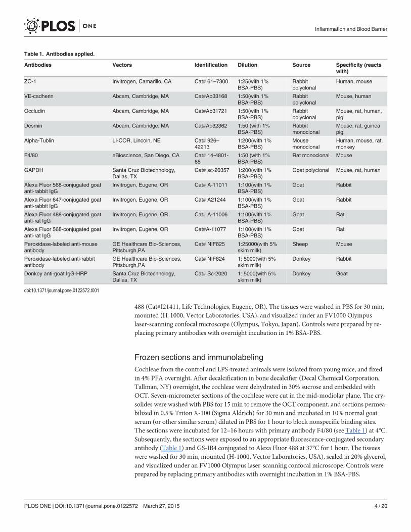
Table 1. Antibodies applied.
Antibodies Vectors Identification Dilution Source Specificity (reactswith)
ZO-1 Invitrogen, Camarillo, CA Cat# 61–7300 1:25(with 1%BSA-PBS)
Rabbitpolyclonal
Human, mouse
VE-cadherin Abcam, Cambridge, MA Cat#Ab33168 1:50(with 1%BSA-PBS)
Rabbitpolyclonal
Mouse, human
Occludin Abcam, Cambridge, MA Cat#Ab31721 1:50(with 1%BSA-PBS)
Rabbitpolyclonal
Mouse, rat, human,pig
Desmin Abcam, Cambridge, MA Cat#Ab32362 1:50 (with 1%BSA-PBS)
Rabbitmonoclonal
Mouse, rat, guineapig,
Alpha-Tublin LI-COR, Lincoln, NE Cat# 926–42213
1:200(with 1%BSA-PBS)
Mousemonoclonal
Human, mouse, rat,monkey
F4/80 eBioscience, San Diego, CA Cat# 14-4801-85
1:50 (with 1%BSA-PBS)
Rat monoclonal Mouse
GAPDH Santa Cruz Biotechnology,Dallas, TX
Cat# sc-20357 1:200(with 1%BSA-PBS)
Goat polyclonal Mouse, rat, human
Alexa Fluor 568-conjugated goatanti-rabbit IgG
Invitrogen, Eugene, OR Cat# A-11011 1:100(with 1%BSA-PBS)
Goat Rabbit
Alexa Fluor 647-conjugated goatanti-rabbit IgG
Invitrogen, Eugene, OR Cat# A21244 1:100(with 1%BSA-PBS)
Goat Rabbit
Alexa Fluor 488-conjugated goatanti-rat IgG
Invitrogen, Eugene, OR Cat# A-11006 1:100(with 1%BSA-PBS)
Goat Rat
Alexa Fluor 568-conjugated goatanti-rat IgG
Invitrogen, Eugene, OR Cat#A-11077 1:100(with 1%BSA-PBS)
Goat Rat
Peroxidase-labeled anti-mouseantibody
GE Healthcare Bio-Sciences,Pittsburgh,PA
Cat# NIF825 1:25000(with 5%skim milk)
Sheep Mouse
Peroxidase-labeled anti-rabbitantibody
GE Healthcare Bio-Sciences,Pittsburgh,PA
Cat# NIF824 1: 5000(with 5%skim milk)
Donkey Rabbit
Donkey anti-goat IgG-HRP Santa Cruz Biotechnology,Dallas, TX
Cat# Sc-2020 1: 5000(with 5%skim milk)
Donkey Goat
doi:10.1371/journal.pone.0122572.t001
Inflammation and Blood Barrier
PLOS ONE | DOI:10.1371/journal.pone.0122572 March 27, 2015 4 / 20
Cell stainingPurified primary cultured PVM/Ms at passage 3 were seeded in a glass well dish, and treatedwith LPS at 1μg/ml for 48 hours. The PVM/Ms were fixed in 4% PFA (pH 7.2) for 15 min, per-meabilized in 0.5% Triton X-100 in PBS-BSA for 3 min, incubated with 10% goat serum, andincubated overnight at 4°C with primary antibody for rat monoclonal F4/80 (eBioscience, SanDiego, CA) diluted 1:50 in 1% PBS-bovine serum albumin. After 3 washes in PBS, the sampleswere incubated with secondary antibodies, Alexa Fluor 568 goat anti-rat IgG (H+L) (Life Tech-nologies, Eugene, OR) or lectin Griffonia simplicifolia IB4 (GS-IB4) conjugated to Alexa Fluor488 (Life Technologies, Eugene, OR) diluted in 1% PBS-bovine serum albumin at 1:100 for 1hour at room temperature. The cells were washed in 2 ml PBS (3 times for 10 min) and imagedunder an FV1000 Olympus laser-scanning confocal microscope.
Purified PCs transfected with pmOrange2-N1 Vector (Clontech Laboratories, Inc., Moun-tain View, CA) were seeded in the glass well dish, and treated with LPS at 1 μg/ml for 48 hours,and imaged under an FV1000 Olympus laser-scanning confocal microscope with a standard559 nm laser excitation line.
Reverse transcription-polymerase chain reaction (RT-PCR) and real-time quantitative RT-PCR (qRT-PCR)The procedure used for quantitative real-time PCR was described previously [28]. For the invitromodels, total RNA from cultured ECs from different groups was extracted separately usingRNeasy (Qiagen, Valencia, CA, USA) per the manufacturer's recommendations. For the in vivomodels, total RNA from strial capillaries of control and LPS-treated groups was extracted sepa-rately using RNeasy (QIAGEN). Each group of 5 mice was analyzed for ZO-1, occludin, ve-cad-herin, and Gapgh mRNA with qRT-PCR. The sample for total RNA was reverse transcribedwith a RETROscript kit (Invitrogen, USA). CDNA synthesized from total RNA was diluted10-fold with DNase-free water, each cDNA sample independently measured 3 times. Transcriptswere quantitated by gene expression assay (Invitrogen): ZO-1 (Mm00493699_m1), occludin(Mm00500912_m1), and ve-cadherin (Mm03053719_s1) on a model 7300 real-time PCR sys-tem (Applied Biosystems, Foster City, CA, USA). The real-time PCR was cycled at 95°C for 20 s,40 cycles at 95°C for 1 s, and 60°C for 20 s. Mouse Gapdh was the endogenous control. Quantita-tive PCR was performed per the guidelines provided by Applied Biosystems and analyzed usingthe comparative cycle threshold method.
Transmission electron microscopy (TEM)The temporal bones were isolated, and the cochlea perfused through the round window. Thiswas followed by immersion in a fixative of 4% (wt/vol) paraformaldehyde -0.1% (vol/vol) glu-taraldehyde in 0.1 M phosphate-buffer overnight. Stria vascularis tissues were dissected andpost fixed in 1% osmium (Electron Microscopy Sciences, Hatfield, PA). Tissues were dehy-drated with a graded alcohol series and embedded in Embed 812 (Electron Microscopy Sci-ences, Hatfield, PA), sectioned, stained with lead citrate (Electron Microscopy Sciences,Hatfield, PA) and uranyl acetate (Electron Microscopy Sciences, Hatfield, PA), and viewed ona Philips CM 100 transmission electron microscope (Philips/FEI Corporation, Eindhoven,Holland).
Western blotTotal protein (50 μg) from each sample was added to a 10% sodium dodecyl sulfate–polyacryl-amide gel to detect ZO-1, occludin, vascular endothelial cadherin (ve-cadherin), and a-tubulin.
Inflammation and Blood Barrier
PLOS ONE | DOI:10.1371/journal.pone.0122572 March 27, 2015 5 / 20
Proteins were electrophoretically transferred to PVDF membranes and blocked with nonfatmilk for 1 hour at room temperature. Specific immunodetection was carried out by incubationwith primary antibodies, either anti-ZO-1 antibody diluted 1:50, or anti-occudin 1:100 andanti-ve-cadherin antibody diluted (1:250) in skim milk overnight at 4°C. After 3 washes withTBST, the membranes were incubated for 1 hour with secondary antibody (information on theantibodies can be found in Table 1), and antigens were assessed using ECL plus Western blot-ting Detection Reagents (Amersham, Arlington Heights, IL, USA).
In-cell Western blottingECs were seeded at the same concentration (1.3×10⁵ cells/ml, 100 μl per well) in a 96-well plateand incubated until the cells consistently adhered to the bottom of the plate. For the LPSgroup, the cells were incubated with LPS at 1μg/ml for 48 hours. The cells were fixed in 4%formaldehyde for 20 min at room temperature. The cells were permeabilized with 1×PBS con-taining 0.1% Triton X-100, blocked in blocking buffer (927–40000,LI-COR Biosciences, Lin-coln, NE, USA) for 90 min, and incubated with primary antibodies for ZO-1, occludin, ve-cadherin, and α-Tublin (Table 1) for 2 hours. The cells were then washed 4 times for 5 min inPBS containing 0.1% Tween-20 and incubated with 1:200 secondary antibodies including don-key anti-mouse IRDye 800CW (926–32212 LI-COR, USA) and donkey anti-rabbit IRDye680RD (926–68073, LI-COR, USA) for 1 hour at room temperature. Cells were washed again3 times in PBS. Stained cells were imaged with an Odyssey Imager (Li-COR Biosciences)and analyzed.
Immunolabeling of TJ-associated proteinsPurified endothelial cells at passage 3 and at a density of 4×10⁴/cm2 were seeded and grown ona glass well dish until an endothelial cell monolayer formed. The cells were incubated with LPSat 1 μg/ml for 24 hours. The cells were then fixed in 4% PFA (pH 7.2) for 15 min at room tem-perature and washed in 2 ml PBS (3 times for 10 min). The cells were permeabilized in 0.5%Triton X-100 in PBS-BSA for 3 min at room temperature, washed with 2 ml PBS (three timesfor 10 min), and incubated with an immunofluorescence blocking solution at room tempera-ture. The cells were incubated with primary antibodies (Table 1) diluted in a 1% PBS-BSA solu-tion overnight at 4°C, washed in 2 ml PBS (three times for 10 min), and incubated withsecondary antibodies (Table 1) diluted in 1% BSA-PBS solution for 1 hour at room tempera-ture. The cells were washed in 2 ml PBS (3 times for 10 min) and imaged under a confocal laserscanning microscope.
Assessment of vascular permeability in vivoVascular permeability in the control and LPS treated group was assessed using a fluoresceinisothiocyanate-conjugated bovine albumin tracer (FITC-albumin, 66 kDa, A-9771; Sigma,Cream Ridge, NJ, USA). The tracer was intravenously administered to the tail vein of anesthe-tized animals 30 min prior to transcardiac perfusion with PBS. The stria vascularis was re-moved and homogenized in 1% Triton X-100 in PBS, and the lysate centrifuged at 13,000 rpmfor 20 min. Relative fluorescence of the supernatant was measured on a TecanGENios Plusmicroplate reader (Tecan Group Ltd, San Jose, CA, USA).
Vascular permeability in vivo was also assessed by recording tracer movement through aspecially prepared vessel window, as described previously [29]. Animals were anesthetized andwrapped in a heating pad, with rectal temperature maintained at 38.5°C. A lateral and ventralapproach was used to open the left bulla and create a rectangular fenestration (0.1×0.1 mm) inthe cochlea at the basal turn. FITC-conjugated to bovine albumin was slowly administrated
Inflammation and Blood Barrier
PLOS ONE | DOI:10.1371/journal.pone.0122572 March 27, 2015 6 / 20
intravenously to the mouse at a concentration of 40 mg/ml in 0.1 ml physiological solution for10 min prior to recording.
Permeability assayECs (5×10⁵) were seeded on the insert of a Transwell filter (Coring 3460, 0.4 μm pore size) in24-well dishes and grown 2–3 days until they reached confluence. After treatment with LPS(1 μg/ml), FITC–dextran (FD10S, Sigma, USA) was added to the upper chamber at a final con-centration of 1mg/mL. After 30 min of incubation at 37°C, 50 μL samples were taken from thelower chamber of each Transwell and transferred to a 96-well plate for fluorescence measure-ments. The fluorescent content of the samples was measured using a fluorescence plate readeroptically filtered for 485nm excitation and 535 nm emission [30].
StatisticsData, presented as means ± SD, were evaluated using the Student's t-test for comparison of twogroups or ANOVA for comparisons of three or more groups. A 95% confidence level was con-sidered statistically significant.
Results
Evaluation of the LPS-induced mouse acute otitis media (OM) modelThe tympanic membrane, middle ear mucosa, and inflammatory cell infiltration in the middleear were observed in the LPS-induced OMmouse model.
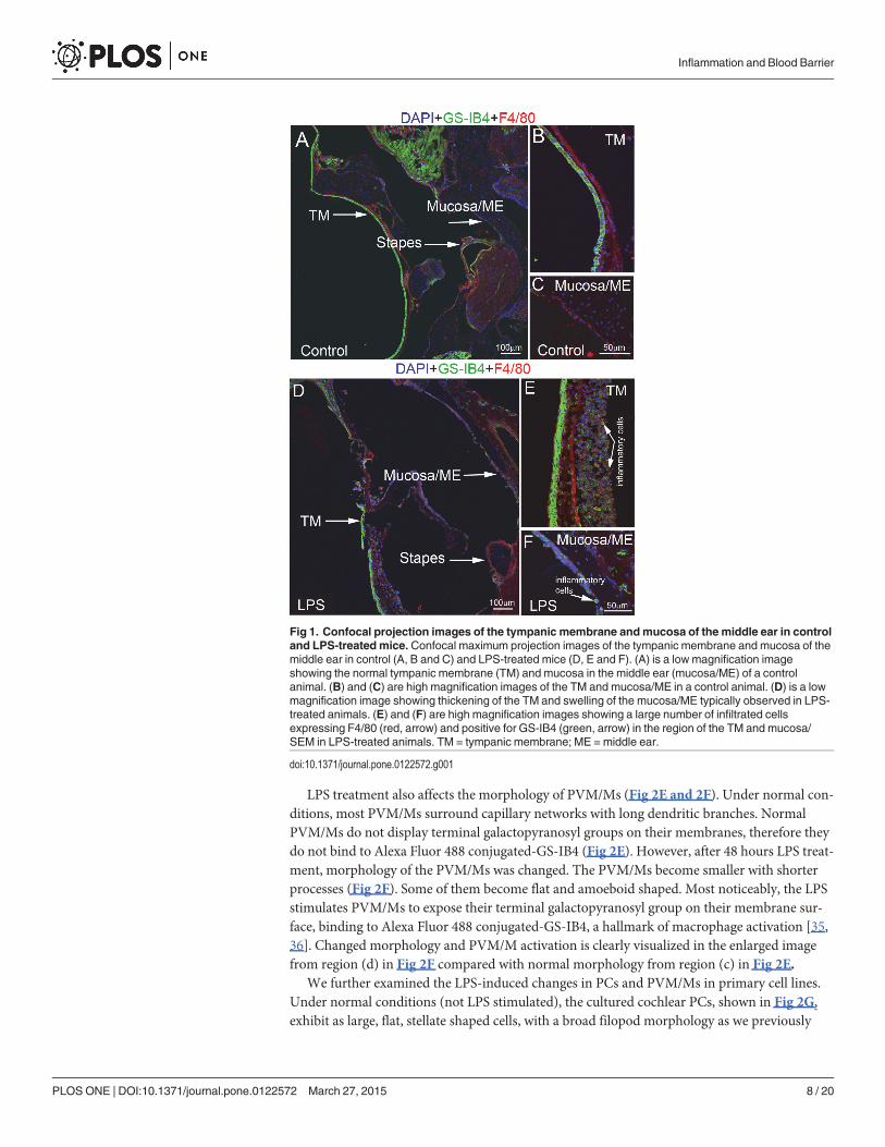
Under normal conditions, the middle ear cavity was clear of inflammation (Fig 1A). Thetympanic membrane and middle ear mucosa were much thinner than in the controls as shownin Figs 1A(low magnification) and 1B and 1C(high magnification). In contrast, the tympanicmembrane and mucosa of the middle ear in the LPS-treated ears were much thicker, bothshowing infiltration of GS-IB4 positive and GS-IB4 negative F4/80 positive inflammatory cellsas shown in Fig 1D. Fig 1E and 1Ffurther display views of the bulkier tympanic membraneand mucosa of the middle ear as well as infiltration of F4/80 positive inflammatory cells in theLPS-treated controls under high magnification.
LPS affects the structure of the intra-strial fluid–blood barrierCompared with non-LPS-treated animal (Fig 2A), a slight endolymphatic hydrops was noticedin the LPS-treated animals (Fig 2B). In addition to causing hearing defects and slightly abnor-mal lymphatic volume, LPS also disrupts accessory component cells and causes changes in thestructure of the intra-strial fluid–blood barrier.
The primary accessory cells in the intra-strial fluid–blood barrier are PCs and PVM/Ms [18,27, 31, 32]. PCs and PVM/Ms are essential for maintaining the physical stability and functionalintegrity of the intra-strial fluid–blood barrier [25]. In this study, we observed how PCs re-spond to LPS treatment in a transgenic mouse model, with the PCs expressing a bright redfluorescence variant (NG2DsRed.T1) under the control of a mouse NG2 (Cspg4) promoter[33]. Under normal conditions, PCs (NG2 positive) exhibit a flat and slender morphology,tightly associated with strial capillaries (Fig 2C). Changed morphology is clearly visualized inthe enlarged image from region (a) in Fig 2C.Morphological changes are seen in the PCs after48 hours of LPS treatment. The LPS-treated PCs display a prominent round body, and arefound peeling away from the capillary wall, previously described as a sign of PC migration[34]. In addition to the migration, the LPS-induced the PCs to release a great number of un-known particles (Fig 2D), shown in an enlarged image (b) from Fig 2D.
Inflammation and Blood Barrier
PLOS ONE | DOI:10.1371/journal.pone.0122572 March 27, 2015 7 / 20
LPS treatment also affects the morphology of PVM/Ms (Fig 2E and 2F). Under normal con-ditions, most PVM/Ms surround capillary networks with long dendritic branches. NormalPVM/Ms do not display terminal galactopyranosyl groups on their membranes, therefore theydo not bind to Alexa Fluor 488 conjugated-GS-IB4 (Fig 2E). However, after 48 hours LPS treat-ment, morphology of the PVM/Ms was changed. The PVM/Ms become smaller with shorterprocesses (Fig 2F). Some of them become flat and amoeboid shaped. Most noticeably, the LPSstimulates PVM/Ms to expose their terminal galactopyranosyl group on their membrane sur-face, binding to Alexa Fluor 488 conjugated-GS-IB4, a hallmark of macrophage activation [35,36]. Changed morphology and PVM/M activation is clearly visualized in the enlarged imagefrom region (d) in Fig 2F compared with normal morphology from region (c) in Fig 2E.
We further examined the LPS-induced changes in PCs and PVM/Ms in primary cell lines.Under normal conditions (not LPS stimulated), the cultured cochlear PCs, shown in Fig 2G,exhibit as large, flat, stellate shaped cells, with a broad filopod morphology as we previously
Fig 1. Confocal projection images of the tympanic membrane andmucosa of the middle ear in controland LPS-treated mice. Confocal maximum projection images of the tympanic membrane and mucosa of themiddle ear in control (A, B and C) and LPS-treated mice (D, E and F). (A) is a low magnification imageshowing the normal tympanic membrane (TM) and mucosa in the middle ear (mucosa/ME) of a controlanimal. (B) and (C) are high magnification images of the TM and mucosa/ME in a control animal. (D) is a lowmagnification image showing thickening of the TM and swelling of the mucosa/ME typically observed in LPS-treated animals. (E) and (F) are high magnification images showing a large number of infiltrated cellsexpressing F4/80 (red, arrow) and positive for GS-IB4 (green, arrow) in the region of the TM and mucosa/SEM in LPS-treated animals. TM = tympanic membrane; ME = middle ear.
doi:10.1371/journal.pone.0122572.g001
Inflammation and Blood Barrier
PLOS ONE | DOI:10.1371/journal.pone.0122572 March 27, 2015 8 / 20
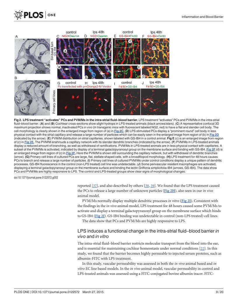
reported [37], and also described by others [38, 39]. We found that the LPS treatment causedthe PCs to release a large number of unknown particles (Fig 2H), also seen in our in vivoanimal model.
PVM/Ms normally display multiple dendritic processes in vitro (Fig 2I). Consistent withthe findings in the in vivo animal model, LPS treatment for 48 hours caused some PVM/Ms toactivate and display a terminal galactopyranosyl group on the membrane surface which bindsto GS-IB4 (Fig 2J). GS-IB4 binding was undetectable in control (non-LPS treated) cell lines.
The data show that PCs and PVM/Ms are highly responsive to LPS.
LPS induces a functional change in the intra-strial fluid–blood barrier invivo and in vitroThe intra-strial fluid–blood barrier restricts molecular transport from the blood into the ear,and is essential for maintaining cochlear homeostasis under normal conditions [22]. In thisstudy, we found that the barrier becomes highly permeable to injected serum proteins, such asalbumin-FITC with LPS treatment.
In this study, vascular permeability was assessed in both the in vivo animal based and invitro EC line based models. In the in vivo animal model, vascular permeability in control andLPS-treated animals was assessed using a FITC-conjugated bovine albumin tracer. FITC-
Fig 2. LPS treatment “activates” PCs and PVM/Ms in the intra-strial fluid–blood barrier. LPS treatment “activates” PCs and PVM/Ms in the intra-strialfluid–blood barrier. (A) and (B) Cochlear cross-sections show slight hydrops in LPS-treated animals (black arrows/stars). (C) A representative confocal 3Dmaximum projection shows normal, inactivated PCs in vivo (in transgenic mice with fluorescent labeled NG2, red) to have a flat and slender cell body. Thecell morphology is clearly shown in the enlarged image from region of (a) in Fig 2C. (D) LPS stimulated PCs display a “prominent round” cell body in lessphysical contact with the strial capillary and release a large number of particles which can be easily seen in the enlarged image from region of (b) in Fig 2D(indicated by the arrow). (E) PVM/M distribution on strial capillaries, shown labeled with GS-IB4 in a control animal. Fig E (c) is an enlarged image from regionof (c) in Fig 2E. The PVM/M enshrouds a capillary network with its slander dendritic branches (indicated by the arrow). (F) PVM/Ms in LPS-treated animalsdisplay a reduced amount of branching, as well as withdrawal of ramifications. PVM/Ms in LPS-treated animals are in less physical contact with capillaries. Asubset of the PVM/Ms is activated, indicated by display of a terminal galactopyranosyl group on the membrane surface and binding with GS-IB4. Fig 2F (d) isan enlarged image from region of (c) in Fig 2F. Here the PVM/M is shown still surrounding the capillary network, but with withdrawal of dendritic branches(arrow). (G) Primary cell lines of cultured PCs are large, flat, stellate shaped cells, with a broadfilopod morphology. (H) LPS treatment for 48 hours causesPCs to branch and release a large number of particles. (I) Primary cell lines of cultured PVM/Ms under control conditions display a unique pattern of dendriticprocesses. GS-IB4 fluorescence in the control (non-LPS treated) cell line was undetectable. (J) Some perivascular resident macrophages are activated,displaying a terminal galactopyranosyl group on the membrane surface and binding the lectin Griffonia simplicifolia-IB4 (arrows, GS-IB4). The data showPCs and PVM/Ms are highly responsive to LPS. The control and LPS-treated groups show clear signs of morphological changes.
doi:10.1371/journal.pone.0122572.g002
Inflammation and Blood Barrier
PLOS ONE | DOI:10.1371/journal.pone.0122572 March 27, 2015 9 / 20
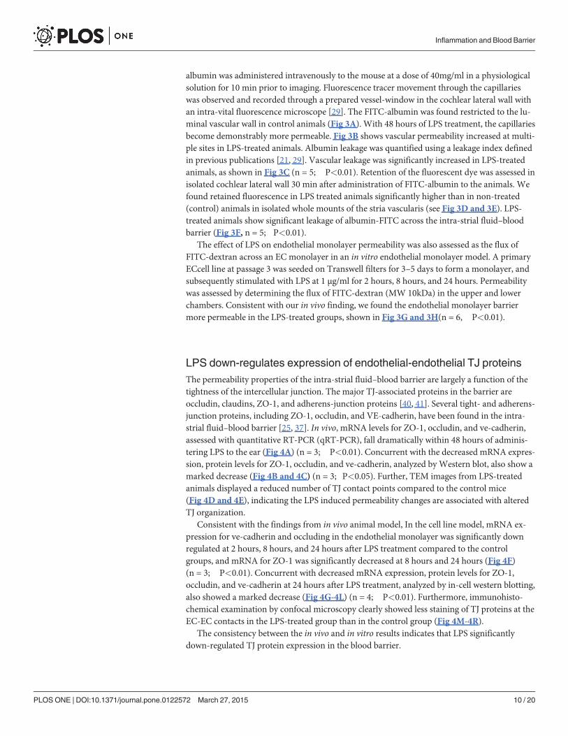
albumin was administered intravenously to the mouse at a dose of 40mg/ml in a physiologicalsolution for 10 min prior to imaging. Fluorescence tracer movement through the capillarieswas observed and recorded through a prepared vessel-window in the cochlear lateral wall withan intra-vital fluorescence microscope [29]. The FITC-albumin was found restricted to the lu-minal vascular wall in control animals (Fig 3A). With 48 hours of LPS treatment, the capillariesbecome demonstrably more permeable. Fig 3B shows vascular permeability increased at multi-ple sites in LPS-treated animals. Albumin leakage was quantified using a leakage index definedin previous publications [21, 29]. Vascular leakage was significantly increased in LPS-treatedanimals, as shown in Fig 3C (n = 5; ��P<0.01). Retention of the fluorescent dye was assessed inisolated cochlear lateral wall 30 min after administration of FITC-albumin to the animals. Wefound retained fluorescence in LPS treated animals significantly higher than in non-treated(control) animals in isolated whole mounts of the stria vascularis (see Fig 3D and 3E). LPS-treated animals show significant leakage of albumin-FITC across the intra-strial fluid–bloodbarrier (Fig 3F, n = 5;��P<0.01).
The effect of LPS on endothelial monolayer permeability was also assessed as the flux ofFITC-dextran across an EC monolayer in an in vitro endothelial monolayer model. A primaryECcell line at passage 3 was seeded on Transwell filters for 3–5 days to form a monolayer, andsubsequently stimulated with LPS at 1 μg/ml for 2 hours, 8 hours, and 24 hours. Permeabilitywas assessed by determining the flux of FITC-dextran (MW 10kDa) in the upper and lowerchambers. Consistent with our in vivo finding, we found the endothelial monolayer barriermore permeable in the LPS-treated groups, shown in Fig 3G and 3H(n = 6, ��P<0.01).
LPS down-regulates expression of endothelial-endothelial TJ proteinsThe permeability properties of the intra-strial fluid–blood barrier are largely a function of thetightness of the intercellular junction. The major TJ-associated proteins in the barrier areoccludin, claudins, ZO-1, and adherens-junction proteins [40, 41]. Several tight- and adherens-junction proteins, including ZO-1, occludin, and VE-cadherin, have been found in the intra-strial fluid–blood barrier [25, 37]. In vivo, mRNA levels for ZO-1, occludin, and ve-cadherin,assessed with quantitative RT-PCR (qRT-PCR), fall dramatically within 48 hours of adminis-tering LPS to the ear (Fig 4A) (n = 3; ��P<0.01). Concurrent with the decreased mRNA expres-sion, protein levels for ZO-1, occludin, and ve-cadherin, analyzed by Western blot, also show amarked decrease (Fig 4B and 4C) (n = 3; �P<0.05). Further, TEM images from LPS-treatedanimals displayed a reduced number of TJ contact points compared to the control mice(Fig 4D and 4E), indicating the LPS induced permeability changes are associated with alteredTJ organization.
Consistent with the findings from in vivo animal model, In the cell line model, mRNA ex-pression for ve-cadherin and occluding in the endothelial monolayer was significantly downregulated at 2 hours, 8 hours, and 24 hours after LPS treatment compared to the controlgroups, and mRNA for ZO-1 was significantly decreased at 8 hours and 24 hours (Fig 4F)(n = 3; ��P<0.01). Concurrent with decreased mRNA expression, protein levels for ZO-1,occludin, and ve-cadherin at 24 hours after LPS treatment, analyzed by in-cell western blotting,also showed a marked decrease (Fig 4G-4L) (n = 4; ��P<0.01). Furthermore, immunohisto-chemical examination by confocal microscopy clearly showed less staining of TJ proteins at theEC-EC contacts in the LPS-treated group than in the control group (Fig 4M-4R).
The consistency between the in vivo and in vitro results indicates that LPS significantlydown-regulated TJ protein expression in the blood barrier.
Inflammation and Blood Barrier
PLOS ONE | DOI:10.1371/journal.pone.0122572 March 27, 2015 10 / 20
Fig 3. LPS treatment increases vascular leakage. LPS treatment increases vascular leakage. (A) Under normal conditions, the FITC-albumin is restrictedto the vascular wall of the stria vascularis, and control animals show no sites of obvious leakage. (B) In LPS-treated animals, however, capillaries arepermeable and multiple sites of vascular leakage are seen in the vessel of the stria vascularis. (C) Significantly more leakage of albumin-FITC is seen in LPS-treated animals than in control animals (**P<0.01). (D) Retention of FITC-albumin in isolated whole mounts of untreated cochlear stria vascularis is relativelylow in non-LPS treated mice, and (E) retention in the LPS-treated mice is much stronger. (F) FITC-albumin fluorescence is significantly higher in the LPS-treated group than in control animals (**P<0.01). (G) and (H) The graph shows the effect of LPS on the permeability of tight junctions in an ECmonolayer toFITC-dextran. Permeability was measured by determining the flux of FITC-dextran from the upper to the lower chamber of the Transwell filter. Data areexpressed as means ± SD (**P<0.01).
doi:10.1371/journal.pone.0122572.g003
Inflammation and Blood Barrier
PLOS ONE | DOI:10.1371/journal.pone.0122572 March 27, 2015 11 / 20
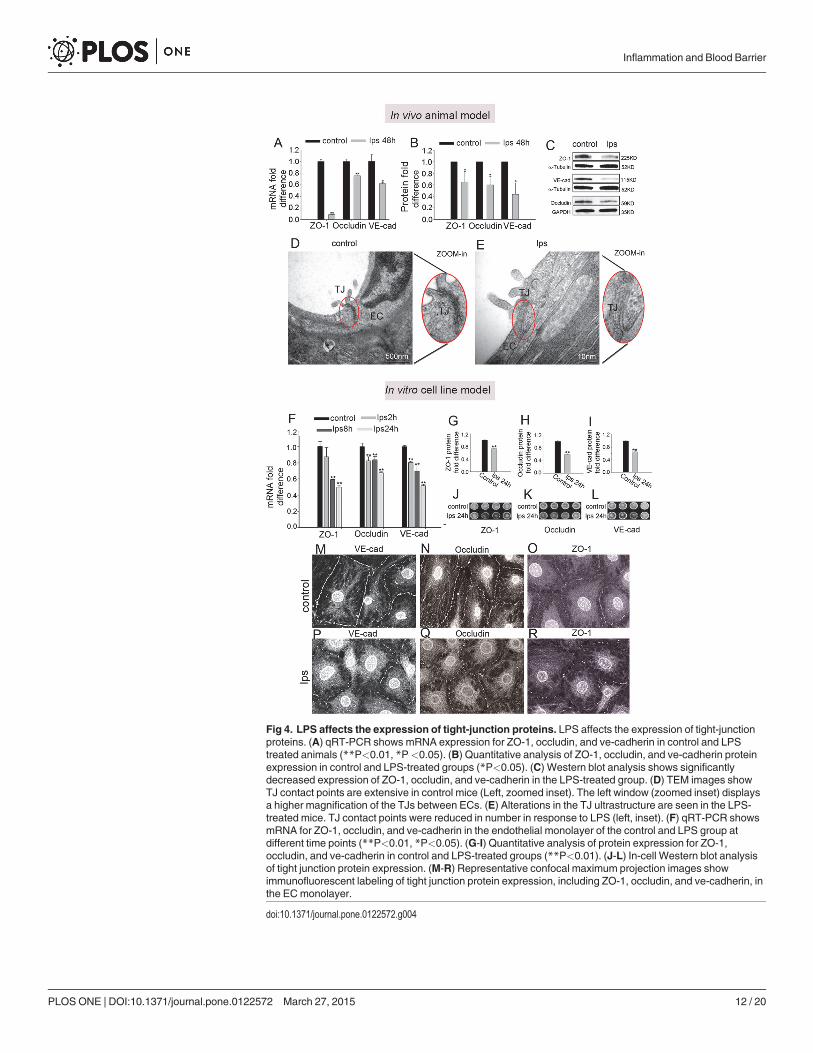
Fig 4. LPS affects the expression of tight-junction proteins. LPS affects the expression of tight-junctionproteins. (A) qRT-PCR shows mRNA expression for ZO-1, occludin, and ve-cadherin in control and LPStreated animals (**P<0.01, *P<0.05). (B) Quantitative analysis of ZO-1, occludin, and ve-cadherin proteinexpression in control and LPS-treated groups (*P<0.05). (C) Western blot analysis shows significantlydecreased expression of ZO-1, occludin, and ve-cadherin in the LPS-treated group. (D) TEM images showTJ contact points are extensive in control mice (Left, zoomed inset). The left window (zoomed inset) displaysa higher magnification of the TJs between ECs. (E) Alterations in the TJ ultrastructure are seen in the LPS-treated mice. TJ contact points were reduced in number in response to LPS (left, inset). (F) qRT-PCR showsmRNA for ZO-1, occludin, and ve-cadherin in the endothelial monolayer of the control and LPS group atdifferent time points (**P<0.01, *P<0.05). (G-I) Quantitative analysis of protein expression for ZO-1,occludin, and ve-cadherin in control and LPS-treated groups (**P<0.01). (J-L) In-cell Western blot analysisof tight junction protein expression. (M-R) Representative confocal maximum projection images showimmunofluorescent labeling of tight junction protein expression, including ZO-1, occludin, and ve-cadherin, inthe ECmonolayer.
doi:10.1371/journal.pone.0122572.g004
Inflammation and Blood Barrier
PLOS ONE | DOI:10.1371/journal.pone.0122572 March 27, 2015 12 / 20
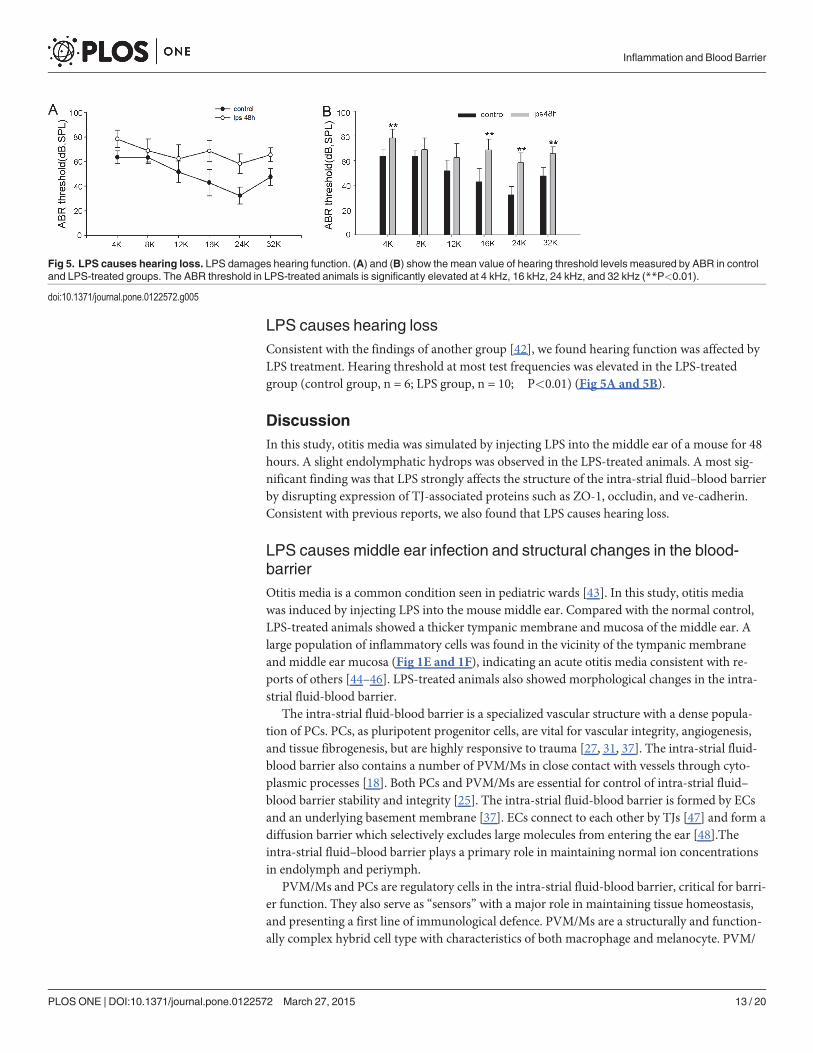
LPS causes hearing lossConsistent with the findings of another group [42], we found hearing function was affected byLPS treatment. Hearing threshold at most test frequencies was elevated in the LPS-treatedgroup (control group, n = 6; LPS group, n = 10; ��P<0.01) (Fig 5A and 5B).
DiscussionIn this study, otitis media was simulated by injecting LPS into the middle ear of a mouse for 48hours. A slight endolymphatic hydrops was observed in the LPS-treated animals. A most sig-nificant finding was that LPS strongly affects the structure of the intra-strial fluid–blood barrierby disrupting expression of TJ-associated proteins such as ZO-1, occludin, and ve-cadherin.Consistent with previous reports, we also found that LPS causes hearing loss.
LPS causes middle ear infection and structural changes in the blood-barrierOtitis media is a common condition seen in pediatric wards [43]. In this study, otitis mediawas induced by injecting LPS into the mouse middle ear. Compared with the normal control,LPS-treated animals showed a thicker tympanic membrane and mucosa of the middle ear. Alarge population of inflammatory cells was found in the vicinity of the tympanic membraneand middle ear mucosa (Fig 1E and 1F), indicating an acute otitis media consistent with re-ports of others [44–46]. LPS-treated animals also showed morphological changes in the intra-strial fluid-blood barrier.
The intra-strial fluid-blood barrier is a specialized vascular structure with a dense popula-tion of PCs. PCs, as pluripotent progenitor cells, are vital for vascular integrity, angiogenesis,and tissue fibrogenesis, but are highly responsive to trauma [27, 31, 37]. The intra-strial fluid-blood barrier also contains a number of PVM/Ms in close contact with vessels through cyto-plasmic processes [18]. Both PCs and PVM/Ms are essential for control of intra-strial fluid–blood barrier stability and integrity [25]. The intra-strial fluid-blood barrier is formed by ECsand an underlying basement membrane [37]. ECs connect to each other by TJs [47] and form adiffusion barrier which selectively excludes large molecules from entering the ear [48].Theintra-strial fluid–blood barrier plays a primary role in maintaining normal ion concentrationsin endolymph and periymph.
PVM/Ms and PCs are regulatory cells in the intra-strial fluid-blood barrier, critical for barri-er function. They also serve as “sensors” with a major role in maintaining tissue homeostasis,and presenting a first line of immunological defence. PVM/Ms are a structurally and function-ally complex hybrid cell type with characteristics of both macrophage and melanocyte. PVM/
Fig 5. LPS causes hearing loss. LPS damages hearing function. (A) and (B) show the mean value of hearing threshold levels measured by ABR in controland LPS-treated groups. The ABR threshold in LPS-treated animals is significantly elevated at 4 kHz, 16 kHz, 24 kHz, and 32 kHz (**P<0.01).
doi:10.1371/journal.pone.0122572.g005
Inflammation and Blood Barrier
PLOS ONE | DOI:10.1371/journal.pone.0122572 March 27, 2015 13 / 20
Ms are highly responsive to a variety of toxic stimuli including ischemia and infection. Previ-ously we demonstrated that PVM/Ms are extremely sensitive to loud sound, and undergo dra-matic morphological and functional changes in response to loud sound stimulation. An earlystudy by Gratton [49] also showed marked increased activity in melanin-bearing cells of theinner ear in response to the stress of LPS-induced middle ear infection. In this study, thePVM/Ms are shown to change their morphology and are activated in response to LPS-inducedmiddle ear infection (Fig 1). In brain research, activation of astrocytes (or microglia) is a majorfactor prolonging the release of pro-inflammatory cytokines in the brain [50]. Targeting (sup-pressing) astrocyte (or microglia) activation attenuates the cytokine release. LPS-triggered acti-vation of PVM/Ms, by its effect on extending cytokine production, could also be extendingLPS-induced inner ear inflammation.
PCs, as “the second line of defense” [51], are in close proximity to ECs, and critical forblood barrier integrity [52, 53]. Normally, PCs are embedded in the basement membrane. PCmigration, resulting in a break away from the basement membrane and vessels, has been seenin the brain and retina in response to various pathogens [54, 55]. Our previous loud soundstudy showed the PCs vulnerable and sensitive to the stress of acoustic trauma. Upon exposureto loud sound, PCs migrate from points of normal endothelial attachment [27] and destabilizethe BLB in the stria vascularis. Consistent with the PC pathology seen with loud sound stimula-tion, PCs migrate from the vessel wall and also release a large number of unknown particleswhen exposed to LPS for 24 hours.
A recent study of PCs in the brain and lung shows the PCs to be a major source of cytokines.They release (secrete) multiple cytokines, including Interleukin (IL)-1 alpha, IL-5, IL-6, Granu-locyte-colony stimulating factor (G-CSF), keratinocyte-derived cytokine (KC), monocyte che-motactic protein 1 (MCP1), and RANTES (a member of the IL-8 superfamily of cytokines)when exposed to LPS [56, 57]. PCs are also reported to release transforming growth factor-beta(TGF-β) in response to glycation end-products [58]. It is unknown whether the particles secret-ed by cochlear PCs on exposure to LPS are cytokines. LPS-induced inflammation in the co-chlea, including recruitment of inflammatory neutrophils, was previously reported by differentlaboratories [7, 59]. Do the particles released by cochlear PCs mediate LPS-induced recruit-ment of inflammatory neutrophils in the stria vascularis?
The specific signals underlying migration of cochlear PCs and PVM/M activation are un-known, as are the sequelae of the migration and activation. The mechanisms governing PC mi-gration and tissue resident macrophage activation are extremely complex, and multiplesignaling pathways, including various cytokines such as vascular endothelial growth factor(VEGF), PDGF-β, and TGF-β are reported to be involved [60–63]. LPS is known to have di-verse effects on the inflammatory cascade through activation of Toll-like receptor 4 (TLR-4)[64, 65]. These receptors are expressed in the inner ear by immune cells such as macrophages,and by non-immune cells, including lateral wall fibroblasts and epithelial cells [13]. TLR-4 sig-naling activates nuclear factor kappa B, the “master switch” for inflammation-mediated cyto-kine production, and releases mediators such as interleukin-1 (IL-1), IL-2, tumor necrosisfactor alpha (TNF-α), and TGF-β, all important cytokines and modulators of the inflammatoryresponse [63, 66]. These receptors might be triggering PC migration and PVM/M activation.
LPS breaks down the blood barrier by down-regulating expression of TJproteinsLike in other barrier systems, TJs form the intra-strial fluid–blood barrier which preventstransport into the ear [67]. The major TJ-associated proteins in the barrier are occludin, clau-dins, ZO-1, and adherens-junction proteins [40, 41]. Several of these tight- and adherens-
Inflammation and Blood Barrier
PLOS ONE | DOI:10.1371/journal.pone.0122572 March 27, 2015 14 / 20
junction proteins, including ZO-1, occludin, and ve-cadherin have previously been identifiedin the intra-strial fluid–blood barrier [24, 25]. Each of the proteins plays a unique molecularand regulatory role in maintaining TJ structure and function. Occludin, an integral plasma-membrane protein in the TJs of epithelial cells, is essential for the integrity of the intra-strialfluid-blood barrier [37]. ZO-1, on the other hand, is a recognition protein for TJ placement,and loss of ZO-1 results in disorganization of TJs [68–70]. Ve-cadherin is an endothelial cellspecific adhesion molecule located at junctions between ECs which moderates EC contacts andaffects the EC through outside-in signaling [71].
In this study, we demonstrated that LPS disrupts the BLB integrity in in vivo animal-basedand in vitro cell line-based models by significantly down-regulating the expression of TJ pro-teins at both the transcript and protein levels. In support of this evidence, we found lessimmuno-staining for occludin, ZO-1, and ve-cadherin proteins in EC contacts of the LPS-treated group and loosened TJs between ECs (confirmed by TEM study of the ultra-structure).Further functional studies have shown EC monolayers more permeable with LPS treatment,corroborating the significantly increased BLB vascular permeability seen in LPS-treated animalmodels.
LPS disruption of TJs could be due to a number of factors: (1) LPS may have a direct detri-mental effect on the barrier via cytokines. A number of studies have shown that LPS causes di-rect damage to the blood barrier by breaking down TJs. This has been shown in organs such asthe gut and milk-blood barrier to be mediated by LPS-induced cytokines such as toll-like recep-tor 4 (TLR4) signaling [64, 72]. (2) LPS may cause damage by inducing PC migration. PCs areextensively branched and tightly embrace the abluminal endothelium wall. PC interaction withthe endothelium is vital for vascular development, blood flow regulation, and vascular integrity(vascular permeability) [73–77]. PC pathology leads to vascular dysfunction, seen in many dis-eases. Evidence is accumulating that PCs control vascular stability by up-regulating TJ proteinexpression, and by suppressing vesicular transport in the endothelium [78]. PC pathologycaused vessel instability and vascular leakage are seen in the brain and other organs [78]. (3)LPS causes damage by inducing PVM/M activation. PVM/Ms in the intra-strial fluid-bloodbarrier maintain barrier integrity by controlling TJ and adhesive junction protein expression[25]. In particular, in a previous report, we demonstrated that PVM/Ms produce pigment epi-thelium growth factor (PEDF), a 50-kDa glycoprotein first identified in retinal pigment epithe-lium cells [21, 25], and essential for stabilizing the intra-strial fluid–blood barrier. PVM/Mactivation and changes in PVM/M phenotype leads to decreased production of PEDF, and toblood barrier breakdown and tissue edema.
LPS effects on hearing functionNormal function of the intra-strial fluid-blood barrier requires blood barrier integrity, includ-ing intact TJs in the endothelial layer [72]. Breakdown of the EC-EC junction leads to vascularleakage (Fig 3). Consistent with earlier reports [79–81], our data showed an elevated hearingthreshold 48 hours after LPS was administered to the middle ear. A hearing loss of ~30 dB-SPLin high frequencies is seen relative to controls.
Several factors may be contributing to the hearing loss. (1) LPS may be directly damagingthe sensory hair cells. LPS, a product of bacteriolysis, has been demonstrated to damage innerand outer hair cell bundles and to cause the tectorial membrane to swell [7]. (2) LPS has beenshown to induce increased cytokine production and reactive free radical damage. Increased cy-tokine production by LPS in the cochlea was previously demonstrated by Juhn et al. [13] andQuintanilla-Dieck et al. [8]. LPS-induced production of free radicals such as nitric oxide wasreported as well by our lab and by other labs [82–84]. In particular, LPS causes inducible nitric
Inflammation and Blood Barrier
PLOS ONE | DOI:10.1371/journal.pone.0122572 March 27, 2015 15 / 20
oxide synthase (iNOS) expression in various cochlear cells, including inner and outer hair cells,pillar cells, hair cell nerve fibers, and marginal cells. (3) LPS may cause a deficiency in cochlearblood flow. Transduction of sound is metabolically demanding, and normal function of the mi-crovasculature in the lateral wall is critical for maintaining ion transport and fluid balance [85–88]. Disruption of the blood-barrier in the stria vascularis causes a shortage of nutrients andoxygen in the tissue and creates a “toxic” environment with accumulation of harmful metabo-lites. In addition, leakage of blood-barrier components into the lateral wall of the cochlea withincreased blood-barrier permeability may be related to sensorineural hearing loss. Juhn et al.[13] suggested the leakage may hinder K+ recycling through the lateral wall, causing an intra-strial electric shunt which disrupts the endocochlear potential EP as reported by Cohen-Salm-on et al. [89].
ConclusionsIn summary, our data show that LPS not only induces middle ear infection, but also causesstructural changes in the intra-strial fluid–blood barrier. LPS is shown to have strong effects onPCs and PVM/Ms in the barrier, causing PCs to migrate and release particles, and activation ofPVM/Ms. LPS also causes a significant down-regulation of TJ-associated protein expression.Down-regulation of ZO-1, occludin, and ve-cadherin weaken the intra-strial fluid-blood barri-er and subsequently leads to blood barrier leakage. The exact mechanisms, however, underlyingPC migration and secretion of particles and PVM/M activation are yet to be determined.
Author ContributionsConceived and designed the experiments: JHZ MMD XS. Performed the experiments: JHZSLC JC ZQH. Analyzed the data: JHZ SLC. Contributed reagents/materials/analysis tools: JHZXS. Wrote the paper: XS JHZ.
References1. van Zon A, van der Heijden GJ, van Dongen TM, Burton MJ, Schilder AG. Antibiotics for otitis media
with effusion in children. Cochrane Database Syst Rev. 2012; 9:CD009163.
2. Park CW, Han JH, Jeong JH, Cho SH, Kang MJ, Tae K, et al. Detection rates of bacteria in chronic otitismedia with effusion in children. J Korean Med Sci. 2004; 19:735–8. PMID: 15483353
3. Khoramrooz SS, Mirsalehian A, Emaneini M, Jabalameli F, Aligholi M, Saedi B, et al. Frequency of alloi-coccus otitidis, streptococcus pneumoniae, moraxella catarrhalis and haemophilus influenzae in chil-dren with otitis media with effusion (OME) in Iranian patients. Auris Nasus Larynx. 2012; 39:369–73.doi: 10.1016/j.anl.2011.07.002 PMID: 21868180
4. Wu TT, Chen TL, Chen RM. Lipopolysaccharide triggers macrophage activation of inflammatory cyto-kine expression, chemotaxis, phagocytosis, and oxidative ability via a toll-like receptor 4-dependentpathway: validated by RNA interference. Toxicol Lett. 2009; 191:195–202. doi: 10.1016/j.toxlet.2009.08.025 PMID: 19735705
5. EricksonMA, Hansen K, BanksWA. Inflammation-induced dysfunction of the low-density lipoprotein re-ceptor-related protein-1 at the blood-brain barrier: protection by the antioxidant N-acetylcysteine. BrainBehav Immun. 2012; 26:1085–94. doi: 10.1016/j.bbi.2012.07.003 PMID: 22809665
6. Buckiova D, Ranjan S, Newman TA, Johnston AH, Sood R, Kinnunen PK, et al. Minimally invasive drugdelivery to the cochlea through application of nanoparticles to the round windowmembrane. Nanomedi-cine (Lond). 2012; 7:1339–54. PMID: 22475648
7. Hirose K, Li SZ, Ohlemiller KK, Ransohoff RM. Systemic lipopolysaccharide induces cochlear inflam-mation and exacerbates the synergistic ototoxicity of kanamycin and furosemide. J Assoc Res Otolar-yngol. 2014; 15:555–70. doi: 10.1007/s10162-014-0458-8 PMID: 24845404
8. Quintanilla-Dieck L, Larrain B, Trune D, Steyger PS. Effect of systemic lipopolysaccharide-induced in-flammation on cytokine levels in the murine cochlea: a pilot study. Otolaryngol Head Neck Surg. 2013;149:301–3. doi: 10.1177/0194599813491712 PMID: 23736811
Inflammation and Blood Barrier
PLOS ONE | DOI:10.1371/journal.pone.0122572 March 27, 2015 16 / 20
9. Zhang F, Zhang J, Neng L, Shi X. Characterization and inflammatory response of perivascular-residentmacrophage-like melanocytes in the vestibular system. J Assoc Res Otolaryngol. 2013; 14:635–43.doi: 10.1007/s10162-013-0403-2 PMID: 23821347
10. Hirose K, Hartsock JJ, Johnson S, Santi P, Salt AN. Systemic lipopolysaccharide compromises theblood-labyrinth barrier and increases entry of serum fluorescein into the perilymph. J Assoc Res Otolar-yngol. 2014; 15:707–19. doi: 10.1007/s10162-014-0476-6 PMID: 24952083
11. Jang CH, Cho YB, Choi CH, Jang YS, Jang SJ, JungWK, et al. ALH-L1005 attenuates endotoxin in-duced inner ear damage. Int J Pediatr Otorhinolaryngol. 2014; 78:465–70. doi: 10.1016/j.ijporl.2013.12.022 PMID: 24418183
12. Kim TH, Bae JS. Ecklonia cava extracts inhibit lipopolysaccharide induced inflammatory responses inhuman endothelial cells. Food Chem Toxicol. 2010; 48:1682–7. doi: 10.1016/j.fct.2010.03.045 PMID:20363280
13. Juhn SK, Jung MK, Hoffman MD, Drew BR, Preciado DA, Sausen NJ, et al. The role of inflammatorymediators in the pathogenesis of otitis media and sequelae. Clin Exp Otorhinolaryngol. 2008; 1:117–38. doi: 10.3342/ceo.2008.1.3.117 PMID: 19434244
14. Scaini G, Morais MO, Galant LS, Vuolo F, Dall'igna DM, Pasquali MA, et al. Coadministration ofbranched-chain amino acids and lipopolysaccharide causes matrix metalloproteinase activation andblood-brain barrier breakdown. Mol Neurobiol. 2014; 50: 358–67. doi: 10.1007/s12035-013-8618-0PMID: 24390570
15. Jaeger LB, Dohgu S, Sultana R, Lynch JL, Owen JB, Erickson MA, et al. Lipopolysaccharide alters theblood-brain barrier transport of amyloid beta protein: a mechanism for inflammation in the progressionof Alzheimer's disease. Brain Behav Immun. 2009; 23:507–17. doi: 10.1016/j.bbi.2009.01.017 PMID:19486646
16. Axelsson A. The vascular anatomy of the cochlea in the guinea pig and in man. Acta Otolaryngol. 1968:Suppl 243:3+.
17. Shi X. Physiopathology of the cochlear microcirculation. Hear Res. 2011; 282:10–24. doi: 10.1016/j.heares.2011.08.006 PMID: 21875658
18. Shi X. Resident macrophages in the cochlear blood-labyrinth barrier and their renewal via migration ofbone-marrow-derived cells. Cell Tissue Res. 2010; 342:21–30. doi: 10.1007/s00441-010-1040-2PMID: 20838812
19. Spicer SS, Schulte BA. Spiral ligament pathology in quiet-aged gerbils. Hear Res. 2002; 172:172–85.PMID: 12361880
20. Wangemann P. K+ cycling and the endocochlear potential. Hear Res. 2002; 165:1–9. PMID: 12031509
21. Zhang F, Dai M, Neng L, Zhang JH, Zhi Z, Fridberger A, et al. Perivascular macrophage-like melano-cyte responsiveness to acoustic trauma—a salient feature of strial barrier associated hearing loss.FASEB J. 2013; 27:3730–40. doi: 10.1096/fj.13-232892 PMID: 23729595
22. Juhn SK, Rybak LP. Labyrinthine barriers and cochlear homeostasis. Acta Otolaryngol. 1981; 91:529–34. PMID: 6791457
23. Juhn SK, Hunter BA, Odland RM. Blood-labyrinth barrier and fluid dynamics of the inner ear. Int TinnitusJ. 2001; 7:72–83. PMID: 14689642
24. Redzic Z. Molecular biology of the blood-brain and the blood-cerebrospinal fluid barriers: similaritiesand differences. Fluids Barriers CNS. 2011; 8:3. doi: 10.1186/2045-8118-8-3 PMID: 21349151
25. ZhangW, Dai M, Fridberger A, Hassan A, Degagne J, Neng L, et al. Perivascular-resident macro-phage-like melanocytes in the inner ear are essential for the integrity of the intrastrial fluid-blood barrier.Proc Natl Acad Sci U S A. 2012; 109:10388–93. doi: 10.1073/pnas.1205210109 PMID: 22689949
26. Neng L, ZhangW, Hassan A, Zemla M, Kachelmeier A, Fridberger A, et al. Isolation and culture of en-dothelial cells, pericytes and perivascular resident macrophage-like melanocytes from the youngmouse ear. Nat Protoc. 2013; 8:709–20. doi: 10.1038/nprot.2013.033 PMID: 23493068
27. Shi X. Cochlear pericyte responses to acoustic trauma and the involvement of hypoxia-inducible factor-1alpha and vascular endothelial growth factor. Am J Pathol. 2009; 174:1692–704. doi: 10.2353/ajpath.2009.080739 PMID: 19349367
28. Yang Y, Dai M, Wilson TM, Omelchenko I, Klimek JE, Wilmarth PA, et al. Na+/K+-ATPase alpha1 iden-tified as an abundant protein in the blood-labyrinth barrier that plays an essential role in the barrier in-tegrity. PLoS One. 2011; 6:e16547. doi: 10.1371/journal.pone.0016547 PMID: 21304972
29. Shi X, Zhang F, Urdang Z, Dai M, Neng L, Zhang J, et al. Thin and open vessel windows for intra-vitalfluorescence imaging of murine cochlear blood flow. Hear Res. 2014; 313:38–46. doi: 10.1016/j.heares.2014.04.006 PMID: 24780131
30. Monaghan-Benson E, Wittchen ES. In vitro analyses of endothelial cell permeability. Methods Mol Biol.2011; 763:281–90. doi: 10.1007/978-1-61779-191-8_19 PMID: 21874459
Inflammation and Blood Barrier
PLOS ONE | DOI:10.1371/journal.pone.0122572 March 27, 2015 17 / 20
31. Shi X, HanW, Yamamoto H, TangW, Lin X, Xiu R, et al. The cochlear pericytes. Microcirculation. 2008;15:515–29. doi: 10.1080/10739680802047445 PMID: 19086261
32. Dai M, Yang Y, Omelchenko I, Nuttall AL, Kachelmeier A, Xiu R, et al. Bone marrow cell recruitmentmediated by inducible nitric oxide synthase/stromal cell-derived factor-1alpha signaling repairs theacoustically damaged cochlear blood-labyrinth barrier. Am J Pathol. 2010; 177:3089–99. doi: 10.2353/ajpath.2010.100340 PMID: 21057001
33. Hall CN, Reynell C, Gesslein B, Hamilton NB, Mishra A, Sutherland BA, et al. Capillary pericytes regu-late cerebral blood flow in health and disease. Nature. 2014; 508:55–60. doi: 10.1038/nature13165PMID: 24670647
34. Dore-Duffy P, Cleary K. Morphology and properties of pericytes. Methods Mol Biol. 2011; 686:49–68.doi: 10.1007/978-1-60761-938-3_2 PMID: 21082366
35. Takacs B, Staehli C. Activated macrophages and antibodies against the plant lectin, GSI-B4, recognizethe same tumor-associated structure (TAS). J Immunol. 1987; 138:1999–2007. PMID: 3819395
36. Tabor DR, Larry CH, Jacobs RF. Differential induction of macrophage GSIB4-binding activity. J LeukocBiol. 1989; 45:452–7. PMID: 2496193
37. Neng L, Zhang F, Kachelmeier A, Shi X. Endothelial cell, pericyte, and perivascular resident macro-phage-type melanocyte interactions regulate cochlear intrastrial fluid-blood barrier permeability. JAssoc Res Otolaryngol. 2013; 14:175–85. doi: 10.1007/s10162-012-0365-9 PMID: 23247886
38. Shepro D, Morel NM. Pericyte physiology. FASEB J. 1993; 7:1031–8. PMID: 8370472
39. Vandenhaute E, Culot M, Gosselet F, Dehouck L, Godfraind C, Franck M, et al. Brain pericytes fromstress-susceptible pigs increase blood-brain barrier permeability in vitro. Fluids Barriers CNS. 2012;9:11. doi: 10.1186/2045-8118-9-11 PMID: 22569151
40. Peppi M, Ghabriel MN. Tissue-specific expression of the tight junction proteins claudins and occludin inthe rat salivary glands. J Anat. 2004; 205:257–66. PMID: 15447685
41. Jiao H, Wang Z, Liu Y, Wang P, Xue Y. Specific role of tight junction proteins claudin-5, occludin, andZO-1 of the blood-brain barrier in a focal cerebral ischemic insult. J Mol Neurosci. 2011; 44:130–9. doi:10.1007/s12031-011-9496-4 PMID: 21318404
42. Choi CH, Jang CH, Cho YB, Jo SY, Kim MY, Park BY. Matrix metalloproteinase inhibitor attenuates co-chlear lateral wall damage induced by intratympanic instillation of endotoxin. Int J Pediatr Otorhinolar-yngol. 2012; 76:544–8. doi: 10.1016/j.ijporl.2012.01.013 PMID: 22326209
43. SteinbachWJ, Sectish TC, Benjamin DK Jr., Chang KW, Messner AH. Pediatric residents' clinical diag-nostic accuracy of otitis media. Pediatrics. 2002; 109:993–8. PMID: 12042534
44. Song JJ, Kwon SK, Cho CG, Park SW, Chae SW. Expression of ENaC in LPS-induced inflammation ofmiddle ear mucosa. Acta Otolaryngol. 2012; 132:1145–50. doi: 10.3109/00016489.2012.697640PMID: 22830999
45. Cho JG, Lee ES, Woo JS, Lee HM, Jung HH, Hwang SJ, et al. Expressions of cyclooxygenase 1 and 2in endotoxin-induced otitis media with effusion in the rat. Int J Pediatr Otorhinolaryngol. 2007; 71:101–6. PMID: 17084463
46. Florea A, Zwart JE, Lee CW, Depew A, Park SK, Inman J, et al. Effect of topical dexamethasone versusrimexolone on middle ear inflammation in experimental otitis media with effusion. Acta Otolaryngol.2006; 126:910–5. PMID: 16864486
47. Morita K, Sasaki H, Furuse M, Tsukita S. Endothelial claudin: claudin-5/TMVCF constitutes tight junc-tion strands in endothelial cells. J Cell Biol. 1999; 147:185–94. PMID: 10508865
48. Sandoval KE, Witt KA. Blood-brain barrier tight junction permeability and ischemic stroke. NeurobiolDis. 2008; 32:200–19. doi: 10.1016/j.nbd.2008.08.005 PMID: 18790057
49. Gratton MA, Wright CG. Hyperpigmentation of chinchilla stria vascularis following acoustic trauma. Pig-ment Cell Res. 1992; 5:30–7. PMID: 1631019
50. Hunter CA, Jennings FW, Kennedy PG, Murray M. Astrocyte activation correlates with cytokine produc-tion in central nervous system of Trypanosoma brucei brucei-infected mice. Lab Invest. 1992; 67:635–42. PMID: 1434541
51. Krause D, Kunz J, Dermietzel R. Cerebral pericytes—a second line of defense in controlling blood-brain barrier peptide metabolism. Adv Exp Med Biol. 1993; 331:149–52. PMID: 8101424
52. Pieper C, Marek JJ, Unterberg M, Schwerdtle T, Galla HJ. Brain capillary pericytes contribute to the im-mune defense in response to cytokines or LPS in vitro. Brain Res. 2014; 1550:1–8. doi: 10.1016/j.brainres.2014.01.004 PMID: 24418464
53. Proebstl D, Voisin MB, Woodfin A, Whiteford J, D'Acquisto F, Jones GE, et al. Pericytes support neutro-phil subendothelial cell crawling and breaching of venular walls in vivo. J Exp Med. 2012; 209:1219–34.doi: 10.1084/jem.20111622 PMID: 22615129
Inflammation and Blood Barrier
PLOS ONE | DOI:10.1371/journal.pone.0122572 March 27, 2015 18 / 20
54. Dore-Duffy P, Owen C, Balabanov R, Murphy S, Beaumont T, Rafols JA. Pericyte migration from thevascular wall in response to traumatic brain injury. Microvasc Res. 2000; 60:55–69. PMID: 10873515
55. Pfister F, Feng Y, vom Hagen F, Hoffmann S, Molema G, Hillebrands J-L, et al. Pericyte migration anovel mechanism of pericyte loss in experimental diabetic retinopathy. Diabetes. 2008; 57:2495–502.doi: 10.2337/db08-0325 PMID: 18559662
56. Dohgu S, BanksWA. Brain pericytes increase the lipopolysaccharide-enhanced transcytosis of HIV-1free virus across the in vitro blood-brain barrier: evidence for cytokine-mediated pericyte-endothelialcell crosstalk. Fluids and barriers of the CNS. 2013; 10:23. doi: 10.1186/2045-8118-10-23 PMID:23816186
57. Edelman DA, Jiang Y, Tyburski JG, Wilson RF, Steffes CP. Cytokine production in lipopolysaccharide-exposed rat lung pericytes. J Trauma. 2007; 62:89–93. PMID: 17215738
58. Shimizu F, Sano Y, Tominaga O, Maeda T, Abe MA, Kanda T. Advanced glycation end-products dis-rupt the blood-brain barrier by stimulating the release of transforming growth factor-beta by pericytesand vascular endothelial growth factor and matrix metalloproteinase-2 by endothelial cells in vitro. Neu-robiol Aging. 2013; 34:1902–12. doi: 10.1016/j.neurobiolaging.2013.01.012 PMID: 23428182
59. Watanabe K, Jinnouchi K, Pawankar R, Yagi T. Expression of myeloperoxidase and cochlear dysfunc-tion in the lipopolysaccharide-treated guinea pig. Eur Arch Otorhinolaryngol. 2001; 258:164–7. PMID:11407446
60. Aguilera KY, Brekken RA. Recruitment and retention: factors that affect pericyte migration. Cell Mol LifeSci. 2014; 71:299–309. doi: 10.1007/s00018-013-1432-z PMID: 23912898
61. Chen Y-T, Chang F-C, Wu C-F, Chou Y-H, Hsu H-L, ChiangW-C, et al. Platelet-derived growth factorreceptor signaling activates pericyte–myofibroblast transition in obstructive and post-ischemic kidney fi-brosis. Kidney Int. 2011; 80:1170–81. doi: 10.1038/ki.2011.208 PMID: 21716259
62. Rodriguez A, Friman T, Kowanetz M, vanWieringen T, Gustafsson R, Sundberg C. Phenotypical differ-ences in connective tissue cells emerging frommicrovascular pericytes in response to overexpressionof PDGF-B and TGF-beta1 in normal skin in vivo. Am J Pathol. 2013; 182:2132–46. doi: 10.1016/j.ajpath.2013.01.054 PMID: 23570836
63. Hamidi A, von Bulow V, Hamidi R, Winssinger N, Barluenga S, Heldin CH, et al. Polyubiquitination oftransforming growth factor beta (TGFbeta)-associated kinase 1 mediates nuclear factor-kappaB activa-tion in response to different inflammatory stimuli. J Biol Chem. 2012; 287:123–33. doi: 10.1074/jbc.M111.285122 PMID: 22069318
64. Kobayashi K, Oyama S, Numata A, Rahman MM, Kumura H. Lipopolysaccharide disrupts the milk-blood barrier by modulating claudins in mammary alveolar tight junctions. PloS One. 2013; 8:e62187.doi: 10.1371/journal.pone.0062187 PMID: 23626786
65. vanWell GT, Sanders MS, Ouburg S, van Furth AM, Morre SA. Polymorphisms in Toll-like receptors 2,4, and 9 are highly associated with hearing loss in survivors of bacterial meningitis. PloS One. 2012; 7:e35837. doi: 10.1371/journal.pone.0035837 PMID: 22662111
66. Huang L, Wang C, Naren G, Aori G. Effect of geniposide on LPS-induced activation of TLR4-NF-kap-paB pathway in RAW264.7 macrophage cell line. Xi Bao Yu Fen Zi Mian Yi Xue Za Zhi. 2013; 29:1012–4. PMID: 24103259
67. Sakagami M, Matsunaga T, Hashimoto PH. Fine structure and permeability of capillaries in the striavascularis and spiral ligament of the inner ear of the guinea pig. Cell Tissue Res. 1982; 226:511–22.PMID: 7139688
68. Youakim A, Ahdieh M. Interferon-gamma decreases barrier function in T84 cells by reducing ZO-1 lev-els and disrupting apical actin. Am J Physiol. 1999; 276:G1279–88. PMID: 10330020
69. Date I, Takagi N, Takagi K, Tanonaka K, Funakoshi H, Matsumoto K, et al. Hepatocyte growth factor at-tenuates cerebral ischemia-induced increase in permeability of the blood-brain barrier and decreasesin expression of tight junctional proteins in cerebral vessels. Neurosci Lett. 2006; 407:141–5. PMID:16973272
70. Yi X, Wang Y, Yu FS. Corneal epithelial tight junctions and their response to lipopolysaccharide chal-lenge. Invest Ophthalmol Vis Sci. 2000; 41:4093–100. PMID: 11095601
71. Vestweber D. VE-cadherin: the major endothelial adhesion molecule controlling cellular junctions andblood vessel formation. Arterioscler Thromb Vasc Biol. 2008; 28:223–32. PMID: 18162609
72. Lei S, Cheng T, Guo Y, Li C, ZhangW, Zhi F. Somatostatin ameliorates lipopolysaccharide-inducedtight junction damage via the ERK-MAPK pathway in Caco2 cells. Eur J Cell Biol. 2014; 93:299–307.doi: 10.1016/j.ejcb.2014.05.003 PMID: 24950815
73. Dore-Duffy P, Katychev A, Wang X, Van Buren E. CNSmicrovascular pericytes exhibit multipotentialstem cell activity. J Cereb Blood FlowMetab. 2006; 26:613–24. PMID: 16421511
Inflammation and Blood Barrier
PLOS ONE | DOI:10.1371/journal.pone.0122572 March 27, 2015 19 / 20
74. Greenhalgh SN, Iredale JP, Henderson NC. Origins of fibrosis: pericytes take centre stage.F1000Prime Rep. 2013; 5.
75. Hall CN, Reynell C, Gesslein B, Hamilton NB, Mishra A, Sutherland BA, et al. Capillary pericytes regu-late cerebral blood flow in health and disease. Nature. 2014; 508:55–60. doi: 10.1038/nature13165PMID: 24670647
76. Peppiatt CM, Howarth C, Mobbs P, Attwell D. Bidirectional control of CNS capillary diameter by peri-cytes. Nature. 2006; 443:700–4. PMID: 17036005
77. Quaegebeur A, Segura I, Carmeliet P. Pericytes: blood-brain barrier safeguards against neurodegen-eration? Neuron. 2010; 68:321–3. doi: 10.1016/j.neuron.2010.10.024 PMID: 21040834
78. Hurtado-Alvarado G, Cabanas-Morales AM, Gomez-Gonzalez B. Pericytes: brain-immune interfacemodulators. Front Integr Neurosci. 2014; 7:80. doi: 10.3389/fnint.2013.00080 PMID: 24454281
79. Tarlow MJ, Comis SD, Osborne MP. Endotoxin induced damage to the cochlea in guinea pigs. ArchDis Child. 1991; 66:181–4. PMID: 1953842
80. Guo Y, Wu Y, ChenW, Lin J. Endotoxic damage to the stria vascularis: the pathogenesis of sensorineu-ral hearing loss secondary to otitis media? J Laryngol Otol. 1994; 108:310–3. PMID: 8182316
81. Darrow DH, Keithley EM, Harris JP. Effects of bacterial endotoxin applied to the guinea pig cochlea. La-ryngoscope. 1992; 102:683–8. PMID: 1602917
82. Hess A, BlochW, Arnhold S, Andressen C, Stennert E, Addicks K, et al. Nitric oxide synthase in thevestibulocochlear system of mice. Brain Res. 1998; 813:97–102. PMID: 9824677
83. Shi X, Dai C, Nuttall AL. Altered expression of inducible nitric oxide synthase (iNOS) in the cochlea.Hear Res. 2003; 177:43–52. PMID: 12618316
84. Watanabe K, Hess A, Bloch W, Michel O. Inhibition of inducible nitric oxide synthase lowers the cochle-ar damage by lipopolysaccharide in guinea pigs. Free Radic Res. 2000; 32:363–70. PMID: 10741857
85. Gratton MA, Schmiedt RA, Schulte BA. Age-related decreases in endocochlear potential are associat-ed with vascular abnormalities in the stria vascularis. Hear Res. 1996; 102:181–90. PMID: 8951461
86. Ohlemiller KK, Rice ME, Gagnon PM. Strial microvascular pathology and age-associated endocochlearpotential decline in NOD congenic mice. Hear Res. 2008; 244:85–97. doi: 10.1016/j.heares.2008.08.001 PMID: 18727954
87. AN, Melichar I, Thalmann R. Mechanisms of endocochlear potential generation by stria vascularis. La-ryngoscope. 1987; 97:984–91. PMID: 3613802
88. Wangemann P. Cochlear blood flow regulation. In: Arnold W, editor. Rational Pharmacotherapy of theInner Ear. München/Wildbad Kreuth: Karger; 2002. p. 51–7.
89. Cohen-Salmon M, Regnault B, Cayet N, Caille D, Demuth K, Hardelin JP, et al. Connexin30 deficiencycauses instrastrial fluid-blood barrier disruption within the cochlear stria vascularis. Proc Natl Acad SciU S A. 2007; 104:6229–34. PMID: 17400755
Inflammation and Blood Barrier
PLOS ONE | DOI:10.1371/journal.pone.0122572 March 27, 2015 20 / 20
Related Documents